Embed Size (px)
Citation preview
Submitted 21 July 2017Accepted 27 October 2017Published 17 November 2017
Corresponding authorEnyi Huang,[email protected],[email protected]
Academic editorRamy Aziz
Additional Information andDeclarations can be found onpage 11
DOI 10.7717/peerj.4057
Copyright2017 Fu et al.
Distributed underCreative Commons CC-BY 4.0
OPEN ACCESS
Comparative analysis of prophages inStreptococcus mutans genomesTiwei Fu1, Xiangyu Fan2, Quanxin Long3, Wanyan Deng4, Jinlin Song1 andEnyi Huang1
1College of Stomatology, Chongqing Medical University, Chongqing Key Laboratory for Oral Diseases andBiomedical Sciences, Chongqing Municipal Key Laboratory of Oral Biomedical Engineering of HigherEducation, Chongqing, China
2 School of Biological Science and Technology, University of Jinan, Jinan, China3Key Laboratory of Molecular Biology for Infectious Diseases of Ministry of Education, Chongqing MedicalUniversity, Chongqing, China
4Key Laboratory of Molecular Biology for Infectious Diseases (Ministry of Education), Institute for ViralHepatitis, Department of Infectious Diseases, The Second Affiliated Hospital, Chongqing Medical University,Chongqing, China
ABSTRACTProphages have been considered genetic units that have an intimate association withnovel phenotypic properties of bacterial hosts, such as pathogenicity and genomicvariation. Little is known about the genetic information of prophages in the genomeof Streptococcus mutans, a major pathogen of human dental caries. In this study,we identified 35 prophage-like elements in S. mutans genomes and performed acomparative genomic analysis. Comparative genomic and phylogenetic analyses ofprophage sequences revealed that the prophages could be classified into threemain largeclusters: Cluster A, Cluster B, and Cluster C. The S. mutans prophages in each clusterwere compared. The genomic sequences of phismuN66-1, phismuNLML9-1, andphismu24-1 all shared similarities with the previously reported S. mutans phagesM102,M102AD, and φAPCM01. The genomes were organized into seven major gene clustersaccording to the putative functions of the predicted open reading frames: packagingand structural modules, integrase, host lysis modules, DNA replication/recombinationmodules, transcriptional regulatory modules, other protein modules, and hypotheticalprotein modules. Moreover, an integrase gene was only identified in phismuNLML9-1prophages.
Subjects Bioinformatics, MicrobiologyKeywords Comparative genomics, Streptococcus mutans, Prophages
INTRODUCTIONA prophage is a temperate bacteriophage genome integrated into a host bacterial DNAchromosome, which has the ability to enter a lysogenic state and replicate vertically with thehost (St-Pierre & Endy, 2008). Prophages are an important source of virulence factors andother determinants that affect bacterial pathogenesis. Whole genome sequencing projectsand comparative genomic analysis have revealed that prophage sequences are widespreadamong bacterial genomes, such as Moraxella catarrhalis (Ariff et al., 2015), Enterococcusspp. (Duerkop, Palmer & Horsburgh, 2014), Lactococcus spp. (Ventura et al., 2007),
How to cite this article Fu et al. (2017), Comparative analysis of prophages in Streptococcus mutans genomes. PeerJ 5:e4057; DOI10.7717/peerj.4057
Mycobacterium spp. (Fan et al., 2014), and Streptococcus suis (Tang et al., 2013). Yet verylittle is known about Streptococcus mutans prophages.
Dental caries are the most prevalent dental disease and an important public healthproblem worldwide (Kidd & Fejerskov, 2013). The development of carious lesions stemsfrom a dynamic process mediated by acid produced by cariogenic bacteria, such asStreptococcus mutans, Streptococcus sobrinus, and Lactobacilli, eventually resulting in de-mineralization and damage to the tooth structure (Argimon et al., 2014; Ericson et al.,2003). S. mutans are Gram-positive and biofilm-forming bacteria that can adhere to thetooth surface and contribute to dental plaque. S. mutans is the major pathogen responsiblefor dental caries in humans (Freires et al., 2017; Motegi et al., 2006). To the best of ourknowledge, there have not been any reports describing S. mutans prophages, and only fiveS. mutans phages have been isolated. Three of them, M102, M102AD, and φAPCM01, havebeen sequenced (Dalmasso et al., 2015; Delisle et al., 2012; Van der Ploeg, 2007). Two otherS. mutans phages, f1 and e10, have previously been isolated and tested for their host rangeand morphology, but not sequenced (Delisle & Rostkowski, 1993). Currently, there are 171S. mutans genomic sequences in theNationalCenter for Biotechnology Information (NCBI)database. Genomic sequencing of S. mutans has made it possible to identify prophagesand perform comparative genomic analysis of prophage sequences and organization.
In this study, we screened all available complete S. mutans genomic sequences andidentified 35 prophage-like elements present in these sequences. We also report thefunctional features of the intact prophages in comparison with another S. mutans phage,M102AD. Comparative genomic analysis and genome content analysis of S. mutansprophages were performed, and genetic information was analyzed.
MATERIALS AND METHODSData collection and prophage sequence analysesIn total, 171 S. mutans genomes were obtained from NCBI. For prophage identification,tools such as PhiSpy (Akhter, Aziz & Edwards, 2012) and VirSorter (Roux et al., 2015) havebeen published as fast, relatively straight forward, and easier to use. We detected putativeprophage DNA sequence data using the previously reported PHAge Search Tool EnhancedRelease (PHASTER) method. PHASTER (http://phaster.ca/) was used to analyze bacterialgenomes to identify and annotate putative prophage sequences (Arndt et al., 2016; Fan etal., 2016).
Genomic and comparative genomic analyses of S. mutans prophagesDot plot comparisons of S. mutans prophage genomes were performed using Geneiousv.10.0.5 (http://www.geneious.com, Kearse et al., 2012). Prophage open reading frames(ORFs) were predicted using PHASTER, GeneMarkS, and BLASP (Zhu, Lomsadze &Borodovsky, 2010). The genomic organization of S. mutans prophage genome maps wasconstructed by SnapGene or SnapGene Viewer (http://www.snapgene.com; GSL Biotech,Chicago, IL, USA) (Sturmberger et al., 2016). The genomes comparison was performedon the DNA level with BLASTn (http://blast.ncbi.nlm.nih.gov/Blast.cgi) based on the
Fu et al. (2017), PeerJ, DOI 10.7717/peerj.4057 2/14
percentage of sequence identity, and results were analyzed using Artemis Comparison Toolsoftware (Carver et al., 2008). Default settings were used in all software.
Phylogenetic analysisAlignments of S. mutans phage and prophage genomic sequences were performed usingMEGA version 7.0 (Tamura et al., 2007). Phylogenetic analysis was performed by theneighbor-joining (NJ)method and visualized usingMEGA software. Phylogenetic distanceswere calculated by the NJ method using the same software.
RESULTS AND DISCUSSIONProphages are prevalent in S. mutans genomesData from 171 available whole S. mutans genomic sequences were downloaded from theNCBI website and analyzed (Table S1). The PHASTER web server was used to identify andannotate putative prophage regions within all S. mutans genomes. Thirty-five prophage-likeelements were identified from 24 S. mutans genomes (13.45%) (Table 1). The genome sizesof S. mutansprophages ranged fromapproximately 4.7 to 68.2 kilobases, and theGCcontentvaried between 35.62 and 44.56%. Only three prophages (phismuNLML9-1, phismuN66-1,and phismu24-1) appeared to represent complete phages with intact genomes. Theremaining prophages were incomplete or questionable. The genomes of S. mutans NG8,S. mutans R221, S. mutans M230, S. mutans N29, S. mutans NLML9, S. mutans N66,and S. mutans 24 were polylysogenic. As many S. mutans genomes have prophages, andclustered regularly interspaced palindromic repeats (CRISPR)/CRISPR-associated (Cas9)can be viewed as a prokaryotic immune system that confers resistance to foreign geneticelements such as phages (Barrangou et al., 2007), we predict that CRISPR may be presentin S. mutans genomes.
Sequence similarities among S. mutans prophagesComparative genomic analyses were carried out through dot plots of 35 S. mutans prophagegenomes. Dot plot analysis revealed that S. mutans prophages sorted into three clustersbased on genomic similarities: Cluster A, Cluster B, and Cluster C (Fig. 1). Cluster Acontains phismuU159, phismuUA159-FR, phismuKK21, phismu5DC8, and phismuN66-2. Cluster B contains phismuN66-1, phismuNLML9-1, and phismu24-1. Cluster Ccontains phismuNG8-3, phismuSF1, and phismu3SN1. Other S. mutans prophages,such as phismuSF14 and phismuW6, phismuM230-1 and phismuSM1, phismuN29-1and phismuNFSM1, and phismu21 and phismuB, shared small fragments of genomicsimilarity, but could not be grouped into a cluster.
Comparative analysis of S. mutans prophagesBased on the similarities of their genomes, S. mutans prophages were divided intothree clusters. Cluster A prophages (phismuUA159, phismuUA159-FR, phismuKK21,and phismu5DC8) shared 100% identity with one another at the genomic sequencelevel. Prophage phismuN66-2 shared one major region (5,854 base pairs [bp]) ofsequence similarity, with 99.98% identity (Fig. 2A). Cluster B prophages (phismuN66-1,
Fu et al. (2017), PeerJ, DOI 10.7717/peerj.4057 3/14
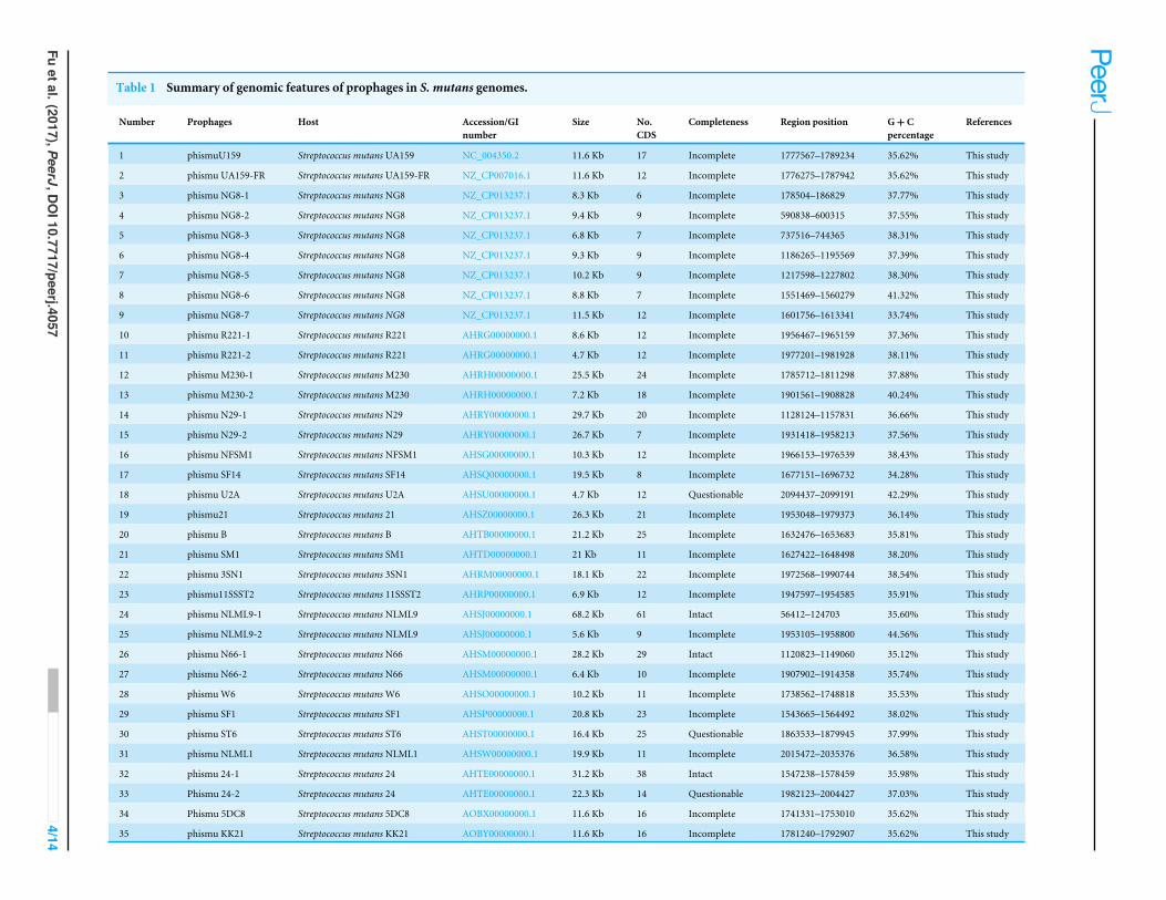
Table 1 Summary of genomic features of prophages in S. mutans genomes.
Number Prophages Host Accession/GInumber
Size No.CDS
Completeness Region position G+ Cpercentage
References
1 phismuU159 Streptococcus mutans UA159 NC_004350.2 11.6 Kb 17 Incomplete 1777567–1789234 35.62% This study
2 phismu UA159-FR Streptococcus mutans UA159-FR NZ_CP007016.1 11.6 Kb 12 Incomplete 1776275–1787942 35.62% This study
3 phismu NG8-1 Streptococcus mutans NG8 NZ_CP013237.1 8.3 Kb 6 Incomplete 178504–186829 37.77% This study
4 phismu NG8-2 Streptococcus mutans NG8 NZ_CP013237.1 9.4 Kb 9 Incomplete 590838–600315 37.55% This study
5 phismu NG8-3 Streptococcus mutans NG8 NZ_CP013237.1 6.8 Kb 7 Incomplete 737516–744365 38.31% This study
6 phismu NG8-4 Streptococcus mutans NG8 NZ_CP013237.1 9.3 Kb 9 Incomplete 1186265–1195569 37.39% This study
7 phismu NG8-5 Streptococcus mutans NG8 NZ_CP013237.1 10.2 Kb 9 Incomplete 1217598–1227802 38.30% This study
8 phismu NG8-6 Streptococcus mutans NG8 NZ_CP013237.1 8.8 Kb 7 Incomplete 1551469–1560279 41.32% This study
9 phismu NG8-7 Streptococcus mutans NG8 NZ_CP013237.1 11.5 Kb 12 Incomplete 1601756–1613341 33.74% This study
10 phismu R221-1 Streptococcus mutans R221 AHRG00000000.1 8.6 Kb 12 Incomplete 1956467–1965159 37.36% This study
11 phismu R221-2 Streptococcus mutans R221 AHRG00000000.1 4.7 Kb 12 Incomplete 1977201–1981928 38.11% This study
12 phismu M230-1 Streptococcus mutansM230 AHRH00000000.1 25.5 Kb 24 Incomplete 1785712–1811298 37.88% This study
13 phismu M230-2 Streptococcus mutansM230 AHRH00000000.1 7.2 Kb 18 Incomplete 1901561–1908828 40.24% This study
14 phismu N29-1 Streptococcus mutans N29 AHRY00000000.1 29.7 Kb 20 Incomplete 1128124–1157831 36.66% This study
15 phismu N29-2 Streptococcus mutans N29 AHRY00000000.1 26.7 Kb 7 Incomplete 1931418–1958213 37.56% This study
16 phismu NFSM1 Streptococcus mutans NFSM1 AHSG00000000.1 10.3 Kb 12 Incomplete 1966153–1976539 38.43% This study
17 phismu SF14 Streptococcus mutans SF14 AHSQ00000000.1 19.5 Kb 8 Incomplete 1677151–1696732 34.28% This study
18 phismu U2A Streptococcus mutans U2A AHSU00000000.1 4.7 Kb 12 Questionable 2094437–2099191 42.29% This study
19 phismu21 Streptococcus mutans 21 AHSZ00000000.1 26.3 Kb 21 Incomplete 1953048–1979373 36.14% This study
20 phismu B Streptococcus mutans B AHTB00000000.1 21.2 Kb 25 Incomplete 1632476–1653683 35.81% This study
21 phismu SM1 Streptococcus mutans SM1 AHTD00000000.1 21 Kb 11 Incomplete 1627422–1648498 38.20% This study
22 phismu 3SN1 Streptococcus mutans 3SN1 AHRM00000000.1 18.1 Kb 22 Incomplete 1972568–1990744 38.54% This study
23 phismu11SSST2 Streptococcus mutans 11SSST2 AHRP00000000.1 6.9 Kb 12 Incomplete 1947597–1954585 35.91% This study
24 phismu NLML9-1 Streptococcus mutans NLML9 AHSJ00000000.1 68.2 Kb 61 Intact 56412–124703 35.60% This study
25 phismu NLML9-2 Streptococcus mutans NLML9 AHSJ00000000.1 5.6 Kb 9 Incomplete 1953105–1958800 44.56% This study
26 phismu N66-1 Streptococcus mutans N66 AHSM00000000.1 28.2 Kb 29 Intact 1120823–1149060 35.12% This study
27 phismu N66-2 Streptococcus mutans N66 AHSM00000000.1 6.4 Kb 10 Incomplete 1907902–1914358 35.74% This study
28 phismu W6 Streptococcus mutansW6 AHSO00000000.1 10.2 Kb 11 Incomplete 1738562–1748818 35.53% This study
29 phismu SF1 Streptococcus mutans SF1 AHSP00000000.1 20.8 Kb 23 Incomplete 1543665–1564492 38.02% This study
30 phismu ST6 Streptococcus mutans ST6 AHST00000000.1 16.4 Kb 25 Questionable 1863533–1879945 37.99% This study
31 phismu NLML1 Streptococcus mutans NLML1 AHSW00000000.1 19.9 Kb 11 Incomplete 2015472–2035376 36.58% This study
32 phismu 24-1 Streptococcus mutans 24 AHTE00000000.1 31.2 Kb 38 Intact 1547238–1578459 35.98% This study
33 Phismu 24-2 Streptococcus mutans 24 AHTE00000000.1 22.3 Kb 14 Questionable 1982123–2004427 37.03% This study
34 Phismu 5DC8 Streptococcus mutans 5DC8 AOBX00000000.1 11.6 Kb 16 Incomplete 1741331–1753010 35.62% This study
35 phismu KK21 Streptococcus mutans KK21 AOBY00000000.1 11.6 Kb 16 Incomplete 1781240–1792907 35.62% This study
Fuetal.(2017),PeerJ,D
OI10.7717/peerj.4057
4/14

Figure 1 Dot plot matrix comparison calculated for the genomes of 35 Streptococcus mutansprophages. Prophage genome comparison of full genomes; the main diagonal represents the alignmentof a sequence with itself. Regions of local similarity or repetitive sequences give rise to further diagonalmatches in addition to the central diagonal, which indicates high similarity of the prophages. The x-and y-axis indicate full genomic sequence comparisons of prophage genomes. The length of the linesrepresents the length of the prophage genomes. The dot plot matrix was calculated using Geneious(http://www.geneious.com, Kearse et al., 2012).
Full-size DOI: 10.7717/peerj.4057/fig-1
phismuNLML9-1, and phismu24-1) possessed at least 83% identity with one another,as determined by multiple genomic sequence alignments (Fig. 2B). In addition, BLASTncomparison of Cluster C (phismuSF1, phismuNG8-3, and phismu3SN1) revealed onesegment (6,852 bp) with identity greater than 99% between phismuSF1 and phismuNG8-3 (Fig. 2C). Sequence comparison showed that phismuSF1 and phismu3SN1 shared two
Fu et al. (2017), PeerJ, DOI 10.7717/peerj.4057 5/14

Figure 2 Genomic sequence comparison of S. mutans prophages. Comparisons were performed usingthe BLASTn and Artemis Comparison Tool visualization programs. Forward and reverse matches are col-ored red and blue, respectively.
Full-size DOI: 10.7717/peerj.4057/fig-2
Fu et al. (2017), PeerJ, DOI 10.7717/peerj.4057 6/14

major regions of reverse complementary sequences (4,253 and 3,352 bp) with a similarity of99.41 and 98.6% identity, respectively (Fig. 2C). Prophages belonging to individual clusterswere more closely related to one another than to phages in the other clusters (Fig. S1).Other S. mutans prophages such as phismuSF14 and phismuW6 shared twomajor sequence(4,199 and 1,427 bp) similarities of 99.19 and 98.8% identity, respectively. PhismuM230-1and phismuSM1 shared one major sequence (8,367 bp) similarity of 99.19% identity.PhismuN29-1 and phismuNFSM1 shared one major reverse complementary sequence(3,765 bp) similarity of 99.15% identity and one small sequence (1,291 bp) similarityof 99.15% identity. Phismu21 and phismuB shared two small reverse complementarysequence (566 and 471 bp) similarities of 99.82 and 97.66% identity, respectively (Fig. 2D).
Phylogeny of S. mutans prophagesTo understand how S. mutans prophages are related to one another, a genome phylogenetictree was constructed based on the complete genomic sequences of some S. mutansprophages, including Cluster A, Cluster B, Cluster C, and three previously reportedS. mutans phages M102 (Delisle & Rostkowski, 1993), M102AD (Delisle et al., 2012), andφAPCM01 (Dalmasso et al., 2015). The same grouping patterns and relationships wereobserved among the three large clusters (Clusters A, B, and C) as in the phylogenetictree (Fig. 3). Cluster A consisted of phismuUA159, phismuUA159-FR, phismuKK21,phismu5DC8, and phismuN66-2. PhismuUA159, phismuUA159-FR, phismuKK21, andphismu5DC8 showed the closest distinct branch in the phylogenetic tree. Cluster Bconsisted ofM102,M102AD,φAPCM01, phismuN66-1, phismuNLML9-1, and phismu24-1. These results suggested that the three sequenced S. mutans phages (M102, M102AD, andφAPCM01) belonged to Cluster B. Cluster C consisted of phismuSF1, phismuNG8-3, andphismu3SN1.
Comparative analysis between M102AD and S. mutans prophagesThe S. mutans phage M102AD, which has a genome length of 30,664 bp and was isolated atthe University of Maryland, was chosen as a reference phage, because it has been sequencedand well annotated (Delisle et al., 2012). The prophages phismuN66-1, phismuNLML9-1,and phismu24-1 all shared sequence similarity with M102AD (Fig. 4). The linear genomiccomparison showed that phismu24-1 shared two major sequence (1,199 and 466 bp)similarities of 84 and 83.2% identity, respectively, with M102AD at the nucleotide level.BLASTn comparison of phismuNLML9-1 and M102AD revealed three major sequences(666, 454, and 423 bp) with 85.6, 82.9, and 82.7% identity at the nucleotide level.PhismuN66-1 shared three major sequences (749, 473, and 124 bp) with 85.6, 83.6,and 84.2% identity in comparison with M102AD genomes.
Summary of features of S. mutans prophage genomic sequencesThree intact prophages (phismu NLML9-1, phismu N66-1, and phismu 24-1) wereidentified in S. mutans, and all three prophages in Cluster B closely resembled the genomeof M102AD. All of the ORFs of the prophages were predicted and annotated by PHASTER,GeneMarkS, and BLASP. PhismuNLML9-1, phismu24-1, and phismu66-1 exhibited thecharacteristic modular arrangement of the M102AD phage, including packaging and
Fu et al. (2017), PeerJ, DOI 10.7717/peerj.4057 7/14
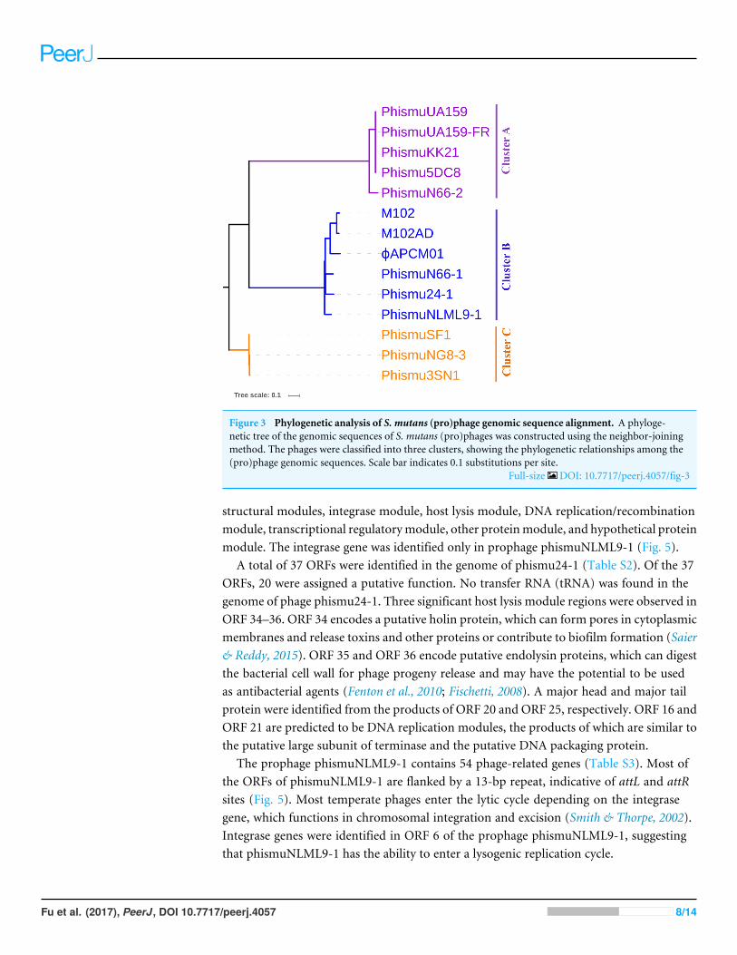
Figure 3 Phylogenetic analysis of S. mutans (pro)phage genomic sequence alignment. A phyloge-netic tree of the genomic sequences of S. mutans (pro)phages was constructed using the neighbor-joiningmethod. The phages were classified into three clusters, showing the phylogenetic relationships among the(pro)phage genomic sequences. Scale bar indicates 0.1 substitutions per site.
Full-size DOI: 10.7717/peerj.4057/fig-3
structural modules, integrase module, host lysis module, DNA replication/recombinationmodule, transcriptional regulatorymodule, other proteinmodule, and hypothetical proteinmodule. The integrase gene was identified only in prophage phismuNLML9-1 (Fig. 5).
A total of 37 ORFs were identified in the genome of phismu24-1 (Table S2). Of the 37ORFs, 20 were assigned a putative function. No transfer RNA (tRNA) was found in thegenome of phage phismu24-1. Three significant host lysis module regions were observed inORF 34–36. ORF 34 encodes a putative holin protein, which can form pores in cytoplasmicmembranes and release toxins and other proteins or contribute to biofilm formation (Saier& Reddy, 2015). ORF 35 and ORF 36 encode putative endolysin proteins, which can digestthe bacterial cell wall for phage progeny release and may have the potential to be usedas antibacterial agents (Fenton et al., 2010; Fischetti, 2008). A major head and major tailprotein were identified from the products of ORF 20 and ORF 25, respectively. ORF 16 andORF 21 are predicted to be DNA replication modules, the products of which are similar tothe putative large subunit of terminase and the putative DNA packaging protein.
The prophage phismuNLML9-1 contains 54 phage-related genes (Table S3). Most ofthe ORFs of phismuNLML9-1 are flanked by a 13-bp repeat, indicative of attL and attRsites (Fig. 5). Most temperate phages enter the lytic cycle depending on the integrasegene, which functions in chromosomal integration and excision (Smith & Thorpe, 2002).Integrase genes were identified in ORF 6 of the prophage phismuNLML9-1, suggestingthat phismuNLML9-1 has the ability to enter a lysogenic replication cycle.
Fu et al. (2017), PeerJ, DOI 10.7717/peerj.4057 8/14
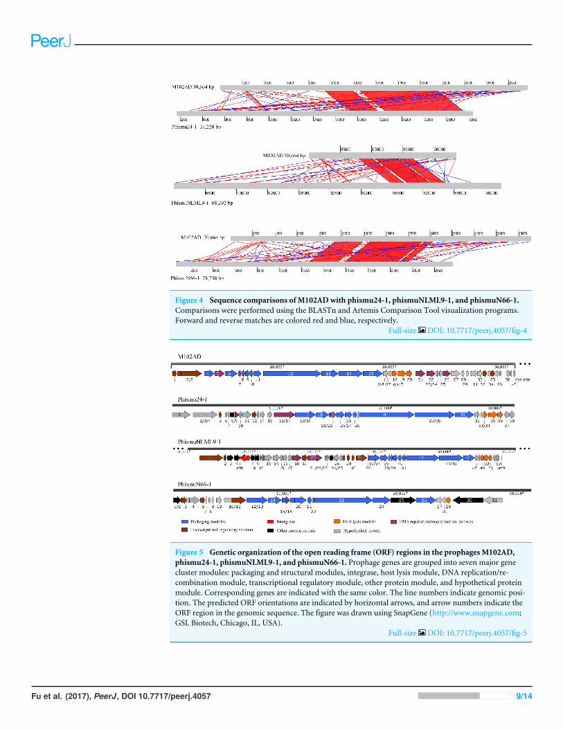
Figure 4 Sequence comparisons of M102ADwith phismu24-1, phismuNLML9-1, and phismuN66-1.Comparisons were performed using the BLASTn and Artemis Comparison Tool visualization programs.Forward and reverse matches are colored red and blue, respectively.
Full-size DOI: 10.7717/peerj.4057/fig-4
Figure 5 Genetic organization of the open reading frame (ORF) regions in the prophages M102AD,phismu24-1, phismuNLML9-1, and phismuN66-1. Prophage genes are grouped into seven major genecluster modules: packaging and structural modules, integrase, host lysis module, DNA replication/re-combination module, transcriptional regulatory module, other protein module, and hypothetical proteinmodule. Corresponding genes are indicated with the same color. The line numbers indicate genomic posi-tion. The predicted ORF orientations are indicated by horizontal arrows, and arrow numbers indicate theORF region in the genomic sequence. The figure was drawn using SnapGene (http://www.snapgene.com;GSL Biotech, Chicago, IL, USA).
Full-size DOI: 10.7717/peerj.4057/fig-5
Fu et al. (2017), PeerJ, DOI 10.7717/peerj.4057 9/14
No putative tRNA or transfer-messenger RNA was recognized. A putative holin proteinwas encoded by ORF 34 and a putative endolysin by ORF 35 and ORF 36. ORF 18, ORF 9,and ORF 20 are predicted to encompass the replication module.
The phismu66-1 prophage genome contains 31 ORFs (Table S4). ORF 30 encodes theABC transporter or permease protein, which functions as a multiple sugar metabolismtransporter and is a promising target for antimicrobial strategies in S. mutans (Nagayamaet al., 2014). ORF 25 encodes a host specificity protein and ORF 29 encodes a lysin-holinprotein. In addition, the packaging and structural modules contained ORF 12, ORF 13,ORF 15, ORF 17–21, ORF 23, ORF 24, and ORF 26, and the transcriptional regulatorymodule was encoded by ORF 3, ORF 6, and ORF 11.
Horizontal gene transfer plays an important role in the adaptation and evolutionof prokaryotes, and bacteriophages, as mobile genetic elements, enable horizontal genetransfer. In our study, we found that ORF 4 of phismuNLML9-1 andORF 30 of phismu66-1both encode an ABC transporter/permease protein, which is a virulence protein associatedwith the development of spontaneous resistance to compound 103 in S. aureus strains(Morisaki et al., 2016). Many unknown functional hypothetical proteins may play animportant role in the acquisition of a specialized set of genes via prophages and horizontaltransfer in S. mutans.
Mapping of the genomic location and comparative analysis ofS. mutans prophagesWe oriented and mapped the ORFs located in the genomic sequences of M102AD,phismu24-1, phismuNLML9-1, and phismuN66-1 (Fig. 6). A total of nine ORFs werelocated in the M102AD, phismuNLML9-1, and phismu24-1 conserved regions, and sevenORFs were located in the conserved regions of phismuN66-1. The major conservedregions of M102AD included ORFs 12–20, which encode a putative tape measure protein,putative tail protein, putative receptor-binding protein, putative minor structural protein,hypothetical protein, hypothetical protein, putative holin, putative endolysin, and putativeendolysin, respectively. According to their putative functions, the ORFs were assigned topackaging/structural modules, hypothetical protein modules, and the host lysis module(Tables S5 and S6). A total of nine ORFs were identified in the conserved regions ofphismu24-1, including ORF 28 and 29 (putative tail component proteins), ORF 30 (tail-host specificity protein), ORF 31 (tail protein), ORF 32 and 33 (hypothetical proteins), ORF34 (putative holin), and ORF 35 and 36 (putative endolysins). These ORFs shared a regionvarying between 57.68 and 95% identity with M102AD (Table S5). The phismuNLML9-1sequence contained nineORFs similar toM102AD:ORF 43 and 44 (putative tail componentproteins), ORF 45 (tail-host specificity protein), ORF 46 (tail protein), ORF 47 and 48(hypothetical proteins), ORF 49 (putative holin), ORF 50 (hypothetical protein), and ORF51 (putative endolysin). The protein sequence identity of these ORFs varied between 57.73and 94% with M102AD (Table S5). The phismuN66-1 sequence contained seven ORFssimilar to M102AD: ORF 23 and 24 (putative tail component proteins), ORF 25 (tail-hostspecificity protein), ORF 26 (tail protein), ORF 27 and 28 (hypothetical proteins), andORF 29 (putative holin). The protein sequence identity of the ORFs varied between 62 and
Fu et al. (2017), PeerJ, DOI 10.7717/peerj.4057 10/14

Figure 6 Schematic representation of the genomic organization and ORF regions of S. mutans phageM102AD compared to the prophages phismu24-1, phismuNLML9-1, and phismuN66-1. The lines rep-resent phage/prophage genomes, and arrows represent ORFs. Regions connected by red shading representconserved genomic identity.
Full-size DOI: 10.7717/peerj.4057/fig-6
95% with M102AD. PhismuN66-1 lost two putative endolysin ORFs compared to otherprophages (Table S5).
CONCLUSIONSIn conclusion, our genome sequencing data analyses identified 35 prophage-like elementspresent in the genome of S. mutans, all of which were identified for the first time. Genomicanalysis of prophages revealed that those belonging to the same cluster displayed sequencesimilarities. The genomes and genetic information of phismuNLML9-1, phismu24-1, andphismu66-1 prophages were analyzed, identifying putative ORFs and functional regions.To the best of our knowledge, this is the first systematic analysis of S. mutans prophages.
ADDITIONAL INFORMATION AND DECLARATIONS
FundingThis work was supported by the Program for Innovation Team Building at Institutionsof Higher Education in Chongqing in 2016 (Project No. CXTDG201602006), the ProjectSupported by Chongqing Municipal Key Laboratory of Oral Biomedical Engineeringof Higher Education, the National Natural Science Foundation of China (Project No.
Fu et al. (2017), PeerJ, DOI 10.7717/peerj.4057 11/14

31600148), the Shandong Excellent Young Scientist Award Fund (BS2014YY031), and theFoundation of University of Jinan (XBS1519, XKY1633). The funders had no role in studydesign, data collection and analysis, decision to publish, or preparation of the manuscript.
Grant DisclosuresThe following grant information was disclosed by the authors:Program for Innovation Team Building at Institutions of Higher Education in Chongqingin 2016: CXTDG201602006.ChongqingMunicipal Key Laboratory ofOral Biomedical Engineering ofHigher Education.National Natural Science Foundation of China: 31600148.Shandong Excellent Young Scientist Award Fund: BS2014YY031.Foundation of University of Jinan: XBS1519, XKY1633.
Competing InterestsThe authors declare there are no competing interests.
Author Contributions• Tiwei Fu conceived and designed the experiments, performed the experiments, analyzedthe data, wrote the paper, prepared figures and/or tables.
• Xiangyu Fan conceived and designed the experiments, contributed reagents/materials/-analysis tools, reviewed drafts of the paper.
• Quanxin Long and Wanyan Deng contributed reagents/materials/analysis tools.• Jinlin Song reviewed drafts of the paper.• Enyi Huang conceived and designed the experiments, reviewed drafts of the paper.
Data AvailabilityThe following information was supplied regarding data availability:
The accession numbers can be found in Table 1.
Supplemental InformationSupplemental information for this article can be found online at http://dx.doi.org/10.7717/peerj.4057#supplemental-information.
REFERENCESAkhter S, Aziz RK, Edwards RA. 2012. PhiSpy: a novel algorithm for finding prophages
in bacterial genomes that combines similarity- and composition-based strategies.Nucleic Acids Research 40:329–334 DOI 10.1093/nar/gks406.
Argimon S, Konganti K, Chen H, Alekseyenko AV, Brown S, Caufield PW. 2014. Com-parative genomics of oral isolates of Streptococcus mutans by in silico genome sub-traction does not reveal accessory DNA associated with severe early childhood caries.Infection, Genetics and Evolution 21:269–278 DOI 10.1016/j.meegid.2013.11.003.
Ariff A, Wise MJ, Kahler CM, Tay CY, Peters F, Perkins TT, Chang BJ. 2015. NovelMoraxella catarrhalis prophages display hyperconserved non-structural genes despitetheir genomic diversity. BMC Genomics 16:860 DOI 10.1186/s12864-015-2104-1.
Fu et al. (2017), PeerJ, DOI 10.7717/peerj.4057 12/14

Arndt D, Grant JR, Marcu A, Sajed T, Pon A, Liang Y,Wishart DS. 2016. PHASTER:a better, faster version of the PHAST phage search tool. Nucleic Acids Research44:W16–W21 DOI 10.1093/nar/gkw387.
Barrangou R, Fremaux C, Deveau H, Richards M, Boyaval P, Moineau S, RomeroDA, Horvath P. 2007. CRISPR provides acquired resistance against viruses inprokaryotes. Science 315:1709–1712 DOI 10.1126/science.1138140.
Carver T, BerrimanM, Tivey A, Patel C, Bohme U, Barrell BG, Parkhill J, RajandreamMA. 2008. Artemis and ACT: viewing, annotating and comparing sequencesstored in a relational database. Bioinformatics 24:2672–2676DOI 10.1093/bioinformatics/btn529.
DalmassoM, De Haas E, Neve H, Strain R, Cousin FJ, Stockdale SR, Ross RP, Hill C.2015. Isolation of a novel phage with activity against streptococcus mutans biofilms.PLOS ONE 10:e0138651 DOI 10.1371/journal.pone.0138651.
Delisle AL, GuoM, Chalmers NI, Barcak GJ, Rousseau GM,Moineau S. 2012. Biologyand genome sequence of Streptococcus mutans phage M102AD. Applied and Environ-mental Microbiology 78:2264–2271 DOI 10.1128/AEM.07726-11.
Delisle AL, Rostkowski CA. 1993. Lytic bacteriophages of Streptococcus mutans. CurrentMicrobiology 27:163–167 DOI 10.1007/bf01576015.
Duerkop BA, Palmer KL, HorsburghMJ. 2014. Enterococcal bacteriophages and genomedefense. Boston: Massachusetts Eye and Ear Infirmary.
Ericson D, Kidd E, McCombD,Mjor I, NoackMJ. 2003.Minimally invasive dentistry–concepts and techniques in cariology. Oral Health & Preventive Dentistry 1:59–72.
Fan X, Li Y, He R, Li Q, HeW. 2016. Comparative analysis of prophage-like elements inHelicobacter sp. genomes. PeerJ 4:e2012 DOI 10.7717/peerj.2012.
Fan X, Xie L, Li W, Xie J. 2014. Prophage-like elements present in mycobacteriumgenomes. BMC Genomics 15:243 DOI 10.1186/1471-2164-15-243.
FentonM, Ross P, McAuliffe O, O’Mahony J, Coffey A. 2010. Recombinant bacterio-phage lysins as antibacterials. Bioengineered Bugs 1:9–16 DOI 10.4161/bbug.1.1.9818.
Fischetti VA. 2008. Bacteriophage lysins as effective antibacterials. Current Opinion inMicrobiology 11:393–400 DOI 10.1016/j.mib.2008.09.012.
Freires IA, Aviles-Reyes A, Kitten T, Simpson-Haidaris PJ, Swartz M, KnightPA, Rosalen PL, Lemos JA, Abranches J. 2017.Heterologous expression ofStreptococcus mutans Cnm in Lactococcus lactis promotes intracellular in-vasion, adhesion to human cardiac tissues and virulence. Virulence 8:18–29DOI 10.1080/21505594.2016.1195538.
Kearse M, Moir R,Wilson A, Stones-Havas S, CheungM, Sturrock S, Buxton S, CooperA, Markowitz S, Duran C, Thierer T, Ashton B, Mentjies P, Drummond A. 2012.Geneious Basic: an integrated and extendable desktop software platform for theorganization and analysis of sequence data. Bioinformatics 28(12):1647–1649DOI 10.1093/bioinformatics/bts199.
Kidd E, Fejerskov O. 2013. Caries control in health service practice. Primary DentalJournal 2:4 DOI 10.1308/205016813807440010.
Fu et al. (2017), PeerJ, DOI 10.7717/peerj.4057 13/14

Morisaki JH, Smith PA, Date SV, Kajihara KK, Truong CL, Modrusan Z, Yan D, KangJ, XuM, Shah IM, Mintzer R, Kofoed EM, Cheung TK, Arnott D, Koehler MF,Heise CE, Brown EJ, TanMW, HazenbosWL. 2016. A putative bacterial ABCtransporter circumvents the essentiality of signal peptidase.MBio 7:e00412–e00416DOI 10.1128/mBio.00412-16.
Motegi M, Takagi Y, Yonezawa H, Hanada N, Terajima J, Watanabe H, Senpuku H.2006. Assessment of genes associated with Streptococcus mutans biofilm morphology.Applied and Environmental Microbiology 72:6277–6287 DOI 10.1128/AEM.00614-06.
Nagayama K, Fujita K, Takashima Y, Ardin AC, Ooshima T, Matsumoto-NakanoM.2014. Role of ABC transporter proteins in stress responses of Streptococcus mutans.Oral Health and Dental Management 13:359–365.
Roux S, Enault F, Hurwitz BL, SullivanMB. 2015. VirSorter: mining viral signal frommicrobial genomic data. PeerJ 3:e985 DOI 10.7717/peerj.985.
Saier Jr MH, Reddy BL. 2015.Holins in bacteria, eukaryotes, and archaea: multifunc-tional xenologues with potential biotechnological and biomedical applications.Journal of Bacteriology 197:7–17 DOI 10.1128/JB.02046-14.
SmithMC, Thorpe HM. 2002. Diversity in the serine recombinases.Molecular Microbiol-ogy 44:299–307 DOI 10.1046/j.1365-2958.2002.02891.x.
St-Pierre F, Endy D. 2008. Determination of cell fate selection during phage lambdainfection. Proceedings of the National Academy of Sciences of the United States ofAmerica 105:20705–20710 DOI 10.1073/pnas.0808831105.
Sturmberger L, Chappell T, Geier M, Krainer F, Day KJ, Vide U, Trstenjak S, SchieferA, Richardson T, Soriaga L, Darnhofer B, Birner-Gruenberger R, Glick BS, Tol-storukov I, Cregg J, Madden K, Glieder A. 2016. Refined Pichia pastoris referencegenome sequence. Journal of Biotechnology 235:121–131DOI 10.1016/j.jbiotec.2016.04.023.
Tamura K, Dudley J, Nei M, Kumar S. 2007.MEGA4: Molecular Evolutionary Ge-netics Analysis (MEGA) software version 4.0.Molecular Biology and Evolution24:1596–1599.
Tang F, Bossers A, Harders F, Lu C, Smith H. 2013. Comparative genomic analysis oftwelve Streptococcus suis (pro)phages. Genomics 101:336–344DOI 10.1016/j.ygeno.2013.04.005.
Van der Ploeg JR. 2007. Genome sequence of Streptococcus mutans bacteriophage M102.FEMS Microbiology Letters 275:130–138 DOI 10.1111/j.1574-6968.2007.00873.x.
VenturaM, Zomer A, Canchaya C, O’Connell-MotherwayM, Kuipers O, Turroni F,Ribbera A, Foroni E, Buist G,Wegmann U, Shearman C, GassonMJ, FitzgeraldGF, Kok J, Van Sinderen D. 2007. Comparative analyses of prophage-like elementspresent in two Lactococcus lactis strains. Applied and Environmental Microbiology73:7771–7780 DOI 10.1128/AEM.01273-07.
ZhuW, Lomsadze A, BorodovskyM. 2010. Ab initio gene identification in metagenomicsequences. Nucleic Acids Research 38:e132 DOI 10.1093/nar/gkq275.
Fu et al. (2017), PeerJ, DOI 10.7717/peerj.4057 14/14


![Genomic and ecological attributes of marine bacteriophages ... · bacterial transcriptome increasing the pathogen’sfitness in the animal-associated environment [15]. Prophages inserted](https://img.pdfslide.us/doc/110x75/5ed1ca21451b173a813900b7/genomic-and-ecological-attributes-of-marine-bacteriophages-bacterial-transcriptome.jpg)