Embed Size (px)
Citation preview
59th Annual Meeting of the
American Association for the Study of Liver Disease Oct. 31st–Nov. 4, 2008 San Francisco, CA, USA
COMMUNICATIONS ABOUT
THE FIBROSCAN®
http://www.echosens.com
59TH AASLD MEETING | COMMUNICATIONS ABOUT FIBROSCAN®
PAGE 2/24
CONTENT POSTGRADUATE COURSE Non-invasive markers of liver fibrosis: FibroScan®, serum markers and other technologies; is biopsy obsolete?
ORAL PRESENTATION 1 Autologous bone marrow cell infusion increased serum albumin and activated the liver progenitor cell compartment in patients with advanced liver cirrhosis
ORAL PRESENTATION 2 Sustained virological response is associated with decrease in liver stiffness using FibroScan® in patients with HCV related cirrhosis
POSTER 1 Transient elastography (FibroScan®) and hepatic venous pressure gradient measurement in patients with cirrhosis and gastrointestinal haemorrhage related to portal hypertension
POSTER 2 Transient elastography (TE) detects graft damage in liver transplanted (LT) patients with aetiologies other than HCV
POSTER 3 Identification of preclinical portal hypertension and staging of fibrosis by transient elastography
POSTER 4 Multiphoton microscopy by second harmonic generation: a new tool for fibrillar collagen quantification in liver fibrosis
POSTER 5 Detection of cirrhosis in heart transplant candidates. Interest of non invasive diagnosis methods and short term survival
POSTER 6 Unexplained severe liver disease in HIV infection: prevalence, histology and potential etiology
POSTER 7 Portal hypertension in HCV cirrhosis is associated with insulin resistance
POSTER 8 Screening for advanced fibrosis using non-invasive biomarkers, FibroTest (FT), and FibroScan® (FS) in a community based population
POSTER 9 Liver stiffness measurement and serum markers for predicting significant fibrosis in chronic hepatitis B
POSTER 10 Clinical usefulness of liver stiffness measurement in HBeAg-positive chronic hepatitis B patients with ALT level < 2 times upper limit of normal
POSTER 11 Utility of liver stiffness measured by transient elastography for determining significant liver fibrosis in patients with chronic hepatitis B
POSTER 12 Changes in liver stiffness during entecavir therapy in patients with chronic hepatitis B
POSTER 13 FibroScan® measures according to intercostal space: validity and agreement
POSTER 14 The impact of steatosis on liver stiffness measurement using FibroScan® in patients with hepatitis B virus-related chronic liver disease in Korea
POSTER 15 Different cutoff values according to ALT level may increase the diagnostic performance of liver stiffness measurement using FibroScan® in patients with hepatitis B virus-related chronic liver disease
POSTER 16 Prospective comparison of transient elastography, ultrasound, APRI and liver biopsy for diagnosis of hepatic fibrosis in a real world population with different forms of chronic liver diseases
POSTER 17 Evaluation of different non-invasive methods for assessment of liver fibrosis in primary biliary cirrhosis (PBC): a comparative study using FibroScan®, MRI, MR-spectroscopy and serum fibrosis markers
POSTER 18 Prospective evaluation of liver stiffness dynamics during and after peginterferon alpha-ribavirin treatment in patients with chronic hepatitis C
POSTER 19 Can liver stiffness measurement be a predictive factor for the development of hepatocellular carcinoma in hepatitis virus-related chronic liver disease?
POSTER 20 Liver Stiffness values in the normal population: a study in voluntary blood donors
POSTER 21 Does BMI influence transient elastography readings?
POSTER 22 Transient elastography is superior to routine screening tests for detection of liver cirrhosis
POSTER 23 TRAF1-C5 as risk locus for severe hepatic fibrosis in chronic liver diseases
POSTER 24 Prevalence and causes of discordance between FibroScan® and Fibrotest for first-line assessment of liver fibrosis in chronic hepatitis C (CHC): A prospective analysis over 4 years in 3148 patients
POSTER 25 Correlation of transient elastography (FibroScan®) and hepatic artery doppler sonography in patients 30 years after single source hepatitis C genotype 1B outbreak in Germany
POSTER 26 Liver stiffness predicts clinical outcomes in patients with chronic liver disease
POSTER 27 Comparison of liver stiffness, FibroTest and liver biopsy for the assessment of liver fibrosis in renal transplant patient with chronic viral hepatitis
POSTER 28 Reduction of liver stiffness by interferon treatment for chronic hepatitis C measured by transient elastography
POSTER 29 Difference of liver stiffness after acute or chronic liver injury due to viral hepatitis
POSTER 30 Diagnostic accuracy of transient elastography: a comparison between chronic hepatitis B and C correlated with optimal-length liver biopsies
59TH AASLD MEETING | COMMUNICATIONS ABOUT FIBROSCAN®
PAGE 3/24
POSTER 31 Acoustic radiation force elastography versus transient elastography for non-invasive assessment of liver fibrosis in viral hepatitis: a new alternative?
POSTER 32 Combination of 13C-methacetin breath test by online molecular correlation spectroscopy and transient elastography improves diagnostic performance of non-invasive fibrosis assessment in chronic hepatitis C
POSTER 33 A sequential predictive algorithm combining APRI and transient elastography (TE) for diagnosing liver fibrosis in chronic hepatitis C
POSTER 34 Error factors for transient elastography in chronic hepatitis C
POSTER 35 Evaluation of transient elastography and King’s score for the diagnosis of fibrosis in patients with chronic hepatitis C infection
POSTER 36 The presence of additional diagnoses in patients with chronic hepatitis B and C. Histological evaluation of liver biopsies used for correlation with transient elastography in 257 patients
POSTER 37 Transient Elastography (TE) as a tool to evaluate acute liver damage: a prospective, multicentric cohort study
POSTER 38 A simple diagnostic algorithm to evaluate liver fibrosis in patients with chronic hepatitis C
59TH AASLD MEETING | COMMUNICATIONS ABOUT FIBROSCAN®
PAGE 4/24
POSTGRADUATE COURSE Start time: Sat, Nov 01 - 12:00 PM
Non-invasive markers of liver fibrosis: FibroScan ®, serum markers and other technologies; is biopsy obsolete? N. H. Afdhal; K. Reddy
ORAL PRESENTATION 1 Final ID/Program Number: 94 Location: Room 3018 (Moscone West Convention Center) Start time: Sun, Nov 02 - 4:45 PM
Autologous bone marrow cell infusion increased serum albumin and activated the liver progenitor cell compartment in patients with advanced liver cirrhosis J. Kim1, 2; Y. Park1, 3; S. Ki1; S. Chung1; H. Chang1; J. Choi1, 2; M. Park4, 6; K. Kim5, 6; Y. Paik1, 2; J. Park1, 2; D. Kim1, 2; S. Ahn1, 2; K. Lee1, 2; B. Moon2, 6; Y. Moon7; C. Chon1, 2; K. Han1, 2 1. Liver Cirrhosis Clinical Research Center, Seoul, South Korea. 2. Dept. of Internal Medicine, Yonsei University College of Medicine, Seoul, South Korea. 3. Dept. of Pathology, Yonsei University College of Medicine, Seoul, South Korea. 4. Dept. of Diagnostic Radiology, Yonsei University College of Medicine, Seoul, South Korea. 5. Dept. of Surgery, Yonsei University College of Medicine, Seoul, South Korea. 6. Yonsei Institute of Gastroenterology, Seoul, South Korea. 7. Dept. of Internal Medicine, Kwandong University Myungji Hospital, Goyang, South Korea.
Background/Aims : Liver Cirrhosis (LC) is the end stage of chronic liver disease and is very difficult to treat. Although recent studies of the role of bone marrow cells (BMCs) in regeneration or fibrosis in a liver fibrosis/cirrhosis animal model produced conflicting results, human clinical trials of BMC infusion for cirrhotic patients had positive results (Esch et al. 2005; Sakaida et al. 2006). This proof-of-concept study evaluated the 1-year effect of autologous BMC infusion (ABMI) on the liver in patients with advanced LC.
Methods : Patients aged between 18 and 75 years with a clinical diagnosis of advanced LC (Child-Pugh class B), a total bilirubin of less than 3.0 mg/dl, a platelet count exceeding 50,000/µL, and no viable hepatocellular carcinoma on magnetic resonance imaging (MRI) were included. Autologous BMCs were harvested from the ilium under general anesthesia and infused into a peripheral vein after RBC depletion and mononuclear cell concentration. Serologic tests, transient elastography, MRI, and biopsies were performed before and 1, 3, and 6 months after the procedure. Patients’ quality of life was surveyed using a questionnaire. Serum markers for liver fibrosis were checked.
Results : Eight patients with a mean age of 55 years (range, 43-64 years) were followed for 1 year. The mean number of infused mononuclear cells was 7.7 × 10⁹. The serum albumin level and prothrombin time were improved significantly at 9 and 12 months and at 6 months after ABMI, respectively (p < 0.05). Other serologic tests showed no significant changes. Ascites improved or disappeared in the patients despite stopping or reducing the oral diuretics. The performance scale and well-being sensation were increased significantly at all time points (p < 0.05). In biopsied tissues, the number
of progenitor cell compartments was increased significantly at 1, 3, and 6 months after ABMI (p < 0.05). Some patients showed an increased liver volume on MRI. There were no serious adverse events.
Conclusions : ABMI for advanced liver cirrhosis improved serum albumin level, protrombin time, and subjective symptoms, it increased the number of progenitor cell compartments in liver. ABMI in selected patients can be used as a bridging modality for the treatment of decompensated LC.
ORAL PRESENTATION 2 Final ID/Program Number: 263 Location: Room 2005/2007 (Moscone West Convention Center) Start time: Tue, Nov 04 - 11:15 AM
Sustained virological response is associated with decrease in liver stiffness using FibroScan ® in patients with HCV related cirrhosis A. F. Cardoso1; C. Stern1; R. Moucari1; N. Giuily1; C. G. Figueiredo-Mendes1; N. Boyer1; M. Ripault1; C. Castelnau1; T. Asselah1; M. Martinot-Peignoux1; S. Maylin1; P. Bedossa2; P. Marcellin1
1. Hepatology and INSERM U773-CRB3, Hôpital Beaujon, Clichy, France. 2. Anatomopathology, Hôpital Beaujon, Clichy, France.
Background and Aim: Liver stiffness measurement
using FibroScan is a non-invasive method proposed for the assessment of hepatic fibrosis in patients with liver diseases. The effect of the response to treatment on liver stiffness has not been well determined. In this study, we evaluated the influence of the response to antiviral therapy on liver stiffness in patients with cirrhosis according to the time after treatment.
Patients and Methods : From September 2006 to April 2008, 136 consecutive CHC patients with bridging fibrosis or cirrhosis were evaluated. They had all received a treatment course with interferon (conventional or pegylated) with or without ribavirin for at least 12 weeks. Liver stiffness measurement using FibroScan was assessed after treatment in patients with or without sustained virological response (SVR). Correlations between results of liver stiffness and genotype, BMI, mean delay between the end of treatment and FibroScan® evaluation, the presence of diabetes, and dyslipidemia were analyzed.
Results : One hundred fourteen of 136 tested patients had reliable FibroScan® results. This subgroup was not different from the overall group. The 114 patients had the following characteristics: male gender (72%), mean age (54±9 years), diabetes (26%), mean BMI (26±6 kg/m²), genotype 1 (59%), 2 (6%), 3 (22%), 4 (12%), 5 (1%). Mean follow-up was 45±33 months. SVR was observed in 33% of patients. Patients with SVR had lower median FibroScan® results (8.4 kPa; range 3.3-45) when compared to non SVR patients (15.7 kPa; range 5.3-75) (p<0.001). In the univariate analyses, the median of liver stiffness was higher in genotype 1 and 4 vs. genotypes 2 and 3 (p=0.02) and in patients with diabetes mellitus (p=0.006). The median of results was 15.4 (5.4-75) when the delay was inferior to 2 years and 10.7 (3.3-75.0) when the delay was more than 2 years between the FibroScan® measurement and the end of therapy (p=0.03). BMI and dyslipidemia did not influence the median of the FibroScan® results. In the logistic
59TH AASLD MEETING | COMMUNICATIONS ABOUT FIBROSCAN®
PAGE 5/24
regression, the median of the FibroScan® results was independently associated with response to the anti-viral treatment (p=0.001) and presence of diabetes (0.002).
Conclusion : In patients with cirrhosis, stage of fibrosis assessed by transient elastography was lower in patients with SVR than in those without SVR and this difference increased with the time, These results suggest the possibility of progressive reduction of fibrosis in patients with SVR and FibroScan® could be an important tool for the assessment of fibrosis stage during the post-treatment follow-up.
POSTER 1 Final ID/Program Number: 708 Location: West Hall (Moscone West Convention Center) Start time: Sat, Nov 01 - 2:00 PM
Transient elastography (FibroScan ®) and hepatic venous pressure gradient measurement in patients with cirrhosis and gastrointestinal haemorrhage related to portal hypertension M. Rudler1; P. Cluzel2; J. Massard1; A. Varaut1; P. Lebray1; M. Auguste2; T. Poynard1; D. Thabut1 1. AP-HP, Service d’Hépato-Gastroentérologie, Hôpital Pitié-Salpêtrière, Paris, France, Paris, France. 2. AP-HP, Service de Radiologie, Hôpital Pitié-Salpêtrière, Paris, France.
Introduction : A Hepatic venous pressure gradient
(HVPG) measurement above 20 mmHg is an independant factor of death in patients with cirrhosis and variceal bleeding. Transjugular intrahepatic porto-systemic shunt (TIPS) placement improves survival in these patients. Although identification of patients with HVPG > 20 mmHg is crucial, HVPG measurement is invasive, and its accessibility is poor. Transient elastometry (FibroScan®) is correlated with HVPG. Our aim was to evaluate if FibroScan® could identify, among patients with cirrhosis admitted for variceal bleeding, those with a HVPG above 20 mmHg.
Methods : All consecutive patients with cirrhosis hospitalised in our Intensive Care Unit of Gastroenterology and Hepatology in Pitié-Salpêtrière hospital for variceal bleeding were prospectively included between October 2007 and April 2008, except those with a history of portal thrombosis or hepatocellular carcinoma. HVPG measurement and FibroScan® were performed within the first 48 hours after admission.
Results : Twenty-eight patients (mean age: 54 ± 13 years, male sex 71%, alcoholic cirrhosis 79%, Child-Pugh score 9.2 ± 2.2) were included. Three (10%) had hepatocellular carcinoma, and 5 (18%) severe acute alcoholic hepatitis. Mean HVPG was 16±4 mmHg. Eight patients (18%) had a HVPG above 20 mmHg. FibroScan® was not performed in 4 patients because of ascites. Among those patients, 3 had a HVPG above 20 mmHg. Mean FibroScan® value was 57 ±19 kPa. Correlation between HVPG and FibroScan® was poor. Mean FibroScan® values were similar among patients with HVPG > 20 mmHg or with HVPG< 20 mmHg (57± 20 kPa vs 58 ± 17 kPa, p= 0.87).
Conclusion : These results suggest that FibroScan®
is not correlated with HVPG in patients with cirrhosis and variceal bleeding. FibroScan® failure was frequent because of the presence of ascites. FibroScan® can not identify patients with a HVPG above 20 mmHg who could benefit from TIPS placement.
POSTER 2 Final ID/Program Number: 571 Location: West Hall (Moscone West Convention Center) Start time: Sat, Nov 01 - 2:00 PM
Transient elastography (TE) detects graft damage in liver transplanted (LT) patients with aetiologies other than HCV C. Rigamonti1; M. Donato1; M. Fraquelli2; F. Agnelli1; P. Reggiani3; G. Rossi3; M. Colombo1 1. First Division of Gastroenterology, IRCCS Fondazione Ospedale Maggiore Policlinico, Mangiagalli e Regina Elena, Milan, Italy. 2. Second Division of Gastroenterology, IRCCS Fondazione Ospedale Maggiore Policlinico, Mangiagalli e Regina Elena, Milan, Italy. 3. Liver Transplant Unit, IRCCS Fondazione Ospedale Maggiore Policlinico, Mangiagalli e Regina Elena, Milan, Italy.
Background and aims: TE reliably predicts severity
of graft damage in LT patients with recurrent hepatitis C; its accuracy in other aetiologies is not validated. We aimed at evaluating TE in LT patients with graft damage due to other than HCV causes.
Methods : 60 recipients were studied (36 males, median age 51 years). Thirty-one hepatitis B/delta recurrence-free, 20 autoimmune/cholestatic disease (8 primary biliary cirrhosis, PBC, 7 primary sclerosing cholangitis, PSC, 5 autoimmune hepatitis, AIH), 6 alcoholic liver disease (ALD), 3 mixed. All underwent protocol/on demand liver biopsy (LB) and concomitant TE examination between September 2005 and October 2007. Histological diagnosis of graft disease was based on criteria defined by Banff Working Group (Hepatology 2006;44:489-501). Patients were divided according to the histological diagnosis into 2 groups, ie. with or without graft damage. TE was considered adequate if ≥10 valid measurements for each patient were obtained with a >65% success rate.
Results : In 5 patients (8%) TE examination was unsuccessful, thus the number of patients investigated was 55 (34 males). Median time from LT was 24 months (range 6-214). Median LB length was 35 mm (range 20-50). LB showed presence of graft damage in 23 patients (42%). Among 10 hepatitis B/delta with graft damage 7 showed idiopathic chronic hepatitis, 1 steatohepatitis, 1 rejection and 1 cholangitis. Among autoimmune/cholestatic disease 8 had recurrent disease (2 PBC, 3 PSC, 3 AIH). Among ALD 2 had steatohepatitis, 1 rejection and 1 cholangitis. One further idiopathic hepatitis was diagnosed (fulminant hepatic failure at transplant). The 23 patients with graft damage had significantly higher serum liver enzymes than the 32 without graft disease. Median TE was 7.5 kPa (range 5.4-27.4) in the patients with graft disease compared to 5.3 kPa (range 3.1-7.4) in those without (p<0.0001). AUROC for detection of graft damage was 0.92 (95%CI 0.81-0.97) with optimal TE cut-off of 6.1 kPa (87% sensitivity, 84% specificity, 90% negative predictive value).
Conclusions : The fact that TE accurately predicts non-HCV related graft damage too extends the applicability of this procedure in the management of LT patients.
59TH AASLD MEETING | COMMUNICATIONS ABOUT FIBROSCAN®
PAGE 6/24
POSTER 3 Final ID/Program Number: 705 Location: West Hall (Moscone West Convention Center) Start time: Sat, Nov 01 - 2:00 PM
Identification of preclinical portal hypertension and staging of fibrosis by transient elastography T. Reiberger1; A. Ferlitsch1; G. Ulbrich1; M. Homoncik1; M. Peck-Radosavljevic1 1. Gastroenterology & Hepatology, Medical University of Vienna, Vienna, Austria.
Background : As liver fibrosis progresses, portal
pressure (PP) rises from normal values (≤5mmHg) to preclinical portal hypertension (6-9mmHg, PCPH) and to clinical significant portal hypertension (≥10mmHg, CSPH). Since transient elastography (FibroScan®, FS) identifies patients with advanced fibrosis, we aimed to determine cut-off values for FS discriminating certain stages of fibrosis (F) and of portal hypertension.
Methods : 350 patients with available data of both PP and FS were included for correlation of FS with PP, while histological data of 94 patients were used for correlation of FS with fibrosis stage. Best cut-off values for PCPH / CSPH and for fibrosis stage were determined by area under the receiver operating characteristic curve (AUROC). Past episodes of variceal bleeding of included patients were documented.
Results : FS significantly correlated with PP (r=0.791, p<0.000001). Significant differences in FS were found between patients with normal PP, PCPH and CSPH (7.8±5.2 vs. 17.5±11.0 vs. 47.2±22.4 kPa, p<0.003). The best FS cut-offs for PCPH and CSPH were identified at 8.1 kPa (AUROC: 0.824; 95%CI: 0.729-0.896) and at 17.8 kPa (AUROC: 0.895; 95%CI: 0.837-0.938), respectively.
AUROC analysis identified a poor FS cut-off value between F1/F2 at 7.2 kPa (AUROC: 0.526; 95%CI: 0.336-0.710), but found well defined FS cut-offs between F2/F3 at 10.7 kPa (AUROC: 0.701; 95%CI: 0.507-0.853) and between F3/F4 at 17.6 kPa (AUROC: 0.880; 95%CI: 0.709-0.968). Patients with a history of variceal bleeding had significantly higher FS than patients without (48.8±27.2 vs. 20.6±13.7 kPa, p<0.001), but stronger statistical differences between patients with and without variceal bleeding were noted in PP (17.9±5.6 vs. 9.6±4.8 mmHg, p<0.00001).
Conclusion : FS is useful for non-invasive monitoring of patients with progressive liver disease, since FS can identify patients at risk for PCPH / CSPH and discriminates between early (F1/F2) and advanced stages of fibrosis (F3) and cirrhosis (F4). FS can help to stratify patients’ risk of variceal bleeding, but measurement of PP provides more sufficient information.
POSTER 4 Final ID/Program Number: 371 Location: West Hall (Moscone West Convention Center) Start time: Sat, Nov 01 - 2:00 PM
Multiphoton microscopy by second harmonic generation: a new tool for fibrillar collagen quantification in liver fibrosis L. Gailhouste1; D. Guyader2 1. INSERM U522, Rennes, France. 2. Liver Department, Hôpital Pontchaillou, Rennes, France.
Background/Aim : A fast and reliable method for collagen quantification would be a valuable tool for accurate study of liver fibrosis. Multiphoton microscopy by Second Harmonic Generation (SHG) allows for the detection of endogenous signals generated by proteins without mesoscopic axis of symmetry like fibrillar collagen. Its principle is based on the use of infrared pulsed lasers allowing, by non-linear excitation processes, for high in-depth imaging of biological tissues. Two types of signals can be collected: the Two Photons Excitation Fluorescence (TPEF) and the SHG selectively collected at the half-wavelength of excitation. An original and rapid method for fibrillar collagen quantification has been developed.
Methods : SHG acquisitions were performed, without labelling, on paraffin embedded histological slices taken from 119 patients by the mean of surgical (n = 46) or needle liver biopsies (n = 73). All the patients had chronic liver disease related to excessive alcohol intake, hepatitis C or hepatitis B. Metavir Fibrosis score were: F0 n = 20; F1 n = 31; F2 n = 19; F3 n = 21; and F4 n = 28. The fibrosis area was determined for each sample imaged and 43 patients with chronic viral hepatitis had an elastometry (FibroScan ®) performed at the time of the liver biopsy.
Results : The specificity of SHG signals for fibrillar collagen type I and III (major forms in fibrotic deposits) was verified using a 810 nm wavelength of excitation and a selective acquisition λ = 405 nm. The data processing established a reliable and precise quantification of fibrosis area for each sample. The results correlated with Metavir fibrosis score (p < 0.001) and liver elasticity (r = 0.6867, p < 0.01). The imaging process was fast (a few minutes per slice) and sensitive (very high SHG/background contrast) allowing for better detection of collagen, standardization and automation of the measure by comparison with the conventional histomorphometry approaches. Moreover, SHG microscopy offered the possibility to image samples with high in-depth resolution (< 500 µm), which could minimize the sampling bias, and allowed for the study of collagen fibers orientation fields, making possible the evaluation of their reticulation degree. Finally, the technique did not need dewaxing or labelling steps, and did not deteriorate samples permitting its classical pathological use after SHG acquisitions.
Conclusion : Multiphoton microscopy by SHG is an original, rapid and efficient technique. SHG microscopy allows for a greater standardization of measurements and provides a powerful tool to assess liver fibrosis progression and the efficiency of antifibrotic therapy.
59TH AASLD MEETING | COMMUNICATIONS ABOUT FIBROSCAN®
PAGE 7/24
POSTER 5 Final ID/Program Number: 568 Location: West Hall (Moscone West Convention Center) Start time: Sat, Nov 01 - 2:00 PM
Detection of cirrhosis in heart transplant candidates. Interest of non invasive diagnosis methods and short term survival P. Lebray1; S. Varnous2; F. Charlotte3; M. Munteanu1; V. Ratziu1; D. Thabut1; J. Vaillant4; D. Eyraud5; C. Vezinet5; T. Poynard1; H. Laurent4; I. Gandjbakhch2 1. Hepatogastroenterology unit, Pitié-Salpétrière Hospital, Paris, France. 2. Cardiothoracic surgery unit, Pitie-Salpetriere Hospital, Pitié-Salpétrière Hospital, Paris, France. 3. Pathology unit, Pitié-Salpétrière Hospital, Paris, France. 4. Liver surgery unit, Pitié-Salpétrière Hospital, Paris, France. 5. Anesthesiology unit, Pitié-Salpétrière Hospital, Paris, France.
Background : Prevalence of cardiac cirrhosis (CIR) is
probably under diagnosed in patients with cardiac insufficiency (CI) but metabolic risk factors (MS) and excessive alcohol intake are frequently found.
Aim : To evaluate the frequency and survival of CIR in heart transplant (HT) candidates.
Methods and Materials : During 3 years (2004-2007), successive patients candidates for HT with Child Pugh score A6 or more (CP6) were evaluated with APRI score, Fibroscan®(FS), FibroTest (FT) and transjugular liver biopsy (TLB). Risks factors of premature death (<3 months) and survival between patients with or without Child-Pugh score > A5 were analysed.
Results : 18/282 pts (6.3%) were included: 89% male, 54.8 years, dilated cardiomyopathy (7); coronaropathy (7); valvulopathy (1) and retransplantation (3). CIR was diagnosed in 5/18 (1.8% of the total cohort) and imputable to alcohol (4), MS (3) or HCV (1). LB was possible in 14/18 patients with 4 (28.5%) displayed non-cardiac CIR, 7 nodular regenerative hyperplasia and 3 minimal fibrosis. For these 14 patients with TLB, APRI score, FS or FT were (1) reliable in 14/14 (100%), 9/14 (64%) and 5/12 pts (41%); (2) suspected cirrhosis in 1/14, 8/9 and 3/5 patients but (3) predicted accurately cirrhosis or not in only 43%, 14% and 80% of them. 7/18 patients were heart transplanted with a high rate of early death (71%). In the control group, 184/264 pts underwent HT. After excluding the 2 successful combined liver/heart transplantation, the 3-months survival rate after HT was significantly different in multivariate analysis if Child-Pugh score > A5 (29% vs. 73%, OR = 4.95, p = 2.10-5) or if recipient age < 44 years (82% vs. 66%, OR = 0.44, p = 0.05).
Conclusions : Frequency of CIR is high (28.5%) in CI patients with signs of decompensated hepatopathy. Non invasive criteria frequently fail to confirm CIR rendering TLB still necessary in patients with Child-Pugh score >6. Moreover, Child-Pugh score >6, suggesting liver deficiency, is associated with a high rate of premature death in heart transplantation.
POSTER 6 Final ID/Program Number: 730 Location: West Hall (Moscone West Convention Center) Start time: Sat, Nov 01 - 2:00 PM
Unexplained severe liver disease in HIV infection: prevalence, histology and potential etiology I. Maida1, 2; E. Vispo1; G. Sotgiu2; P. Barreiro1; L. Martin-Carbonero1; M. Rios3; V. Soriano1 1. Hospital Carlos III, Madrid, Spain. 2. University of Sassari, Sassari, Italy. 3. Hospital Virgen de la Macarena, Seville, Spain.
Background : Cases of unexplained liver disease
(ULD) in HIV patients without any known cause of liver damage have recently been described. Severe portal hypertension (SPH) with preserved parenchimal function is a distinctive feature. Histological findings suggest primary vascular damage, ranging from periportal fibrosis (PPF) to nodular regenerative hyperplasia (NRH).
Methods : 13 subjects with SPH out of 41 HIV individuals with ULD were identified at 3 European clinics, where 3,287 HIV patients are on routine follow-up. All subjects had elevated AST/ALT for >12 months in the absence of any recognizable cause (HCV, HBV, alcohol, drugs, hemochromatosis, autoimmunity, protein S deficiency, etc.).
Results : 11/13 (85%) were male (all but one MSM), median age, 50 years; median time since HIV diagnosis, 7.5 years. Mean follow-up since the diagnosis of SPH, 4 years. At the last evaluation, all but none were on HAART. Median values were: CD4 count 240 cells/mm3, plasma HIV-RNA 4.6 log, ALT 80 IU/mL, and liver stiffness using FibroScan 11.3 kPa (Metavir F3 estimate). A liver biopsy informed NRH in 31%, PPF in 8%, drug-induced hepatitis in 8%, NASH in 31%, and unspecific findings in 22%. Since first diagnosis of SPH, 8 patients (61%) experienced hepatic decompensation, including variceal bleeding in 6 (46%) and portal thrombosis in 5 (38%). Prolonged exposure to didanosine (median 50 months) was recognised in all but one, and drug removal was followed by ALT reductions in almost all cases.
Discussion : HIV-associated ULD is a new and relatively rare condition in HIV patients. Prolonged didanosine exposure seems to play a major role. Of interest, other adenosine analogues (vincristine, 6-thioguanine, azathioprine, etc) also have demonstrated to produce hepatic perivascular abnormalities. The recognition of SPH, including oesophageal varices, in the absence of significant impairment in the liver synthetic function may alert about this condition. Its early identification is crucial, as adequate management of SPH may prevent potentially fatal upper gastrointestinal bleeding.
59TH AASLD MEETING | COMMUNICATIONS ABOUT FIBROSCAN®
PAGE 8/24
POSTER 7 Final ID/Program Number: 729 Location: West Hall (Moscone West Convention Center) Start time: Sat, Nov 01 - 2:00 PM
Portal hypertension in HCV cirrhosis is associated with insulin resistance C. Camma'1; S. Petta1; V. di Marco1; F. Bronte1; S. Ciminnisi1; G. Licata1; S. Peralta1; F. Simone1; A. Craxì1 1. Cattedra ed U.O.C. di Gastroenterologia ed Epatologia, Palermo, Italy.
Background and aims : Non-endoscopic methods to
assess portal hypertension in cirrhotic patients, specifically oesophageal varices (OV), are not sensitive enough for widespread use. Liver stiffness measurement by Transient Elastography (TE) and metabolic factors affecting development of fibrosis, particularly insulin resistance (IR), could improve the ability to predict varices presence.
Methods : One hundred-four consecutive patients with newly diagnosed Child A HCV cirrhosis underwent upper GI endoscopy to search for OV. Clinical, anthropometric, biochemical, ultrasonographic and metabolic characteristics, including IR by the homeostasis model assessment (HOMA), and liver stiffness by TE were recorded at the time of endoscopy.
Results : OV were detected in 63 of 104 patients (60%). Large OV (>/= F2) were observed in 10 (16%). At multivariate analysis presence of OV was independently associated with a low platelet count/spleen diameter ratio (OR 0.998 95% CI 0.996-0.999) and with a high HOMA score (OR 1.296; 95%CI 1.018-1.649) but not with liver stiffness (OR 1.009; 95%CI 0.951-1.070). This independent association between low platelet/spleen ratio (OR 0.998 95%CI 0.996-1.000), high HOMA score (OR 1.373; 95%CI 1.014-1.859) and OV presence was confirmed in the sub-group of 77 non-diabetic subjects. It is noteworthy that 9 out off 10 patients with large OV had platelet/spleen ratio < 792 or HOMA > 3.5.
Conclusions : In patients with compensated HCV cirrhosis, regardless of diabetes presence, a low platelet/spleen ratio and a high HOMA score are the strongest independent predictors of the presence of OV. Liver stiffness by TE does not contribute significantly to risk assessment in this setting.
POSTER 8 Final ID/Program Number: 1105 Location: West Hall (Moscone West Convention Center) Start time: Sun, Nov 02 - 8:00 AM
Screening for advanced fibrosis using non-invasive biomarkers, FibroTest (FT), and FibroScan ® (FS) in a community based population T. Poynard1; P. Lebray1; P. Ingiliz1; A. Varaud1; B. Varsat2; Y. Ngo1; P. Norha1; M. Munteanu 3; R. Morra1; D. Messous1; F. Imbert-Bismut1; J. Massard1; V. Ratziu1; J. Giordanella2 1. APHP UPMC, Paris, France. 2. Caisse Primaire Assurance Maladie, Paris, France. 3. Biopredictive, Paris, France.
Background : FT and elastography by FS have been
validated as biomarkers of advanced liver fibrosis (AF)
(bridging fibrosis) in the most frequent chronic liver diseases: chronic hepatitis C, B, alcoholic and non-alcoholic steatosis, and recently in the fibrosis screening of patients with diabetes.
Aims and Methods : To identify AF using FT and FS, in a community based population. We prospectively studied consecutive informed subjects patients, of 40 years or older, screened at random in 2 social security prevention centres in Paris. Seventy epidemiological, clinical, biological characteristics and the biomarker data were obtained and subjects with presumed advanced fibrosis were re-investigated in tertiary center by a hepatologist using FS, and if necessary, ultrasonography, endoscopy or liver biopsy. Security algorithms permitted to exclude high-risk profiles of false negative and positive FT. In a subgroup of consecutive subjects the FS was also performed as screening test and patients with liver stiffness measurements >7.1 kPa were re-investigated as well. The protocol was designed to fully investigate at least 100 patients with presumed AF.
Results : 6,877 subjects were pre-included and 6,819 (97%) had interpretable FT, 55% male, median 58 years of age, median BMI 25 kg/m², waist circumference 86 cm, 21% drunk alcohol every day, and 42% smoked. FT predicted AF in 203 (2.98%)], including cirrhosis in 28 (0.41%). A total of 100 subjects with presumed AF accepted re-investigation and AF was confirmed in 58 subjects, was still suspected in 40 and in two it was probably a FT false positive. The cause of liver disease in the 98 reinvestigated cases, was NAFLD in 45%, ALD in 21%, HCV in 19%, HBV in 2% and others in 13%; Among the 16 patients with cirrhosis HCV 50%, ALD 25%, and NAFLD 19%. Among the 82 patients without cirrhosis: NAFLD 51%, ALD 21%, and HCV in 12%. The main factors associated with AF in multivariate regression analysis (odds ratio [OR]) were male gender (OR=4.4; P<0.0001), age (OR=1.15; P<0.0001) and waist circumference (OR=1.02; P=0.006). For 245 patients FT and FS were used together in a prevention center and the concordance for the prediction of advanced fibrosis was 87%.
Conclusions : FibroTest may be used for the detection of advanced fibrosis in general populations, with elastography being used as a confirmatory test. This strategy was fully accepted by 50% of patients with suspected fibrosis. In the Paris area, among subjects 40 years of age or older, the prevalence of suspected advanced fibrosis was 3% and suspected cirrhosis 0.4%. NAFLD was the main cause of intermediate fibrosis but HCV the main cause of cirrhosis.
POSTER 9 Final ID/Program Number: 836 Location: West Hall (Moscone West Convention Center) Start time: Sun, Nov 02 - 8:00 AM
Liver stiffness measurement and serum markers for predicting significant fibrosis in chronic hepatitis B Y. Seo1, 3; E. Jung1; H. An2; B. Keum1; Y. Kim1; H. Yim1, 3; Y. Jeen1; H. Chun1; C. Kim1; H. Ryu1; S. Um1
, 3
1. Internal Medicine, Korea University College of Medicine, Seoul, South Korea. 2. Biostatistics, Korea University College of Medicine, Seoul, South Korea. 3. Liver Cirrhosis Clinical Research Center, Seoul, South Korea.
59TH AASLD MEETING | COMMUNICATIONS ABOUT FIBROSCAN®
PAGE 9/24
Background and aims : Various non-invasive
methods were reported to be very useful for predicting fibrosis stage in patients with chronic hepatitis. However, most published studies focused on the patients with chronic hepatitis C. Therefore, it is still uncertain which non-invasive method is most accurate for fibrosis assessment in patients with chronic hepatitis B (CHB). This study was performed to evaluate the non-invasive indicators for predicting significant fibrosis in patients with CHB.
Methods : We enrolled patients with CHB who performed liver biopsy. Fibrosis stage on liver biopsy was determined according to METAVIR scoring system. Liver stiffness measurement (LSM) and laboratory tests (AST, ALT, prothrombin time [PT], gamma-GT, cholesterol, triglyceride, platelet, haptoglobin, collagen-IV, hyaluronic acid, apolipoprotein-A1, α2-macroglobulin, and procollagen III N-terminal peptide [PIIINP]) were performed on biopsy-performed day. Fibrosis models including AST/ALT ratio (AAR), AST-to-platelet ratio index (APRI), age-platelet index (API), Forns index, and PGA index were determined using the results of laboratory tests. Patients were divided into two groups according to the fibrosis stage: mild fibrosis group,
Results : Eighty-five patients with CHB were enrolled (66 males and 19 females; age, 35±10 years). Significant fibrosis was noted in 58 patients (68.2%): F0-1, 27 patients (31.8%); F2, 25 (29.4%); F3, 23 (27.1%); F4, 10 (11.8%). Liver stiffness was 6.1±2.0 kPa, 11.5±6.6 kPa, 15.2±6.7 kPa, and 18.3±7.6 kPa in patients with F0-1, F2, F3, and F4, respectively. Liver stiffness, platelet, PT, AST, ALT, bilirubin, gamma-GT, triglyceride, haptoglobin, collagen-IV, hyaluronic acid, α2-macroglobulin, and PIIINP were significantly different between two groups. When binary logistic analysis was performed with the results of laboratory tests, bilirubin, PIIINP, and PT were the significant independent indicators for significant fibrosis. However, when liver stiffness was added into the variables, only liver stiffness (P=0.009) and PT (P=0.014) were independent predictive factors. In area under ROC curve analysis for significant fibrosis, LSM was superior to APRI, AAR, API, Forn’s index, and PGA index.
Conclusions : LSM was useful method for predicting significant fibrosis in patients with CHB. PT might improve the diagnostic accuracy of LSM for predicting significant fibrosis.
POSTER 10 Final ID/Program Number: 987 Location: West Hall (Moscone West Convention Center) Start time: Sun, Nov 02 - 8:00 AM
Clinical usefulness of liver stiffness measurement in HBeAg-positive chronic hepatitis B patients with ALT level < 2 times upper limit of normal J. Choi13, D. Kim1, 2; J. Park1, 2; S. Ahn1, 2; K. Yoon1,
3; J. Lee1,3; J. Kim1, 3; Y. Paik14, K. Lee1, 2; B. Moon1, 2; C. Chon1, 2; K. Han1, 4 1. Department of Internal Medicine, Yonsei University College of Medicine, Seoul, South Korea. 2. Institute of Gastroenterology, Yonsei University College of Medicine, Seoul, South Korea. 3. Liver Cirrhosis Clinical Research Center, Seoul, South Korea. 4. Brain Korea 21 Project for Medical Science of Medicine, Seoul, South Korea.
Background/Aims : In patients with HBeAg positive chronic hepatitis B (CHB) having alanine aminotransferase (ALT) levels less than two 2 times normal, antiviral treatment may be initiated if moderate or severe necroinflammation or significant fibrosis is seen on liver biopsy. We investigated the usefulness of liver stiffness measurement (LSM) using FibroScan® in patients with CHB who were HBeAg-positive and had ALT levels less than two times normal and asked whether we could determine the need for antiviral therapy in these patients using LSM instead of liver biopsy.
Subjects & Methods : Liver biopsy and LSM were carried out in patients with CHB who were HBeAg-positive in Severance Hospital between December 2005 and May 2008. Patients with a serum HBV DNA ≥5log10copies/ml and an ALT level less than two times normal were enrolled. Patients suspected of having liver cirrhosis clinically were excluded.
Results : Forty-eight patients underwent liver biopsy and LSM. Their mean age was 41.7±13.3 years and 28 (58.3%) were men. The mean body mass index (BMI) was 23.3±3.1 kg/m² and the mean ALT level was 43.2±18.0 IU/L. The mean platelet count was 192,670±68,680 /mm3, the mean size of the spleen was 9.6±1.4cm, and the mean HBV DNA level was 8.0±8.3 log10copies/ml. LSM was correlated with hepatic inflammation and fibrosis (r=0.66, p<0.001 and r=0.308, p=0.039). The areas under the receiver operating characteristic curve (AUROC) of the LSM for ≥F2, ≥F3, and F4 were 0.88 (95% CI: 0.76-1.00), 0.86 (0.75-0.97), and 0.86 (0.76-0.97), respectively. The cutoff value of ≥F2 with the optimal diagnostic accuracy was 7.7 kPa (sensitivity 88%, specificity 88%). The cutoff value of liver cirrhosis was 10.4 kPa (sensitivity 79%, specificity 83%). The AUROC values for the diagnosis of hepatic inflammation ≥A2 or hepatic fibrosis ≥F2 was 0.88 (0.75-1.00). The cutoff value was 7.7 kPa (sensitivity 85%, specificity 86%).
Discussion : LSM using FibroScan® was effective for predicting inflammatory activity and fibrosis in patients with CHB who were HBeAg-positive and had ALT levels less than two times the upper limit of normal (ULN). LSM offered the best diagnostic performance for significant fibrosis (F≥2). The cutoff value was 7.7 kPa. LSM has the potential to reduce the number of liver biopsies in patients with CHB who require antiviral therapy.
POSTER 11 Final ID/Program Number: 899 Location: West Hall (Moscone West Convention Center) Start time: Sun, Nov 02 - 8:00 AM
Utility of liver stiffness measured by transient elastography for determining significant liver fibrosis in patients with chronic hepatitis B T. Tanwandee1; P. Charatcharoenwitthaya1; V. Viboolsirikul1; W. Chotiyaputta1; S. Chainuvati1; M. Maneerattanaporn1; V. Prachayakul1; S. Pongprasobchai1; S. Manatsathit1; S. Leelakusolvong1; N. Pausawasdi1; W. Srikureja1; U. Kachintorn1 1. Medicine, Siriraj Hospital, Mahidol University, Bangkok, Thailand.
Background : Transient Elastography (TE) is a
reliable non-invasive predictor of hepatic fibrosis but
59TH AASLD MEETING | COMMUNICATIONS ABOUT FIBROSCAN®
PAGE 10/24
data on TE in patients with chronic hepatitis B (CHB) is still limited.
Aim : To prospectively evaluate the accuracy of TE for diagnosis of hepatic fibrosis in patients with CHB.
Methods: One hundred and four consecutive patients with CHB who underwent liver biopsy prior to antiviral therapy at Siriraj Hospital between May 2007 and December 2007 were enrolled. Liver stiffness measurement with TE (Fibroscan®) was performed by an experienced operator who was unaware of the clinical, biochemical, and radiological data. Liver histology done on the same day was graded by pathologists using the METAVIR classification. Spearman rank correlation was used to evaluate the association between liver stiffness and liver histology. Area-under-receiver-operating-curves (AUROC) was used to evaluate the accuracy of TE in diagnosing significant fibrosis (F≥2) and advanced fibrosis (F≥3).
Results : Patients had a mean age of 44 years and 63% were male. Mean body mass index at baseline (standard deviation) was 23.6 (4.2) kg/m². The HBeAg was positive in 27 (26%) patients, and 77 (74%) patients were HBeAg-negative. Mean level of serum aspartate aminotransferase, albumin, and platelet count was 49 U/L, 4.4 g/dl, and 219,000/ml, respectively. The median liver stiffness was 6.9 kPa (range 3.3-46.4 kPa). The median liver stiffness measurement value correlated well with histological fibrosis grade (r=0.719, p<0.001) and good with necroinflammation activity (r=0.656, p<0.001). AUROC for diagnosis of significant fibrosis was 0.757 (95% CI: 0.66, 0.84) and advanced fibrosis was 0.793 (95% CI: 0.70, 0.87). Optimal liver stiffness value was 6.9 kPa for diagnosis of significant fibrosis, which yielded a sensitivity of 70%, specificity of 79%, positive predictive value (PPV) of 82%, and negative predictive value (NPV) of 66%. Optimal liver stiffness value was 7.3 kPa for diagnosis of cirrhosis, which provided a sensitivity of 93%, specificity of 61%, PPV of 31%, and NPV of 98%.
Conclusion : Liver stiffness is a reliable predictor of hepatic fibrosis in patients with CHB. This non-invasive measurement should help clinicians in identification of significant fibrosis in this population.
POSTER 12 Final ID/Program Number: 939 Location: West Hall (Moscone West Convention Center) Start time: Sun, Nov 02 - 8:00 AM
Changes in liver stiffness during entecavir therapy in patients with chronic hepatitis B S. Lim1; J. Cheong1; S. Cho1 1. Gastroenterology, Ajou University School of Medicine, Suwon, South Korea.
Background/Aims : Liver Stiffness (LS)
measurement is a non-invasive method for assessment of fibrosis in patients with liver disease. The usefulness of LS measurement for the follow-up of fibrosis in hepatitis B patients receiving antiviral therapy is unknown. The aim of this study was to evaluate changes in LS and factors associated with LS changes in chronic hepatitis B (CHB) patients receiving entecavir.
Methods : Study population included 24 CHB patients and 22 cirrhotic (LC) patients. All patients received entecavir more than 12 months. LS and HBV DNA levels were measured by FibroScan® and b-DNA assay,
respectively. LS was measured at baseline and 48 weeks of therapy.
Results : Among 46 patients treated, 42 patients (91%) achieved HBV DNA < 2×10³ copies/mL, and 31 patients (67%) achieved ALT normalization at 48 weeks of therapy. Mean LS value at baseline was 28 kPa (13 kPa in CHB, 28kPa in LC). After 48 weeks of therapy, mean LS value decreased significantly to 14.5 kPa (7 kPa in CHB, 14 kPa in LC). High AST level at baseline was significantly associated with decrease in LS value during therapy (P=0.05), LS value tended not to decrease in patients with abnormal ALT level at 48 weeks of therapy. Decrease in LS value during therapy was significantly correlated with decrease in AST level (P=0.02), ALT level (P=0.01), and APRI score(P=0.04).
Conclusions : LS is significantly reduced during entecavir therapy. Regression of hepatic inflammation and fibrosis during therapy may result in reducing LS.
POSTER 13 Final ID/Program Number: 1393 Location: West Hall (Moscone West Convention Center) Start time: Mon, Nov 03 - 8:00 AM
FibroScan ® measures according to intercostal space: validity and agreement A. Abergel1; C. Bonny1; K. Randl1; C. Nicolas1; L. Roszyk2; C. Noirfalise3; S. Massoulier1; B. Chanteranne1; V. Sapin2; G. Bommelaer1 1. Service d'Hépato-gastroentérologie, CHU Clermont-Ferrand, Clermont-Ferrand Cedex 1, France. 2. Service de Biochimie, CHU Clermont-Ferrand, Clermont-Ferrand Cedex 1, France. 3. Service d'Epidémiologie, CHU Clermont-Ferrand, Clermont-Ferrand Cedex 1, France.
Background : Several studies have shown that
overweight was a major factor influencing the validity of FibroScan®. To our knowledge, elasticity and validity according to measure levels has never been studied.
Methods : From January 2006 to September 2007, 1011 patients had FibroScan®, Fibrotest® and complete biological test with an etiological aim. Superior border of the liver (SBL) was located by percussion at the anterior axillary line and intercostal spaces were numbered from the SBL and named IC1 to IC4. Statistical analysis used kappa test to study agreement between Fibrotest® (F0-1<0.32, F2=0.32-0.58, F3=0.58-0.74, >0.74=F4) and FibroScan ® (criteria of Castera).
Results : Characteristics of the patients were: mean age 53.6 years, male 56%, chronic hepatitis C 60% and overweight 45%. Mean value of elasticity was respectively 9.8, 9.2, 8.9, and 8.5 kPa for level IC1, IC2, IC3, and IC4. The IC1 value was significantly higher than the three other values (p=0.003).
The percentage of valid measures was respectively 81%, 80%, 70% and 67% for levels IC1, IC2, IC3, and IC4 (p<0.001 between IC1 and IC3, IC1 and IC4, IC2 and IC3, IC2 and IC4). The validity varied according to the level, in patients with BMI<28 kg/m² as well as in patients with BMI>28 kg/m². The validity reached 94% for patients with BMI<28 kg/m² and 81% for patients with BMI>28 kg/m² when elasticity was measured on two levels.
Agreement between FibroScan® and Fibrotest® was inferior to 0.4 in all the intercostal spaces. The agreement between the different measure levels (IC1 to 4) was good (>0.6).
59TH AASLD MEETING | COMMUNICATIONS ABOUT FIBROSCAN®
PAGE 11/24
Conclusion : These results suggest that 1) elasticity and validity measures decrease from level IC1 to level IC4; the wideness of the different intercostal spaces could explain these results, 2) IC2 seems to offer the best compromise between validity rate and agreement, 3) the success rate is only 85% for patients with BMI>28 kg/m², 4) agreement between Fibrotest® and FibroScan® is low (kappa=0.4). Thus, it will be useful 1) to standardize measure level in studies evaluating fibrosis regression after an antiviral treatment, 2) to study agreement between FibroScan® and other biochemical tests.
POSTER 14 Final ID/Program Number: 1548 Location: West Hall (Moscone West Convention Center) Start time: Mon, Nov 03 - 8:00 AM
The impact of steatosis on liver stiffness measurement using FibroScan ® in patients with hepatitis B virus-related chronic liver disease in Korea S. Kim1; D. Kim1, 2; J. Park1, 2; S. Ahn1, 2; Y. Paik1, 2; E. Choi3; K. Lee12; C. Chon1, 2; B. Moon1, 2 J. Kim1,
4 K. Han1,5 1. Department of Internal Medicine, Yonsei University College of Medicine, Seoul, South Korea. 2. Institute of Gastroenterology, Yonsei University College of Medicine, Seoul, South Korea. 3. Department of Biostatistics, Yonsei University College of Medicine, Seoul, South Korea. 4. Liver Cirrhosis Clinical Research Center, Seoul, South Korea. 5. Brain Korea 21 Project for Medical Science, Seoul, South Korea.
Background/Aims : Recently, several studies have
indicated that liver stiffness measurement (LSM) using FibroScan® predicts liver fibrosis accurately. This study investigated the impact of steatosis on LSM in patients with hepatitis B virus (HBV) – related chronic liver disease (CLD) in South Korea.
Subjects & Methods : The study prospectively enrolled 200 patients with HBV-related CLD who underwent both liver biopsy (LB) and LSM between January 2005 and March 2008. The biopsy specimen was evaluated according to the Batts and Ludwig scoring system and the amount of steatosis was classified as nonsignificant (ns; 0-5%), mild (mi; 6-33%), moderate (mo; 34-66%), and severe (se; >66% of the liver parenchyma). First, we checked the overall correlation between the amount of steatosis and LSM for all patients. Subsequently, we selected patients at equivalent fibrosis stages and activity grade (F2A2) for subgroup analysis and stratified them into four subgroups: A (F2A2-ns), B (F2A2-mi), C (F2A2-mo), and D (F2A2-se). The LSM of the subgroups were compared.
Results : The mean age of the patients was 45.4 years (143 men and 57 women), and the mean body mass index (BMI) was 23.4±2.91 kg/m². The fibrosis stage was F1 to F4 in 20 (10.4%), 63 (32.6%), 18 (9.3%), and 92 (47.7%) patients, respectively. Steatosis was nonsignificant in 178 patients (89.0%), mild in 19 (9.5%), moderate in 3 (1.5%), and severe in none. No overall significant correlation was noted between the LSM and steatosis (r=0.085, p=0.231). In further subgroup analysis excluding groups C and D due to the small sample size, the mean LSM of groups A and B was 7.2±2.3 and 7.5 ±3.9 kPa (p=0.872), respectively.
Conclusions : Steatosis does not seem to have a significant impact on LSM in patients with HBV-related CLD, especially in Korea, where obesity is not prevalent.
POSTER 15 Final ID/Program Number: 1543 Location: West Hall (Moscone West Convention Center) Start time: Mon, Nov 03 - 8:00 AM
Different cutoff values according to ALT level may increase the diagnostic performance of liver stiffness measurement using FibroScan ® in patients with hepatitis B virus-related chronic liver disease S. Kim1; D. Kim1, 2; J. Park1, 2; S. Ahn1, 2; Y. Paik12; E. Choi3; K. Lee1, 2; C. Chon1, 2; B. Moon1, 2; J. Kim1,
4; K. Han15 1. Department of Internal Medicine, Yonsei University College of Medicine, Seoul, South Korea. 2. Institute of Gastroenterology, Yonsei University College of Medicine, Seoul, South Korea. 3. Department of Biostatistics, Yonsei University College of Medicine, Seoul, South Korea. 4. Liver Cirrhosis Clinical Research Center, Seoul, South Korea. 5. Brain Korea 21 Project for Medical Science, Seoul, South Korea.
Background/Aims : Recently, several studies have
indicated that liver stiffness measurement (LSM) using FibroScan® can be significantly influenced by major changes in transaminases in patients with chronic viral hepatitis. This study sought to enhance the performance of LSM by defining cutoff values according to the alanine aminotransferase (ALT) level for hepatitis B virus (HBV)-related chronic liver disease (CLD) in a large population.
Subjects & Methods : The study prospectively enrolled 200 patients (143 men and 57 women, mean age 45.4 years) with HBV-related CLD who underwent both a liver biopsy and LSM between January 2005 and March 2008. We calculated different cutoff values according to the ALT level that influenced the performance of LSM.
Results : The fibrosis stage was F1 to F4 in 20 (10.4%), 63 (32.6%), 18 (9.3%), and 92 (47.7%) patients, respectively. The numbers of patients with ALT below the upper limit of normal (ULN), ≥ ULN and <2x ULN, and ALT ≥2x ULN were 104 (52.0%), 52 (26.0%), and 44 (22.0%), respectively. When the patients with ALT < ULN were selected, the area under the receiver operating characteristic curve (AUROC) of predicting cirrhosis using the LSM was higher compared to those for ALT ≥ ULN and <2x ULN or all patients (AUROC=0.874 vs. 0.857 and 0.849). The cutoff values for F2-4, F3-4, and F4 were 6.0, 7.5, and 10.1 kPa, respectively, in patients with ALT < ULN. In contrast, the cutoff values in patients with ALT ≥ULN and <2x ULN tended to increase and was 8.9, 11.0, and 15.5 kPa, respectively. When the LSM value was combined with the age-spleen-platelet ratio index (ASPRI), the performance of predicting cirrhosis became much higher (AUROC=0.917 for patients with ALT < ULN).
Conclusions : Our data suggest that the performance of LSM can change and different cutoff values according to the ALT level can increase its performance in patients with HBV-related CLD.
59TH AASLD MEETING | COMMUNICATIONS ABOUT FIBROSCAN®
PAGE 12/24
POSTER 16 Final ID/Program Number: 1539 Location: West Hall (Moscone West Convention Center) Start time: Mon, Nov 03 - 8:00 AM
Prospective comparison of transient elastography, ultrasound, APRI and liver biopsy for diagnosis of hepatic fibrosis in a real world population with different forms of chronic liver diseases B. Schlosser1; A. Felder1; M. Biermer1; H. Müller1; F. Van Bömmel1; V. Weich1; K. Neumann1; E. Schott1; A. Bergk1; T. Berg1 1. Departement of Gastroenterology and Hepatology , Charite Campus Virchow, Berlin, Germany.
Background/Aims : FibroScan® (TE) is a non
invasive, rapid and reproducible method for assessment of hepatic fibrosis. Most data available rely on studies focussing chronic hepatitis C patients. Cut off values for Metavir stages differ in these studies and need further evaluation. Best accuracy of TE was shown by adding non-invasive markers. We evaluated the accuracy of FibroScan®, Ultrasound (US) and APRI in patients with different origins of liver diseases and whether combination of these methods could improve accuracy.
Methods : A total of consecutive 126 patients were included who routinely underwent liver biopsy in our clinic. TE failed in 10 patients (8%) either because of obesity or small intercostals space: Liver biopsy also failed in three of 93 cases. TE was performed at same day of biopsy and 66 had an ultrasound at the same day or at least within 6 months after the biopsy.
Predominant aetiologies were hepatitis C (53%) and B (20%) and patients with NASH, PBC, AIH or undefined elevated enzymes (27%).
Histological, most of the patients (58%), had mild fibrosis (Metavir F0/F1) 25% had significant (F2) and 14% severe fibrosis (F3) and only three patients had cirrhosis (3%).
Results : FibroScan® values ranged from 3.4 - 42.2 (median 7.3 kPa). AUROC for significant Fibrosis was 0.742 for TE, 0.62 for US and for APRI 0.65. Optimal cut off value with highest sensitivity and specifity for significant fibrosis was 7.25 kPa in our population. With this cut off, significant fibrosis could be detected with a sensitivity of 76% (95% KI: 58%-88%) and specificity of 68 %( 95% KI: 54%-80%) leading to a negative and positive predictive value of 80% (95% KI: 64% - 90%) and 63% (95% KI: 47%-77%), respectively. With US, sensitivity for detection of significant fibrosis was just 36%, (specificity 83%; PPV vs.NPV 62 vs 65%). In comparison to histology, a correct classification of the presence or absence of significant fibrosis was achieved in 72% with TE, 64% with US and 60% with APRI. In our cohort an optimized cut off defining significant fibrosis by APRI was 0.89 (sensitivity 33%, specificity 93%, PPV 79% NPV 66%). The accuracy of the prediction of significant fibrosis did not improve significantly when TE was combined with US or APRI (AUROC: 0.764 vs. 0.718).
Conclusion : When performed in patients with different forms of chronic liver diseases in early fibrotic stages the use of TE alone or in combination with other non-invasive measures (APRI, US) was overall reliable with an AUROC of 0.76 but showed less accuracy as compared to some previous reports which were undertaken only in patients with chronic hepatitis C and
included a higher proportion of patients with advanced fibrosis.
POSTER 17 Final ID/Program Number: 1536 Location: West Hall (Moscone West Convention Center) Start time: Mon, Nov 03 - 8:00 AM
Evaluation of different non-invasive methods for assessment of liver fibrosis in primary biliary cirrhosis (PBC): a comparative study using FibroScan ®, MRI, MR-spectroscopy and serum fibrosis markers M. Friedrich-Rust1; C. Mueller2; A. Winckler1; S. Kriener3; E. Herrmann4; J. Holtmeier1; R. Hammerstingl2; S. Zeuzem1; C. Sarrazin1 1. Department of Internal Medicine, J.W.Goethe University Hospital, Frankfurt a. M., Germany. 2. Department of Radiology, J.W.Goethe University Hospital, Frankfurt, Germany. 3. Institute of Pathology, J.W.Goethe University Hospital, Frankfurt, Germany. 4. Institute of Biostatistics and Math. Modeling, Faculty of Medicine, J.W.Goethe University, Frankfurt, Germany.
Background and aims : In recent years non-invasive
methods have been evaluated for assessment of liver fibrosis. However, typically two or three methods are used in the different studies performed, while direct comparison of multiple different methods was rarely performed. Furthermore, only few studies have evaluated these methods in a selected group of patients with PBC. Thus, the aim of the present study was to compare the value of transient elastography (FS), MRI, MR-spectroscopy (MRS) and serolgicyl fibrosis markers for the assessment of liver fibrosis and steatosis in PBC.
Methods : 45 patients with PBC and present histological assessment of liver fibrosis (Ludwig’s classification) were included in the study. All patients received FS(FibroScan, Echosens), as well as detailed blood examinations (incl FibroMax, Biopredictive). In addition, 41/45 patients received contrast-enhanced MRI (Gd-EOB-DTPA, primovist) and 38/45 patients proton MRS(choline-to-lipid ratio).
Results : Significant correlations between the non-invasive methods and histological fibrosis stage are shown in the attached table. However, no significant correlation was found between the histological fibrosis and MRS, the serological FibroTest, and APRI score. In addition, histological steatosis significantly correlated with BMI (0.46,p<0.005), the serologic SteatoTest (0.39,p<0.05), HOMA-IR (0.46,p<0.005), and MRS (-0.76,p<0.001). Excellent correlations were found between the non-invasive methods with each other (r=0.58-0.80,p<0.001).
Conclusion : Not only as previously reported in viral hepatitis, but also in patients with PBC primovist uptake (MRI) is significantly reduced in the hepatocyte-specific phase (5-10 min after contrast injection) in patients with significant fibrosis. However, transient elastography was superior to MRI in the prediction of liver fibrosis in the present study. MR-spectroscopy seems to be an excellent method to determine the degree of steatosis non –invasively, better than present serological markers. The good correlation of histological fibrosis with histological steatosis and necroinflammatory activity suggests a close pathophysiologic relationship in
59TH AASLD MEETING | COMMUNICATIONS ABOUT FIBROSCAN®
PAGE 13/24
patients with PBC, and might explain the high correlation of non-invasive methods with each other.
Test Correlation p-value
Transient elastography (FibroScan)
0.64 <0.001
MRI-contrast enhancement after 5 minutes
-0,32 <0.05
MRI-contrast enhancement after 10 minutes
-0.42 <0.01
Serological Forns index 0.45 <0.005
Serological SteatoTest 0.41 <0.05
Histological steatosis 0.41 <0.01
Histological necroinflammatory activity
0.50 <0.001
POSTER 18 Final ID/Program Number: 1215 Location: West Hall (Moscone West Convention Center) Start time: Mon, Nov 03 - 8:00 AM
Prospective evaluation of liver stiffness dynamics during and after peginterferon alpha-ribavirin treatment in patients with chronic hepatitis C C. Hezode1; L. Castera2; I. Rosa3; D. Roulot4; V. Leroy5; M. Bouvier-Alias6; A. Mallat1; F. Roudot-Thoraval7; J. Pawlotsky6 1. Hepatology, Hopital Henri Mondor, Creteil, France. 2. Hepatology, Hopital Haut Leveque, Bordeaux, France. 3. Hepatology, Centre Hospitalier Intercommunal, Creteil, France. 4. Hepatology, Hopital Avicenne, Bobigny, France. 5. Hepatology, Hopital Michallon, Grenoble, France. 6. Virology, Hopital Henri Mondor, Creteil, France. 7. Public Health, Hopital Henri Mondor, Creteil, France.
Liver Stiffness (LS) measurement by transient
elastography is a well-validated non-invasive method for the assessment of fibrosis in patients with chronic hepatitis C (CHC). The interest of LS measurement during and after the treatment of patients with CHC is not established.
The objective of this prospective study was to evaluate the dynamics of LS during peginterferon and ribavirin combination therapy according to the virological responses in patients with CHC.
Methods : 115 patients (83 men, 32 women, mean age: 51.9+11.4 years, mean BMI: 25.3+3.5 kg/m², HCV genotype 1: 43.5%) with fibrosis (LS > 7 kPa) were included. 105 patients were treated with the standard regimen of pegylated interferon alpha-2a and ribavirin, according to the HCV genotype. Ten untreated patients served as controls. LS and HCV RNA levels were monitored at baseline, weeks 4, 8, 12, 18, 24 and 48 of therapy in all patients. Determinations were also performed at weeks 32, 40, and 72 in patients treated for 48 weeks.
Results : At interim analysis, 78 treated patients had achieved 6 months of follow-up after treatment discontinuation and were compared to 10 untreated patients. A sustained virologic response (SVR) was observed in 56.4 % of the patients (44/78). a) At baseline, LS was not different between treated and untreated patients. b) Baseline LS did not allow to discriminate among patients with or without an SVR. c) On treatment, LS dynamics did not significantly differ between end-of-treatment virological responders (HCV
RNA < 12 IU/mL at the end of treatment, n=85), non responders (n=18) and untreated controls (n=10). d) 6 months post-treatment, LS had significantly decreased in the patients with an SVR (13.9 to 10.1 kPa, p=0.012), but there were no significant decrease in the patients without an SVR (18.8 to 17.4 kPa, p=0.33) and in the controls (18.7 to 20.9 kPa, p=0.10). In the 43 sustained virologic responders, LS improvement was maintained 12 months after therapy (10.2 and 9.9 kPa at 6 and 12 months, respectively, NS).
Conclusions : LS is significantly reduced by therapy in the patients with chronic hepatitis C who achieve a SVR to peginterferon and ribavirin therapy. However, the on-treatment dynamics of LS do not parallel HCV RNA dynamics. These results suggest that: a) LS assessment measures the improvement of fibrosis due to viral clearance but not the improvement of inflammation related to viral replication inhibition; b) LS monitoring has little utility during antiviral therapy; c) A prolonged follow-up is needed to evaluate the long-term improvement of fibrosis in the patients with a SVR.
POSTER 19 Final ID/Program Number: 1471 Location: West Hall (Moscone West Convention Center) Start time: Mon, Nov 03 - 8:00 AM
Can liver stiffness measurement be a predictive factor for the development of hepatocellular carcinoma in hepatitis virus-related chronic liver disease? J. Shin1; S. Kim1; D. Kim1, 2; J. Park1, 2; J. Kim1, 2; S. Ahn1, 3; Y. Paik1, 4; K. Lee1, 2; C. Chon1, 2; B. Moon1,
2; K. Han1, 4 1. Department of Internal Medicine, Yonsei University College of Medicine, Seoul, South Korea. 2. Institute of Gastroenterology, Yonsei University College of Medicine, Seoul, South Korea. 3. Liver Cirrhosis Clinical Research Center, Seoul, South Korea. 4. Brain Korea 21 Project for Medical Science, Seoul, South Korea.
Background/Aims : Liver stiffness measurement
(LSM) using FibroScan® can predict liver fibrosis, which is closely correlated with the development of hepatocellular carcinoma (HCC). This study evaluated the ability of LSM to predict the development of HCC.
Subjects & Methods : This study enrolled 203 patients with hepatitis B and C virus-related chronic liver disease (148 hepatitis B virus-related, 49 hepatitis C virus-related, and 6 hepatitis B and C virus coinfection-related patients) who underwent LSM between July 2006 and August 2007. Data on demographics, underlying liver disease, and laboratory findings were collected retrospectively by reviewing medical records. Group A was defined as patients who developed HCC and group B was defined as those who did not develop HCC.
Results : Twenty-two patients (group A) developed HCC within a median follow-up duration of 19.1 months (range 9.3–22.1). The median interval from the day of LSM to HCC occurrence was 8.7 months (range 6.7–17.3). The mean age of all patients was 51.8 ± 11.5 years (132 men and 71 women). Group A had a greater mean age (62.5 vs. 50.0 years, p<0.001), LSM (19.3 vs. 10.2, p=0.012 kPa), median α-fetoprotein (12.9 vs. 4.3 ng/mL, p=0.069), and proportion of background liver cirrhosis (68 vs. 44.2%, p=0.042), and a lower proportion of antiviral therapy (22.8 vs. 48.1%, p=0.025) than group B in the univariate analysis. In the subsequent
59TH AASLD MEETING | COMMUNICATIONS ABOUT FIBROSCAN®
PAGE 14/24
multivariate logistic regression analysis to investigate risk factors predicting HCC development, α-fetoprotein [p=0.017, odds ratio (OR): 6.792, confidence interval (CI): 1.413–32.649], LSM (p=0.031, OR: 6.449, CI: 1.180–35.246), age (p=0.016, OR: 4.662, CI: 1.333–16.304), and antiviral agent (p=0.019, OR: 0.223, CI: 0.064–0.783) were independent risk factors for developing HCC. The cutoff value of LSM for predicting the development of HCC from our data was 10.4 kPa.
Conclusions : Our data suggest that LSM is a significant predictor of HCC development in addition to α-fetoprotein, antiviral therapy, and age.
POSTER 20 Final ID/Program Number: 1550 Location: West Hall (Moscone West Convention Center) Start time: Mon, Nov 03 - 8:00 AM
Liver Stiffness values in the normal population: a study in voluntary blood donors S. Colombo1; L. Belloli3; M. Buonocore1; C. Jamoletti1; M. Zaccanelli2; E. Badia2; P. del Poggio1 1. Hepatology Unit, Treviglio Hospital, Treviglio (Bg), Italy. 2. Blood Bank, Treviglio Hospital, Treviglio (Bg), Italy. 3. Italian Association of Voluntary Blood Donors (AVIS), Treviglio (Bg), Italy.
Background : Transient Elastography (TE) is a
promising non invasive technology measuring liver stiffness, which was found to be correlated to fibrosis stage in chronic liver disease. However normal values in the general population are still undefined.
Aim : To determine the normal range of liver stiffness in healthy subjects and to study potential factors influencing its measurement.
Methods : We studied 327 healthy volunteer blood donors (206 males, 121 females; median age 45 years, range 21-71). Infection by hepatotropic virus was excluded by antibody testing and real time PCR, ALT was less than 1.5 upper normal.
TE, abdominal ultrasound and a medical interview were performed on the same day by 2 physicians with good concordance in TE readings. Only procedures with at least 10 acquisitions and at least 60% success rate were considered. Fatty liver was present in 21%, alcohol abuse (> 30 g/daily) in 7% and overweight (BMI >28%) in 12%. Statistical analysis was performed with Sigma Stat 3.5 package.
Results: 2.4% of the donors were excluded because of unreliable measurements, in the others the mean success rate was 88%. Mean liver stiffness of the whole population was 4.9 kPa (95% CI 4.6-5.1) with no difference between sexes. Higher BMI, but not older age was related to increased stiffness (Spearman-Rank correlation). Blood donors with fatty liver had higher values (median 5.3 kPa range 2.6-18) than the others (median 4.4, range 1.8-10.5, p=0.01 Rank Sum Test). At multiple linear regression analysis only the severity of steatosis was significantly related to increase liver stiffness, while BMI, sex, age and ALT levels were not. Only 2.1-6.5 % of the donors had values higher than 8.8-7.1 kPa which is commonly considered as the cut off for significant fibrosis. This percentage decreased to 1.1- 4.3% in the donors without fatty liver.
Conclusions : 1) Normal Blood Donors have a mean liver stiffness of 4.9, well below the minimal cut off for
significant fibrosis 2) Fatty liver, but not age, sex and ALT levels can slightly increase liver stiffness.
POSTER 21 Final ID/Program Number: 1374 Location: West Hall (Moscone West Convention Center) Start time: Mon, Nov 03 - 8:00 AM
Does BMI influence transient elastography readings? C. M. Reddy1; S. Gaddam2; Z. Leibovici1; P. A. Bejarano1; L. A. Servin-Abad3; M. De Medina1; C. G. Nudo1; L. Jeffers1; E. R. Schiff1 1. Center for Liver Diseases, , University of Miami, Leonard M. Miller School of Medicine,, Miami, FL, USA. 2. Department of Internal Medicine, St. John's Mercy Medical Center, St Louis, MO, USA. 3. Division of Gastroenterology, University of Kentucky, Lexington, KY, USA.
Background : Transient elastography (TE) is a
relatively new instrument to assess liver fibrosis, while liver biopsy remains the gold standard to determine the degree of fibrosis. The accuracy of liver biopsies is limited by Inter/intra-observer and sampling variability. Laparoscopic liver biopsy (LLB) reduces sampling error; however this is an invasive procedure. General consensus is that obesity may limit the accuracy of TE.
Aim : The purpose of this study is to determine if body mass index (BMI) and skin thickness affect TE score.
Methods : Charts of patients who had TE, LLB, and skin thickness measured by an ultrasound from November 2004 to September 2007 were reviewed. The biopsy specimen was read by a pathologist who was blinded to the clinical presentation of the patient. To be included in this study, patients must have had a documented BMI within ±4 months of undergoing LLB. Pearson correlation and linear regression were performed to analyze the data.
Results : Eleven out of 95 patients who underwent LLB and TE were excluded from the analysis as the necessary data was not available. Out of the 84 patients, the average age was 51 and 50% were male. Indications for LLB included Hepatitis C (64.3%); Abnormal liver chemistries (10.7%); Hepatitis B (9.5%); Non-alcoholic steato-hepatitis (6%); and others (9.5%). BMI correlated with TE score (r= 0.237, p = 0.03), while skin thickness did not (r=0.124, p = .290). On a scatter plot, TE score was noted to be linearly related to BMI. Stepwise linear regression was performed to look for variables that could explain the variability in the TE score. Both BMI (p = 0.027) and LLB fibrosis staging (p < 0.001) contributed to the statistically significant model (p < 0.001, R = 0.61). The following equation was obtained: TE score = (5.156 * LLB Fibrosis Stage) + (0.446 * BMI) – 11.137 TE readings may potentially be influenced by the type of liver pathology. In turn, liver pathology may be influenced by disorders associated with a high BMI. Therefore, we performed a subgroup analysis on 54 HCV patients. Both BMI (p = 0.183) and LLB fibrosis staging (p <0.001) were included in the model (p <0.001, R = 0.649). The following equation was obtained: TE score = (6.626 * LLB Fibrosis Stage) + (0.356 * BMI) – 13.739 Skin thickness did not contribute to the above regression models.
Conclusions : From the above analysis, it appears that BMI significantly affects TE readings, however skin
59TH AASLD MEETING | COMMUNICATIONS ABOUT FIBROSCAN®
PAGE 15/24
thickness does not. It may be possible to develop a model which accounts for BMI so that we can assess liver fibrosis more accurately using TE. It is unclear whether the effect of BMI is due to steatosis or fibrosis.
POSTER 22 Final ID/Program Number: 1538 Location: West Hall (Moscone West Convention Center) Start time: Mon, Nov 03 - 8:00 AM
Transient elastography is superior to routine screening tests for detection of liver cirrhosis A. Erhardt1; J. Schadewaldt1; C. Poremba2; A. Sagir1; D. Haussinger1 1. Clinic for Gastroenterology, Hepatology and Infectiology, Heinrich-Heine-University, Duesseldorf, Germany. 2. Institute for Pathology, Heinrich-Heine-University, Duesseldorf, Germany.
Background and Aims : Diagnosis of early liver
cirrhosis is often difficult in routine clinical practice. We evaluated the usefulness of transient elastography compared to ubiquitous available diagnostic tools and signs like laboratory parameters, liver skin signs and ultrasound.
Methods : Transient elastography and other screeeing test were assessed in 127 patients with histologically confirmed liver cirrhosis. Criteria for diagnosis of liver cirrhosis were as follows: a) liver stiffness ≥13 with transient elastography (at least 6 valid measurements, 60% success rate), b) platelet count below 150000/µl, c) APRI score >2 (only for viral induced cirrhosis), d) presence of one of the following cutaneous liver signs: spider angiomas, caput medusae, smooth tongue, Terry`s nails, gynaecomastia) or e) at least two of the following ultrasound signs: nodular appearance of the liver surface, inhomogeneous liver texture, rarefaction or tortuosity of liver veins, dilatation of portal vein, splenomegaly, ascites, presence of collaterals.
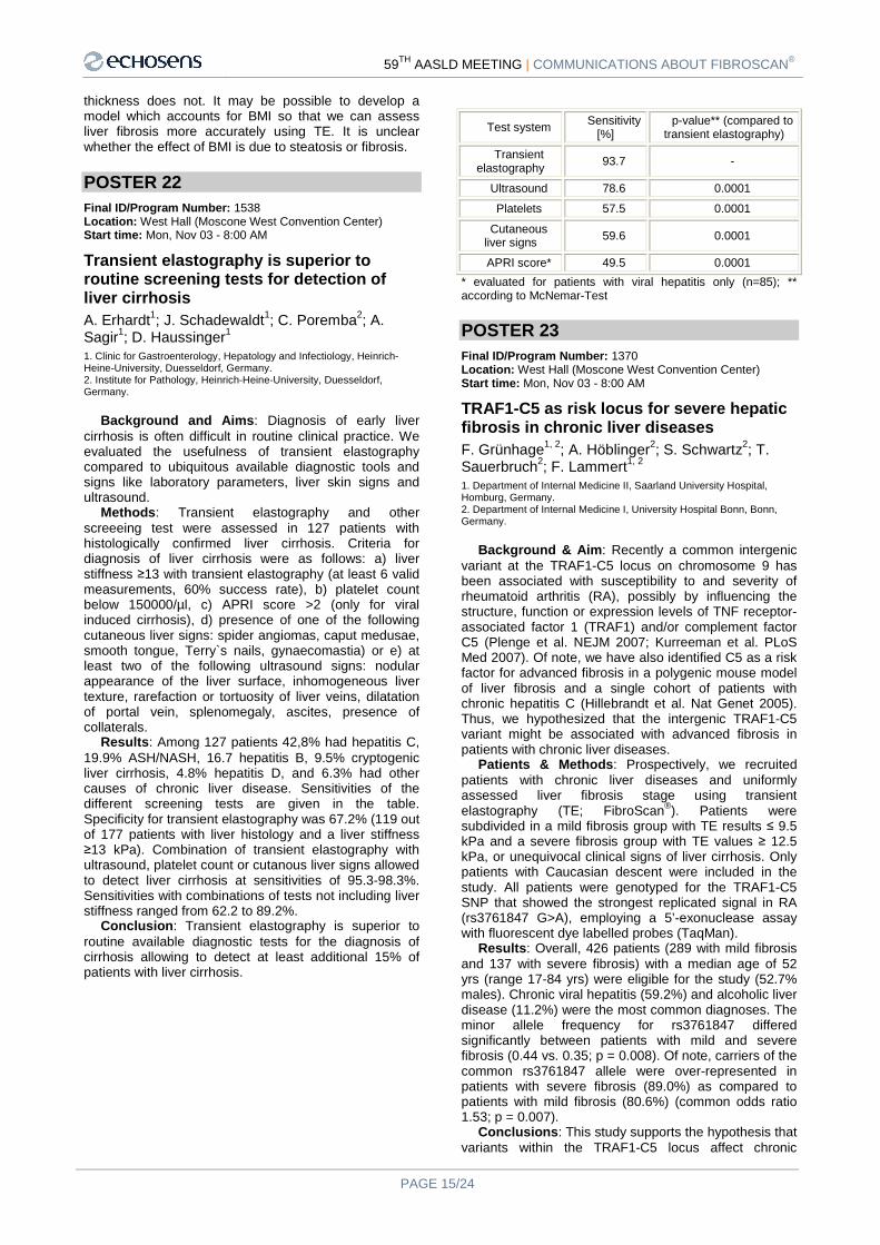
Results : Among 127 patients 42,8% had hepatitis C, 19.9% ASH/NASH, 16.7 hepatitis B, 9.5% cryptogenic liver cirrhosis, 4.8% hepatitis D, and 6.3% had other causes of chronic liver disease. Sensitivities of the different screening tests are given in the table. Specificity for transient elastography was 67.2% (119 out of 177 patients with liver histology and a liver stiffness ≥13 kPa). Combination of transient elastography with ultrasound, platelet count or cutanous liver signs allowed to detect liver cirrhosis at sensitivities of 95.3-98.3%. Sensitivities with combinations of tests not including liver stiffness ranged from 62.2 to 89.2%.
Conclusion : Transient elastography is superior to routine available diagnostic tests for the diagnosis of cirrhosis allowing to detect at least additional 15% of patients with liver cirrhosis.
Test system Sensitivity [%]
p-value** (compared to transient elastography)
Transient elastography
93.7 -
Ultrasound 78.6 0.0001
Platelets 57.5 0.0001
Cutaneous liver signs 59.6 0.0001
APRI score* 49.5 0.0001
* evaluated for patients with viral hepatitis only (n=85); ** according to McNemar-Test
POSTER 23 Final ID/Program Number: 1370 Location: West Hall (Moscone West Convention Center) Start time: Mon, Nov 03 - 8:00 AM
TRAF1-C5 as risk locus for severe hepatic fibrosis in chronic liver diseases F. Grünhage1, 2; A. Höblinger2; S. Schwartz2; T. Sauerbruch2; F. Lammert1, 2 1. Department of Internal Medicine II, Saarland University Hospital, Homburg, Germany. 2. Department of Internal Medicine I, University Hospital Bonn, Bonn, Germany.
Background & Aim : Recently a common intergenic
variant at the TRAF1-C5 locus on chromosome 9 has been associated with susceptibility to and severity of rheumatoid arthritis (RA), possibly by influencing the structure, function or expression levels of TNF receptor-associated factor 1 (TRAF1) and/or complement factor C5 (Plenge et al. NEJM 2007; Kurreeman et al. PLoS Med 2007). Of note, we have also identified C5 as a risk factor for advanced fibrosis in a polygenic mouse model of liver fibrosis and a single cohort of patients with chronic hepatitis C (Hillebrandt et al. Nat Genet 2005). Thus, we hypothesized that the intergenic TRAF1-C5 variant might be associated with advanced fibrosis in patients with chronic liver diseases.
Patients & Methods : Prospectively, we recruited patients with chronic liver diseases and uniformly assessed liver fibrosis stage using transient elastography (TE; FibroScan®). Patients were subdivided in a mild fibrosis group with TE results ≤ 9.5 kPa and a severe fibrosis group with TE values ≥ 12.5 kPa, or unequivocal clinical signs of liver cirrhosis. Only patients with Caucasian descent were included in the study. All patients were genotyped for the TRAF1-C5 SNP that showed the strongest replicated signal in RA (rs3761847 G>A), employing a 5’-exonuclease assay with fluorescent dye labelled probes (TaqMan).
Results : Overall, 426 patients (289 with mild fibrosis and 137 with severe fibrosis) with a median age of 52 yrs (range 17-84 yrs) were eligible for the study (52.7% males). Chronic viral hepatitis (59.2%) and alcoholic liver disease (11.2%) were the most common diagnoses. The minor allele frequency for rs3761847 differed significantly between patients with mild and severe fibrosis (0.44 vs. 0.35; p = 0.008). Of note, carriers of the common rs3761847 allele were over-represented in patients with severe fibrosis (89.0%) as compared to patients with mild fibrosis (80.6%) (common odds ratio 1.53; p = 0.007).
Conclusions : This study supports the hypothesis that variants within the TRAF1-C5 locus affect chronic
59TH AASLD MEETING | COMMUNICATIONS ABOUT FIBROSCAN®
PAGE 16/24
inflammatory responses and modify fibrogenesis in chronic liver diseases irrespective of the underlying etiology, possibly by modulating the Th1/Th2 balance.
POSTER 24 Final ID/Program Number: 1766 Location: West Hall (Moscone West Convention Center) Start time: Tue, Nov 04 - 8:00 AM
Prevalence and causes of discordance between FibroScan ® and Fibrotest for first-line assessment of liver fibrosis in chronic hepatitis C (CHC): A prospective analysis over 4 years in 3148 patients L. Castera1, 2; M. Haaser1; B. Le Bail3; J. Foucher1; P. Bernard2; W. Merrouche1; V. de Ledinghen 1; P. Couzigou1 1. Hepatogastroenterology, CHU Bordeaux, Hopital Haut Leveque, Pessac, France. 2. Hepatogastroenterology, CHU Bordeaux, Hopital St Andre, Bordeaux, France. 3. Pathology, CHU Bordeaux, Hopital Pellegrin, Bordeaux, France.
Background : Combining 2 independent non invasive
methods (liver stiffness measurement (LSM) using FibroScan® (FS) and serum index such as FibroTest (FT)) increases accuracy for liver fibrosis assessment in CHC patients. However, the issue of discordance between FS and FT remains to be solved before implementation in clinical practice.
Aim : to prospectively analyse the prevalence and causes of discordance between FS and FT in 3148 consecutive CHC patients who underwent FS and FT the same day over the past 4 years.
Methods : Prevalence of discordance was determined for significant fibrosis (Metavir F≥ 2) (FS≥7.1 kPa and FT>0.48) and cirrhosis (F4) (FS≥12.5 kPa and FT≥0.75). The following risk factors were analysed: age, gender, BMI, transaminases, manufacturer’s recommendations for FS (IQR/LSM<0.30, success rate<60%) and FT. For the analysis of causes of discordance, liver biopsy (LB) was taken as reference.
Results : Discordance was observed in 24.8% of cases for F≥2 (FS≥7.1 + FT≤0.48: 12.6% and FS<7.1 + FT>0.48: 12.2%) and in 12.9% of cases for F4 (FS≥12.5 + FT<0.75: 5% and FS<12.5 + FT≥0.75: 7.9%), respectively. In multivariate analysis, discordance for F≥2 was independently associated with: older age (OR: 1.01, 95% CI (1.01-1.0.2); p<0.001); male gender (1.57 (1.32-1.87); p<0.0001) and increased IQR/LSM (2.45 (1.37-4.39); p<0.002). Discordance for F4 was independently associated with: IQR/LSM (OR: 4.16 (1.96-8.83); p=0.002), higher FT (222 (124-400); p<0.0001), AST (0.99 (0.98-0.99); p<0.0001) and ALT (1.01 (1.01-1.0.2); p<0.0001). Among the 1499 patients who underwent LB, 314 patients (181 males, mean age 52±11yrs) with LB performed the same day as FS and FT (mean length 21±7mm; read by a single pathologist blinded to FS and FT results) were analysed. Discordance for F≥2 was observed in 28% of cases and was more frequently related to FT than FS (false negative (FN): 41% vs. 33%; false positive (FP): 16% vs. 9% ; correctly classified (CC): 43% vs. 57%, respectively; p<0.0001). Discordance for F4 was observed in 21% of cases and more frequently related to FT than FS (FP: 46% vs. 15%; FN: 28% vs. 10%; CC: 25% vs. 73%, respectively; p<0.0001). Excluding
patients not satisfying to manufacturer’s recommendations decreased discordance rate.
Conclusion : Discordance between FS and FT is more frequent for the diagnosis of significant fibrosis than cirrhosis. In both cases, discordance is more often related to FT than FS. Our results suggest that first-line assessment of liver fibrosis, using the combination of FS and FT, is implementable in clinical practice in patients with CHC, provided manufacturer’s recommendations for FS and FT are satisfied.
POSTER 25 Final ID/Program Number: 1769 Location: West Hall (Moscone West Convention Center) Start time: Tue, Nov 04 - 8:00 AM
Correlation of transient elastography (FibroScan ®) and hepatic artery doppler sonography in patients 30 years after single source hepatitis C genotype 1B outbreak in Germany I. Schiefke1; M. Wiese1; J. Wiegand2 1. Community Hospital "St. George", Leipzig, Germany. 2. Internal Medicine II, University of Leipzig, Leipzig, Germany.
Introduction : The cohort of German women infected
with hepatitis C virus (HCV) genotype 1b via contaminated anti-D immunoglobulin in 1978/79 represent a well characterized group to investigate the natural course of HCV infection. Progression to cirrhosis is rare in this group. Therefore non invasive studies to identify patients with rapid progression of fibrosis have increasingly gained attention.
Methods : On a single occasion in the 30th year after the defined HCV 1b infection (from August 1978 until March 1979) due to HCV contaminated anti-D immunoglobulin we performed transient elastography (FibroScan®), sonography and doppler sonography of the hepatic artery (parameter: hepatic artery pulsatility index (HAPI) and hepatic artery resistance index (HARI)). Results were correlated to corresponding liver biopsies (within last 5 years) if available.
Results : A total of 44 females (mean age 52 years, mean body mass index 24.91 kg/m²) were included. Four groups (G) were defined according to liver stiffness (kPa)-G0: <4.2; G1: 4.2-6,25; G2: 6.25-7.8; G3: 7.8-13.7; G4 >13.7. The distribution of patients was G0=34%, G1=32%; G2=18%; G3=14%; G4=2%. HAPI and HARI were well correlated with liver stiffness measurement (HARI: r=0.35; HAPI: r=0.46; both p<0.001). In a subgroup of patients (n=20/44 liver biopsies were available (Ishak fibrosis score: F0-1: n= 14; F2-4: n=6; F5-6: n=0). Independently of the Ishak score, neither transient elastography (r=-0.27, p=0.24) nor doppler measurements (HARI r=0.14; p=0.55; HAPI r=-0.17; p=0.47) were able to predict the amount of fibrosis. A body mass index greater or less then 25 kg/m² did not correlate with liver stiffness according to FibroScan® or the Ishak fibrosis score (r=-0.1; p=0.37).
Conclusions : Measurement of liver stiffness with transient elastography correlates well with doppler sonography indices. In the subgroup of patients with liver biopsy data failed to show a close correlation between FibroScan®, doppler indices and histological staging probably due to the high proportion of cases with mild fibrosis. Further studies are warranted which validate FibroScan® and doppler sonography in cases with mild fibrosis to substitute follow-up liver biopsies.
59TH AASLD MEETING | COMMUNICATIONS ABOUT FIBROSCAN®
PAGE 17/24
POSTER 26 Final ID/Program Number: 1681 Location: West Hall (Moscone West Convention Center) Start time: Tue, Nov 04 - 8:00 AM
Liver stiffness predicts clinical outcomes in patients with chronic liver disease D. Klibansky1; P. G. Blanco1; E. Kelly1; A. Brown1; N. H. Afdhal1 1. Beth Israel Deaconess Medical Center, Boston, MA, USA.
Introduction : Determining prognosis and optimal
treatment in patients with chronic liver disease has depended upon histology and liver biopsy. Non-invasive measures to determine the presence or absence of underlying fibrosis and assess subsequent risk for morbidity and mortality are needed. Previous research has demonstrated that Transient Elastography (TE) can accurately determine stage of fibrosis or cirrhosis. The purpose of this study was to determine whether this technology can reliably predict clinical outcomes in a population of patients with chronic liver disease.
Methods : 600 patients with liver disease (HCV 334, HBV 32, male 70%, cirrhosis 167) were followed at a single liver center and underwent TE. Time to clinical events was recorded as defined by death, development of ascites or encephalopathy, variceal bleed, increase in Child score by 2, diagnosis of hepatocellular carcinoma or liver transplant. Patients were followed for an average of 626 days after elastography. 43 of 600 patients achieved a predetermined clinical outcome during the observed surveillance period. Univariate logistic regression was used to determine which patient related variables were predictive of the occurrence of a clinical event in an individual subject. The strength of association between each variable studied and the occurrence of clinical event during the study was determined. Positive variables underwent multivariate analysis using a forward method of variable inclusion.
Results : Univariate logistic regression analysis revealed significant differences between those with and without events for the following variables: elasticity, age, MELD, Child score, platelet count, albumin, AST level, APRI, AST/ALT, histological stage and grade, and presence of varices. Multivariate logistic regression analysis revealed the only independent predictors of outcome included Child score (OR 3.35; [2.33 to 4.80]) and elasticity (OR 1.06 [1.07 to 1.08]). ROC analysis for transient elastography revealed an AUC of .88 +/- .021 (p< .0001) with resultant sensitivity and specificity of 97% and 61% respectively as well as a NPV and PPV of 99% and 16% using a score of 10kPa as a threshold value.
Conclusion : TE is superior to liver biopsy for predicting clinical outcomes. Both the Child score and TE score independently predicted clinical outcomes and may be combined to generate a model to best predict risk clinical outcome in patients with chronic liver disease.
POSTER 27 Final ID/Program Number: 1795 Location: West Hall (Moscone West Convention Center) Start time: Tue, Nov 04 - 8:00 AM
Comparison of liver stiffness, FibroTest and liver biopsy for the assessment of liver
fibrosis in renal transplant patient with chronic viral hepatitis L. Alric1; N. Kamar2; M. Danjou3; S. THebault1; L. Rostaing2 1. Service de Medecine Interne- Pôle Digestif CHU Purpan, Toulouse, France. 2. Service de Medecine INterne- Pôle Digestif CHU Purpan, Toulouse, France. 3. Laboratoire d'Anatomopathologie CHU Purpan, Toulouse, France.
Aims : because liver biopsy (LB) is an invasive
procedure, the aims of this study was to assess the accuracy of two non-invasive tools, FibroTest (FT) and liver stiffness measurement (LSM), in the assessment of liver fibrosis in renal transplant patients with chronic Hepatitis virus B (HBV) or C (HCV) infection.
Methods : 38 renal transplant patients with HCV (n=26) or HBV (n=12) infection prospectively followed, underwent LB followed by FT and LSM. Diagnostic performance of FT and LSM were assessed using ROC curves.
Results : The corresponding area under the ROC curve for mild fibrosis stage < F2 was 0.69 (0.47-0.91) for Fibrotest and 0.68 (0.45-0.90) for LSM. For severe fibrosis stage F3-F4, areas under curves were 0.55 (0.35-0.76) and 0.69 (0.50-0.87) for FT and LSM, respectively, with respect to no significant liver fibrosis.
Conclusion : As compared to LB, accuracy of FT and LSM is satisfactory for renal transplant patients with chronic HCV or HBV infection and mild liver fibrosis. The diagnostic value of FT and LSM for the prediction of severe liver disease is not confirmed by our study.
POSTER 28 Final ID/Program Number: 1902 Location: West Hall (Moscone West Convention Center) Start time: Tue, Nov 04 - 8:00 AM
Reduction of liver stiffness by interferon treatment for chronic hepatitis C measured by transient elastography Y. Arima1; N. Kawabe1; S. Hashimoto1; M. Harata1; Y. Nitta1; M. Murao1; T. Nakano1; H. Shimazaki1; N. Komura1; K. Kobayashi1; K. Yoshioka1 1. Internal Medicine, Fujita Health University, Toyoake, Aichi, Japan.
Background : Transient Elastography (TE) enables
non-invasive measurement of liver stiffness (LS), correlates with fibrosis assessed in liver biopsy. In chronic hepatitis C, interferon (IFN) treatment may reduce liver fibrosis, although it is difficult to perform liver biopsies repeatedly after IFN treatment. To assess the reduction of liver fibrosis after IFN treatment, LS was measured repeatedly by TE.
Methods : One hundred and seventeen of patients with chrnonic hepatitis C treated by various IFNs with or without ribavirin (RBV). 25 patients without IFN treatment were also examined. LS was measured before treatment, at the end of treatment, and one year after treatment. The reduction of LS was calculated by the equation; reduction rate= 100-(LS after treatment)/(pretreatment LS) x100.
Results : Seventy one patients became sustained virological responders (SVRs), 25 patients became virological relapsers and 21 patients sustained to be positive for HCV RNA during treatment(virological nonresponders, NRs ). LS was significantly reduced at
59TH AASLD MEETING | COMMUNICATIONS ABOUT FIBROSCAN®
PAGE 18/24
the end of treatment either in SVRs (10.8±45.9%, p=0.021), in virological relapsers (28.5±25.5%, p=0.017 ), or in NRs (17.9±32.1%, p=0.027). One year after treatment, LS maintain to be significantly reduced either in SVRs (24.7±44.1%, p=0.004) or in relapsers ( 20.8±37.2%, p=0.0055 ) compared with pretreatment values, but not in NRs (-0.8±44.5%). LS did not change significantly in patients without IFN treatment (observation period; 776±252 days);
Conclusion : LS was reduced in SVRs, relapsers, or NRs at the end of treatment. One year after treatment, LS maintained to be low levels in SVRs and relapsers, while LS reversed to the pretreatment values in NRs. IFN treatment was demonstrated to reduce LS in most of the patients, even in NRs. The reduction of LS sustained in SVRs and even in relapsers at least for one year after the end of treatment. Further studies are needed to clarify whether the reduction of LS observed in the present study actually indicates alleviation of fibrosis.
POSTER 29 Final ID/Program Number: 1960 Location: West Hall (Moscone West Convention Center) Start time: Tue, Nov 04 - 8:00 AM
Difference of liver stiffness after acute or chronic liver injury due to viral hepatitis J. Kim1; H.. Yim1; Y. Jung1; J. Kim1; Y. Seo1; J. Yeon1; S. Um1; K. Byun1 1. Department of Internal Medicine, Korea University Medical Center, Seoul, South Korea.
Background/Aims : Liver Stiffness (LS)
measurement by transient elastography is a noninvasive method that can estimate degrees of liver fibrosis in patients with chronic liver disease. Recently, there have been several reports that LS is increased in acute viral hepatitis patients. However, long term data of LS after recovery of acute viral hepatitis are still lacking. This needs to be assessed to know if there is any sequel in terms of fibrosis in these patients. In the present study, we evaluated difference of LS between patients who recovered from acute viral hepatitis more than 1 year ago and healthy control group, acute viral hepatitis patients, chronic hepatitis patients.
Patients/Method : Among patients who had admitted at Korea University Ansan Hospital between January 2006 and January 2007 due to acute viral hepatitis (A, B or C) and recovered were enrolled (Group A, n=22). We measured the LS and compared with those of healthy control group (Group B, n=17), current acute viral hepatitis group (Group C, n=31) and chronic hepatitis group - the patients who have viral replication over 1 year but show normal sonographic finding - (Group D, n=31). The statistical methods used were Mann-Whitney U-test in continuous variables and Chi-square test or Fisher's exact test in categorial variables. P values less than 0.05 were considered statistically significant.
Result : In Group A, mean ALT level was 24.4 (8-51) IU/L and there was no statistic difference from ALT level of Group B, 20.5 (8-50) IU/L (p=0.318). LS was also not significantly different (Group A, 5.02 (3.3-7.2) kPa vs. Group B, 4.37 (1.8-6.3) kPa, p=0.154). But when compared with Group C (ALT 1649.9 (15-5514), p<0.001) and LSM (12.9 (6.3-44.3), p<0.001) and with Group D (ALT 101.9 (12-476) IU/L, p=0.047 and LSM 10.7 (3.2-48.8) kPa, p<0.001), Group A had lower ALT and LSM values.
Conclusion : LS of patients who recovered from acute viral hepatitis more than 1 year ago was shown to be similar to healthy control group but significant difference was observed in comparison with current acute viral hepatitis patients or chronic hepatitis patients. It is considered that chronic liver fibrosis is not developed in patients who recovered from acute viral hepatitis
POSTER 30 Final ID/Program Number: 1791 Location: West Hall (Moscone West Convention Center) Start time: Tue, Nov 04 - 8:00 AM
Diagnostic accuracy of transient elastography: a comparison between chronic hepatitis B and C correlated with optimal-length liver biopsies C. Verveer1; B. E. Hansen2; P. E. Zondervan3; H. L. Janssen1; R. J. de Knegt1 1. Dept. of Gastroenterology and Hepatology, Erasmus MC University Medical Centre Rotterdam, Rotterdam, Netherlands. 2. Biostatistics, Erasmus MC, University Medical Centre, Rotterdam, Rotterdam, Netherlands. 3. Pathology, Erasmus MC, University Medical Centre, Rotterdam, Rotterdam, Netherlands.
Background : Transient Elastography (TE) is used to
assess hepatic fibrosis non-invasively, mostly in chronic viral hepatitis C (CHC). The efficacy in chronic viral hepatitis B (CHB) is less well established. The diagnostic performance of TE in severe fibrosis and cirrhosis is good, but less reliable in the Metavir fibrosis stages F0-F2. We investigated whether the diagnostic performance and accuracy improved when correlated with liver biopsies of optimal length and whether outcome was influenced by inflammatory activity according to the Ishak HAI score, steatosis and iron. In addition, a comparison between CHC and CHB was made.
Methods : 257 consecutive patients with a liver biopsy measuring at least 25mm and concomitant TE (FibroScan) were enrolled. Liver specimens were scored for fibrosis (both Metavir and Ishak classification) with the assessment of necro-inflammatory activity according to the modified HAI system. In addition the amount of steatosis (Brunt classification) and iron were scored.
Results : 137 patients with CHB, 117 patients with CHC and 3 patients with combined CHC and CHB were evaluated. Patients were categorized as follows in the Metavir classification: 22 patients F0, 98 F1, 68 F2, 47 F3 and 22 F4. There were no significant differences in the reliability of elastographical measurements between patients with CHB or CHC, when corrected for age, BMI, Metavir classification, steatosis, iron and inflammatory activity. The ROC-curves for the different Metavir classifications were: F1 0.75, F2 0.81, F3 0.90 and F4 0.89. Necro-inflammatory activity, which was significantly correlated with ALT, was an independent factor to influence the TE measurements. With increasing inflammatory activity, the FibroScan outcome can be 1.4 kPa higher than expected. Steatosis and the amount of iron had no significant effect on TE measurement.
Conclusion : Diagnostic accuracy of TE by FibroScan® in CHB and CHC is similar. TE correlates very well with severe fibrosis (F3) and cirrhosis (F4) as well with moderate fibrosis (F2). Active inflammation of the liver produces a significant increase in TE outcome and should therefore be taken into account.

59TH AASLD MEETING | COMMUNICATIONS ABOUT FIBROSCAN®
PAGE 19/24
POSTER 31 Final ID/Program Number: 1792 Location: West Hall (Moscone West Convention Center) Start time: Tue, Nov 04 - 8:00 AM
Acoustic radiation force elastography versus transient elastography for non-invasive assessment of liver fibrosis in viral hepatitis: a new alternative? M. Friedrich-Rust1; K. Wunder1; F. Sotoudeh1; S. Kriener3; S. Martens2; T. Poynard4; E. Herrmann2; J. Bojunga1; S. Zeuzem1; C. Sarrazin1 1. Department of Internal Medicine, J.W.Goethe University Hospital, Frankfurt a. M., Germany. 2. Institute of Biostatistics and Math. Modeling, Faculty of Medicine, J.W.Goethe University, Frankfurt, Germany. 3. Institute of Pathology, J.W.Goethe University Hospital, Frankfurt, Germany. 4. Hospital Pitié-Salpêtrière, Paris, France.
Aims : We present a first prospective study comparing
a novel ultrasound-based Acoustic Radiation Force Impulse (ARFI) imaging technology (Siemens Acuson S2000 Virtual TouchTM tissue quantification) with transient elastography (FibroScan®, Echosens) for the non-invasive assessment of liver fibrosis in patients infected with chronic viral hepatitis. ARFI imaging technology is integrated in an ultrasound machine and can be performed with conventional ultrasound probes during routine ultrasound examination.
Methods : ARFI imaging involves the mechanical excitation of tissue using short-duration acoustic pulses to generate localized, micron-scale displacements in tissue. The displacements result in shear-wave propagation away from the region of excitation which is tracked using ultrasonic, correlation-based methods. The results are expressed by the shear wave velocity (m/s); the stiffer the tissue the faster the shear wave velocity. 89 patients with chronic viral hepatitis and either present histological assessment of the fibrosis stage (n=74) or proven liver cirrhosis (n=15) were assessed. All patients received the ultrasound-based methods transient elastography and ARFI imaging, as well as detailed blood examinations. 20 healthy volunteers were examined as control group. The results were compared to liver biopsy as reference method, as well as to each other.
Results : The median velocity measured with ARFI imaging technology in the 20 healthy volunteers was 1.10 m/s (range 0.64 – 2.52). The median velocity in the study population ranged from 0.80 to 4.26 m/s. The Spearman correlation coefficient between the ARFI imaging, transient elastography, and FibroTest on the one hand and the histological fibrosis stage on the other hand was highly significant with 0.66, 0.74, and 0.61, respectively (p<0.001). The diagnostic accuracy expressed as areas under ROC curves (AUROC) for the different fibrosis stage is shown in the attached table. At the time of this analysis laboratory and FibroTest results were only available in 71/89 patients.
Discussion : ARFI based shear wave velocity quantification is a new and promising ultrasound-based, non-invasive method for the assessment of liver fibrosis in chronic viral hepatitis. Diagnostic accuracy was highly comparable to transient elastography. Furthermore, a complete abdominal ultrasound examination can be performed and with additional 5 minutes for 10 measurements the liver stiffness can be measured simultaneously.
AUROC for F≥2
AUROC for F≥3
AUROC for F=4
FibroScan® 0.87 0.88 0.91
ARFI-imaging 0.87 0.90 0.93
FibroTest 0.81 0.86 0.77
POSTER 32 Final ID/Program Number: 1796 Location: West Hall (Moscone West Convention Center) Start time: Tue, Nov 04 - 8:00 AM
Combination of 13C-methacetin breath test by online molecular correlation spectroscopy and transient elastography improves diagnostic performance of non-invasive fibrosis assessment in chronic hepatitis C O. Goetze1; A. Grau1; A. Geier 1; T. Gerlach1; M. Fried1; B. Mullhaupt1 1. Department of Internal Medicine, Division of Gastroenterology and Hepatology, University Hospital Zurich, Zurich, Switzerland.
Background : Stable isotope breath tests have been
developed for the non-invasive assessment of microsomal liver function in patients with chronic liver disease. Among different tests, the 13C-methacetin breath test (MBT) is suitable for the assessment of hepatic functional reserve. Aims: To assess prospectively the diagnostic performance of fibrosis detection by MBT in patients with chronic hepatitis C infection using validated molecular correlation spectroscopy with a continuous, online, automatic breath collection and analysis system (Goetze et al., APT 2007) and to compare MBT results with transient elastography.
Methods : The METAVIR fibrosis score of a liver biopsy specimen was used as a reference and was obtained within one week of MBT and elastography. 62 patients (42 M, 44.7 ± 13 y., BMI 24.6 ± 3.6 kg/m², AST 1.2 ± 0.8, ALT 1.8 ± 1.5 x ULN) with chronic hepatitis C were studied (fibrosis stage F0, n=6; F1, n=35; F2, n=8; F3, n=2; F4, n=11) Each patient received 75 mg of 13C-methacetin dissolved in 50 ml of water. The 13C/12C ratio was determined over 90 minutes by molecular correlation spectroscopy (BreathID, Exalenz, Israel) as delta over baseline (DOB [‰]) and was expressed as maximal DOB (DOBmax [‰]). Liver stiffness (FS [kPa]) was measured by transient elastography (FibroScan®, Echosens, France). Diagnostic performance of each test as well as the combination of MBT and elastography (DOBmax/FS [‰/kPa]) were compared by receiver operator characteristics (ROC) analysis and statistical ROC curve comparisons.
Results : Mean areas under the ROC curve for DOBmax and stiffness values were similar for F≤1(0.78 vs. 0.85, p=0.3), F≤2 (0.84 vs. 0.89, p=0.5) and F≤3 (0.85 vs. 0.93, p=0.2). Method combination (DOBmax/FS) increased the diagnostic performance for both methods depending on the clinical context: F≤1 (0.86: p=0.05 vs. MBT, p=0.8 vs. elastography), F≤2 (0.94: p=0.09 vs. MBT, p<0.05 vs. elastography) and F≤3 (0.97: p<0.05 vs. MBT, p=0.07 vs. elastography).
Conclusions : MBT by continuous automatic molecular correlation spectroscopy is an easy to use and effective method for assessing liver fibrosis, with a
59TH AASLD MEETING | COMMUNICATIONS ABOUT FIBROSCAN®
PAGE 20/24
similar diagnostic performance compared to elastography. A combination of methacetin breath test and elastography results increases the diagnostic performance of each method alone for non-invasive fibrosis detection in chronic HCV infection.
POSTER 33 Final ID/Program Number: 1765 Location: West Hall (Moscone West Convention Center) Start time: Tue, Nov 04 - 8:00 AM
A sequential predictive algorithm combining APRI and transient elastography (TE) for diagnosing liver fibrosis in chronic hepatitis C M. Fraquelli1; C. Rigamonti2; G. Casazza3; A. Colli4; M. Rumi2; D. Conte1; M. Donato2; G. Ronchi2; M. Colombo2
1. II Division Gastroenterology, Fondazione IRCCS Ospedale Policlinico, Mangiagalli e Regina Elena, Milano, Italy. 2. I Division Gastroenterology, Fondazione IRCCS Ospedale Policlinico, Mangiagalli e Regina Elena, Milano, Italy. 3. Institute of Statistic and Biometry, University of Milan, Milano, Italy. 4. Internal Medicine, Ospedale A Manzoni, Lecco, Italy.
Background/Aims : In the search for non-invasive
diagnostic tools for hepatic fibrosis in patients with chronic hepatitis C, Transient Elastography (TE) has gained popularity. It, in fact, measures liver stiffness as marker for hepatic fibrosis and could therefore applied to the selection of the best candidates for further investigations with liver biopsy. We assessed the diagnostic performance of a sequential algorithm combining the AST to platelet ratio index (APRI) with TE using liver biopsy as the reference standard in the evolution of patients with chronic hepatitis C.
Methods : 459 consecutive patients with chronic hepatitis C were evaluated for APRI and TE (FibroScan®; Echosens, Paris, France) at same day of the liver biopsy. TE examinations with at least 10 validated measurements and a success rate of >60% were considered adequate. Ultrasound guided-liver biopsy was performed with a 16 G needle. Necroinflammation and fibrosis were scored by METAVIR and compared with APRI and TE results to generate their operative characteristics. The tests were sequentially combined to obtain a predictive rule for F>=2, F>3 and F=4 with at least a post-test probability > 90%, for confirmation and <10% for exclusion.
Results : 257 (56%) patients had F0-1, 110 (24%) had F2, 47 (10%) had F3 and 45 (10%) had F4 (cirrhosis).The APRI score was <0.5 in 170 patients, >0.5-<1.5 in 214 and >1.5 in the remaining 75. In diagnosing F>2 the predictive algorithm including APRI and TE (7 kPa cut off) had an area under the curve (AUROC) of 0.82 (95% CI 0.76-0.87);. in diagnosing F>3 and F=4 the predictive algorithms including APRI and TE (14 kPa cut off) had an AUROC of 0.81 (0.76-0.87) and 0.85 (0.78-0.92), respectively. The clinical utility model showed that 39%, 83% and 83% of liver biopsies, i.e. from 179 to 380 procedures, could be avoided accordingly.
Conclusions : Stepwise application of APRI and TE prior to liver biopsy improves the diagnostic algorithm in patients with chronic hepatitis C, reducing by 39–83 % the need for a liver biopsy with a significant reduction of risk and costs, in parallel. Interestingly, the predictive algorithm was more efficient in reducing the need for
liver biopsies in F3/F4 patients for which the risks of an invasive procedure are substantially higher.
POSTER 34 Final ID/Program Number: 1776 Location: West Hall (Moscone West Convention Center) Start time: Tue, Nov 04 - 8:00 AM
Error factors for transient elastography in chronic hepatitis C V. Calvaruso1, 2; C. Cammà1; V. Di Marco1; S. Maimone3; F. Bronte1; M. Enea1; M. Pleguezuelo2; E. Xirouchakis2; M. Misseri2; P. Manousou2; G. M. Dusheiko3; A. K. Burroughs2; A. Craxì1 1. Gastroenterology and Hepatology Unit, DIBIMIS, University of Palermo, Palermo, Italy. 2. Liver Transplantation and Hepatobiliary Unit, London, United Kingdom. 3. Centre for Hepatology, Royal Free and University College School of Medicine, London, U.K.
Background : Transient elastography (TE) has a
good diagnostic accuracy for cirrhosis. However the AUROC to assess significant fibrosis (Metavir ≥ F2) has a high variation depending from features of disease (necro-inflammation) independent from fibrillar extracellular matrix. To improve the accuracy of TE correction should be made for these factors.
Aim : To establish how TE varies according to individual patient profiles, particularly when TE is discordant with histological fibrosis.
Methods : 300 consecutive HCV-RNA positive patients (200 Italy, 100 UK), all HIV and HBV negative, abstinent from alcohol and treatment-naïf were enrolled. All had liver tests and TE performed at the day of biopsy. Sixteen subjects were excluded from analysis due to high BMI.
Results : Independent predictors of stiffness by multivariate linear regression analysis were AST and Metavir’s stage score. F ≥ 2 was independently associated with age, stiffness and grade score (p<0.05). Using 7.1 kPa as stiffness cut-off for F ≥ 2, 96 patients (32%) had discordant liver stiffness and fibrosis. By marginal logistic model AST value was the only significant variable (p=0.046) in the discordance between liver fibrosis and liver stiffness by TE. 68/214 patients (32%) with AST ≤ 80 UI/l had stage ≥ 2 with a liver stiffness < 7.1 kPa. In 12 patients (6%) liver stiffness was ≥ 7.1 kPa without significant fibrosis at histology. Among the 70 patients with AST ≥ 80 UI/l, 7(10%) were false negatives and 9(13%) false positives of TE. An algorithm to improve the performance of TE for moderate fibrosis (AUROC:0.78) is proposed in fig.1. Adjusting stiffness value according to age and AST value achieves an AUROC of 0.801 for F ≥ 2.
Conclusion : In clinical practice, each liver stiffness measure must be interpreted taking into account the patient‘s clinical and biochemical profile. Simple adjustments for age and ALT may significantly improve diagnostic accuracy.
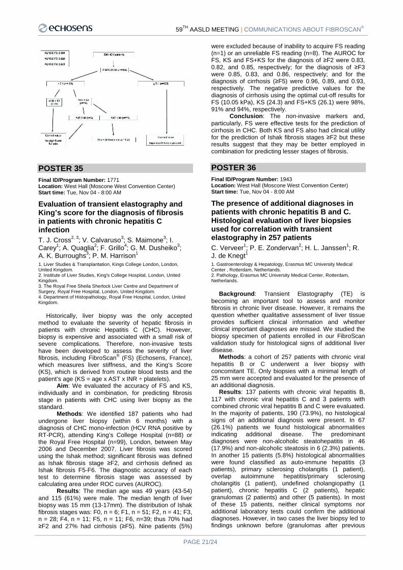
59TH AASLD MEETING | COMMUNICATIONS ABOUT FIBROSCAN®
PAGE 21/24
POSTER 35 Final ID/Program Number: 1771 Location: West Hall (Moscone West Convention Center) Start time: Tue, Nov 04 - 8:00 AM
Evaluation of transient elastography and King’s score for the diagnosis of fibrosis in patients with chronic hepatitis C infection T. J. Cross2, 3; V. Calvaruso3; S. Maimone3; I. Carey1; A. Quaglia2; F. Grillo4; G. M. Dusheiko3; A. K. Burroughs3; P. M. Harrison1 1. Liver Studies & Transplantation, Kings College London, London, United Kingdom. 2. Institute of Liver Studies, King's College Hospital, London, United Kingdom. 3. The Royal Free Sheila Sherlock Liver Centre and Department of Surgery, Royal Free Hospital, London, United Kingdom. 4. Department of Histopathology, Royal Free Hospital, London, United Kingdom.
Historically, liver biopsy was the only accepted
method to evaluate the severity of hepatic fibrosis in patients with chronic Hepatitis C (CHC). However, biopsy is expensive and associated with a small risk of severe complications. Therefore, non-invasive tests have been developed to assess the severity of liver fibrosis, including FibroScan® (FS) (Echosens, France), which measures liver stiffness, and the King’s Score (KS), which is derived from routine blood tests and the patient’s age (KS = age x AST x INR ÷ platelets).
Aim : We evaluated the accuracy of FS and KS, individually and in combination, for predicting fibrosis stage in patients with CHC using liver biopsy as the standard.
Methods : We identified 187 patients who had undergone liver biopsy (within 6 months) with a diagnosis of CHC mono-infection (HCV RNA positive by RT-PCR), attending King’s College Hospital (n=88) or the Royal Free Hospital (n=99), London, between May 2006 and December 2007. Liver fibrosis was scored using the Ishak method; significant fibrosis was defined as Ishak fibrosis stage ≥F2, and cirrhosis defined as Ishak fibrosis F5-F6. The diagnostic accuracy of each test to determine fibrosis stage was assessed by calculating area under ROC curves (AUROC).
Results : The median age was 49 years (43-54) and 115 (61%) were male. The median length of liver biopsy was 15 mm (13-17mm). The distribution of Ishak fibrosis stages was: F0, n = 6; F1, n = 51; F2, n = 41; F3, n = 28; F4, n = 11; F5, n = 11; F6, n=39; thus 70% had ≥F2 and 27% had cirrhosis (≥F5). Nine patients (5%)
were excluded because of inability to acquire FS reading (n=1) or an unreliable FS reading (n=8). The AUROC for FS, KS and FS+KS for the diagnosis of ≥F2 were 0.83, 0.82, and 0.85, respectively; for the diagnosis of ≥F3 were 0.85, 0.83, and 0.86, respectively; and for the diagnosis of cirrhosis (≥F5) were 0.96, 0.89, and 0.93, respectively. The negative predictive values for the diagnosis of cirrhosis using the optimal cut-off results for FS (10.05 kPa), KS (24.3) and FS+KS (26.1) were 98%, 91% and 94%, respectively.
Conclusion : The non-invasive markers and, particularly, FS were effective tests for the prediction of cirrhosis in CHC. Both KS and FS also had clinical utility for the prediction of Ishak fibrosis stages ≥F2 but these results suggest that they may be better employed in combination for predicting lesser stages of fibrosis.
POSTER 36 Final ID/Program Number: 1943 Location: West Hall (Moscone West Convention Center) Start time: Tue, Nov 04 - 8:00 AM
The presence of additional diagnoses in patients with chronic hepatitis B and C. Histological evaluation of liver biopsies used for correlation with transient elastography in 257 patients C. Verveer1; P. E. Zondervan2; H. L. Janssen1; R. J. de Knegt1 1. Gastroenterology & Hepatology, Erasmus MC University Medical Center , Rotterdam, Netherlands. 2. Pathology, Erasmus MC University Medical Center, Rotterdam, Netherlands.
Background : Transient Elastography (TE) is
becoming an important tool to assess and monitor fibrosis in chronic liver disease. However, it remains the question whether qualitative assessment of liver tissue provides sufficient clinical information and whether clinical important diagnoses are missed. We studied the biopsy specimen of patients enrolled in our FibroScan validation study for histological signs of additional liver disease.
Methods : a cohort of 257 patients with chronic viral hepatitis B or C underwent a liver biopsy with concomitant TE. Only biopsies with a minimal length of 25 mm were accepted and evaluated for the presence of an additional diagnosis.
Results : 137 patients with chronic viral hepatitis B, 117 with chronic viral hepatitis C and 3 patients with combined chronic viral hepatitis B and C were evaluated. In the majority of patients, 190 (73.9%), no histological signs of an additional diagnosis were present. In 67 (26.1%) patients we found histological abnormalities indicating additional disease. The predominant diagnoses were non-alcoholic steatohepatitis in 46 (17.9%) and non-alcoholic steatosis in 6 (2.3%) patients. In another 15 patients (5.8%) histological abnormalities were found classified as auto-immune hepatitis (3 patients), primary sclerosing cholangitis (1 patient), overlap autoimmune hepatitis/primary sclerosing cholangitis (1 patient), undefined cholangiopathy (1 patient), chronic hepatitis C (2 patients), hepatic granulomas (2 patients) and other (5 patients). In most of these 15 patients, neither clinical symptoms nor additional laboratory tests could confirm the additional diagnoses. However, in two cases the liver biopsy led to findings unknown before (granulomas after previous
59TH AASLD MEETING | COMMUNICATIONS ABOUT FIBROSCAN®
PAGE 22/24
tuberculosis, and hepatic iron overload with heterozygous beta-thalassemia).
Conclusion : In case TE (FibroScan®) replaces the liver biopsy in the evaluation of patients with chronic hepatitis B or C, important additional liver disease might be missed, being non-alcoholic steatohepatitis or hepatic steatosis in the majority of these cases. Therefore a liver biopsy should still be considered in a new patient presenting with chronic hepatitis B or C.
POSTER 37 Final ID/Program Number: 1958 Location: West Hall (Moscone West Convention Center) Start time: Tue, Nov 04 - 8:00 AM
Transient Elastography (TE) as a tool to evaluate acute liver damage: a prospective, multicentric cohort study V. di Marco1; A. Iacò1; F. Bronte1; V. Calvaruso1; M. Puoti2; L. Biasi2; K. Prestini2; R. Bruno3; D. Achilli3; G. Squadrito4; G. Amaddeo4; A. Craxì1 1. Gastroenterologia & Epatologia, DIBIMIS, University of Palermo, Palermo, Italy. 2. Dipartimento di Malattie Infettive e Tropicali, University of Brescia, Brescia, Italy. 3. Divisione di Malattie Infettive e Tropicali, University of Pavia, Pavia, Italy. 4. Unità di Epatologia Clinica e Biomolecolare, University of Messina, Messina, Italy.
Background and Aim : Discrimination between acute
liver damage and flares of chronic hepatitis may be problematic. Albeit Liver Stiffness Measurement (LSM) by TE is mostly linked to fibrosis, necro-inflammation and tissue edema are known to affect its values thus reducing its diagnostic ability in acute processes. Since the latter lesions are rapidly reversible, contrary to fibrosis, we aimed to assess the longitudinal behaviour of LSM in patients with acute hepatitis (AH).
Patients and Methods : 70 patients (74% males; median age 39 years) with AH (AST/ALT ≥ 10 u.n.l) and no history of previous liver disease seen at 4 Liver Units between 9.06 and 3.08 underwent LSM by TE at presentation and after 2, 4, 8, 12 weeks. Liver damage was due to acute HBV (n=29) or HCV (n=18) infection, drug toxicity (n=14), HAV, CMV or EBV infection (n=9).
Results : The median value of AST, ALT and LSM at presentation were 1090 IU/l, 553 IU/l and 11 kPa respectively. Twenty-eight patients (52%) had an LSM ≥ 12.5 kPa at the presentation. HBV patients had higher median AST/ALT and LSM than those with other aetiology (AST: 890 IU/l vs. 397 IU/l, p = 0.001; ALT: 1787 IU/l vs. 787 IU/l, p = 0.004; LSM: 12.3 kPa vs. 9.8 kPa, p = 0.016). No significant differences of AST/ALT values and LSM were found between patients of viral and non viral aetiology (AST: 649 IU/l vs. 337 IU/l; ALT: 1202 IU/l vs. 785 IU/l; LSM: 11,1 kPa vs. 10 kPa, p = n.s.). By logistic regression analysis the only variable independently associated to LSM ≥ 12.5 kPa was the AST value (OR = 1.003: CI: 1.001-1.005; p = 0.005). Median values of AST, ALT and Stiffness at week 2 after presentation were respectively 140 IU/l, 467 IU/l and 9,2 kPa; 59 IU/l, 114 IU/l and 7,9 kPa at week 4; 37 IU/l, 56 IU/l and 7,2 kPa at week 8 and 33(range:15-140 IU/l), 40(range:7-320 IU/l) and 6(range:4-11 kPa) at week 12. At the end of follow up 12 patients did not achieve the normalization of aminotransferases and stiffness value.
Conclusion : High LSM values at the onset of acute liver damage reflect the degree of necro-inflammation,
regardless of aetiology. The reduction of LSM parallels the fall of aminotranferases confirms the absence of fibrosis. TE can thus be useful in this setting only if repeated during follow up.
POSTER 38
Final ID/Program Number: 1783 Location: West Hall (Moscone West Convention Center) Start time: Tue, Nov 04 - 8:00 AM
A simple diagnostic algorithm to evaluate liver fibrosis in patients with chronic hepatitis C S. M. Martinez1; M. Bruguera1; J. M. Barrera1; X. Forns1; J. M. Sánchez-Tapias1 1. Hospital Clinic, Barcelona, Spain.
Background : Patients with chronic hepatitis C (CHC)
and significant liver fibrosis (F ≥ 2) should be priorized for antiviral treatment with pegylated interferon and ribavirin (NIH consensus statement, 2002). Although liver biopsy remains the reference standard for fibrosis evaluation, several non- invasive methods have been proposed to predict significant fibrosis.
Aim: to evaluate the performance of a simple algorithm to identify patients with CHC who are candidates for antiviral treatment in routine clinical practice.
Methods : The performance of non-invasive methods to predict significant liver fibrosis was investigated in a cohort of 215 consecutive patients with CHC who underwent antiviral therapy from September 2004 to April 2008. Diagnostic accuracy of the following non-invasive methods was assessed by the area under the ROC curve (AUROC): Forns score, FIB-4 index, APRI index and transient elastography (TE). After evaluation of sensitivity, specificity, and positive and negative predictive values, a diagnostic algorithm was designed. Liver fibrosis stage was assessed according to Scheuer classification.
Results : The median age of the cohort was 50 years (18-69), and 132 (61.4%) were male. The distribution of liver fibrosis stages was: F0: 9.8% , F1: 19.1%, F2: 23.7%, F3: 9,3% and F4: 38.2%. AUROC for diagnosis of F≥ 2 were 0.83, 0.85, 0.86, 0.88 for Forns score, FIB-4, APRI and TE, respectively. The best diagnostic algorithm to identify patients with significant fibrosis was achieved by first using the Forns score (cut-off > 6.9) followed by TE (cut-off > 7.5 kPa). The performance of this algorithm in the discrimination between F 0-1 vs F 2-4 is shown in table 1. A total of 185 patients (86%) could be correctly classified as either with or without significant liver fibrosis.
Conclusion : A sequential calculation of the Forns score followed by TE could avoid a liver biopsy in ¾ of patients with CHC in routine clinical practice.
Table 1
Entire CHC cohort (n=215) Forns Score F0-1 (n=62) F2-4 (n= 153) Cut-off > 6.9
(n=71) 3 (4.2%) 68 (95.8%)
Cut-off ≤ 6.9 (n=144) 59 (41%) 85 (59%)

59TH AASLD MEETING | COMMUNICATIONS ABOUT FIBROSCAN®
PAGE 23/24
Patients with Forns Score Cut-off point ≤ 6.9 (n=144) TE F0-1 (n=59) F2-4 (n=85)
Cut-off ≤ 7.5 (n=52) 42 (80.8%) 10 (19.2%)
Cut-off > 7.5 (n=92) 17 (18.5%) 75 (81.5%)

59TH AASLD MEETING | COMMUNICATIONS ABOUT FIBROSCAN®
Echosens, the Echosens logo, FibroScan, the FibroScan logo are registered trademarks of Echosens Company.
© 2007, Echosens, Paris, 75013 www.echosens.com
NOTES