Embed Size (px)
Citation preview
Dr. Tudor H. Hughes M.D., FRCR
Department of Radiology
University of California School of Medicine
San Diego, California
Commonly Missed Injuries of
the Extremities
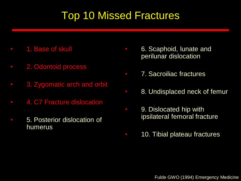
Top 10 Missed Fractures
• 1. Base of skull
• 2. Odontoid process
• 3. Zygomatic arch and orbit
• 4. C7 Fracture dislocation
• 5. Posterior dislocation of humerus
• 6. Scaphoid, lunate and perilunar dislocation
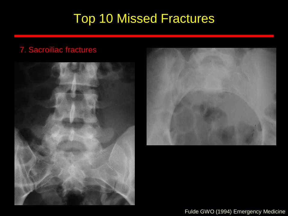
• 7. Sacroiliac fractures
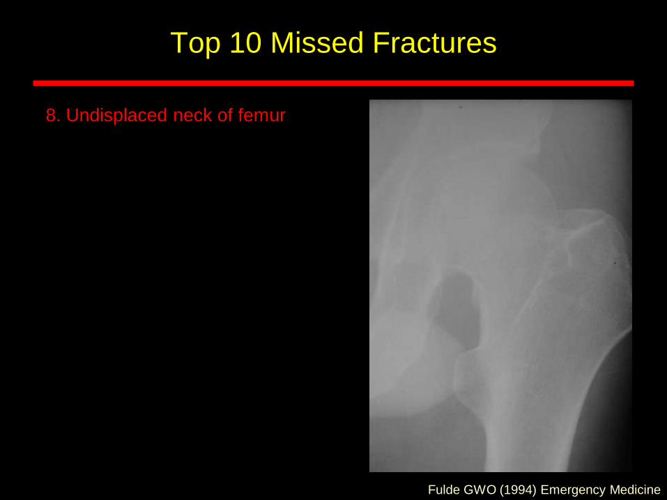
• 8. Undisplaced neck of femur
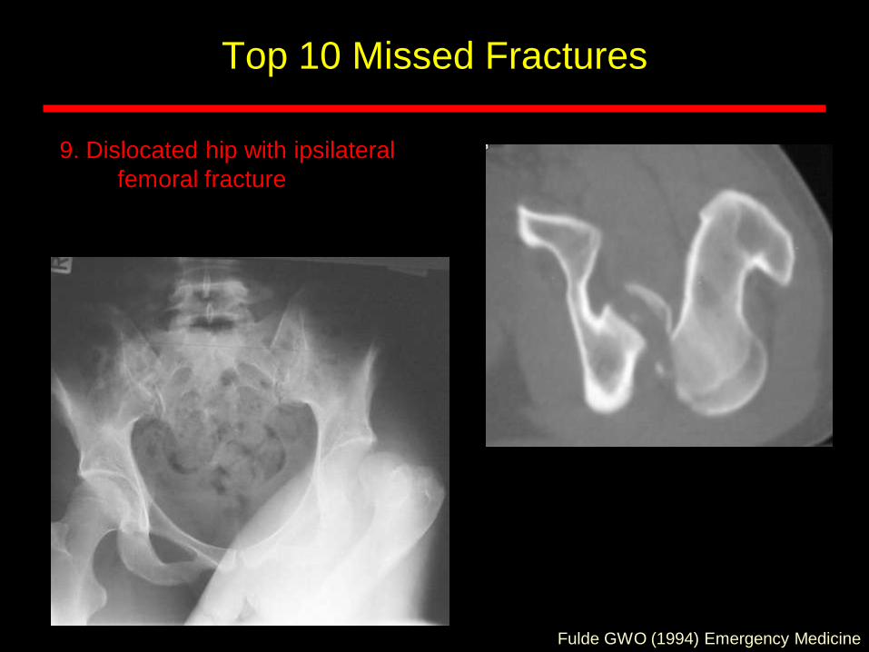
• 9. Dislocated hip with ipsilateral femoral fracture
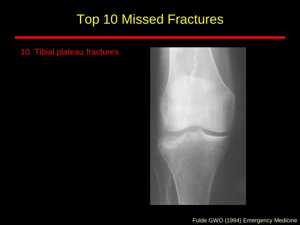
• 10. Tibial plateau fractures
Fulde GWO (1994) Emergency Medicine
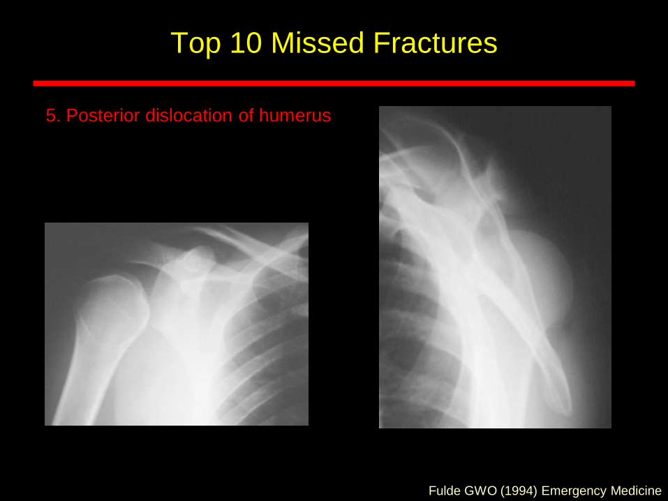
Top 10 Missed Fractures
5. Posterior dislocation of humerus
Fulde GWO (1994) Emergency Medicine
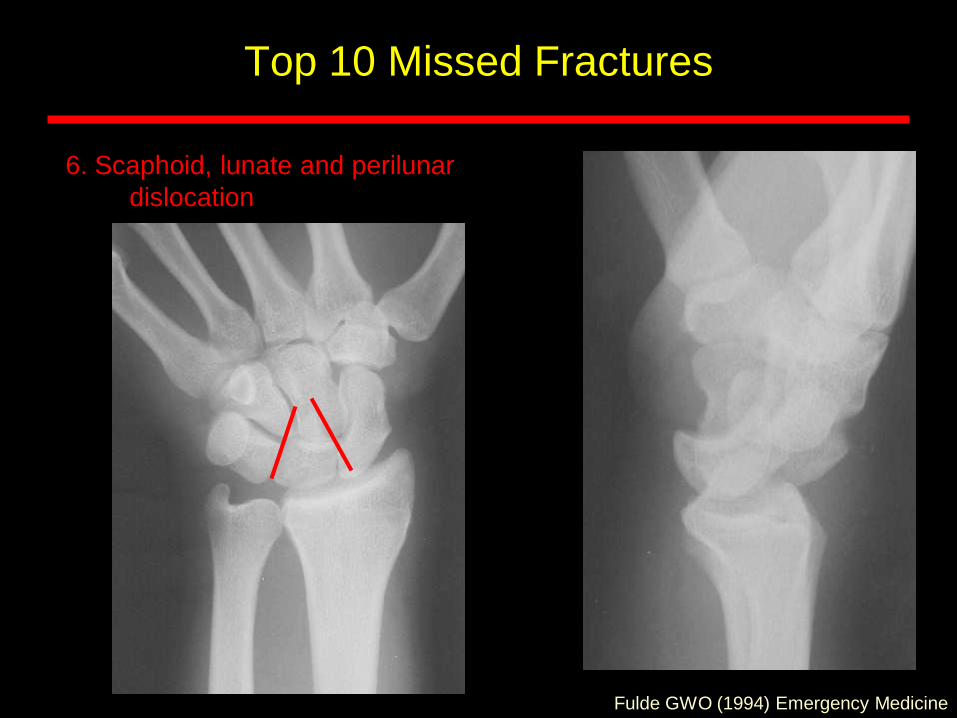
Top 10 Missed Fractures
6. Scaphoid, lunate and perilunar
dislocation
Fulde GWO (1994) Emergency Medicine
Top 10 Missed Fractures
7. Sacroiliac fractures
Fulde GWO (1994) Emergency Medicine
Top 10 Missed Fractures
8. Undisplaced neck of femur
Fulde GWO (1994) Emergency Medicine
Top 10 Missed Fractures
9. Dislocated hip with ipsilateral
femoral fracture
Fulde GWO (1994) Emergency Medicine
Top 10 Missed Fractures
10. Tibial plateau fractures
Fulde GWO (1994) Emergency Medicine
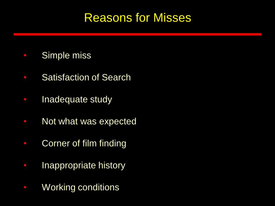
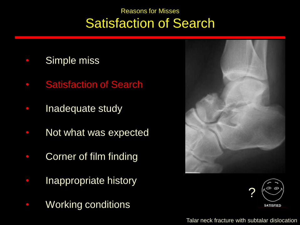
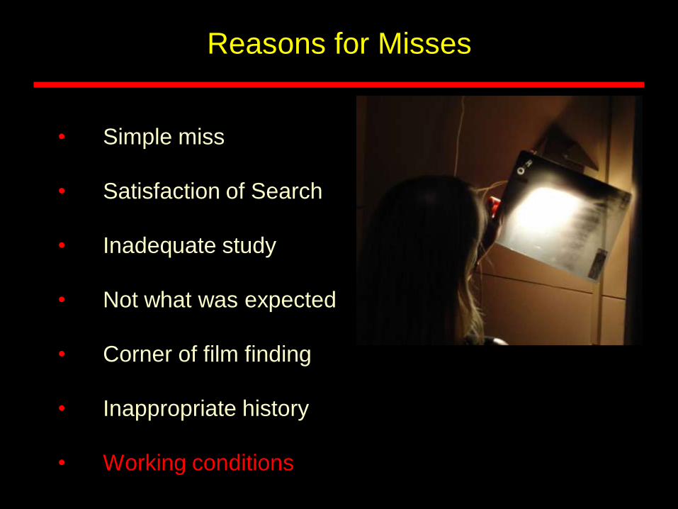
Reasons for Misses
• Simple miss
• Satisfaction of Search
• Inadequate study
• Not what was expected
• Corner of film finding
• Inappropriate history
• Working conditions
Reasons for Misses
Satisfaction of Search
• One of the commonest reasons to miss injuries
• See most obvious injury
• Miss other (more significant) injury
?
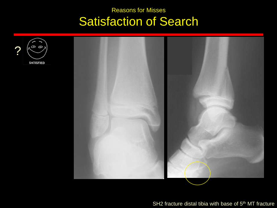
Reasons for Misses
Satisfaction of Search
SH2 fracture distal tibia with base of 5th MT fracture
?
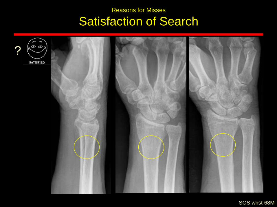
Reasons for Misses
Satisfaction of Search
SOS wrist 68M
?
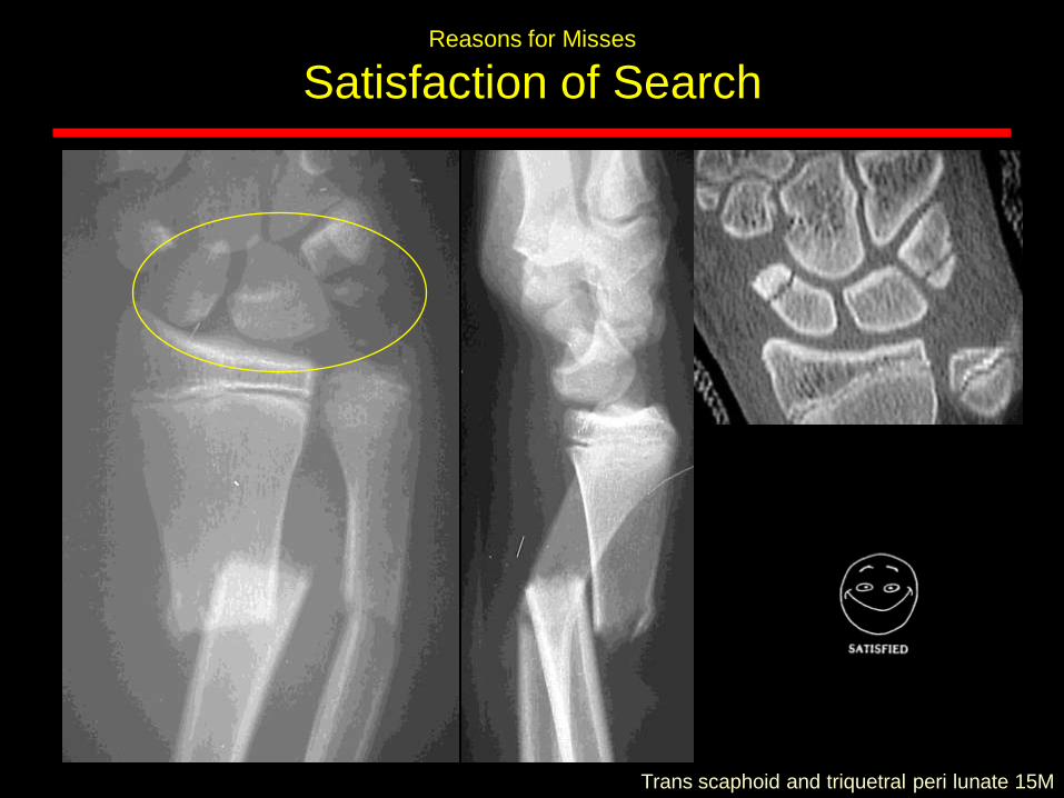
Reasons for Misses
Satisfaction of Search
Trans scaphoid and triquetral peri lunate 15M
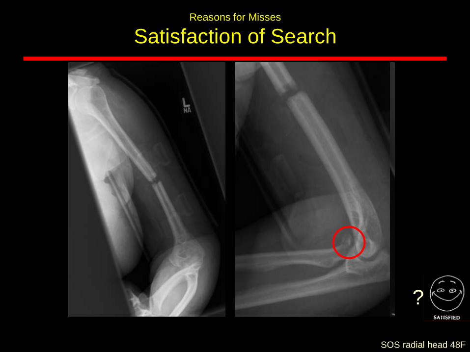
Reasons for Misses
Satisfaction of Search
SOS radial head 48F
?
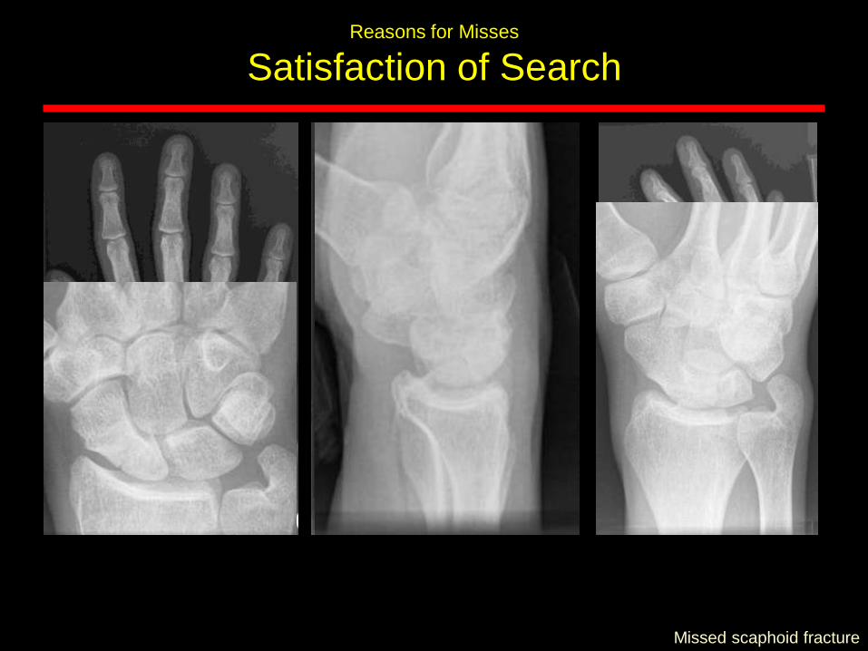
Reasons for Misses
Satisfaction of Search
Missed scaphoid fracture
• Simple miss
• Satisfaction of Search
• Inadequate study
• Not what was expected
• Corner of film finding
• Inappropriate history
• Working conditions
Talar neck fracture with subtalar dislocation
Reasons for Misses
Satisfaction of Search
?
Reasons for Misses
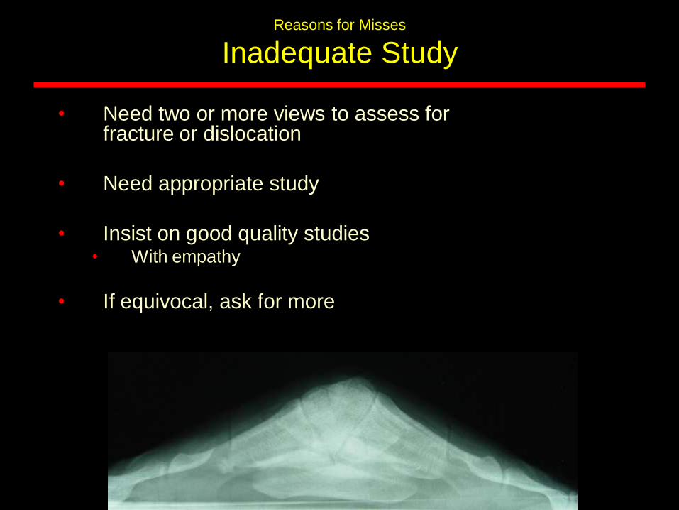
Inadequate Study
• Need two or more views to assess for fracture or dislocation
• Need appropriate study
• Insist on good quality studies• With empathy
• If equivocal, ask for more
Reasons for Misses
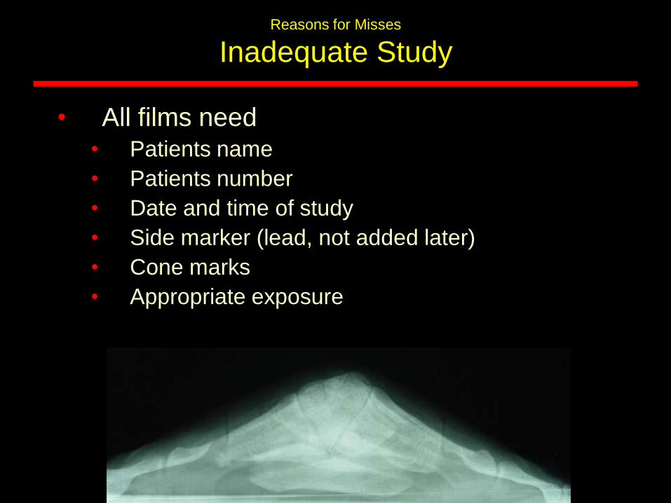
Inadequate Study
• All films need• Patients name
• Patients number
• Date and time of study
• Side marker (lead, not added later)
• Cone marks
• Appropriate exposure
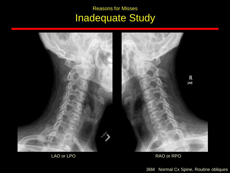
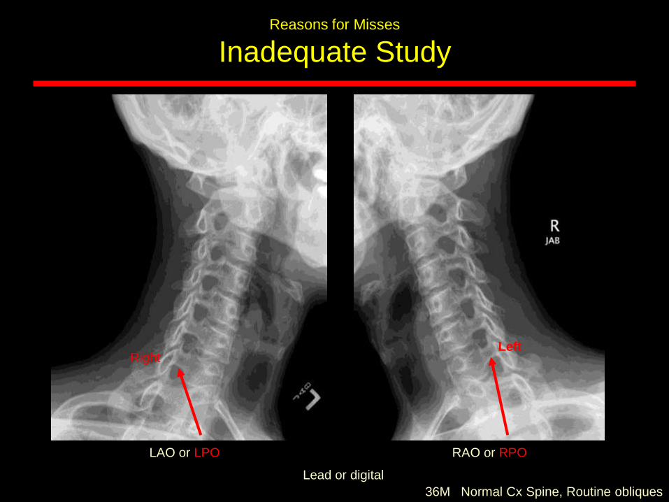
36M Normal Cx Spine, Routine obliques
LAO or LPO RAO or RPO
Reasons for Misses
Inadequate Study
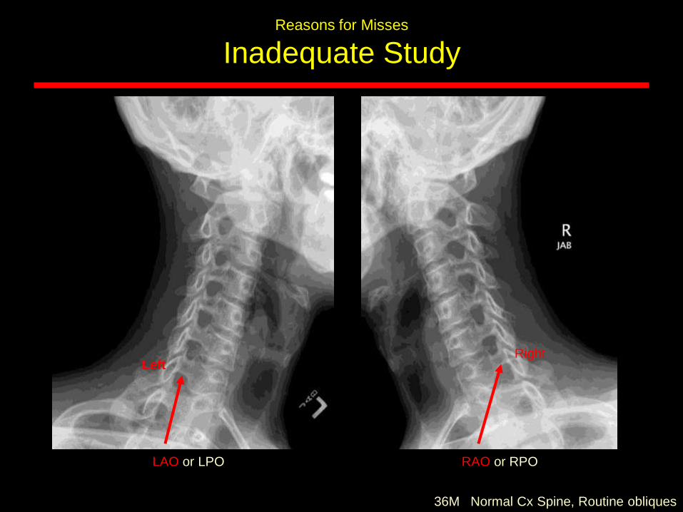
36M Normal Cx Spine, Routine obliques
LAO or LPO RAO or RPO
LeftRight
Reasons for Misses
Inadequate Study
36M Normal Cx Spine, Routine obliques
LAO or LPO RAO or RPO
Lead or digital
LeftRight
Reasons for Misses
Inadequate Study
Reasons for Misses
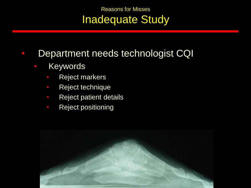
Inadequate Study
• Department needs technologist CQI
• Keywords
• Reject markers
• Reject technique
• Reject patient details
• Reject positioning
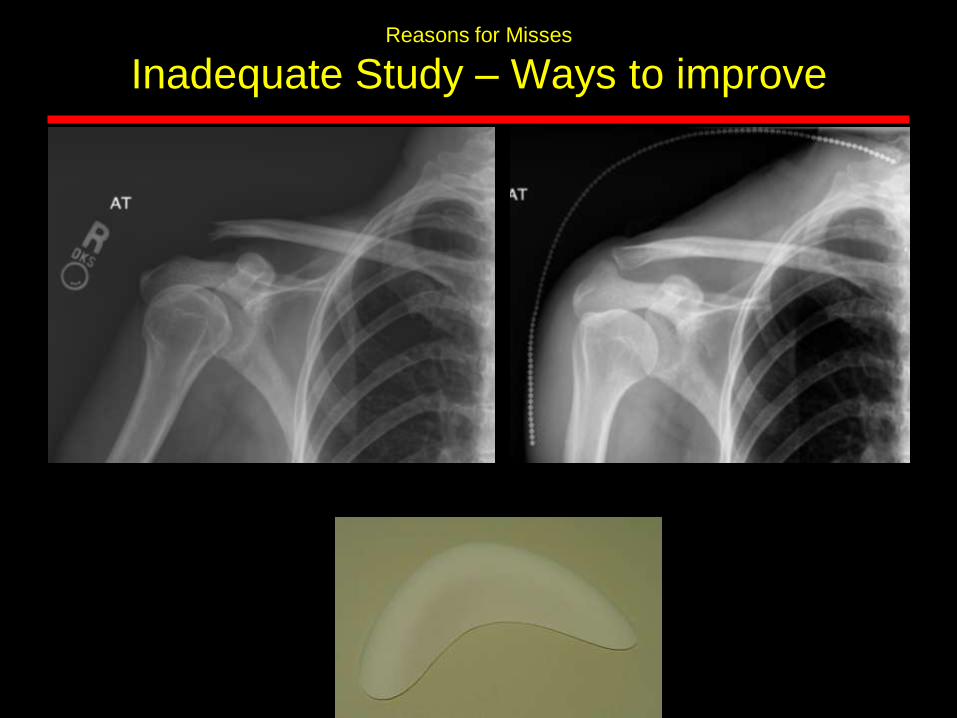
Reasons for Misses
Inadequate Study – Ways to improve
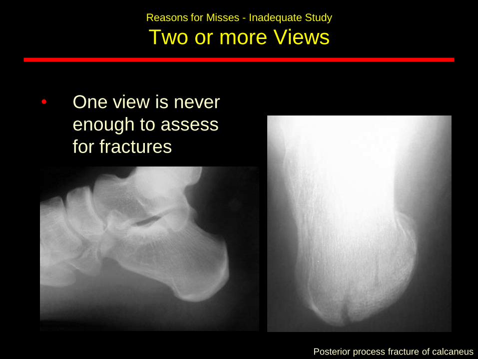
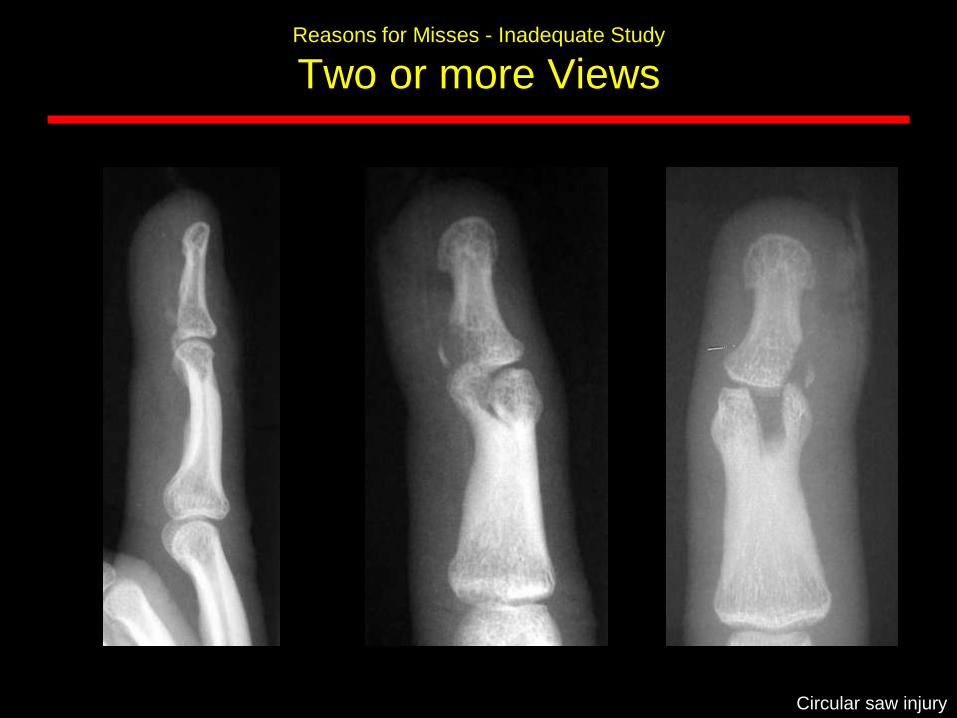
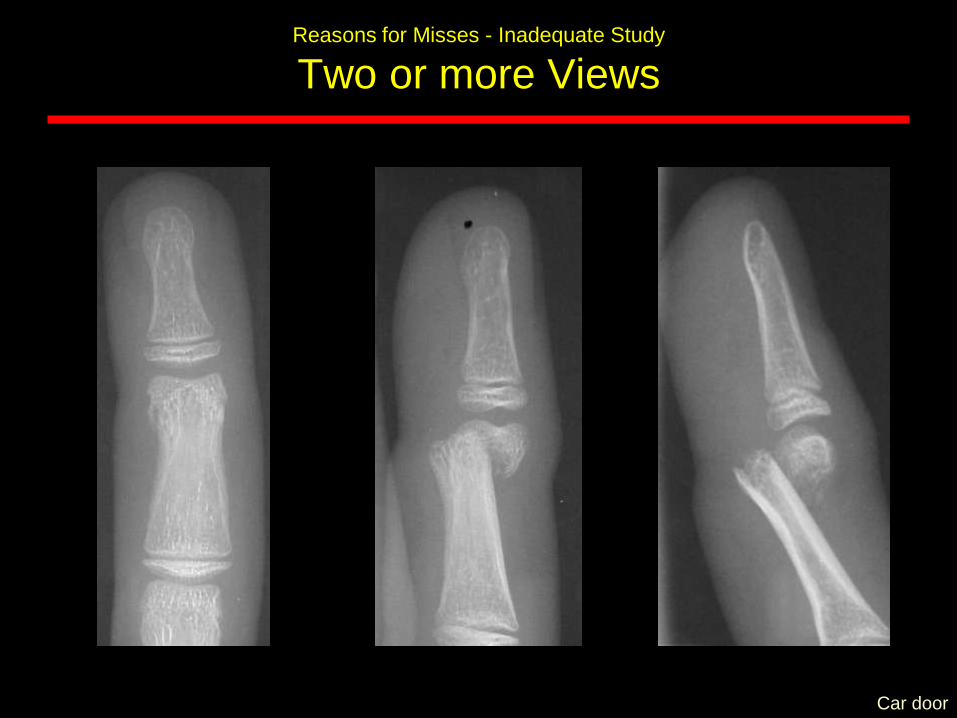
Reasons for Misses - Inadequate Study
Two or more Views
• One view is never
enough to assess
for fractures
Posterior process fracture of calcaneus
Reasons for Misses - Inadequate Study
Two or more Views
Circular saw injury
Reasons for Misses - Inadequate Study
Two or more Views
Car door
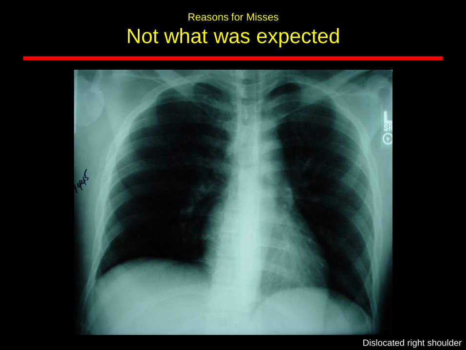
Reasons for Misses
Not what was expected
Dislocated right shoulder
Reasons for Misses
• Simple miss
• Satisfaction of Search
• Inadequate study
• Not what was expected
• Corner of film finding
• Inappropriate history
• Working conditions
Ways to Avoid Missing Fractures
• Look for fracture patterns
• Look at regions that should align
• Look for secondary signs of fracture
• Look for the common sites of fractures
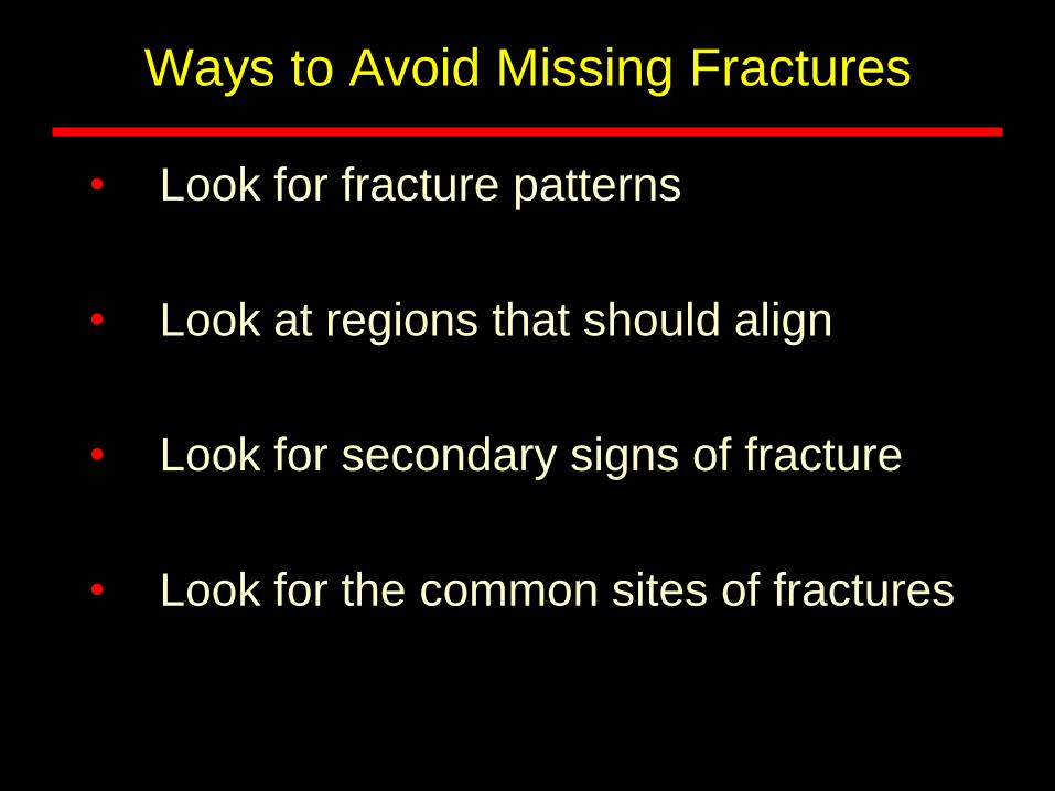
Fracture Patterns
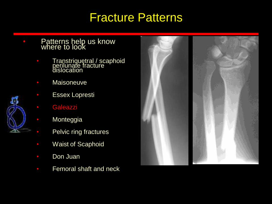
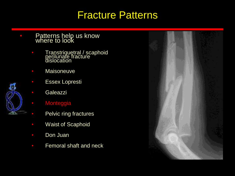
• Patterns help us know where to look
• Transtriquetral / scaphoidperilunate fracture dislocation
• Maisoneuve
• Essex Lopresti
• Galeazzi
• Monteggia
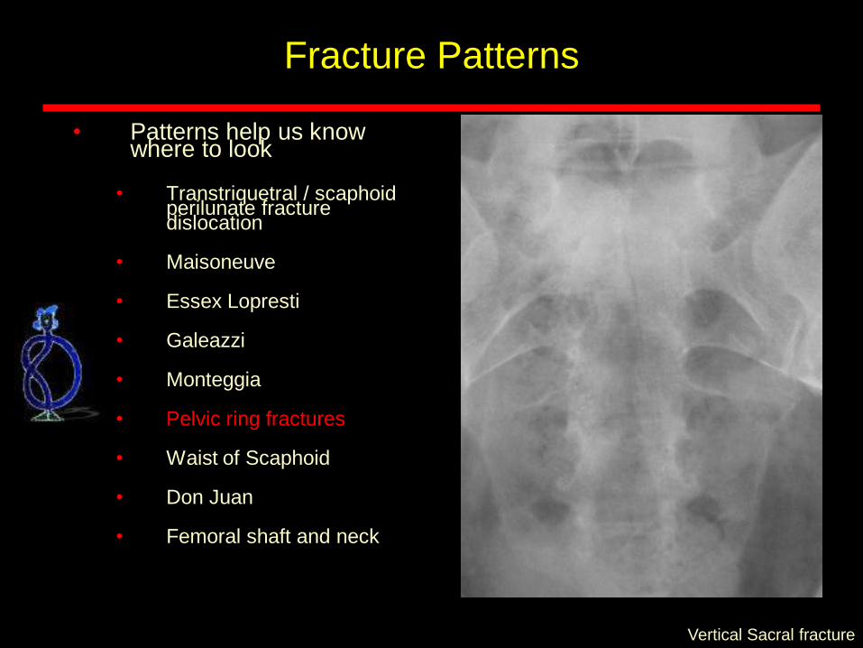
• Pelvic ring fractures
• Waist of Scaphoid
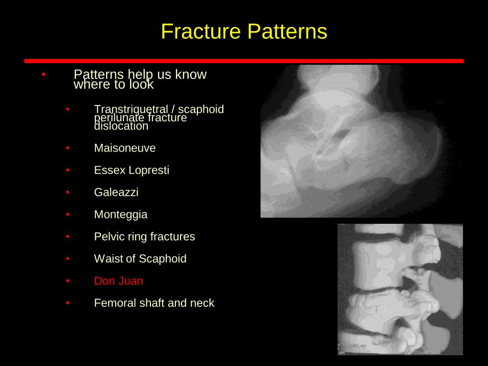
• Don Juan
• Femoral shaft and neck
27M
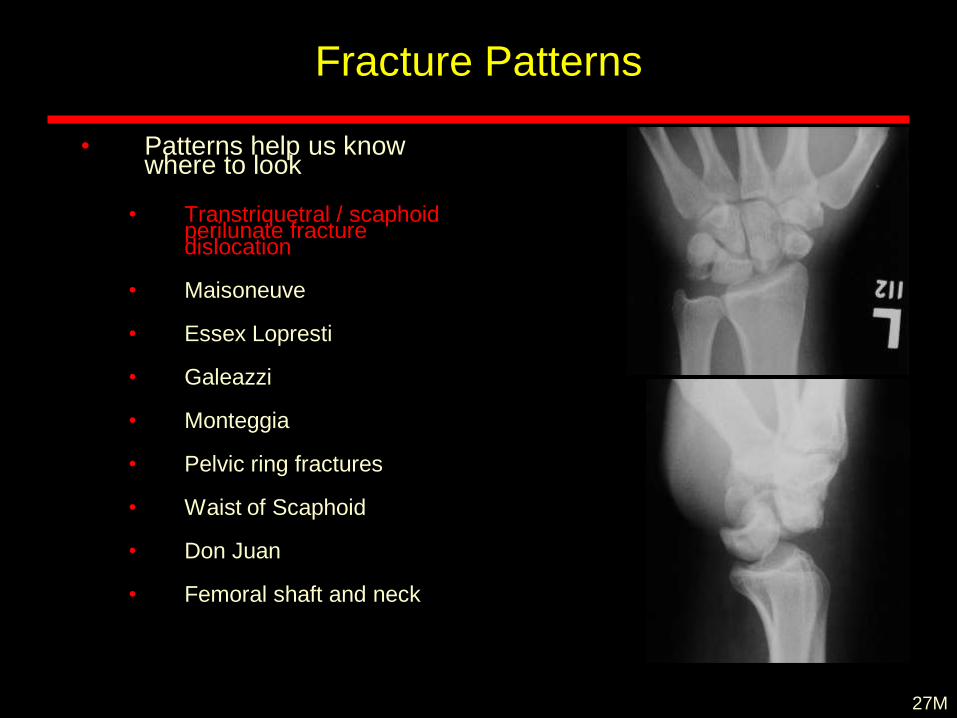
Fracture Patterns
• Patterns help us know where to look
• Transtriquetral / scaphoidperilunate fracture dislocation
• Maisoneuve
• Essex Lopresti
• Galeazzi
• Monteggia
• Pelvic ring fractures
• Waist of Scaphoid
• Don Juan
• Femoral shaft and neck
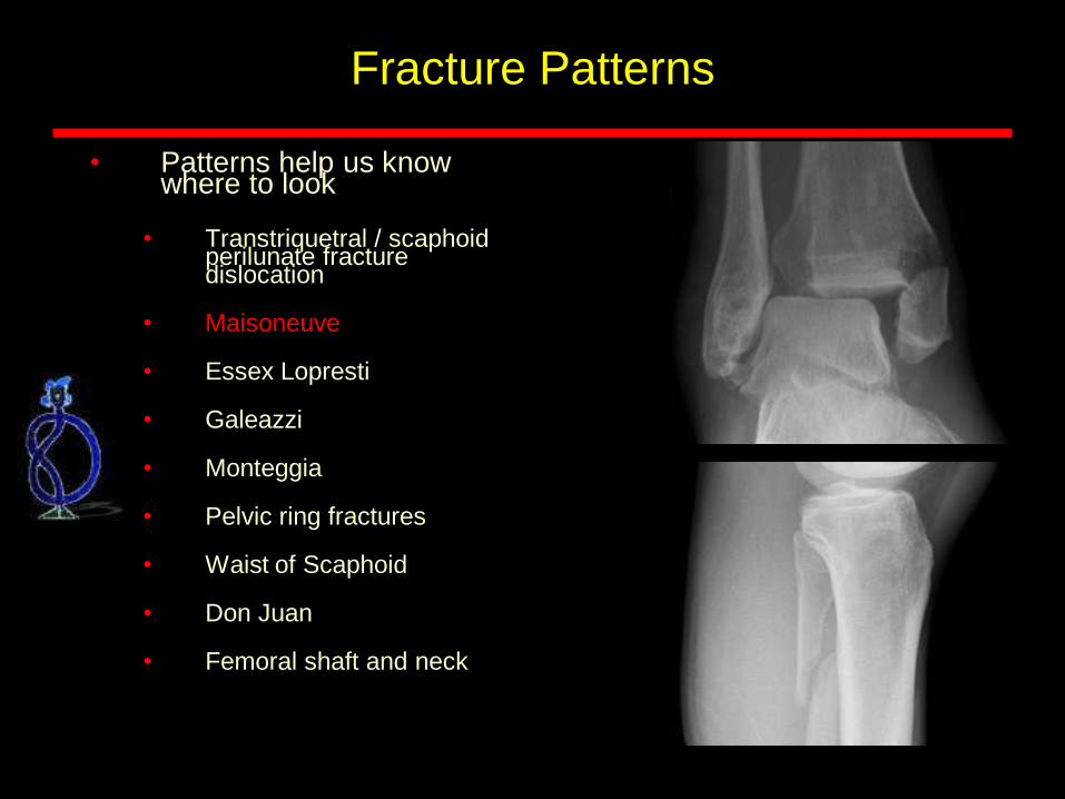
Fracture Patterns
• Patterns help us know where to look
• Transtriquetral / scaphoidperilunate fracture dislocation
• Maisoneuve
• Essex Lopresti
• Galeazzi
• Monteggia
• Pelvic ring fractures
• Waist of Scaphoid
• Don Juan
• Femoral shaft and neck
Fracture Patterns
• Patterns help us know where to look
• Transtriquetral / scaphoidperilunate fracture dislocation
• Maisoneuve
• Essex Lopresti
• Galeazzi
• Monteggia
• Pelvic ring fractures
• Waist of Scaphoid
• Don Juan
• Femoral shaft and neck
Fracture Patterns
• Patterns help us know where to look
• Transtriquetral / scaphoidperilunate fracture dislocation
• Maisoneuve
• Essex Lopresti
• Galeazzi
• Monteggia
• Pelvic ring fractures
• Waist of Scaphoid
• Don Juan
• Femoral shaft and neck
Fracture Patterns
• Patterns help us know where to look
• Transtriquetral / scaphoidperilunate fracture dislocation
• Maisoneuve
• Essex Lopresti
• Galeazzi
• Monteggia
• Pelvic ring fractures
• Waist of Scaphoid
• Don Juan
• Femoral shaft and neck
Vertical Sacral fracture

Fracture Patterns
• Patterns help us know where to look
• Transtriquetral / scaphoidperilunate fracture dislocation
• Maisoneuve
• Essex Lopresti
• Galeazzi
• Monteggia
• Pelvic ring fractures
• Waist of Scaphoid
• Don Juan
• Femoral shaft and neck
Fracture Patterns
• Patterns help us know where to look
• Transtriquetral / scaphoidperilunate fracture dislocation
• Maisoneuve
• Essex Lopresti
• Galeazzi
• Monteggia
• Pelvic ring fractures
• Waist of Scaphoid
• Don Juan
• Femoral shaft and neck
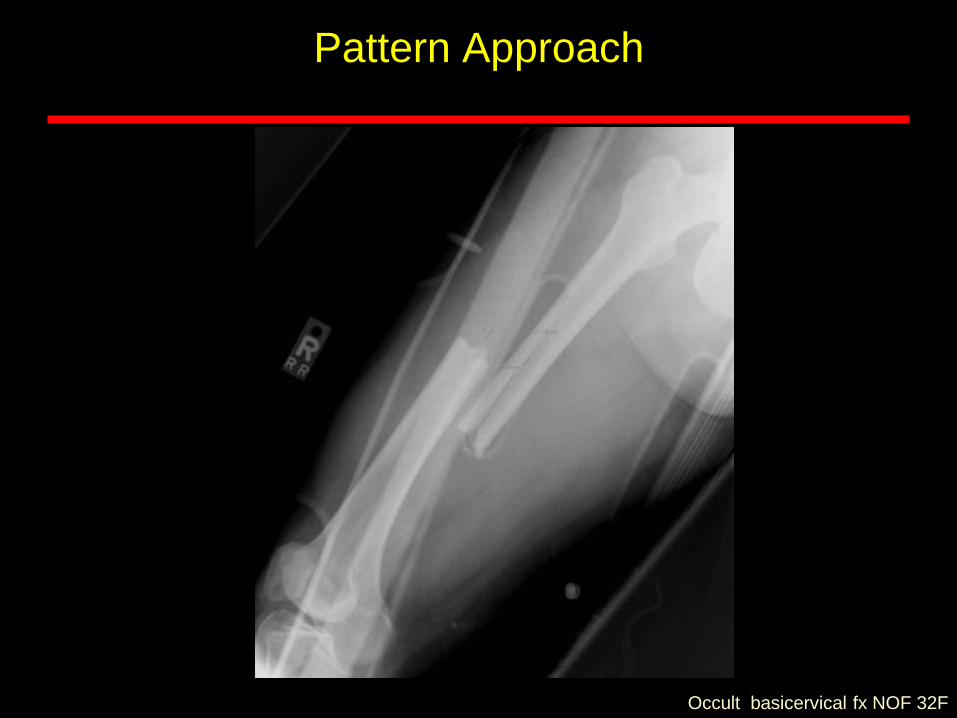
Occult basicervical fx NOF 32F
Pattern Approach
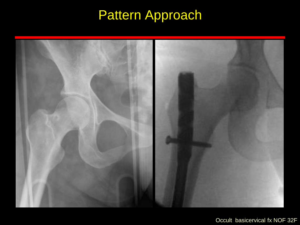
Pattern Approach
Occult basicervical fx NOF 32F
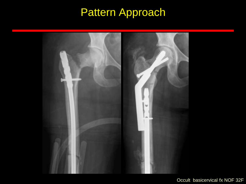
Pattern Approach
Occult basicervical fx NOF 32F
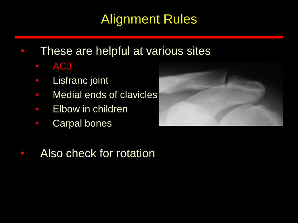
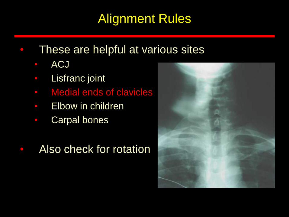
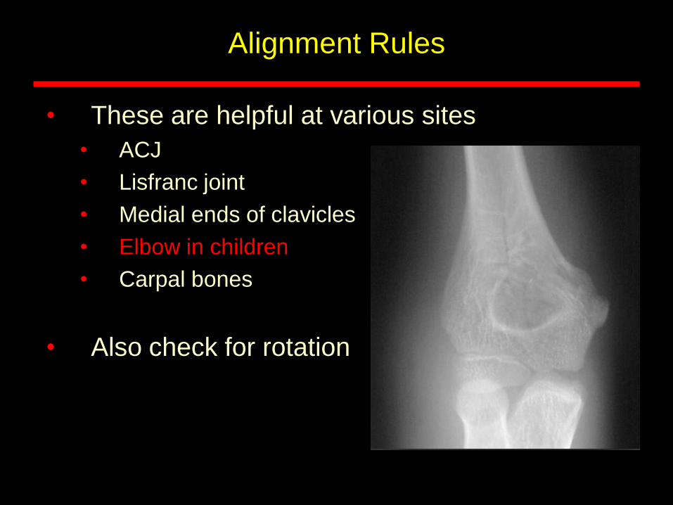
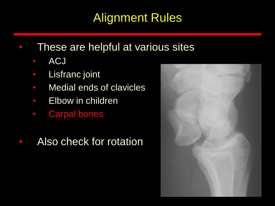
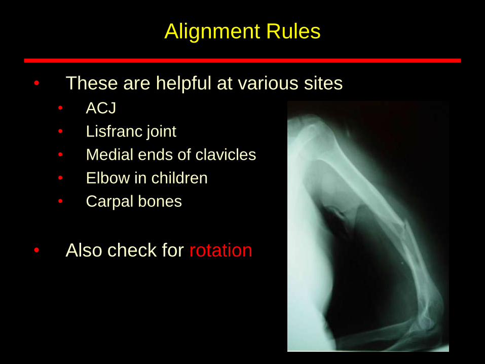
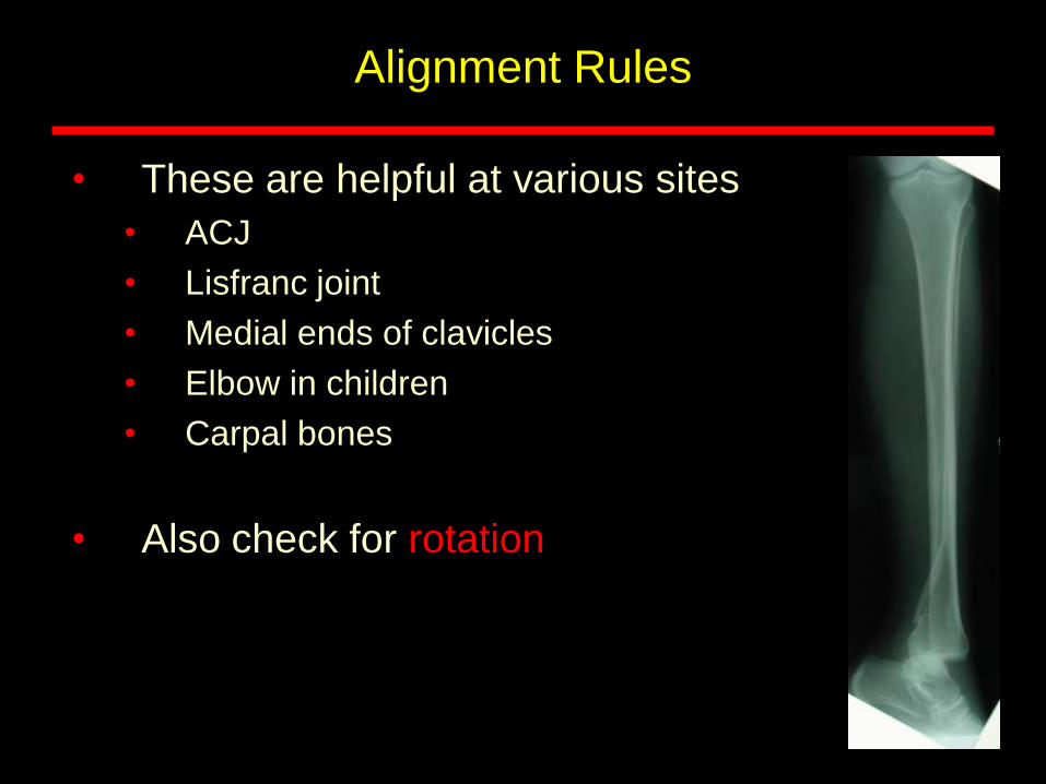
Alignment Rules
• These are helpful at various sites
• ACJ
• Lisfranc joint
• Medial ends of clavicles
• Elbow in children
• Carpal bones
• Also check for rotation

Alignment Rules
• These are helpful at various sites
• ACJ
• Lisfranc joint
• Medial ends of clavicles
• Elbow in children
• Carpal bones
• Also check for rotation
Alignment Rules
• These are helpful at various sites
• ACJ
• Lisfranc joint
• Medial ends of clavicles
• Elbow in children
• Carpal bones
• Also check for rotation
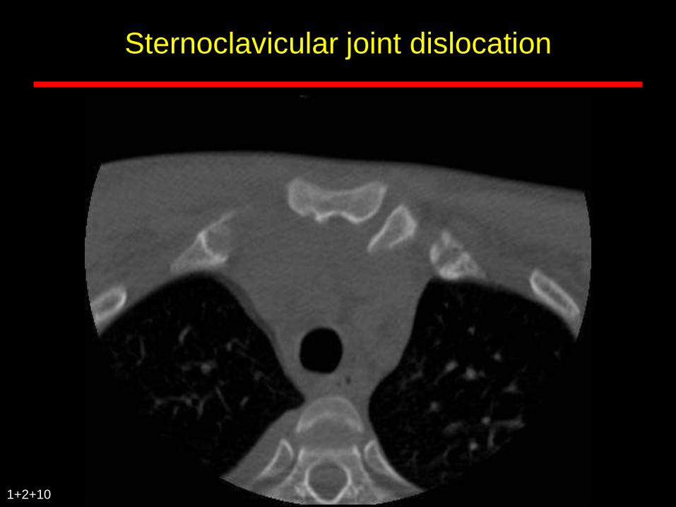
Sternoclavicular joint dislocation
1+2+10
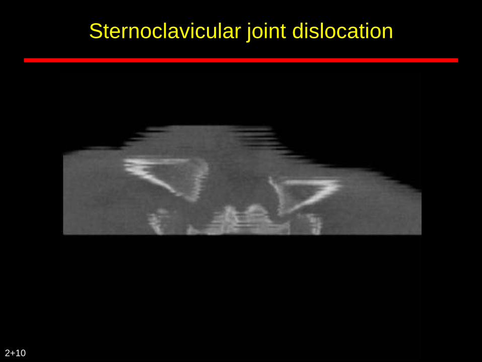
Sternoclavicular joint dislocation
2+10
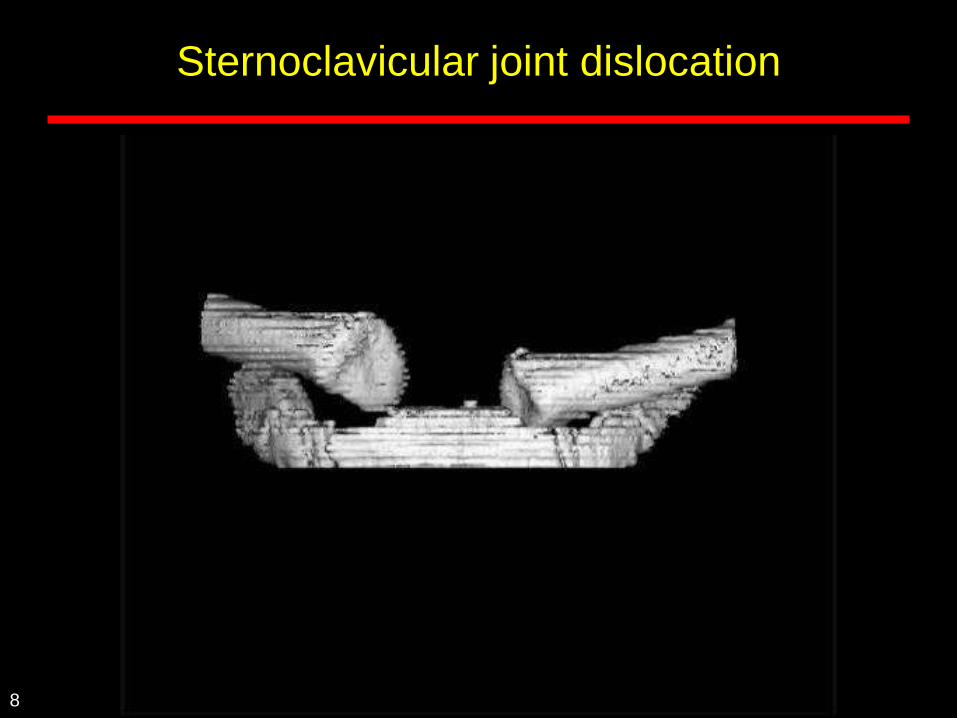
Sternoclavicular joint dislocation
8
Alignment Rules
• These are helpful at various sites
• ACJ
• Lisfranc joint
• Medial ends of clavicles
• Elbow in children
• Carpal bones
• Also check for rotation
Alignment Rules
• These are helpful at various sites
• ACJ
• Lisfranc joint
• Medial ends of clavicles
• Elbow in children
• Carpal bones
• Also check for rotation
Alignment Rules
• These are helpful at various sites
• ACJ
• Lisfranc joint
• Medial ends of clavicles
• Elbow in children
• Carpal bones
• Also check for rotation
Alignment Rules
• These are helpful at various sites
• ACJ
• Lisfranc joint
• Medial ends of clavicles
• Elbow in children
• Carpal bones
• Also check for rotation
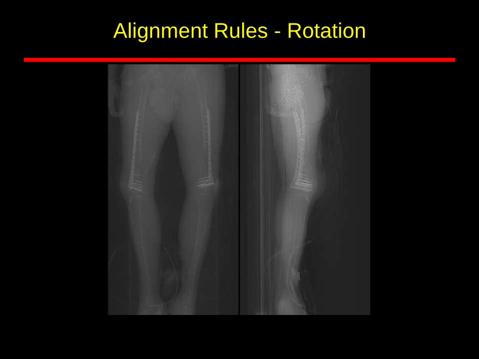
Alignment Rules - Rotation
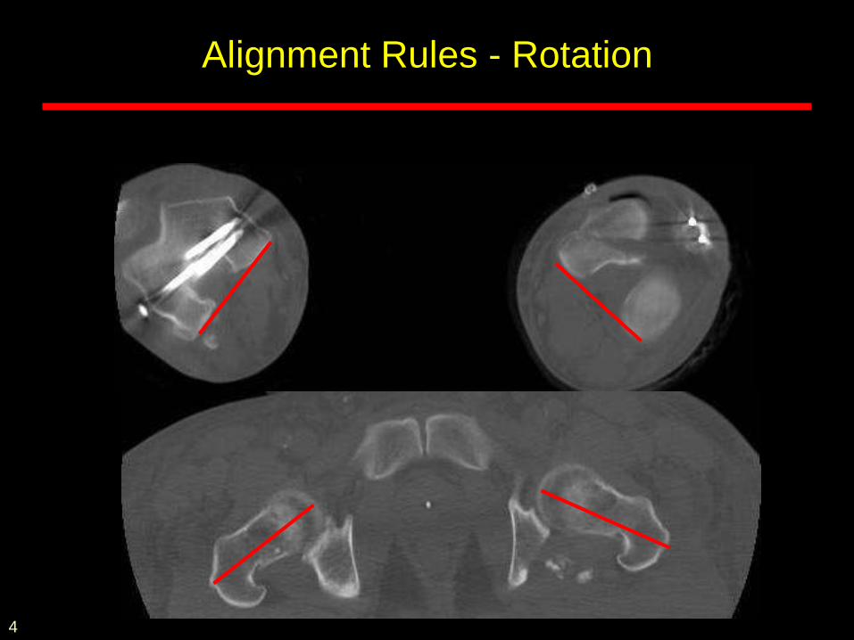
Alignment Rules - Rotation
4
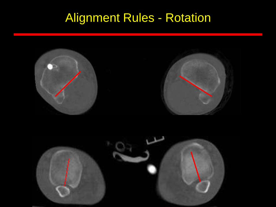
Alignment Rules - Rotation
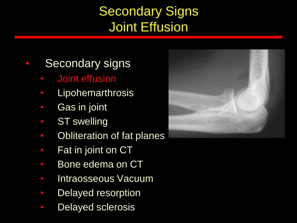
Secondary Signs
Joint Effusion
• Secondary signs
• Joint effusion
• Lipohemarthrosis
• Gas in joint
• ST swelling
• Obliteration of fat planes
• Fat in joint on CT
• Bone edema on CT
• Intraosseous Vacuum
• Delayed resorption
• Delayed sclerosis
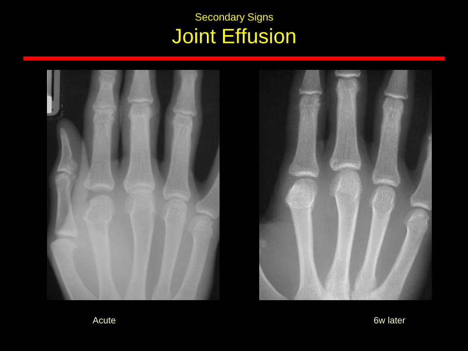
Secondary Signs
Joint Effusion
Acute 6w later

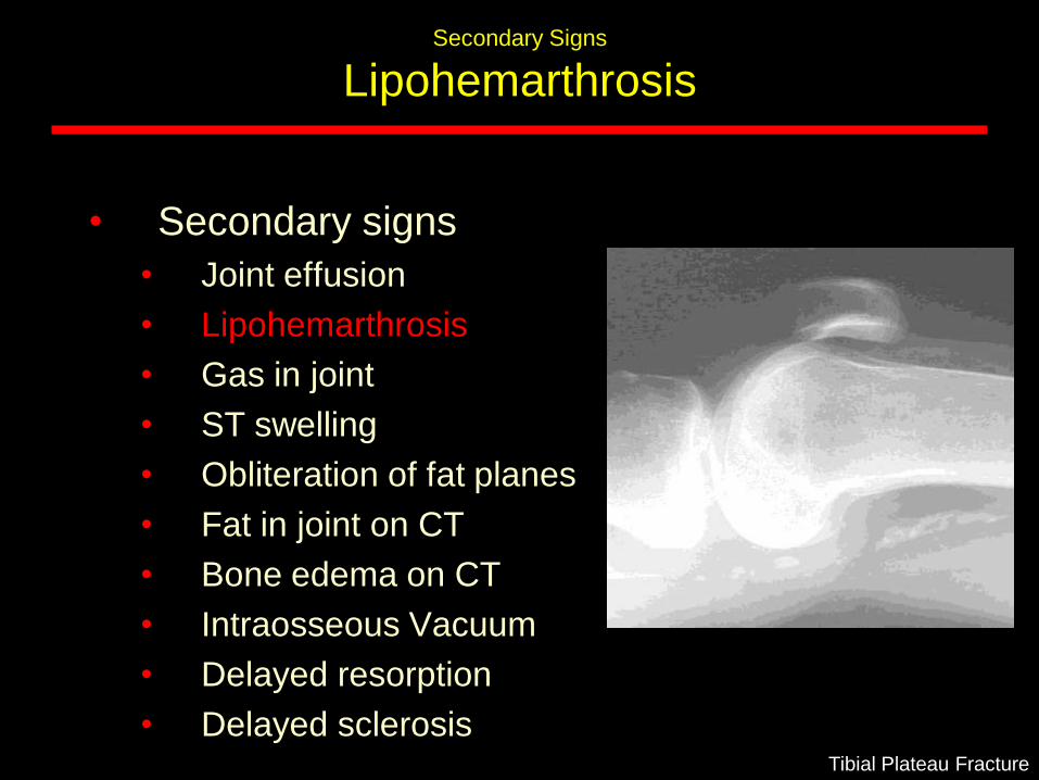
Secondary Signs
Lipohemarthrosis
Tibial Plateau Fracture
• Secondary signs
• Joint effusion
• Lipohemarthrosis
• Gas in joint
• ST swelling
• Obliteration of fat planes
• Fat in joint on CT
• Bone edema on CT
• Intraosseous Vacuum
• Delayed resorption
• Delayed sclerosis
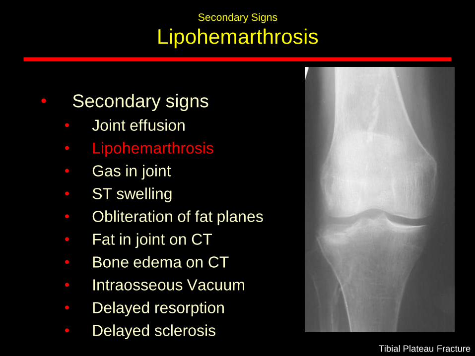
Secondary Signs
Lipohemarthrosis
• Secondary signs
• Joint effusion
• Lipohemarthrosis
• Gas in joint
• ST swelling
• Obliteration of fat planes
• Fat in joint on CT
• Bone edema on CT
• Intraosseous Vacuum
• Delayed resorption
• Delayed sclerosisTibial Plateau Fracture
Secondary Signs
Lipohemarthrosis
• Secondary signs
• Joint effusion
• Lipohemarthrosis
• Gas in joint
• ST swelling
• Obliteration of fat planes
• Fat in joint on CT
• Bone edema on CT
• Intraosseous Vacuum
• Delayed resorption
• Delayed sclerosisTibial Plateau Fracture
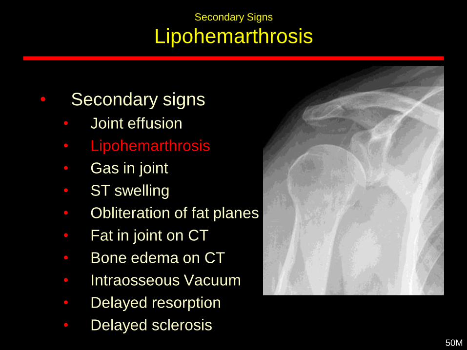
Secondary Signs
Lipohemarthrosis
• Secondary signs
• Joint effusion
• Lipohemarthrosis
• Gas in joint
• ST swelling
• Obliteration of fat planes
• Fat in joint on CT
• Bone edema on CT
• Intraosseous Vacuum
• Delayed resorption
• Delayed sclerosis50M
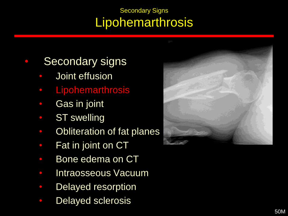
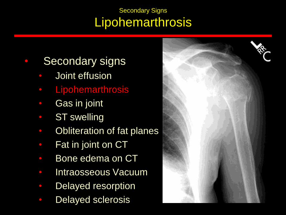
Secondary Signs
Lipohemarthrosis
• Secondary signs
• Joint effusion
• Lipohemarthrosis
• Gas in joint
• ST swelling
• Obliteration of fat planes
• Fat in joint on CT
• Bone edema on CT
• Intraosseous Vacuum
• Delayed resorption
• Delayed sclerosis50M
Secondary Signs
Lipohemarthrosis
• Secondary signs
• Joint effusion
• Lipohemarthrosis
• Gas in joint
• ST swelling
• Obliteration of fat planes
• Fat in joint on CT
• Bone edema on CT
• Intraosseous Vacuum
• Delayed resorption
• Delayed sclerosis
Secondary Signs
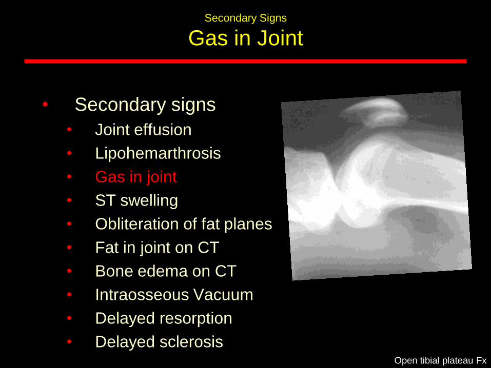
Gas in Joint
• Secondary signs
• Joint effusion
• Lipohemarthrosis
• Gas in joint
• ST swelling
• Obliteration of fat planes
• Fat in joint on CT
• Bone edema on CT
• Intraosseous Vacuum
• Delayed resorption
• Delayed sclerosisOpen tibial plateau Fx
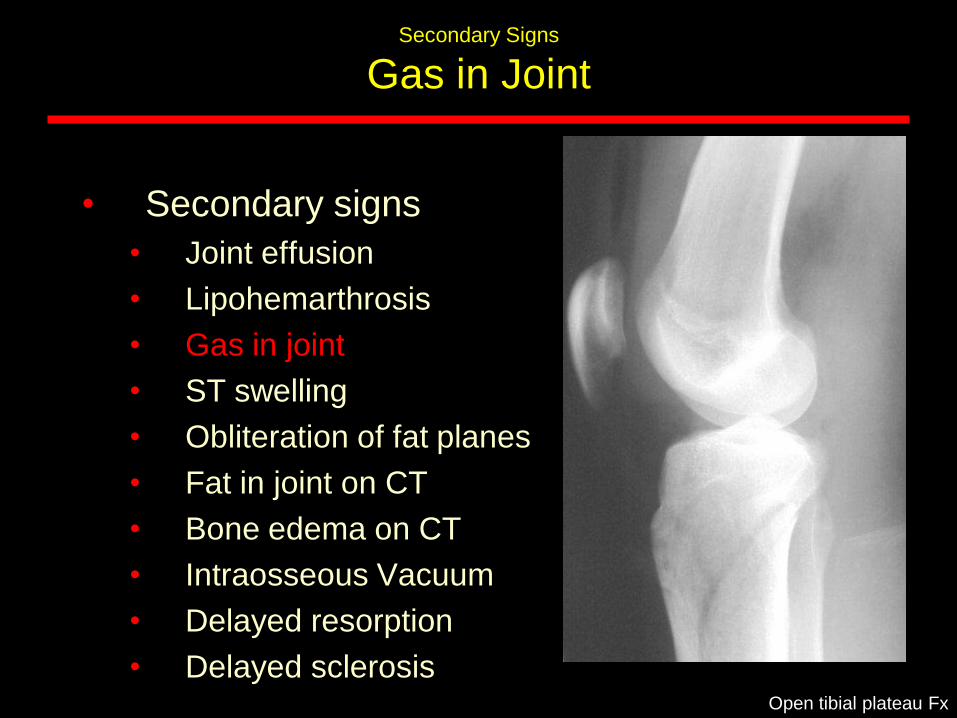
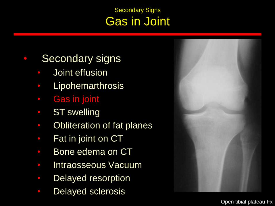
Secondary Signs
Gas in Joint
• Secondary signs
• Joint effusion
• Lipohemarthrosis
• Gas in joint
• ST swelling
• Obliteration of fat planes
• Fat in joint on CT
• Bone edema on CT
• Intraosseous Vacuum
• Delayed resorption
• Delayed sclerosisOpen tibial plateau Fx
Secondary Signs
Gas in Joint
• Secondary signs
• Joint effusion
• Lipohemarthrosis
• Gas in joint
• ST swelling
• Obliteration of fat planes
• Fat in joint on CT
• Bone edema on CT
• Intraosseous Vacuum
• Delayed resorption
• Delayed sclerosisOpen tibial plateau Fx
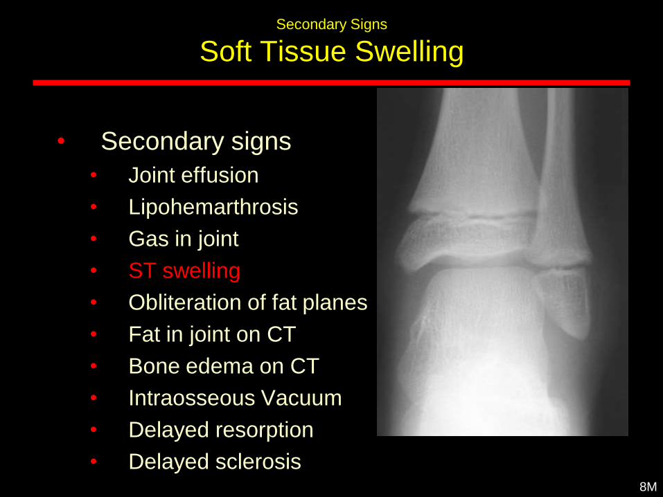
Secondary Signs
Soft Tissue Swelling
• Secondary signs
• Joint effusion
• Lipohemarthrosis
• Gas in joint
• ST swelling
• Obliteration of fat planes
• Fat in joint on CT
• Bone edema on CT
• Intraosseous Vacuum
• Delayed resorption
• Delayed sclerosis8M
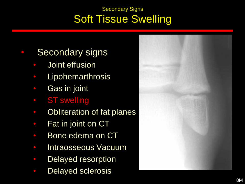
Secondary Signs
Soft Tissue Swelling
• Secondary signs
• Joint effusion
• Lipohemarthrosis
• Gas in joint
• ST swelling
• Obliteration of fat planes
• Fat in joint on CT
• Bone edema on CT
• Intraosseous Vacuum
• Delayed resorption
• Delayed sclerosis8M
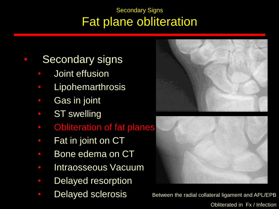
Secondary Signs
Fat plane obliteration
Between the radial collateral ligament and APL/EPB
Obliterated in Fx / Infection
• Secondary signs
• Joint effusion
• Lipohemarthrosis
• Gas in joint
• ST swelling
• Obliteration of fat planes
• Fat in joint on CT
• Bone edema on CT
• Intraosseous Vacuum
• Delayed resorption
• Delayed sclerosis
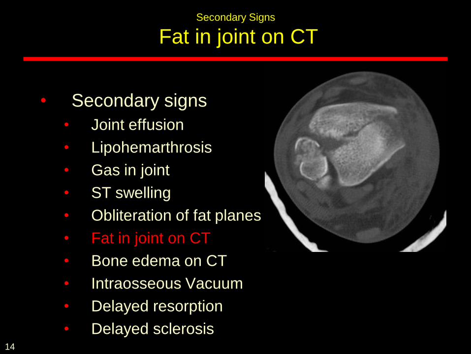
Secondary Signs
Fat in joint on CT
• Secondary signs
• Joint effusion
• Lipohemarthrosis
• Gas in joint
• ST swelling
• Obliteration of fat planes
• Fat in joint on CT
• Bone edema on CT
• Intraosseous Vacuum
• Delayed resorption
• Delayed sclerosis14
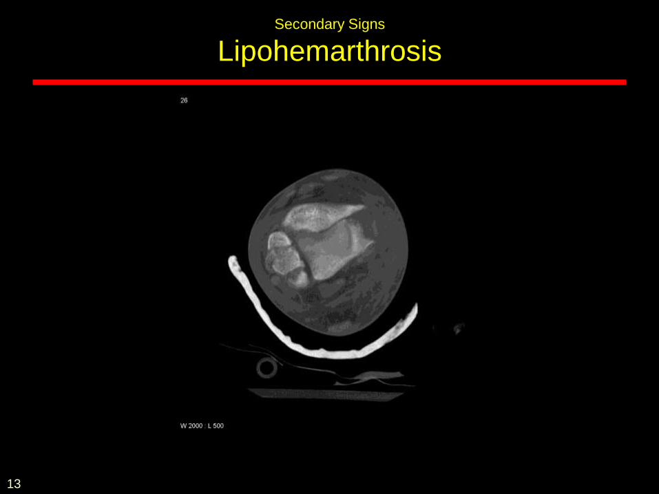
Secondary Signs
Lipohemarthrosis
13
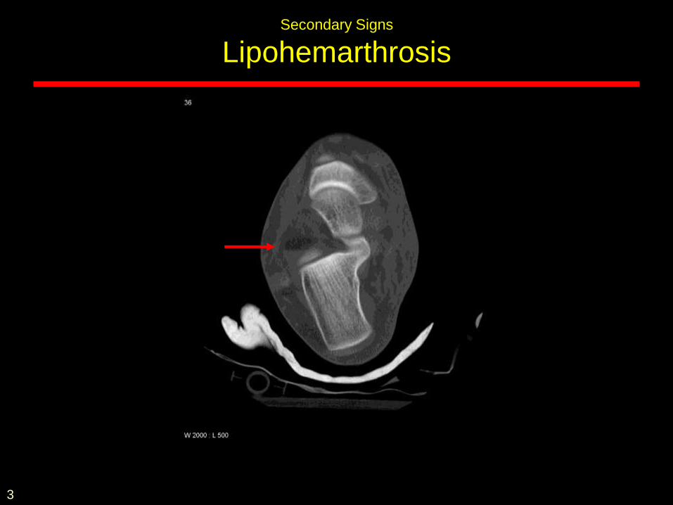
Secondary Signs
Lipohemarthrosis
3
10.12.2002
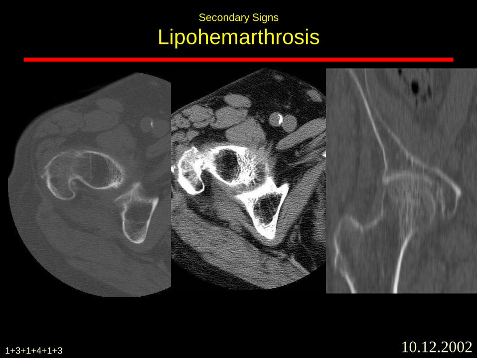
Secondary Signs
Lipohemarthrosis
1+3+1+4+1+3
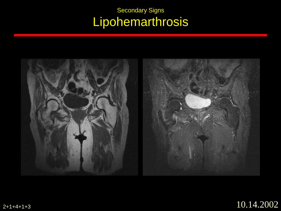
Secondary Signs
Lipohemarthrosis
10.14.20022+1+4+1+3
8.9.2003
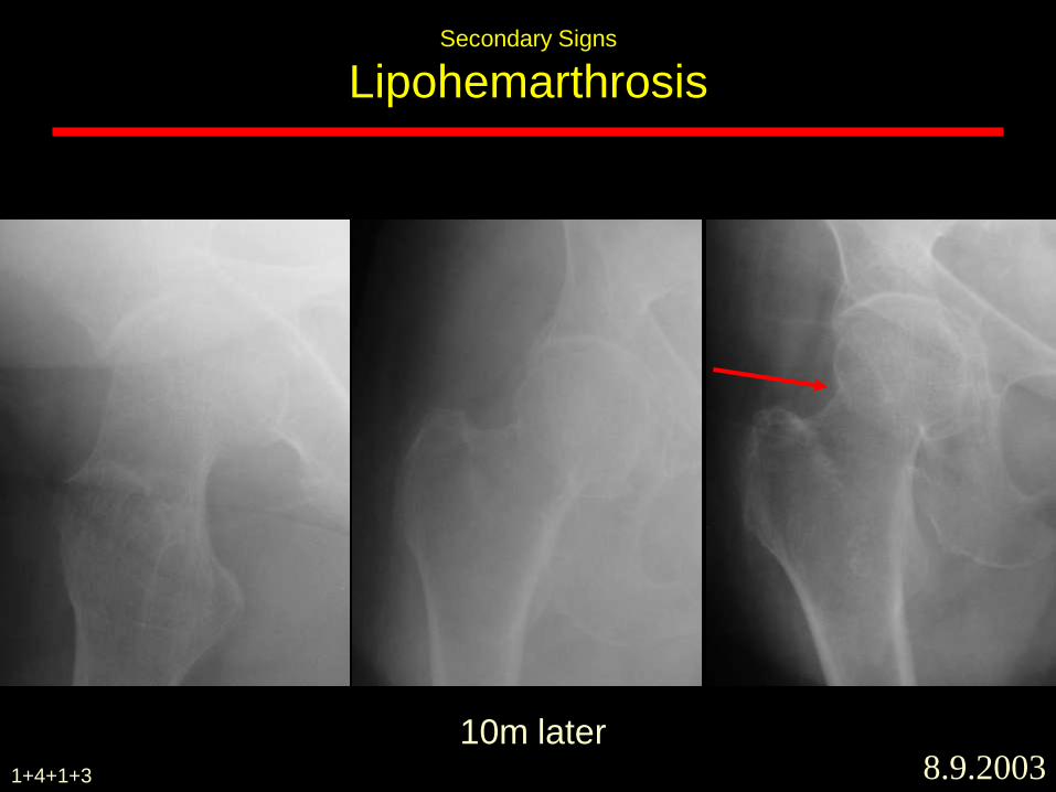
Secondary Signs
Lipohemarthrosis
10m later1+4+1+3
8.9.2003
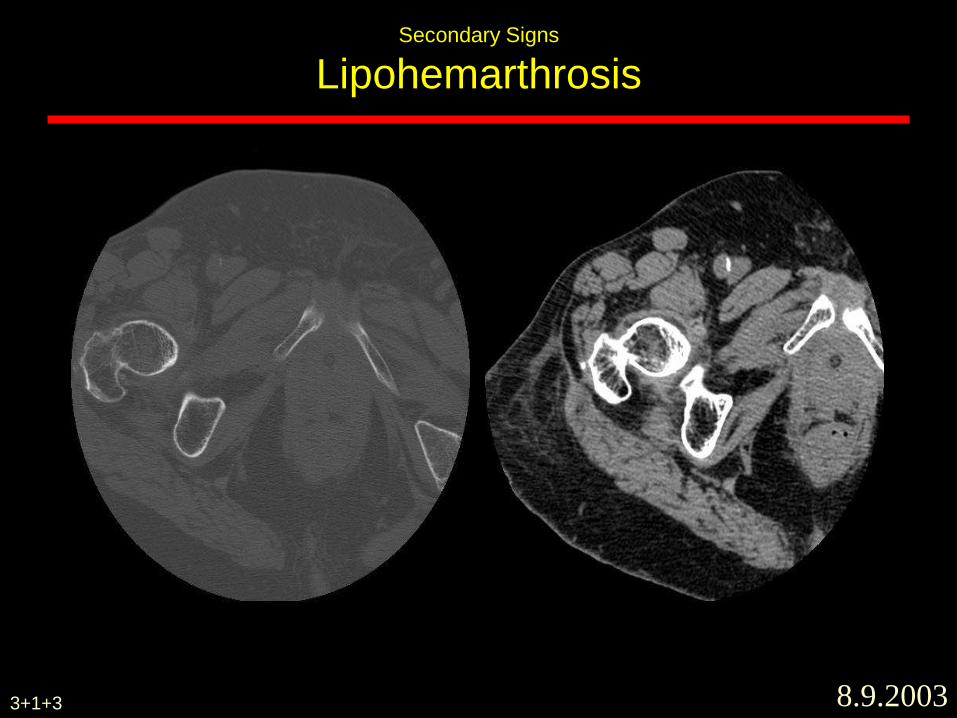
Secondary Signs
Lipohemarthrosis
3+1+3
8.9.2003
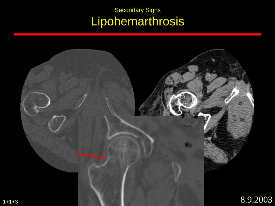
Secondary Signs
Lipohemarthrosis
1+1+3
8.11.2003
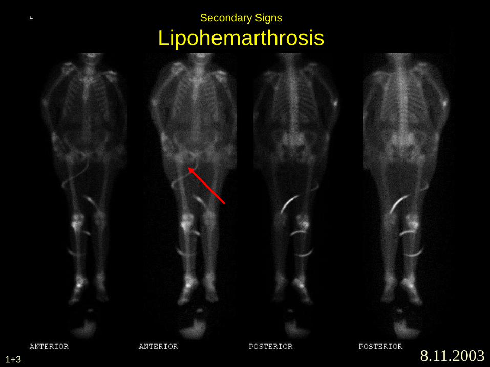
Secondary Signs
Lipohemarthrosis
1+3
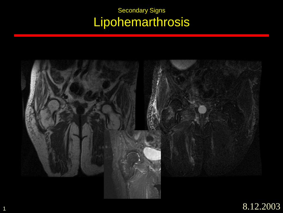
Secondary Signs
Lipohemarthrosis
8.12.20031
Bone window ST window
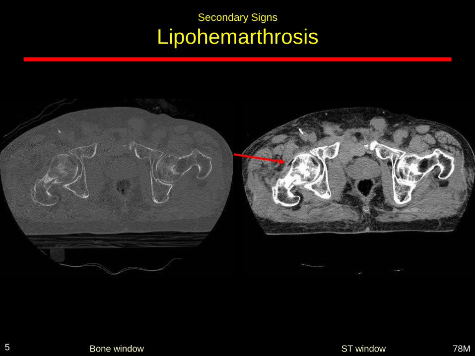
Secondary Signs
Lipohemarthrosis
78M5
60 y/o female with pain s/p fall
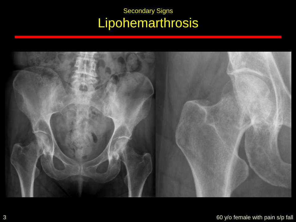
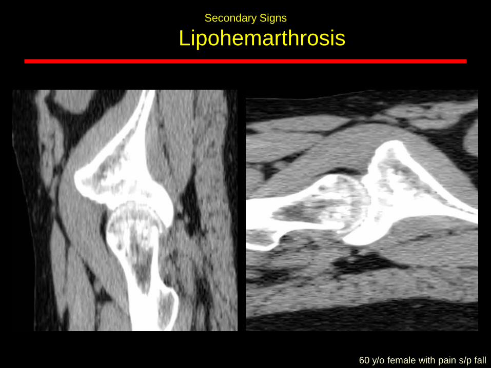
Secondary Signs
Lipohemarthrosis
3
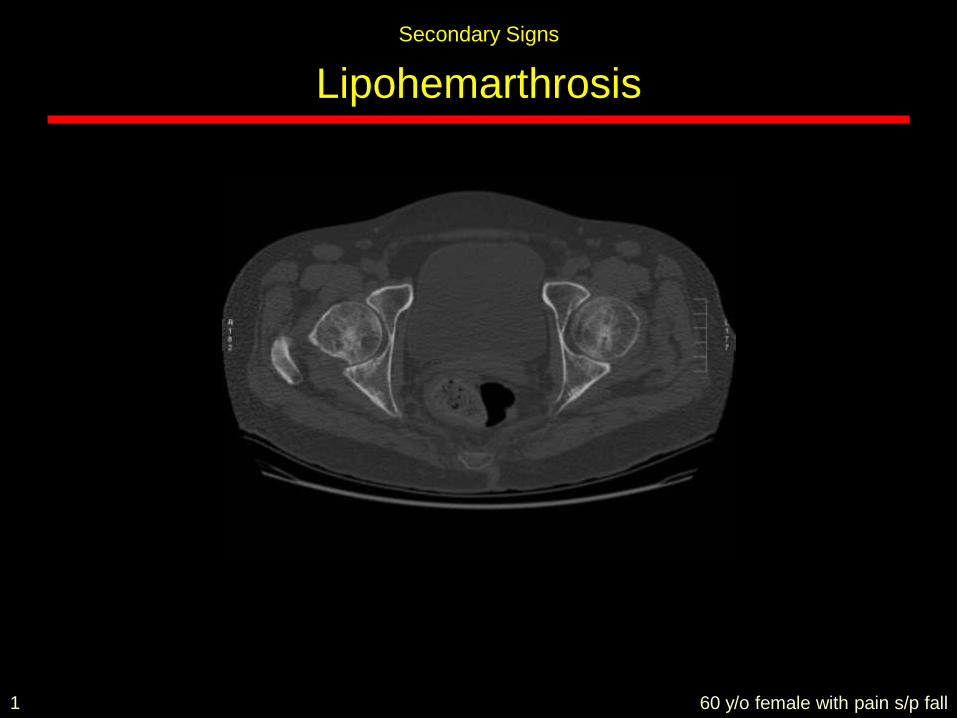
60 y/o female with pain s/p fall
Secondary Signs
Lipohemarthrosis
1
Secondary Signs
Lipohemarthrosis
60 y/o female with pain s/p fall
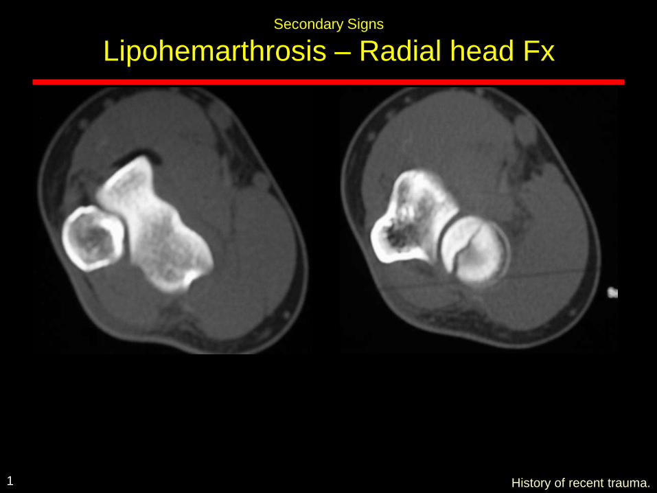
Secondary Signs
Lipohemarthrosis – Radial head Fx
History of recent trauma.1
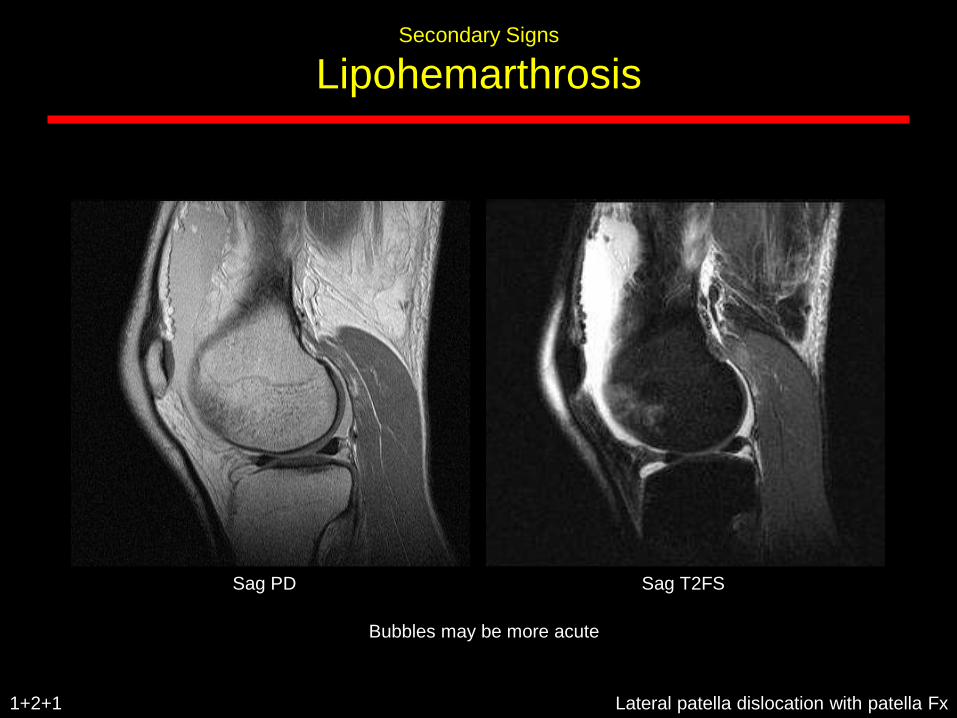
Secondary Signs
Lipohemarthrosis
Lateral patella dislocation with patella Fx1+2+1
Sag PD Sag T2FS
Bubbles may be more acute
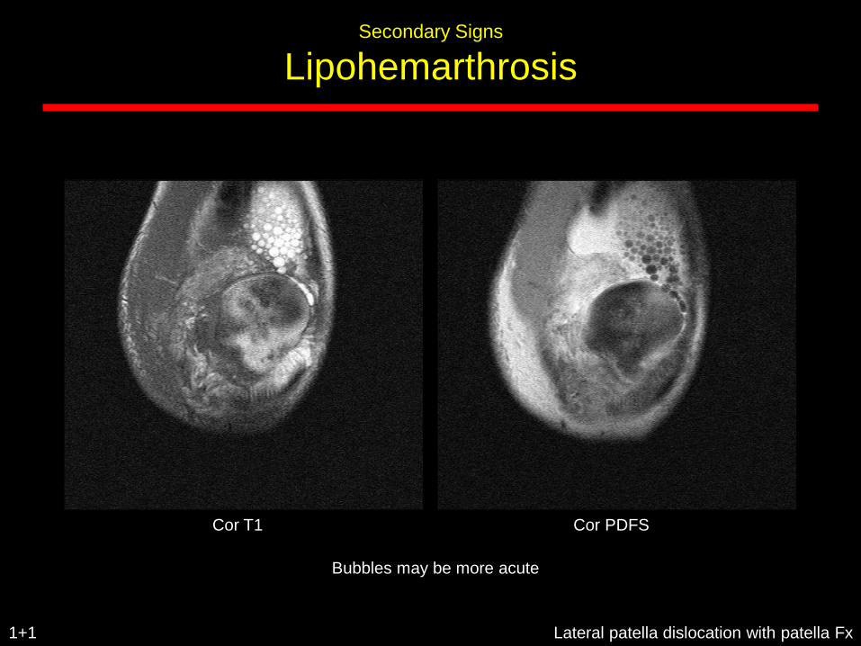
Secondary Signs
Lipohemarthrosis
Lateral patella dislocation with patella Fx1+1
Cor T1 Cor PDFS
Bubbles may be more acute
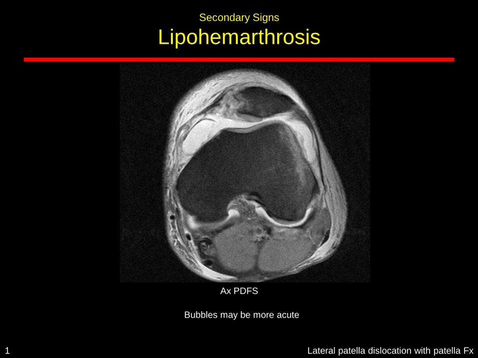
Secondary Signs
Lipohemarthrosis
Lateral patella dislocation with patella Fx1
Ax PDFS
Bubbles may be more acute
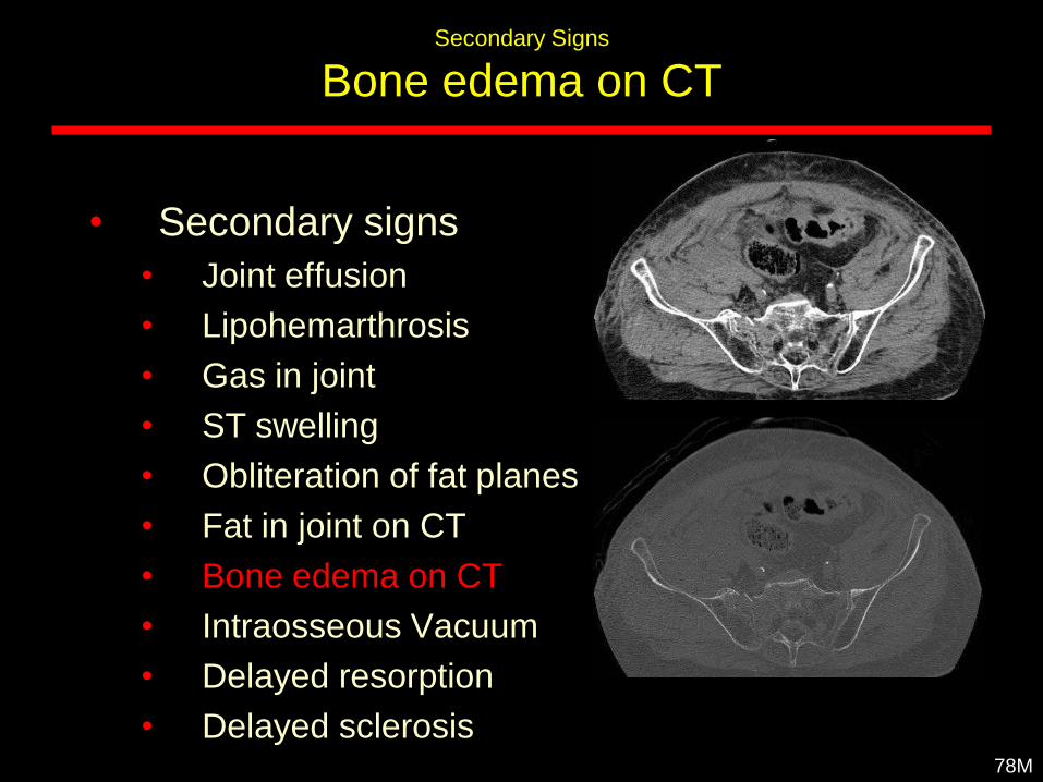
Secondary Signs
Bone edema on CT
• Secondary signs
• Joint effusion
• Lipohemarthrosis
• Gas in joint
• ST swelling
• Obliteration of fat planes
• Fat in joint on CT
• Bone edema on CT
• Intraosseous Vacuum
• Delayed resorption
• Delayed sclerosis78M
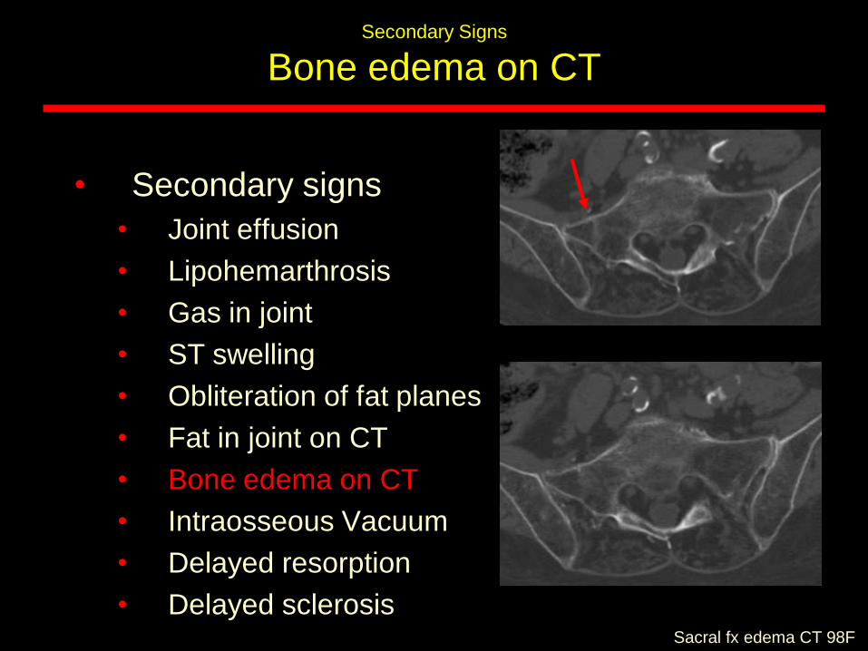
Secondary Signs
Bone edema on CT
• Secondary signs
• Joint effusion
• Lipohemarthrosis
• Gas in joint
• ST swelling
• Obliteration of fat planes
• Fat in joint on CT
• Bone edema on CT
• Intraosseous Vacuum
• Delayed resorption
• Delayed sclerosisSacral fx edema CT 98F
Secondary Signs
Lipohemarthrosis
• Secondary signs
• Joint effusion
• Lipohemarthrosis
• Gas in joint
• ST swelling
• Obliteration of fat planes
• Fat in joint on CT
• Bone edema on CT
• Intraosseous Vacuum
• Delayed resorption
• Delayed sclerosisLipohem knee bone edema 62F
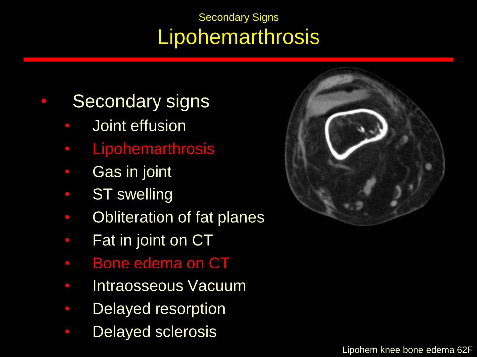
Secondary Signs
Lipohemarthrosis
• Secondary signs
• Joint effusion
• Lipohemarthrosis
• Gas in joint
• ST swelling
• Obliteration of fat planes
• Fat in joint on CT
• Bone edema on CT
• Intraosseous Vacuum
• Delayed resorption
• Delayed sclerosisLipohem knee bone edema 62F
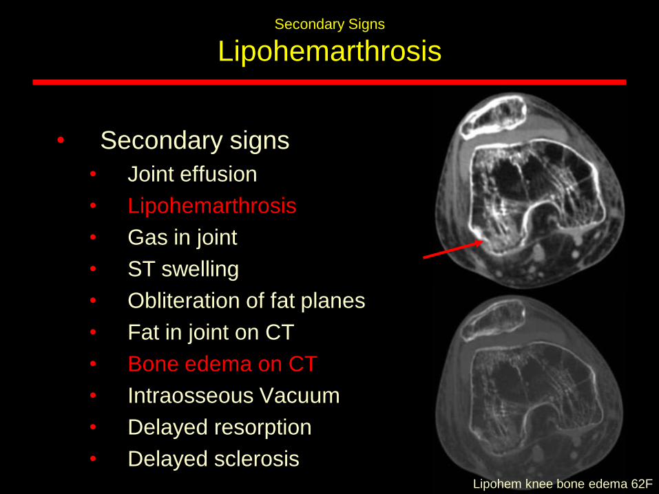
Secondary Signs
Intraosseous Vacuum
• Secondary signs
• Joint effusion
• Lipohemarthrosis
• Gas in joint
• ST swelling
• Obliteration of fat planes
• Fat in joint on CT
• Bone edema on CT
• Intraosseous Vacuum
• Delayed resorption
• Delayed sclerosisLipohem knee bone edema 62F
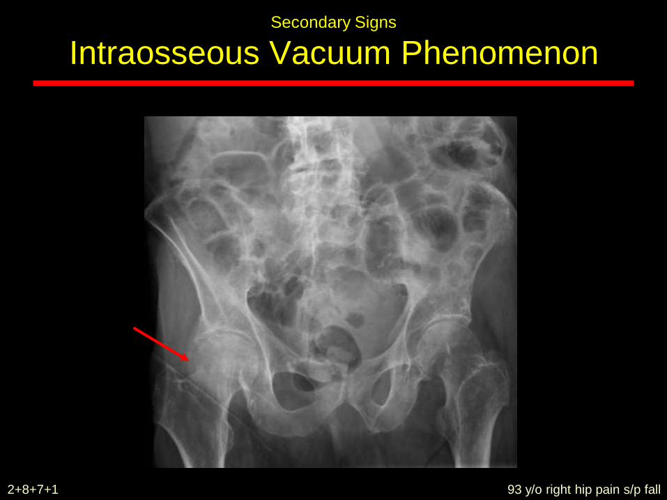
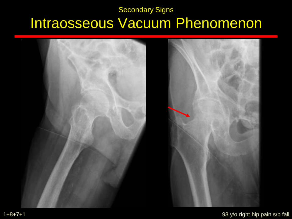
93 y/o right hip pain s/p fall
Secondary Signs
Intraosseous Vacuum Phenomenon
2+8+7+1
Secondary Signs
Intraosseous Vacuum Phenomenon
93 y/o right hip pain s/p fall1+8+7+1
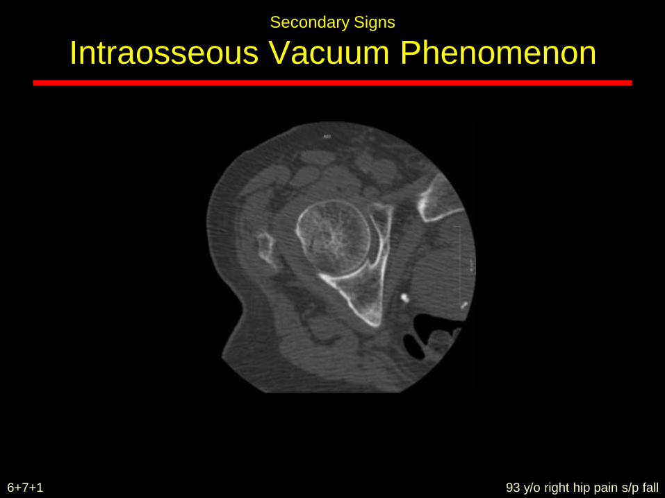
Secondary Signs
Intraosseous Vacuum Phenomenon
93 y/o right hip pain s/p fall6+7+1
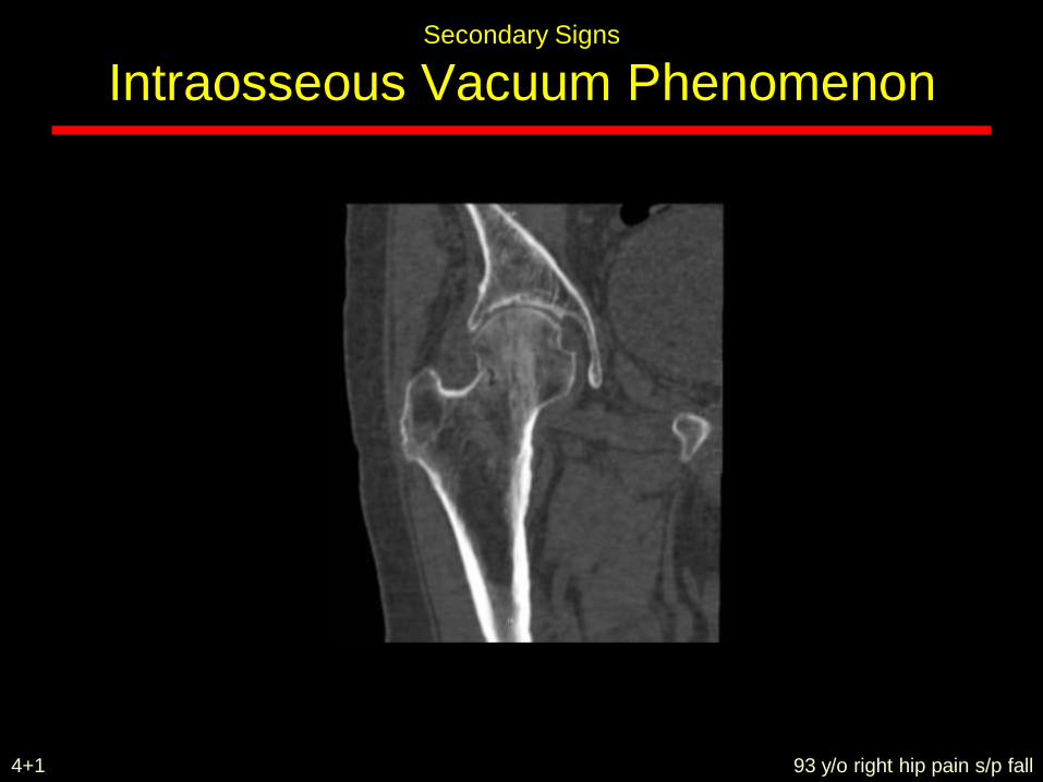
Secondary Signs
Intraosseous Vacuum Phenomenon
93 y/o right hip pain s/p fall4+1
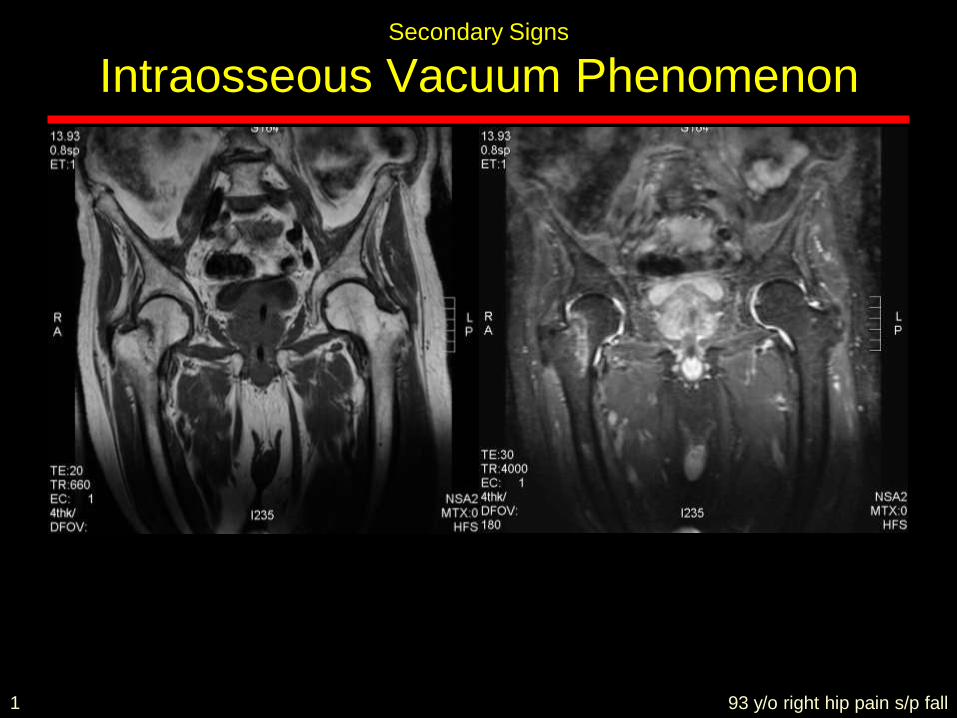
Secondary Signs
Intraosseous Vacuum Phenomenon
93 y/o right hip pain s/p fall1
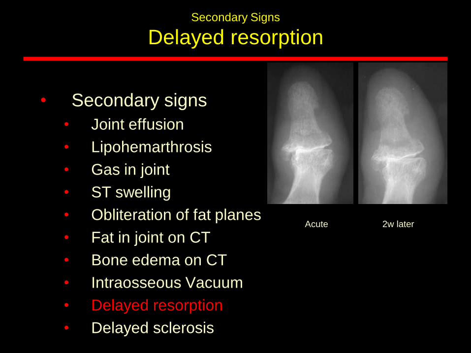
Secondary Signs
Delayed resorption
• Secondary signs
• Joint effusion
• Lipohemarthrosis
• Gas in joint
• ST swelling
• Obliteration of fat planes
• Fat in joint on CT
• Bone edema on CT
• Intraosseous Vacuum
• Delayed resorption
• Delayed sclerosis
Acute 2w later
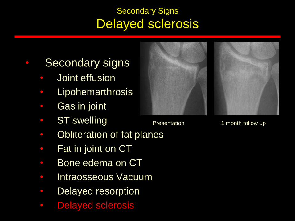
Secondary Signs
Delayed sclerosis
• Secondary signs
• Joint effusion
• Lipohemarthrosis
• Gas in joint
• ST swelling
• Obliteration of fat planes
• Fat in joint on CT
• Bone edema on CT
• Intraosseous Vacuum
• Delayed resorption
• Delayed sclerosis
Presentation 1 month follow up
Elderly
• Fractures often hard to see
• Degenerative changes obscure fractures
• Fatty marrow makes bone edema useful sign
• Fractures more often fatal
• If alters management, low threshold for MRI
Childhood Fractures
• Tendons stronger than bone• Apophyseal avulsion
• Fracture patterns different• Salter Harris
• Incomplete fractures more common• Plastic bowing
• Torus / Buckle
• Greenstick
• Remember NAI
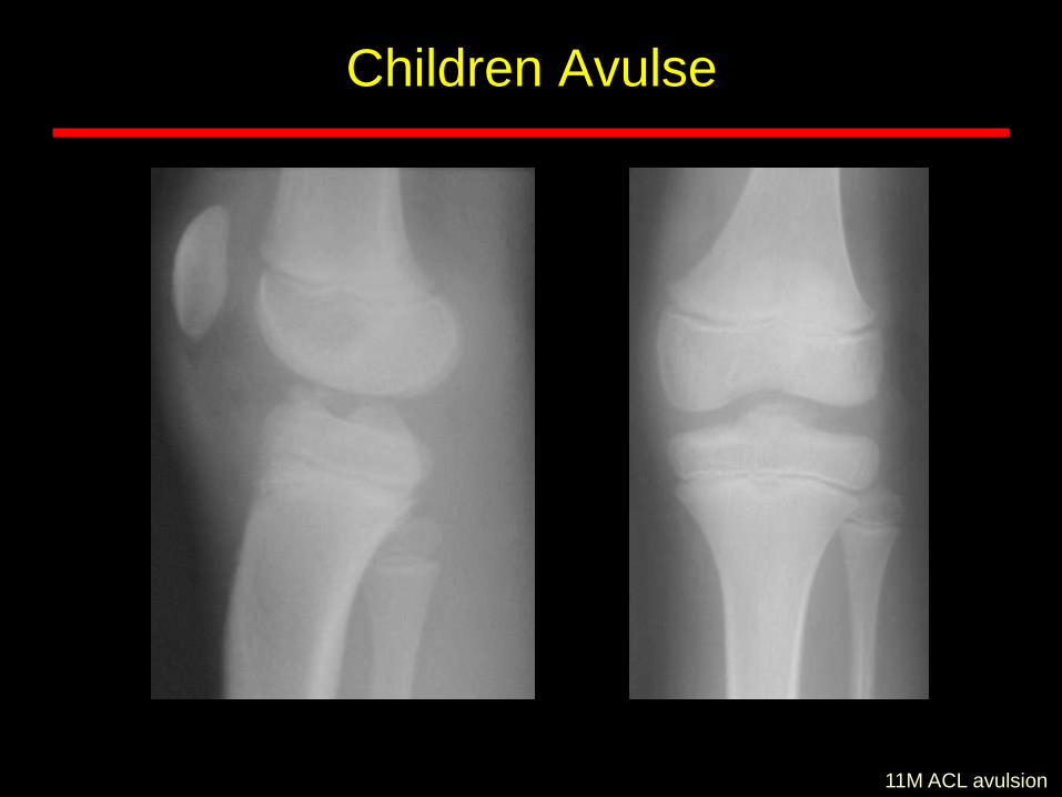
Children Avulse
11M ACL avulsion
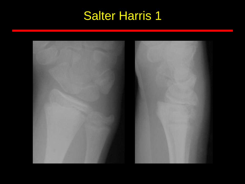
Salter Harris 1
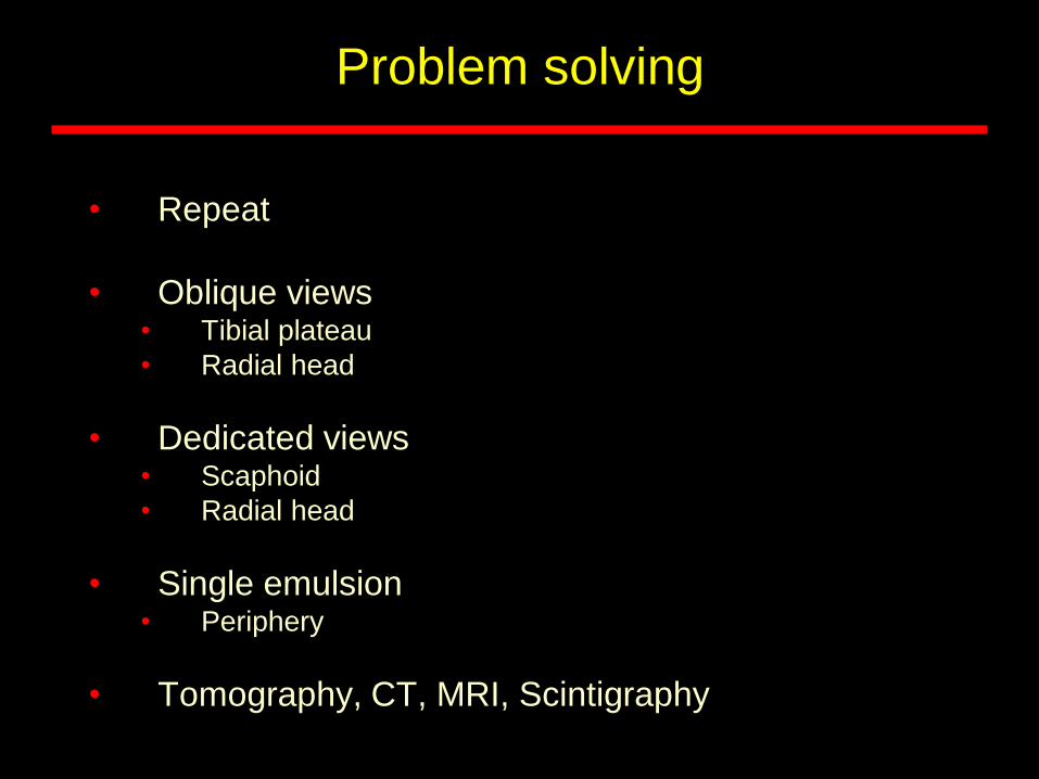
Problem solving
• Repeat
• Oblique views• Tibial plateau
• Radial head
• Dedicated views• Scaphoid
• Radial head
• Single emulsion• Periphery
• Tomography, CT, MRI, Scintigraphy
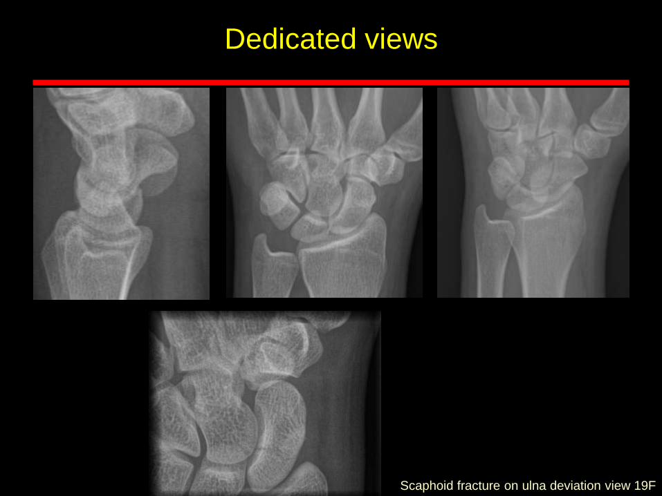
Scaphoid fracture on ulna deviation view 19F
Dedicated views
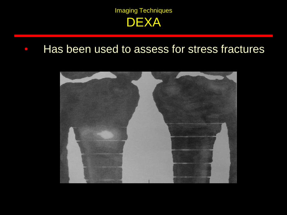
Imaging Techniques
DEXA
• Has been used to assess for stress fractures
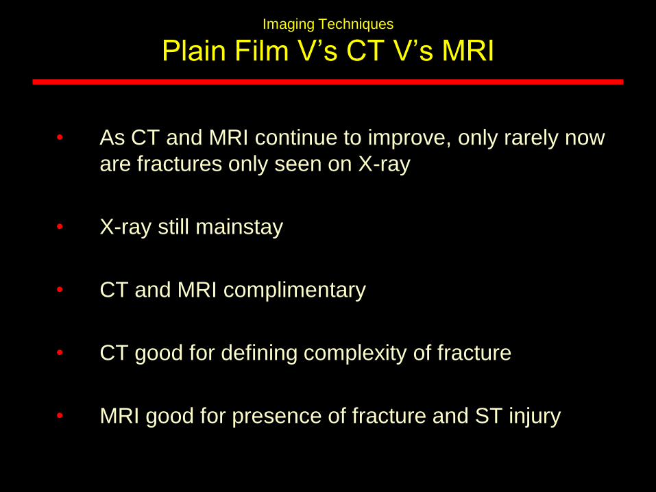
Imaging Techniques
Plain Film V’s CT V’s MRI
• As CT and MRI continue to improve, only rarely now
are fractures only seen on X-ray
• X-ray still mainstay
• CT and MRI complimentary
• CT good for defining complexity of fracture
• MRI good for presence of fracture and ST injury

![New Brunswick Health Indicators · head injuries or concussions. Hockey injuries are the most commonly reported injury among school-aged boys [20]. In New Brunswick, approximately](https://img.pdfslide.us/doc/110x75/5f7023d204a50125214b036a/new-brunswick-health-indicators-head-injuries-or-concussions-hockey-injuries-are.jpg)