Embed Size (px)
Citation preview

COMMON SKIN LESIONS
IN CHILDREN
Dr.Amanda Woods
Consultant Dermatologist
St. Georges NHS Trust

Colour Skin Lesion Red Spider naevus, salmon patch, port-wine
stain, haemangiomas, Spitz naevi
Purple Pyogenic granuloma
Skin coloured Viral wart, molluscum, pilomatricoma,
epidermal naevus, dermatofibroma,
granuloma annulare
Brown Benign naevus, Becker’s Naevus,
Naevus Spilus, Café au lait patch
Black Spindle cell naevus of Reed,
melanoma
Blue Blue naevus, Mongolian blue spot, blue
rubber bleb naevus syndrome
White Milia, halo naevus, naevus
depigmentosus
Yellow Sebaceous naevus, mastocytoma
Orange Juvenile Xanthogranuloma

Red Skin Lesions in Children
• Spider Naevi
• Salmon Patch
• Port-wine Stain
• Haemangiomas
• Spitz Naevi

Spider Naevi
• 10 -15% children
• Common on the face and upper chest.
• Central red papule with
feeding capillary ’legs’
• Compressing the
central point will
blanche the arcade of vessels
• Usually resolve
spontaneously

CAPILLARY VASCULAR
MALFORMATIONS • Malformed dilated blood vessels
• Always present at birth
Two common capillary vascular
malformations:
Salmon Patch Port wine stain

SALMON PATCH
•Very common – 40% of all newborns
•Small flat patches of pink or red
skin with poorly defined borders
•Nape of neck ( Stork Bite), on the
forehead between eyebrows
(Angel’s Kiss) or on the eyelids.
•Most lesions spontaneously
disappear within first year of life
•Stork bites tend to be more
persistent and remain unchanged
in 50% of cases

PORT WINE STAIN
•Less common – 0.3% of newborns
•Large flat patch of purple or dark
red skin with well-defined borders
•Flat at birth – becomes raised with
time
•Face more commonly affected
•Some fade, some deepen in
colour
•Most remain unchanged
•Respond to pulsed dye laser –
45% improve by at least 75% after
5-25 treatments

STURGE-WEBER SYNDROME
•Capillary vascular malformation affecting the skin supplied by one
branch of the trigeminal nerve
• Defects in underlying tissues:
• Cerebral atrophy
• Calcification of skull
• Epilepsy
• Meningeal angioma
• Glaucoma, optic atrophy

HAEMANGIOMAS
•Present at birth or shortly
after
•10% infants affected
•Formed from proliferating
endothelial cells
•80% occur on head and
neck
•Most of the growth is in
first 3 months
•Most stop growing at 5
months

HAEMANGIOMAS
Predisposing Factors
•Low birth weight
•Prematurity
•Females
•Caucasians
•Multiple births
•Advanced maternal
age
•Family history of
Infantile Haemangiomas

HAEMANGIOMAS
Superficial Deep Mixed

ERUPTIVE NEONATAL
HAEMANGIOMATOSIS
•Multiple infantile haemangiomas
present at birth or within early
neonatal period
•Benign eruptive neonatal
haemangiomatosis – if only skin
involved
•Disseminated eruptive neonatal
haemangiomatosis - when
lesions present on internal organs
( GI tract, lung, brain, eyes)
•Death occurs within first few
months of life
•If a neonate has more than 6
haemangiomas should be further
investigated.

REGRESSION
2 months 1 year 2 years
30% regress by the 3rd birthday
50% regress by the 5th birthday
70% regress by the 7th birthday
90% regress by the 9th birthday

WHEN IS TREATMENT
NECESSARY?
• Very large and unsightly lesions
• Ulcerating haemangiomas (5 – 25%)
• Lesions that impair vision, hearing, breathing or feeding
• If they fail to resolve by school age

MANAGEMENT
• The majority of lesions need no
treatment.
• Regular monitoring in Primary Care
may reassure parents.
• Photographs at 6-12 month intervals are very useful

Which lesions should be
referred?
• Interfering with breathing, feeding, or vision Amblyopia can occur within 10 days of lesions interfering with vision
• Facial lesions
• Lesions complicated by bleeding, ulceration or causing functional impairment
• Lumbosacral lesions - up to 50% of such cases are associated with significant problems such as tethering or compression of the spinal cord, and external genital abnormalities. MRI imaging should be performed in such patients
• Kasabach-Merritt syndrome (consumption coagulopathy) is an uncommon complication, mainly in larger lesions

TREATMENT OF SEVERE
INFANTILE HAEMANGIOMAS
• Oral Propranolol - N Eng J Med 2008
• Mechanism of action: inhibits the growth of
blood vessels and constrict existing blood
vessels within the haemangioma. It acts on
beta adrenergic receptors to decrease the
release of blood vessel growth-signalling
molecules (VEGF) and (BFGF) and by
triggering programmed cell death.

Indications for Propranolol
• High risk sites where they may interfere with normal function such as breathing, feeding, vision and hearing.
• Within the airway
• Around the eye
• Around the mouth or on the lips
• Within the ear canal
• On the tip of the nose
• Large lesions on the face
• Napkin area
• Skin creases
• Multiple haemangiomas including visceral (internal) lesions.

How effective is Propranolol?
• Most effective when started during the growth phase of the haemangioma, in infants up to 6 months of age
• They may begin to respond within 24 to 48 hours. The haemangioma softens (decrease in volume) and darkens in colour.
• The optimal duration of treatment is yet to be established, though most reports are of use for 3-12 months. Rebound growth may occur on cessation and gradual weaning may be required.
• NEJM 2015 – 460 infants studied 88% of treated group showed improvement at 5 weeks compared to 5% in control group


Side Effects of Propranolol
• Hypotension
• Bradycardia
• Hypoglycaemia
• Bronchial hyper-reactivity
• Restless sleep
• Cold extremities
• Constipation

TREATMENT OF INFANTILE
HAEMANGIOMS
• Small lesions
• No treatment
• Very potent topical and or intralesional
corticosteroids
• Topical beta blockers
• Lasers

TOPICAL BETABLOCKERS
• Timolol maleate ( Timoptol –LA gel 0.5% )
• Apply once a day
• Treatment is usually for six months
• Side effects are very rare – bradycardia,
hypotension, bronchospasm,
hypoglycaemia, sleep disturbance,
peripheral vasoconstriction

Topical Timolol for Infantile
Haemangiomas

Topical Timolol for Infantile
Haemangiomas

SPITZ NAEVUS (Juvenile Melanoma)
•Well-circumscribed, smooth
surfaced, firm, dome-shaped
pink to dark brown papules and
nodules
•Head, neck and limbs
•2-20mm diameter
•70% < 2years
•Commoner in fair skinned
•Grow rapidly over first 3-6
months
•Remain static for years
•Spontaneously disappear
•Local excision with 2mm
margin
•Benign

PURPLE SKIN LESIONS IN
CHILDREN
• Pyogenic granuloma

PYOGENIC GRANULOMA
• Sudden in onset, grows rapidly and bleeds after minimal trauma
• Can arise on any part of the body but the most common sites are the fingers / hands, head and upper trunk

Dermoscopy Pyogenic
Granuloma
• A distinct keratinised
border or collarette.
• Vascular structures
are usually present
but there is no clear
lacunar pattern.
• White linear 'rail
lines' are often seen.

Management of Pyogenic
Granuloma • Excision may be required if lesion is bleeding or
diagnosis is in doubt
• Recurrence rate is high – 15%
• Topical Timolol has been used
7 cases – 6 topical
All partial response by 2 months
3 bleeding lesions – bleeding stopped
No adverse events
Paediatric Dermatology 2014

Skin coloured lesions in children
• Viral warts
• Molluscum
• Pilomatricoma
• Epidermal naevus
• Dermatofibroma
• Granuloma annulare

VIRAL WARTS
Common warts – single or multiple flat-
topped, skin –coloured hyper-keratotic
lesions
Plane warts – slightly raised, circular or
ovoid lesions on the face and dorsum of
the hands
Plantar warts – hyperkeratotic lesions
that are subject to constant pressure.
Grow inwards and produce discomfort

TREATMENT OF WARTS
1. Nothing!
• 50% warts disappear in 6 months
• 90% warts disappear in 2 years
2. Occlusion – duct tape
3. Chemical treatments – salicylic acid, podophyllin, 3% formalin
4. Cryotherapy
5. Curettage and cautery
6. Imiquimod
7. Immunotherapy with DCP (diphencyprone)

MOLLUSCUM CONTAGIOSUM
•Common viral skin
infection
•Pox virus
•Clusters of pink, white or
brown papules in axillae,
groin and behind knees
•Appear waxy and are
umbilicated
•Spread by direct skin
contact
•Commoner and more
severe in atopic eczema
and HIV
•May persist for months or
years

MOLLUSCUM CONTAGIOSUM
Dermatitis
Scarring
Treatment
•Most cases no specific
treatment is necessary
•Surgery
•Cryotherapy
•Imiquimod
•Salicylic acid or podophyllin
•Crystacide – 1% hydrogen
peroxide cream.
Apply twice a day for three
weeks
In 2/3 patients, lesions had
resolved
•5% Potassium Hydroxide

5% Potassium Hydroxide
• A double-blind, randomised, placebo-
controlled trial of 20 children .
In 70% of patients treated with potassium
hydroxide the lesions cleared, whereas only
20% of the placebo group showed clearance
of lesions.
Short et al. (2006).
• 20 children whose Molluscum contagiosum
all cleared within 6 weeks using a 5%
solution of potassium hydroxide
Romiti et al. (2000)
• 29 children. 44% resolution after 2 months
Ucmak et al 2014

Pilomatricoma
• Skin coloured, firm
papule on head
and neck usually
• Calcification within
lesion – ‘tenting’ of
overlying skin.
• Derived from hair
matrix cells

Pilomatricoma Diagnosis
• Ultrasound shows a
‘doughnut’ in the
dermis – a
hyperechoic area
with calcification
within.
• Plain X ray

Pilomatricoma Treatment
• Treatment is surgical
• Lesions will not
resolve
spontaneously
• Very small risk of
malignant change

Pilomatricoma Histology
• There are darkly
stained ‘basophilic’
cells and ‘ghost’ cells
with missing nuclei.
• Calcium deposits are
found in most lesions.

EPIDERMAL NAEVUS
•0.01% neonates
•Early lesions are macular
•Become
hyperpigmented,
papillomatous linear
plaques
•Extensive lesions may be
part of Epidermal Naevus
Syndrome
•No treatment is required
•Excision is preferable to
superfical treatments
•Malignant change is rare

DERMATOFIBROMA
Dimple test
•Firm, minimally elevated, pink
to yellow brown papules or
nodules
•2mm to 20mm diameter
•Pinching of skin produces a
‘dimple’
•Benign tumour of dermal
dendritic histiocytes
•Common on arms and legs at
sites of minor injury i.e. insect
bite
•Persistent
•Treatment is not usually
required

GRANULOMA ANNULARE
•Skin-coloured or red, non-scaly,
arciform or annular plaques with
a beaded edge
•Ring can vary between 1 – 5 cm
•Single or multiple
•Dorsum of hands and feet
common
•Develop slowly and persist for
years
•Unknown aetiology
•Resolve spontaneously without
scarring
•Generalised GA may be
associated with diabetes mellitus
•Topical steroids may hasten
resolution

Brown Skin Lesions in Children
•Benign naevus
•Becker’s Naevus
• Naevus Spilus
•Café au lait patch
CONGENITAL MELANOCYTIC
NAEVI
Multi – shaded pigmented patches, oval-shaped and
uniform
Larger naevi have satellite lesions
Present at birth
Small – up to 1.5cm diameter
Medium – up to 10cm
Large – between 10 and 20cm
Giant – over 20cm – bathing
trunk naevi
Common – 1 in 100 (small and
medium)

RISK OF MELANOMA
•Small or medium
naevi under 1%
•Giant Naevi – 5%

NEUROCUTANEOUS
MELANOCYTOSIS
•Many congenital naevi
with abnormal
melanocytes in the central
nervous system.
•Affects 10% patients with
giant melanocytic naevi.
•MRI scan detects
melanocytes
•Hydrocephalus
•Epilepsy
•Developmental delay

BECKER’S NAEVUS
•Solitary, unilateral
•Childhood, more
prominent in adolescence
•Initial irregular macular
pigmentation becoming
darker and well
demarcated
•Skin becomes thickened,
hairy, comedones,
papules and cystic
nodules develop
•Upper quadrant of trunk
or upper arm
•M:F 5:1
•Up to 15cm diameter
•Malignant transformation
not reported

NAEVUS SPILUS
•A speckled lentiginous
naevus with dark spots
scattered on a flat tan
background
•Occasionally occurs in
neurofibromatosis
•Melanoma is rare – 1%
•Risk factors: >4cm diameter
•Present since birth

CAFÉ AU LAIT PATCHES
•Flat tan mark usually oval in
shape
•Familial
•Six or more café au lait
patches more than 0.5cm in
diameter may indicate
neurofibromatosis

Black Skin Lesions in Children
• Spindle cell naevus of Reed
• Melanoma

Spindle Cell Naevus of Reed
• Spindle Cell Naevus
of Reed is a well
defined heavily
pigmented lesion.
• Often seen on the
hands and feet.

Spindle Cell Naevus of Reed
• Dermoscopy: ‘Starburst’ pattern
• Histology: Characteristic nests of
spindle-shaped cells with heavy pigmentation

Malignant Melanoma in
Childhood
• Rare paediatric neoplasm
• 1 -3% of all childhood cancers
• 1 -4% malignant melanomas are in children and adolescents less than 20
• 7 x more frequent in 2nd than 1st decade
• Diagnosed at a thicker level than in adults – often a delay in diagnosis
• Same prognostic factors as adults

Blue Skin Lesions in Children
• Blue naevus
• Mongolian blue spot
• Blue rubber bleb syndrome

Blue Naevus
• Benign
• Blue because the
melanocytes are
deeper in the
dermis
• Scalp, face, hands
and feet
• Older children and
adolescents

Dermoscopy Blue Naevus
• Uniform steel blue
pattern

MONGOLIAN BLUE SPOT
•Blue –grey marking of the skin
that usually affects the lower
back and buttock region
•Few centimetres in diameter
but can be larger
•Present at birth in more than
90% children of East Asians,
Polynesians and Indonesians
•Aberrant Mongolian spots on
face and limbs – can be
confused with bruises
•Entrapment of melanocytes in
dermis
•Usually disappear by the age
of 4

Blue Rubber Bleb Naevus
Syndrome
• Rare form of venous
malformation with skin
and gastrointestinal tract
lesions
• Iron deficiency anaemia
• AD
• compressible blue or
purple rubbery nodules
with a wrinkled surface
• Laser or sclerotherapy

White Skin Lesions in Children
• Milia
• Halo naevus
• Naevus depigmentosus

Milia
• Eyelids, cheeks,
forehead and genitalia
• Histology: small
epidermoid cyst
coming from a vellus
hair follicle.
• Resolve spontaneously-
may take months
• Topical retinoids for
widespread lesions,
single lesions deroofed
with sterile needle

Halo Naevus
• Autoimmune process
against the
melanocytes
• Older children
• Often multiple
• Trunk usually affected
• Central naevus
disappears over
months or years
• No treatment

Differential Diagnosis
Halo naevus – uniform
depigmentation around
benign naevus
Regression in a melanoma
- Irregular naevus with
irregular depigmentation

Naevus Depigmentosus
• Well defined but irregular
pale patch
• Usually noted at birth or
early childhood
• Naevi remain stable over
time.
• Trunk and limbs
• Cutaneous mosaicism
• Clone of melanocytes
that are unable to form
melanin

Yellow Skin Lesions in Children
• Sebaceous Naevus
• Mastocytoma
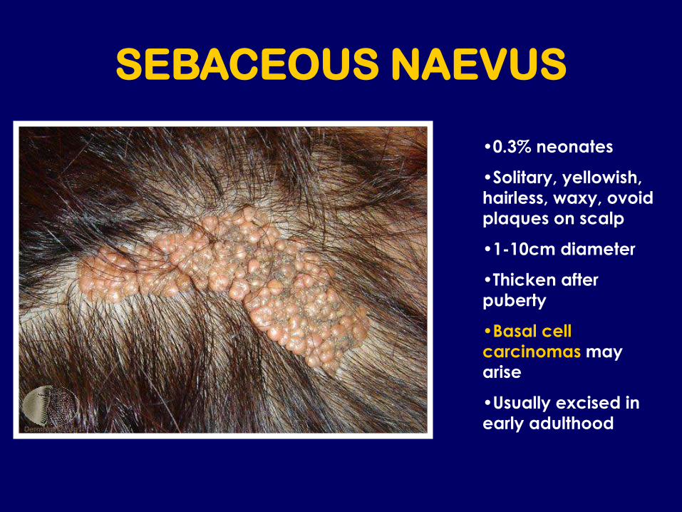
SEBACEOUS NAEVUS
•0.3% neonates
•Solitary, yellowish,
hairless, waxy, ovoid
plaques on scalp
•1-10cm diameter
•Thicken after
puberty
•Basal cell
carcinomas may
arise
•Usually excised in
early adulthood

MASTOCYTOMA
Darier’s Sign
•Red, pink or tan nodules
•Up to 4cm in diameter
•Solitary or multiple
•May be pruritic and blister
•Increased mast cells in skin
•When rubbed produce an
urticarial wheal – Darier’s Sign
•Resolve by adolescence
•Urticaria Pigmentosa – multiple
mastocytomas
•Systemic mastocytosis is rare in
children

Orange Skin Lesions in Children
• Juvenile Xanthogranuloma

JUVENILE
XANTHOGRANULOMA
•Soft orange/pink dome
shaped papules
•Head, neck, trunk and
proximal limbs
•Single or multiple
•15% present at birth
•75% appear in first year
•Benign form of
histiocytosis
•0.5% have ocular
involvement - glaucoma
•Resolve over 3-6 year
period by puberty
•No treatment required

Juvenile Xanthogranuloma
• ‘Setting sun’ sign on dermoscopy

Which childhood lesions should be referred to
secondary care?
•Any changing lesion
•Haemangiomas – ulcerated, bleeding or obscuring
vision, hearing
•Port-wine stains
•Pyogenic granulomas
•?Spitz naevi
•?Pilomatricomas
•?Mastocytomas
•?Juvenile Xanthogranulomas
•?Spindle Cell Naevus of Reed
•More than six café au lait patches