Embed Size (px)
Citation preview

Common mechanisms for regulated exocytosis in thechromaffin cell and the synapseAlan Morgan and Robert D. Burgoyne
Chromaffin granule exocytosis differs in many physiologicalrespects from neuronal synaptic vesicle exocytosis, which hasled to the assumption that the two processes occur by distinctmechanisms. While different mechanisms are certainly inoperation for the biogenesis of granules and synaptic vesicles,it is now becoming clear that similar mechanisms are used byboth beyond this stage. The similarities extend to variousaspects of regulated exocytosis, including regulation of thenumber of vesicles released in response to cell stimulation.Most strikingly, it now appears that the same proteinsmediate the docking and fusion of both chromaffin granulesand synaptic vesicles, and that homologues of these proteinsact similarly in constitutive membrane traffic throughoutevolution.
Key words: calcium / exocytosis / secretory granule /synaptic vesicle
©1997 Academic Press Ltd
ADRENAL MEDULLARY chromaffin cells are derivedembryonically from the neural crest, from the sameprecursors that give rise to sympathetic neurons, andare known to express a large number of ‘neuronal-specific’ proteins. For this reason, chromaffin cellsand their tumour cell derivatives, PC12 cells, havebeen extensively used as neuronal cell models. Thelarge dense-core vesicles present in certain neuronsare very similar to the catecholamine-containingchromaffin granules (see review by Apps, this issue)present in these neuroendocrine cells. Indeed, mostof the information on neuronal large dense-corevesicle biogenesis and exocytosis has been inferredfrom studies of chromaffin granules over many years.More recently, it has become clear that neuroendo-crine cells also possess another class of regulatedsecretory vesicle, synaptic-like microvesicles (SLMVs),which are very similar to neuronal synaptic vesicles.Again, much of the information on neuronal synapticvesicle biogenesis has come from studies of these
neuroendocrine cells (especially PC12 cells),although little is known about SLMV exocytosis. Thephysiology of synaptic vesicles and large-dense corevesicles varies greatly (see Table 1), precipitating thenotion that the vast wealth of data on granuleexocytosis in chromaffin and PC12 cells could beextrapolated to the exocytosis of neuronal largedense-core vesicles, but not synaptic vesicles. However,recent evidence points to similar mechanisms beingused in the Ca2-triggered exocytosis of both vesicleclasses.
The life cycle of granules and synaptic vesicles
Although both classes of vesicle share the ability toundergo Ca2+ -triggered exocytosis, they differ inmany respects, including the kinetics and Ca2+ -dependency of exocytosis (Table 1). The clearestdifference between the two vesicle classes, however, isin their biogenesis. Immature granules bud directlyfrom the trans-Golgi network (TGN) and are thoughtto develop into mature granules via fusion with otherimmature granules, with excess membrane possiblybeing removed by a budding process.1 Upon anincrease in cytosolic free Ca2+ concentration, aproportion of granules undergo exocytosis. Sincegranules contain large amounts of lumenal protein,which can only be taken up in the TGN, it appearsthat exocytosis represents the end of the granule lifecycle. However, there are some indications that thegranule membrane may be recycled back to the TGNfor this purpose rather than being synthesized denovo.2
In contrast, synaptic vesicles are thought to origi-nate from constitutive secretory vesicles whichundergo exocytosis and are then recycled to aspecialized endosome.1,3 Here, synaptic vesicle pro-teins are sorted away from constitutive recyclingproteins into synaptic vesicles, which only undergoexocytosis in response to an increase in cytosolic freeCa2+ concentration. Following regulated exocytosis,synaptic vesicles (which do not contain lumenal
The Physiological Laboratory, University of Liverpool, P.O. Box147 Crown Street, Liverpool L69 3BX, UK
seminars in CELL & DEVELOPMENTAL BIOLOGY, Vol 8, 1997: pp 141–149
©1997 Academic Press Ltd1084-9521/97/020141 + 09 $25.00/0/sr960133
141

Table 1. Characteristics of regulated secretory vesicles in neurons
Synaptic vesicles Large dense-core vesicles
Vesicle diameter 50 nm 200 nmSpeed of transmission 200 µs MillisecondsNeurotransmitter Fast neurotransmitters PeptidesCells affected Post-synaptic contact only Cells in surrounding areaDuration of effect Short-lived Longer-livedStimulation Low-frequency High-frequencyCa2+ concentration Hundreds of micromolar Tens of micromolarLocal vesicle recycling? Yes No
protein) are thought to be recycled via the endosometo replenish the vesicle pool.1,3 In addition to endoso-mal recycling, it may be that synaptic vesicles can bereused without returning to the endosome by a ‘kissand run’ process whereby the vesicle fuses briefly withthe plasma membrane and immediately pinches off toreform itself, reload with neurotransmitter and await afurther signal for regulated exocytosis.4
Since the life cycles of granules and synaptic vesiclesdiffer so dramatically it has, quite reasonably, beenassumed that they each employ distinct mechanismswhich give rise to their differing physiologies. Whilethis is almost certainly true for their biogenesis, it isnow becoming clear that analogous mechanisms areemployed for the regulation of the releasable pool ofvesicles and identical mechanisms for the final stagesof exocytosis. Moreover, it now appears that themechanism of vesicle docking and fusion is conservednot only in granule and synaptic vesicle exocytosis, butalso in constitutive membrane traffic throughout thesecretory and endocytic pathways from yeast to thehuman brain.
Regulation of the releasable pool of vesicles
In many regulated secretory cell types, including thechromaffin cell, the vast majority of granules areprevented from reaching the plasma membrane by adense subplasmalemmal cytoskeletal network com-posed predominantly of actin 5,6 (Figure 1). Clearly,this barrier to exocytosis must be overcome in someway in order to allow granules to fuse with the plasmamembrane upon receipt of an appropriate signal(usually Ca2+ ). This is achieved by the reversibledisassembly of the cortical cytoskeleton to allow thegranules to move to the plasma membrane. Bio-chemical and microscopic analyses support this the-ory, and in chromaffin cells cytoskeletal breakdownoccurs in discrete patches which correlate with exocy-totic release sites.6 The protein(s) which Ca2+ acts on
to trigger cytoskeletal disassembly are unclear butthere is evidence that scinderin, a Ca2+ -dependentactin-severing protein, is involved in this process inchromaffin cells.6 At least one protein kinase Csubstrate is likely to be required since phorbol estersare able to cause cytoskeletal disassembly in chro-maffin cells. However, as phorbol esters do not elicitsecretion in these cells, cytoskeletal disassembly isnecessary but not sufficient for regulated exocytosis,and hence Ca2+ acts at least two distinct stages inexocytosis: an early stage of actin rearrangement anda late stage of membrane fusion.
In neuronal exocytosis, a similar mechanism mayoperate for large dense-core vesicles (although thishas not been actively studied in neurons), but synapticvesicles are not directly bound to the actin cytoskele-ton. Rather, while a small proportion of synapticvesicles are tightly docked to neurotransmitter releasesites at the presynaptic plasma membrane (‘the activezone’), the majority of synaptic vesicles are cross-linked by the extrinsic vesicle protein synapsin I,which is itself associated with the actin cytoskele-ton.3,6,7 Following the action potential, synaptic vesi-cles at the active zone undergo exocytosis within200 µs in response to the extremely high Ca2+
concentrations at the mouth of presynaptic Ca2+
channels to which synaptic vesicles are thought to betethered.3,7 Evidently, some mechanism must exist toallow the replenishment of vesicles lost from the activezone as a consequence of such rapid exocytosis.Abundant evidence suggests that this mechanisminvolves the liberation of vesicles from the reservepool of synapsin I-linked synaptic vesicles (Figure 1).Ca2+ is known to have very limited diffusion in cytosoldue to the efficiency of Ca2+ binding and extrusionmechanisms, and so although the concentration ofCa2+ may be several hundred micromolar at theplasma membrane, a short distance within the pre-synaptic terminal it would be sub-micromolar. Suchsub-micromolar Ca2+ concentrations activate calm-odulin-dependent protein kinase II (present on syn-
A. Morgan and R. D. Burgoyne
142

aptic vesicles) which phosphorylates synapsin I, lead-ing to its dissociation from the vesicles and so allowingthe released synaptic vesicles to move to and dock atthe active zone.3,6,7 Thus, as in chromaffin granuleexocytosis, Ca2+ acts at an early stage of filamentdisassembly and at a late stage of membrane fusion.Synapsin I would then be dephoshorylated by phos-phatases to allow cross-linking of new endosome-derived (or possibly ‘kiss and run’-derived) synapticvesicles. In this way, a steady supply of synaptic vesiclesis kept ready for fusion in order that the neuron canrespond to repetitive stimulation.
Although the regulation of the pool of releasablegranules and synaptic vesicles is mediated by distinctsets of proteins, the two mechanisms fulfil the samepurpose. Patch-clamp flash photolysis studies in chro-maffin cells have revealed multiple kinetic phases ofexocytosis (see Gillis and Chow, this issue), which canbe simplified into fast and slow phases. The fastphases, lasting from milliseconds to seconds, com-prise a small secretory response representing thefusion of only a few hundred granules per cell;
whereas the slow phase, lasting from seconds tominutes, is a much larger secretory response repre-senting the fusion of up to 10 000 granules per cell(this slow exocytosis is what is measured in bio-chemical assays of secretion). It is thought that thesekinetically distinct phases of release are related to thedistribution of granules in the cell, since electronmicroscopic studies have revealed that of the 30 000granules in chromaffin cells, all but around 500 arekept away from the plasma membrane by the corticalactin barrier mentioned above.5 Thus, it may be thatthese 500 granules undergo fast exocytosis as a resultof their proximity to the plasma membrane, while therequirement for cytoskeletal disassembly retards theexocytotic fusion of the remaining granules. This maybe important in vivo since it is likely that only a smallnumber of vesicles are released per chromaffin cell toprovoke the ‘fight or flight’ response (secretion of 10000 vesicles per cell by the whole adrenal medullawould almost certainly release a fatal dose of adrena-line). Thus, chromaffin cells use a similar mechanismto that employed by neurons to ensure that a limited
Figure 1. Ca2+ -activated stages in regulated exocytosis.Synapse: Following an action potential, highCa2+ concentrations (EC50 = 190 µM) cause synaptic vesicles docked at the active zone to undergoultrafast exocytosis. Lower Ca2+ concentrations deeper in the synapse cause the liberation ofsynaptic vesicles from the synapsin-linked reserve pool to replenish the readily-releasable pool atthe active zone. Chromaffin cell: Following cell stimulation, moderate Ca2+ concentrations(EC50 = 15 µM) cause granules located close to the plasma membrane to undergo fast exocytosis.This Ca2+ signal also causes disassembly of the cortical actin barrier, enabling the liberation ofgranules from the reserve pool to replenish the readily-releasable pool near to the plasmamembrane.
Mechanisms of regulated exocytosis
143
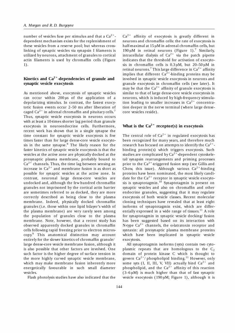
number of vesicles fuse per stimulus and that a Ca2+ -dependent mechanism exists for the replenishment ofthese vesicles from a reserve pool; but whereas cross-linking of synaptic vesicles via synapsin I filaments isutilized by neurons, attachment of granules to corticalactin filaments is used by chromaffin cells (Figure1).
Kinetics and Ca2+ -dependencies of granule andsynaptic vesicle exocytosis
As mentioned above, exocytosis of synaptic vesiclescan occur within 200 µs of the application of adepolarizing stimulus. In contrast, the fastest exocy-totic fusion events occur 2–50 ms after liberation ofcaged Ca2+ in adrenal chromaffin and pituitary cells.7
Thus, synaptic vesicle exocytosis in neurons occurswith at least a 10-times shorter lag period than granuleexocytosis in neuroendocrine cells. Furthermore,recent work has shown that in a single synapse thetime constant for synaptic vesicle exocytosis is fivetimes faster than for large dense-core vesicle exocyto-sis in the same synapse.8 The likely reason for thefaster kinetics of synaptic vesicle exocytosis is that thevesicles at the active zone are physically docked at thepresynaptic plasma membrane, probably bound toCa2+ channels. Thus, the time lag between sensing anincrease in Ca2+ and membrane fusion is as short aspossible for synaptic vesicles at the active zone. Incontrast, neuronal large dense-core vesicles areundocked and, although the few hundred chromaffingranules not imprisoned by the cortical actin barrierare sometimes referred to as docked, they are morecorrectly described as being close to the plasmamembrane. Indeed, physically docked chromaffingranules (i.e. those within one lipid bilayer’s width ofthe plasma membrane) are very rarely seen amongthe population of granules close to the plasmamembrane. Note, however, that a recent study hasobserved apparently docked granules in chromaffincells following rapid freezing prior to electron micros-copy.9 This anatomical distinction may accountentirely for the slower kinetics of chromaffin granule/large dense-core vesicle membrane fusion, although itis also possible that other factors are involved. Onesuch factor is the higher degree of surface tension inthe more highly curved synaptic vesicle membrane,which may make membrane fusion inherently moreenergetically favourable in such small diametervesicles.
Flash photolysis studies have also indicated that the
Ca2+ affinity of exocytosis is greatly different inneurons and chromaffin cells: the rate of exocytosis ishalf-maximal at 15 µM in adrenal chromaffin cells, but190 µM in retinal neurons (Figure 1).7 Similarly,intracellular dialysis of Ca2+ via the patch pipetteindicates that the threshold for activation of exocyto-sis in chromaffin cells is 0.3 µM, but 20–50 µM inretinal neurons.7 This large difference in Ca2+ affinityimplies that different Ca2+ -binding proteins may beinvolved in synaptic vesicle exocytosis in neurons andgranule exocytosis in chromaffin cells (see later). Itmay be that the Ca2+ affinity of granule exocytosis issimilar to that of large dense-core vesicle exocytosis inneurons, which is induced by high-frequency stimula-tion leading to smaller increases in Ca2+ concentra-tion deeper in the nerve terminal (where large dense-core vesicles reside).
What is the Ca2+ receptor(s) in exocytosis
The central role of Ca2+ in regulated exocytosis hasbeen recognized for many years, and therefore muchresearch has focused on attempts to identify the Ca2+ -binding protein(s) which triggers exocytosis. Suchstudies are complicated by Ca2+ -dependent cytoskele-tal/synapsin rearrangements and priming processesprior to the Ca2+ -triggered fusion step (see Gillis andChow, this issue). Although various Ca2+ -bindingproteins have been nominated, the most likely candi-date for the Ca2+ receptor in synaptic vesicle exocyto-sis is synaptotagmin.10 Synaptotagmin is present onsynaptic vesicles and also on chromaffin and otherendocrine granules, suggesting that it may regulateexocytosis of both vesicle classes. Recent molecularcloning techniques have revealed that at least eightisoforms of synaptotagmin exist, which are differ-entially expressed in a wide range of tissues.11 A rolefor synaptotagmin in synaptic vesicle docking/fusionhas been suggested based on its interaction withN-type Ca2+ channels, the α-latratoxin receptor andsyntaxin: all presynaptic plasma membrane proteinswhich have been implicated in synaptic vesicleexocytosis.
All synaptotagmin isoforms (syts) contain two cyto-plasmic repeats that are homologous to the C2
domain of protein kinase C which is thought togovern Ca2+ /phospholipid binding.11 However, onlysome syts (I, II, III, V, VII) actually bind Ca2+ andphospholipid, and the Ca2+ affinity of this reaction(3–6 µM) is much higher than that of fast synapticvesicle exocytosis (190 µM; Figure 1), although it is
A. Morgan and R. D. Burgoyne
144

similar to that of granule exocytosis (15 µM; Figure 1).Syts I, II and V bind syntaxin at Ca2+ concentrationsof 200–500 µM, while syts III and VII bind at < 10 µMCa2+ , whereas syts IV, VI and VIII do not bind tosyntaxin.11 Since syntaxin is a key player in vesicledocking/fusion (see later), the Ca2+ dependence ofthis protein–protein interaction may be of moresignificance for exocytosis than phospholipid binding.The presence of synaptotagmin I on the synapticvesicle suggests that it may be the low-affinity Ca2+
receptor in fast neurotransmitter release, which isknown to require high Ca2+ concentrations. Indeed,cultured neurons from transgenic mice carrying a lossof function mutation in the syt I gene have beenfound to be defective in fast synaptic transmission,although the slow component of synaptic transmis-sion was unaffected.12 This suggests that syt I is thelow-affinity Ca2+ -receptor active in fast neurotransmis-sion, but that a different high-affinity Ca2+ -bindingprotein mediates slower synaptic vesicle exocytosis.Furthermore, syt mutants in Drosophila show reducedCa2+ cooperativity, consistent with the proposed roleof syt as a vesicle Ca2+ sensor.13 In addition to its likelyrole in synaptic vesicle exocytosis, it seems thatsynaptotagmin also functions in the recycling ofsynaptic vesicles after exocytosis, since all syts havenanomolar affinities for the AP2 complex involved inendocytosis11 and since synaptic vesicle endocytosis isinhibited in C. elegans syt mutants,14 thus complicatingthe interpretation of neurotransmission data.
If syt I really is the low-affinity Ca2+ -receptor insynaptic vesicle exocytosis, what is the high-affinityreceptor? This is an important question since, asdiscussed earlier, large dense-core vesicle exocytosis inneurons and secretory granule exocytosis requiremuch lower Ca2+ concentrations than fast synapticvesicle exocytosis, and so it is possible that the sameprotein(s) regulate exocytosis in all these cases. SytsIII and VII are candidates for such a protein sincetheir Ca2+ affinity for syntaxin binding ( < 10 µM) issimilar to that of granule exocytosis and as PC12 cellsexpressing syt III, but lacking syts I and II, exhibitgranule exocytosis.15 However, the wealth of evidencefor a role of synaptotagmin in neurotransmissionmust be contrasted with the paucity of data support-ing its putative role in granule exocytosis,16 which isitself controversial.15,17 No published functional dataare available regarding syt’s role in chromaffin cellexocytosis, and the only isoform definitely expressedin these cells is syt I (see Apps, this issue), whose Ca2+
affinity is poorly matched to granule exocytosis. Inorder to properly investigate synaptotagmin’s possible
role in chromaffin granule exocytosis, it will benecessary to first identify which isoforms are presentand to subsequently selectively disrupt their functionand study the effect on secretion.
Alternatively, the high-affinity Ca2+ -receptor maynot be a syt isoform and two extrinsic membraneproteins found on both synaptic vesicles and granules,rabphilin-3A and Doc2, are potential candidates. Likesyts, rabphilin-3A and Doc2 each contain two repeatsof the C2 domain of protein kinase C and bind Ca2+ /phospholipid. The Ca2+ affinity of Doc2 is unknown,but that of rabphilin-3A is consistent with a high-affinity Ca2+ receptor (maximal at 1-10 µM).18 A rolefor rabphilin-3A in exocytosis has been suggestedbased on its binding in a GTP-dependent manner torab3A,18 which is itself thought to act in exocytosis(see later), and overexpression of both rabphilin-3Ain chromaffin cells19 and Doc2 in PC12 cells20
enhances Ca2+ -dependent granule exocytosis. Thereare functional data supporting a potential role forvarious other Ca2+ -binding proteins, includingannexin II,21 p14522 and calmodulin,23 in granuleexocytosis and so it may be that exocytosis is notcontrolled by a single Ca2+ receptor, but rather that anumber of different Ca2+ binding proteins integratethe Ca2+ signal to provide sophisticated control overexocytosis. This concept of multiple Ca2+ receptors isconsistent with the high degree of cooperativity forCa2+ exhibited by regulated exocytosis.
A conserved mechanism for vesicle dockingand fusion
Tetanus and botulism are fatal diseases caused byextremely potent neurotoxin proteins released byClostridium tetani and Clostridium botulinum, respec-tively. Tetanus toxin and the seven botulinum neu-rotoxins (designated A–G) were known to poisonnerve terminals by blocking neurotransmitter releaseat an intracellular site. However, their mechanism ofaction was mysterious until recently, when the pio-neering work of Schiavo et al.24 revealed that tetanusand botulinum B toxins are zinc-dependent endopro-teases which block neurotransmitter release by thespecific cleavage of the synaptic vesicle protein,synaptobrevin (also known as VAMP). The proteolyticsubstrates for the other botulinum toxins were subse-quently identified as synaptobrevin (D, F, G), SNAP-25(synaptosomal-associated protein, 25 kDa; A, E) andsyntaxin (C).25 Thus, the synaptic vesicle proteinsynaptobrevin and the presynaptic plasma membrane
Mechanisms of regulated exocytosis
145

proteins SNAP-25 and syntaxin (Figure 2) are essen-tial for synaptic vesicle exocytosis. The clostridialneurotoxin substrates are present in chromaffincells26,27 and neurotoxins specific for each substratehave been shown by many laboratories to inhibitgranule exocytosis in chromaffin and PC12 cells. Theimportance of synaptobrevin-like proteins in vesicularfusion is underscored by the observation that itshomologues Snc1p and Snc2p are present on post-Golgi vesicles and are required for exocytosis of thesevesicles in yeast.28 Yeast homologues of syntaxin(Sso1p and Sso2p)29 and SNAP-25 (Sec9p)30 are alsothought to function in exocytosis, indicating thatsimilar vesicle and plasma membrane proteins medi-ate docking and/or fusion of secretory vesicles inyeast and mammalian brain.
In apparently unconnected work, a search forcytosolic proteins involved in vesicular fusion in theGolgi stack in vitro identified NSF (N-ethylmaleimide-sensitive fusion protein) and SNAPs (soluble NSFattachment proteins) as essential proteins.31 Theseproteins are thought to function in membrane fusionevents at all stages of the constitutive secretory andendocytic pathways, and homologues of NSF (Sec18p)and α-SNAP (Sec17p) are required for vesiculartransport in yeast.31 NSF and SNAPs were known toassemble into a 20 S complex in the presence ofdetergent-solubilized membranes, prompting asearch for the membrane receptors for SNAPs.Recently, the SNAP receptors (‘SNAREs’) in brainmembranes were identified as synaptobrevin, syntaxinand SNAP-25.32 Since these are all substrates for theclostridial neurotoxins, this suggested to the authorsthat NSF and SNAPs also acted in regulated exocytosisin addition to their known function in constitutivemembrane fusion. Indeed, SNAPs have recently beenshown to stimulate granule exocytosis in chromaffincells33 and synaptic vesicle exocytosis in the squidgiant synapse.34 NSF is assumed to act in concert withSNAPs in both granule and synaptic vesicle exocytosis(as it does in all known processes), and this notion issupported by the recent discovery that the tem-perature-induced block of neurotransmission exhib-ited by the Drosophila mutant, comatose, is due tomutations in the NSF-1 gene.35
There is little doubt that synaptobrevin, syntaxin,SNAP-25, NSF and SNAPs are required for regulatedexocytosis in chromaffin cells as well as synapses, andfunction as part of an evolutionarily ancient ubiqui-tous mechanism for vesicle docking and fusion.However, controversy has raged in recent years as tothe roles of the individual proteins and the precise
point at which they function in the complex processof docking/fusion (see ref 36 for review). At present itis unclear what the precise roles of synaptobrevin,syntaxin and SNAP-25 are, but they are likely to beessential for docking, fusion or both. Consensus isnow emerging that SNAPs and NSF act prior todocking36-38 possibly by SNAPs stimulating the ATPaseactivity39 of the putative molecular chaperone NSF toinduce conformational changes in the SNARE pro-teins to facilitate their function in docking/fusion.36,37 Work on constitutive membrane fusionhas indicated that following the priming action ofNSF/SNAPs, a rab protein is subsequently required toact,37 possibly by further affecting SNARE function.40
Rab3 (four isoforms, A–D) is localized to bothsynaptic vesicles and chromaffin granules and func-tional studies have indicated the involvement of rab3Ain exocytosis of both vesicle classes,41,42 although theprecise molecular function of rab proteins remainsunknown.
While much attention has been focused on theneurotoxin substrates, SNAPs and NSF, several otherpresynaptic proteins have been implicated as keycomponents of the exocytotic machinery (Figure 2)and some of these are capable of interacting with theNSF/SNAP/SNARE core complex. All of the proteinsshown in Figure 2 are expressed in chromaffin cells aswell as synapses. Synaptotagmin has been shown tobind to the complex of synaptobrevin, SNAP-25 andsyntaxin.43 In addition, it has recently been claimedthat syt independently binds â-SNAP, but notα-SNAP,44 suggesting that α- and â-SNAP may havedistinct functions. In permeabilized chromaffin cells,however, α- and â-SNAPs appear to function asinterchangeable isoforms.45 Munc18, identified inchromaffin cells as mSec1,26 binds to syntaxin andfunctional evidence for a role for this protein inneurotransmitter release comes from studies of itshomologues in nematodes and Drosophila46 althoughno functional data exist for granule exocytosis. Syn-aptophysin has been shown to interact with VAMP,47
but it is difficult to draw conclusions on the role ofthis abundant synaptic vesicle protein since its localiz-ation to granule membranes and, indeed, its require-ment for synaptic vesicle exocytosis48 remaincontroversial.
Other putative components of the conservedmachinery for exocytosis which are not known toassociate with the core complex include the previouslydiscussed rab3 and rabphilin3A and also cysteinestring protein (Csp; Figure 2). Functional evidencefrom Drosophila supports a role for Csp, a synaptic
A. Morgan and R. D. Burgoyne
146

vesicle protein, in neurotransmitter release.49 Morerecently, Csps have been shown to be expressed inchromaffin cells,50 where they are associated withchromaffin granules51 suggesting a general role inexocytosis. However, it is also possible that Csp isinvolved in endocytic recycling after exocytosis,52
since it interacts with hsc70, itself required foruncoating clathrin-coated vesicles. Nevertheless, thisis yet another supposedly ‘neuronal specific’ proteinthat is expressed in chromaffin cells.
Although poorly understood, it is likely that thelipid environment of both vesicle and plasma mem-branes is critical to allow the exocytotic machinery tofunction. Compelling evidence has emerged in recentyears that the polyphosphoinositide content in chro-maffin cell membranes has to be maintained to allowexocytosis to occur,53 and a number of proteins thatstimulate exocytosis in permeabilized PC12 cells havebeen identified as components of the polyphosphoi-nositide biosynthetic pathway54,55 (Figure 2).
Although the precise role of these lipids is unclear, itis likely that their requirement is common to allvesicle docking/fusion events.56
Finally, the feature that distinguishes exocytosis ofgranules and synaptic vesicles from other membranefusion events is its regulation in response to cellstimulation. Such regulatory mechanisms have to beintegrated to suit the physiologically differentresponse required by neurons and chromaffin cells,and so one cannot generalize about receptor/secondmessenger control of exocytosis. Nevertheless, similarregulatory mechanisms can be seen downstream ofthese events (Figure 2). Protein kinases of varioustypes have been shown to regulate both synapticvesicle and granule exocytosis.3,5 While it is becomingincreasingly clear that a major action of kinases is inincreasing the size of the releasable pool of vesicles(see previously), it is likely that phosphorylation ofkey components of the exocytotic machinery alsoplays a significant part. 14-3-3 proteins also regulate
Figure 2. Proteins expressed in both synapses and chromaffin cells which are involved inexocytosis. Abbreviations: PI, phosphatidylinositol; PITP, phosphatidylinositol transfer protein;PIP, phosphatidylinositol 4-phosphate; PIP2 > , phosphatidylinositol 4,5-bisphosphate; PI4,5K,phosphatidylinositol 4-phosphate 5-kinase; PKC, protein kinase C; PKA, protein kinase A; PTK,protein tyrosine kinases.
Mechanisms of regulated exocytosis
147

both granule57 and synaptic vesicle exocytosis58 and itappears that their major action is in regulating thesize of the releasable pool of vesicles,59 by facilitatingcortical actin disassembly in chromaffin cells to allowgranule recruitment to the plasma membrane60 andby an uncharacterized mechanism in neurons asindicated by the phenotype of the Drosophila mutant,leonardo.58
Conclusion
Despite apparently contradictory physiological evi-dence, it is now clear that exocytosis of synapticvesicles and chromaffin granules occurs by similarmechanisms. In particular, the fundamental mecha-nism of docking and fusion appears virtually identicalfor both classes of vesicle and so it seems safe toconclude that findings from chromaffin cells can beextrapolated to neurons, thus justifying their histor-ical use as neuronal models. Despite the hugeadvances made in recent years, many fundamentalquestions remain about the mechanism of membranedocking/fusion (not least the identity of the mem-brane fusogen itself) and it is likely that studies ofgranule exocytosis in chromaffin and PC12 cells willcontinue to provide information on this ubiquitousmechanism.
References
1. Bauerfeind R, Huttner WB (1993): Biogenesis of constitutivesecretory vesicles, secretory granules and synaptic vesicles. CurrOpin Cell Biol 5:628-635
2. Patzak A, Winkler H (1986) Exocytotic exposure and recyclingof membrane antigens of chromaffin granules: Ultrastructuralevaluation after immunolabelling. J Cell Biol 102:510-515
3. Calakos N, Scheller RH (1996) Synaptic vesicle biogenesis,docking, and fusion: A molecular description. Physiol Rev76:1-29
4. Fesce R, Grohvaz F, Valtorta F, Meldolesi J (1994) Neuro-transmitter release: Fusion or “kiss and run”. Trends Cell Biol4:1-4
5. Burgoyne RD, Morgan A (1993) Regulated exocytosis. BiochemJ 293:305-316
6. Trifaro J-M, Vitale ML (1993): Cytoskeleton dynamics duringneurotransmitter release. Trends Neurosci 16:466-472
7. Burgoyne RD, Morgan A (1995) Ca2+ and secretory vesicledynamics. Trends Neurosci 18:191-196
8. Bruns D, Jahn R (1995) Real-time measurement of transmitterrelease from single synaptic vesicles. Nature 377:62-65
9. Parsons TD, Coorssen JR, Horstmann H, Almers W (1995)Docked granules, the exocytic burst, and the need for ATPhydrolysis in endocrine cells. Neuron 15:1085-1096
10. Littleton JT, Bellen HJ (1995) Synaptotagmin controls andmodulates synaptic-vesicle fusion in a Ca2+ -dependent manner.Trends Neurosci 18:177-183
11. Li C, Ullrich B, Zhang JZ, Anderson RGW, Brose N, Sudhof TC(1995) Ca2+ dependent and independent activities of neuraland non-neural synaptotagmins. Nature 375:594-599
12. Geppert M, Goda Y, Hammer RE, Li C, Rosahl TW, Stevens CF,Sudhof TC (1994) Synaptotagmin I: a major Ca2+ sensor fortransmitter release at a central synapse. Cell 79:717-727
13. Littlejohn JT, Stern M, Perin M, Bellen HJ (1994): Calciumdependence of neurotransmitter release and rate of sponta-neous vesicle fusions are altered in Drosophila synaptotagminmutants. Proc Natl Acad Sci USA 91:10888-10892
14. Jorgensen EM, Hartweig E, Schuske K, Nonet ML, Jin Y, HorvitzHR (1995) Defective recycling of synaptic vesicles in synapto-tagmin mutants of Caenorhabditis elegans. Nature 378:196-199
15. Shoji-Kasai Y, Yoshida A, Sato K, Hoshino T, Ogura A, Kondo S,Fujimoto Y, Kuwahara R, Kato R, Takahashi M (1992):Neurotransmitter release from synaptotagmin-deficient clonalvariants of PC12 cells. Science 256:1820-1823
16. Elferink LA, Petersen MR, Scheller RH (1993) A role forsynaptotagmin (p65) in regulated exocytosis. Cell 72:153-159
17. Wendland B, Scheller RH (1994) Secretion in AtT-20 cellsstably transfected with soluble synaptotagmins. Mol Endocrinol8:1070-1082
18. Yamaguchi T, Shirataki H, Kishida S, Miyazaki M, Nishikawa J,Wada K, Numata S, Kaibuchi K, Takai Y (1993) Two functionallydifferent domains of rabphilin-3A, Rab3A p25/smgp25A-bind-ing and phospholipid- and Ca2+ -binding domains. J Biol Chem268:27164-27170
19. Chung S-H, Takai Y, Holz RW (1995) Evidence that the rab3a-binding protein, rabphilin3a enhances regulated secretion. JBiol Chem 270:16714-16718
20. Orita S, Sasaki T, Komuro R, Sakaguchi G, Maeda M, IgarashiH, Takai Y (1996) Doc2 enhances Ca2+ -dependent exocytosisfrom PC12 cells. J Biol Chem 271:7257-7260
21. Ali SM, Geisow MJ, Burgoyne RD (1989): A role for calpactin incalcium-dependent exocytosis in adrenal chromaffin cells.Nature 340:313-315
22. Walent JH, Porter BW, Martin TFJ (1992) A novel 145 kd braincytosolic protein reconstitutes Ca2+ -regulated secretion inpermeable neuroendocrine cells. Cell 70:765-775
23. Okabe T, Sugimoto N, Matsuda M (1992): Calmodulin isinvolved in catecholamine secretion from digitonin-permeabi-lized bovine adrenal medullary chromaffin cells. BiochemBiophys Res Commun 186:1006-1011
24. Schiavo G, Benfenati F, Poulain B, Rosetto O, Polverino deLaureto P, DasGupta BR, Montecucco C (1992): Tetanus andbotulinum B neurotoxins block neurotransmitter release byproteolytic cleavage of synaptobrevin. Nature 359:832-835
25. Montecucco C, Schiavo G (1994) Mechanism of action oftetanus and botulinum neurotoxins. Mol Microbiol 13:1-8
26. Hodel A, Schafer T, Gerosa D, Burger MM (1994): Inchromaffin cells, the mammalian Sec1p homologue is asyntaxin 1A-binding protein associated with chromaffin gran-ules. J Biol Chem 269:8623-8626
27. Roth D, Burgoyne RD (1994) SNAP-25 is present in a SNAREcomplex in adrenal chromaffin cells. FEBS Lett 351:207-210
28. Protopopov V, Govindan B, Novick P, Gerst JE (1993) Homo-logs of the synaptobrevin/VAMP family of synaptic vesicleproteins function on the late secretory pathway in S. cerevisiae.Cell 74:855-861
29. Aalto MK, Ronne H, Keranen S (1993) Yeast syntaxins Sso1pand Sso2p belong to a family of related membrane proteinsthat function in vesicular transport. EMBO J 12:4095-4104
30. Couve A, Gerst JE (1994) Yeast Snc proteins complex with Sec9.J Biol Chem 269:23391-23394
31. Rothman JE (1994) Mechanisms of intracellular proteintransport. Nature 372:55-63
32. Sollner T, Whiteheart SW, Brunner M, Erdjument-Bromage H,Geromanos S, Tempst P, Rothman JE (1993)SNAP receptorsimplicated in vesicle targeting and fusion. Nature 362:318-324
A. Morgan and R. D. Burgoyne
148

33. Morgan A, Burgoyne RD (1995) A role for soluble NSFattachment proteins (SNAPs) in regulated exocytosis in adrenalchromaffin cells. EMBO J 14:232-239
34. DeBello WM, O’Connor V, Dresbach T, Whiteheart SW, WangSS-H, Schweizer FE, Betz H, Rothman JE, Augustine GJ (1995):SNAP-mediated protein-protein interactions essential for neu-rotransmitter release. Nature 373:626-630
35. Pallanck L, Ordway RW, Ganetzky B (1995) A drosophila NSFmutant. Nature 376:25
36. Morgan A, Burgoyne RD (1995) Is NSF a fusion protein?Trends Cell Biol 5:335-339
37. Mayer A, Wickner W, Haas A (1996) Sec18p (NSF)-drivenrelease of sec17p (α-SNAP) can precede docking and fusion ofyeast vacuoles. Cell 85:83-94
38. Morgan A (1996) Classic clues to NSF function. Nature382:680
39. Morgan A, Dimaline R, Burgoyne RD (1994) The ATPaseactivity of N-ethylmaleimide-sensitive fusion protein (NSF) isregulated by soluble NSF attachment proteins. J Biol Chem269:29347-29350
40. Lian JP, Stone S, Jiang Y, Lyons P, Ferro-Novick S (1994) Ypt1pimplicated in v-SNARE activation. Nature 372:698-701
41. Holz RW, Brondyk WH, Senter RA, Kuizon L, Macara IG (1994)Evidence for the involvement of Rab3A in Ca2+ -dependentexocytosis from adrenal chromaffin cells. J Biol Chem269:10229-10234
42. Geppert M, Bolshakov VY, Siegelbaum SA, Takei K, De CamilliP, Hammer RE, Sudhof TC (1994) The role of Rab3A inneurotransmitter release. Nature 369:493-497
43. Sollner T, Bennett MK, Whiteheart SW, Scheller RH, RothmanJE (1993) A protein assembly-disassembly pathway in vitro thatmay correspond to sequential steps of synaptic vesicle docking,activation and fusion. Cell 75:409-418
44. Schiavo G, Gmachl MJS, Stenbeck G, Sollner T, Rothman J(1995) A possible docking and fusion particle for synaptictransmission. Nature 378:733-736
45. Sudlow AW, McFerran BW, Bodill H, Barnard RJO, Morgan A,Burgoyne RD (1996) Similar effects of α- and â-SNAP on Ca2+ -regulated exocytosis. FEBS Lett 393:185-188
46. Harrison SD, Broadie K, van de Goor J, Rubin GM (1994)Mutations in the drosophila rop gene suggest a function ingeneral secretion and synaptic transmission. Neuron13:555-566
47. Calakos N, Scheller RH (1994) Vesicle-associated membraneproteins and synaptophysin are associated on the synapticvesicle. J Biol Chem 269:24534-24537
48. Eshkind LG, Leube RE (1995) Mice lacking synaptophysinreproduce and form typical synaptic vesicles. Cell Tissue Res282:423-433
49. Zinsmaier KE, Eberle KK, Buchner E, Walter N, Benzer S(1994) Paralysis and early death in cysteine string proteinmutants of drosophila. Science 263:977-980
50. Chamberlain LH, Burgoyne RD (1996) Identification of a novelcysteine string protein variant and expression of cysteine stringproteins in non-neuronal cells. J Biol Chem 271:7320-7323
51. Chamberlain LH, Henry J, Burgoyne RD (1996) Cysteine stringproteins are associated with chromaffin granules. J Biol Chem271:19514-19517
52. Sudhof TC (1995): The synaptic vesicle cycle: a cascade ofprotein-protein interactions. Nature 375:645-653
53. Eberhard DA, Cooper CL, Low MG, Holz RW (1990): Evidencethat the inositol phospholipids are necessary for exocytosis.Biochem J 268:15-25
54. Hay JC, Martin TFJ (1993) Phosphatidylinositol transfer pro-tein required for ATP-dependent priming of Ca2+ activatedsecretion. Nature 366:572-575
55. Hay JC, Fisette PL, Jenkins GH, Anderson RA, Fukami K,Takenawa T, Martin TFJ (1995) ATP-dependent inositidephosphorylation required for Ca2+ -activated secretion. Nature374:173-177
56. De Camilli P, Emr SD, McPherson PS, Novick P (1996)Phosphoinositides as regulators in membrane traffic. Science271:1533-1539
57. Morgan A, Burgoyne RD (1992) Exo1 and Exo2 proteinsstimulate calcium-dependent exocytosis in permeabilized adre-nal chromaffin cells. Nature 355:833-835
58. Broadie KS (1996) Regulation of the synaptic vesicle cycle inDrosophila. Biochem Soc Trans 24:639-645
59. Chamberlain LH, Roth D, Morgan A, Burgoyne RD (1995)Distinct effects of α-SNAP, 14-3-3 proteins, and calmodulin onpriming and triggering of regulated exocytosis. J Cell Biol130:1063-1070
60. Roth D, Burgoyne RD (1995) Stimulation of catecholaminesecretion from adrenal chromaffin cells by 14-3-3 proteins isdue to reorganisation of the cortical actin network. FEBS Lett374:77-81
Mechanisms of regulated exocytosis
149