Embed Size (px)
Citation preview

COMMON FOOT AND ANKLE PROBLEMS FOR PRIMARY CARE
Dr. Navin Prasad MSc, MD, MCFP (SEM)
Dip Sports Med (CASEM)

I have no actual or potential conflict of interests relevant to
this presentation
DISCLOSURES

Allan McGavin Sports Medicine Clinic at the Chan Gunn Pavilion on UBC campus
St. Paul’s Foot and Ankle clinic 2009-2019
GOALS
▪ Review common foot and ankle problems by region
▪ Review “don’t miss conditions”
▪ Review shoewear and insole options for foot and ankle problems
GOALS OF PRESENTATION

COMMON FOREFOOT PROBLEMS
Hallux valgus/bunion Hallux rigidus/limitus Morton’s neuroma
CASE STUDY
25F has had a painful great toe for the past 6 months with increasing difficulty finding dress shoes for work. She has tried Advil and flat shoes but pain persists and she is noticing redness and swelling around the great toe now.

▪ Pain over prominent bump ▪ Pain over ball of foot
▪ Transfer metatarsalgia ▪ Problem not just at 1st MTP but also 1st TMT ▪ Worse with restrictive shoewear ▪ Family history/genetics
▪ 80% have first degree relative with bunion ▪ Conservative treatments effective 75%
HALLUX VALGUS/BUNION CLINCAL PRESENTATION

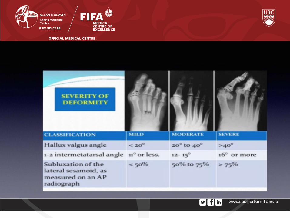
• Important to have standing xrays • Measure hallux valgus and intermetatarsal
angles • Often medial shift occurs first at TMT joint
and then MTP joint drifts laterally • Important to rule out associated 1st MTP OA
as this changes surgical procedure
HALLUX VALGUS DIAGNOSIS

▪ This is a pain management problem ▪ Conservative treatment effective ▪ Bunion pads and toe spacers can help
in short term ▪ Dynamic splint at night ▪ Shoe wear modifications
▪ Best to fit shoes at end of day ▪ Orthotics if foot pronation ▪ OTC meds effective if additional OA
HALLUX VALGUS TREATMENT

▪ Pain Management NOT cosmesis • Surgical results best in those with moderate-severe pain
▪ Failure rate of surgery 10-30% ▪ Minor procedures dealing only with 1st MTPJ often result in
recurrence of deformity. TMTJ must also be addressed ▪ Surgery involves both a bony procedure and a soft tissue procedure
▪ Typically osteotomy of first metatarsal at TMTJ ▪ Lengthening or loosening tight or loose tendons and ligaments
▪ Often cannot drive for 3 months post surgery and up to 1 year for full recovery of function
▪ Goal of surgery is not to be able to wear narrow shoes
BUNIONS SURGICAL INDICATIONS

CASE STUDY
60M presents with worsening pain in great toe both at rest and with activity. He has increasing difficulty walking especially on hills or stairs and feels he is shifting his weight laterally. He feels worse in his running shoes and dress shoes and better barefoot on carpet.

HALLUX LIMITUS/RIGIDUS
• Ultimately a progressive loss of articular cartilage at the 1st MTPJ
• Can be genetic but also influenced by shoewear, overuse injuries, gout, infection
• Creates pain and stiffness at great toe often with a bone spur on dorsum of 1st MTPJ

▪ Pain, stiffness, swelling at 1st MTPJ • Often bump mistaken for bunion or gout swelling
▪ Difficulty with toe off portion of walking or running gait ▪ N=60° extension but limitus <40° and rigidus < 20°
▪ Compensatory pains in hip, knee, back ▪ Best managed with stiff soled rocker shoes
CLINICAL FINDINGS

• Limit Great Toe Motion • Stiff soled rocker • Rigid steel shank for 1st ray • Spring plate • Orthotic with Morton’s extension • Turf toe plate
NON-OPERATIVE TREATMENT

Forefoot Rocker Sole ➢ 1st MTP and forefoot pain
Heel to Toe Rocker Sole ➢ ankle and midfoot OA

SURGICAL TREATMENT Cheilectomy
• Effective only if OA in dorsum of joint • Effective for limitus more than rigidus • Allows for joint motion as opposed to fusion • Day surgery, and home same day with stiff soled shoe • Wound healing at 2/52 and driving 3-4/52 • High recurrence rate

• Best for hallux rigidus- Gold Standard • Most successful procedure for pain control • Limits function for active individuals • NWB 6-12 weeks and still need a stiff soled
rocker shoe afterwards • Complications
– Malunion, non-union, hardware failure
SURGICAL TREATMENT 1ST MTPJ Fusion
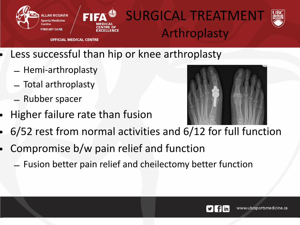
• Less successful than hip or knee arthroplasty – Hemi-arthroplasty – Total arthroplasty – Rubber spacer
• Higher failure rate than fusion • 6/52 rest from normal activities and 6/12 for full function • Compromise b/w pain relief and function
– Fusion better pain relief and cheilectomy better function
SURGICAL TREATMENT Arthroplasty
CASE STUDY
50F hairdresser and recreational runner has had increased difficulty running as she feels she has a pebble in her shoe and has to remove her shoe to massage the ball of her foot. Her pain is worse with standing all day at work especially with tighter shoes. She describes a burning pain and some numbness between her 3rd and 4th toe. She gets relief with Advil and feels fine when she is shoeless at home.

• Tibial nerve branches into medial and lateral plantar nerves distal to medial malleolus
• Tethering branch between 3-4th interspaces
• 4th ray more mobile attached to cuboid than 3rd ray attached to cuneiforms
• Distal transverse metatarsal ligament can be taut and compress interdigital nerves
DTML
Lateral plantar n
Medial plantar n
MORTON’S NEUROMA

MORTON’S NEUROMA
• Injured or compressed nerve most often between the 3rd and 4th toes. – 66%-3rd, 32%-2nd, 2%-4th
• Burning / tingling pain on the ball of the foot or toes.
• Patients may feel fullness or a mass in the area when they walk.
• Worse with tight shoes or high heels • Clinical diagnosis more than imaging
– Mulder’s sign Se-94-98%
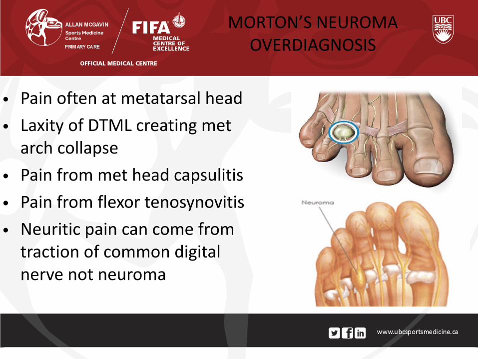
• Pain often at metatarsal head • Laxity of DTML creating met
arch collapse • Pain from met head capsulitis • Pain from flexor tenosynovitis • Neuritic pain can come from
traction of common digital nerve not neuroma
MORTON’S NEUROMA OVERDIAGNOSIS

• Conservative treatments buy time but don’t solve problem
• Cortisone injection provides short term pain relief ≤ 50% and long term 30%
• ETOH injections 50-60% short term success but perineural fibrosis may affect future surgical outcomes
• Shockwave, laser, botox no long term benefits • Orthotics for pronation/supination no evidence
– Shoewear/met pad more helpful
TREATMENT PLANS

DON’T MISS MIDFOOT PROBLEMS
Base of 5th metatarsal fractures Navicular stress fractures Lisfranc Injuries
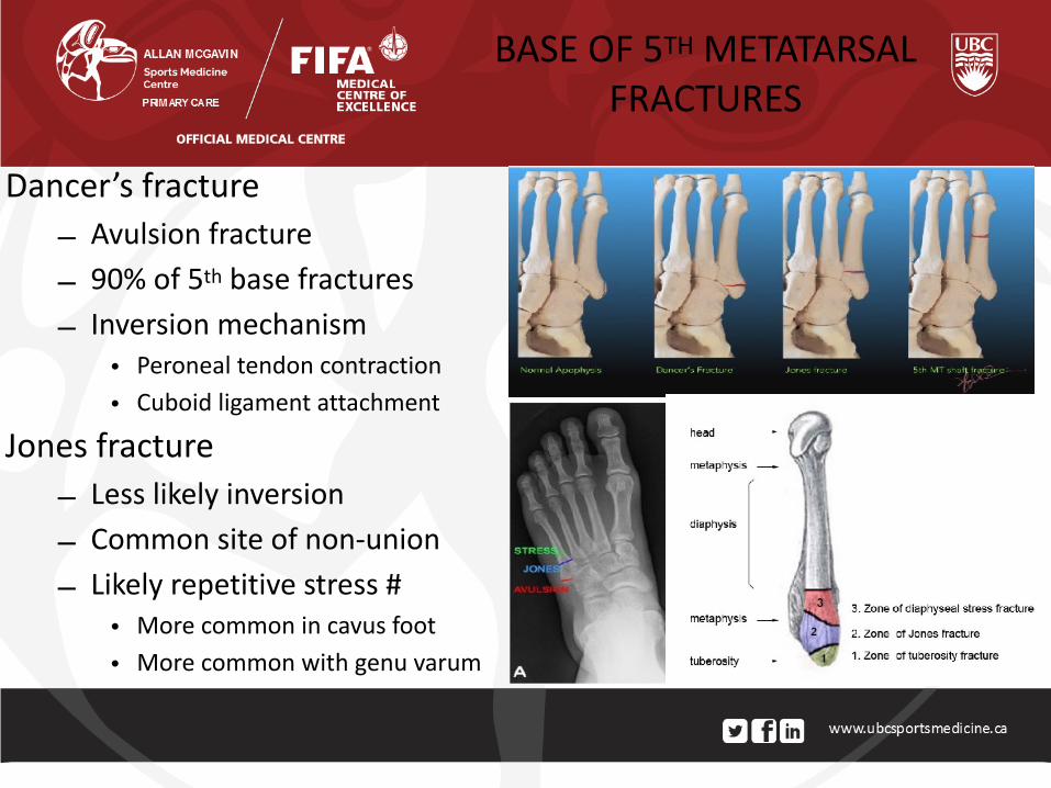
Dancer’s fracture – Avulsion fracture – 90% of 5th base fractures – Inversion mechanism
• Peroneal tendon contraction • Cuboid ligament attachment
Jones fracture – Less likely inversion – Common site of non-union – Likely repetitive stress #
• More common in cavus foot • More common with genu varum
BASE OF 5TH METATARSAL FRACTURES
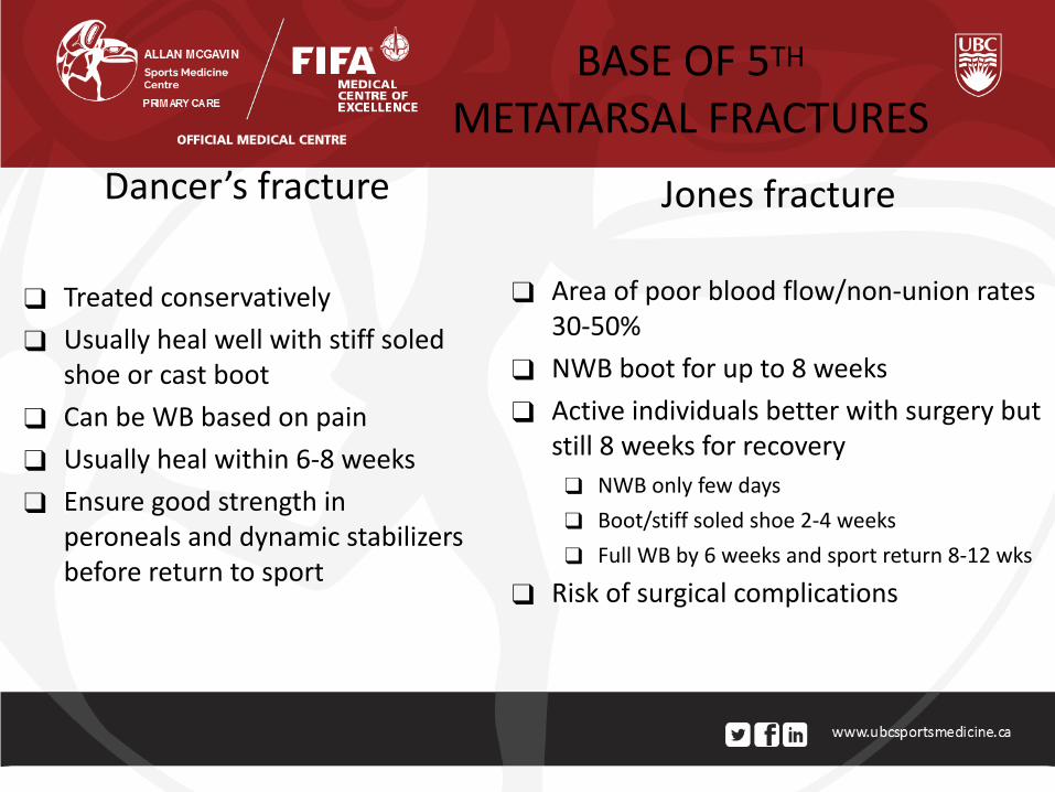
Dancer’s fracture
❑ Treated conservatively ❑ Usually heal well with stiff soled
shoe or cast boot ❑ Can be WB based on pain ❑ Usually heal within 6-8 weeks ❑ Ensure good strength in
peroneals and dynamic stabilizers before return to sport
Jones fracture
❑ Area of poor blood flow/non-union rates 30-50%
❑ NWB boot for up to 8 weeks ❑ Active individuals better with surgery but
still 8 weeks for recovery ❑ NWB only few days ❑ Boot/stiff soled shoe 2-4 weeks ❑ Full WB by 6 weeks and sport return 8-12 wks
❑ Risk of surgical complications
BASE OF 5TH METATARSAL FRACTURES
CASE STUDY
18M varsity runner has been noticing right anteromedial ankle pain for the past 6 months. He has been increasing his running with the varsity team but also started playing some intramural soccer this year. There is no swelling or bruising but he has not responded to physiotherapy, orthotics or taping. He did rest for 1 month but symptoms recurred as soon as he got back to running.

• Overuse injury often missed – Average time to diagnosis was 6 months – Running, tennis, hurdles, ultimate frisbee
• Area of non-union due to poor blood supply • Rarely seen on xray unless cortical disruption • MRI is best for diagnosing injury but CT scan is more
helpful for characterizing fracture line – Complete vs partial – Dorsal vs volar
• Saxena classification – Type I, II, III based on whether dorsal/volar cortex
NAVICULAR STRESS FRACTURES

• Important to recognize “N” spot • Controversy over surgical versus non-
operative management • NWB cast 6/52 and return to sport 6/12
for Type 1 injury • Surgery for Type 2 & 3 had earlier return
to sport but controversy as to long term outcomes improved or not.
TREATMENT
CASE STUDY
18M sustained an injury to his right foot and ankle during a tackle in football. He was able to weight bear initially and was told he had an ankle sprain. Pain persisted with walking and toe-off and he could not return play. No swelling or locking or instability feeling but he is now 3 weeks post injury and not able to run.

KEYSTONE ROMAN ARCH

• 20-40% are missed especially in context of ankle sprain mechanism
• 2 common mechanisms in sports – Hindfoot fixed and midfoot rotation
• Cyclist, equestrian
– Plantarflexed forefoot and axial load to heel
• Football, soccer, wrestling
LISFRANC INJURY

Bruising in different locations than typical ankle sprain
LISFRANC INJURY

Weight bearing xrays critical WB film may be difficult in ER setting
More than 2mm displacement at 1st TMT Possible flex sign on xray b/w 1st and 2nd MT
WB xrays sometimes better than CT CT has 3 mm slices
XRAYS

• Non-weight bearing immobilization boot cast for 4-6 weeks.
• Good to have arch support in boot • Gradual transition to shoes and
activity after about 12 weeks • Surgery for displaced ligament injury
or fracture • Arthrodesis may be required for
missed injuries
TREATMENT

COMMON REARFOOT PROBLEMS
• Achilles tendinopathy – Insertional – Non-insertional
• Plantar fasciopathy
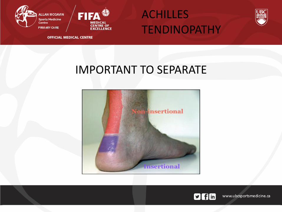
IMPORTANT TO SEPARATE
ACHILLES TENDINOPATHY
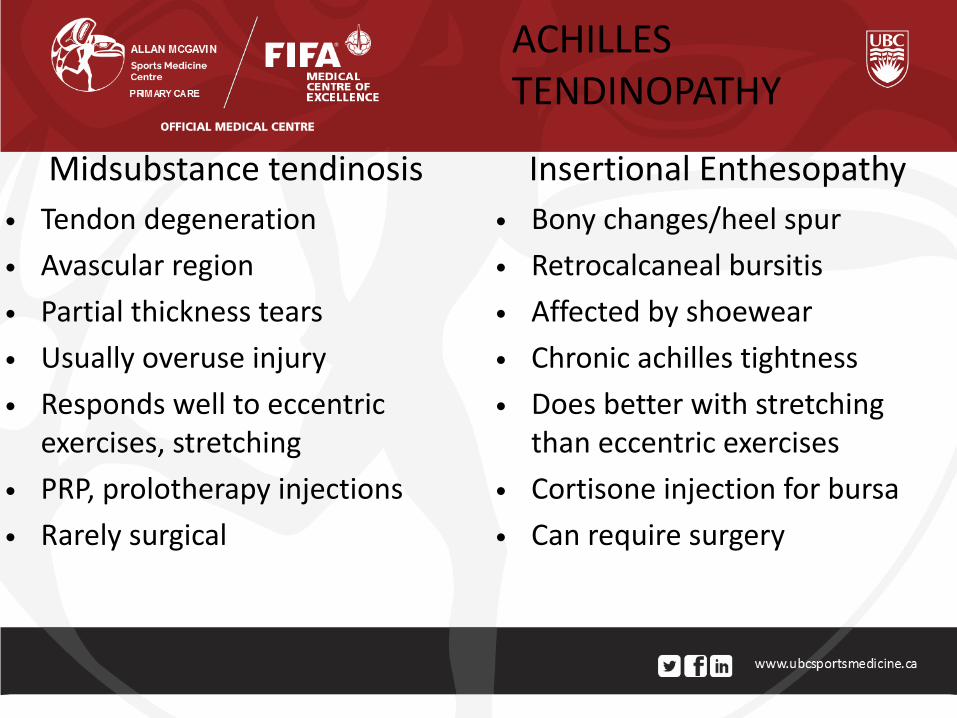
Midsubstance tendinosis • Tendon degeneration • Avascular region • Partial thickness tears • Usually overuse injury • Responds well to eccentric
exercises, stretching • PRP, prolotherapy injections • Rarely surgical
Insertional Enthesopathy • Bony changes/heel spur • Retrocalcaneal bursitis • Affected by shoewear • Chronic achilles tightness • Does better with stretching
than eccentric exercises • Cortisone injection for bursa • Can require surgery
ACHILLES TENDINOPATHY

• Initially starts as a tendinitis but progresses to tendinosis
• Foot pronation/supination can be factors
• NSAID’s less effective as condition progresses
• Respond well to eccentric exercises and stretching
MID-SUBSTANCE TENDINOSIS
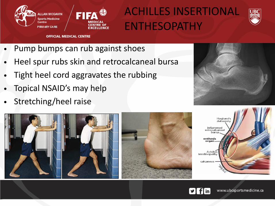
• Pump bumps can rub against shoes
• Heel spur rubs skin and retrocalcaneal bursa
• Tight heel cord aggravates the rubbing
• Topical NSAID’s may help
• Stretching/heel raise
ACHILLES INSERTIONAL ENTHESOPATHY
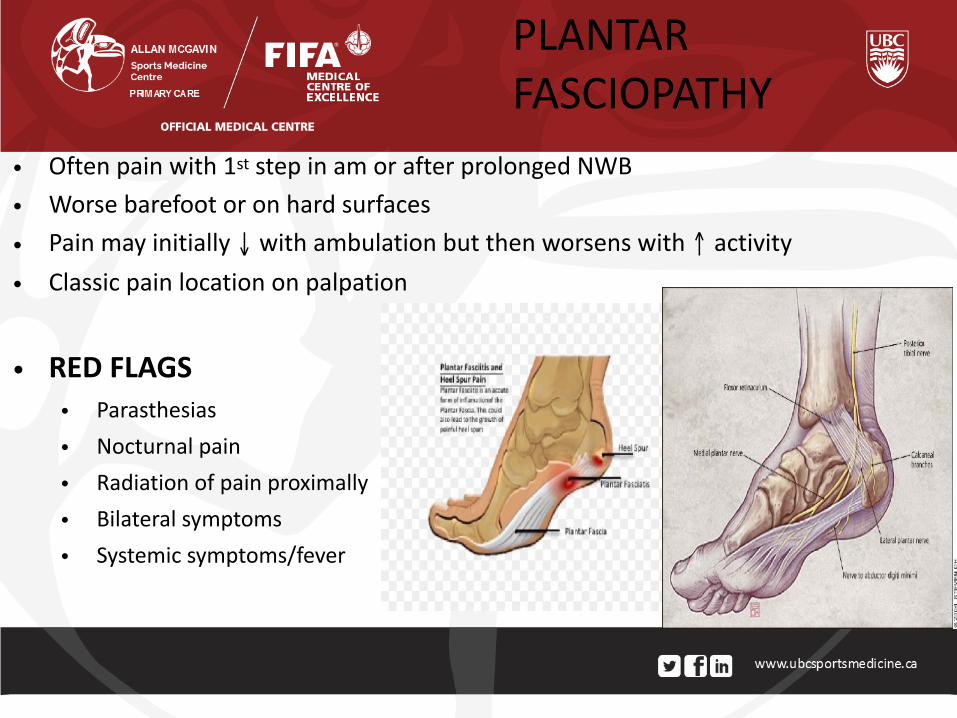
• Often pain with 1st step in am or after prolonged NWB
• Worse barefoot or on hard surfaces
• Pain may initially ↓ with ambulation but then worsens with ↑ activity
• Classic pain location on palpation
• RED FLAGS
• Parasthesias
• Nocturnal pain
• Radiation of pain proximally
• Bilateral symptoms
• Systemic symptoms/fever
PLANTAR FASCIOPATHY

ACUTE PHASE (≤ 6 Weeks) – Physiotherapy – Cryotherapy – NSAID (oral/topical) – Stretching exercises – Shoewear changes – Low dye taping – Orthotics/heel pads – Activity modifications
CHRONIC PHASE (≥ 6-12weeks) – Strengthen foot intrinsics – Night splints – ESWT – Prolotherapy
• Hyperosmolar dextrose
– PRP injections – Surgery
PLANTAR FASCIOPATHY TREATMENT
THANK YOU