Embed Size (px)
Citation preview

Committee on Medical
Aspects of Radiation in
the Environment
(COMARE)
SIXTEENTH REPORT
Patient radiation dose issues resulting from the use of CT in the UK
Chairman: Professor A Elliott

© Crown copyright 2014
Produced by Public Health England for the
Committee on Medical Aspects of Radiation in the Environment
ISBN 978-0-85951-756-0

3
CONTENTS
Page
Foreword 5
Chapter 1 Introduction 7
Chapter 2 Applications and benefits of CT scanning 12
Chapter 3 Detriments associated with radiation dose from CT scans 19
Chapter 4 Developments in CT and technological strategies for dose
reduction 31
Chapter 5 Dose measurements, dose surveys and diagnostic reference
levels 41
Chapter 6 Clinical strategies for dose reduction 53
Chapter 7 Governance 61
Chapter 8 Conclusions 68
Chapter 9 Recommendations 69
References 71
Acknowledgements 82
Appendix A Glossary and Abbreviations 85
Appendix B CT versus MRI versus Ultrasound 91
Appendix C History of CT 92
Appendix D Letter from the Royal College of Radiologists 94
Appendix E COMARE Reports 95
Appendix F COMARE Membership 97
Appendix G Declarations of Interest 100


5
FOREWORD
i The Committee on Medical Aspects of Radiation in the Environment
(COMARE) was established in November 1985 in response to the final
recommendation of the report of the Independent Advisory Group chaired by
Sir Douglas Black (Black, 1984). The terms of reference for COMARE are:
‘to assess and advise government and the devolved authorities on the
health effects of natural and man-made radiation and to assess the
adequacy of the available data and the need for further research’
ii In the course of providing advice to government and the devolved
authorities for over 28 years, COMARE has published to date 15 major reports
and many other statements and documents mainly related to exposure to
naturally occurring radionuclides, such as radon and its progeny, or to man-
made radiation, usually emitted by major nuclear installations. The most recent
COMARE report focused on the radium contamination in the area around
Dalgety Bay.
iii In August 2011, the Department of Health asked COMARE to produce
a report on medical radiation dose issues with CT scanners. Issues to be
considered in the report related to the increased use, justification and
optimisation of CT exposures on patients. The report would focus on diagnostic
CT applications, as well as including PET-CT, SPECT-CT and cone beam CT,
but would not consider exposure of staff or asymptomatic individuals. A future
report will investigate the use of CT in interventional radiology. COMARE
reconstituted its Medical Practices Subcommittee, with a new membership
consisting of committee members and external experts, to conduct this work.
The subcommittee’s terms of reference are:
‘to advise COMARE on the health effects arising from medical and
similar practices involving the use of ionising and non-ionising
radiation through assessment of the available data and to inform
COMARE of further research priorities’
The investigation of the increased use of CT, together with the potential risks
associated with the radiation exposure from the examinations detailed in this
report, lies within the remit of the subcommittee.
iv The aim of this COMARE report has been to provide advice to the
Department of Health on the increased use of diagnostic CT within the UK,
with consideration of the potential dose reduction benefits of practical
approaches and supporting initiatives.
v When the subcommittee had finished its review, the report was
presented to COMARE for consideration by the full committee, with the aim
that the information would be presented to the Department of Health in due
course. That information is contained in this, our sixteenth report.


7
CHAPTER 1
INTRODUCTION
1.1 Medical imaging is ranked within the top five recent medical
developments in numerous surveys: for example, see the survey by the Royal
College of Physicians of Edinburgh (RCPE, 2010). A variety of imaging
modalities are currently available for clinical use, including radiography,
fluoroscopy, computed tomography (CT), nuclear medicine (including hybrid
imaging with CT), ultrasound and magnetic resonance imaging (MRI). Each has
its advantages and disadvantages (see Appendix B for a comparison of CT, MRI
and ultrasound).
1.2 The development of cross-sectional imaging (CT and MRI) has
revolutionised the manner in which the body can be visualised to detect disease
(eg cancer) and to inform treatment in a range of disease areas.
1.3 The principal advantages of CT are:
(a) Rapid acquisition of images
(b) Impressive spatial resolution
(c) A wealth of clear and specific information answering a wide range of
clinical questions
(d) A view of a large portion of the body
(e) The ability to discriminate between structures on the basis of density
No other imaging procedure combines these features into a single modality. In
addition, as with most digital technologies, these developments have been
achieved with a decrease in the real cost of equipment. The result has been
increased patient throughput at lower cost per caput, which has consolidated CT
as a major, first-line, diagnostic modality.
1.4 CT, originally known as computed axial tomography (CAT), uses
specialised X-ray equipment to obtain image data from different angles around
the body. Digital processing of this information results in detailed cross-
sectional images of body tissues and organs in either two- or three-dimensional
format. It was the first technique to produce such images and as the diagnostic
advantages were immediately obvious, it rapidly became established as a
valuable diagnostic tool.
1.5 The idea of CT was conceived in 1961 by W H Oldendorf (Oldendorf,
1961), although the first clinical application was envisaged in 1967 in England
by G N Hounsfield at the Thorn EMI Central Research Laboratories, being
publicly announced in 1972. The system was independently developed by
A M Cormack at Tufts University and both he and Hounsfield shared the Nobel
Prize for Medicine in 1979. The original test rig, developed in 1971, used
americium as a gamma source, and took 160 parallel readings over 180 angles,
each 1° apart. It took 9 days to collect sufficient information about the object
being scanned and a further 2.5 hours to reconstruct the data into an image.
8
Later, the gamma source was replaced by a more powerful X-ray source, which
reduced the scanning time (Hounsfield, 1973).
1.6 The first prototype clinical CT scanner (the forerunner of the commercial
version known as the EMI scanner) was installed in Atkinson Morley’s Hospital,
Wimbledon. It was limited to making tomographic sections of the brain, and the
first patient brain scan using the machine was obtained in 1971. To reduce the
dynamic range of the radiation reaching the detectors, the machine required
the use of a water-filled Perspex™ tank, with a pre-shaped rubber head cap
enclosing the patient’s head. The machine took between 4.5 and 20 minutes per
180° scan to acquire the image data, with 7 minutes required to process each
image (Beckmann, 2006).
1.7 CT scanners have gone through many phases of technological develop-
ment, from single-slice static machines to single-slice spiral/helical machines,
and in the last decade there have been further significant advances in CT
technology (see Appendix C for a more detailed history). In particular, the
continued development of multi-detector (often referred to as multi-slice) CT
and developments in gantry technology have increased the speed of scanning
and enabled high resolution reconstruction of images in all planes. Other recent
developments allow consideration of volume CT acquisition. These improve-
ments mean that more complex and extensive scans can be performed within
timeframes tolerable to most patients. Large volumes of the chest can be
scanned within a breath hold and cardiac examinations achieved within a single
heart beat. An additional dimension has been provided by time-based exposures,
which make it possible to investigate the perfusion characteristics of organs for
a range of diseases.
1.8 Increased computing power and data processing capacity have
contributed significantly to the expanded capability of CT systems and this is
expected to continue. Software packages have been developed to make use of
these benefits, with modern scanners being able to reconstruct a study yielding
around 1000 images in less than 30 seconds. Multi-planar and three-dimensional
imaging, created from the exposed volume, are now common techniques that
have contributed to an increased demand for CT.
1.9 These developments have resulted in a dramatic increase in the number
of clinical applications of CT. The first systems were designed only for
examination of the brain. When body systems were first developed, they were
restricted to selective examination of the trunk, largely in relation to the
management of cancer. As the modality became more widely available, more
accurate and more flexible, the number of applications in benign disease and in
young people increased dramatically.
1.10 Today, there are many applications and benefits in the clinical use of
CT, aiding more effective care management by:
(a) Determining the necessity for surgery
(b) Reducing the need for ‘exploratory’ surgery
(c) Improving diagnosis, staging and treatment of cancer
(d) Reducing the length of hospitalisations
(e) Reducing the need for examination under sedation, especially in very
young patients
(f) Guiding the treatment of common conditions such as injury, cardiac
disease and stroke

9
1.11 Of around 130 applications of CT recommended by the Royal College
of Radiologists (RCR) in its guidelines ‘iRefer’, 70% now relate to benign or
potentially benign conditions (RCR, 2012). In many conditions, which although
benign can be life threatening, CT has been recommended as a standard investi-
gation: for example, in the investigation of acute appendicitis (Raptopoulos
et al, 2003; Saito et al, 2013).
1.12 CT has now replaced a large number of conventional examinations, such
as barium studies and angiograms (Lederlin et al, 2011; Makayama et al, 2001).
Additionally, CT can be used to obtain data from which virtual endoscopy can be
created, obviating the need for invasive endoscopy (Summers, 2010; Vining,
1997). As a consequence, in many cases better diagnostic information has also
been accompanied by a reduction in the radiation dose or the morbidity (and in
some cases mortality) associated with traditional procedures. This is not always
the case, however, as in some benign cases the use of CT in place of other
investigations results in significantly higher radiation exposure, for example, in
Crohn’s disease (Jaffe et al, 2007). CT pulmonary angiography has in many
places replaced nuclear medicine ventilation-perfusion lung scanning, but with
increased breast radiation dose, especially for pregnant and lactating women
(Anderson, 2007; Shahir et al, 2010).
1.13 Wider use of CT in benign disease and in younger patients, especially
children, whose tissues have a greater radiosensitivity, highlights the need for
conscientious radiation protection. Children have a longer predicted lifespan in
which potential harmful effects of radiation exposure have more opportunity to
emerge. The current success of treatments for a range of cancers and the
increasing lifespan of the population as a whole means that radiation protection
has become a matter of importance for all patients receiving CT scans.
1.14 Comparing data from the USA between 1980 and 2006 shows a
significant increase in both the average annual effective dose to the US
population and the contribution from medical radiation sources (Figure 1.1).
The data for the UK population in 2003 show a similar distribution to that of
the 1980s US population; however, more recent data suggest that the UK is
following a similar trend to the USA, with an increasing contribution from
medical exposures.
Figure 1.1 Average annual effective radiation dose to the US and UK populations from all sources (Mettler et al,
2009; Watson et al, 2005)

10
1.15 For the UK population, the annual per caput effective radiation dose
from all diagnostic X-rays has increased from 0.33 millisievert (mSv) in 1997 to
0.4 mSv in 2008 (Hart et al, 2010). For some time, medical X-rays have been
the largest single artificial source of radiation exposure for the UK population.
In 2008, they contributed 15% of the average annual effective dose to the
population from ionising radiation*. It has been recognised that CT makes a
disproportionate contribution to the radiation exposure of patients compared
with other radiation-based imaging techniques. The then National Radiological
Protection Board (NRPB)† noted in 1989 that while CT comprised around 2.5%
of all examinations, it contributed around 25% of the collective dose to the
population from imaging (Shrimpton et al, 1991). By 2008, CT accounted for
around 7% of all medical and dental X-ray examinations, but produced 68% of
the collective dose (Hart et al, 2010).
1.16 According to statistics from the NHS‡, the number of CT scans
conducted in hospitals in England each year for the period 1996/97 to 2012/13
steadily increased from just over 1 million to almost 5 million, with no sign of
reaching a plateau (see Figure 1.2).
1.17 The increasingly extensive examinations and more widespread use of
CT are major contributors to the observation that diagnostic uses of radiation
have almost doubled the average radiation exposure to the population in some
20 years, as has been shown for the USA (Hricak et al, 2011).
Figure 1.2 Number of NHS CT examinations performed from 1995/96 to
2012/13 in England
* The average annual effective dose from natural and artificial sources of ionising radiation is
estimated at 2.7 mSv for the UK, with the majority of this attributed to natural radiation (2.2 mSv)
(Watson et al, 2005).
† The NRPB was subsequently incorporated into the Health Protection Agency (HPA).
On 1 April 2013 the HPA was abolished and its functions transferred to Public Health England.
‡ www.england.nhs.uk/statistics/statistical-work-areas/diagnostics-waiting-times-and-
activity/imaging-and-radiodiagnostics-annual-data/

11
1.18 Documented average exposures to the population in reality reflect even
higher exposure of a smaller number of people, namely patients. The role of CT
in the clinical management of some diseases may result in a patient receiving a
large number of scans in the course of one illness (Katz et al, 2006; Meeson
et al, 2009; Sodickson et al, 2009). Restricting doses in CT is, therefore, widely
perceived as an important objective in the practice of the technique and is held by
some authors to be medicine’s major current challenge in radiation protection
(Golding, 2005).
1.19 The rising use of CT, the radiation exposure used in the examination
and the potential risks associated with it are described in this report. These
circumstances indicate that there is potential benefit in recommending steps to
reduce population exposure due to CT. These are considered in detail in the
following chapters.

12
CHAPTER 2
APPLICATIONS AND BENEFITS OF
CT SCANNING
2.1 CT is a powerful, highly flexible clinical tool, capable of making radical
changes to the management of patients. On the basis of the current state of
alternative technologies, it is possibly the most important cross-sectional
imaging modality in the diagnosis and management of a broad array of
conditions. There is a strong evidence base which demonstrates that CT changes
patient management in a wide range of applications and disease states (RCR,
2012). In some conditions it may be the only investigation the patient requires.
2.2 As is common to all investigations in medicine, the correct application
of CT is based on weighing the potential benefit of reliable investigation against
the inherent risk. While this principle supports the widespread use of CT, active
measures to achieve greater dose constraint are required in view of the current
position of CT as a major source of patient irradiation.
Principles of radiation
protection relevant to
CT and regulation
2.3 The governance of CT adheres to the established radiation protection
principles of the International Commission on Radiological Protection (ICRP).
Three key principles of radiation protection were reaffirmed in 1990 (ICRP, 1991):
Justification – exposure to radiation must produce sufficient benefit to
the exposed individuals, or to society, to offset the potential radiation
detriment
Optimisation – implementing procedures and techniques to keep
exposures as low as reasonably practicable, economic and social factors
being taken into account
Dose limitation – keeping radiation doses received within specified
limits
2.4 The ICRP issued new recommendations on radiation protection in 2007
(ICRP, 2007), which formally replaced its 1990 recommendations (ICRP, 1991).
2.5 Only justification and optimisation apply in the context of medical
radiation exposures for patients, whereas all three key principles apply to the
occupational exposure of medical staff. Occupational exposure is not considered
further in this report; rather it focuses on patient exposure. The concept of
diagnostic reference levels (DRLs) has been introduced to support the control
and periodic reduction of radiation doses from diagnostic procedures. These are
based on dose data for a range of commonly requested procedures in the UK
and are regularly updated.
2.6 In the UK, legislation has been put in place implementing key European
Council Directives, to address the hazards associated with ionising radiation.
For CT scanning there are three sets of regulations of particular importance:
The Justification of Practices Involving Ionising Radiation Regulations
2004, which address justification of practices at the highest level

13
The Ionising Radiations Regulations 1999, which address protection of
workers and the public*
The Ionising Radiation (Medical Exposure) Regulations 2000, which
address protection of patients and others
2.7 The guidance on the application and administration of the Justification
of Practices Involving Ionising Radiation Regulations 2004 includes medical
exposures using CT for diagnosis as an existing type of practice. The Ionising
Radiation Regulations 1999 require employers to establish a framework to
ensure exposures arising from work activities are kept as low as reasonably
practicable and below dose limits.
2.8 The Ionising Radiation (Medical Exposure) Regulations 2000 (IR(ME)R
2000) (Department of Health, 2000) continue these themes at the individual
patient level and require that all individual medical exposures are referred,
justified and optimised. Both the referrer and the practitioner must be registered
healthcare professionals.
2.9 IR(ME)R 2000 require an identified person, known as the ‘practitioner’
in the UK, to take legal responsibility for deciding whether an individual
medical exposure is justified. Such a person must be adequately trained to carry
out the task of justification and be entitled to do so by their employer.
Justification of exposures must take into account medical information about the
individual provided by a referrer and should be based on the available scientific
evidence. Justification cannot be retrospective. The practitioner’s decision as to
whether an individual medical exposure is justified must be made prior to the
exposure, and must be valid whether the test result is subsequently positive or
negative. Procedures can only be justified if the individual for whom the
exposure is proposed will receive a benefit that outweighs the detriment, or if
there is an overall net benefit to society.
2.10 Optimisation of every medical exposure is the responsibility of the
practitioner and of the ‘operator’ who undertakes the practical aspects of a
medical exposure, to the extent of their respective involvement. To assist in
optimisation, the employer must ensure that written protocols are in place for all
standard procedures which must be specific to each piece of equipment. Such
protocols should include exposure factors for each routine examination.
2.11 Effective strategies for reducing radiation exposure due to CT require
attention to several aspects of clinical, technical and operational management
and practice (as part of serial and parallel processes):
(a) Justification of examinations, including consideration of the use of
alternative technologies
(b) Optimisation of examinations
(c) Maximising the capability of the equipment
(d) Maximising the capability of staff (practitioners and operators)
2.12 These and other governance issues are discussed in further detail in the
following chapters.
Applications of CT 2.13 In the past 15 years, the major breakthrough in CT technological
development has been the introduction of multi-slice or multi-detector helical
CT, which has made it possible to acquire and analyse patient data within
* The Ionising Radiations Regulations (Northern Ireland) 2000 for Northern Ireland.

14
seconds rather than minutes. Representation of a volume of body tissue is now
possible with uniform resolution images reformatted in multiple planes, or in
three dimensions. As a consequence, new investigations are now possible – for
example, investigation of blood flow through organs or blood vessels.
2.14 General clinical indications for CT scanning include, but are not
limited to:
(a) Staging of cancer – response, assessment and surveillance
(b) Gastroenterology – bowel and organ pathology, including masses, sepsis,
bleeding and trauma
(c) Thorax – lung and mediastinal disease, masses, vasculature and trauma
(d) Urology – identification/characterisation of masses, sepsis, anatomical
abnormalities, ureteric and renal colic
(e) Gynaecology – masses and sepsis
(f) Colo-rectal – large bowel pathology, extent and spread of disease
(g) Trauma – assessment of injuries
(h) Musculoskeletal – structural abnormalities, disease or trauma
(i) Neurology – trauma, disease, sepsis, vascular and to exclude
haemorrhage
(j) ENT/head and neck – disease, masses, sepsis and anatomical abnormalities
(k) Cardiology – angiography
(l) Vascular – abnormalities, disease and trauma
(m) Intervention – guide treatment; biopsy, drainage and ablation
2.15 CT has also enhanced the capabilities of nuclear medicine imaging by
providing an anatomical dimension previously missing in this functional
imaging modality, leading to the production of hybrid imaging systems. CT
provides a means of localising disease processes shown by the nuclear medicine
techniques of positron emission tomography (PET) CT and single photon
emission computed tomography (SPECT) CT. The advent of hybrid PET CT and
SPECT CT systems has simplified image registration – the PET or SPECT and
CT datasets are collected sequentially, on the same system, without the need for
the patient to move to another scanner. This removes the image registration
problems introduced by different patient set-up positions.
2.16 The acquisition parameters of the CT component will be determined by
the clinical question to be answered by the procedure, and whether a diagnostic
CT is performed before, or after, the PET/SPECT imaging. The CT in PET CT/
SPECT CT systems provides information to assist in the correction of the
attenuation of photons from the radionuclide as they pass through the body
in order to improve the PET imaging. A relatively low dose CT examination
may be appropriate for the purpose of co-registration and localisation of
abnormalities detected on the PET component (where images of lower quality/
higher noise than diagnostic CT are acceptable). A higher dose CT will provide a
diagnostic quality image, making a separate CT examination unnecessary unless
contrast is required.
2.17 In other cases, a diagnostic CT examination may sometimes be acquired
in conjunction with the PET imaging, resulting in a study of comparable, and
sometimes higher, radiation exposure. Clinical indications for the use of
PET CT include, but may not be restricted to (RCP/RCR, 2013):

15
(a) Malignancies
(b) Neurological abnormalities
(c) Cardiological abnormalities
(d) Vasculitis
(e) Sarcoidosis
(f) Infection imaging
2.18 A recent development has been so-called ‘cone beam’ CT (CBCT). This
particular imaging technique uses a flat panel detector (in contrast to the array
of individual detectors of conventional CT), coupled with beam exposure that is
similar to that of conventional radiography. It is essentially a form of rotation
radiography and its dosimetry differs significantly from that of CT. CBCT
images contain inherently fewer image data than conventional CT and are
acquired at lower radiation exposure. The technique was developed for specific
clinical applications, such as dentistry, but is now being used more widely
wherever it can be applied. In view of the fundamental difference between
CBCT and conventional CT, the technique is not considered in detail in this
report. However, CBCT, where it is clinically applicable, may offer a dose-
sparing alternative to conventional CT and its use should, therefore, be
considered when justifying referrals for CT.
2.19 CT also has applications in veterinary medicine and in industry;
however, these are not considered in this report.
2.20 In the clinical environment CT is now established in a range of differing
roles. These may be categorised as:
(a) Diagnostic and staging
(b) Guiding
(c) Planning
(d) Monitoring
Additionally, CT has been employed in health assessments of asymptomatic
individuals. This practice was the focus of the 12th COMARE report
(COMARE, 2007).
Diagnostic and staging CT 2.21 CT is the most frequently used imaging technique where cross-sectional
information is necessary for diagnosis or staging. It is applicable to diseases of
the brain, musculoskeletal system, chest, abdomen and pelvis, either as a
complement to other imaging investigations such as radiography, or ultrasound,
or increasingly as the primary investigatory technique.
2.22 Early use of CT provided information within the head that was previously
unavailable. Rapid development of body scanners quickly established that the
technique was an accurate method of detecting or excluding many diseases, and
in showing the extent of disease. This led to changing treatment to a more
appropriate regime, with the possibility of an improved health outcome.
Research studies have confirmed the clinical effectiveness of diagnostic CT in
influencing patient management (Kumta et al, 2002; Moore et al, 1987).
2.23 Multi-planar and three-dimensional imaging created from the exposure
volume are now common approaches that have further extended the role and
contributed to an increased demand for CT. Time-based exposures make it
possible to investigate the perfusion characteristics of disease and the combina-
tion of speed and multi-planar reformatting has allowed CT to replace a large
number of conventional angiograms (Rathbun et al, 2000; Wittram et al, 2004).
16
2.24 The capability of demonstrating or excluding disease within the trunk
has established CT as a means of investigating patients before, or instead of,
surgery. CT has, in effect, become a non-invasive alternative to investigatory
laparotomy – for example, in suspected critical damage in abdominal trauma
(Salim et al, 2006). Many of these applications have been supported by research
studies confirming both the clinical value and cost-effectiveness of this approach
(Kocher et al, 2011). However, it is perceived that the value of this application
of CT has led to an impression among some surgeons that all patients under-
going surgery to the trunk merit CT beforehand. Further research is required to
determine whether such an extended application of CT has an appropriate
evidence base.
2.25 The accuracy of CT has also led to important changes in documenting
the extent of disease, especially malignant tumours. The margins of the primary
lesion can be evaluated and the involvement of other organs, by direct extension
or by vascular spread, can be assessed. Disease staging rapidly became, and has
remained, a leading use of CT.
2.26 The high diagnostic accuracy of CT has meant that it is becoming
increasingly essential in the early diagnosis phase of trauma care. CT is more
often being used in place of conventional radiographic imaging and there is
emerging use of whole-body CT for trauma patients (Smith and Mason, 2012).
The value of immediate whole-body CT of severely injured trauma patients is
still to be determined (Sierink et al, 2012a). An initial review of the current data
suggests that the practice reduces the time spent by the patient in the emergency
department when compared with conventional radiography (Sierink et al, 2012b).
CT provides complex imaging that answers a wide range of clinical questions in
all regions of the body. It affords the ability to reconstruct in any plane while
being able to focus on small areas of concern. It can include venous and arterial
phases in a fast single scan and is becoming common practice to use in place of
conventional imaging when a trauma patient has been stabilised.
2.27 Magnetic resonance imaging (MRI) is an alternative imaging modality,
free from ionising radiation, which is used for many applications. It was
developed after CT was already established. MRI is now the investigation of
choice in many clinical situations involving the brain, musculoskeletal system
and trunk (RCR, 2012). It is arguable that the timing of the development of
MRI, combined with the relative speeds of acquisition and comparatively
limited availability and higher cost, has caused the clinical use of MRI to lag
behind that of CT. The relative use of CT and MRI is considered in more detail
in Chapter 6.
Guiding interventions 2.28 The cross-sectional display afforded by CT has been used to guide the
percutaneous placement of instruments for diagnostic or therapeutic use. These
so-called ‘interventional’ uses of CT include guided biopsy, catheter placement
and drainage of pathological fluid collections such as abscesses, nerve blocking
or other tissue ablative techniques. They represent a particular use of CT in
assisting a procedure. A specialised form of interventional guidance is real-time
CT using specialised equipment (CT fluorography). This technique poses
particular protection issues relating to its individual technology. Interventional
techniques including CT will be reviewed separately by COMARE and are not
covered within this report.
Treatment planning 2.29 Research studies in the 1970s quickly established the advantages of
using cross-sectional imaging in cancer treatment planning. The technique
shows the spatial relationships of lesions such as tumours, and can be used to
determine the appropriate treatment regime. If a tumour is shown to be
17
localised, its resectability can be predicted pre-operatively and the appropriate
surgical approach defined in advance.
2.30 Radiotherapy planning represents a particular use of this aspect of CT,
for which a separate examination is usually needed under standardised
conditions. Radiotherapy planning uses CT in two distinct activities, as follows.
Accurate localisation of the volume to be treated – three-dimensional
localisation is now the standard for all but a few radiotherapy episodes.
The clinical target volume and adjacent critical organs are defined on
the CT images and these are then fed into the dosimetry planning
system
Dosimetry – the CT images are used to define the patient contour incident
to the treatment beams and provide information for the planning
algorithm on tissue inhomogeneities. Variation between tissues will
influence absorption of radiation as the beam passes through and,
therefore, the dose distribution achievable within the treatment volume
Monitoring the effects of
treatment
2.31 The advantages of CT in defining disease make it suitable for
demonstrating how diseases respond to treatment, when this information is
important for further clinical management of the patient. Advantages apply to
both benign and malignant disease, as follows.
Defining response to ongoing treatment – a major impetus for the
increased use of imaging in follow up arises from a wider range of
treatment options being available for many diseases, including
malignant tumours. The frequency of monitoring depends on the nature
of the disease and whether treatment management decisions rest on the
result. In acute illnesses such as refractory abdominal abscesses, repeat
CT may be required every few days, whereas in malignant tumours the
examination may be needed to assess progress every few weeks.
Monitoring examinations may be required frequently or over a long
period and represent an important source of cumulative exposure in
individual patients. Hybrid imaging has the potential to provide further
information during a particular treatment episode, eg within a course
of chemotherapy
Early recognition of relapse – where treatment options exist for
relapsed disease there may be clinical advantage in early detection. CT
is widely used for this purpose in both benign and malignant disease.
The frequency of investigation should be determined by current
knowledge of the natural history of the individual disease and
evaluation of the clinical situation of the individual patient.
Population screening
or individual health
assessment
2.32 In recent years, CT has been used on individuals who are asymptomatic.
The diagnostic accuracy of CT has led to proposals for its use for screening
populations for disease, or for screening apparently healthy individuals for
possible disease. The radiation exposure of healthy subjects can only be justified
on the basis of demonstrable benefit to the population or the individual, balanced
against the risks involved.
2.33 Research studies are proceeding to determine whether screening
programmes using CT are justifiable, but currently there is no strong evidence
base supporting this use of the technique for the general population. The
National Cancer Institute National Lung Cancer Screening Trial (NLST) in the
USA published findings that low dose CT shows promise as a method for
detecting lung cancer in highest risk individuals who have yet to show
symptoms, reducing deaths by 20% compared with chest X-rays (NLST, 2011).

18
The American Lung Association only recommends lung cancer screening using
low dose CT scans for people meeting specific criteria – current or former
smokers (aged 55 to 74 years) with a smoking history of an average of a pack a
day for 30 years and with no history of lung cancer*.
2.34 A recent publication (Melgies et al, 2013) has considered the place
of CT coronary angiography (CTCA) as a screening tool for the diagnosis of
coronary artery disease. The authors conclude that, as yet, CTCA is unable
to predict the potential vulnerability of plaque and that there is insufficient
evidence to support a screening role.
2.35 COMARE previously reviewed personally initiated CT scanning
services (individual health assessments) in its 12th report (COMARE, 2007).
The report concluded that the practice may provide benefits to the individual,
but these would not be the same as those associated with the use of diagnostic
CT in a symptomatic patient. The justification for this practice could, therefore,
not be considered in the same way as justification for patients. Based on the
available evidence, it was not possible to recommend the use of whole-body CT
scanning on asymptomatic individuals. It was recognised, with the constant
developments in the field, that the recommendations from the report should be
reviewed as new evidence was presented. As a consequence of the COMARE
report, the Ionising Radiation (Medical Exposure) Regulations were amended in
2011 to specifically include the provision of individual health assessments as an
exposure requiring justification (Department of Health, 2011).
Summary 2.36 CT is a powerful, highly flexible clinical tool with its use governed by
the radiation protection principles of justification and optimisation. In the UK,
CT scanning is governed largely by three sets of regulations: the Justification
of Practices Involving Ionising Radiation Regulations 2004, the Ionising
Radiations Regulations 1999 and the Ionising Radiation (Medical Exposure)
Regulations 2000.
2.37 Technological developments in CT have greatly expanded its
applications. In a clinical environment, CT has a distinct role in the diagnosis
and staging of disease, guiding interventions, treatment planning and
monitoring the effects of treatment. It is applicable to a range of clinical
conditions, including diseases of the brain, musculoskeletal system, chest,
abdomen and pelvis. The use of CT is becoming increasingly common in the
early diagnosis phase of trauma care and it also has a role in investigating
patients before, or instead of, surgery.
2.38 The increase in scanning speed means that it is now possible to
investigate perfusion characteristics of disease.
2.39 CT is often performed in conjunction with other modalities: for example,
to enhance the capabilities of nuclear medicine with PET CT and SPECT CT.
2.40 CT has been used in recent years in scanning asymptomatic individuals,
aimed as a form of preventive medicine. COMARE previously reviewed these
services in its 12th report and determined that the benefits would differ from
those associated with symptomatic patients. COMARE was not able to support
the use of whole-body CT scanning on asymptomatic individuals. Research
studies are in progress to determine if the use of CT is justifiable in population
screening programmes, but there is currently no strong evidence to support it.
* www.lung.org/about-us/our-impact/top-stories/guidance-on-ct-lung-cancer.html

19
CHAPTER 3
DETRIMENTS ASSOCIATED WITH
RADIATION DOSE FROM CT SCANS
Benefit versus detriment
with diagnostic CT scans
3.1 While the use of CT has had a clearly beneficial effect on advancing the
accuracy of diagnostic radiology (see Chapter 2), there are potential detriments
associated with its use as well. One of the fundamental principles in the practice
of medicine is ‘First do no harm’. In the context of this report this means
balancing the potential benefit to be gained by having a CT scan against the
potential harm that might be caused by the radiation exposure (Lautin et al,
2008; Pilling, 2008). Although other risks from CT scans exist, such as those
inherent in the use of contrast agents (Martin and Bradley, 2012), this report
focuses on the principal risk associated with the exposure to ionising radiation,
where the dose received by the patient may be significant when compared with
those from other diagnostic imaging procedures.
3.2 In general, the risks associated with ionising radiation can be divided
into those defined as stochastic effects and deterministic effects.
Stochastic effects – generally somatic effects (cancer) in the directly
exposed population, and potential genetic effects to their offspring. Those
effects which occur by chance, affecting the probability of a change
rather than the severity and are a function of dose without a threshold
Deterministic (tissue reaction) effects – radiation injury due to cell killing
and radiation disease, defined by the ICRP in its 1990 recommendations
(ICRP, 1991). These are now termed tissue reaction effects through
recognition that some effects are not determined solely at the time of
irradiation, but can be modified after radiation exposure (ICRP, 2007).
It is assumed that there is a threshold dose, below which there is no
effect, and the response (probability of effect) smoothly increases above
that point
3.3 For diagnostic exposures, the potential deleterious effects are usually
considered to be stochastic and consist of potential malignancy arising many
years after exposure. In addition, benign effects (primarily cataracts) and
cardiac effects may occur earlier. Deterministic (tissue reaction) effects are not
expected to occur following exposure from diagnostic CT scans carried out
correctly. However, rare instances of equipment or administration error have
resulted in a greater radiation exposure than denoted for the particular
examination, with visible tissue reaction effects.
Levels of radiation
exposure in medicine
3.4 Ionising radiation is a potent mutagen and carcinogen. Exposure to
ionising radiation is a known factor for the induction of human malignancies
(Cardis et al, 2007; Gilbert, 2009; Mullenders et al, 2009). Radiation can induce
germline mutations in a variety of experimental systems (Dubrova, 2003;
Morgan, 2003).
3.5 Epidemiological studies have demonstrated that malignant disease,
particularly leukaemia, is a significant risk for occupational groups such as
radiologists who have received substantial doses (see the review by Goodhead,

20
2009). The risk of heritable genetic mutations was also considered to be the
primary concern for human populations exposed to radiation. These investigations
continue to form a part of the current system of radiation protection, although
direct demonstrations of such risks in humans remain elusive (Goodhead, 2009).
3.6 For low dose exposures, such as from medical X-rays used in radio-
graphs, there are substantial uncertainties in the magnitude of the health risk. It
has generally been assumed that ionising radiation risks at moderate to low
doses and dose rates are dominated by stochastic cancer risks in exposed
individuals. For protection purposes, it is assumed that the risk increases in line
with increasing dose – the linear no-threshold (LNT) model – but the evidence
for this is unclear (for more detail see the review by Little et al, 2009a).
3.7 As outlined by Harris and others (Harris, 2005; UNSCEAR, 1993, 2000)
there are biological data to suggest that cancer arises from a failure of cell
differentiation, and that it may predominantly originate from mutagenic damage
to a single cell, via damage to the DNA (UNSCEAR, 1993, 2000), although a
role for non-DNA targeted effects cannot be ruled out (Morgan, 2003).
3.8 At high radiation doses, such as those received by patients treated with
radiotherapy, a variety of other (so-called deterministic or tissue reaction)
effects are observed, resulting from inactivation of large numbers of cells and
associated functional impairment of the affected tissue.
3.9 The effective radiation dose from CT scans has a broad range,
depending on the examination undertaken (see Table 3.1).
3.10 Reports concerning radiation risk tend to be population-based risk
projection studies and make no allowance for the age or medical prognosis of
the individual patient (ARSAC, 2006; Berrington de Gonzalez and Darby,
2004). Population studies to assess the risk from exposure to ionising radiation
are different from studies relating to medical exposure where the risk is to an
individual patient. Relevant factors which influence the risk can be taken into
account, which is not possible in population studies.
3.11 The benefit is also individualised. It may be argued that other sources
of potential radiation exposure benefit the population, such as nuclear power
Table 3.1 Examples of typical effective doses from diagnostic procedures, equivalent number of chest X-rays and
equivalent period of natural background radiation (RCR, 2012)
Examination
Typical effective dose
(mSv)
Equivalent number
of chest X-rays
Equivalent period of natural
background radiation*
Chest X-ray (single PA film) 0.015 1 2.5 days
Skull X-ray 0.07 5 12 days
Abdomen X-ray 0.4 30 2 months
CT head 1.4 90 7.5 months
CT abdomen 5.6 370 2.5 years
CT chest 6.6 440 3 years
CT chest, abdomen and pelvis 10 670 4.5 years
PET CT head 7 460 3.2 years
PET CT body 18 1200 8.1 years
* UK average = 2.2 mSv per year

21
installations which provide electricity, but in medical exposures the benefit is to
an individual patient and thus can be more readily appreciated. Patients having
CT scans (or other investigations using ionising radiation) are expected to
derive some benefit from the examination in terms of directing the management
of their illness.
3.12 The potential harm that might be caused to a patient is determined by a
range of factors. The radiation dose is dependent on the size of the patient, the
body position and extent of the scan and also the number of scans undertaken.
The gender and age of the patient will affect the risk level, with lifetime cancer
mortality risks from radiation exposure decreasing with increasing age. The
radiosensitivity of the patient and the prognosis of the disease for which the
patient requires the CT scan should also be taken into account when considering
the potential harm.
3.13 Since imaging with ionising radiation is just a part of the diagnostic
process and management of the patient, the risk caused by a CT scan has to be
judged in relation to the sum of the other risks that occur. For instance, if the
patient is to have radiotherapy, then the radiation dose from a CT scan might
be perceived to be negligible in relation to the radiotherapy dose. However, if
the patient has a benign disease with a good prognosis then the radiation risk
assumes more importance.
Radiation dose from
CT scans
3.14 The organ or effective radiation dose from a CT scan can vary depending
on the area(s) of the body scanned and the type of procedure performed. A scan
to image the chest, abdomen and pelvis has a typical effective dose of 10 mSv
(Wall et al, 2011). This type of scan is associated with a lifetime cancer risk in
a 30 year old of 520 per million for males and 740 per million for females
(see Table 3.2).
3.15 However, this risk is age dependent. For patients below the age of
10 years the risk for males is 960 per million and 1500 per million for females
for a CT examination of the chest, abdomen and pelvis. Conversely, for patients
aged 60–69 years the risk is 240 per million for males and 360 per million for
females for the same examination (Wall et al, 2011). Thus, the risk of developing
cancer following an effective dose of 10 mSv varies by three- to five-fold,
depending on the age at which the exposure occurs.
3.16 Lifetime cancer risk associated with CT scans can be categorised into
broad risk bands according to a scheme proposed in 1995 by the Chief Medical
Officer of the Department of Health (Department of Health, 1995).
Negligible Less than 1 in a million risk (<10–6
)
Minimal 1 in a million – 1 in 100,000 risk (10–6
– 10–5
)
Very low 1 in 100,000 – 1 in 10,000 risk (10–5
– 10–4
)
Low 1 in 10,000 – 1 in 1,000 risk (10–4
– 10–3
)
Moderate 1 in 1,000 – 1 in 100 risk (10–3
– 10–2
)
3.17 For most of the examinations listed in Table 3.2, the risks typically lie
in the upper half of the ‘low’ risk band for younger patients. However, in some
cases (CT of the chest in girls aged 0–9 years and CT of the chest, abdomen and
pelvis in girls aged 0–19 years) the risk moves into the ‘moderate’ band.
3.18 All patient risks must be taken in their clinical context. Most CT scans
are performed for diagnosis or to follow the treatment of a serious disease. In

22
Table 3.2 Typical total lifetime cancer risk as a function of age at exposure and sex for selected CT examinations
(per million) (taken and abridged from Table 20 in Wall et al, 2011)
Examination Sex
Total lifetime cancer risk (per million)
Age at exposure (years)
0–9 10–19 20–29 30–39 40–49 50–59 60–69 70–79 80–89 90–99
CT head M 250 190 130 100 80 57 36 20 9.0 1.2
F 190 140 100 77 71 46 27 13 4.8 0.3
CT chest M 530 440 350 300 260 220 160 99 42 2.2
F 1100 860 680 560 490 390 290 180 68 1.7
CT abdomen M 670 530 400 310 240 170 110 56 21 1.5
F 610 480 380 300 240 170 110 59 20 0.6
CT abdomen and
chest
M 850 670 520 410 320 230 150 78 29 1.9
F 740 590 470 370 310 230 150 80 28 0.8
CT chest, abdomen
and pelvis
M 960 780 630 520 440 340 240 140 58 3.3
F 1500 1100 910 740 640 500 360 210 80 2.1
the context of, for example, pancreatic cancer – with a 5-year survival of 2%
(Cancer Research UK, 2009) – the radiation risk to the individual is of minimal
relevance to life expectancy. However, other types of cancer have good, and
increasingly better, prognoses. In these patients, while there may be little
difficulty justifying an initial CT scan, multiple follow-up scans can result in a
significant radiation dose and hence increased risk, which needs careful
consideration, particularly if there is a prospect of cure. In some conditions the
use of CT in place of conventional investigations results in significantly higher
radiation exposure, as in the case of Crohn’s disease (Jaffe et al, 2007). It
should be determined whether the additional information provided by the CT
scan is relevant to management decisions for these patients.
3.19 The use of CT in some diseases may result in patients receiving a large
number of scans – for example, in excess of 10 – in the course of a single illness
(Meeson et al, 2009). These higher exposures may be documented by
departmental dose audit, which is a crucial part of dose monitoring. Limiting the
number of examinations to only essential events offers an important means of
dose constraint.
3.20 The wider use of CT in patients with advanced malignancy may
have little impact on late radiation effects since long-term survivors are rare.
Of greater concern is the use of CT and PET CT in curable malignancies in
young patients. This applies in particular to those individuals with germ cell
tumours and lymphoproliferative conditions including Hodgkin’s and non-
Hodgkin lymphoma.
3.21 In the case of disease which does not affect longevity or have a good
long-term prognosis, the risk from the radiation dose must be considered in
terms of the dose against the benefit. This judgement can be difficult since the
justification process is, to a large degree, subjective.
Stochastic effects
associated with
radiation exposure
3.22 Stochastic effects are those effects which are thought to occur as a
result of a chance mutagenic damage to a single cell. The probability of
occurrence is proportional to radiation dose, while the severity is independent of
dose. In the context of radiation protection, the linear no-threshold (LNT) model

23
is accepted as a core principle and is fundamental to the additive system of
radiation protection. It also allows the use of effective dose as a surrogate for
risk (Goodhead, 2009). The main stochastic effects are cancer in the directly
exposed population and heritable effects (genetic risks to offspring).
3.23 For any individual the exposure to radiation can be thought of as
increasing the probability that stochastic effects will occur: the higher the dose
absorbed, the higher the chance of developing cancer and other stochastic
effects (Holmberg et al, 2010). Two recent large epidemiological studies
provide strong supporting evidence for low dose cancer risk. A UK CT study
(Pearce et al, 2012a) suggests that there are excess leukaemia and brain cancer
risks associated with a dose-response finding of about 50 mGy exposure in
childhood. The risks of such exposure are consistent with those for childhood
exposures in the Life Span Study (LSS)* of Japanese atomic bomb survivors,
and suggest that there is little sparing effect of low dose rate exposure. This
view is reinforced by the results of a large UK case-control study suggesting an
excess leukaemia risk at even lower levels of dose (about 5 mSv), associated
with natural background (gamma) radiation exposure (Kendall et al, 2012).
However, uncertainties in risks in the UK CT study, and to a lesser extent also
those of Kendall et al, may be substantial. Although recall bias is not an issue in
the CT study, other sorts of bias (eg confounding by indication) cannot be
discounted, although their contribution to the observed leukaemia risk is likely
to be modest.
Deterministic
(tissue reaction) effects
associated with
radiation exposure
3.24 A deterministic (tissue reaction) effect describes damage induced by
ionising radiation where a dose threshold exists, below which there is no effect,
and for which the severity of damage increases with increasing dose above that
threshold (ICRP, 2007, 2011). Examples include radiation burns (skin reddening),
hair loss, radiation sickness (nausea, vomiting and diarrhoea), depression of
blood cell formation, decrease in fertility, and teratogenic effects. All of these
effects result from acute high doses of radiation to either a part of the body or to
the whole body. For whole-body exposure it is generally thought that an absorbed
dose of between 3 and 5 Gy will cause 50% of those exposed to die within
30 days if medical intervention is not given. This is known as the LD50 dose.
Deterministic effects depend on the rate at which the dose is absorbed in the
tissue. Cells affected by a lower dose rate may be repaired or replaced more
quickly if they are damaged.
3.25 Cell killing is thought to be central to all tissue reaction effects,
although it is not clear whether this is the case for cataracts (Ainsbury et al,
2009) or for circulatory disease (Little et al, 2008, 2010, 2012; Schultz-Hector
and Trott, 2007). When a sufficiently large number of cells are damaged within
a certain critical time period in which the body cannot replace them (Edwards
and Lloyd, 1998), a loss of function in the tissue or organ is observed. Harm to
a tissue or organ should be nearly zero at low doses, but once the dose increases
above a minimum level or threshold, detrimental effects would be seen.
3.26 The ICRP has reviewed recent epidemiological evidence suggesting that
there are some tissue reaction effects, particularly those with very late
manifestation, where threshold doses are, or might be, lower than previously
considered (ICRP, 2012). For example, the absorbed dose threshold for
circulatory disease may be as low as 0.5 Gy for potential damage to the heart or
brain. Patient doses of this magnitude could be reached during some complex
interventional procedures, but are unlikely in CT.
* www.rerf.jp/glossary_e/lss.htm

24
3.27 The ICRP continues to recommend that optimised protection should be
applied in all exposure situations and for all categories of exposure. With the
recent evidence, the ICRP further emphasises that protection should be optimised
not only for whole-body exposures, but also for exposures to specific tissues,
particularly the lens of the eye, and to the heart and the cerebrovascular system.
3.28 With the complexity of current CT systems, there are rare and extreme
cases recorded when patients receive significantly more radiation than would
be indicated for a particular examination. This can be due to either operator
error or equipment error. In Mad River Community Hospital in Northern
California in 2008, a radiologic technologist (radiographer) was reported to
have administered 151 cervical spine CT scans to a toddler in 68 minutes. There
was obvious evidence of the overexposure after the examination, with skin
reddening extending downward from a clearly defined line just below the boy’s
eyes. The technologist reported administering the examination several times,
but not 151, in response to table movement errors reported by the CT system.
3.29 The standard warnings used in a CT system at the Cedars-Sinai Hospital
in Southern California were somehow bypassed in a specially programmed
examination for CT brain perfusion used in the diagnosis and management of
strokes. The sequence correctly reported the radiation dose emitted during the
examination (up to eight times greater than required); however, the system did
not provide the failsafe warning about potential overdose. In total, 385 patients
from six hospitals were identified as having been exposed to excess radiation
during CT brain perfusion scans. Some patients reported obvious deterministic
effects of radiation overexposure, such as hair loss or skin reddening*.
Cataracts 3.30 A cataract is a clouding of the lens of the eye that affects vision. Most
cataracts are related to ageing. However, the lens is one of the most radiosensitive
tissues in the body and radiation-induced cataracts have been demonstrated in
staff involved in interventional procedures using X-rays (Vano et al, 1998).
Radiation-induced cataracts may take many months or years to appear.
3.31 Cataracts can be induced by acute doses of less than 2 Gy of low linear
energy transfer (LET) ionising radiation and less than 5 Gy of protracted
radiation. Recent evidence from the LSS cohort indicates a radiation effect for
vision-impairing cataracts at doses less than 1 Gy (Neriishi et al, 2012). The
ICRP currently categorises a radiation-induced cataract as a deterministic (tissue
reaction) effect, only appearing when a threshold dose is exceeded. Although
some work has been conducted in this area, the exact mechanisms of radiation
cataractogenesis are not fully understood, with factors such as genetics and cell
communication yet to be resolved. Several lines of evidence have suggested that
radiation cataracts may be stochastic (see the review by Ainsbury et al, 2009).
3.32 The latest guidance from the ICRP gives the threshold dose for
radiation-induced eye cataracts as around 0.5 Gy for both acute and fractionated
exposures (ICRP, 2012). In a study using multi-detector row CT scanners and a
human head phantom, the dose to the lens received from a single whole-brain
CT scan was estimated as 50–100 mGy (Suzuki et al, 2010). Research from
Taiwan reported that repeated head and neck CT exposure is significantly
associated with an increased risk of cataract (Yuan et al, 2013). Cumulative lens
dose from a series of CT head scans should, therefore, be a consideration from
the perspective of radiation protection.
* www.fda.gov/MedicalDevices/Safety/AlertsandNotices/ucm185898.htm

25
Cardiac effects 3.33 It has generally been assumed that ionising radiation risks at moderate
to low doses and dose rates are dominated by cancer risks in the directly
exposed individuals. At high radiation doses, such as those used in radiotherapy,
a variety of other effects are observed, presumably resulting from inactivation
(via cell killing) of large numbers of cells and associated functional impairment
of the affected tissue. Among such effects are direct damage to the structures of
the heart – including marked diffuse fibrotic damage, especially of the peri-
cardium and myocardium, pericardial adhesions, microvascular damage and
stenosis of the valves and to the coronary arteries (Adams et al, 2003).
3.34 There are plausible, if not completely understood, mechanisms of effects
at the high doses relevant to radiotherapy (Schultz-Hector and Trott, 2007).
However, there is emerging evidence of an excess risk of cardiovascular damage
at much lower radiation doses and occurring over much longer intervals after
radiation exposure in the LSS cohort (Wong et al, 1993; Yamada et al, 2004) and
in other groups (Azizova et al, 2010; Howe et al, 2004; Ivanov et al, 2006;
Laurent et al, 2010; McGale and Darby, 2005, 2008; McGeoghegan et al, 2008;
Muirhead et al, 2009). A number of recent systematic reviews have highlighted
the accumulating evidence of an excess risk of circulatory disease in different
occupationally exposed groups, with cases at both very low dose rates and high
cumulative doses (Little et al, 2008, 2010, 2012). If these associations are inter-
preted causally, the circulatory disease risks in a general population are expected
to be similar to those of cancer (Little et al, 2012). Therefore, the radiation
exposure levels associated with repeated diagnostic scans could be relevant.
3.35 A mechanistic (mathematical) model of cardiovascular risk following
low dose and low dose rate radiation exposure has been proposed (Little et al,
2009b). The radiation-induced risks predicted by the model are quantitatively
consistent with the magnitude of excess risk observed in occupational groups
(Little et al, 2009b). However, the detailed assumptions made by the model
have yet to be verified.
Groups at high risk for
radiation exposure
Children
3.36 Children have a greater radiosensitivity than adults at the same effective
dose due to more proliferating tissue and different distribution of tissue within
the body (Vock, 2005). Children not only have smaller organs than adults, but
usually also have less fat acting as a contrast layer between organs of similar
density. Additionally, the lifetime cancer mortality risks attributable to the
radiation exposure from a paediatric CT scan are estimated to be considerably
higher than from a comparable scan for adults (BEIR V Committee, 1990). A
single abdominal CT examination on a 1-year-old child is estimated to give a
lifetime cancer mortality risk of 1 in 550, which is an order of magnitude higher
than for the equivalent examination on an adult (Brenner et al, 2001). The
cancer risk is cumulative over a lifetime, with a contribution due to each
radiation exposure. Many radiation-induced solid cancers will not be evident for
decades and, therefore, radiation exposure in older adults does not carry the
same risk level as does exposure in children.
3.37 The use of CT examinations in children is increasing and reflects the
growing availability and technological developments in CT. For the 10-year
period from 1993 to 2002 in the hospitals included in the UK CT study of
Pearce and colleagues, the number of examinations approximately doubled,
from an estimated 25,000 to 48,000 scans per year in patients under 22 years of
age (Pearce et al, 2012b). The most common examination was of the head,
particularly in infants. For examinations in young children, the increasing speed
of CT scans is a notable advantage of CT over other imaging modalities,
particularly MRI, which may require the use of sedation or anaesthesia to obtain
the required image.

26
3.38 There is evidence of excess leukaemia and brain cancer risks associated
with cumulative radiation doses of about 50 mGy in childhood from CT scans in
the UK (Pearce et al, 2012a). In a retrospective study, almost 180,000 patients
under 22 years of age who underwent a CT scan between 1985 and 2002 were
studied and the excess incidence of leukaemia and brain tumours calculated. A
total of 74 patients were diagnosed with leukaemia and 135 with brain cancer.
The authors calculated that the relative risk of leukaemia increased by 0.036 per
extra milligray received and for brain tumours the increased risk was 0.023.
When compared with patients receiving a dose of less than 5 mGy, patients
receiving a cumulative mean dose of 50 mGy had around three times the risk of
developing leukaemia, while those receiving a cumulative mean dose of around
60 mGy had triple the risk of developing brain tumours. Further increased
follow-up and analysis of other cancer types is required to identify the total
excess risk for all cancers associated with CT scans of children.
3.39 A recent study in Australia considered the cancer risk from diagnostic
CT scans carried out during childhood or adolescence between 1985 and 2005
in 680,000 people (Mathews et al, 2013). A 24% increase in the incidence of all
cancers was reported when compared with over 10 million unexposed people
and the increase was greater for people exposed at younger ages. The incidence
was also significantly increased for many types of solid cancers, leukaemia,
myelodysplasia and some lymphoid cancers specifically. For leukaemias and
myelodysplasias, the estimated excess rate ratio per milligray was 0.039 (based
on a one-year lag) and for brain cancer the estimate was 0.021, both of which
are comparable to the UK estimates by Pearce and colleagues (discussed above).
This is the largest population-based study on diagnostic medical radiation
exposure to date and suggests evidence of increases in other cancers in addition
to leukaemias, myelodysplasias and brain cancers following exposure to ionising
radiation from CT scans.
3.40 Due to the rising concerns with radiation protection in young people
undergoing CT scans, an epidemiological study to quantify the risks in paediatric
computed tomography and to optimise doses (EPI‐CT) has been set up to
investigate the relationship between the exposure to ionising radiation from CT
scans in childhood and adolescence and possibly attributable late health effects
(Thierry-Chef et al, 2013). This multinational collaborative study is bringing
together the national studies already in progress in France, Germany, Sweden
and the UK, and has established additional studies in four other European
countries, with the initial results expected in 2015. For each country-specific
study, cohorts of paediatric and adolescent patients are assembled from the
records of radiology departments, as in the original UK CT study. The patients
will be followed over time to ascertain information on the incidence of leukaemia,
brain tumours and possibly other cancers. Similarly, there are also studies
underway in Australia, Canada and Israel (Hricak et al, 2011), with a new CT
study planned for Brazil.
3.41 Radiation exposure from fixed parameters results in a relatively higher
dose for a child’s smaller cross-sectional area compared with that of an adult.
Technological parameters, such as tube current, tube voltage and collimation,
can be adjusted to minimise the radiation dose. At a minimum, basic scanning
parameters should be adjusted to manage the radiation dose to a paediatric
patient (Strauss et al, 2010). With the great variability in body size in the
paediatric population these adjustments are important and necessary to reduce
the radiation dose received. There is evidence in some countries of the use
of adult exposure parameters and protocols on paediatric patients (Muhogora
et al, 2010).

27
3.42 The ICRP has produced some guiding principles for referring clinicians
and clinical staff when performing diagnostic imaging on paediatric patients
(ICRP, 2013). One of the unique aspects of paediatric imaging is the wide range
of size and weight in children. These are far more important factors than age
alone and should invoke special attention to the optimisation and modification of
equipment, techniques and imaging parameters. Use of up-to-date dose reduction
technology, when appropriate, is also recommended for paediatric CT scans.
Genetic susceptibility 3.43 There are some genetic conditions associated with an increased
susceptibility to ionising radiation, characteristically due to DNA repair defects.
Several of these cancer susceptibility conditions are due to inherited alterations
in genes which collaborate in DNA repair and cell cycle checkpoint control.
3.44 Many of the conditions are rare, and are recognised because of the
clinical phenotype. Some of these conditions are also associated with immune
deficiency. Individuals with an inherited susceptibility to specific cancers due to
the inheritance of germline alterations in certain cancer-predisposing genes may
have an abnormal response to external irradiation, which could promote the
initiation of cancer.
3.45 Several of these genetic conditions have been characterised and include
ataxia telangiectasia, Fanconi’s anaemia, Bloom’s syndrome, Werner syndrome,
Nijmegen breakage syndrome and xeroderma pigmentosum (see Table 3.3).
They are predominantly autosomal recessive conditions. Ionising radiation
exposure should be avoided in affected individuals (eg homozygotes for recessive
mutations). Clinical radiosensitivity is evident in most of these syndromes
(Digweed et al, 1999; Turnbull et al, 2006). The position regarding any radio-
sensitivity in individuals heterozygous for the mutation in autosomal recessive
conditions is unclear. For example, ataxia telangiectasia is an autosomal recessive
disorder characterised by radiosensitivity and an increased risk of lymphoid
malignancies, but it is uncertain whether or not heterozygotes (individuals
carrying one copy of a faulty ataxia mutated (ATM) gene) have increased radio-
sensitivity (Taylor et al, 2004). However, there is accumulating evidence that
some heterozygotes may indeed have an increased risk of breast cancer (Renwick
et al, 2006).
3.46 There are also some autosomal, dominantly inherited, conditions
which predispose strongly to certain cancers. Gorlin syndrome is one example,
which predisposes the individual to basal cell carcinomas (BCCs) of the skin,
with demonstrable radiosensitivity through increases in the development of BCCs
in the irradiated area (Strong, 1977). Li-Fraumeni syndrome is another example
of an autosomal dominant cancer susceptibility with radiosensitivity that is linked
to a predisposition to breast cancer, lymphomas, leukaemias, brain tumours,
adrenal carcinoma and many other early onset cancer. Sarcomas and solid
cancers were found in individuals with this syndrome after radiotherapy (Li and
Fraumeni, 1982; Turnbull et al, 2006). Radiation exposure should be kept to a
minimum in such individuals.
3.47 Children with a germline alteration in the tumour suppressor gene Rb
have a high risk of developing one or more retinoblastoma of the retina in
childhood. Long-term follow-up of such children has shown an increased risk of
a second cancer in the irradiated patients, predominantly in the radiation field
(Kleinerman et al, 2005, 2007).
3.48 Individuals who are carriers of germline mutations in the BRCA1 or
BRCA2 genes have significantly increased risks of developing breast and
ovarian cancer or prostate cancer, and a smaller increased risk of certain other

28
cancers. Women carrying such germline mutations have up to an 80% lifetime
risk of developing breast cancer, and a smaller, but significant risk of
developing ovarian cancer (Thompson and Easton, 2004; Turnbull et al, 2006).
A recent publication reported that women with germline mutations in BRCA1
and BRCA2 exposed to any diagnostic radiation before the age of 30 years had
an increased subsequent risk of developing breast cancer (hazard ratio 1.90,
95% CI 1.20–3.00), with a clear dose-response effect. If accurate, this gives
clear clinical implications that diagnostic radiation exposure should be kept to a
minimum in carriers of germline mutations in these genes (Pijpe et al, 2012).
3.49 About 30% of women with breast cancer also show slightly increased
lymphocyte radiosensitivity compared with 10% in the general population. The
observation suggests that such individuals are more likely to develop cancer
if exposed to radiation, as in CT scans (Scott et al, 1998). However, this has
not been examined further in any research study to date and the conclusions
remain speculative.
3.50 Increased susceptibility has also been demonstrated in a variety of other
rare conditions in addition to those listed in Table 3.3. In these conditions the
evidence is based mainly on increased relative risks of cancers after radiotherapy,
but it is also possible that smaller doses such as from CT scans could have some
effect on cancer risks.
Other groups 3.51 There are other groups, such as pregnant women, who require additional
consideration with regards to radiation protection when undergoing CT scans.
Maternal and fetal radiation exposure and dose are clearly affected by gestational
age, anatomical site, modality and technique. Imaging should be used to evaluate
pregnant patients only when the benefits outweigh the risks (Wang et al, 2012).
Summary 3.52 The potential benefit to a patient gained through a CT scan must be
balanced against the potential detriments. The principal risk is associated with
the exposure to ionising radiation, where the level of the dose received may be
significant compared with those from other diagnostic imaging procedures. The
potential harm may be determined by three factors – the radiation dose, the age
of the patient and the prognosis of the disease. The radiation dose is dependent
on the size of the patient, the body position and the extent of the scan.
3.53 Risks associated with ionising radiation are divided into stochastic
effects (genetic effects which occur by chance, eg cancer) and tissue reaction
(deterministic) effects (radiation injuries with a threshold dose). For diagnostic
exposures, the potential effects are usually considered to be stochastic and may
arise years after the exposure. Cataracts and cardiac effects may also occur.
3.54 The radiation dose from a CT scan can vary depending on the type of
procedure performed and the area(s) of the body scanned. The management of
some diseases can require patients to undergo multiple scans and it would
be advantageous to limit the number of CT scans to only essential events during
the care pathway.
3.55 Certain groups are at higher risk from radiation exposure. Children have
a greater radiosensitivity than adults at the same effective dose. Cancer risk is
cumulative over a lifetime, with a contribution from each radiation exposure,
resulting in CT scans giving a higher risk to children than to older adults. In the
UK, there is evidence of excess leukaemia and brain cancers being associated
with an exposure of approximately 50 mGy in patients under 22 years of age.
Children present a unique wide range of size and weight against age and these
factors require special consideration in optimisation and modification of equip-
ment, techniques and imaging parameters.

29
Table 3.3 Examples of some genetic disorders characterised by genomic instability and predisposition to cancer
(reproduced in part from Little, 2003)
Clinical disorder Gene Function
Major cellular
abnormalities Cancer types
Ataxia telangiectasia ATM DNA damage sensor Chromosomal instability,
radiosensitivity, cell cycle
abnormalities
Primarily leukaemia and
lymphoma and some solid
tumours
Bloom’s syndrome BS Helicase (DNA
replication)
Chromosomal instability,
elevated sister chromatid
exchanges
Multiple cancers of all types –
in vitro studies show impaired
accuracy of repair of double
strand breaks in breast cancer
cells in this syndrome‡
Fanconi’s anaemia FA* DNA damage sensing
and repair
Chromosomal instability,
sensitivity to DNA cross-
linking agents
Leukaemia and solid tumours
Familial breast
cancer
BRCA1,
BRCA2
Recombinational DNA
repair
Chromosomal instability,
radiosensitivity
Breast and ovarian cancer
Hereditary non-
polyposis colon
cancer
MMR† Mismatch DNA repair Microsatellite instability,
mutational instability
Colon and certain other solid
tumours
Li-Fraumeni
syndrome (1)
TP53 Control of cell division Cell cycle abnormalities Breast cancer, sarcoma,
adrenocortical carcinoma,
astrocytoma and glioblastoma
Nevoid basal cell
carcinoma syndrome
(Gorlin syndrome)
PTCH1 Tumour suppressor Production of an abnormal
version of receptor
(patched-1 protein),
uncontrolled proliferation
Basal cell carcinoma and
medulloblastoma
Nijmegen breakage
syndrome
NBS1 Recombinational DNA
repair
Chromosomal instability,
radiosensitivity, cell cycle
abnormalities
Lymphoma and leukaemia
Schwachman-
Diamond syndrome
SBDS Ribosome biogenesis
and RNA processing/
RNA metabolism
Increase apoptosis Myeloid hematological
malignancy (leukaemia,
myelodysplastic syndrome)
Werner syndrome WRN Critical for DNA
replication and
maintaining DNA at the
end of chromosomes
(telomere)
Disruption in DNA
replication, repair and
transcription
Sarcoma, melanoma and
thyroid cancer
Xeroderma
pigmentosum (A)
XPA Nucleotide-excision repair Mismatch repair activity,
cell cycle abnormalities
Basal cell carcinoma,
squamous cell carcinoma
and melanoma
Xeroderma
pigmentosum (C)
XPC DNA repair/nucleotide
excision repair pathway
Recognition of bulky
DNA adducts in
nucleotide excision repair
Basal cell carcinoma,
squamous cell carcinoma
and melanoma
* There are seven interacting FA genes
† Mismatch repair. There are several different MMR genes, inactivation of any one of which will give rise to the disorder
‡ Tachibana (2004)

30
3.56 Some genetic conditions are associated with increased sensitivity to
ionising radiation. Most conditions are rare and radiation exposure in homo-
zygotes of autosomal recessive disorders of this type should be avoided. Certain
dominantly inherited disorders also confer radiosensitivity, notably Gorlin
syndrome and Li-Fraumeni syndrome, and radiation exposure should be
minimised in individuals with these conditions. There is evidence of an increased
risk of developing breast cancer for women carrying germline mutations in the
BRCA1 or BRCA2 genes if exposed to any diagnostic radiation before the age of
30 years.

31
CHAPTER 4
DEVELOPMENTS IN CT AND TECHNOLOGICAL
STRATEGIES FOR DOSE REDUCTION
CT technology 4.1 CT uses X-rays to produce cross-sectional images of the body. X-rays
are produced when electrons, emitted by the cathode, strike a target (the anode)
made of a high atomic number material, often tungsten. The energy with which
the electrons strike the target is dictated by the potential difference (tube
voltage) between the anode and the cathode. In diagnostic and interventional
radiography this is expressed as kilovoltage (or kV) – the greater the tube
voltage, the greater the maximum possible energy of the X-rays and, therefore,
the more penetrating the resultant X-rays will be.
4.2 The production of X-rays is directly proportional to the tube current. In
diagnostic and interventional radiography, the tube current is expressed as
milliamperes (or mA) – the greater the tube current, the greater the quantity
of X-rays produced. The time over which the tube is generating X-rays is
important, and therefore, in CT, the product of tube current and time expressed
as milliampere seconds (or mAs) is a useful parameter as it relates to the total
amount of X-rays produced. It may be required to be known for a single rotation
of the tube or for the total examination, being referred to as the mAs per rotation
or the total mAs, respectively. The tube-current–time product is often used when
discussing radiation dose, as dose is proportional to mAs when other parameters
are constant for a particular examination.
4.3 CT has undergone technological developments rapidly from slice-by-slice
image acquisition to continuous helical scanning, with volume data acquisition
providing flexible image manipulation (see Appendix C for a timeline of CT
development). These advances are well documented in the literature*.
4.4 In October 1975 CT scan times were approximately 20 seconds per
slice for a 320 320 pixel image matrix, although early scanners took minutes
to perform an image slice. By the late 1980s scan times were down to only
3 seconds and matrix sizes were up to 1024 1024 pixels, reducing movement
artefacts and improving resolution. The early 1990s saw the introduction of
helical (continuous) scanning and the development of multi-slice scanners, with
the availability of four-slice (per rotation) scanners and scan times of 0.5 second
by the end of the century. These advances increased both the speed of scanning
and the volume covered in a single breath hold. Developments in technology in
the 21st century have included 320-slice scanners, dual-source and dual-energy
CT scanners and modern iterative reconstruction techniques.
4.5 While the development of CT in the mid-1990s and following decade
could be described as relentless, the introduction of dose reduction technology
within CT only became a major focus in the second half of this period.
Innovation in CT dose reduction technology is expected to continue as the
number of CT scans performed worldwide increases and new applications, such
as cardiac CT, become commonplace. Sub-millisievert scanning has already
* www.impactscan.org/bluecover.htm

32
become a reality for a few CT applications (cardiac and paediatric) (Kalender,
2011; Schuhbaeck et al, 2013), while the goal of sub-millisievert scanning in
general will require a significant reduction in dose. Developments in hardware
are helping to play a part in this.
4.6 Technical advances in CT that have an impact on dose include:
(a) Detector technology
(b) X-ray beam width
(c) Tube current modulation
(d) Tube voltage optimisation
(e) Iterative reconstruction
(f) Dynamic scanning/perfusion
(g) Dual energy
(h) Cardiac scanning and reconstruction techniques
Each has an effect on dose, dependent on how it is used. Some features are built
into scan protocols which may not be changed on a daily or individual basis.
However, the operator needs to understand these technologies and their implica-
tions to ensure patient safety and maximise the use of the equipment.
4.7 In this report it is assumed that hospital trusts and departments will
have met their legal requirement to ensure that the equipment they install is
appropriately selected, correctly installed and adequately maintained and
monitored, with appropriate quality assurance programmes.
Detector technology 4.8 The first-generation CT scanner used a single sodium iodide scintillation
detector in the scan plane, and a single narrow X-ray beam. This design also
incorporated a detector in a neighbouring plane to create a two-slice system.
This concept gave the first multi-slice scanner, but was discontinued until modern
multi-slice scanners were developed. Second-generation scanners were introduced
in the mid-1970s with multiple detectors (eg 20) against a small angle fan beam.
Development of a fan-beam X-ray and the use of an array of detectors resulted in
the third generation of scanners. Initial models incorporated around 250 detectors
in the array; later designs expanded this to around 750 detectors. This generation
also introduced xenon gas detector arrays, although these have been superseded
by solid-state detectors. Fourth-generation scanners (single-slice) used a ring of
detectors (Goldman, 2007). However, this design was not favoured and the
third-generation design proved to have lasting value.
4.9 Modern multi-slice scanners are based on the third-generation design
and were introduced in the early 1990s, with four-slice systems becoming
available in about 1998. Multiple detector rows enable multiple image or data
slices to be acquired simultaneously. These detector rows are manufactured for
the individual detector elements to be extremely small (around 0.5 mm) to allow
for very narrow image widths. The detector elements are separated by septa to
prevent photons crossing to neighbouring detectors.
4.10 The number of detector rows does not necessarily match the number of
data or image slices acquired. There may be more rows – particularly for the
lower-slice scanners (eg four) where the number of data acquisition channels
was limited – but by having more detector rows there was a greater flexibility
in terms of acquired image widths. The number of detector rows can also be
less than the number of slices – this is where the tube technology allows an

33
oscillating focal spot to acquire double the number of projections – each at a
slightly different angle from its neighbour. Alternatively, three-dimensional
reconstruction techniques with the large cone beam scanners can allow for a
greater number of slices to be produced from one acquisition.
4.11 Modern CT scanners use ceramic or crystal-based scintillators as the
detector material. These have greater detection efficiency than the older – no
longer used – xenon gas detectors.
4.12 Manufacturers continue to develop their detectors for increased
sensitivity, faster response times and less afterglow – all intended to afford dose
reduction opportunities and increase the accuracy of image data with higher
spatial and temporal resolution.
4.13 Consideration should be given to detector performance when procuring
a new system. Involving the medical physicists in the procurement process, as
well as the radiologists and radiographers, is invaluable.
X-ray beam width 4.14 Traditionally, the extent of the beam along the patient length is called
the beam width. As the beams have extended beyond fan beams with narrow
beam widths to those with wider beam widths, it is more correct to talk about a
‘cone beam’– from both an imaging and a dosimetric perspective.
4.15 Typically, the beam width might be about 40 mm for modern systems;
however, there are currently two scanners which have greater beam coverage, at
80 and 160 mm. These systems allow a greater extent of the patient to be scanned
at any one time, which is advantageous, especially for single organ coverage,
with a particular application in dynamic scanning.
4.16 For the wider beam scanners to provide continuous image coverage, a
small amount of overlap is required in successive irradiations along the patient
length, due to the narrower projection of the beam at the patient’s surface
compared to the iso-centre (centre of rotation).
4.17 In multi-slice scanning, primarily only the main beam is used to cover
the active detectors involved in image reconstruction, and the penumbra of the
beam is not used for image reconstruction. This is to ensure uniformity of data
projections and reconstructed slices. The penumbra extends about 2–4 mm
either side of the main beam along the z-axis, and is generally a fixed amount
regardless of the beam width. This means that the dose efficiency along the
z-axis improves with wider beam widths.
4.18 In helical scanning, data are required outside the image volume to
reconstruct the first and last images in that volume, and this is achieved by one
or more rotations of the tube and detector assembly at each end beyond the
required image limits. Wider beam widths, therefore, add an additional amount
of unnecessary irradiation since only part of the beam is required, namely the
portion that irradiates the detectors whose signal is being used to reconstruct the
end images. Some manufacturers have developed technology that temporarily,
and dynamically with the table movement, blocks the part of the X-ray beam
not used for image reconstruction so that only targeted tissue is irradiated,
affording dose reduction. Dose reductions of up to 40% have been reported,
particularly for high pitch and small scanning ranges (Christner et al, 2010).
4.19 Additional dose reduction opportunities offered with advanced collima-
tion technology should be taken into consideration and evaluated when procuring
a CT system.

34
4.20 Operators also need to be familiar with the optimal beam width required
for the appropriate clinical scan. For example, the majority of scans would be
undertaken with the widest beam as it is more dose efficient.
Tube current (mA)
modulation
4.21 Traditionally, a tube current was established for a given scan protocol
and subsequently used for all patients (including children). Over time, separate
protocols were sometimes established for paediatrics, but with both these and
the adult protocols the fixed tube current was used. With concerns beginning to
emerge on the use of the same tube current for different patient sizes, some sites
established protocols for ranges of patient sizes. This ensured that slimmer
patients did not receive unnecessary radiation dose and larger patients received
an appropriate amount to ensure an adequate image quality.
4.22 Consideration of the variation of attenuation due to overall patient size,
along a given patient’s length, and around a cross-sectional view, led to the use
of variable tube current for, and during, an examination. This concept of using
tube current variation to reduce radiation dose while still maintaining image
quality was introduced by Haaga and colleagues in 1981 (Haaga et al, 1981). In
1994, GE Medical Systems produced the first commercial machine to incorporate
a tube current modulation system, with which dose reductions of up to 20% were
achievable (McCollough et al, 2006).
4.23 Tube current modulation, also known as automatic exposure control
(AEC), is available on all new CT systems and, since it has tended to be added
with software upgrades, also to much of the installed base. It automatically
adjusts the tube current according to patient size, differences in attenuation
along the patient axis (z-axis modulation) and differences around the patient
(angular modulation).
4.24 AEC systems have a number of potential advantages, including better
control of patient dose, avoidance of photon starvation artefacts (for example,
through the pelvis region), reduced load on the X-ray tube, and more uniform
image noise both on a single patient examination and over a wide range of
patients (Lee et al, 2008).
4.25 Each manufacturer has a slightly different implementation of AEC, with
some systems enabling all three aspects of modulation to occur at once. The level
of adjustment is based on measurements taken from one or two (anteroposterior
and lateral) scan projection radiographs (SPRs), and in two instances, for the
z-axis modulation, it is also adjusted according to the measured attenuation on a
previous rotation of the X-ray tube through the patient. Tube current is then
modulated in response to rapidly changing patient size or attenuation within
a slice position or from one slice position to the next. This potentially can
reduce the mAs through the shoulder regions, for example, by 50% (Kalender
et al, 1999).
4.26 The overall adjustment to the tube current is made to a nominated
required image quality figure (related to image noise), or to a suggested
reference value tube current – which in turn relates to a pre-determined required
image quality. Some systems allow the user to specify a maximum and minimum
tube current limit, which can assist in the overall control of the tube current.
4.27 Manufacturers are taking into account that it is not always required – or
even possible – to adjust the tube current to achieve the same image noise for
large patients. Conversely, if the tube current is adjusted accordingly for small
patients, the level of image noise may not be suitable for diagnosis as such
patients have less fat around the organs. The tendency is now to adjust the tube

35
current to a higher noise level for larger patients and a lower noise level for
small patients. This has already been implemented for some time by one
manufacturer, and the others all have this feature in development.
4.28 Implementation of the AEC systems requires understanding of each
system particular to its manufacturer. Tube current modulation can be
customised for each protocol and site. It is often guided by the applications
training specialists, working closely with the users to understand individual site
preferences and image quality requirements.
4.29 Reducing tube current is a valuable manoeuvre when issues of image
resolution are not paramount. For example, an increase in image noise does not
compromise diagnosis when looking for renal stones. This applies especially to
examinations of tissues with inherent high contrast, such as the lung or bone, and
some evidence is available from research studies indicating those applications
where reduced current can be used without critically compromising diagnostic
quality (Diederich et al, 2004; Gurung et al, 2005; Hu et al, 2011).
4.30 Tube current may also be reduced in follow-up examinations when the
initial examination has indicated that demonstrating the disease process does not
require maximal resolution.
4.31 Automatic exposure control has demonstrated reductions in dose of
about 20–40% when image quality is appropriately specified (McCollough et al,
2009). Although there is some variation in the dose reduction seen with each
of the manufacturers’ systems, dose modulation software claims dose reduction
of up to 50% (Raman et al, 2013).
4.32 Clear explanation of how modulation is achieved, and the factors
influencing this, is crucial. At installation and applications training, a focus on
the importance of exact positioning at iso-centre is key to understanding the
correct use of this dose reduction tool.
4.33 Patient centring is important for optimal dose and image quality
distribution even without AEC. For example, patients positioned in the antero-
posterior position and placed too high in the gantry will receive a lower anterior
surface dose and associated higher noise in the anterior region (and conversely a
higher posterior dose and lower noise in the posterior region). The explanation
for this can be understood by considering the X-ray tube in the lateral position.
At this point in the rotation of the tube, the X-ray beam through the iso-centre of
the patient will be attenuated by the thicker region of the beam shaping filter.
This type of effect will occur for both vertical and lateral mis-centring, and
the extent of the effect will also be dependent on the beam shaping filter (Toth
et al, 2007). With AEC the requirement of patient centring is even more
important as there is the additional scope for inaccurate calculations for the
AEC (Gudjonsdottir et al, 2009; Singh et al, 2011).
4.34 If the scan projection radiograph (SPR) does not cover the entire area for
examination it is important that the operators are aware that modulation may not
be applied to the area missing from the SPR. For example, a default tube current
may be implemented which may be inappropriate for the region scanned or for
the size of patient, or the tube current at the last slice position may be used, which
again, may be inappropriate. In some situations it may be appropriate to repeat the
SPR to include all the anatomy required. This highlights the need for operators to
be adequately trained and familiar with the technology.
4.35 All manufacturers offer tube current modulation on their latest
equipment and it is widely used throughout the CT community. Manufacturers

36
have different implementations of the AEC; one manufacturer integrates tube
current modulation with simultaneous automatic tube voltage (kV) optimisation,
offering consistent image quality with the two dose reduction programmes
working together.
4.36 It is essential that operators and physics support have full understanding
of the operation and implications of the use of these AEC systems, since
inappropriate use can easily result in increased radiation dose (Keat, 2005).
Therefore, continuous specialist training throughout the life of the equipment
is essential to maintain and promote awareness of new techniques, technology
and innovations.
X-ray tube voltage (kV)
optimisation
4.37 The X-ray tube voltage may be increased in large patients to achieve
adequate image quality without needing to raise other exposure factors such as
tube current. Raising the tube voltage to give a required noise value can have a
smaller increase in patient dose than the equivalent process of applying an
increased tube current (Nagel, 2000). It can also be advantageous where there is
an upper tube current limitation.
4.38 Lowering the tube voltage allows for greater image contrast with the
use of intravenous iodine contrast and, therefore, this introduces the potential
for lower dose scanning. It also allows for the automatic adjustment of exposure
according to patient size (including children) circumventing any tube current
limitation at the lower limit.
4.39 Tube voltage in CT examinations should be varied more often than is
common practice today and done, not only based on patient size, but also
according to the substance imaged to minimise dose (Kalender et al, 2009). For
imaging involving iodine or bone, the optimal values are typically 80 kV and
lower, due to the greater image contrast of these substances, compared with
tissue. Similarly, low tube voltage values are appropriate for paediatric imaging
and offer a potential for dose reduction.
4.40 An overall dose reduction of 25% has been demonstrated when using
100 kV (compared with the standard 120 kV protocol) for CT angiography on
a per-patient basis using automatic tube voltage and current modulation
(Winklehner et al, 2011a). Similarly, when the tube voltage was reduced from
120 kV to 100 kV in a paediatric patient, a dose reduction of 23% was achieved,
demonstrating improved contrast and bowel visualisation (Yu et al, 2009).
4.41 Some manufacturers offer the ability to automatically change the tube
voltage. However, if this is not the case, it is still possible to manipulate (reduce
or increase) the tube voltage on an individual basis to answer specific clinical
questions or relating to patient size. The use of this potential dose reduction tool
is closely linked with balancing acceptable image quality. Tube current
modulation is often used in conjunction with a reduction in tube voltage to
ensure adequate image quality. Manufacturers have recognised tube voltage
adaptation as a dose reduction opportunity and some are offering options from
70 kV through to 140 kV.
4.42 This approach to dose optimisation has not always been common practice
for various reasons. Operators may have felt reluctant to reduce the tube
voltage, concerned for the effect on image quality, producing an image unfit for
diagnostic purposes. Historically, there were fewer options available to change
the tube voltage and a more limited portfolio of examinations. As technology
has developed and facilitated new applications (such as cardiac CT and CT
colonography), the dynamics of the operator’s role have evolved. Operators are
37
now presented with more options to use technology, change parameters and
reduce dose.
4.43 Clinical staff can embrace this change in practice, with the associated
potential dose reduction, where there are supporting data available to confirm
optimal tube voltage for specific tissues/compounds, patient size (particularly
for paediatrics), and where there is appropriate training. Ongoing applications
training by manufacturers is essential to encourage the appropriate use of all the
dose reduction tools offered on each individual system.
Iterative reconstruction 4.44 The first CT scanner model used standard algebraic iterative
reconstruction techniques, which were unsatisfactory in terms of the length of
time required to process the images. Subsequently, CT images have been
reconstructed using the analytical filtered back projection technique. With wider
beam widths and multi-slice acquisitions (greater than about 12 slices), modified
versions of filtered back projection are used and, with even wider beams,
modified three-dimensional complex reconstructions are required.
4.45 Iterative reconstruction was introduced in modern scanners in 2008 and
all manufacturers have such packages available (Karpitschka et al, 2013;
Neroladaki et al, 2013).
4.46 Iterative reconstruction works on the basis of taking the filtered back
projected image, forward projecting from this to compare with the actual
measured attenuation profiles, applying a correction and repeating the process
a number of times. There are two approaches, a statistical approach and a
model-based approach – the latter is the more complex whereby the forward
projection process uses real dimensions of the focal spot and detectors. This
approach has longer computational times, and its implementation has to be
considered in the context of the workflow. Iterative reconstruction affects the
look of the image, which can influence its clinical acceptance, but uses more of
the projection data to the extent that there is lower noise in the images compared
to filtered back projection.
4.47 Many iterative reconstruction packages allow for a combined approach
of filtered back projection and partial iteration. This approach reduces the
reconstruction time and also keeps the appearance of the images similar to that
of filtered back projection to enable clinical acceptability.
4.48 Generally, the post-reconstruction reduction in noise enables the
protocol to use a lower mAs than normally used. This has the potential for dose
reduction, for example, quoted in the order of 30–50% (Winklehner et al,
2011b) for CT pulmonary angiogram. A greater than 45% dose reduction, at
maintained image quality, has been evaluated in oncological patients
(Karpitschka et al, 2013), demonstrating the potential dose savings achievable
with iterative reconstruction techniques.
4.49 Each manufacturer achieves this by a slightly different route; however,
this process can have one of three aims:
(a) Keep image quality the same and lower doses
(b) Keep doses the same and improve image quality
(c) A balance of lower doses and better image quality
4.50 The challenge for operators is in setting up iterative reconstruction for
each individual protocol and gaining a consensus of agreement from clinicians
as to acceptable image quality. It is crucial to ‘ring fence’ sufficient time and

38
effort with applications training specialists, physicists and radiologists to choose,
and then objectively evaluate, image quality. Dose audits should be completed
to ensure a measurable evaluation can be undertaken.
Dynamic scanning/
perfusion
4.51 The ability to take successive images of blood or contrast perfusion
through organs or vessels is dependent on the scanner’s ability to use sequences
of sets of projections overlapping in time to provide a suitable image refresh
rate, while scanning the same volume of tissue. Special software packages are
required to undertake this function. This ability is also dependent on the extent
of organ coverage provided by the scanner; a wider beam scanner can provide a
wider region for investigation.
4.52 The widest beam scanners can perform dynamic scanning for single
organs, such as the brain or liver. However, other scanners can achieve the same
volume coverage by moving the couch rapidly from one neighbouring scan
position to another (either axially or helically) to provide continuous imaging
for dynamic studies. Special software and hardware facilities are required for
this; two of the terms used are ‘jog scan’ and ‘helical shuttle’.
4.53 Dynamic scanning for perfusion studies can provide extensive single-
site irradiation and, therefore, has the potential to give high doses if standard
scan parameters are used (Imanishi et al, 2005). Close attention needs to be given
to the scan protocols. Most scanners by default will operate at a low tube current,
with a limited time period for the examination exposure. If standard tube currents
are used accidentally, there is scope for overdose to a single region*.
4.54 Operators and physicists need to work closely with applications
specialists to develop protocols to reflect the dose implications with this
technique. These should include default limits for patient safety and regular
reviews of the protocol.
Dual energy 4.55 Dual-energy CT techniques have become possible with the new tech-
nologies of fast tube voltage switching and dual X-ray source scanners. Some
manufacturers refer to this as spectral imaging.
4.56 Dual-energy techniques were primarily developed to give improved
image quality. By scanning the same volume at two different tube voltages,
materials whose composition gives rise to a significant variation in CT number
with energy, can be separated from other materials. In particular, iodine contrast
has a significant difference in CT number when scanned with the range of tube
voltages (ie peak energies) available on a CT scanner. Various technologies and
methods are available to achieve this. The options include: performing a scan
at one tube voltage value, then undertaking a repeat scan at a different value;
scanning using two tubes simultaneously – each operating at a different tube
voltage; switching tube voltage rapidly during a scan; or using a detector that can
discriminate between low and high energy photons in one irradiation.
4.57 Dual-energy CT, therefore, might allow for better discrimination of
certain tissues and pathology. For example, it can potentially provide accurate
differentiation between urinary stones that do and do not contain uric acid, and
improve the visualisation of tendons of the hand and foot; it might also support
bone removal, provide an additional method that can remove bony structures from
CT angiography scans or provide differentiation between plaque and contrast
media in arteries.
* www.fda.gov/medicaldevices/safety/alertsandnotices/ucm185898.htm

39
4.58 There is strong evidence that dual-energy CT with dual-source CT is not
associated with increased radiation dose levels. Radiation dose data on dual-
energy techniques based on rapid tube voltage switching to date are inconclusive.
Judicious use of dual-energy techniques holds the potential of drastically reducing
radiation exposure – for example, by the elimination of unenhanced CT scans
(Henzler et al, 2012).
Cardiac 4.59 Cardiac imaging is a demanding application of multi-slice CT and is only
possible due to recent technological advances. Two of these advances include
faster rotation times and data acquisition at sub-millimetre slice thickness.
These techniques afford high temporal (for motion-free images) and spatial
resolution (for visualisation of small coronary segments).
4.60 To image the rapidly moving heart, data must be acquired as fast as
possible to freeze the heart motion. This can be achieved either by prospective
ECG triggering or retrospective ECG gating.
4.61 Prospective ECG triggering is similar to conventional CT, step-and-
shoot. The patient’s cardiac rhythm is ECG monitored and the scanner starts
the scan at pre-determined intervals to acquire sufficient data for image
reconstruction. X-rays are on for a limited period and these sequential scans
typically result in a lower radiation dose.
4.62 When retrospective ECG gating is used, the X-rays are on continuously
and scan data are collected throughout the heart cycle. Retrospectively, data
from selected points in the cardiac cycle are selected for image reconstruction.
The radiation dose is greater with this type of scan mode compared to that from
prospective triggering.
4.63 Technological improvements have enabled cardiac CT angiography to
operate at much reduced doses from its first implementation. For example, the
fast pitch scanning mode, which allows full coverage of the whole heart in one
cardiac cycle offers markedly reduced doses, averaging approximately 1 mSv
(Achenbach et al, 2010; Yu et al, 2009).
Scanning time 4.64 Radiation dose rises in line with the time the X-ray exposure is applied.
In practice, this parameter is usually dictated by the need to cover the examina-
tion volume in a short enough time to avoid problems from patient movement.
Exposure time can, however, be reduced by limiting the examination volume or
manipulating pitch factors, as below.
Pitch factors 4.65 The term ‘pitch’ relates to the speed of table movement during image
acquisition. Setting a pitch factor greater than one allows a larger volume to be
covered in a shorter time, with proportionately less exposure to the tissues
contained within the imaging volume. With single-slice scanners this resulted in
a dose reduction and also a decrease in image resolution due to a wider image
width. This may not be suitable where high resolution volume acquisition is
required. For example, to facilitate high resolution multi-planar post-processing
(three-dimensional image manipulation) or where image resolution does not
need to be maximal, increasing the pitch factor is a valuable method of reducing
exposure (Kalender, 2004).
4.66 However, in multi-slice scanning on some scanners the tube current
is automatically adjusted to allow for the average reduced dose along a
scanned volume. With multiple detectors available the image width will generally
be unaffected.

40
Summary 4.67 CT has undergone dramatic technical advances, with the introduction
of helical and also multi-slice scanners, dual-source and dual-energy scanners
and modern iterative reconstruction techniques. The speed of scanning has also
increased, allowing a greater volume to be scanned during a single breath hold.
4.68 There has been a focus on dose reduction technology since the turn of
the century, with sub-millisievert scanning already a reality for some applications
of CT. Technological advances that have the potential for dose reduction
include detector technology, X-ray beam width, tube current modulation, tube
voltage optimisation, iterative reconstruction, dynamic scanning/perfusion, dual
energy and new approaches to cardiac scanning and reconstruction techniques.
Each of these developments offers potential dose reductions depending on how
it is employed.
4.69 It is, therefore, important that the operators understand the different
technologies and how to maximise their use, while being aware of the
implications to ensure patient safety. This is best achieved by education, training
and continuing professional development related to the concepts of CT and their
specific application for individual scanners. This, in turn, requires manufacturers
to ensure that their applications specialists are fully familiar with the capabilities
of the equipment and the changes enabled by any software upgrades installed as
part of routine servicing.

41
CHAPTER 5
DOSE MEASUREMENTS, DOSE SURVEYS AND
DIAGNOSTIC REFERENCE LEVELS
Dose measurements 5.1 In general, CT has the same dose quantities as other imaging
technologies that use ionising radiation. However, there are some additional
quantities that are specific to CT.
General exposure and dose
quantities
5.2 Absorbed dose (expressed in gray, Gy) is the radiation energy imparted
per unit mass of irradiated material, eg air or tissue. In CT, it is proportional to
the intensity of the emitted X-ray beam and the time for which the material is
irradiated. Differences in scanned lengths will contribute scatter to the absorbed
dose at a given position.
5.3 A related quantity is kerma, which stands for kinetic energy released in
matter. It depends on the same factors and has the same units as absorbed dose
and, at the X-ray energies used in radiography and CT, kerma is equal to
absorbed dose.
5.4 In projection radiography, air kerma per unit mAs may be measured
with a calibrated ionisation chamber at a fixed distance from the X-ray tube for
a range of tube voltage and filtration values. Such data may be used to calculate
the entrance surface kerma and entrance surface (absorbed) dose for specific
radiographic projections. Subsequently, it is possible to estimate absorbed dose
at depth or organ dose for a generic patient. The latter requires simulation of
radiation interactions in tissue (using Monte Carlo methods) and a mathematical
model of patient anatomy. For practical purposes, organ doses for particular
investigations are presented in tabular form. Sensitivity to the effects of radiation
varies between organs, and organ dose is regarded as a good indicator of
radiation risk (Bushberg et al, 2012).
5.5 Equivalent dose (expressed in sievert, Sv) takes account of the fact that
different types of radiation cause different biological effects for the same
absorbed dose; it is given by the absorbed dose multiplied by the radiation
weighting factor (ICRP, 2007). For X-rays, this weighting factor is one and so
the equivalent dose is numerically equal to the absorbed dose in CT.
5.6 A further quantity, effective dose (also expressed in Sv), is calculated as
the sum of the product of equivalent dose and a tissue weighting factor; this factor
represents the relative radiosensitivity of the tissue or organ. The summation is
done over all exposed tissues and organs. Clearly, calculation in this way requires
an estimate of organ dose. In addition to the factors that determine absorbed
and equivalent dose, effective dose in CT depends on the scan length and the
anatomical region imaged.
5.7 Effective dose is a useful means of expressing radiation detriment for
partial-body irradiation, which is characteristic of diagnostic medical exposures
to ionising radiation. It is the uniform whole-body dose that carries the same
risk of stochastic biological effects as an actual irradiation. However, effective
dose is not intended as an indication of risk to an individual patient who has

42
been subjected to a particular radiological investigation, but may be used, for
example, to compare one type of investigation with another.
5.8 In projection radiography and fluoroscopy, the quantity dose-area
product (DAP) is widely used. It is the product of absorbed dose in air and
X-ray beam area; typical units are cGy cm2. DAP is easily measurable at the
point where the beam leaves the X-ray tube, and, since the X-ray beam is
usually well collimated to the clinical area of interest, it will represent all the
dose the patient receives.
CT-specific exposure and
dose quantities
5.9 The geometrical features of patient irradiation in CT differ from those
that apply in projection radiography. First, the radiation source (X-ray tube)
rotates so that irradiation is continuous around the patient. Second, at any
instant only a relatively short length of the patient is exposed to radiation, but
the exposed region moves along the patient until the required volume of tissue
has been imaged.
5.10 The rotational irradiation geometry means that dose distribution within
the patient is much more uniform in CT than is the case for radiography and
fluoroscopy. Both of the latter are characterised by large dose gradients from the
point at which the X-ray beam enters the patient to the exit point. Dose uniformity
in CT is further enhanced by the use of a beam-shaping (bow-tie) filter.
5.11 In CT the whole beam is not incident on the patient, indeed it is designed
such that the outer edges of the fan beam are incident on reference detectors at
the end of the arc of the imaging detector array. Therefore it is not possible to
have a DAP-equivalent parameter in CT.
5.12 Instead there are two dose quantities that have been specifically
developed for use with CT: the CT dose index (CTDI) and dose-length product
(DLP). These have been introduced because of the unique geometrical features
of patient irradiation in CT.
5.13 Organ dose in CT is estimated using a reference measurement of air
kerma at the iso-centre of the scanner. As with projection radiography, organ
doses for specific exposure factors are tabulated using the results of Monte
Carlo simulations.
CT dose index (CTDI) 5.14 The CTDI is a measure of the absorbed dose (in Gy) from a single
rotation of the CT scanner gantry (with no movement of the patient couch); it
depends on the output of the X-ray tube and the width of the X-ray beam in the
axial direction (along which the patient lies) (Hufton, 2002). It is defined as the
integral (with respect to distance) of the dose profile in the axial direction, divided
by the nominal collimated width of the X-ray beam in the same direction. The
existence of beam divergence, beam penumbra and scattered radiation means
that the dose profile has long tails on both sides of the central maximum (at zero
distance), and so the limits of integration are infinite (Hsieh, 2003; IPEM, 2003;
Shope et al, 1981).
5.15 In practice, the CTDI is measured with a calibrated pencil-shaped
ionisation chamber which is 100 mm long, thus measuring the integration of
the single-slice dose profile over 100 mm. With this pencil chamber, the
dose profile is integrated with limits of ±50 mm, which overcomes the difficulty
of evaluating an integral over infinite distance. The resulting index is known
as CTDI100.
5.16 The CTDI parameters in common use are the CTDI measured in air
(CTDIfree-in-air) or in standard phantoms (CTDIw – weighted, CTDIvol – volume).

43
5.17 For the measurement of the CTDIfree-in-air the ionisation chamber is placed
at the iso-centre of the scan field of view.
5.18 For the measurement of the CTDIw and CTDIvol, the ionisation chamber
is placed in a cylindrical phantom (test object) made of poly(methylmethacrylate)
(PMMA, also known as Perspex™) which is positioned at the centre of the CT
scanner gantry during exposure.
5.19 There are two standard adult CT dosimetry phantoms for the
measurement of CTDI100: a body phantom with a diameter of 32 cm and a head
phantom with a diameter of 16 cm. Both are 15 cm long. The head phantom
also serves as a torso phantom for children. The phantoms are used purely for
the determination of CTDI; more sophisticated phantoms are available for the
optimisation of dose and image quality.
5.20 The ionisation chamber may be inserted into either a central or
peripheral hole in the phantoms. Adding one-third of the central CTDI100
measurement and two-thirds of that at the periphery gives a CTDIw value. This
is a good estimate of the average dose to the phantom at the central CT slice of
an examination, as though it were scanned with contiguous slices for a range of
100 mm.
5.21 Dose is inversely related to helical pitch, which is defined as the axial
movement of the patient couch for one complete rotation of the gantry divided
by the nominal X-ray beam width. Dividing the CTDIw by the pitch gives the
CTDIvol, an approximation to the average absorbed dose within the volume that
has been scanned (Allisy-Roberts and Williams, 2008).
5.22 Wide beam scanners have a modified approach to the measurement
of the CTDIvol. This definition has gone through a few iterations by the IEC
and, therefore, requires close attention when looking at the CTDIvol for scanners
with beams wider than about 60 mm (IEC, 2010). The current accepted
definition of CTDI for wide beam scanners requires the CTDIvol to be measured
using a beam width of less than or equal to 40 mm. This is corrected using
the ratio of CTDIfree-in-air values measured for the wide beam and the narrower
beam (for which the CTDIvol has been measured). The CTDIfree-in-air for the
wider beam can be measured by stepping the chamber through the beam,
thereby measuring the integration of the dose profile. This is described in
an IAEA report on the status of CT dosimetry for wide cone beam scanners
(IAEA, 2011).
5.23 The CTDIvol is unique for the particular beam shaping filter that is used
for an examination. Sometimes a 16 cm phantom is used for the quotation of the
CTDIvol for paediatric body examinations. Whenever paediatric CTDIvol values
are compared, the specifying phantom must also be quoted.
Dose-length product (DLP) 5.24 Dose-length product (DLP), the second CT-specific dose quantity, is
defined as the product of CTDIvol and the scanned length of the patient. It is a
measure of the total radiation delivered by the CT scanner. Usually CT scanners
display both the predicted and actual DLP.
Applications of CTDI
and DLP
5.25 The CTDI and DLP have specific applications and uses in CT, as well as
some limitations.
5.26 The CTDIfree-in-air is a useful measure of tube output and it can also be
normalised to the tube-current–time product, ie expressed as mGy per unit mAs.
In this form, it may be used as an alternative to air kerma as a reference

44
measurement for quality control (IPEM, 2003), as well as with Monte Carlo
generated organ dose datasets in order to estimate organ doses and hence effective
dose (ImPACT CTDosimetry calculator*, NRPB-SR250
†).
5.27 The CTDIvol and its precursors were originally developed as a means
of comparing different CT scanners and protocols, and as such the CTDIvol is a
very valuable tool.
5.28 DLP is approximately proportional to effective dose for a particular
type of CT investigation and factors are available to convert DLP (in mGy cm)
to effective dose (in mSv). These values are available for specific scanned
regions of the body, and for adults and children. They originated with European
quality criteria (European Commission, 1999) and have since been updated
(AAPM, 2008; Shrimpton et al, 2005).
5.29 Although there have been isolated reports of deterministic effects, the
major concern as regards patient safety in CT is the stochastic risk of cancer
induction at some time in the future. Effective dose, whether calculated with
knowledge of organ doses or estimated from DLP, allows the estimation of risk
to a member of the general population; the accepted risk coefficient for radiation-
induced fatal cancer is 5.7% per Sv. However, effective dose is not suitable for
risk estimation in individual patients since it does not take account of factors
such as the patient’s age, sex and body size.
Pre- and post-scan dose
estimates
5.30 The IEC has a requirement for the display of CTDIvol and DLP
(IEC, 2010).
Display of CTDIvol and DLP 5.31 The majority of CT scanners have the facility to display the predicted
CTDIw (and/or CTDIvol) and DLP, prior to the patient scan after the scan protocol
has been set (see Table 5.1). This is possible because scanner manufacturers have
measured the CTDIw for each scanner model over a range of conditions, such as
tube current and voltage, X-ray beam filter combinations, beam collimations and
focal spot sizes. The displayed quantity (CTDIw or CTDIvol) is calculated using
the relevant measurement and the values of exposure factors and pitch for the
scan protocol as appropriate.
5.32 In some cases, the actual CTDI is shown after the scan has been
completed; this may differ from that predicted due to the operation of automatic
exposure control (AEC) during the scan acquisition. The actual CTDI (usually
expressed as an average) from the scan and the associated DLP are usually
presented in the protocol page, and the DICOM dose reporting object. The
average tube current, and rotation time, can also be extracted from the DICOM
headers for each image for more exact calculation of the CTDI.
Notifications and alerts 5.33 Following a number of high profile CT incidents in the USA‡, a US
technical standard (XR 25) was published in 2010 by the National Electrical
Manufacturers Association (NEMA, 2010). CT scanners in compliance with this
standard can be configured to inform users when scan settings would probably
yield values of CTDIvol or DLP that would exceed pre-assigned values. Compliant
scanners allow users, before proceeding with scanning, to confirm or correct
settings that might otherwise lead to unnecessarily high exposures. Manufacturers
may include pre-assigned values in their default protocols, but all values are
user-configurable.
* www.impactscan.org/ctdosimetry.htm
† www.hpa.org.uk/Publications/Radiation/NPRBArchive/NRPBSoftware/
‡ www.fda.gov/medicaldevices/safety/alertsandnotices/ucm185898.htm

45
Table 5.1 Normalised values of effective dose per dose-length product (DLP)
over various body regions and (standard) patient age (Shrimpton et al, 2005)
Effective dose per DLP (mSv(mGy cm)–1
) by age
Region of body 0*
1 y*
5 y*
10 y*
Adult†
Head and neck 0.013 0.0085 0.0057 0.0042 0.0031
Head 0.011 0.0067 0.0040 0.0032 0.0021
Neck 0.017 0.012 0.011 0.0079 0.0059
Chest 0.039 0.026 0.018 0.013 0.014
Abdomen and pelvis 0.049 0.030 0.020 0.015 0.015
Trunk 0.044 0.028 0.019 0.014 0.015
* All data normalised to CTDIw in the standard head CT dosimetry phantom
† Data for the head and neck regions normalised to CTDIw in the standard head CT dosimetry
phantom; data for other regions normalised to CTDIw in the standard body CT dosimetry
phantom
5.34 There are two important definitions in the XR 25 standard – a ‘notifi-
cation’ value and an ‘alert’ value. The notification value is where a value of
CTDIvol (in units of mGy) or DLP (in units of mGy cm) is used to trigger a
notification when the value would probably be exceeded by the prescribed
scans. The alert value is where a value of CTDIvol (in units of mGy) or DLP
(in units of mGy cm) is used to trigger an alert when the system projects that the
prescribed scans within an ongoing examination would result in a cumulative
dose index value that exceeded the user-configured alert value.
5.35 The cumulative dose index value is compared with the alert value at
each anatomical position throughout an examination. While any individual scan
might not trigger a notification or alert, if the cumulative dose index value at
any anatomical position were expected to exceed the alert value when the next
scan was performed, an alert would be triggered prior to scanning. An alert
value is associated with a complete examination protocol, not with individual
scans. On some systems, it may be possible to set different alert values for
different examination protocols.
5.36 The NEMA Standards Publication XR 25-2010 Computed Tomography
Dose Check (NEMA, 2010) supplements IEC Standard 60601-2-44 Editions 2.1
and 3 (Particular Requirements for the Basic Safety and Essential Performance
of X-ray Equipment for Computed Tomography) (IEC, 2010) until the latter is
updated to include a version of the features specified.
Limitations of CTDI and
DLP
Patient size and scanned
length
5.37 The CTDIvol is valuable for the purpose intended, and it is not meant to
represent the dose given to an individual patient since it does not match the size
or composition of the patient, nor does it represent the typical scanned length.
Therefore, it should not be used to represent the dose to an individual patient
(McCollough et al, 2011).
5.38 However, for a scanner operator (a radiographer) the CTDIvol presented
on the scanner console can be misleading in that it represents the dose to a
specific size of phantom, regardless of the size of the patient (Table 5.2).
Therefore, when image quality is estimated to be similar for two patients of
different sizes, a large patient would appear to have a higher dose than a slim
patient, since a higher tube current is used. However, if actual organ doses were
measured, the doses would be similar.

46
Table 5.2 Display of CTDI and DLP on the operator’s console for models
reported in 2009 (Note: this is provided for information purposes only and is not
designed to demonstrate any advantage of one machine over another)
Scanner CTDI DLP
Wide bore CT scanners (NHS CEP, 2009a)
GE LightSpeed Xtra, RT 16 CTDIvol Predicted
Philips Brilliance Big Bore Yes Yes
Siemens SOMATOM Sensation Open
(24/40)
CTDIvol Predicted before scan
Actual after scan
Toshiba Aquilion LB CTDIw or CTDIvol Planned
Actual after scan
128- to 320-slice CT scanners (NHS CEP, 2009b)
Philips Brilliance iCT Yes Yes
Siemens SOMATOM Definition AS+ CTDIvol Predicted before scan
Actual after scan
Toshiba Aquilion ONE CTDIw.e* or CTDIvol.e
* Planned
Actual after scan
64-slice CT scanners (NHS CEP, 2009c)
GE LightSpeed VCT Predicted
Actual
Predicted
Actual
GE LightSpeed VCT XT Predicted
Actual
Predicted
Actual
Philips Brilliance CT 64 Yes Yes
Siemens SOMATOM Sensation 64,
Definition AS 64, Definition Dual
Source
CTDIvol Predicted before scan
Actual after scan
Toshiba Aquilion 64 CTDIw or CTDIvol Planned
Actual after scan
16-slice CT scanners (NHS CEP, 2009d)
GE BrightSpeed Elite Yes Yes
Philips Brilliance CT 16 Yes Yes
Siemens SOMATOM Emotion 16 CTDIvol Predicted before scan
Actual after scan
Toshiba Activion 16, Aquilion 16 CTDIw or CTDIvol Planned
Actual after scan
* Termed by the manufacturer as the ‘extended’ CT dose index
5.39 Body size has a strong influence on organ dose derived from the CTDIvol
and this is a limitation of the index. It arises because the CTDIvol is based on the
measurement of absorbed dose (or kerma) in air at two locations in a PMMA
phantom whose diameter is greater than the average diameter of most members of
the general population. Hence the CTDIvol as a dose index would also under-
estimate dose for the majority of patients. However, it is possible to derive
correction factors based on effective patient diameter (AAPM, 2011). These are
called the size-specific dose estimates (SSDE) for paediatric and adult body CT
examinations, taking into account a typical scanned length for an abdomen scan.
5.40 Another limitation of the CTDIvol arises because scattered radiation is an
important contributor to radiation dose in CT. Scatter dose profiles extend long
distances from the primary beam in the axial direction. As the length of the

47
scanned patient region increases, the dose at its centre increases due to an
increased scatter contribution. However, this increase is asymptotic and as the
scan length approaches the range of the scattered radiation dose profile tails, the
dose at the centre approaches a limit. It is possible to address this problem by
deriving correction factors based on scan length (AAPM, 2010).
5.41 Dose may be estimated for a representative patient by correcting the
CTDI and DLP for patient diameter and scan length. The more accurate
estimates of organ doses obtained in this way may be multiplied by age- and sex-
specific risk coefficients and these products summed to give a more appropriate
estimation of the risk of radiation-induced fatal cancer (BEIR VII Committee,
2005). However, even this cannot be considered as more than a probabilistic
risk associated with a representative patient, derived from accepted models.
Image quality measures 5.42 Dose cannot be considered in isolation from image quality, and there
are a number of objective approaches to image quality determination in addition
to the clinical interpretation of the final images.
5.43 As with other radiological imaging technologies, the information about
an object that may be retrieved from a CT image may be expressed in terms of
contrast, spatial resolution and noise as indices of image quality. Contrast depends
on the physical properties of the object, while spatial resolution depends on the
construction and operation of the scanner hardware and software. Statistical noise,
however, depends on radiation dose.
5.44 A figure of merit is a quantity that relates dose and image quality and is
defined for CT by the Q-factor (ImPACT, 2000). The Q-factor is given by
Q = √(f 3
/σ 2 z CTDIw)
where f is the spatial resolution in the plane of the image slice (expressed as a
spatial frequency), σ is the statistical noise (expressed as a percentage standard
deviation in CT number) and z is the width of the image slice profile (expressed
as a full width at half maximum). The Q-factor is useful for comparing the dose
efficiency of different CT scanners, with a high Q-factor indicating good image
quality at low dose. It is appropriate to be applied for standard resolution imaging.
5.45 Noise power spectra calculations can be made to quantify the frequency
content of the noise, thus more comprehensively combining spatial resolution
together with noise. The DQE (detection quantum efficiency) and the NEQ
(noise equivalent quanta) also can provide valuable information. Further work
is underway in the area of subjective assessments of image quality including
objective ‘subjective’ approaches using ‘model observer’ techniques. These are
all more complex and comprehensive approaches, but there are no standardised
approaches as yet.
National and international
dose surveys
5.46 National CT dose surveys can provide data for use in determining
population dose from medical X-rays, as well as offering a snapshot of clinical
practice for diagnostic CT scans on adults and children.
5.47 In the absence of a methodology necessary for more comprehensive
assessments of patient exposure, doses from CT were initially thought to be
broadly comparable with those from corresponding conventional X-ray
examinations (Perry and Bridges, 1973). Early CT dose surveys concentrated on
quality control measurements in standard dosimetry phantoms (Conway et al,
1992; McCrohan et al, 1987). The high patient doses from CT compared with
conventional radiography were first established by a national survey for 1989

48
conducted in the UK by the then National Radiological Protection Board
(NRPB) that included assessments of organ and effective doses from typical CT
procedures (Shrimpton et al, 1991). By 1992, data from national surveys in
eight other countries had confirmed, as a general pattern, the increasing
importance of CT as a significant source of exposure for populations and so a
necessary focus for efforts in patient protection (Shrimpton and Wall, 1995).
Accordingly, diagnostic reference levels (DRLs) were developed to promote
improved CT practice in Europe (European Commission, 1999). The values
were updated in 2004 on the basis of a European survey for multi-slice CT
(MSCT) that included 53 CT centres in eight countries (MSCT, 2004). As with
conventional radiological examinations, DRLs are dose levels for typical
examinations for groups of standard-sized patients for broadly defined types of
equipment. They are not intended to be applied to individual patients.
5.48 A second CT survey for the UK (for 2003) demonstrated the continuing
existence of wide variations in practice between CT centres for 12 common
types of CT examination and their associated specific clinical indications. The
overall levels of exposure were in general lower by 10–40% than the previous
UK survey data for 1991 (Shrimpton et al, 2005). However, there was an
apparent trend for slightly increased doses from MSCT (four or more slices)
relative to single-slice CT scanners. On the basis of these survey data, an
updated assessment of population dose from medical X-rays in the UK for 2008
reported an increase in the dominance from CT examinations, to 68% of the
total medical population dose (Hart et al, 2010). Similar patterns are now
prevalent in the USA ((Mettler et al, 2009; NCRP, 2009), elsewhere in Europe
(Jarvinen, 2012) and worldwide (UNSCEAR, 2010). A third CT survey for the
UK (for 2011) is shortly to be published by Public Health England (Shrimpton
et al, 2014).
5.49 The periodic assessment of dose is an essential part of quality assurance
and routine performance testing within X-ray departments in support of
patient protection. In the UK, the recommended frequency for conducting local
surveys of typical doses from CT (values of CTDIvol and DLP) is on a three-year
basis (IPEM, 2005), in support of the local setting and application of DRLs
(IPEM, 2004).
5.50 The national CT surveys for the UK (for 1989, 2003 and 2011) have so
far involved voluntary participation in the submission of data, but with
reasonably robust sample sizes (in excess of 25%). Completion of the 2011 CT
survey will provide a timely opportunity to review and revise methods for
streamlined data collection. Imminent further developments in European
legislation concerning radiation protection for patients may impact on the
electronic health care information systems used for the national monitoring of
patient doses. The 2011 survey will allow consideration of analyses that will
make the best use of this information system.
5.51 The UK dose surveys were also used in establishing a database of organ
doses for paediatric and young adult CT scans in the UK, with the objective of
quantifying the magnitude of the cancer risk in relation to the radiation dose
(Kim et al, 2012). The younger children received higher doses in the pre-2001
period, when the use of adult CT settings for children was more common.
Role of national surveys 5.52 Regular national radiation dose surveys report levels and trends in
population exposure and are the main strategic tool in planning safe practice.
National surveys establish a framework within which operational safe practice
can be defined. To date, only a minority of UK radiology departments contribute
data to such surveys.

49
5.53 Evidence from surveys suggests that many CT installations operate
considerably above the threshold exposure that delivers adequate image quality
(Hausleiter et al, 2009). There is a natural anxiety to avoid low exposure CT for
fear of risking an inadequate and unsuccessful examination, resulting in the
patient requiring repeat examination and therefore increased exposure.
However, published research studies document selected applications in which
exposure reduction may be achieved without adversely affecting diagnostic
quality (Iannaccone et al, 2003; Newton et al, 2011; Tamm et al, 2011;
Winklehner et al, 2011b). Results continue to be published, but the number of
applications for which this information is available remains small compared
with the overall applications of CT. Wider research is required to provide a
stronger evidence base on which standard protocols may be recommended for
each application of CT.
5.54 Reviews of these standard protocols could be used to establish DRLs for
a much wider range of CT applications. DRLs can then be used as a trigger for
assessing examination protocols in use at other institutions (Mohiy et al, 2012;
Moss and McLean, 2006). The approach would help to standardise practice and
reduce the wide variations in exposure observed between institutions.
5.55 Currently, national DRLs are available for the most common CT
procedures and local radiology departments, together with associated medical
physics or radiation protection services, should produce both site-specific and
scanner-specific dose data for these examinations. The employer has a duty to
establish DRLs under the Ionising Radiation (Medical Exposure) Regulations
2000 (IR(ME)R 2000) which should be available to staff working in an area.
The availability of dose data nationally can help to encourage higher standards
of practice.
5.56 National surveys are also key to establishing current experience of best
practice, resulting in information which can be conveyed to radiology departments
as recommended performance criteria.
5.57 A challenge exists in CT dosimetry technology in that it has to adapt
continuously to emerging technology. Survey bodies, therefore, require adequate
research capacity to extend and update their methods.
Diagnostic reference levels
(DRLs)
5.58 IR(ME)R 2000 require that employers establish DRLs and undertake
appropriate reviews if these are consistently exceeded.
5.59 Typical patient doses for the same type of X-ray examination can vary
considerably between hospitals. Reference doses for specific examinations can
give an indication of unusually high values. The NRPB recommended national
reference doses for common diagnostic X-ray examinations from 1990; in recent
years these recommendations have been provided by the Health Protection
Agency (HPA) and now by Public Health England (PHE). The recommended
reference doses are reviewed by the Department of Health (DH) and formally
adopted as DRLs.
5.60 The reference doses were originally based on a national patient dose
survey conducted by the NRPB in the mid-1980s (Shrimpton et al, 1986) and
are now based on the regular reviews of the (now) PHE National Patient Dose
Database (Hart and Wall, 2003). Reference doses are set at about the third-
quartile value of the distribution of typical doses seen in this database from
hospitals all over the country. Hospitals found to be consistently exceeding the
national reference doses should investigate the reasons for using such abnormally

50
high doses. If they cannot be clinically justified, the hospitals should carry out
corrective action to bring their doses more into line with the majority.
5.61 National dose surveys are of primary importance in providing the
underpinning data required to set national DRLs. Subsequent local and regional
surveys enable local and regional DRLs to be established, and also to be
compared to national DRLs. The ICRP encourages the development and regular
updating of local/regional/national DRLs to assist in the optimisation process,
particularly with paediatric patients (ICRP, 2013).
5.62 The current national DRLs for CT examinations, as agreed by the
DH DRL working party, are based on the 2003 review of the National Patient
Dose Database (Shrimpton et al, 2005). These DRLs include twelve types of CT
examination on adult patients and four types of CT examination on paediatric
patients (for three age ranges) (see Tables 5.3 and 5.4).
Table 5.3 National DRLs for CT examinations on adult patients (Shrimpton et al, 2005)
Examination
(clinical indication)
CTDIvol (mGy) DLP (mGy cm)
SSCT MSCT SSCT MSCT
Routine head
(acute stroke)
Posterior fossa 65 100 – –
Cerebrum 55 65 – –
Whole examination – – 760 930
Chest
(lung cancer or metastases)
Lung 10 13 – –
Liver 11 14 – –
Whole examination – – 430 580
Chest – high resolution
(diffuse lung disease)
Whole examination 3 7 80 170
Abdomen
(liver metastases)
Whole examination 13 14 460 470
Abdomen and pelvis
(abscess)
Whole examination 13 14 510 560
Chest, abdomen and pelvis
(lymphoma staging or
follow-up)
Lung 10 12 – –
Abdomen/pelvis 12 14 – –
Whole examination – – 760 940
Table 5.4 National DRLs for CT examinations on paediatric patients (Shrimpton et al, 2005)
Examination
(clinical indication) Age (y) CTDIvol (mGy) DLP (mGy cm)
Head
(trauma)
Posterior fossa 0–1 35 –
5 50 –
10 65 –
Cerebrum 0–1 30 –
5 45 –
10 50 –
Whole examination 0–1 – 270
5 – 470
10 – 620
Chest
(detection of malignancy)
Whole examination 0–1 12 200
5 13 230
10 20 370

51
5.63 A new set of national DRLs for CT examinations will be based on the
review of CT doses for 2011 (Shrimpton et al, 2014). The survey has collected
data from about 30% (183) of the scanners in the UK. This is a similar
percentage to the previous survey, and represents a slightly higher number of
scanners (Table 5.5). While the 2003 survey contained data from a mixture of
single- and multi-slice scanners, all the scanners contributing data to the 2011
survey are multi-slice scanners.
Table 5.5 Data collected for the three national CT dose surveys
Survey Main scanner type Sample size
Sample type
Doses
First (1989) 100% single slice 144 scanners
(83% UK)
Protocol data
– adult
Organ + effective dose
(CTDIvol, DLP in 1999*)
Second (2003) 63% single slice 126 scanners
(27% UK)
Protocol data
– adult + paediatric
CTDIvol, DLP
UK mean effective dose
Third (2011) 100% multi-slice 183 scanners
(30% UK)
Patient data
– adult + paediatric
CTDIvol, DLP
UK mean effective dose
* CTDIvol and DLP values were derived retrospectively from the first survey data (European Commission, 1999)
Table 5.6 Comparison of national reference dose data from 2003 and 2011 (Shrimpton et al, 2014)
Examination
(clinical indication)
CTDIvol (mGy) DLP (mGy cm)
2003 2011 2003 2011
SSCT MSCT MSCT SSCT MSCT MSCT
Routine head
(acute stroke)
Posterior fossa 65 100 80 – – –
Cerebrum 55 65 60 – – –
All sequences – – 60 760 930 970
Chest
(lung cancer or metastases)
Lung 10 13 – – – –
Liver 11 14 – – – –
All sequences – – 12 430 580 610
Chest – high resolution
(interstitial disease)
Axial – – 4 – – 140
Helical – – 12 – – 350
All sequences 3 7 – 80 170 –
Abdomen
(liver metastases)
13 14 14 460 470 910
Abdomen and pelvis
(abscess)
All sequences 13 14 15 510 560 745
Chest, abdomen and pelvis
(lymphoma staging or
follow-up)
All sequences 12 14 – 760 940 1000
Paediatric head: 0–1 y
(trauma)*
Posterior fossa 35†
– – –
Cerebrum 30† – – –
All sequences – 25 270† 350
Paediatric head: >1–5 y
(trauma)*
Posterior fossa 50† – – –
Cerebrum 45† – – –
All sequences – 40 470† 650
Paediatric head: >5 y
(trauma)*
Posterior fossa 65† – – –
Cerebrum 50† – – –
All sequences – 60 620† 860
* Doses refer to measurements in the 16 cm standard CT dosimetry phantom
† Analysis over all practice (single-slice CT and multi-slice CT scanners together)

52
5.64 In the 2011 survey CTDIvol and DLP data were collected from actual
patient studies, with the focus on 14 clinical protocols covering 14 clinical
indications. Data were collected for both adult and paediatric patients.
5.65 Preliminary results show CTDIvol values to be approximately equivalent
to 2003 levels, with DLP values from some examinations showing a small
(around 5%) increase, and others demonstrating a more significant increase
(over 30%). The full results are in Table 5.6, demonstrating the trends against
the 2003 survey.
5.66 While it may be preferable to consider that new technology in CT
scanners has brought a decrease in dose levels, the 2011 data suggest
alternative interpretations. For example, the similarity of CTDIvol levels would
support the view that new technology has resulted in improved image quality,
and therefore improved diagnostic efficacy. The increase in some of the DLP
values (essentially implying increased or repeated coverage) demonstrates a
change in usage of the scanners for certain conditions, where the benefit of
this change is considered to be a diagnostic advantage.
5.67 During the period between the last two CT surveys some examinations,
such as cardiac scanning, have grown in frequency and have undergone
significant dose reduction as a consequence of improvements in technology
(Gosling et al, 2013). Therefore, future developments in CT will require more
regular surveys or real-time access to dose data to provide an accurate picture
of CT dose in the UK.
Summary 5.68 There is evidence from dose surveys that the radiation exposure from
similar CT investigations can vary widely between different hospitals and,
sometimes, even within the same radiology department. Optimisation of
examination protocols is often regarded as one of the most pressing needs in
modern CT practice.
5.69 The ALARP principle (as low as reasonably practicable) is used in the
optimisation process in the UK, on the understanding that examinations are
performed to an adequate diagnostic quality without excessive radiation dose.
Research into applications where exposure reduction could be achieved
without affecting diagnostic quality could be used to establish DRLs for a
wider range of CT applications than currently available. National DRLs are
currently available for most common CT investigations.
5.70 A variety of emerging technologies are focusing on CT dose reduction
and, for a few applications, sub-millisievert scanning has become a reality.
Developments such as the optimisation of X-ray spectra (tube voltage), more
efficient detectors, dose management (tube current modulation), image
reconstruction techniques (dose reduction software) and X-ray beam collimation
can afford dose reduction potential, if available and if used appropriately.
5.71 Since image quality is a key factor determining the extent of use of
these dose reduction techniques, the evaluation of image quality, in conjunction
with the measurement of dose, is paramount for quantification of these effects.

53
CHAPTER 6
CLINICAL STRATEGIES FOR DOSE REDUCTION
Justification and
optimisation of
examinations
6.1 As described in Chapter 2, justification and optimisation are key elements
used to protect the patient from the hazards associated with ionising radiation.
These processes are particularly important in the practice of CT owing to the
relatively high level of radiation exposure compared with many other radiological
investigations (Brenner and Hall, 2007; Hricak et al, 2011).
6.2 Adequate patient protection requires that the medical and other staff of
radiology, radiotherapy and nuclear medicine departments take an active and
practical approach to implementing these principles in each individual case when
using CT. They should also be able to demonstrate that their practice is in accord
with statutory requirements. To serve the needs of patient protection effectively,
the process of justification is applied on an individual basis to each referral for
any examination that involves radiation exposure.
Optimisation 6.3 Optimisation requires that the examination is conducted as efficiently and
effectively as possible using the lowest reasonably practicable radiation exposure,
consistent with the intended purpose. The optimisation process consists of a
chain of responsibilities extending from appropriate manufacture, selection and
maintenance of equipment to the exposure parameters selected for the individual
examination (Lewis and Edyvean, 2005; Meeson et al, 2012). For the purpose
of this report, only those aspects of optimisation which come under the control of
staff in the radiology department are considered.
Justification 6.4 Justification is a responsibility of the practitioner who carries out the
CT examination and is based on information provided by the referrer. It is a
fundamental principle of medical investigation that any health risks associated
with the investigation, including exposure to ionising radiation, are outweighed
by the putative medical benefit (Lautin et al, 2008). The process of justification is
conducted to demonstrate that the radiation exposure incurred in the examination
is justified by the probability of conferring benefit to the individual patient.
Importance of referral
information in ensuring
appropriate justification
6.5 The referrer who requests CT must ensure that they provide correct and
adequate information concerning the clinical needs of the individual patient so
that the decision on whether radiation exposure is justified may be fully informed.
6.6 It is inevitable, in practice, that on occasion it is necessary for the person
justifying the CT examination to obtain additional clinical information from the
referrer by enquiry. It is recognised that this may be time-consuming and
operationally undesirable in busy departmental practice, but this must not be
allowed to impede the justification process. To accept a referral for CT when it
may not be the optimal modality, on the basis that it is easier to do so than to seek
the relevant information, is inappropriate practice.
6.7 Justification of examinations must be carried out by staff who possess
adequate knowledge and experience of the imaging investigation and its clinical
application, to make a fully informed judgement for each individual case. Under

54
ideal circumstances the radiologist should be responsible for supervising or
reporting that CT examination. Where this is not practicable, other staff may
provide the justification or authorise a procedure under written guidelines.
Clinical radiology departments should ensure that they have appropriate
clinical governance arrangements in place to guarantee competent justification
in every case.
6.8 The process of justification in CT should be based on the following key
questions:
Is the investigation required?
Does the clinical problem require CT, or would another imaging
modality using less or no ionising radiation be as effective?
Does the patient require the volume of CT requested?
Does the patient require the exposure usually employed, or would
reduced exposure give equal clinical information?
What is the risk of adverse effects from the scan (radiation,
contrast, etc)?
Is the investigation
required?
6.9 The criterion to be employed in deciding if CT is necessary at all is based
on the impact on the clinical management of the patient. The person justifying the
CT scan should consider what action may result depending on the probable range
of findings from the examination.
6.10 In 1990, a joint publication by the Royal College of Radiologists and the
National Radiological Protection Board suggested that approximately 20% of
X-ray studies may be clinically unhelpful (RCR/NRPB, 1990). It is quite possible
that this situation persists in CT. A comprehensive study in Sweden investigated
the degree of justification of all CT examinations performed in a single day
(Almén et al, 2009). Referrals were retrospectively evaluated. The main finding
was that approximately one in five of all examinations was not justified. The
degree of justification also varied considerably depending on the organ examined.
The study concluded that if unjustified examinations could be avoided, a large
dose reduction for the population would result.
Does the clinical problem
require CT?
6.11 If the clinical problem can be adequately answered by an investigation
that does not employ ionising radiation, then CT may not be appropriate. Answers
to the clinical problem may be obtained through other diagnostic investigations
such as biochemical laboratory tests or, if imaging is required, by ultrasound
or magnetic resonance imaging (MRI), neither of which employs ionising
radiation. However, there may be occasions when alternative techniques would
be preferable, but other factors such as availability, local expertise and time
constraints may result in CT being the best option. Wherever possible, every
investigation should yield a result which is adequate and relevant to the clinical
problem in a timely manner. Misleading, incomplete or inaccurate results may
impede clinical care, increasing treatment duration and costs.
6.12 Radiology departments should take active steps to guarantee that they
have in place a process ensuring that they have the appropriate knowledge
and skills to provide informed advice on the alternative investigations. This
might involve consultation with the referring clinician or appropriate multi-
disciplinary team.
6.13 Effective justification requires responsible action from clinical referrers.
There may be lower acceptance among referring clinicians for ultrasound or
MRI. Greater preference of clinicians for CT can be a significant source of an

55
unjustified increase in exposure (Boland et al, 2009). Some clinicians may regard
CT as an appropriate confirmatory investigation following abnormal findings on
ultrasound. This may be neither appropriate nor necessary; if information
appropriate to the clinical problem has been adequately revealed by ultrasound
or MRI then CT is, by definition, unjustifiable. Radiology departments should
ensure that inappropriate referral patterns do not become established in their
practice, by providing appropriate advice to referring clinicians.
Use of alternative
technologies
6.14 In 1992 the NRPB drew attention to the rising contribution to population
exposure from CT, indicating that the technique made a disproportionate
contribution to population exposure compared to the incidence of use (NRPB,
1992). It advised that increasing the use of ultrasound and MRI would result in
decreased exposure to radiation from CT, particularly in children and in other
groups at risk. Substitution of CT with techniques that do not use ionising
radiation remains fertile ground for patient protection.
6.15 To provide a diagnostically viable substitute, techniques must be of
comparable reliability to CT. In imaging practice objective comparison of
techniques is made by determining their sensitivity and specificity to the presence
of disease. These two measures define the clinical reliability of an imaging
investigation by analysing the extent to which the technique detects or excludes
disease, ie whether positive or negative findings are true or false. A high
sensitivity score indicates that a test detects disease reliably. A high specificity
score indicates that the test excludes disease reliably.
6.16 Ultrasound has comparable sensitivity and specificity compared with CT
in many applications involving the abdomen, pelvis, neck, chest wall and soft
tissues of the limbs. Ultrasound is also a simpler, less expensive alternative to CT
and it is logical that it should be used as the primary investigation in the areas
where it provides a clinically reliable alternative.
6.17 The applications of diagnostic ultrasound technology include, but are not
limited to*:
(a) Cardiology (echocardiography)
(b) Endocrinology
(c) Evaluation of structures such as breast, thyroid, testicle and skin
(d) Obstetrics
(e) Gynaecology
(f) Gastroenterology
(g) Musculoskeletal
(h) Urology
(i) Vascular
(j) Intervention
6.18 As indicated above, disease findings on ultrasound, if conclusive, should
not be regarded as an indication for the use of CT for confirmation.
6.19 MRI offers the most comparable cross-sectional technique to CT in
more complex examinations of the trunk and is also applicable to examination
of the brain, head, neck and limbs.
* www.bmus.org/about-ultrasound/GoingforanUltrasoundScan1.pdf

56
6.20 Areas of common application where CT is conventionally regarded as the
investigation of choice, but where MRI may be regarded as an acceptable
alternative include:
(a) Abdominal sepsis
(b) Abdominal masses and tumours
(c) Pancreatic disease
(d) Liver disease
(e) Vascular disease
(f) Mediastinal tumours
(g) Renal disease, including renal transplants
(h) Pelvic tumours, abscesses and fistulas
(i) Disease of the pharynx, larynx and neck
(j) Congenital anomalies of the skeleton and face
6.21 Throughout Europe MRI has been regarded as constrained by its limited
availability, greater cost and greater complexity of operation, and the acceptability
of the test to the patient when compared with CT. Patient throughput is also much
lower with MRI compared with CT. However, these perceived disadvantages have
been progressively obviated by advancing technology and may be minimised by
operational change. MRI offers a greater challenge in substituting for CT, but is
better adapted to the situations of complex anatomy and disease in which CT has
made its major diagnostic impact. This approach is especially important where the
extent of the CT examination usually delivers high radiation exposure (Clarke
et al, 2001).
6.22 Clinical research studies have already demonstrated applications in which
MRI has a greater sensitivity and/or specificity than CT: for example, in the
examination of the lumbar spine (Forristall et al, 1988; Janssen et al, 1994). In
these circumstances, MRI should be regarded as the investigation of choice on the
grounds of both clinical effectiveness and radiation protection. In applications
where the sensitivity and specificity of MRI and CT are comparable, MRI may be
regarded as the investigation of choice on the grounds of radiation protection.
6.23 Where sufficient capacity exists, departments may also consider using
MRI as an initial investigation despite reduced sensitivity or specificity compared
to CT. This approach would result in some patients needing to proceed to CT for
conclusive investigation, but others would not require an examination using
ionising radiation when the information from MRI had proved adequate for the
clinical problem. How effective this approach can be in practice depends upon the
number of patients who would need to proceed to CT, and would need to be
established by clinical research studies.
6.24 A proactive approach in radiology departments to replace CT with MRI
where practicable would make a significant impact on the levels of population
radiation exposure. Departments might not be able to adopt this policy in the
short term. However, there should be the capacity to change patient pathways as
time allows services to be restructured: for example, by transferring trained staff
from one technique to the other. In the longer term, departments should be
encouraged to develop business plans for substituting some CT installations
with MRI scanners when equipment comes up for replacement.
6.25 Further clinical research is needed to establish how effectively
substitution of CT may be made across the whole spectrum of its applications.

57
Studies should concentrate on establishing the relative effectiveness of techniques
in clinical practice, and the extent to which radiation may be avoided in each
application to clinical problems.
6.26 A proactive approach to the substitution of CT by other imaging
techniques would represent a significant change of culture in UK hospitals, but
offers a large degree of scope for exposure reduction and an effective response
to the continual rise in population exposure resulting from CT. To support
this, radiology departments should ensure that their staff are adequately trained
to realise the full potential of all of their imaging equipment in offering an
alternative to CT.
Role of clinical guidelines 6.27 Clinical guidelines which advise on the relevant investigation pathway
for different diseases and clinical circumstances, such as those produced by the
Royal College of Radiologists (RCR, 2012), have an important role in ensuring
that unjustifiable or irrelevant patient pathways are not followed and that patients
are not subjected to unwarranted investigation. However, guidelines make general
recommendations, and the justification process is applied on an individual basis.
The fact that CT is recommended by guidelines for the clinical circumstances of a
patient does not automatically mean that CT is justified in that case. As always,
justification should be applied at the level of the individual referral.
6.28 Patients should not be denied appropriate investigation on the grounds
of radiation protection alone. In these cases it is advisable, for the interests of
the patient, the person providing the justification, and the institution, that the
circumstances supporting justification are adequately documented.
Does the patient
require the volume
of CT requested?
6.29 Successive international surveys of practice (Shrimpton et al, 2014;
Zanca et al, 2012) have shown a growing tendency to extend CT examinations
to a larger volume than that immediately relevant to the clinical problem under
consideration. This tendency has been facilitated by the ease and speed with
which CT can now be carried out as a result of advances in technology. It also
appears to be fuelled by a wish to avoid overlooking additional or incidental
disease (Dixon and Goldstone, 2002). However, this approach is a failure in
justification unless supported by adequate published evidence.
6.30 The total absorbed dose rises in direct proportion to the volume of
the body included in CT (Kalender, 2004; Zanca et al, 2012). Limiting the
examination to the area of the clinical problem can achieve a significant reduction
in the absorbed dose. All examination protocols should be designed to cover only
the areas of the body relevant to the individual disease process in the application
and irrelevant areas should be excluded. The clinical value of finding incidental
abnormalities by otherwise unwarranted extension of CT has not been established.
6.31 In nuclear medicine hybrid imaging, the tendency to extend the CT
component of the examination to cover the whole body is rarely helpful
clinically and produces unnecessary radiation exposure. The misconception that
the CT component provides a small element of the total dose compared with
that arising from the radiopharmaceutical may influence this practice. These
protocols should be reviewed.
6.32 Departments must have in place standard protocols for the conduct of
CT, based on application to recognised clinical problems. The protocols should
specify the volume of examination appropriate to the clinical problem, using an
evidence-based approach. Extension of examinations with no evidence base
should be resisted. Repeat or follow-up examinations for monitoring known
disease offer a particular opportunity to limit the volume of examination. This

58
makes a significant contribution to the protection of patients whose clinical
management requires exposure to CT on multiple occasions.
Does the patient require
the exposure usually
employed, or would
reduced exposure give
equal clinical information?
6.33 This is an issue affecting both justification and optimisation. A decision
on whether a standard or reduced exposure can be employed may be taken when
CT is optimised at the beginning of the examination. However, an evaluation of
clinical need at the time of justification, when appropriate clinical information is
available, may be valuable in exposure constraint by indicating in advance that
a reduced exposure would be appropriate.
6.34 Circumstances where this approach is possible include:
(a) The patient is undergoing follow-up CT for monitoring disease progress,
where the volume of the examination may be limited to the area of
interest only
(b) Where previous CT has indicated that disease depiction is of sufficient
clarity that optimal image resolution is not required (Lewis and Edyvean,
2005; Tamm et al, 2011)
(c) When CT is used to guide interventional procedures such as biopsy or
drainage and in hybrid imaging
Relationship between
justification and
optimisation
6.35 Decisions on the above questions have implications as to how some
examinations may be optimised, particularly regarding the volume of examination
and reduced exposure. However, it is important that a decision on justification
does not preclude further consideration of dose constraint at the stage of
optimisation. The justification decision clears the patient for examination and may
define the appropriate examination technique, but circumstances influencing
optimisation may be evident when the patient attends for examination.
6.36 Evidence from surveys has indicated that there is wide variation in the
radiation exposure employed in CT for similar applications in different hospitals
and sometimes within the same radiology department (Golding et al, 2008; Koller
et al, 2003; Olerud, 1997; Shrimpton et al, 1991, 2005). While some variation in
exposure necessarily results from differences in examination technique dictated
by individual circumstances, the extent of the variation is not adequately
explained by this alone. Some variation will be due to differences in dose
efficiency of the systems (ImPACT, 2006), which could potentially account for
the difference in dose for the same image quality. Other variations will be from
the individual radiologist’s accepted benchmark of subjective image quality from
their own training, preference or hospital culture. Optimisation of examination
protocols is commonly regarded as one of the most pressing needs in the modern
practice of CT (Golding, 2005; Scheck et al, 1998).
6.37 Underlying the process of optimisation is the principle that examinations
performed are of adequate diagnostic quality and obtained without excessive
radiation dose. Adequate diagnostic image quality is not an objectively defined
term and is dependent on the individual radiologist’s preference, training or
radiology culture. The aspect of ‘without excessive radiation dose’ is expressed
for all radiation-based investigations by the ALARP principle (as low as
reasonably practicable) used in the UK. The clinical radiology officers of the
Royal College of Radiologists agree in principle with the COMARE Medical
Practices Subcommittee’s statement (see Appendix D):
‘Where a specific clinical question has been asked by the referrer of a
patient for CT scan, the CT examination should be carried out at the
lowest dose required to answer the question, accepting that other organs
in the scan field may not be optimally visualised.’

59
6.38 In the case of CT, as described above, a large number of factors influence
the radiation exposure employed during the examination. CT technology has a
high in-built tolerance to radiation dose and overexposure of the patient is not
revealed by obvious changes in the resulting images.
6.39 Nevertheless, staff supervising or operating CT scanners have
considerable scope for limiting exposure of the individual patient by
manipulating exposure factors according to established principles, as noted in
Chapter 5. The ideal approach is to establish a standard examination protocol
that uses a minimal threshold exposure which has been established by prior
research. Even in a large number of applications where this is not available,
staff may adapt a range of exposure factors to limit exposure, particularly where
maximal image quality is not required for diagnostic purposes.
Staff training and
optimisation of site-specific
CT protocols
6.40 CT has evolved into a flexible and challenging modality that provides
varying imaging solutions. These may offer less invasive, safer, day-case
investigations that afford improved patient acceptability and potential cost
savings. With the introduction of innovative technologies, new applications such
as CT coronary angiography and CT colonography are now being performed.
These bring with them the challenge for operators of training in these techniques,
to understand the radiation dose implications and have robust processes to ensure
the whole team is competent in all applications. If the full potential of new CT
technology is to be realised, the engagement of radiologists, radiographers and
technicians, physicists and manufacturers’ training specialists is required to
optimise site-specific protocols. The clinical question to be answered, dose and
image quality need balance when establishing protocols and all parties should
afford appropriate time to communicate and reflect on this.
6.41 A core CT team is imperative, with an ongoing training plan and
continuing professional development (CPD) sessions. On-site and remote
applications training and support throughout the life of the equipment is a
requirement to ensure continuing optimisation. Focus and training on CT dose,
groups at high risk for radiation exposure and areas for optimisation should be
made available for all CT operators, across all modalities using CT. User group
meetings, workshops and web-based educational material are key to encouraging
communication between sites and sharing best practice.
6.42 A representative training programme should reflect an understanding of
key optimisation features and may include:
(a) Importance of patient positioning and iso-centre
(b) Understanding clinical questions being asked
(c) Ensuring only the area required for diagnosis of the clinical question is
included
(d) Awareness of when low dose scans are appropriate
(e) Minimising multi-phase scans
(f) Understanding dose implications when manipulating parameters including
pitch
(g) Understanding features of tube current (mA) modulation and, where
appropriate, automatic tube voltage (kV) software
(h) Implementating and using dose reduction software
(i) Understanding and using dose alert and predicted dose software
(j) Familiarity with local diagnostic reference levels (DRLs) for common CT
examinations

60
Summary 6.43 Justification and optimisation are key elements of radiation protection
for CT examinations. Justification is the responsibility of the practitioner
carrying out the CT examination and the process should be individually applied
to each referral. The health risks associated with the medical investigation must
be outweighed by the medical benefit to the patient. Optimisation requires the
examination to be carried out as effectively and efficiently as possible, using the
lowest radiation exposure practicable.
6.44 Justification must be carried out by staff with adequate knowledge and
experience of the examination and with sufficient correct referral information, so
that a fully informed judgement can be made for each individual case.
Justification should answer whether the investigation is required, if CT is the most
appropriate modality, if the patient requires the volume of CT requested and
whether a reduced exposure would suffice for the investigation. This may also
influence the optimisation of the examination, particularly regarding the volume
of the examination and reduced exposure.
6.45 Alternatives to the use of CT in an investigation may include biochemical
laboratory tests or other imaging modalities, such as ultrasound and MRI (neither
of which uses ionising radiation). Use of alternative technologies should consider
availability, local expertise and time constraints and there may be occasions when
CT is the best option. Radiology departments should ensure that their staff are
adequately trained to realise the full potential of all of the imaging equipment, so
that the most appropriate modality is used in an investigation.
6.46 Clinical guidelines exist, such as those from the Royal College of
Radiologists, which advise on the relevant investigation pathway for different
diseases. However, recommendation of the use of CT for a specific circumstance
may not mean that the use of CT is justified at the individual level.
6.47 There is evidence of a wide variation in radiation exposure between
similar CT applications undertaken in different hospitals. The ALARP principle
underlies the process of optimisation for CT applications, with the aim of
achieving adequate diagnostic quality without excessive radiation dose. However,
there is no definition of ‘adequate diagnostic quality’, no objective image quality
parameter that adequately mimics the radiologist’s eye – though there are many
parameters that are along the spectrum towards that goal. ‘Acceptable image
quality’ is also dependent on the clinical task, on the individual radiologist’s
training, preference and radiology culture. This is the primary challenge still to be
achieved in CT.
6.48 CT is continuing to evolve. For the full potential of new technologies to
be realised, it is important for radiologists, radiographers and technicians,
physicists and manufacturers’ training specialists to be engaged in optimising the
site-specific protocols for a department. An ongoing training plan is imperative to
ensure continuing optimisation, not only for image optimisation, but also for dose
optimisation. This should be supported throughout the life of the equipment.

61
CHAPTER 7
GOVERNANCE
7.1 In addition to the specific legal requirements identified through
regulation and legislation discussed previously, there is the potential for an
array of other factors and initiatives to influence the governance of diagnostic
CT scanning.
Role of industry
Transparency of industry
7.2 As described, rapid technological developments have shaped the clinical
use of CT. Industry has developed dose reduction technology within its scanners.
However, it has given insufficient emphasis on the training of radiology
department staff to ensure they are fully aware of the functions of their scanners
and how to implement appropriately the protocols and dose reduction technology
now available.
7.3 Initial applications training is often focused on acquiring the best
images from new technology. Emphasis should also be placed on the provision
of suitable images to answer the clinical question, even if this means a reduction
in image quality. Initial training should be supplemented by review visits
and updates and follow-up training within six months and throughout the
lifetime of a scanner. At every stage, a key objective of training should be the
full understanding of potential dose reduction strategies. Employers need to
demonstrate their commitment to training by making clinical staff available
whenever this is scheduled.
Role of dose monitoring
and audit
7.4 In the UK, IR(ME)R 2000 require radiology departments to document
factors relating to radiation doses from individual examinations and industry
can facilitate this by providing clear and unambiguous data as part of the
examination record. These data can be used as part of a regular audit against
established practice*. Examinations where the radiation dose departs significantly
from national levels should be reviewed.
7.5 As more information becomes available concerning threshold examina-
tion protocols and diagnostic reference levels (DRLs) it should become possible
to define best practice for most routine applications of CT. Industry can help
to provide and disseminate information, and departments should monitor this
emerging evidence and audit their practice against it, adapting their protocols
as necessary.
7.6 To further reduce the likelihood of widely varying exposures between
different institutions, hospitals should be encouraged to share their audit data
with other hospitals and industry alike.
National, European and
international initiatives
UK initiatives
7.7 The UK has a long history in radiation protection and in CT dose
reduction in particular, and has produced a range of documents, training and
advice through the NRPB, HPA and now PHE, and other medical and scientific
bodies and organisations. Many of these have attracted international interest
and acclaim.
* www.rcr.ac.uk/content.aspx?PageID=293

62
7.8 As part of a range of activities, over 25 years ago the Department of
Health established and supported ImPACT (Imaging Performance Assessment of
CT scanners), an independent evaluation group. The group provided a wide range
of support and services to the CT scanning community in the NHS, including:
(a) Technical evaluation of the imaging and dose performance of CT scanners
(b) User evaluation of the overall CT system function
(c) Dose and image quality optimisation
(d) Dose issues in CT
(e) Consultancy for CT scanner purchases
(f) Educational courses
(g) Market guides
7.9 The evaluations were designed to be objective, independent and
comparative and the reports produced were used worldwide by professionals
and by industry. CT courses were rated highly by professionals in the CT and
regulatory communities. ImPACT was closed in September 2011* and the
international reaction demonstrated the depth of feeling regarding the loss of
this valuable asset.
HERCA/COCIR
collaboration
7.10 In 2010 the Heads of the European Radiological protection Competent
Authorities (HERCA) proposed a collaboration with CT manufacturers because
of their unique role in the optimisation of medical exposures in a healthcare
setting†. Following meetings between the two groups, a voluntary self-
commitment regarding CT dose was produced by the European Radiological,
Electromedical and Healthcare IT Industry (COCIR)‡. This document included
four commitments:
(a) Development of a standardised benchmarking to characterise a specific
CT system
(b) Implementation of dose reduction measures in CT
(c) Improved user-friendly dose management and reporting system
(d) Provision of specific training curricula for CT users
COCIR believes that these measures will help reduce patient dose from CT
examinations. HERCA continues to work with industry at a general and
technical level with regard to CT.
FDA 7.11 In 2010, the US Food and Drug Administration (FDA) investigated the
radiation overexposure of 206 patients at the Cedars-Sinai Hospital in Southern
California from CT brain perfusion scans. The investigation uncovered a total of
385 patients in six hospitals who were exposed to excess radiation from these
specific scans§. The investigation also revealed improvements that could be
made by industry to the equipment, user information and training to improve
safety and reduce the likelihood of occurrence of overexposures.
7.12 As a consequence, the FDA launched an initiative to reduce unnecessary
radiation exposure from medical imaging¶. Through this initiative, the FDA
* www.impactscan.org/
† www.herca.org/WGs.asp?WGnr=3
‡ www.cocir.org/site/fileadmin/5_Initiatives/COCIR_CT_MANUFACTURER_Commitment_
Version_2_13_May_2011_released.pdf
§ www.fda.gov/MedicalDevices/Safety/AlertsandNotices/ucm185898.htm
¶ www.fda.gov/Radiation-
EmittingProducts/RadiationSafety/RadiationDoseReduction/default.htm

63
aims to promote patient safety by extrapolating two of the principles of
radiation protection – justification and optimisation – developed by the ICRP.
7.13 Using its unique position as a regulatory authority, the FDA is pursuing
key partnerships with professional organisations and industry, and other govern-
mental agencies, with an aim to aid the incorporation of radiation protection
principles into quality assurance and training requirements for facilities.
7.14 This initiative aims to help ensure that each patient is getting the correct
imaging examination, at the most suitable time, with the appropriate radiation
dose. The FDA hopes to provide a comprehensive approach for this effort with
collaborative activities in the following areas:
(a) Facility guidelines and personnel qualifications
(b) Education and communication
(c) Appropriate use
(d) Equipment safety features
(e) Monitoring dose data and adverse events
(f) Research and development
7.15 In 2012, the FDA launched an initiative to help reduce unnecessary
radiation exposure of children*. In its draft guidance
†, the FDA recommended
that manufacturers design new X-ray imaging devices with protocols and
instructions that address use for paediatric patients. It also proposed that
manufacturers who do not adequately demonstrate that their new X-ray imaging
devices are safe and effective for paediatric patients should include a label on
their device that cautions against use with paediatric populations.
7.16 In addition to this initiative, the FDA launched a website on paediatric
imaging that includes information on the benefits and risks of imaging using
ionising radiation, recommendations for parents and health care providers to
help reduce unnecessary radiation exposure, and information for manufacturers
of X-ray imaging devices‡.
Image Gently and
Image Wisely
7.17 Also in the USA, two initiatives have been set up by the imaging
community to promote radiation awareness and radiation protection – Image
Gently and Image Wisely. The Image Gently campaign was established by the
Alliance for Radiation Safety in Pediatric Imaging, with the aim to change
practice by increasing awareness of the opportunities to promote radiation
protection in the imaging of children§. The Alliance chose to focus first on CT
scans based on the dramatic increase in the number of paediatric CT scans
performed in the USA in the past five years and the rapid evolution, change and
availability of CT technology and equipment. The campaign’s second focus
concerns safety in paediatric interventional radiology and began in late August
2009. Image Gently provides information for staff and parents as well as
providing guidance on how to develop CT protocols for children.
7.18 Image Wisely (which focuses on adult imaging) is run by the American
College of Radiology, the Radiological Society of North America, the American
* www.fda.gov/NewsEvents/Newsroom/PressAnnouncements/ucm303386.htm
† www.fda.gov/MedicalDevices/DeviceRegulationandGuidance/GuidanceDocuments/ucm3008
50.htm
‡ www.fda.gov/Radiation-
EmittingProducts/RadiationEmittingProductsandProcedures/MedicalImaging/ucm298899.htm
§ www.pedrad.org/associations/5364/ig/

64
Association of Physicists in Medicine and the American Society of Radiologic
Technologists, with the objective of lowering the amount of radiation used in
medically necessary imaging studies and eliminating unnecessary procedures*.
The campaign offers resources and information to radiologists, medical
physicists, other imaging practitioners and patients.
7.19 Both campaigns invite staff to pledge to promote radiation protection
and reduce the radiation dose from imaging.
Local governance
IR(ME)R
7.20 As described previously, regulation of radiation protection of those
subject to medical exposures is provided by IR(ME)R 2000. These regulations
follow a health and safety format and require employers to provide a framework
of procedures under which professionals can act as further duty holders. These
duty holders have responsibilities relating to justification and optimisation.
7.21 Responsibilities regarding justification are placed on a single practitioner,
but those relating to practical aspects of an examination, including those elements
which ensure optimisation, are placed on the operator. In practice, it falls on a
number of operators who undertake a range of activities, including calibrating
equipment, performing scans and evaluating the resulting images.
7.22 While the regulations require individual duty holders to be identified,
they also require duty holders to work together on optimisation. By recognising
the operator as a duty holder, the regulations establish the importance of all the
professional groups and individuals involved in optimisation.
Governance within
departments
Governance of protocols
7.23 Increasingly, consistent and good quality imaging is delivered through
adherence to written protocols, required by employers, but established by
professionals, taking into account clinical requirements, service provision,
equipment availability and capability, and staffing structures. Protocols need to be
reviewed annually by the key professional groups involved in their management,
unless significant practice or equipment changes require more frequent revision.
7.24 Areas that need attention are the image quality and dose levels required,
and consistent protocol nomenclature, as well as the actual scan parameters in
the protocols.
7.25 Robust protocol management systems should be in place. It is important
to consider all scanners within the same organisation – in particular taking into
consideration variations in scanner type and model, staff training and expertise,
etc – to ensure consistent services and standard of care for all patients,
regardless of where and when a patient presents to the organisation.
Guidance to staff as to
what is available for
monitoring dose, eg dose
monitoring software
7.26 To support these requirements, a number of commercial companies have
launched dose monitoring software products. These have a range of functions
which include monitoring protocols, patient doses and performing dose
benchmarking within or between organisations. These products also allow for
alerts to be set which notify staff when DRLs or other dose levels are exceeded.
Radiology department
rules
7.27 It may be possible for radiology departments to establish good practice
guidelines for undertaking examinations that would aid dose reduction.
7.28 A representative set of guidelines for optimisation may include:
(a) Ensure the exposure is justified
(b) Plan your approach to optimisation
* www.imagewisely.org/

65
(c) Know your equipment
(d) Know what is required from the image to answer the clinical question
(e) Ensure all staff are appropriately trained to be competent in all aspects
and applications of the equipment
(f) If changes are implemented, make sure all staff are aware and have a
robust system in place to ensure all staff are informed
(g) Do not simply adopt manufacturer setting/protocols – develop locally
optimised protocols to answer the clinical question, focusing on dose
image quality
(h) Establish local DRLs
(i) Share good practice
(j) Get advice from specialist centres
7.29 Vock (2005) suggested seven rules for optimised CT dose reduction in
children:
(a) Justify CT examination rigorously
(b) Prepare the patient
(c) Accept noise as long as the scan is diagnostic
(d) Optimise scan parameters within the axial plane
(e) Optimise scan parameters for volume coverage
(f) Scan minimal length
(g) Minimise repeated scanning of identical area
Importance of key
professional groups and
the appointment of
‘radiation protection
champions’
7.30 While optimisation covers a range of activities within CT services,
protocol development and review offers perhaps the greatest opportunity to
optimise and improve on existing safe practices. Within the process, input will
be required from a number of key professional groups if this is to be successful
and the key components and stages of protocol development are reviewed,
revised and adopted.
7.31 Physicists have a knowledge of the scanners and their performance in
different scan parameter conditions. Radiographers implement the protocols and
are uniquely aware of the limitations of any protocol and the effect on image
quality at the time of scanning. Radiologists have a requirement for the appropriate
image quality to be able to interpret the image and to make a satisfactory diagnosis.
There may also be the requirement for a specialist in radiology information and
picture archiving and communication systems (RIS/PACS).
7.32 All these professionals should work together to ensure the optimal
management of protocols and their implementation. However, to reflect the
importance of each of these staff groups, the employer should allocate time
within job plans to work together to improve the optimisation of clinical
protocols. To support these activities, the employer should consider appointing
these individuals to form a team of radiation protection champions.
7.33 The role of champion should be on a par with that of the radiation
protection adviser, medical physics expert or radiation protection supervisor
(RPA, MPE or RPS). The champions should report to the radiation protection or
IR(ME)R committee. The period of greatest activity for these staff will always
be at the introduction of a new CT scanner. However, they should be involved
whenever new hardware or software enhancements are discussed, when
techniques and protocols are modified or introduced, and when new services are
proposed and introduced.

66
7.34 To maximise the impact of their role, the champions should:
(a) Review literature
(b) Attend national and international meetings
(c) Liaise with the MPE and RPS
(d) Represent the department as appropriate regarding the development of
new services
(e) Act as an advocate to ensure that all colleagues are aware of
developments within the department
(f) Implement any protocol changes designed to improve optimisation
Current requirements for
the UK
7.35 The establishment of an independent evaluation group would provide
impartial and objective assessments of CT scanners that are available in the UK,
to take account of manufacturer bias and inconsistency. Scanner evaluation
reports that are independent and detailed would help to support purchasing
decisions for investment in high cost capital equipment for the NHS and other
health care providers. Diagnostic imaging and radiotherapy departments have
specific requirements for the applications of a scanner and guidance on these
aspects would be of great value, both for radiation protection and for value for
money. Departments preparing specifications for the purchase of a scanner
require information on many aspects, from its ease of use to its imaging and
dosimetric capabilities. The latter two are vital for the optimisation of the image
and dose reduction. With further technological developments in CT equipment
by all manufacturers, automatic exposure control and dose measurement become
essential aspects and will facilitate future dose surveys.
Summary 7.36 Governance of CT works on a variety of different levels, from industry,
through to national, European and international initiatives, to local governance
and down to the departmental level.
7.37 Industry has a role in developing dose reduction technology and in
ensuring that the associated protocols and software are implemented and used
appropriately. Data from examination records can be used as part of a regular
audit against established practice and help in the definition of best practice for
most routine CT investigations.
7.38 A number of initiatives exist at national, European and international
levels, which consider radiation protection and dose reduction in CT investi-
gations, some of which are targeted specifically at children, such as the Image
Gently initiative in the USA.
7.39 In the UK a wide range of services and support were provided to the
CT scanning community by an independent evaluation group (ImPACT) until
2011. It may be argued that there still exists a need for a group that is able to
provide impartial and objective advice on CT scanners, balanced against
manufacturers, to support equipment purchasing decisions for the NHS and
other health care providers.
7.40 For the UK, local governance involves IR(ME)R 2000, which place
responsibilities both on the practitioner and operator as well as on the employer.
The regulations require the duty holders to work together on optimisation.
7.41 Within radiology departments, the use of protocols establishes another
level of governance. These should be reviewed annually by key professional
groups involved in protocol management, with particular focus on image quality,

67
required dose levels and consistent protocol nomenclature. There should be
consideration of providing a consistent service and standard of care for all
scanners within an organisation. Departments could consider establishing good
practice guidelines for examinations, which may be especially applicable to
optimising CT dose reduction in children.
7.42 Within radiology departments, input is required from a number of key
professional groups (physicists, radiographers and radiologists) to ensure the
optimal management of protocols and their implementation. Individuals within
these groups could be appointed as ‘champions’ to optimise and improve on
existing safe practices, to support the introduction of new equipment into the
department, and maintain an awareness of current research and technological
developments in the field.

68
CHAPTER 8
CONCLUSIONS
8.1 The use of CT in a clinical environment has expanded dramatically
since its introduction, as have its technological developments. CT provides a
unique combination of features in an imaging modality which makes it
applicable to a wide range of clinical examinations.
8.2 The increased use of CT has raised concerns regarding the radiation
dose to patients from CT scans. There is evidence that CT makes a larger
contribution to the radiation exposure of patients when compared with other
imaging modalities based on ionising radiation. There is an associated concern
for the increase in the number of younger patients undergoing CT scans, due to
their greater radiosensitivity.
8.3 In recent years there has been an emerging use of CT to scan
asymptomatic individuals. This issue was covered in the 12th COMARE report
(COMARE, 2007), which determined that the benefits to the individual would
not be the same as those for a symptomatic patient and that the practice of
whole-body scanning on asymptomatic individuals could not be supported.
8.4 The use of CT is governed by the radiation protection principles of
justification and optimisation. The potential benefit to a patient must be
balanced against the potential detriments. Although other risks exist, the
principal risk is from the ionising radiation exposure. For diagnostic exposures
the potential effects from the radiation exposure are considered to be stochastic,
although cataracts and cardiac effects can also occur. There have been reports of
tissue reaction (deterministic) effects being observed in extreme cases.
8.5 Specific groups are at greater risk from radiation exposure. Children have
a much higher radiosensitivity than adults at the same effective dose. Some
genetic conditions are associated with an increased sensitivity to radiation.
8.6 Since the turn of the century, manufacturers have been developing dose
reduction technologies, which offer potential reductions of radiation dose to
varying degrees depending on their deployment. Each CT scan undertaken
should be optimised to achieve adequate diagnostic quality without excessive
radiation exposure. Operators should understand the equipment available to them
(including the use of dose reduction technologies) and how to maximise the use
of that equipment, while considering the clinical question to be answered and the
radiation protection of the patient. Children present a unique range of size and
weight against age, which requires special consideration in optimisation and
modification of equipment, techniques and imaging parameters.
8.7 Despite developments by manufacturers regarding dose reduction in
CT, the modality remains a major contributor to individual and population dose.
The demise of the ImPACT group in 2011 has removed the amount of
independent cross-manufacturer information available to purchasers and users
of CT scanners. Current international initiatives may improve manufacturer
transparency, but are not intended to substitute for all the work previously
provided by ImPACT.

69
CHAPTER 9
RECOMMENDATIONS
Recommendation 1 During the next 10 years, the importance of the radiosensitivity of high risk
groups is expected to become more widely recognised as a factor in a range of
clinical applications involving ionising radiation, including CT. We recommend
that the UK is actively involved in further research in this area. Professional
bodies and medical and scientific societies should continue to provide educational
opportunities to increase the understanding of clinical staff regarding all of the
potential risks to patients, and not just the dose received, from CT scans. This is
particularly relevant for CT scans on children and other high risk groups.
Recommendation 2 The continuing development of technology and the growing range of clinical
applications in CT suggest that individual and population dose from CT will
continue to rise. We recommend that Public Health England should undertake
more frequent UK dose surveys to provide data to support regular updating of
national diagnostic reference levels, including those specifically regarding
children. To facilitate this, the Department of Health should include within
regulations a requirement for health care providers to submit patient dose data at
a frequency which reflects the changes in the application of the modality.
Recommendation 3 Optimisation of CT scanning can be best achieved when scanners include a full
range of dose reduction features. We recommend hospitals should be required to
include these features and options as part of any procurement process for new
equipment. Manufacturers and suppliers should ensure the application and
performance of these features is fully understood by customers and should be a
major feature of initial and ongoing applications training for radiographers and
radiologists. Employers should recognise the value of continued training as part
of continuing professional development as well as for patient safety and should
release staff so that the benefits of manufacturer training are maximised.
Recommendation 4 Although we recognise the value of a range of international initiatives on
radiation dose in CT, there remains a need for detailed independent information
on CT scanner performance. We recommend that the Department of Health
reviews the sources of available information and, if necessary, provides funding
to support an independent evaluation group, acting collaboratively where
appropriate, but also providing assessments of CT scanners as and when required.
Recommendation 5 Modern CT scanners are capable of providing precise detail of patient anatomy,
but this is not always required. Requests for imaging should include a clear
statement regarding the clinical question to be answered and the scan should be
performed to provide this. We recommend that the Royal College of Radiologists
should continue to work with referrers and its own fellows and members to
ensure an appreciation that CT scans should be optimised, taking into account
both image quality and dose.
Recommendation 6 The most appropriate use of CT relies upon a range of factors involving the
referring clinician and the radiologist or other clinician who justifies the scan. In
many cases, the most appropriate outcome of a referral may be that the CT scan
is not performed as an alternative diagnostic procedure may be more effective.

70
We recommend that the Royal College of Radiologists, together with other
appropriate organisations, continues to review and produce referral guidelines
and includes within these an even greater emphasis on alternative imaging
techniques using less or no ionising radiation. The Department of Health should
continue to actively support this process by facilitating the availability of
referral guidelines and, while doing so, highlight the importance of alternative
techniques for patient groups who may have enhanced radiosensitivity.
Recommendation 7 Optimisation of scanning protocols offers significant potential for dose
reduction. This can only be achieved at local level through active promotion and
cooperation between professional groups. We recommend that in conjunction
with the production of new regulations for medical exposures, the Department
of Health provides supporting guidance on optimisation, including a
requirement for radiology services to consider formally appointing a team of
radiation protection champions, consisting of a radiologist, a radiographer and a
medical physicist.
We recognise that some of the recommendations may impact on organisations
other than the Department of Health, including professional bodies and the NHS.
The recommendations are aimed at promoting good practice and encouraging
a more proactive approach to protecting the patient and reducing radiation
dose, recognising the patient benefit associated with a reduced incidence of
radiation‐induced disease. Implementation of the recommendations should,
where possible, consider equipment and procedures already in place and is not
expected to result in a significant additional cost burden. It should be noted,
however, that some of the dose reduction features described in this report are
only available on newer CT scanners and the use of old machines can result in a
significantly higher dose to the patient from some types of examination; this
should be borne in mind when formulating capital equipment programmes. Any
impact on the NHS that is incurred through the recommendations being
implemented should be balanced against the overall costs of diagnostic services
and the predicted costs to the service of radiation‐induced disease. Stakeholders,
such as Public Health England and scanner manufacturers, may be expected to
help minimise any impact.

71
REFERENCES
AAPM (2008). The Measurement, Reporting and Management of Radiation Dose.
Report 96. American Association of Physicists in Medicine.
www.aapm.org/pubs/reports/RPT_96.pdf (accessed June 2012).
AAPM (2010). Comprehensive Methodology for the Evaluation of Radiation Dose in
X-ray Computed Tomography. Report 111. American Association of Physicists in
Medicine. www.aapm.org/pubs/reports/RPT_111.pdf (accessed June 2012).
AAPM (2011). Size-specific Dose Estimates (SSDE) in Pediatric and Adult Body CT
Examinations. Report 204. American Association of Physicists in Medicine.
www.aapm.org/pubs/reports/RPT_204.pdf (accessed June 2012).
Achenbach S, Marwan M, Ropers D, Schepis T, Pflederer T, Anders K, Kuettner A,
Daniel W G, Uder M and Lell M M (2010). Coronary computed tomography
angiography with a consistent dose below 1 mSv using prospectively
electrocardiogram-triggered high-pitch spiral acquisition. Eur Heart J 31, 340–46.
Adams M J, Hardenbergh P H, Constine L S and Lipshultz S E (2003). Radiation-
associated cardiovascular disease. Crit Rev Oncol Hematol 45, 55–75.
Ainsbury E A, Bouffler S D, Dorr W, Graw J, Muirhead C R, Edwards A A and Cooper J
(2009). Radiation cataractogenesis: a review of recent studies. Radiat Res 172, 1–9.
Allisy-Roberts P and Williams J (2008). Farr’s Physics for Medical Imaging,
2nd Edition. Saunders Ltd, pp116–19.
Almén A, Leitz W and Richter S (2009). National survey on justification of
CT-examinations in Sweden. 2009:03. SSM (Swedish Radation Safety Authority).
Anderson D R, Kahn S R, Rodger M A, Kovacs M J, Morris T, Hirsch A, Lang E, Stiell
I, Kovacs G, Dreyer J, Dennie C, Cartier Y, Barnes D, Burton E, Pleasance S, Skedgel
C, O'Rouke K and Wells P S (2007). Computed tomographic pulmonary angiography vs
ventilation-perfusion lung scanning in patients with suspected pulmonary embolism: a
randomized controlled trial. JAMA 298, 2743–753.
ARSAC (2006). Notes for Guidance on the Clinical Administration of
Radiopharmaceuticals and Use of Sealed Radioactive Sources. HPA, for the
Administration of Radioactive Substances Advisory Committee, Chilton.
Azizova T V, Muirhead C R, Druzhinina M B, Grigoryeva E S, Vlasenko E V, Sumina
M V, O'Hagan J A, Zhang W, Haylock R G and Hunter N (2010). Cerebrovascular
diseases in the cohort of workers first employed at Mayak PA in 1948–1958. Radiat Res
174, 851–64.
Beckmann E C (2006). CT scanning the early days. Br J Radiol 79(937), 5–8.
BEIR V Committee (1990). Health Effects of Exposure to Low Levels of Ionizing
Radiation. Report of the BEIR V Committee. National Academy of Sciences/National
Academies Press, Washington DC.
BEIR VII Committee (2005). Health Risks from Exposure to Low Levels of Ionizing
Radiation. Report of the BEIR VII Committee. National Academy of Sciences/National
Academies Press, Washington DC. www.nap.edu/catalog.php?record_id=11340
(accessed June 2012).
Berrington de Gonzalez A and Darby S (2004). Risk of cancer from diagnostic X-rays:
estimates for the UK and 14 other countries. Lancet 363, 345–51.

72
Black D (1984). Investigation of the possible increased incidence of cancer in West
Cumbria. Report of the Independent Advisory Group. HMSO, London.
Boland G W, Guimaraes A S and Mueller P R (2009). The radiologist’s conundrum:
benefits and costs of increasing CT capacity and utilization. Eur Radiol 19, 9–11.
Brenner D J and Hall E J (2007). Computed tomography: an increasing source of
radiation exposure. NEJM 357, 2277–84. http://dx.doi.org/10.1056/NEJMra072149
(accessed June 2012).
Brenner D, Elliston C, Hall E and Berdon W (2001). Estimated risks of radiation-
induced fatal cancer from pediatric CT. Am J Roentgenol 176, 289–96.
Bushberg J T, Seibert J A, Leidholdt E M and Boone J M (2012). The Essential Physics
of Medical Imaging. 3rd Edition. Wolters Kluer/Lippincott Williams and Wilkins,
pp52–59 and 375–401.
Cancer Research UK (2009). Cancer survival statistics by cancer site.
http://info.cancerresearchuk.org/cancerstats/survival/siteandsex/ (accessed June 2012).
Cardis E, Vrijheid M, Blettner M, Gilbert E, Hakama M, Hill C, Howe G, Kaldor J,
Muirhead C R, Schubauer-Berigan M, Yoshimura T, Bermann F, Cowper G, Fix J,
Hacker C, Heinmiller B, Marshall M, Thierry-Chef I, Utterback D, Ahn Y O,
Amoros E, Ashmore P, Auvinen A, Bae J M, Bernar J, Biau A, Combalot E, Deboodt P,
Diez S A, Eklof M, Engels H, Engholm G, Gulis G, Habib R R, Holan K, Hyvonen H,
Kerekes A, Kurtinaitis J, Malker H, Martuzzi M, Mastauskas A, Monnet A, Moser M,
Pearce M S, Richardson D B, Rodriguez-Artalejo F, Rogel A, Tardy H,
Telle-Lamberton M, Turai I, Usel M and Veress K (2007). The 15-country collaborative
study of cancer risk among radiation workers in the nuclear industry: estimates of
radiation-related cancer risks. Radiat Res 167, 396–416.
Christner J A, Zavaletta V A, Eusemann C D, Walz-Flannigan A I and McCollough
C H (2010). Dose reduction in helical CT: dynamically adjustable z-axis X-ray beam
collimation. Am J Roentgenol 194, W49–55.
Clarke J C, Cranley K, Kelly B E, Bell K and Smith P H (2001). Provision of MRI can
significantly reduce CT collective dose. Br J Radiol 74, 926–31.
COMARE (2007). Twelfth Report. The Impact of Personally Initiated X-ray Computed
Tomography Scanning for the Health Assessment of Asymptomatic Individuals. HPA,
Chilton. www.comare.org.uk/documents/COMARE12thReport.pdf (accessed June
2012).
Conway B J, McCrohan J L, Antonsen R G, Rueter F G, Slayton R J and Suleiman O H
(1992). Average radiation dose in standard CT examinations of the head: results of the
1990 NEXT survey. Radiology 184, 135–40.
Department of Health (1995). On the State of Public Health 1995. In Introduction to the
Annual Report of the Chief Medical Officer of the Department of Health for the year
1995. HMSO, London.
Department of Health (2000). The Ionising Radiation (Medical Exposure) Regulations.
IR(ME)R (2000). Department of Health. Statutory Instrument 2000 No. 1059.
Department of Health (2011). The Ionising Radiation (Medical Exposure) (Amendment)
Regulations 2011. IR(ME)R (2011). Department of Health. Statutory Instrument 2011
No. 1567.
Diederich S, Thomas M, Semik M, Lenzen H, Roos N, Weber A, Heindel W and
Wormanns D (2004). Screening for early lung cancer with low-dose spiral computed
tomography: results of annual follow-up examinations in asymptomatic smokers. Eur
Radiol 14, 691–702.
Digweed M, Reis A and Sperling K (1999). Nijmegen breakage syndrome:
consequences of defective DNA double strand break repair. Bioessays 21, 649–56.
Dixon A K and Goldstone K E (2002). Abdominal CT and the Euratom Directive. Eur
Radiol 12, 1567–70.

73
Dubrova Y E (2003). Radiation-induced transgenerational instability. Oncogene 22,
7087–93.
Edwards A A and Lloyd D C (1998). Risks from ionising radiation: deterministic
effects. J Radiol Prot 18, 175–83.
European Commission (1999). European guidelines on quality criteria for computed
tomography. EUR 16262 EN. Office for Official Publications of the European
Communities, Luxembourg. www.drs.dk/guidelines/ct/quality/index.htm (accessed
June 2012).
Forristall R M, Marsh H O and Pay N T (1988). Magnetic resonance imaging and
contrast CT of the lumbar spine. Comparison of diagnostic methods and correlation
with surgical findings. Spine 13, 1049–54.
Gilbert E S (2009). Ionising radiation and cancer risks: what have we learned from
epidemiology? Int J Radiat Biol 85, 467–82.
Golding S J (2005). Multi-slice computed tomography (MSCT): the dose challenge of
the new revolution. Radiat Prot Dosim 114, 303–7.
Golding S J, Ying-Lie O, Alvey C M, Benjamins E, Chlapoutakis K, Moreno E F,
Hansen J, Meeson S, Meijer A, Niemann T, Patel R, Ruiz-Lopez L, Salmeron I,
Theocharopoulos N, Bongartz G, Calzado A, Damilakis J, Geleijns J, Jurik A, Ferreira
V A and Alves C C (2008). Practice in multislice computed tomography: how do we
perform? Eur Radiol 18(Suppl 1), 206 (abstract).
Goldman L W (2007). Principles of CT and CT technology. J Nucl Med Technol 35,
115–28.
Goodhead D T (2009). Fifth Warren K Sinclair Keynote Address: Issues in quantifying
the effects of low-level radiation. Health Phys 97, 394–406.
Gosling O, Morgan-Hughes G, Iyengar S, Strain W, Loader R, Shore A and
Roobottom C (2013). Computed tomography to diagnose coronary artery disease:
a reduction in radiation dose increases applicability. Clin Radiol 68, 340–45.
Gudjonsdottir J, Svensson J R, Campling S, Brennan P C and Jonsdottir B (2009).
Efficient use of automatic exposure control systems in computed tomography requires
correct patient positioning. Acta Radiol 50, 1035–41.
Gurung J, Khan M F, Maataoui A, Herzog C, Bux R, Bratzke H, Ackermann H and
Vogl T J (2005). Multislice CT of the pelvis: dose reduction with regard to image
quality using 16-row CT. Eur Radiol 15, 1898–905.
Haaga J R, Miraldi F, MacIntyre W, LiPuma J P, Bryan P J and Wiesen E (1981).
The effect of mAs variation upon computed tomography image quality as evaluated by
in vivo and in vitro studies. Radiology 138, 449–54.
Harris H (2005). A long view of fashions in cancer research. Bioessays 27, 833–8.
Hart D and Wall B F (2003). The UK national patient dose database: now and in the
future. Br J Radiol 76, 361–5.
Hart D, Wall B F, Hillier M C and Shrimpton P C (2010). Frequency and Collective
Dose for Medical and Dental X-ray Examinations in the UK, 2008. HPA-CRCE-012.
HPA, Chilton. www.hpa.org.uk (accessed June 2012).
Hausleiter J, Meyer T and Hermann F (2009). Estimated radiation dose associated with
cardiac CT angiography. JAMA 301, 500–507.
Henzler T, Fink C, Schoenberg S O and Schoepf U J (2012). Dual-energy CT: radiation
dose aspects. Am J Roentgenol 199, S16–25.
Holmberg O, Malone J, Rehani M, McLean D and Czarwinski R (2010). Current issues
and actions in radiation protection of patients. Eur J Radiol 76, 15–19.
Hounsfield G N (1973). Computerized transverse axial scanning (tomography). 1.
Description of system. Br J Radiol 46(552), 1016–22.

74
Howe G R, Zablotska L B, Fix J J, Egel J and Buchanan J (2004). Analysis of the
mortality experience amongst US nuclear power industry workers after chronic low-
dose exposure to ionizing radiation. Radiat Res 162, 517–26.
Hricak H, Brenner D J, Adelstein S J, Frush D P, Hall E J, Howell R W, McCollough
C H, Mettler F A, Pearce M S, Suleiman O H, Thrall J H and Wagner L K (2011).
Managing radiation use in medical imaging: a multifaceted challenge. Radiology 258,
889–905.
Hsieh J (2003). Computed Tomography: Principles, Design, Artifacts and Recent
Advances, pp136–41. SPIE Press.
Hu X H, Ding X F, Wu R Z and Zhang M M (2011). Radiation dose of non-enhanced
chest CT can be reduced 40% by using iterative reconstruction in image space. Clin
Radiol 66, 1023–9.
Hufton A P (2002). Diagnostic radiology: patient dosimetry. In Practical Radiation
Protection in Healthcare, pp232–5. Oxford University Press.
IAEA (2011). Status of Computed Tomography Dosimetry for Wide Cone Beam
Scanners. IAEA Human Health Report No. 5. International Atomic Energy Agency,
Vienna. www-pub.iaea.org/MTCD/Publications/PDF/Pub1528_web.pdf (accessed
June 2012).
Iannaccone R, Laghi A, Catalano C, Mangiapane F, Piacentini F and Passariello R
(2003). Feasibility of ultra-low-dose multislice CT colonography for the detection of
colorectal lesions: preliminary experience. Eur Radiol 13, 1297–302.
ICRP (1991). The 1990 Recommendations of the International Commission on
Radiological Protection. ICRP Publication 60. Ann ICRP 21(1–3).
ICRP (2007). The 2007 Recommendations of the International Commission on
Radiological Protection. ICRP Publication 103. Ann ICRP 37(2–4).
ICRP (2012). ICRP Statement on Tissue Reactions/Early and Late Effects of Radiation
in Normal Tissues and Organs – Threshold Doses for Tissue Reactions in a Radiation
Protection Context. ICRP Publication 118. Ann ICRP 41(1–2).
ICRP (2013). Radiological Protection in Paediatric Diagnostic and Interventional
Radiology. ICRP Publication 121. Ann ICRP 42(2).
IEC (2010). Medical Electrical Equipment – Part 2-44 Edition 3, Amendment 1:
Particular requirement for basic safety and essential performance of X-ray equipment
for computed tomography. IEC-60601-2-44, 62B/804/CD committee draft. International
Electrotechnical Commission, Geneva. www.iec-normen.de/dokumente/preview-
pdf/info_iec60601-2-44%7Bed2.1%7Den.pdf (accessed June 2012).
Imanishi Y, Fukui A, Niimi H, Itoh D, Nozaki K, Nakaji S, Ishizuka K, Tabata H,
Furuya Y, Uzura M, Takahama H, Hashizume S, Arima S and Nakajima Y (2005).
Radiation-induced temporary hair loss as a radiation damage only occurring in patients
who had the combination of MDCT and DSA. Eur Radiol 15, 41–6.
ImPACT (2000). Comparison of the imaging performance of CT scanners. Issue 12.
MDA/00/11.
ImPACT (2006). 16 slice CT scanner comparison report version 14 Mar-06.
Report 06012. www.impactscan.org/reports/Report06012.htm (accessed June 2012).
IPEM (2003). Computed Tomography X-Ray Scanners – Measurement of the
Performance Characteristics of Diagnostic X-Ray Systems. Report 32 – Part III.
Institute of Physics and Engineering in Medicine, York.
IPEM (2004). Guidance on the Establishment and Use of Diagnostic Reference Levels
for Medical X-ray Examinations. Report 88. Institute of Physics and Engineering in
Medicine, York.
IPEM (2005). Recommended Standards for the Routine Performance Testing of
Diagnostic X-ray Imaging Systems. Report 91. Institute of Physics and Engineering in
Medicine, York.

75
Ivanov V K, Maksioutov M A, Chekin S Y, Petrov A V, Biryukov A P, Kruglova Z G,
Matyash V A, Tsyb A F, Manton K G and Kravchenko J S (2006). The risk of
radiation-induced cerebrovascular disease in Chernobyl emergency workers. Health
Phys 90, 199–207.
Jaffe T A, Gaca A M, Delaney S, Yoshizumi T T, Toncheva G, Nguyen G and Frush
D P (2007). Radiation doses from small-bowel follow-through and abdominopelvic
MDCT in Crohn’s disease. Am J Roentgenol 189, 1015–22.
Janssen M E, Bertrand S L, Joe C and Levine M I (1994). Lumbar herniated disk
disease: comparison of MRI, myelography, and post-myelographic CT scan with
surgical findings. Orthopedics 17, 121–7.
Jarvinen H (2012). Summary of the population dose for X-ray procedures. Presented at
the Workshop on European Population Doses from Medical Exposure, April 2012,
Athens. http://ddmed.eu/workshop:programme (accessed June 2012).
Kalender W (2004). Dose management in multi-slice spiral computed tomography. Eur
Radiol Supple 14, 40–49.
Kalender W A (2011). Computed Tomography: Fundamentals, System Technology,
Image Quality, Applications. John Wiley and Sons.
Kalender W A, Wolf H and Suess C (1999). Dose reduction in CT by anatomically
adapted tube current modulation. II. Phantom measurements. Med Phys 26, 2248–53.
Kalender W A, Deak P, Kellermeier M, van S M and Vollmar S V (2009).
Application- and patient size-dependent optimization of X-ray spectra for CT. Med Phys
36, 993–1007.
Karpitschka M, Augart D, Becker H C, Reiser M and Graser A (2013). Dose reduction
in oncological staging multidetector CT: effect of iterative reconstruction. Br J Radiol
86(1021), 20120224.
Katz S I, Saluja S, Brink J A and Forman H P (2006). Radiation dose associated with
unenhanced CT for suspected renal colic: impact of repetitive studies. Am J Roentgenol
186, 1120–24.
Keat N (2005). CT scanner automatic exposure control systems. ImPACT, London.
www.impactscan.org/reports/Report 05016 (accessed June 2012).
Kendall G M, Little M P, Wakeford R, Bunch K J, Miles J C, Vincent T J, Meara J R
and Murphy M F (2012). A record-based case-control study of natural background
radiation and the incidence of childhood leukaemia and other cancers in Great Britain
during 1980–2006. Leukemia 27(1), 3–9.
Kim K P, Berrington de G A, Pearce M S, Salotti J A, Parker L, McHugh K, Craft A W
and Lee C (2012). Development of a database of organ doses for paediatric and young
adult CT scans in the United Kingdom. Radiat Prot Dosim 150, 415–26.
Kleinerman R A, Tucker M A, Tarone R E, Abramson D H, Seddon J M, Stovall M,
Li F P and Fraumeni J F, Jr (2005). Risk of new cancers after radiotherapy in long-term
survivors of retinoblastoma: an extended follow-up. J Clin Oncol 23, 2272–9.
Kleinerman R A, Tucker M A, Abramson D H, Seddon J M, Tarone R E and
Fraumeni J F, Jr (2007). Risk of soft tissue sarcomas by individual subtype in survivors
of hereditary retinoblastoma. J Natl Cancer Inst 99, 24–31.
Kocher K E, Meurer W J, Fazel R, Scott P A, Krumholz H M and Nallamothu B K
(2011). National trends in use of computed tomography in the emergency department.
Ann Emerg Med 58, 452–62.
Koller C J, Eatough J P and Bettridge A (2003). Variations in radiation dose
between the same model of multislice CT scanner at different hospitals. Br J Radiol 76,
798–802.
Kumta N D, Park G, Toms A, Housden B and Dixon A K (2002). Body computed
tomography in critically ill patients. Anaesthesia 57, 544–8.

76
Laurent O, Metz-Flamant C, Rogel A, Hubert D, Riedel A, Garcier Y and Laurier D
(2010). Relationship between occupational exposure to ionizing radiation and mortality
at the French electricity company, period 1961–2003. Int Arch Occup Environ Health
83, 935–44.
Lautin E M, Novick M K and Jean-Baptiste R (2008). Tailored CT: primum non nocere.
Br J Radiol 81, 442–3.
Lederlin M, Thambo J B, Latrabe V, Corneloup O, Cochet H, Montaudon M and
Laurent F (2011). Coronary imaging techniques with emphasis on CT and MRI. Pediatr
Radiol 41, 1516–25.
Lee C H, Goo J M, Ye H J, Ye S J, Park C M, Chun E J and Im J G (2008). Radiation
dose modulation techniques in the multidetector CT era: from basics to practice.
Radiographics 28, 1451–9.
Lewis M A and Edyvean S (2005). Patient dose reduction in CT. Br J Radiol 78,
880–83.
Li F P and Fraumeni J F, Jr (1982). Prospective study of a family cancer syndrome.
JAMA 247, 2692–4.
Little J B (2003). Genomic instability and radiation. J Radiol Prot 23, 173–81.
Little M P, Tawn E J, Tzoulaki I, Wakeford R, Hildebrandt G, Paris F, Tapio S and
Elliott P (2008). A systematic review of epidemiological associations between low and
moderate doses of ionizing radiation and late cardiovascular effects, and their possible
mechanisms. Radiat Res 169, 99–109.
Little M P, Wakeford R, Tawn E J, Bouffler S D and Berrington de G A (2009a). Risks
associated with low doses and low dose rates of ionizing radiation: why linearity may
be (almost) the best we can do. Radiology 251, 6–12.
Little M P, Gola A and Tzoulaki I (2009b). A model of cardiovascular disease giving a
plausible mechanism for the effect of fractionated low-dose ionizing radiation exposure.
PLoS Comput Biol 5, e1000539.
Little M P, Tawn E J, Tzoulaki I, Wakeford R, Hildebrandt G, Paris F, Tapio S and
Elliott P (2010). Review and meta-analysis of epidemiological associations between
low/moderate doses of ionizing radiation and circulatory disease risks, and their
possible mechanisms. Radiat Environ Biophys 49, 139–53.
Little M P, Azizova T V, Bazyka D, Bouffler S D, Cardis E, Chekin S, Chumak V V,
Cucinotta F A, de V F, Hall P, Harrison J D, Hildebrandt G, Ivanov V, Kashcheev V V,
Klymenko S V, Kreuzer M, Laurent O, Ozasa K, Schneider T, Tapio S, Taylor A M,
Tzoulaki I, Vandoolaeghe W L, Wakeford R, Zablotska L B, Zhang W and Lipshultz
S E (2012). Systematic review and meta-analysis of circulatory disease from exposure
to low-level ionizing radiation and estimates of potential population mortality risks.
Environ Health Perspect 120(11), 1503–11.
McCollough C H, Bruesewitz M R and Kofler J M, Jr (2006). CT dose reduction and
dose management tools: overview of available options. Radiographics 26, 503–12.
McCollough C H, Primak A N, Braun N, Kofler J, Yu L and Christner J (2009).
Strategies for reducing radiation dose in CT. Radiol Clin North Am 47, 27–40.
McCollough C H, Leng S, Yu L, Cody D D, Boone J M and Nitt-Gray M F (2011).
CT dose index and patient dose: they are not the same thing. Radiology 259, 311–16.
McCrohan J L, Patterson J F, Gagne R M and Goldstein H A (1987). Average radiation
doses in a standard head examination for 250 CT systems. Radiology 163, 263–8.
McGale P and Darby S C (2005). Low doses of ionizing radiation and circulatory
diseases: a systematic review of the published epidemiological evidence. Radiat Res
163, 247–57.
McGale P and Darby S C (2008). Commentary: A dose-response relationship for
radiation-induced heart disease – current issues and future prospects. Int J Epidemiol 37,
518–23.

77
McGeoghegan D, Binks K, Gillies M, Jones S and Whaley S (2008). The non-cancer
mortality experience of male workers at British Nuclear Fuels plc, 1946–2005. Int J
Epidemiol 37, 506–18.
Makayama Y, Yamashita Y and Takahahi M (2001). CT of the aorta and its major
branches. In Multislice CT (M Reiser et al, Eds). Springer-Verlag.
Martin D and Bradley A (2012). Risk of developing acute kidney injury (AKI)
following the administration of iodinated contrast medium. Clin Med 12, 607–8.
Mathews J D, Forsythe A V, Brady Z, Butler M W, Goergen S K, Byrnes G B, Giles
G G, Wallace A B, Anderson P R, Guiver T A, McGale P, Cain T M, Dowty J G,
Bickerstaffe A C and Darby S C (2013). Cancer risk in 680,000 people exposed to
computed tomography scans in childhood or adolescence: data linkage study of
11 million Australians. BMJ 346, f2360.
Melgies J, Hamilton M C and Manghat N E (2013). Computed tomographic coronary
angiography – is it ready as a screening tool for coronary artery disease? Clin Med
13(5), 465–71.
Meeson S, Alvey C M and Golding S J (2009). Justifying multidetector CT in
abdominal sepsis: time for review? Br J Radiol 82, 190–97.
Meeson S, Patel R and Golding S J (2012). Clinical expansion of CT and radiation
dose. In Radiation Dose from Multidetector CT (D Tack et al, Eds). Springer-Verlag,
pp21–34.
Mettler F A, Bhargavan M, Faulkner K, Gilley D B, Gray J E, Ibbott G S, Lipoti J A,
Mahesh M, McCrohan J L, Stabin M G, Thomadsen B R and Yoshizumi T T (2009).
Radiologic and nuclear medicine studies in the United States and worldwide: frequency,
radiation dose, and comparison with other radiation sources – 1950–2007. Radiology
253, 520–31.
Mohiy H A, Sim J, Seeram E, Annabell N, Geso M, Mandarano G and Davidson R
(2012). A dose comparison survey in CT departments of dedicated paediatric hospitals
in Australia and Saudi Arabia. World J Radiol 4, 431–8.
Moore A T, Dixon A K, Rubenstein D and Wheeler T (1987). Cost-benefit evaluation
of body computed tomography. Health Trends 19, 8–12.
Morgan W F (2003). Non-targeted and delayed effects of exposure to ionizing radiation:
II. Radiation-induced genomic instability and bystander effects in vivo, clastogenic
factors and transgenerational effects. Radiat Res 159, 581–96.
Moss M and McLean D (2006). Paediatric and adult computed tomography practice and
patient dose in Australia. Australas Radiol 50, 33–40.
MSCT (2004). 2004 Multislice CT (MSCT) Quality Criteria. Appendix B: European
Field Survey on MSCT. www.msct.eu/ (accessed June 2012).
Muhogora W E, Ahmed N A, Alsuwaidi J S, Beganovic A, Ciraj-Bjelac O, Gershan V,
Gershkevitsh E, Grupetta E, Kharita M H, Manatrakul N, Maroufi B, Milakovic M,
Ohno K, Ben O L, Ptacek J, Schandorf C, Shaaban M S, Toutaoui N, Sakkas D,
Wambani J S and Rehani M M (2010). Paediatric CT examinations in 19 developing
countries: frequency and radiation dose. Radiat Prot Dosim 140, 49–58.
Muirhead C R, O'Hagan J A, Haylock R G, Phillipson M A, Willcock T, Berridge G L
and Zhang W (2009). Mortality and cancer incidence following occupational radiation
exposure: third analysis of the National Registry for Radiation Workers. Br J Cancer
100, 206–12.
Mullenders L, Atkinson M, Paretzke H, Sabatier L and Bouffler S (2009). Assessing
cancer risks of low-dose radiation. Nat Rev Cancer 9, 596–604.
Nagel H D (2000). Radiation Exposure in Computed Tomography. European
Coordination Committee of the Radiological and Electromedical Industries, Frankfurt.
NCRP (2009). Ionizing Radiation Exposure of the Population of the United States.
Report 160. National Council on Radiation Protection and Measurements,
Bethesda MD.

78
NEMA (2010). Computed Tomography Dose Check. XR 25-2010. National Electrical
Manufacturers Association.
Neriishi K, Nakashima E, Akahoshi M, Hida A, Grant E J, Masunari N, Funamoto S,
Minamoto A, Fujiwara S and Shore R E (2012). Radiation dose and cataract surgery
incidence in atomic bomb survivors, 1986–2005. Radiology 265, 167–74.
Neroladaki A, Botsikas D, Boudabbous S, Becker C D and Montet X (2013). Computed
tomography of the chest with model-based iterative reconstruction using a radiation
exposure similar to chest X-ray examination: preliminary observations. Eur Radiol 23,
360–66.
Newton T D, Mehrez H, Wong K, Menezes R, Wintersperger B J, Crean A, Nguyen E
and Paul N (2011). Radiation dose threshold for coronary artery calcium score with
MDCT: how low can you go? Eur Radiol 21, 2121–9.
NHS CEP (2009a). Comparative specifications: Wide bore CT scanners. CEP08029.
NHS Purchasing and Supply Agency.
nhscep.useconnect.co.uk/ShowDocument.ashx?id=241&i=true (accessed June 2012).
NHS CEP (2009b). Comparative specifications: 128 to 320 slice CT scanners.
CEP08028. NHS Purchasing and Supply Agency.
nhscep.useconnect.co.uk/ShowDocument.ashx?id=147&i=true (accessed June 2012).
NHS CEP (2009c). Comparative specifications: 64 slice CT scanners. CEP08027. NHS
Purchasing and Supply Agency.
nhscep.useconnect.co.uk/ShowDocument.ashx?id=146&i=true (accessed June 2012).
NHS CEP (2009d). Comparative specifications: 16 slice CT scanners. CEP08025. NHS
Purchasing and Supply Agency.
nhscep.useconnect.co.uk/ShowDocument.ashx?id=144&i=tr (accessed June 2012).
NLST (2011). Reduced lung-cancer mortality with low-dose computed tomographic
screening. NEJM 365, 395–409.
NRPB (1992). Protection of the Patient in X-ray Computed Tomography. Prepared by
P C Shrimpton. Doc NRPB 3(4), 1–16. NRPB, Chilton.
Oldendorf W H (1961). Isolated flying spot detection of radiodensity discontinuities –
displaying the internal structural pattern of a complex object. Ire Trans Biomed Electron
BME-8, 68–72.
Olerud H M (1997). Analysis of factors influencing patient doses from CT in Norway.
Radiat Prot Dosim 71, 123–33.
Pearce M S, Salotti J A, Little M P, McHugh K, Lee C, Kim K P, Howe N L,
Ronckers C M, Rajaraman P, Craft A W, Parker L and Berrington de G A (2012a).
Radiation exposure from CT scans in childhood and subsequent risk of leukaemia and
brain tumours: a retrospective cohort study. Lancet 380, 499–505.
Pearce M S, Salotti J A, Howe N L, McHugh K, Kim K P, Lee C, Craft A W,
Berrington de G A and Parker L (2012b). CT scans in young people in Great Britain:
temporal and descriptive patterns, 1993–2002. Radiol Res Pract 2012, 594278.
Perry B J and Bridges C (1973). Computerized transverse axial scanning (tomography).
3. Radiation dose considerations. Br J Radiol 46, 1048–51.
Pijpe A, Andrieu N, Easton D F, Kesminiene A, Cardis E, Nogues C, Gauthier-Villars M,
Lasset C, Fricker J P, Peock S, Frost D, Evans D G, Eeles R A, Paterson J, Manders P,
van Asperen C J, Ausems M G, Meijers-Heijboer H, Thierry-Chef I, Hauptmann M,
Goldgar D, Rookus M A and van Leeuwen F E (2012). Exposure to diagnostic radiation
and risk of breast cancer among carriers of BRCA1/2 mutations: retrospective cohort
study (GENE-RAD-RISK). BMJ 345, e5660.
Pilling D W (2008). Are we doing more harm than good? Br J Radiol 81, 441.
Raman S P, Johnson P T, Deshmukh S, Mahesh M, Grant K L and Fishman E K (2013).
CT dose reduction applications: available tools on the latest generation of CT scanners.
J Am Coll Radiol 10, 37–41.

79
Raptopoulos V, Katsou G, Rosen M P, Siewert B, Goldberg S N and Kruskal J B
(2003). Acute appendicitis: effect of increased use of CT on selecting patients earlier.
Radiology 226, 521–6.
Rathbun S W, Raskob G E and Whitsett T L (2000). Sensitivity and specificity of
helical computed tomography in the diagnosis of pulmonary embolism: a systematic
review. Ann Intern Med 132, 227–32.
RCP/RCR (2013). Evidence-based indications for the use of PET-CT in the UK.
Royal College of Physicians and the Royal College of Radiologists, London.
www.rcplondon.ac.uk/sites/default/files/pet-ct_guidelines_2013.pdf
RCPE (2010). Top 20 most important medical developments of the last 50 years.
Royal College of Physicians of Edinburgh. www.rcpe.ac.uk/Whats-
New/archive/2010/rcpe-top20-list.pdf (accessed June 2012).
RCR (2012). iRefer – Making the Best Use of Clinical Radiology. Royal College of
Radiologists, London.
RCR/NRPB (1990). Patient Dose Reduction in Diagnostic Radiology. Doc NRPB 1(3),
1–46. NRPB,Chilton.
Renwick A, Thompson D, Seal S, Kelly P, Chagtai T, Ahmed M, North B, Jayatilake H,
Barfoot R, Spanova K, McGuffog L, Evans D G, Eccles D, Easton D F, Stratton M R
and Rahman N (2006). ATM mutations that cause ataxia-telangiectasia are breast cancer
susceptibility alleles. Nat Genet 38, 873–5.
Saito J M, Yan Y, Evashwick T W, Warner B W and Tarr P I (2013). Use and accuracy
of diagnostic imaging by hospital type in pediatric appendicitis. Pediatrics 131, e37–44.
Salim A, Sangthong B, Martin M, Brown C, Plurad D and Demetriades D (2006).
Whole body imaging in blunt multisystem trauma patients without obvious signs of
injury: results of a prospective study. Arch Surg 141, 468–73.
Scheck R J, Coppenrath E M, Kellner M W, Lehmann K J, Rock C, Rieger J,
Rothmeier L, Schweden F, Bauml A A and Hahn K (1998). Radiation dose and image
quality in spiral computed tomography: multicentre evaluation at six institutions. Br J
Radiol 71, 734–44.
Schuhbaeck A, Achenbach S, Layritz C, Eisentopf J, Hecker F, Pflederer T, Gauss S,
Rixe J, Kalender W, Daniel W G, Lell M and Ropers D (2013). Image quality of ultra-
low radiation exposure coronary CT angiography with an effective dose <0.1 mSv using
high-pitch spiral acquisition and raw data-based iterative reconstruction. Eur Radiol 23,
597–606.
Schultz-Hector S and Trott K R (2007). Radiation-induced cardiovascular diseases: is
the epidemiologic evidence compatible with the radiobiologic data? Int J Radiat Oncol
Biol Phys 67, 10–18.
Scott D, Barber J B, Levine E L, Burrill W and Roberts S A (1998). Radiation-induced
micronucleus induction in lymphocytes identifies a high frequency of radiosensitive
cases among breast cancer patients: a test for predisposition? Br J Cancer 77, 614–20.
Shahir K, Goodman L R, Tali A, Thorsen K M and Hellman R S (2010). Pulmonary
embolism in pregnancy: CT pulmonary angiography versus perfusion scanning. Am J
Roentgenol 195, W214–20.
Shope T B, Gagne R M and Johnson G C (1981). A method for describing the doses
delivered by transmission X-ray computed tomography. Med Phys 8, 488–95.
Shrimpton P C and Wall B F (1995). The increasing importance of X ray computed
tomography as a source of medical exposure. Radiat Prot Dosim 57, 413–15.
http://rpd.oxfordjournals.org/content/57/1-4/413.abstract (accessed June 2012).
Shrimpton P C, Wall B F, Jones D G, Fisher E S, Hillier M C, Kendall G M and
Harrison R M (1986). A National Survey of Doses to Patients undergoing a Selection of
Routine X-ray Examinations in English Hospitals. NRPB-R200. NRPB, Chilton.
80
Shrimpton P C, Jones D G, Hillier M C, Wall B F, Le Heron J and Faulkner K (1991).
Survey of CT Practice in the UK. Part 2: Dosimetric Aspects. NRPB-R249. NRPB,
Chilton.
Shrimpton P C, Hillier M C, Lewis M A and Dunn M (2005). Doses from Computed
Tomography (CT) Examinations in the UK – 2003 Review. NRPB-W67. NRPB,
Chilton. www.hpa.org.uk.
Shrimpton P C, Hillier M C, Meeson S and Golding S J (2014). Doses from Computed
Tomography (CT) Examinations in the UK – 2011 Review. PHE-CRCE-013.
PHE CRCE, Chilton. www.gov.uk/phe. In preparation.
Sierink J C, Saltzherr T P, Beenen L F, Luitse J S, Hollmann M W, Reitsma J B,
Edwards M J, Hohmann J, Beuker B J, Patka P, Suliburk J W, Dijkgraaf M G and
Goslings J C (2012a). A multicenter, randomized controlled trial of immediate total-
body CT scanning in trauma patients (REACT-2). BMC Emerg Med 12, 4.
Sierink J C, Saltzherr T P, Reitsma J B, Van Delden O M, Luitse J S and Goslings J C
(2012b). Systematic review and meta-analysis of immediate total-body computed
tomography compared with selective radiological imaging of injured patients. Br J Surg
99(Suppl 1), 52–8.
Singh S, Kalra M K, Thrall J H and Mahesh M (2011). Automatic exposure control in
CT: applications and limitations. J Am Coll Radiol 8, 446–9.
Smith C M and Mason S (2012). The use of whole-body CT for trauma patients: survey
of UK emergency departments. Emerg Med J 29, 630–34.
Sodickson A, Baeyens P F, Andriole K P, Prevedello L M, Nawfel R D, Hanson R and
Khorasani R (2009). Recurrent CT, cumulative radiation exposure, and associated
radiation-induced cancer risks from CT of adults. Radiology 251, 175–84.
Strauss K J, Goske M J, Kaste S C, Bulas D, Frush D P, Butler P, Morrison G, Callahan
M J and Applegate K E (2010). Image gently: ten steps you can take to optimize image
quality and lower CT dose for pediatric patients. Am J Roentgenol 194, 868–73.
Strong L C (1977). Genetic and environmental interactions. Cancer 40, 1861–6.
Summers R M (2010). Polyp size measurement at CT colonography: what do we know
and what do we need to know? Radiology 255, 707–20.
Suzuki S, Furui S, Ishitake T, Abe T, Machida H, Takei R, Ibukuro K, Watanabe A,
Kidouchi T and Nakano Y (2010). Lens exposure during brain scans using multidetector
row CT scanners: methods for estimation of lens dose. Am J Neuroradiol 31, 822–6.
Tachibana A (2004). Genetic and physiological regulation of non-homologous end-
joining in mammalian cells. Adv Biophys 38, 21–44.
Tamm E P, Rong X J, Cody D D, Ernst R D, Fitzgerald N E and Kundra V (2011).
Quality initiatives: CT radiation dose reduction: how to implement change without
sacrificing diagnostic quality. Radiographics 31, 1823–32.
Taylor A M, Stewart G, Stankovic T, Man S and Byrd P J (2004). Clinical, cellular
and mutational variations in ataxia telangiectasia. In Genetic Predisposition to Cancer
(R A Eeles et al, Eds). Arnold, pp169–79.
Thierry-Chef I, Dabin J, Friberg E G, Hermen J, Istad T S, Jahnen A, Krille L, Lee C,
Maccia C, Nordenskjold A, Olerud H M, Rani K, Rehel J L, Simon S L, Struelens L and
Kesminiene A (2013). Assessing organ doses from paediatric CT scans – a novel
approach for an epidemiology study (the EPI-CT study). Int J Environ Res Public
Health 10, 717–28.
Thompson D and Easton D F (2004). The BRCA1 and BRCA2 genes. In Genetic
Predisposition to Cancer (R A Eeles et al, Eds). Arnold, pp256–76.
Toth T, Ge Z and Daly M P (2007). The influence of patient centering on CT dose and
image noise. Med Phys 34, 3093–101.
Turnbull C, Mirugaesu N and Eeles R (2006). Radiotherapy and genetic predisposition
to breast cancer. Clin Oncol (R Coll Radiol ) 18, 257–67.

81
UNSCEAR (1993). 1993 Report: Sources and Effects of Ionizing Radiation. Report to
the General Assembly, with Scientific Annexes. United Nations Scientific Committee
on the Effects of Atomic Radiation, United Nations, New York.
www.unscear.org/unscear/en/publications/1993.html (accessed June 2012).
UNSCEAR (2000). 2000 Report: Sources and Effects of Ionizing Radiation, Volume II.
Report to the General Assembly, with scientific annexes. United Nations Scientific
Committee on the Effects of Atomic Radiation, United Nations, New York.
www.unscear.org/unscear/en/publications/2000_2.html (accessed June 2012).
UNSCEAR (2010). 2008 Report: Sources and Effects of Ionizing Radiation, Volume 1.
United Nations Scientific Committee on the Effects of Atomic Radiation, United
Nations, New York.
Vano E, Gonzalez L, Beneytez F and Moreno F (1998). Lens injuries induced by
occupational exposure in non-optimized interventional radiology laboratories. Br J
Radiol 71, 728–33.
Vining D J (1997). Virtual colonoscopy. Gastrointest Endosc Clin N Am 7, 285–91.
Vock P (2005). CT dose reduction in children. Eur Radiol 15, 2330–40.
Wall B F, Haylock R, Jansen J T M, Hillier M C, Hart D and Shrimpton P C (2011).
Radiation Risks from Medical X-ray Examinations as a Function of the Age and Sex of
the Patient. HPA-CRCE-028. HPA, Chilton. www.hpa.org.uk.
Wang P I, Chong S T, Kielar A Z, Kelly A M, Knoepp U D, Mazza M B and Goodsitt
M M (2012). Imaging of pregnant and lactating patients: Part 1, evidence-based review
and recommendations. Am J Roentgenol 198, 778–4.
Watson S, Jones A, Oatway W and Hughes J (2005). Ionising Radiation Exposure of the
UK Population: 2005 Review. HPA-RPD-001. HPA, Chilton. www.hpa.org.uk.
Winklehner A, Goetti R, Baumueller S, Karlo C, Schmidt B, Raupach R, Flohr T,
Frauenfelder T and Alkadhi H (2011a). Automated attenuation-based tube potential
selection for thoracoabdominal computed tomography angiography: improved dose
effectiveness. Invest Radiol 46, 767–73.
Winklehner A, Karlo C, Puippe G, Schmidt B, Flohr T, Goetti R, Pfammatter T,
Frauenfelder T and Alkadhi H (2011b). Raw data-based iterative reconstruction in body
CTA: evaluation of radiation dose saving potential. Eur Radiol 21, 2521–6.
Wittram C, Maher M M, Yoo A J, Kalra M K, Shepard J A and McLoud T C (2004).
CT angiography of pulmonary embolism: diagnostic criteria and causes of
misdiagnosis. Radiographics 24, 1219–38.
Wong F L, Yamada M, Sasaki H, Kodama K, Akiba S, Shimaoka K and Hosoda Y
(1993). Noncancer disease incidence in the atomic bomb survivors: 1958–1986. Radiat
Res 135, 418–30.
Yamada M, Wong F L, Fujiwara S, Akahoshi M and Suzuki G (2004). Noncancer
disease incidence in atomic bomb survivors, 1958–1998. Radiat Res 161, 622–32.
Yu L, Liu X, Leng S, Kofler J M, Ramirez-Giraldo J C, Qu M, Christner J, Fletcher J G
and McCollough C H (2009). Radiation dose reduction in computed tomography:
techniques and future perspective. Imag Med 1, 65–84.
Yuan M-K, Tsai D-C, Chang S-C, Yuan M-C, Chang S-J, Chen H-W and Leu H-B
(2013). The risk of cataract associated with repeated head and neck CT studies:
a nationwide population-based study. Am J Roentgenol 201, 626–30.
Zanca F, Demeter M, Oyen R and Bosmans H (2012). Excess radiation and organ dose
in chest and abdominal CT due to CT acquisition beyond expected anatomical
boundaries. Eur Radiol 22, 779–88.

82
ACKNOWLEDGEMENTS
We are grateful to Ms Sue Edyvean, Dr Paul Shrimpton and Mrs Gail Woodhouse
(Medical Exposure Department, at the Centre for Radiation, Chemical and
Environmental Hazards of Public Health England) for the provision of the 2011
dose survey data and technical expertise for Chapters 4 and 5.

83
THE APPENDICES


85
APPENDIX A
GLOSSARY AND ABBREVIATIONS
ABSORBED DOSE The quantity of energy imparted by ionising radiation to a unit mass of matter
such as tissue. Absorbed dose has the units of joules per kilogram (J kg–1
) and
the specific name gray (Gy), where 1 Gy = 1 J kg–1
ALARP As low as reasonably practicable: the principle used in radiation protection in
the UK that doses to people should be as low as possible once all the
‘reasonable’ methods of dose reduction have been employed
ANGIOGRAM An X-ray of one or more blood vessels, used in diagnosing pathological
conditions
ASYMPTOMATIC Without obvious symptoms of disease
AXIAL Relating to, forming, or characteristic of an axis. For CT scans, the term applies
to slices through the body
BENIGN Non-cancerous or non-malignant. A benign tumour may grow but it does not
invade surrounding tissue or spread to other parts of the body
CARCINOGEN An agent that causes cancer
CARDIAC Pertaining to the heart
CATARACT An opacity, partial or complete, on the lens of the eye which may impair vision
and, if dense enough, can cause blindness
COMPUTED
TOMOGRAPHY (CT)
A special radiographic technique that uses a computer to assimilate multiple
X-ray images into a two-dimensional cross-sectional image
CONTRAST AGENT A substance that is introduced into or around a structure and, because of the
difference in absorption of X-rays by the contrast medium and the surrounding
tissues, allows radiographic visualisation of the structure
CT DOSE INDEX
(CTDI)
A description of the ionising radiation dose from a single rotation of a CT
scanner
CT NUMBER In volumetric (three-dimensional) digital radiology, the radiographic density
(= X-ray attenuation power) in each voxel of the volume of interest is expressed
by a number called the CT number
DETERMINISTIC A deterministic health effect has a severity that is dependent on dose and is
believed to have a threshold level below which no effect is seen
DIAGNOSTIC
REFERENCE LEVELS
(DRLs)
Dose levels in medical radiodiagnostic practices for typical examinations for
groups of standard-sized patients or standard phantoms for broadly defined
types of equipment
DOSE A measure of the amount of radiation received. More strictly it is related to the
energy absorbed per unit mass of tissue (see Absorbed Dose). Doses can be
estimated for individual organs or for the body as a whole

86
DOSE-AREA PRODUCT
(DAP)
A measure of radiation risk calculated by multiplying the absorbed dose by the
area irradiated (in Gy per cm2)
DOSE-LENGTH
PRODUCT (DLP)
A basic measure of radiation risk calculated by multiplying the CTDI for a scan
sequence by the length of coverage (along the patient’s length)
EFFECTIVE DOSE Effective dose is the sum of the weighted equivalent doses in all the tissues and
organs of the body. It takes into account the biological effectiveness of different
types of radiation and variation in the susceptibility of different organs and
tissues to radiation damage. Thus it provides a common basis for comparing
exposures from different sources. Unit = sievert (Sv)
ENDOSCOPY An endoscopy is a procedure where the inside of a person’s body is examined
internally using an endoscope. An endoscope is a thin, long, flexible tube that
has a light source and a video camera at one end
EPIDEMIOLOGY The study of factors affecting health and illness of populations, regarding the
causes, distribution and control
EQUIVALENT DOSE The quantity obtained by multiplying the absorbed dose by a factor to allow for
the different effectiveness of the various ionising radiations in causing harm to
tissue. Unit = sievert (Sv)
GENERIC Something that is general, common, or inclusive rather than specific, unique or
selective; relating to or descriptive of an entire group or class
GERMLINE Usually used to refer to those cells called germ cells as well as the final egg and
sperm
GRAY (Gy) The international (SI) unit of absorbed dose. One gray is equivalent to one joule
of energy absorbed per kilogram of matter such as body tissue
HELICAL CT Combines continuously rotating X-ray tube and table/patient movement through
the gantry aperture. The path traced by X-ray tube describes a spiral and
produces a volume of data
HETEROZYGOTE An organism having two different alleles of a particular gene
HOMOZYGOTE An organism having two identical alleles of a particular gene
ICRP International Commission on Radiological Protection. It consists of experts in
radiology, genetics, physics, medicine and radiological protection from a
number of countries. Established in 1928 it meets regularly to consider the
research on the effects of radiation and publishes recommendations on all
aspects of radiation protection including dose limits to man
INCIDENCE This is the number of new cases of a disease arising in a population over a
specific period of time, usually one year
IONISING RADIATION Radiation that is sufficiently energetic to remove electrons from atoms in its
path. In human or animal exposures ionising radiation can result in the
formation of highly reactive particles in the body which can cause damage to
individual components of living cells and tissues
IRRADIATION The process by which an item is exposed to radiation, either intentionally or
accidentally
ISO-CENTRE The intersection of the central scan plane with the axis of rotation of the X-ray
tube and detector around the patient

87
JUSTIFICATION Consideration that a medical exposure shall show a sufficient net benefit,
weighing the total potential diagnostic or therapeutic benefits it produces,
including the direct health benefits to an individual and the benefits to society,
against the individual detriment that the exposure might cause, taking into
account the efficacy, benefits and risks of available alternative techniques
having the same objective but involving no or less exposure to ionising
radiation
LINEAR
NO-THRESHOLD
HYPOTHESIS
The hypothesis used in radiation protection to estimate the long-term, biological
damage caused by ionising radiation, which assumes that the damage is directly
proportional (‘linear’) to the dose of radiation, at all dose levels and that any
radiation exposure is always considered harmful with no safety threshold
LONGEVITY An individual’s lifespan or the duration of an individual life beyond the norm
for the species
LEVEL OF RADIATION
DOSE
Radiation dose can be defined into different levels. An example is used in
Kadhim et al (2013)*:
Very high doses above 15 Gy
High doses of 5–15 Gy
Medium doses of 0.5–5 Gy
Low doses of 0.05–0.5 Gy
Very low doses below 0.05 Gy
MAGNETIC
RESONANCE
IMAGING (MRI)
The use of nuclear magnetic resonance of protons to produce proton density
images
MALIGNANT Cancerous growth, a mass of cells showing uncontrolled growth, a tendency to
invade and damage surrounding tissues and an ability to seed daughter growths
to sites remote from the primary growth
MEDICAL PHYSICS
EXPERT (MPE)
An MPE is a physicist, expert in an area of medical radiation, appointed to
support and advise the employer in the safe use of radiation for patients
(Ionising Radiation (Medical Exposure) Regulations 2000, IR(ME)R 2000). The
MPE and RPA may be same individual
MODALITY The method of application of a therapeutic agent or regimen
MONTE CARLO
METHODS
Monte Carlo methods are a statistical approach for modelling X-ray interactions
in and through tissue, and are used to determine an estimate of radiation dose
MULTI-DETECTOR
CT/ MULTI-SLICE CT
(MDCT/MSCT)
A form of CT technology used in diagnostic imaging, where a two-dimensional
array of detector elements replaces the linear array typically used in
conventional and helical CT scanners. This arrangement allows the acquisition
of multiple slices or sections simultaneously and therefore greatly increases the
speed of image acquisition
MUTAGEN Any chemical or physical environmental agent that induces a genetic mutation
or increases the mutation rate
MUTATION A permanent transmissible change in the genetic material, which may alter a
characteristic of an individual or manifest as disease
* Kadhim M et al (2013). Non-targeted effects of ionising radiation – implications for low dose risk. Mutat Res 752, 84–98.

88
OPERATOR Any person who is entitled to carry out the practical aspects of a medical
exposure
OPTIMISATION Consideration that a medical exposure be conducted as efficiently and
effectively as possible using the lowest reasonably practicable radiation
exposure, consistent with the intended purpose. The optimisation process
consists of a chain of responsibilities extending from appropriate manufacture,
selection and maintenance of equipment to the exposure parameters selected for
the individual examination
PAEDIATRIC Of, or relating to, the medical care of children
PATIENT DOSE The ionising radiation dose to a patient or other individual undergoing a medical
exposure
PERFUSION The passage of fluid (such as blood) through a specific organ or area of the
body (such as the heart)
PHANTOM Object generally comprised of tissue substitute materials used to simulate a
patient or part thereof
PICTURE ARCHIVING
AND
COMMUNICATION
SYSTEM (PACS)
PACS (picture archiving and communication system) is a standard healthcare
technology for short- and long-term storage, retrieval, management, distribution
and presentation of medical images
PITCH FACTOR Pitch is the ratio of the distance travelled for one complete rotation of the X-ray
tube. When the distance the table travels during one rotation of the tube equals
the slice thickness or beam collimation, the pitch ratio is one
POSITRON EMISSION
TOMOGRAPHY (PET)
SCAN
A diagnostic examination involving the acquisition of physiological images
based on the detection of radiation through the emission of positrons. The
positrons are emitted from a short-lived radionuclide incorporated into a
metabolically active substance administered to the patient prior to the
examination
PRACTITIONER A registered health care professional, who is entitled to take clinical
responsibility for an individual medical exposure in accordance with national
requirements
PROGNOSIS A prediction of the probable course and outcome of a disease and the prospects
of recovery as indicated by the nature of the disease and the symptoms of the
case
RADIATION
PROTECTION
ADVISER (RPA)
An RPA is an expert in radiation protection, certified as competent by an HSE
approved body, to advise the employer in radiation safety for the public and
staff (under the Ionising Radiations Regulations 1999, IRR99). The RPA and
the MPE may be same individual
RADIATION
PROTECTION OR
IR(ME)R COMMITTEE
Local hospital or trust group of experts (eg RPA, MPE, RPS, senior clinicians,
risk manager and radiology services manager) who meet on a regular basis to
discuss radiation safety issues, and ensure the compliance with IR(ME)R 2000
and IRR99 policies and procedures
RADIATION
PROTECTION
SUPERVISOR (RPS)
An RPS is a line manager, or person of similar status, working in and having
knowledge of the equipment and practices in a radiation controlled area –
appointed under IRR99 to ensure local rules are adhered to in that area
RADIODIAGNOSTIC Pertaining to in vivo diagnostic nuclear medicine, medical diagnostic radiology
and dental radiology

89
RADIOLOGIST A medically qualified doctor who specialises in the use of imaging techniques
(X-rays, ultrasound, CT, MR, fine needle biopsy, etc) for diagnosis (diagnostic
radiologist) or one who specialises in the use of imaging techniques in assisting
treatment – for example, in inserting catheters into blood vessels or in choking
the blood supply of a tumour by injection of a type of glue (interventional
radiologist)
RADIOLOGY
INFORMATION
SYSTEM (RIS)
A radiology information system is networked software used for managing
radiological records and associated data in a multiple locations. It is often seen
used in conjunction with a picture archiving and communication system (PACS)
to manage workflow
RADIONUCLIDE A type of atomic nucleus which is unstable and which may undergo
spontaneous decay to another atom by emission of ionising radiation (usually
alpha, beta or gamma)
RADIOSENSITIVITY The relative susceptibility of cells, tissues, organs, organisms, or any other
substances to the effects of radiation
RADIOTHERAPY The treatment of disease with ionising radiation. The purpose of radiotherapy is
to deliver an optimal dose of either particulate or electromagnetic radiation to a
particular area of the body with minimal damage to normal tissues. The source
of radiation may be outside the body of the patient (external radiotherapy) or it
may be a radionuclide that has been implanted or instilled into abnormal tissue
or a body cavity
RECONSTRUCTION The computerised creation of images from a series of X-ray projections in
computed tomography
REFERRER A registered health care professional who is entitled in accordance with the
employer’s procedures to refer individuals for medical exposure to a
practitioner
RISK The probability that an event will occur, eg that an individual will become ill or
die before a stated period of time or age. This is also a non-technical term
encompassing a variety of measures of the probability of a (generally)
unfavourable outcome
SENSITIVITY A measure for assessing the results of diagnostic and screening tests. Sensitivity
is the proportion of diseased people who are identified as being diseased by the
test. It is the probability of correctly diagnosing a condition in a person who has
that disease
SIEVERT (Sv) The international (SI) unit of effective dose obtained by weighting the
equivalent dose in each tissue in the body with the ICRP-recommended tissue
weighting factors and summing over all tissues. Because the sievert is a large
unit, effective dose is commonly expressed in millisieverts (mSv) – ie
one-thousandth of one sievert. The average annual radiation dose received by
members of the public in the UK is 2.7 mSv
SPECIFICITY A measure for assessing the results of diagnostic and screening tests. Specificity
is the proportion of normal individuals who are so identified by the screening
test. It is the probability of correctly excluding a disease in a normal individual
STAGING A CT scan to assess the extent to which a cancer has spread from its original
source. Staging is used to inform treatment and prognosis
STOCHASTIC Stochastic effect or ‘chance effect’ is a classification of radiation effects that
refers to the random, statistical nature of the damage. The severity is
independent of dose. Only the probability of an effect increases with dose

90
TERATOGENIC Of, or relating to, substances or agents that can interfere with normal embryonic
development
TORSO The main part of the human body, without the limbs and head; the trunk
TRANSVERSE In anatomy, lying in a crosswise direction
TUMOUR Mass of tissue formed by unregulated growth of cells; can be benign or
malignant
ULTRASOUND The use of ultrasonic waves for diagnostic or therapeutic purposes, specifically
to visualise an internal body structure, monitor a developing fetus, or generate
localised deep heat to the tissues
VASCULAR Of, relating to, or containing blood vessels
X-RAY An image obtained using high energy radiation with waves shorter than those
of visible light. X-rays possess the properties of penetrating most substances
(to varying extents), of acting on a photographic film or plate (permitting
radiography), and of causing a fluorescent screen to give off light (permitting
fluoroscopy). In low doses X-rays are used for making images that help to
diagnose disease, and in high doses to treat cancer

91
APPENDIX B
CT VERSUS MRI VERSUS ULTRASOUND
CT MRI Ultrasound
Principle CT imaging uses X-rays
to create visual images
of the body. Three-
dimensional images can
be produced through
computer reconstructions
MRI uses strong
magnetic fields along
with radiofrequency
pulses to create visual
images of the body
Ultrasound uses high
frequency sound waves
to create visual images
of the body
Details of bony
structures and soft
tissue
Provides good details of
bony structures. Gives less
tissue contrast compared
to MRI
MRI scans give the best
soft tissue contrast of all
the imaging modalities.
Bony structures may be
less detailed compared
to CT
Allows visualisation of
detailed tissue structure
with advanced technology.
Not used for bony
structures
Contrast agents Contrast media often used May be used Occasionally used
Ionising radiation use Yes No No
Contraindications Not routinely used for
pregnant women but may
be considered in certain
circumstances
Cannot be used if patient
has implanted metal
(eg pacemaker)
No current evidence of
any contraindications
Expense (equipment)
Timing (relative)* ~ 5–10 minutes ~30 minutes ~10–15 minutes
Availability in the NHS Widely available Available Very widely available
Portability in the NHS No No Yes
Number of machines in
the NHS in the UK
462 369
Total number of NHS
examinations in
England in 2010/11†
3.98 million 2.12 million 8.60 million
Disadvantages Involves ionising
radiation; often requires
contrast enhancement
with risks of
nephrotoxicity or
anaphylaxis; weight
limitations
Claustrophobia; noisy
exams, possibly lengthy;
some nephrotoxic
contrast agents; weight
limitations; pacemakers
and some medical
equipment cannot be put
in a magnetic field safely
Does not show function,
only anatomy; difficult
with obese, immobile
patients; hard to see
deep structures; difficult
to scan through bone or
gaseous areas (eg lung) in
the body
* Timings are examination and patient dependent. Times shown are for indicative purposes only
† http://www.dh.gov.uk/en/Publicationsandstatistics/Statistics/Performancedataandstatistics/HospitalActivityStatistics/DH_077487

92
APPENDIX C
KEY EVENTS IN THE HISTORY OF CT
AND X-RAYS
Adapted from Impactscan.org
(http://www.impactscan.org/CThistory.htm)
1895 Wilhelm Conrad Röntgen discovers X-rays (Würzburg, Bavaria, Germany)
1896 Francis Henry Williams takes the first successful chest X-ray (Boston MA, USA)
Carl Schleussner, working with Wilhelm Röntgen, develops the photographic X-ray plate
(Frankfurt am Main, Germany)
First X-ray department established, at Glasgow Royal Infirmary (Scotland, UK)
Skin burns reported resulting from use of X-rays
1902 First case of skin cancer reported associated with ionising radiation
1913 William David Coolidge invents the hot cathode X-ray tube
1917 Johann Radon demonstrates that the image of a three-dimensional object can be reconstructed from
an infinite number of two-dimensional projections of the object, providing the mathematical basis
for CT image construction (Vienna, Austria)
1921 André Bocage develops focal-plane tomography (Paris, France)
1930–
1931
Alessandro Vallebona develops ‘stratigraphy’ (Genova, Italy) and Bernard Ziedes des Plantes
develops ‘planigraphy’ (Utrecht, Netherlands): both are forms of tomography
1937 William Watson patents axial transverse tomography and obtains the first radiographic images using
this technique (London, UK)
1940 Gabriel Frank patents back-projection (Budapest, Hungary)
1961 William Oldendorf builds a model tomographic scanner that uses the techniques later developed
independently by Hounsfield and Cormack (Los Angeles, USA)
First PET scanner demonstrated by James Robertson and associates (New York, USA)
1963–
1964
Allan Cormack publishes a theoretical analysis and the results from experimental scanner using a
computer to reconstruct cross-sectional images from data (Medford MA, USA)
1966 David Kuhl, John Hale and Walter Eaton publish a paper with the transmission images of a subject’s
thorax, using an external radiation source (Philadelphia, USA)
1968 Godfrey Hounsfield’s original project proposal at EMI (London, UK)
1971 First patient (head scan) at Atkinson Morley’s Hospital using prototype EMI head scanner
(London, UK)
1972 Godfrey Hounsfield and James Bull lecture, New York, showing first clinical CT images
(New York, USA)
The first CT scanner demonstration in the USA, at the Mayo Clinic (Rochester MN, USA)
1973 320 320 image matrix
First clinical patient scan in the USA, at the Mayo Clinic (Rochester MN, USA)
Robert Ledley designs ACTA, a whole-body CT scanner (Washington DC, USA)

93
1974 First body CT scan (of Hounsfield) in a prototype of the EMI body scanner (London, UK)
35 EMI head scanners installed worldwide; 60 more on order
1975 Prototype of the EMI CT1010 body scanner installed at Atkinson Morley’s Hospital (London, UK)
First commercial PET scanner installed (Los Angeles, USA)
First scans from EMI body scanner shown, at first international conference on CT (Bermuda)
Research body scanner installed at Northwick Park Hospital (London, UK)
First body scan in the USA, under the direction of Ron Evans at the Mallinkrodt Institute (St Louis,
USA)
1976 5 second scan time for an image
17 companies now offering ‘third-generation’ CT scanners
650 scanners now installed worldwide; 450 supplied by EMI
1977 First MRI body scan on humans, by Raymond Vahan Damadian, Larry Minkoff and
Michael Goldsmith (Nottingham, UK)
1978 512 512 image matrix
200 scanners sold in the USA
ECG-synchronised CT scanning
1979 Hounsfield and Cormack jointly awarded the Nobel Prize for Medicine (Stockholm, Sweden)
Around 1000 scanners in operation worldwide
1980 FONAR markets the first commercial MRI scanner (Melville NY, USA)
1981 3 second scan time for an image
1983 800 CT scanners sold in the USA
1985 1 second scan time
‘Superfast CT’ (electron beam tomography) is developed by Douglas Boyd (San Francisco, USA)
1987 1024 1024 image matrix
1989 First spiral (helical) CT, manufactured by Siemens, enters the market (Erlangen, Germany)
1992 Elscint CT Twin, first modern multi-slice scanner; sub-millimetre slices (Haifa, Israel)
1994 0.75 second scan time
1998 4-slice scanners
0.5 second scan time
1999 PET CT developed, by David Townsend and Ron Nutt (Pittsburgh, USA)
2002 8- and 16-slice scanners introduced
2004 640-slice scanners
2005 Dual X-ray source scanner
2007 320-detector row scanner
72 million CT scans performed in the USA
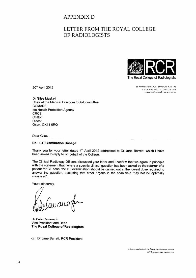
94
APPENDIX D
LETTER FROM THE ROYAL COLLEGE
OF RADIOLOGISTS

95
APPENDIX E
REPORTS OF THE COMMITTEE ON
MEDICAL ASPECTS OF RADIATION
IN THE ENVIRONMENT
COMARE Fifteenth
Report
Radium contamination in the area around Dalgety Bay. PHE, Chilton,
May 2014
COMARE Fourteenth
Report
Further consideration of the incidence of childhood leukaemia around nuclear
power plants in Great Britain. HPA, Chilton, May 2011
COMARE Thirteenth
Report
The health effects and risks arising from exposure to ultraviolet radiation from
artificial tanning devices. HPA, Chilton, June 2009
COMARE Twelfth
Report
The impact of personally initiated X-ray computed tomography scanning for the
health assessment of asymptomatic individuals. HPA, Chilton, December 2007
COMARE Eleventh
Report
The distribution of childhood leukaemia and other childhood cancer in Great
Britain 1969–1993. HPA, Chilton, July 2006
COMARE Tenth Report The incidence of childhood cancer around nuclear installations in Great Britain.
HPA, Chilton, June 2005
COMARE Ninth Report Advice to Government on the review of radiation risks from radioactive internal
emitters carried out and published by the Committee Examining Radiation
Risks of Internal Emitters (CERRIE). NRPB, Chilton, October 2004
COMARE Eighth Report A review of pregnancy outcomes following preconceptional exposure to
radiation. NRPB, Chilton, February 2004
COMARE Seventh
Report
Parents occupationally exposed to radiation prior to the conception of their
children. A review of the evidence concerning the incidence of cancer in their
children. NRPB, Chilton, August 2002
COMARE and RWMAC
Joint Report
Radioactive contamination at a property in Seascale, Cumbria. NRPB, Chilton,
June 1999
COMARE Sixth Report A reconsideration of the possible health implications of the radioactive particles
found in the general environment around the Dounreay nuclear establishment in
the light of the work undertaken since 1995 to locate their source. NRPB,
Chilton, March 1999
COMARE Fifth Report The incidence of cancer and leukaemia in the area around the former Greenham
Common Airbase. An investigation of a possible association with measured
environmental radiation levels. NRPB, Chilton, March 1998
COMARE Fourth Report The incidence of cancer and leukaemia in young people in the vicinity of the
Sellafield site, West Cumbria: further studies and an update of the situation
since the publication of the report of the Black Advisory Group in 1984.
Department of Health, London, March 1996
Radioactive Waste Management Advisory Committee.

96
COMARE and RWMAC*
Joint Report
Potential health effects and possible sources of radioactive particles found in the
vicinity of the Dounreay nuclear establishment. HMSO, London, May 1995
COMARE Third Report Report on the incidence of childhood cancer in the West Berkshire and North
Hampshire area, in which are situated the Atomic Weapons Research
Establishment, Aldermaston and the Royal Ordnance Factory, Burghfield.
HMSO, London, June 1989
COMARE Second Report Investigation of the possible increased incidence of leukaemia in young people
near the Dounreay nuclear establishment, Caithness, Scotland. HMSO, London,
June 1988
COMARE First Report The implications of the new data on the releases from Sellafield in the 1950s for
the conclusions of the report on the investigation of the possible increased
incidence of cancer in West Cumbria. HMSO, London, July 1986
* Radioactive Waste Management Advisory Committee.

97
APPENDIX F
COMMITTEE ON MEDICAL ASPECTS OF
RADIATION IN THE ENVIRONMENT
CHAIRMAN
Professor A Elliott BA PhD DSc CPhys FInstP FIPEM University of Glasgow
PRESENT MEMBERS
Dr P Darragh MD PhD MSc FRCP FFPHM Public Health Agency for Northern Ireland, Belfast
Professor W Evans MA PhD FInstP FIPEM HonMRCR University Hospital of Wales, Cardiff
Dr B Howard MBE Centre for Ecology and Hydrology, Lancaster Environment Centre
Professor M Kadhim PhD Department of Biological and Medical Sciences, Oxford Brookes University,
Oxford
Professor S McKeown MA PhD FSB CBiol School of Biomedical Sciences, University of Ulster, Coleraine
Professor P Marsden MSc PhD FSRP MIPEM MInstP CRadP UCL Hospitals NHS Foundation Trust, London
Dr G Maskell MA FRCP FRCR Department of Radiology, Royal Cornwall Hospital, Truro
Dr T Nunan MD FRCP FRCR Nuclear Medicine Physician
Dr M Pearce BSc MSc PhD Institute of Health and Society, Newcastle University, Newcastle upon Tyne
Mr I Robinson BSc CRadP FSRP FNucI Consultant on Nuclear and Radiation Safety (formerly HM Superintending
Inspector of Nuclear Installation, Office for Nuclear Regulation)
Professor R Taylor MA FRCPE FRCP FRCR School of Medicine, Swansea University
Professor R Wakeford BSc PhD CSci CPhys FInstP CStat CEng MINucE CRadP FSRP Dalton Nuclear Institute, University of Manchester
Professor P Warwick BA MSc PhD DSc CChem FRSC Centre for Environmental Studies, Loughborough University
Professor Catharine West BA PhD University of Manchester

98
FORMER MEMBERS WHO SERVED DURING THE PREPARATION OF
THIS REPORT
Dr J Bithell BA MA DPhil Childhood Cancer Research Group, Oxford
Professor S Hodgson BM BCh DM FRCP Department of Clinical Development Sciences,
St George’s University of London
Professor P Hoskin
Mount Vernon Cancer Centre, Northwood
Professor P Jeggo BSc PhD Genome Damage and Stability Centre, University of Brighton
SECRETARIAT
Mr S Ebdon-Jackson BSc MSc FRCR HonFRCP (Scientific)
Dr J Meara FFPH (Scientific)
Dr E Petty BSc PhD (Scientific)
Dr K Broom BSc DPhil CBiol FSB (Scientific)
Ms K Stonell (Minutes)
Ms J Humphries (Administrative)
ASSESSORS IN ATTENDANCE REPRESENTING THE FOLLOWING
ORGANISATIONS
Department for Education
Department for Communities and Local Government
Department of Energy and Climate Change
Department of the Environment, Food and Rural Affairs
Department of Health
Department of Health, Social Services and Public Safety (Northern Ireland)
Department for Innovation, Universities and Skills
Environment Agency
Food Standards Agency
Health and Safety Executive
Medical Research Council
Ministry of Defence
Nuclear Decommissioning Authority
Office for National Statistics
Public Health England
Public Health and Intelligence, NHS National Services Scotland
Scottish Environment Protection Agency
Scottish Government
Welsh Government

99
MEDICAL PRACTICES SUBCOMMITEE (CT)
CHAIRMAN
Dr G Maskell MA FRCP FRCR Department of Radiology, Royal Cornwall Hospital, Truro
MEMBERS
Professor W Evans MA PhD FInstP FIPEM HonMRCR University Hospital of Wales, Cardiff
Dr S Golding MA MB BS DMRD FRCR FBIR FHEA University of Oxford
Professor S Hodgson BM BCh DM FRCP Department of Clinical Development Sciences,
St George’s University of London
Professor M Kadhim PhD Department of Biological and Medical Sciences, Oxford Brookes University,
Oxford
Dr T Nunan MD FRCP FRCR Nuclear Medicine Physician
Dr M Pearce BSc MSc PhD Institute of Health and Society, Newcastle University, Newcastle upon Tyne
Ms H Warner
Patient Representative

100
APPENDIX G
DECLARATION OF MEMBERS’ INTERESTS
CODE OF PRACTICE
1 Introduction This code of practice guides members of COMARE as to the circumstances in
which they should declare an interest in the course of the Committee’s work.
To avoid any public concern that commercial interests of members might affect
their advice to Government, Ministers have decided that information on
significant and relevant interests of members of its advisory committees should
be on the public record. The advice of the Committee frequently relates to
matters which are connected with the radiation industry generally and, less
frequently, to commercial interests involving radioactivity. It is therefore
essential that members should comply with the code of practice which is set
out below.
2 Scope and definitions This code applies to members of COMARE and its subcommittees, subgroups,
working groups and working parties which may be formed.
For the purposes of this code of practice, the ‘radiation industry’ means:
(a) companies, partnerships or individuals who are involved with the
manufacture, sale or supply of products processes or services which are
the subject of the Committee’s business. This will include nuclear
power generation, the nuclear fuel reprocessing industry and associated
isotope producing industries, both military and civil and also medical
service industries
(b) trade associations representing companies involved with such
products
(c) companies, partnerships or individuals who are directly concerned
with research or development in related areas
(d) interest groups or environmental organisations with a known
interest in radiation matters
This excludes government departments, professional bodies, international
organisations and agencies.
It is recognised that an interest in a particular company or group may, because
of the course of the Committee’s work, become relevant when the member had
no prior expectation this would be the case. In such cases, the member
should declare that interest to the Chairman of the meeting and thereafter to
the Secretariat.
In this code, ‘the Department’ means the Department of Health, and ‘the
Secretariat’ means the secretariat of COMARE.
3 Different types of
interest – definitions
The following is intended as a guide to the kinds of interests which should
be declared. Where a member is uncertain as to whether an interest should be
declared they should seek guidance from the Secretariat or, where it may
concern a particular subject which is to be considered at a meeting, from the

101
Chairman at that meeting. Members of the Committee and the Secretariat are
under no obligation to search out links between one company and another, for
example where a company with which a member is connected has a relevant
interest of which the member is not aware and could not reasonably be
expected to be aware.
If members have interests not specified in these notes but which they believe
could be regarded as influencing their advice they should declare them to the
Secretariat in writing and to the Chairman at the time the issue arises at
a meeting.
3.1 Personal interests A personal interest involves current payment to the member personally. The
main examples are:
(a) Consultancies and/or direct employment: any consultancy,
directorship, position in or work for the radiation industries which
attracts regular or occasional payments in cash or kind.
(b) Fee-paid work: any work commissioned by those industries for
which the member is paid in cash or kind.
(c) Shareholdings: any shareholding in or other beneficial interest in
shares of those industries. This does not include shareholdings through
unit trusts or similar arrangements where the member has no influence
on financial management.
(d) Membership or affiliation: any membership role or affiliation that
the member or close family member has to clubs or organisations with
an interest or involvement in the work of the Department. This will not
include professional bodies, organisations and societies.
3.2 Non-personal interests A non-personal interest involves current payment which benefits a department
to which a member is responsible, but is not received by the member
personally. The main examples are:
(a) Fellowships: the holding of a fellowship endowed by the radiation
industry.
(b) Support by industry: any payment, other support or sponsorship by
the radiation industry which does not convey any pecuniary or material
benefit to a member personally, but which does benefit their position or
department, eg:
(i) a grant from a company for the running of a unit or
department for which a member is responsible;
(ii) a grant or fellowship or other payment to sponsor a post or a
member of staff in a unit or department for which a member is
responsible. This does not include financial assistance for
students, but does include work carried out by postgraduate
students and non-scientific staff, including administrative and
general support staff;
(iii) the commissioning of research or work by, or advice from,
staff who work in a unit or department for which a member is
responsible.
(c) Support by charities and charitable consortia: any payment, other
support or sponsorship from these sources towards which the radiation
industry has made a specific and readily identifiable contribution. This
does not include unqualified support from the radiation industry
towards the generality of the charitable resource.

102
(d) Trusteeships: where a member is trustee of a fund with
investments in the radiation industry, the member may wish to consult
the Secretariat about the form of declaration which would be
appropriate.
3.3 Specific interests A specific interest relates explicitly to the material, product, substance or
application under consideration by the Committee.
A member must declare a personal, specific interest if they currently receive a
payment, in any form, for any significant fundamental development work
undertaken previously or at this time, on a material, product or substance or its
application under consideration. This will include the production of radioactive
substances and devices designed to use ionising or non-ionising radiation for
diagnostic, treatment or other purposes.
A member must declare a non-personal, specific interest if they are aware that
the department to which they are responsible currently receives payment for
significant fundamental development work undertaken previously or at this
time, on a material, product or substance or its application under consideration,
but they have not personally received payment for that work in any form.
This will include the production of radioactive substances and devices
designed to use ionising or non-ionising radiation for diagnostic, treatment or
other purposes.
3.4 Non-specific interests A non-specific interest relates to a company or associated material, product,
substance or application, but not to the specific material, product, substance or
application under consideration by the Committee.
A member must declare a personal, non-specific interest if they have a current
personal interest in a material, product, substance or application from a
particular company, which does not relate specifically to the material, product,
substance or application from that company under consideration.
A member must declare a non-personal, non-specific interest if they are aware
that the department to which they are responsible is currently receiving
payment from the company concerned which does not relate specifically to a
material, product, substance or application under discussion.
If a member is aware that a material, product, substance or their application
under consideration is or may become a competitor of a material, product or
substance manufactured, sold or supplied by a company in which the member
has a current personal interest, they should declare their interest in the company
marketing the rival material, product or substance.
Members are under no obligation to seek out knowledge of such work done for
or on behalf of the radiation industry within departments to which they are
responsible if they would not reasonably expect to be informed. This applies to
all non-personal, specific and non-specific interests.
4 Declaration of interests
4.1 Declaration of
interests to the Secretariat
Members should inform the Secretariat in writing when they are appointed of
their current personal and non-personal interests and annually in response to a
Secretariat request. Only the name of the company (or other body) and the
nature of the interest is required; the amount of any salary, fees, shareholding,
grant, etc, need not be disclosed. An interest is current if the member has a
continuing financial involvement with the industry, eg if they hold shares in a
radiation company, have a consultancy contract, or if the member or the

103
department to which they are responsible is in the process of carrying out work
for the radiation industry. Members are asked to inform the Secretariat at the
time of any change in their personal interests, and may be invited to complete a
form of declaration when required. It would be sufficient if changes in non-
personal interests are reported at the next annual declaration following the
change. (Non-personal interests involving less than £5000 from a particular
company in the previous year need not be declared.)
The register of interests should be kept up-to-date and be open to the public.
4.2 Declaration of
interests at meetings and
participation by members
Members are required to declare relevant interests at Committee meetings and
to state whether they are personal or non-personal interests. The declaration
should include an indication of the nature of the interest.
(a) If a member has a current (personal or non-personal) interest in
the business under discussion, they will not automatically be debarred
from contributing to the discussion subject to the Chairman’s
discretion. The Chairman will consider the nature of the business under
discussion and of the interest declared (including whether it is personal
or non-personal) in deciding whether it would be appropriate for the
relevant member to participate in the item.
(b) If a member has an interest which is not current in the business
under discussion, this need not be declared unless not to do so might be
seen as concealing a relevant interest. The intention should always be
that the Chairman and other members of the Committee are fully aware
of relevant circumstances.
A member who is in any doubt as to whether they have an interest which
should be declared, or whether to take part in the proceedings, should ask the
Chairman for guidance. The Chairman has the power to determine whether or
not a member with an interest shall take part in the proceedings.
If a member is aware that a matter under consideration is or may become a
competitor of a product, process or service in which the member has a current
personal interest, they should declare the interest in the company marketing the
rival product. The member should seek the Chairman’s guidance on whether to
take part in the proceedings.
If the Chairman should declare a current interest of any kind, they should stand
down from the chair for that item and the meeting should be conducted by the
Deputy Chairman or other nominee if the Deputy Chairman is not there.

104
4.3 Members’ declarations
of interests – 2013 Member Company
Personal
interest
Company
Non-personal
interest
Dr J Bithell None None
Dr P Darragh None None
Prof A Elliott None None
Prof W Evans None None
Prof S Hodgson None None
Prof P Hoskin None None
Dr B Howard None None
Prof M Kadhim None None
Prof S McKeown None None
Prof P Marsden None None
Dr G Maskell None None
Dr T Nunan None None
Dr M Pearce None None
Prof R Taylor None None
Mr I Robinson AMEC Consultancy None
Prof R Wakeford 1 Sellafield Ltd Consultancy None
2 Compensation
Scheme for Radiation-
linked Diseases
Consultancy
3 Canadian Nuclear
Safety Commission
Contract
4 Augean Contract
Prof P Warwick 1 Enviras Ltd Director and
shareholder
NDA Grants
2 Sellafield Ltd/
Golder
Contract
3 NNL/LLWR Ltd Consultancy
Prof C West None None