Embed Size (px)
Citation preview
Commercial crocodile farming in Bangladesh, past, present and future possibilities
Mushtaq AhmedBanCroc Ltd., 2-B, 1/8 Block D, Lalmatia, Dhaka-1207, Bangladesh
E-mail: [email protected]; [email protected]
thProposal for crocodile farm in Bangladesh came in the year 1982, by the legend Rom Whitaker. In the 7 CSG meeting
1984 a survey on crocodile farming around the world was published. All over the world crocodile farming initially began
with ranching operation, which eventually converted to captive breeding. Bangladesh started crocodile farming with F2
generation in the year 2004. Starting with 75 heads Reptiles Farm Ltd. now has approximately 1500 heads of saltwater
crocodile (Crocodylus porosus). RFL got registered with CITES in 2007 and made its first export in 2010. Through out
the process RFL faced new challenges as it is first such venture in Bangladesh. The company has bright future potentials;
at the same time there are plenty of challenges from the other stakeholders. This study is based on the observation made
by different stakeholders in the industry and Bangladesh Forest Department. It is precisely focused on the policy issue,
which needs to be addressed for the future smooth functioning of the industry.
123Sri Lanka 20-23 May 2013
Proceedings : World Crocodile Conferencend -22 Working Meeting of the IUCN SSC Crocodile Specialist Groupnd -22 Working Meeting of the IUCN SSC Crocodile Specialist Groupnd -22 Working Meeting of the IUCN SSC Crocodile Specialist Group
Proceedings : World Crocodile Conferencend -22 Working Meeting of the IUCN SSC Crocodile Specialist Groupnd -22 Working Meeting of the IUCN SSC Crocodile Specialist Groupnd -22 Working Meeting of the IUCN SSC Crocodile Specialist Group
p 123

Microbial investigation of captive gharial hatchlings in Chitwan National Park, Nepal
1 2 3, 4, 5K.P. Gairhe , I.P. Dhakal , D.K. Singh H.B. Basnet and J.B. Sherchand1. Chitwan National Park, Nepal. 2, 3, 4.Institute of Agriculture and Animal Sciences, Rampur,
Chitwan, Nepal. 5. Health Research Laboratory, Institute of Medicine, Maharajgunj, Kathmandu
Abstract
Dead gharial hatchlings (137) aged between 6 to 42 weeks were collected from Gharial Breeding Center (GBC), autopsied and examined. Four organs (Liver, lungs, heart blood and kidney) of 102 hatchlings subjected to aerobic bacterial cultures identified Gram-positive bacteria and Gram-negative rods. Gram positive organism isolated were Staphylococcus aureus, Streptococcus spp., Actinomyces spp., Bacillus spp. and Clostridium spp. Gram negative organisms were Citrobacter freundii, Escherichia coli, Providencia rettgeri, Pseudomonas spp., Proteus vulgaris, P. mirabilis, Aeromonas spp., Klebsiella oxytoca, Morganella morganii, Salmonella spp. and Shigella spp. Fourty samples (hatchlings infected with Surahi fluke (Exotidendrium sp.) and having a distinct "Sphincter Cap") from the rectocloacal area were cultured aerobically. It resulted in the isolation of two organisms (Bacillus subtilis and Strep. viridans) of the Gram-positive group and nine organisms of the Gram-negative group. The dominant isolates were Citrobacter freundii (30%) and E. coli (20%). One hundred and forty eight culture positive samples based upon monthly mortality were analyzed and it revealed 25% infection in September, 16.89% in August, and less than 14% infection in other months. Bacterial infections in different organs detected on 148 culture positive gharial hatchlings were found to be 27.03, 42.57, 15.54 and 14.86% in liver, lungs, kidney and heart respectively. Spectrum of bacteria on 90 culture positive gharial hatchlings were only one species in 70%, two species in 21.11% and three or more bacterial species in 8.89% of the carcasses. Presence of infection in particular age group of hatchlings led to the finding that the highest percentage of infection (75%) was recorded in the 30-34 weeks old hatchlings and in an average 38.64% hatchlings had been infected by one or more bacteria.
Out of 102 hatchlings subjected to bacterial culture, 12 hatchlings (11.76%) were found negative to cultures. Culture of skin scrapings from suspected skin lesions led to the isolation of six environmental fungi without a higher prevalence of any particular species. It is concluded that higher mortality of gharial hatchlings in Chitwan was multifactorial. Bacteria, fungi, coccidia and parasites were significantly associated to these deaths, but environmental factors such as extreme temperatures and managemental deficiencies could not be ignored.
Keywords: Gharial, hatchling mortality, Citrobacter freundii, Cutaneous fungal lesions, Sphincter Cap, Gharial Breeding Centre.
Introduction and literature review
The gharial (Gavialis gangeticus) was literally brought back from the brink of extinction by restocking programs initiated in India (1975) and Nepal (1978). This program strategically protected gharial nest in the wild, collected eggs, raised the hatchlings in captivity and released them back into the main rivers (Cadi and Maskey, 2005). Predation by fishes, birds, jackal, civets and monitor lizards significantly reduces the survivability of the young gharial hatchlings in the wild limiting the survival rate to adulthood just to about one per cent (Dhungel, 1987); likewise, the mortality rate in captivity exceeding 30 per cent during the first year of life have been reported as a major constraint in crocodile farming (Ladds et al., 1995).
The mortality rate of gharial hatchlings (hatching to 1-year age) at GBC has ranged between 42.95 to 97.20%. The causes behind such high mortality include occasional predation (by mongooses), pilling up and suffocation, ant attacks, infection of skin and teeth by fungus, etc. This emphasized a need of a thorough investigation work to reveal out the major errors in management and husbandry along with the biological agents involved in the death of young hatchlings for increasing the survival rate which in turn may efficiently help in restoring and augmenting wild gharial populations.
Twenty five to 80% of the gharial hatchlings die within the first year of their life in captive breeding facilities because of skin disease, neurological disorders, retention and infection of the yolk sac and prolapse of the rectum where as the mortality rate in the second year do not exceed 10% (Maskey, 1989). Rigorous work is still important to know the involvement of pathological agents in higher percentage of hatchling mortally. The causes have not been explored well due to the difficulty in obtaining gharial specimens from the wild or because of the inaccessibility of the gharial breeding centers to the research institutions. The difficulty is further increased by the protected status of the species requiring
Sri Lanka 20-23 May 2013
Proceedings : World Crocodile Conferencend -22 Working Meeting of the IUCN SSC Crocodile Specialist Groupnd -22 Working Meeting of the IUCN SSC Crocodile Specialist Groupnd -22 Working Meeting of the IUCN SSC Crocodile Specialist Group
124
pp 124 -132
permissions from the respective governments for activities related to scientific research and study. This has led to the poor improvements in reducing the mortalities of hatchlings particularly in the first year. Several studies related to gharial ecology, habitat and movement were carried out in the 1980's; however, studies concerning the diseases, its effect on mortality and ways to increase their viability through improved health, hygiene and veterinary interventions, were less than a handful of scientific work. Very few scientists have reported the results of investigation on gharial mortality. Clostridium spp. and E. coli were found responsible for hatchling deaths (Mihsra et al., 1993) and the need of clean water and sound management practice were emphasized to raise disease free gharial hatchlings. Pseudomonas spp. infections were found predominantly responsible for hatchling deaths (Mishra et al., 1996) in India. Maskey et al. (1998) evaluated the disastrous impact of intestinal infection in captive bred gharial hatchlings in Nepal where as Mehrotra et al. (2000) reported E. coli, Pseudomonas aeruginosa, Staphylococcus, Corynebacterium spp., Bacillus spp. and some fungal agents emphasizing the need of fresh running water for gharials instead of stagnant ponds. Providencia rettgeri (Ladds et al., 1996) Morganella morganii (Heard et al., 1988) Proteus spp. (Chakraborty et al. 1988), Klebsiella oxytoca (Flandry et al., 1989) Aeromonas hydrophila (Gorden et al. 1979), Citrobacter freundii (Novak and Seigel, 1986), Escherichia coli (Xuesong et al. 2002), Mycoplasma (Mohan et al., 1995), Chlamydia (Australia, 2006) were reported involved to cause death of other farmed species of crocodiles likewise many fungi , parasites and prorozoa were responsible to kill hatchlings crocodiles. Similarly, metabolic and nutritional diseases and death due to skeletal deformities have also been reported. Stressful phenomenon such as piling up at one corner of the enclosure or on top of each other (a normal reaction of the hatchlings to fear) may have contributed to death by suffocation or by permitting infectious agents through resulting scratches (Huchzermeyer, 2003).
This research of captive gharial hatchlings aimed at investigation of the causes of mortality of young captive gharial hatchlings to improve their viability in captivity.
Material and methods
The study was carried out at the Veterinary Teaching Hospital (VTH), Institute of Agriculture and Animal Science (IAAS), Rampur, Chitwan (August, 2006-April, 2007). Dead gharial hatchlings that hatched between June 15-28, 2006 were collected from Gharial Breeding Center. A total of 137 (six to 42 weeks old) dead hatchlings constituted the study population. These carcasses in whole were transported to IAAS Veterinary Teaching Hospital (VTH) in ice, examined immediately or wrapped individually in a plastic bag, labeled with indelible ink and stored again in the freezer (-20ºC) until necropsy. Before necropsy, each carcass was taken in a 41.50 x 30.50 x 6.50 cm plastic tray, washed thoroughly with tap water to remove all the dirt and sand particles. The hatchlings were then thoroughly washed with distilled water and soaked for five minutes in 70% dehydrated alcohol (Ethanol, B. P., Bengal Chemicals and Pharmaceuticals Ltd., Calcutta, India). The alcohol was drained thoroughly and every carcass was assigned a label written in a clean paper with dates of death, necropsy examination and its weight and length measurements recorded and photographed. External examination was followed by internal examination. Necropsy was carried out by common method and by the method described by Huchzermeyer (2003). Samples for microbiological examination were taken asceptically and inoculated immediately in nutrient, McConkey and blood agar and incubated at 37ºC for 24-48 hours and the colony characters were recorded. The colonies obtained on the agar surface were stained with Gram's stain for morphological studies and subjected to conventional biochemical tests for identification of the organisms (Barrow and Feltham, 2004). The specific single colony were also inoculated in 1 ml of nutrient broth (HiMedia Laboratories Pvt. Ltd., Mumbai, India) in a 5 ml sterilized screw capped glass vial and incubated till development of cloudiness to which was then added 1 ml of 40% glycerol (Qualigens Fine Chemicals, Glaxo Smithkline Pharmaceuticals Ltd., Mumbai, India). These were stored in a freezer and dispatched to Health Research Laboratory, Institute of Medicine, Maharajgunj, Kathmandu, in ice for further confirmatory results. Skin scrapings were treated with 10% KOH and stained with Lactophenol cotton blue (LCB) for observation of fungal elements. The samples were also inoculated in Sabouraud's dextrose agar and potato dextrose agar (HiMedia Laboratories Pvt. Ltd., Mumbai, India) and the resulting fungal growth were identified through slide culture and respective morphology of the spores and hyphae.
Results
Bacterial cultures Internal organs (liver, lungs, kidney and heart or its blood) were subjected to bacterial cultures. Cultures revealed 19 species of bacteria-five genus of Gram positive and 10 genus of the Gram-negative rods. The Gram-positive genus were Actinomyces, Bacillus, Clostridium, Staphylococcus and Streptococcus where as the Gram-negative rods were Aeromonas, Citrobacter, Escherichia, Klebsiella, Shigella, Morganella, Proteus, Providencia, Pseudomonas and Salmonella spp. Twelve hatchlings (48 organ samples), however, did not produce any growth of organisms. The frequencies of the isolates obtained from 148 culture positive organs of 102 dead hatchlings are presented in Table 1.
125
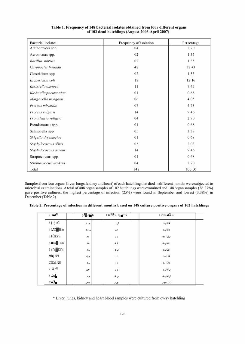
Table 1. Frequency of 148 bacterial isolates obtained from four different organs of 102 dead hatchlings (August 2006-April 2007)
Samples from four organs (liver, lungs, kidney and heart) of each hatchling that died in different months were subjected to microbial examinations. A total of 408 organ samples of 102 hatchlings were examined and 148 organ samples (36.27%) gave positive cultures, the highest percentage of infection (25%) were found in September and lowest (3.38%) in December (Table 2).
Table 2. Percentage of infection in different months based on 148 culture positive organs of 102 hatchlings
* Liver, lungs, kidney and heart blood samples were cultured from every hatchling
Bacterial isolates Frequency of isolation Percentage
Actinomyces spp. 04 2.70
Aeromonas spp. 02 1.35
Bacillus subtilis 02 1.35
Citrobacter freundii 48 32.43
Clostridium spp. 02 1.35
Escherichia coli 18 12.16
Klebsiella oxytoca 11 7.43
Klebsiella pneumoniae 01 0.68
Morganella morganii 06 4.05
Proteus mirabilis 07 4.73
Proteus vulgaris 14 9.46
Providencia rettgeri 04 2.70
Pseudomonas spp. 01 0.68
Salmonella spp. 05 3.38
Shigella dysenteriae 01 0.68
Staphylococcus albus 03 2.03
Staphylococcus aureus 14 9.46
Streptococcus spp. 01 0.68
Streptococcus viridans 04 2.70
Total 148 100.00
a ◘■Ċ╙ {Ă▓♫▄ś ℓا t ◘ℓ╜Ċ╜ōś ľĵ ▄Ċĵ ʼnś t śʼnľś■ĊĂ┼ś
! ĵ ┼ĵ ℓĊ ي ييو يو ي آل
{ś♫Ċś▓Ľśʼn و يى ىه ههيو
h ľĊ◘Ľśʼn و ي و ه و ي ىى
b ◘ōś▓Ľśʼn و ه ه ه آل يهي
5 śľś▓Ľśʼn ه ىو ه ي يىى
WĂ■ĵ ĂʼnŦ آلوىو وو يي
CśĽʼnĵ ĂʼnŦ ه وو يو ي وو
a Ăʼnľ╙ و هي يىو و ي
! ♫ʼn╜▄ ه ىو ه ي يهى
Ç◘ĊĂ▄ يهى يىو و هه .00
126
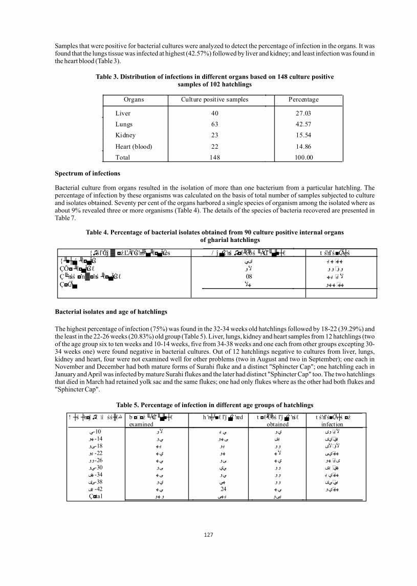
Samples that were positive for bacterial cultures were analyzed to detect the percentage of infection in the organs. It was found that the lungs tissue was infected at highest (42.57%) followed by liver and kidney; and least infection was found in the heart blood (Table 3).
Table 3. Distribution of infections in different organs based on 148 culture positive samples of 102 hatchlings
Spectrum of infections
Bacterial culture from organs resulted in the isolation of more than one bacterium from a particular hatchling. The percentage of infection by these organisms was calculated on the basis of total number of samples subjected to culture and isolates obtained. Seventy per cent of the organs harbored a single species of organism among the isolated where as about 9% revealed three or more organisms (Table 4). The details of the species of bacteria recovered are presented in Table 7.
Table 4. Percentage of bacterial isolates obtained from 90 culture positive internal organs of gharial hatchlings
Bacterial isolates and age of hatchlings
The highest percentage of infection (75%) was found in the 32-34 weeks old hatchlings followed by 18-22 (39.29%) and the least in the 22-26 weeks (20.83%) old group (Table 5). Liver, lungs, kidney and heart samples from 12 hatchlings (two of the age group six to ten weeks and 10-14 weeks, five from 34-38 weeks and one each from other groups excepting 30-34 weeks one) were found negative in bacterial cultures. Out of 12 hatchlings negative to cultures from liver, lungs, kidney and heart, four were not examined well for other problems (two in August and two in September); one each in November and December had both mature forms of Surahi fluke and a distinct "Sphincter Cap"; one hatchling each in January and April was infected by mature Surahi flukes and the later had distinct "Sphincter Cap" too. The two hatchlings that died in March had retained yolk sac and the same flukes; one had only flukes where as the other had both flukes and "Sphincter Cap".
Table 5. Percentage of infection in different age groups of hatchlings
Organs Culture positive samples Percentage
Liver 40 27.03 Lungs 63 42.57
Kidney 23 15.54
Heart (blood) 22 14.86 Total 148 100.00
{♫śľĊʼnĵ ▓ ◘ź ĽĂľĊśʼn╜Ă▄ ╜ℓ◘▄ĂĊes / ĵ ▄Ċĵ ʼnś ♫◘ℓ╜Ċ╜ōś ╙ĂĊľ╙▄╜■┼ℓ t śʼnľś■ĊĂ┼ś {╜■┼▄ś ╜ℓ◘▄ĂĊś ي ىي ه ه هÇŎ◘ ╜ℓ◘▄ĂĊśℓ وووو آلو Ç╙ʼnśś ◘ʼn ▓◘ʼnś ╜ℓ◘▄ĂĊśℓ 08 ه ي ي آلÇ◘ĊĂ▄ هو هآل ه هه
! ┼ś ┼ʼn◘ĵ ♫ℓ �í śś╫ℓهللا b ◘ ◘ź ╙ĂĊľ╙▄╜■┼ℓ examined
h ʼn┼Ă■ℓ ľĵ ▄Ċĵ ʼned t ◘ℓ╜Ċ╜ōś ľĵ ▄Ċĵ ʼnśℓ obtained
t śʼnľś■ĊĂ┼ś ◘ź infection
ي آلو 10-ي يوى يو ي آلهو هو يو 14- يى ى يييى ه 18-ىو ي و ي آلوآلى وو يو ه 22- ه هو ي ههيى آل وو ه 26- ه ىو ي هو ي ي ىيى وو يي ىو 30-يو هي هى ه 34- ي وو يو ى ههي يييى وو هي يو 38-ىى يى ه 42- ه 24 ي ههيو ي Ç◘tal هو هى و ي يىو
127

Isolates from colon and "Sphincter Cap"
Swabs from colorectal mucosa and sphincter surface of the gharial hatchlings infected with Surahi flukes (Exotidendrium spp.) and having thickened mucosa were subjected to bacteriological culture. The results are summarized in Table 6.
Table 6. Colon and "Sphincter Cap" isolates from 40 gharial hatchlings
* Only hatchlings with specific lesion were cultured
Mycological examination
Skin scrapings from six dead hatchlings were subjected to mycological studies. Cultures revealed six different species of saprophytic fungus. The result suggested that the lesions were produced by opportunistic fungal pathogens. Out of 137 hatchlings examined grossly, 26 (18.98%) hatchlings had fungal lesions on the skin (Table 8).
Table 8. Fungal isolates obtained from skin scrapings of gharial hatchlings
Organisms No. of isolates* Percentage Aeromonas spp. 3 7.50 Bacillus subtilis 1 2.50 Citrobacter freundii 12 30.00 E. coli 8 20.00 Klebsiella oxytoca 2 5.00 Morganella morganii 2 5.00 Proteus mirabilis 2 5.00 Proteus vulgaris 5 12.50 Providencia rettgeri 1 2.50 Pseudomonas spp. 1 2.50 Streptococcus viridans 3 7.50 Total 40 100.00
! ┼ś ◘ź ╙ĂĊľ╙▄╜■┼ℓ (weeks)
Number Cĵ ■┼ĵ ℓ ╜ℓ◘▄ĂĊśŕ
و ىو / ĵ ʼnōĵ ▄Ăʼn╜Ă ℓ♫♫ w╙╜ū◘♫ĵ ℓ ℓ♫♫ و وو Çʼn╜ľ╙◘ŕśʼn▓Ă ℓ♫♫ و وو t ś■╜ľ╜▄▄╜ĵ ▓ ℓ♫♫ و يو Cĵ ℓĂʼn╜ĵ ▓ ℓ♫♫ و يو t ℓśĵ ŕĂ▄▄śℓľ╙śʼn╜Ă Ľ◘Ŧŕ ╜╜
128
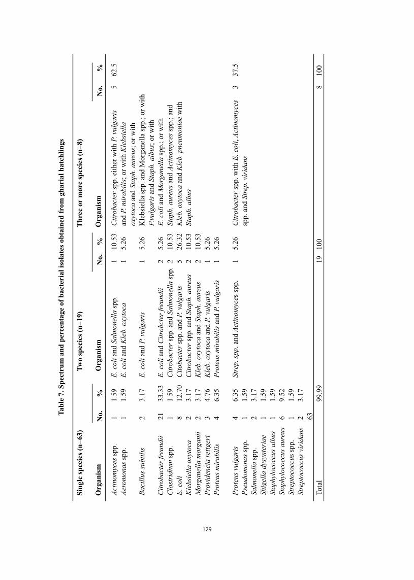
Tab
le 7
. Sp
ectr
um
an
d p
erce
nta
ge o
f b
acte
rial
iso
late
s ob
tain
ed f
rom
gh
aria
l h
atch
lin
gs
Sin
gle
spec
ies
(n=
63)
Org
anis
m
Tw
o sp
ecie
s (n
=19
)
Org
anis
m
Th
ree
or m
ore
spec
ies
(n=
8)
Org
anis
mN
o.%
No.
%
Act
inom
yces
spp
.
Aer
omon
as s
pp.
Bac
illu
s su
btil
is
Cit
roba
cter
fre
undi
i
Clo
stri
dium
spp
.
E. c
oli
Kle
bsie
lla
oxyt
oca
Mor
gane
lla
mor
gani
i
Pro
vide
ncia
ret
tger
i
Pro
teus
mir
abil
is
Pro
teus
vul
gari
s
Pse
udom
onas
spp
.
Salm
onel
la s
pp.
Shi
gell
a dy
synt
eria
e
Sta
phyl
ococ
cus
albu
s
Sta
phyl
ococ
cus
aure
us
Stre
ptoc
occu
s sp
p.
Stre
ptoc
occu
s vi
rida
ns
Tot
al
1 1 2 21 1 8 2 2 3 4 4 1 2 1 1 6 1 2 63
1.59
1.59
3.17
33.3
3
1.59
12.7
0
3.17
3.17
4.76
6.35
6.35
1.59
3.17
1.59
1.59
9.52
1.59
3.17
99.9
9
E. c
oli
and
Salm
onel
la s
pp.
E. c
oli
and
Kle
b. o
xyto
ca
E. c
oli
and
P. v
ulga
ris
E. c
oli
and
Cit
robc
ter
freu
ndii
Cit
roba
cter
spp
. and
Sal
mon
ella
spp
.
Cit
obac
ter
spp.
and
P. v
ulga
ris
Cit
roba
cter
spp
. and
Sta
ph. a
ureu
s
Kle
b. o
xyto
ca a
nd S
taph
. aur
eus
Kle
b. o
xyto
ca a
nd P
. vul
gari
s
Pro
teus
mir
abil
is a
nd P
. vul
gari
s
Stre
p. s
pp. an
d A
ctin
omyc
es s
pp.
No.
%
1 1 1 2 2 5 2 2 1 1 1 19
10.5
3
5.26
5.26
5.26
10.5
3
26.3
2
10.5
3
10.5
3
5.26
5.26
5.26
100
Cit
roba
cter
spp
. eit
her
wit
h P.
vul
gari
s
and
P. m
irab
ilis
; or
wit
h K
lebs
iell
a
oxyt
oca
and
Stap
h. a
ureu
s; o
r w
ith
Kle
bsie
lla
spp.
and
Mor
gane
lla
spp.
; or
wit
h
P.vu
lgar
is a
nd S
taph
. alb
us;
or w
ith
E. c
oli
and
Mor
gane
lla
spp.
; or
wit
h
Stap
h. a
ureu
s an
d A
ctin
omyc
es s
pp.;
and
Kle
b. o
xyto
ca a
nd K
leb.
pne
umon
iae
wit
h
Stap
h. a
lbus
Cit
roba
cter
spp
. wit
h E
. col
i, A
ctin
omyc
es
spp.
and
Str
ep. v
irid
ans
5 3 8
62.5
37.5
100
129
Discussion
This study aimed at finding the involvement of microorganisms to cause deaths in gharial hatchlings in their first year of life. Data based on four batches of hatchlings revealed 2.79-57.05% hatchling survival rate with a clear indication of high mortality and variations. Hatchling mortality rates of 97.20, 42.60, 76.90 and 61.50% in the year 2003, 2004, 2005 and 2006 respectively showed large variations. This could be due to the variations in the extent of congenital anomalies, environmental extremes precipitating a particular disease or due to the variation in the degree of care and management of hatchlings.
Many researchers are in the opinion that bacterial isolation from a crocodile should not be regarded as solely responsible for disease or death, because all the crocodile specific pathogens are opportunists, waiting for a weakened or stressed animal to produce the specific disease. Septicaemia is often produced by the fact that crocodilians are devoid of lymph nodes (Huchzermeyer, 2003). Crocodiles with septicaemic lesions due to intestinal bacteria have been reported suffering from severe stress (Huchzermeyer, 2000; Huchzermeyer and Cooper, 2000). This study also cannot exclude the probability of involvement of intestinal bacteria in causing septicemic death in gharial hatchlings due in period of stress.
Pseudomonas aeruginosa (Mishra et al., 1996; Mehrotra et al., 2000) and Clostridium spp. (Mishra et al., 1993) caused massive deaths of gharial hatchlings and E. coli (Sinha et al., 1988) was associated with septicemic conditions in mugger crocodiles even though such bacteria have been isolated from apparently healthy gharial hatchlings (Mishra et al., 1993); Gram negative rods including Salmonella spp., Proteus spp., E. coli, Providencia rettgeri, Morganella morganii, Serratia marcescens and Aeromonas hydrophila have been isolated from crocodilians and are implicated as responsible for diseases and death in many instances.
Citrobacter spp. has been isolated from captive Nile crocodile and from the faeces of a Caiman crocodylus (Roggendorf and Muller, 1976). Citrobacter freundii has been found associated with septicemia in American alligators (Novak and Seigel, 1986) and C. koseri has also caused meningitis in newborn human babies (Gross et al., 1973; Ross, 1979).
Aeromonas hydrophila is one of the most common bacteria associated with the aquatic environment and has caused skin lesions and septicaemia in a Nile crocodile (C. niloticus), yielding pure cultures from skin, internal organs and blood (Turutoglu et al., 2005). It was reported earlier as the sole agent but later reports showed Proteus spp., Morganella morganii, Serratia marcescens and Klebsiella oxytoca equally responsible for septicaemic lesions in crocodiles. It is emerging as a potential pathogen for the immune-compromised host (Chang et al., 1997). Various aeromonal infections, including septicaemia, have also been reported in apparently healthy individuals; the septicaemic course is often fulminant and fatal and may lack an obvious focus.
Morganella morganii is an opportunistic secondary invader originally thought responsible for summer diarrhoea in humans. Several reports implicate this organism for causing septicaemia and abscess in the brain and ovary in neonates. It had been found in a case of chorioamninonitis and meningitis in an immune-compromised patient. It has been an important cause of nosocomial infection, though it was regarded as a relatively unimportant human pathogen in the past. Morganella morganii has been found in cases of septic arthritis of African dwarf crocodiles (Heard et al., 1988) and was also isolated from juvenile Crocodylus porosus (Hibberd et al., 1996).
Providencia rettgeri infection have caused neurological disorders (swaying, swimming in circle and head tilting) and death in Crocodylus porosus (Ladds et al., 1996) and American alligator (Camus and Hawke, 2002) hatchlings in association with overcrowding or severe temperature stress respectively. The neurological disorders and death in gharial hatchlings described by Maskey (1989) seems to have caused by this species of bacteria. The frequency of infection and death of gharial hatchlings due to this organism was almost similar (2.50-2.70%) with the reports of Ladds et al. (1996) in Crocodylus porosus hatchlings. Providencia spp. has also been recovered from human urine, throat, faeces, blood and wound specimens (O'Hara et al., 2000). Proteus mirabilis and P. vulgaris have been isolated from captive Nile crocodile and there are reports of septicemia in crocodiles due to these organisms. Proteus mirabilis has also been implicated in bacteremia, meingo-encephalitis, empyema and osteomylitis particularly in very young babies (O'Hara et al., 2000).
Klebsiella pneumoniae and K. oxytoca are opportunistic pathogens found in the environment and in mammalian mucosal surfaces. They can infect neonates having impaired respiratory host defenses and produce septicaemia. Escherichia coli form a part of normal intestinal flora and it is the predominant bacteria responsible for urinary tract infections, neonatal meningitis, gastroenteritis and septicemia in man and animals (Chakraborty, 1995). Poor hygiene, intensive husbandry practices and younger age are common predisposing factors for E. coli infection in animals (Quinn et al., 1994). E. coli was found responsible for septicaemic death in gharial hatchlings (Mehrotra et al., 2000) and it was also recovered from severely stressed African dwarf crocodiles (Huchzermeyer and Agnagna, 1994). In this study E. coli comprised 12.16% among all the isolated bacteria from the internal organs of the hatchlings where as isolates from the culture of colon swabs reached 30%.
130

Staphylococcus is ubiquitous organism primarily found on mammalian skin and mucosal surfaces. Staphylococcus aureus is associated with most suppurative lesions; however, it is the common cause of bacteremia, the infection reaching the blood through lungs, gastrointestinal tract, urinary tract and skin abrasions. Mihsra et al. (1993) thought Clostridium spp. responsible for the death of gharial hatchlings as they isolated it from the oedematous fluid of swollen limbs but such swelling in the carcasses were not observed in this study. Actinomyces spp. isolated from gharial hatchlings at present study are probably normal flora of oropharynx or gastrointestinal tract. Several bacterial species from apparently healthy gharials (Mishra et al., 1993) and wild caught African dwarf crocodiles (Huchzermeyer and Agnagna, 1994) have been isolated but the opportunistic bacteria can cause serious infections in reptiles under stress condition (Ebani and Fratini, 2005). Because of this fact, bacteria isolated from the gharial hatchlings at present study may be regarded as the evidences of the mortalities. The number of skin scrapings of the fungal lesions that were cultured and identified in this study was very few but it revealed that all of them were involved only externally. Four species of fungi that were identified in this study were also reported to occur as oral flora of American alligator and intestinal flora of African dwarf crocodiles (Huchzermeyer, 2003) and two species of fungi isolated in this study are probably new records from gharial skin lesions. Extensive hatchling mortality due to fungal infections (>50%) as described by Hibberd and Harrower (1993) in C. porosus was not recorded in this study. Similarly, systemic involvement in other species of crocodiles as reported by others (Fromtling et al.,1979; Frelier et al., 1985; Maslen et al., 1988; Hibberd and Harrower, 1993; Hibberd et al., 1996; Thomas et al., 2002) were not recorded both on gross and microscopic examination of the carcasses and tissue sections. Therefore, it can be concluded that all the isolated fungi were opportunistic pathogens and invasion was secondary to stress or debilitating diseases similar to the report of Migaki et al. (1984).
Pseudomonas spp., E. coli, Providencia rettgeri, Morganella morganii, Staphylococcus aureus, Salmonella spp., Proteus spp., Aeromonas spp., Citrobacter spp. Klebsiella spp., were among the isolates obtained from other species of crocodile hatchlings.
Gharial hatchlings in Chitwan were infected highly by Exotidendrium spp. and concurrent infections with Gram-negative bacteria may be responsible for high mortality. The relationship between heavy parasitism, bacterial infection and mortality may be similar to the systemic illness and death of Green sea turtles (Chelonia mydas) infected with higher number of spirorchid cardiovascular flukes, other internal parasites and simultaneous infections of Salmonella, Escherichia coli and other Gram-negative bacteria (Radial et al., 1998).
Acknowledgement
We are thankful to Dr. J.B. Serchand and his colleagues at Health Research Laboratory for the identification and confirmation of bacterial and fungal isolates. We sincerely acknowledge the financial support of Rufford Maurice Laing Foundation for the research work. We appreciated the kind support of many colleagues across the world for literature search on gharial research. We are thankful to WWF Nepal Program for travel and accommodation support enabling us to participate and present the paper during the World Crocodile Conference in Sri Lanka.
Literature cited
01. Ariel, E., P.W. Ladds and G.N. Buenviaje. 1997a. Concurrent gout and suspected hypovitaminosis A in crocodile hatchlings. Australian Veterinary Journal 75:247- 249.
02. Australia, 2006. Deaths in juvenile farmed crocodiles associated with Chlamydial infection. Animal Health News. Northern Territory, Department of Primary Industry, Fisheries and Mines, Northern Territory Government, Australia.
03. Barrow, G.I. and R.K.A. Feltham. 2004. Cowan and Steel's manual for the identification of medical bacteria. 3rd ed. Cambridge, Cambridge University Press.
05. Cadi, A. and T. Maskey. 2005. Gharial re-enforcement in Royal Chitwan National Park, Nepal. Reintroduction News. Newsletter of the Reintroduction Specialist Group of IUCN's Species Survival Commission (SSC) 24:45-46.
06. Camus, A.C. and J.P. Hawke. 2002. Providencia rettgeri associated septicaemia and meningoencephalitis in juvenile farmed American Alligators (Alligator mississippiensis). Journal of Aquatic Animal Health 14:149-153.
07. Chakraborty, P. 1995. A text book of Microbiology. Kolkata. New Central Book Agency (P) Ltd. India.
08. Chakraborty, T., D.K. Basak and B.K. Majumder. 1988. Septicaemia in a crocodile due to Proteus spp. Indian Journal of Veterinary Pathology 12:72-73.
09. Chang, C.Y., H. Thompson, N. Rodman, J. Bylander and J. Thomas. 1997. Pathogenic analysis of Aeromonas hydrophila septicaemia. Annals of Clinical Laboratory Science 27(4):254-259.
10. Dhungel, S. 1987. Reintroduction of Gharial (Gavialis gangeticus) in Nepal. Tiger Paper 14(4):11-15.
11. Ebani, V.V. and F. Fratini. 2005. Bacterial zoonoses among domestic reptiles. Annali della Facolta di Medicina Veterinaria LVIII:86-91.
12. Flandry, F., E.J. Lisecki, G.J. Domingue, R.L. Nichols, D.L. Greer and R.J. Haddad. 1989. Initial antibiotic therapy for alligator bites: characterization of the oral flora of Alligator mississippiensis. Southern Medical Journal 82:262-266.
131
13. Frelier P.F., L. Sigler and P.E. Nelson. 1985. Mycotic pneumonia caused by Fusarium moniliforme in an alligator. Sabouraudia 23(6):399-402.
14. Fromtling, R.A., S.D. Kosanke, J.M. Jensen and G.S. Bulmer. 1979. Fatal Beauveria bassiana infection in a captive American alligator. Journal of the American Veterinary Medical Association 175(9):934-936.
15. Gorden, R.W., T.C. Hazen, G.W. Esch and C.B. Fliermans. 1979. Isolation of Aeromonas hydrophila from the American Alligator, Alligator mississippiensis. Journal of Wildlife Diseases 15:239-243.
16. Gross, R.J., B. Rowe and J.A. Easton. 1973. Neonatal meningitis caused by Citrobacter koseri. Journal of Clinical Pathology 26:138-139.
17. Heard, D.J., E.R. Jacobson, R.E. Clemmons and G.A. Campbell. 1988. Bacteremia and septic arthritis in a West African dwarf crocodile. Journal of the American Veterinary Medical Association 192(10):1453-54.
18. Hibberd, E.M.A. and K.M. Harrower. 1993. Mycoses in crocodiles. The Mycologist 7(1):32-37.
19. Hibberd, E.M.A., R.J. Pierce, B.D. Hill and M.A. Kelly. 1996. Diseases of juvenile farmed crocodiles Crocodylus porosus. In: Crocodiles: Proceedings of the 13th working Meeting of the Crocodile Specialist Group, 13-17 May, Argentina, IUCN-The World Conservation Union, Gland, Switzerland. pp 303-312.
20. Huchzermeyer, F.W. 2000. A case of stress septicaemia in farmed Nile crocodiles. Crocodile Specialist Group Newsletter 19(4):20-25.
21. Huchzermeyer, F.W. 2003. Crocodiles: Biology, husbandry and diseases. Wallingford, CABI Publishing. pp 167-170.
22. Huchzermeyer, F.W. and J.E. Cooper. 2000. Fibriscess, not abscess, resulting from a localized inflammatory response to infection in reptiles and birds. Veterinary Record 147:515-517.
23. Huchzermeyer, F.W. and M. Agnagna. 1994. A survey of parasites and pathology of African dwarf crocodiles Osteolaemus tetraspis in the Congo Republic. In: Crocodiles. Proceedings of the 12th Working Meeting of the Crocodile Specialist Group. IUCN-The World Conservation Union, Gland, Switzerland 2:309-313.
24. Ladds, P.W., H. Mangunwirjo, D. Sebayang and P.W. Daniels. 1995. Diseases of young farmed crocodiles in Irian Jaya. Veterinary Record 136:121-124.
25. Ladds, P.W., J. Bradley and R.G. Hirst. 1996. Providencia rettgeri meningitis in hatchling saltwater crocodiles (Crocodylus porosus). Australian Veterinary Journal 74:397-98.
26. Maskey, T.M. 1989. Movement and survival of captive reared Gharial (Gavialis gangeticus) in the Narayani River, Nepal. Thesis, Ph D. University of Florida. 187p.
27. Maskey, T.M., P. Kolle, R. Hoffmann, C.C. Anders and H.H. Schleich. 1998. Disastrous impact of intestinal infection in captive bred gharial hatchlings. Veroffentlichungen aus dem Fuhlrott- Museum 4: 291-294.
28. Maslen, M., J. Whitehead, W.M. Forsyth, H. McCracken and A.D. Hocking. 1988. Systemic mycotic disease of captive crocodile hatchling (Crocodylus porosus) caused by Paecilomyces lilacinus. Journal of Medical Veterinary Mycology 26(4):219-25.
29. Mehrotra, P.K., B.B.L. Mathur, S. Bhargava and S. Chaudhary. 2000. Mortality in gharial (Gavialis gangeticus) hatchlings at Jaipur Zoo. Zoos' Print Journal 15(5):267-268.
30. Migaki, G., E.R. Jacobson and H.W. Casey. 1984. Fungal diseases in reptiles. In: G.L. Hoff, F.L. Frye and E.R. Jacobson (eds.). Diseases of amphibians and reptiles. New York, Plenum Press. pp 183-204.
31. Mishra, P.R., D. Kumar, G.M. Patnaik, R.P. Rahman and A. Sinha. 1993. Bacterial isolates from apparently healthy and diseased crocodiles (Gavialis gangeticus). Indian Veterinary Journal 70:375-376.
32. Mishra, P.R., S.K. Patra, H.K Mohapatra, K.C. Patra and S. Mohapatra. 1996. Aetiopathological findings from young gharial mortality cases. Indian Veterinary Journal 73(8):888-889.
33. Mohan., K., C.M. Foggin, P. Muvavarirwa, J. Honywill and A. Pawandiwa. 1995. Mycoplasma-associated polyarthritis in farmed crocodiles (Crocodilus niloticus) in Zimbabwe. Onderstepoort Journal of Veterinary Research 62:45-49.
34. Novak, S.S. and R.A. Seigel. 1986. Gram negative septicaemia in American alligators (Alligator mississippiensis). Journal of Wildlife Diseases 22(4):484-487.
35. O'Hara, C.M., F.W. Brenner and J.M. Miller. 2000. Classification, identification and clinical significance of Proteus, Providencia and Morganella. Clinical Microbiology Reviews 13(4):534-546.
36. Quinn, P.J., M.E. Carter, B. Markey and G.R. Carter. 1994. Enterobacteriaceae. In: P.J. Quinn, B. Markey and G. R. Carter (eds.) Clinical Veterinary Microbiology. Madrid, Wolfe Publishing Co. pp 209-236.
38. Radial, S.R., M. Ohara, R.P. Hobbs and R.I.T. Prince. 1998. Gram-negative bacterial infections and cardiovascular parasitism in green see turtles (Chelonia mydas). Australian Veterinary Journal 76(6):415-417.
39. Roggendorf, M. and H.E. Muller. 1976. Enterobacterien bei reptilien. Zentralblatt fur Bacteriologie und Hygiene, I. Original series A236:22-35.
40. Ross, J.P. 1998. Crocodile status, survey and conservation action plan. (2nd ed.) IUCN/SSC, Gland, Switzerland.
41. Ross, S.J. 1979. Neonatal meningitis due to Citrobacter koseri. Journal of Perinatal Medicine 7(5):273-275.
42. Sinha, R.P., J.P. Soman, G.J. Jha, A. Prasad, H.V.S. Chauhan and R.S. Prasad. 1988. An outbreak of Escherichia coli enteritis in crocodiles. Indian Journal of Animal Sciences 58(3):338-340.
43. Thomas, A.D., L. Sigler, S. Peucker, J.H. Norton and A. Neilan. 2002. Chrysosporium anamorph of Nannizziopsis vriesii associated with fatal cutaneous mycoses in the salt-water crocodile (Crocodylus porosus). Medical Mycology 40:143-151.
44. Turutoglu, H., S. Ercelik and M. Corlu. 2005. Aeromonas hydrophila-associated skin lesions and septicaemia in a Nile crocodile (Crocodylus niloticus). Journal of the South African Veterinary Association 76(1): 40-42.
45. Xuesong, Ye-Wide and Z.Yongkang. 2002. Occurrence and prevention of diseases caused by E. coli in the Yangtze crocodile. Journal of Economic Animal 3 (4): 39-41.
132
Crocodile conservation breeding programme in India past and future
Brij Kishor Gupta
Evaluation & Monitoring Officer Central Zoo Authority (Ministry of Environment & Forests) Annexe VI, Bikaner House Shahajahan Road, New Delhi 110 011 India
Email: [email protected]
Background
The first ever programme for conservation breeding of species in India initiated was of the crocodile species, whose population in the wild has been on decline. Dr. H.R.Bustard, a FAO expert on crocodiles, on the invitation by the Government of India, initiated the programme 1975 at Tikerpada in Satkoshia Gorge sanctuary (Orissa).
The Government of India with help of the UNDP set up a Central Crocodile Breeding & Management Training Institute (CCBMTI), at Hyderbad, Andhra Pradesh as part of the Project. CCBMTI offered a 9 month Diploma Course to young forest officers in all aspects of crocodile conservation including sanctuary management. The Project funded a gharial breeding complex in the Nandankanan Zoological Park in Orissa. The enclosure was designed by Dr. Bustard, which had a huge pool 9m deep (capacity 180,000 litres) with flowing and re-circulating water. The viewing point was only 9m wide on one side of the huge enclosure, the rest having a high wall to provide these shy animals with total protection. The Zoo had three adult gharials, but the male suffered repeated penile prolapse, and it was decided to obtain a large male from Frankfurt Zoo in (then) West Germany rather than capture one from the wild in India. This male reached Nandankanan and despite never having seen another gharial since a baby, mated with the Oriyan females. Thus the world's first captive-breeding of the gharial took place.
The Crocodile Conservation Project has seen the creation of first few wetland sanctuaries of the country under the provision of the Wildlife (Protection) Act, 1972. At the beginning of 1980s, the project boasted thirteen crocodile sanctuaries. Later, several other protected areas highlighted their attention to the management of crocodilians. e.g. Corbett National Park, Dudhwa National Park and Similipal Sanctuary and National Park, all of which are tiger reserves covered under Project Tiger.
Out of the initial list of crocodile sanctuaries, the Nagarjuna Sagar Srisailam Sanctuary (Andhra Pradesh) was later declared a tiger reserve, and the Satkoshia Gorge Sanctuary (Orissa) has been nominated for declaration as either the second tiger reserve or an elephant sanctuary. Satkoshia Gorge Sanctuary and Bhitarkanika Sanctuary have also been proposed for declaration as Biosphere Reserves.
One of the most striking features of the Crocodile Conservation Project has been the building up of a base for wildlife research in the country - beginning with the state of Orissa, and followed by Uttar Pradesh and Andhra Pradesh. The project started a trend of involving fulltime research personnel, propagating the idea that successful conservation and research must go hand in hand.
There are four species of crocodiles recorded in the zoos. The Mugger was the most common reptiles on display in the zoos. Over 2505 individuals of Mugger Crocodylus palustris are housed in 62 zoos in India. The endangered Long-Snouted Crocodile Gavialis gangeticus are housed in 40 zoos with total population of 495 individuals. The Gharials are housed in zoos of all five regions but not in Island. There are 89 nos. of Salt Water Crocodile are housed in 14 zoos of all the regions except north India. All species are listed in Schedule- I of the Wild Life (Protection) Act, 1972.
The ex situ conservation of the following crocodilian species highly recommended by the Central Zoo Authority by providing better upkeep and veterinary care for the scientific management of crocodiles species so that zoos can play significant role in conservation of crocodiles.
1. Mugger Crocodylus palustris 2. Gharial Gavialis gangeticus 3. Estuarine crocodile Crocodylus porosus
Sri Lanka 20-23 May 2013 133
Proceedings : World Crocodile Conferencend -22 Working Meeting of the IUCN SSC Crocodile Specialist Groupnd -22 Working Meeting of the IUCN SSC Crocodile Specialist Groupnd -22 Working Meeting of the IUCN SSC Crocodile Specialist Group
pp 133 -138

1. Introduction
India is one of the rich biodiversity country of the world which harbours a large number of mammals (350 species), birds (1224 species), reptiles (4808 species), amphibians (197 species), fishes (2546 species), insects (57548 species) and plants (46284 species) in a large landscape of 77.47 million hectare of forest cover. It is one of the twelve mega biodiversity country of the world and has 8% of world biodiversity.
The country faces huge challenges on account of population growth coupled with expansion of agriculture and human settlements, industrialization and resulting in environmental degradation and loss of prime/ critical habitat for a large number of species. The growing pressure on the wild population due to shrinkages of habitat and loss of critical resources for the fauna as led to dwindling of population of many species which are on the verge of extinction in various parts of the country. India has established a large protected area network comprising of 4.58% of the total geographical area and for ex-situ conservation, there are 197 recognized zoological parks housing more than 40000 wild animals in captivity across the country, which includes 2505 nos. of Muggers and 495 nos. of Gharials (as on 31.03.2013).
A study of the status of the population in captivity in zoos reveals that a large number of species are not of important conservation value. There are few species in the category, endangered and threatened which are housed in the zoos. Moreover, the species do not occur in natural social group and with unknown lineage and therefore the task of initiating a conservation breeding programme with the available population is a challenge.
Zoos in India are regulated as per the Recognition of Zoo Rules, 1992/2009 framed under the provision of the Wild Life (Protection) Act, 1972 and reflects the policy enshrined in the National Zoo Policy, 1998. The Wild Life (Protection) Act, 1972 was amended in 1992 and a Central Zoo Authority was created to oversee the functioning and management of zoo and to provide technical support to facilitate the development of zoos in the country. The main objectives of zoos as per the National Zoo Policy, 1998 is to strengthen the national efforts in conservation of rich biodiversity of the country by supporting conservation of endangered wild animals species by giving species which has no chance of surviving in the wild, a last chance to coordinated breeding programme under ex-situ conditions and raise stocks for rehabilitating them in wild as and when it is appropriate and desirable. The National Wildlife Action Plan (2002-2016) also lays emphasizes on ex-situ breeding.
Captive breeding programmes are initiated to conserve a population of endangered species which is in danger of becoming extinct but it is not known with certainty whether such efforts can really conserve genetic diversity and produce healthy offspring for re-establishing a stable self-sustaining population in the wild. Conservation biology research suggests that in breeding and loss of fitness and health of animals can occur very rapidly, with such high magnitude with the increasing number of years of an animal in captivity. Nevertheless, there are successful examples of captive bred individuals release in the wild which maintain healthy genetic diversity and continue to sustain a healthy population. There are several scientific technologies which assist in captive breeding which stored the genetic material through cryo-preservation and artificially reproduction. There are still lots of research and studies required to investigate to what extent captive breeding procedures might ultimately help in species recovery programmes and the specific genetic factor necessary to help success captive breeding programme and alternate solutions required for crocodiles. The present conservation breeding programme will also prove an important tool to collate and collect data on the following:
2. Concept and theme of conservation breeding programme for Crocodiles
Captive breeding programmes on Crocodiles was initiated in 1975 to prevent the imminent population collapse in the wild due to a large number of eliminative pressures. The ultimate aim is to conserve to genetic diversity and re-establish self sustaining population in the wild.
134
3. The need for Conservation breeding programme for Crocodiles
As we all know that Conservation breeding programme for species recovery in the wild should be initiated after careful field research to assess the status of population of a particular species in wilderness and of comprehensive assessment of the reason for decline of the species as a judgement is to be made whether the species can on its own recover in the wilderness through a species recovery strategy based on mitigation of the factors which in the first place cause the decline of the species which could be habitat degradation, change in hydrological regime of the tract, natures balance in maintaining prey-predator ration, fire and poaching. If a determination has been made that conservation alternatives are not immediately available and that captive breeding is essential for long-term survival of species. Can it be included to initiate captive breeding programme? Not as a long term conservation strategy but as a recovery technique integrated with supplementary efforts to augment and re-establish wild population. Every proposal to establish a captive population for recovery merits thorough evaluation and review. Captive breeding should not be constitute as a rehabilitation and recovery measures for species whose number has crashed in the wild below a minimum viable population. This population may still be far more viable and captive population, given the many limitations associated with captive breeding and reintroduction. Proponents of the programme justify captive breeding based on population viability enhances but regress assessment of the viability of wild in captive population is necessary. It is possible that alternative non-captive approaches may be more effective and safe than the captive approaches. The National Zoo Policy, 1998 reiterates that if population has decline in the wild it is necessary to supplement in-situ population with the captive stock bred in ex-situ facility. This should be qualified by the fact that each species needs to be assessed whether it truly needs captive interventions. Many zoos feed that they must carry out the mandate of the policy by involving in captive breeding of Crocodiles. However, this is not the end all they may contribute by developing husbandry, reproduction, social behaviour and dietary protocols which will ultimately help raising a captive breeding stock whenever required for reintroduction.
4. Breeding Programme for Muggers
Past HistoryThe breeding programme was initiated in 1975 a Central Crocodile Breeding & Management Training Institute was established in Hyderabad, Andhra Pradesh. The first ever programme for conservation breeding of species initiated in India for the crocodile species from Nandankanan, Odisha. This Programme is considered among the successful Conservation Initiative.
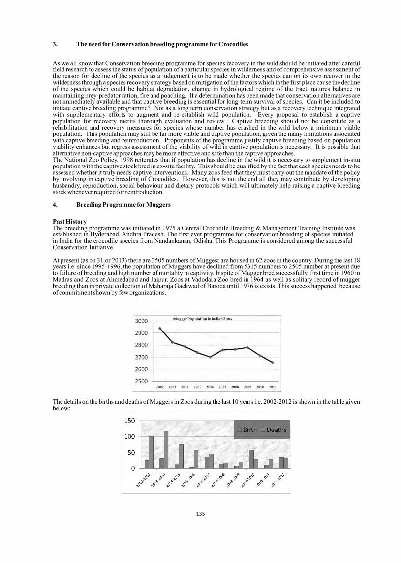
At present (as on 31.or.2013) there are 2505 numbers of Muggesr are housed in 62 zoos in the country. During the last 18 years i.e. since 1995-1996, the population of Muggers have declined from 5315 numbers to 2505 number at present due to failure of breeding and high number of mortality in captivity. Inspite of Mugger bred successfully, first time in 1960 in Madras and Zoos at Ahmedabad and Jaipur. Zoos at Vadodara Zoo bred in 1964 as well as solitary record of mugger breeding than in private collection of Maharaja Gaekwad of Baroda until 1976 is exists. This success happened because of commitment shown by few organizations.
The details on the births and deaths of Muggers in Zoos during the last 10 years i.e. 2002-2012 is shown in the table given below:
135
5. Breeding Programme for Gharials
Past HistoryThe Conservation Breeding of Gharials has the creation of first few wetland sanctuaries of the country under the provision of the Wildlife (Protection) Act, 1972. Dr. H.R.Bustard, a FAO expert on crocodiles, on the invitation by the Government of India, initiated the programme in 1975 at Tikerpada in Satkoshia Gorge Sanctuary (Odisha). The Project funded a Gharial Breeding Enclosure in the Nandankanan Zoological Park in Orissa. The Nandankanan Zoo in Odisha, had 3 adult gharials in 1947, but the male suffered repeated penile prolapse, and it was decided to obtain a large male from Frankfurt Zoo in (then) West Germany rather than capture one from the wild in India. This male reached Nandankanan and mated and bred first time in 1980. This started similar breeding programme at at Kukrail, Uttar Pradesh under Endangered Species Project. More than 4000 reared gharial released in National Chambal Sanctuary.
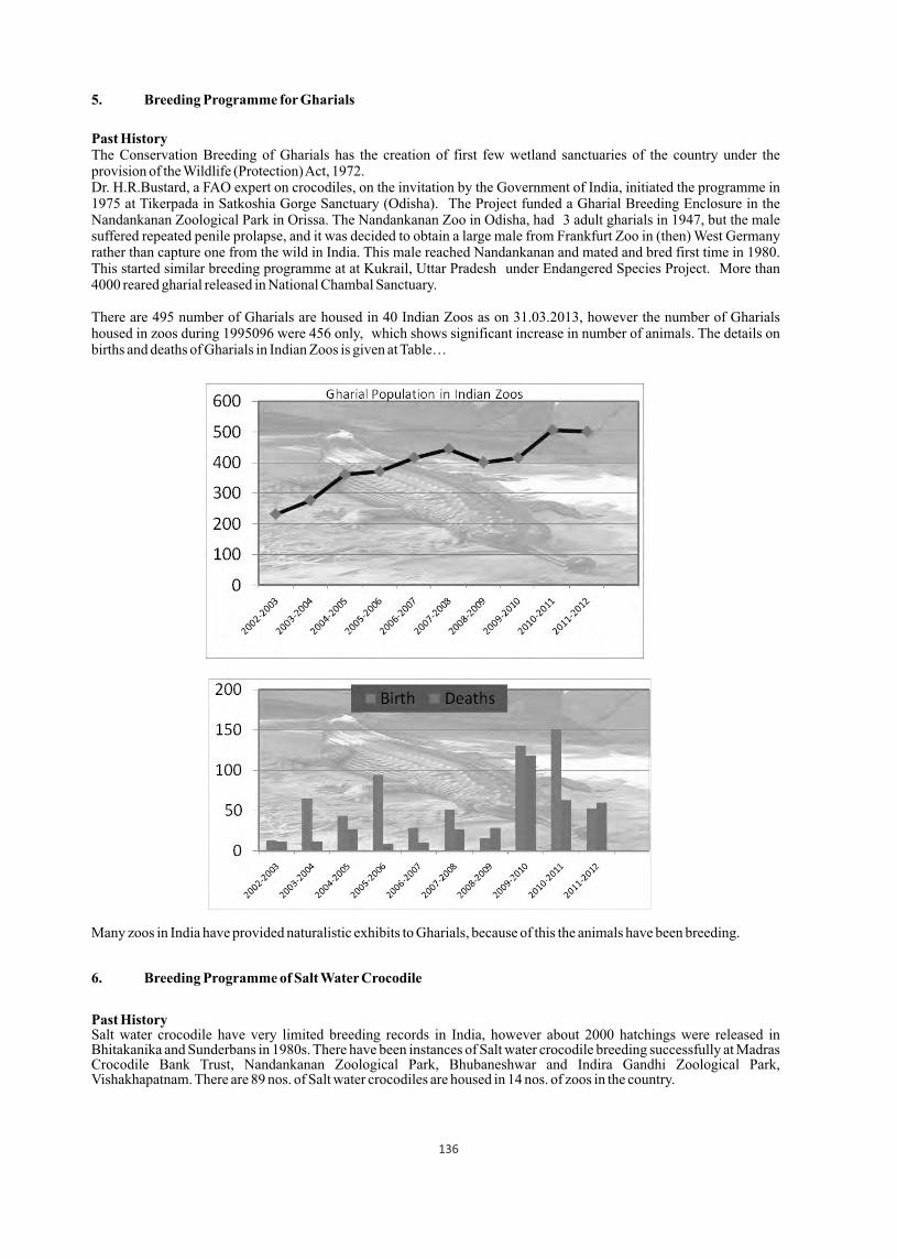
There are 495 number of Gharials are housed in 40 Indian Zoos as on 31.03.2013, however the number of Gharials housed in zoos during 1995096 were 456 only, which shows significant increase in number of animals. The details on births and deaths of Gharials in Indian Zoos is given at Table…
Many zoos in India have provided naturalistic exhibits to Gharials, because of this the animals have been breeding.
6. Breeding Programme of Salt Water Crocodile
Past HistorySalt water crocodile have very limited breeding records in India, however about 2000 hatchings were released in Bhitakanika and Sunderbans in 1980s. There have been instances of Salt water crocodile breeding successfully at Madras Crocodile Bank Trust, Nandankanan Zoological Park, Bhubaneshwar and Indira Gandhi Zoological Park, Vishakhapatnam. There are 89 nos. of Salt water crocodiles are housed in 14 nos. of zoos in the country.
136
7. Short comings/ Limitations of Captive Breeding of Crocodiles:
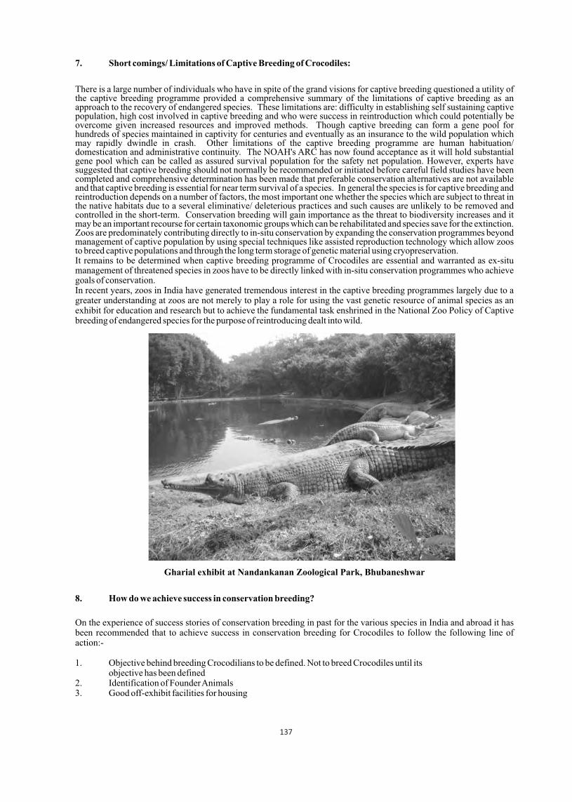
There is a large number of individuals who have in spite of the grand visions for captive breeding questioned a utility of the captive breeding programme provided a comprehensive summary of the limitations of captive breeding as an approach to the recovery of endangered species. These limitations are: difficulty in establishing self sustaining captive population, high cost involved in captive breeding and who were success in reintroduction which could potentially be overcome given increased resources and improved methods. Though captive breeding can form a gene pool for hundreds of species maintained in captivity for centuries and eventually as an insurance to the wild population which may rapidly dwindle in crash. Other limitations of the captive breeding programme are human habituation/ domestication and administrative continuity. The NOAH's ARC has now found acceptance as it will hold substantial gene pool which can be called as assured survival population for the safety net population. However, experts have suggested that captive breeding should not normally be recommended or initiated before careful field studies have been completed and comprehensive determination has been made that preferable conservation alternatives are not available and that captive breeding is essential for near term survival of a species. In general the species is for captive breeding and reintroduction depends on a number of factors, the most important one whether the species which are subject to threat in the native habitats due to a several eliminative/ deleterious practices and such causes are unlikely to be removed and controlled in the short-term. Conservation breeding will gain importance as the threat to biodiversity increases and it may be an important recourse for certain taxonomic groups which can be rehabilitated and species save for the extinction. Zoos are predominately contributing directly to in-situ conservation by expanding the conservation programmes beyond management of captive population by using special techniques like assisted reproduction technology which allow zoos to breed captive populations and through the long term storage of genetic material using cryopreservation. It remains to be determined when captive breeding programme of Crocodiles are essential and warranted as ex-situ management of threatened species in zoos have to be directly linked with in-situ conservation programmes who achieve goals of conservation. In recent years, zoos in India have generated tremendous interest in the captive breeding programmes largely due to a greater understanding at zoos are not merely to play a role for using the vast genetic resource of animal species as an exhibit for education and research but to achieve the fundamental task enshrined in the National Zoo Policy of Captive breeding of endangered species for the purpose of reintroducing dealt into wild.
Gharial exhibit at Nandankanan Zoological Park, Bhubaneshwar
8. How do we achieve success in conservation breeding?
On the experience of success stories of conservation breeding in past for the various species in India and abroad it has been recommended that to achieve success in conservation breeding for Crocodiles to follow the following line of action:-
1. Objective behind breeding Crocodilians to be defined. Not to breed Crocodiles until its objective has been defined2. Identification of Founder Animals 3. Good off-exhibit facilities for housing
137
4. Standardize the husbandry practices to be followed5. Animals nutrition, behavior, and their reproductive biology should be recorded6. Veterinary Care of all animals should be taken on priority7. Training for staff involved in the programme 8. Strengthening of Data, Records Keeping systems9. Long term Management plans for breeding and post breeding of animals10. Partners Zoos and similar organizations to be identified.
9. Future Strategies:
The Central Zoo Authority which is regulatory for all zoos in India, desired to complement and strengthen the national efforts in conservation of the Crocodiles in the country, particularly the ex-situ conservation linked with in-situ practices. It also desired that to provide better upkeep and veterinary care to the Crocodiles housed in zoos in India to ensure their conservation through best practices of management and bringing education & awareness among the people.
The following future strategies are of the Central Zoo Authority for the ex situ conservation of Crocodiles in India:
1. Continue to review the ex situ population of Crocodile and their scientific management.2. Develop integrated long-term co-ordinated monitored programs, including crocodiles biology, behaviour and post release monitoring programme, including crocodiles biology, behaviour and post release monitoring programme with in situ managers.3. Expand communication networks with allied professional in India and abroad.4. Complete the marking of individuals housed in Zoos.5. Improve the housing and enrich the enclosures to meet species requirement and husbandry protocols.6. Look more broadly for opportunities for supplementing wild population by releasing.
10. Acknowledgements
I acknowledge my sincere thanks to Shri B. S. Bonal, Member Secretary, Central Zoo Authority (Ministry of Environment & Forests), New Delhi for his kind support and permission to attend the World Crocodile Conference held at Negombo, Sri Lanka.
138

High hatching success of saltwater crocodile (Crocodylus porosus) in a commercial crocodile farm of Bangladesh
Md. Sakhawat Hossain, M. Firoj Jaman, Mushtaq Ahmed, Md. Mokhlesur Rahman
and Md. Saidur RahmanDepartment of Zoology, University of Dhaka, Dhaka-1000 ([email protected])
An extensive study was conducted from March 2007 to February 2012 on hatching success of saltwater crocodile (Crocodylus porosus) in the Reptiles Farm Ltd. (RFL) located at Hatiber village of Uthura union under Bhaluka upazila in Mymensingh. Probably, this is one of the most successful crocodile breeding programs in the world. The study was mainly based on direct field observation and some previous data collected by the farm's technicians. A special type of
0incubator having 98-100% moisture and 31-33 c temperature was maintained to improve the hatching success. Yearly hatching success in captivity was 94.53%, 96.03%, 97.44%, 95.15% and 95.8% in 2011, 2010, 2009, 2008 and 2007 respectively. The average rate of hatching success in RFL was 95.8 ± 1.09%. In this study, 100% hatching success was found in 29 clutches out of 56 clutches. It was noted that clutch size was 19-68 eggs. Non hatching rate in the farm was 4.19% where most of the embryos had died before hatching. The average time required for incubation was 79 ± 3, 79.5 ± 4.5, 80 ± 4, 80.5 ± 4.5 and 78.5 ± 3.5 days in 2007, 2008, 2009, 2010 and 2011 respectively. This study revealed that compared to the wild habitat, captive environment in controlled conditions and predation might improve hatching rates. Crocodylus porosus is a critically endangered (CR) species in Bangladesh due to destruction of habitats, breeding incapability, illegal poaching, lack of awareness and other pressures. This study put emphasis on improving hatching success and creating successful crocodile farm in the world.
Food consumption and feeding habits of hatchlings and adult saltwater Crocodile (Crocodylus Porosus) in a crocodile farm of Bangladesh
Md. Sakhawat Hossain, M. Firoj Jaman*, Mushtaq Ahmed, Md. Mokhlesur Rahman and Moyen Uddin
Department of Zoology, University of Dhaka, Dhaka-1000 |* [email protected]
An extensive study was conducted on food consumption and feeding habits of hatchlings and adult (breeders) saltwater crocodile (Crocodylus porosus) from March 2011 to September 2012 in the Reptiles Farm Ltd (RFL) located at Hatiber village, Bhaluka, Mymensingh. The study was mainly based on direct field observation and some previous data collected by the technicians of the RFL. All crocodiles in the farm depend on provisioned food supplied by the technicians of the farm. In total 46 breeder crocodiles in 32 breeder ponds and one hatchery for hatchlings with three tubs for rearing of
0hatchlings up to one year were kept under observation. Average temperature of nature ( C) in the farmed area was significantly correlated with average minced food consumption (g) of the hatchlings. Average food consumption was the
0highest in August (45.02 ±13.05 g) while temperature was the highest (30.5 ±5.5 C). Monthly feeding quantity of hatchlings were different in 3 individual's tubs with the highest consumption in August while hatchery's temperature was
0 032 C but air temperature of nature was the highest at 36 C. Chicken, fish and beef were mainly supplied to the adults (breeders). Food was given in summer 41.4 kg/indiv and in monsoon 51.72 kg/indiv. No any provisioned food was given in winter. The highest food was given in October, probably in order to accumulation fat in their body which would provide energy in the whole winter seasons. Yearly, adult crocodiles were provisioned by 93.1 kg/individual.
Sri Lanka 20-23 May 2013 139
Proceedings : World Crocodile Conferencend -22 Working Meeting of the IUCN SSC Crocodile Specialist Groupnd -22 Working Meeting of the IUCN SSC Crocodile Specialist Groupnd -22 Working Meeting of the IUCN SSC Crocodile Specialist Group
p 139
Towards developing animal welfare standards for saltwater crocodiles in Northern Australia
1,2 2,3Sally R. Isberg and John W. Finger Jr.
1 PO Box 329, Noonamah, Northern Territory, Australia 08372Faculty of Veterinary Science, University of Sydney, NSW, Australia 2006
3Department of Environmental Health Science, University of Georgia, Athens, GA, USA 30602
As crocodile farmers, we are becoming more sensitive towards the needs of our animals with the understanding that the less “stress” our management practices impose, the better our economic productivity indicators will be achieved; namely growth, survival and ultimately skin quality. However, before we can improve our management practices, we need to understand the stress dynamics imposed by our current production systems over age-cohorts and seasons. As such, a series of projects have been undertaken at two crocodile farms in the Northern Territory using plasma corticosterone as the indicator of stress. The first study showed that there was no difference between plasma corticosterone in near-harvest size communally- (n=20) or individually-housed (n=20) animals (p=0.69). However, this was conducted at one time point (July cool, dry season),butsubsequently raised further questions of seasonality and size relationships. Thus, the study was expanded to include all age categories on the farms (hatchlings, yearling, grow-outs and individual pens) with repeated measures on individuals to account for individual variation. The hatchling (n=40) and individual pen (n=100) animals were also assayed for testosterone and oestradiol levels to evaluate any interactions between these hormones. To quantify the effect of stress on innate immunosuppression, bacterial killing assays have been conducted on the hatchlings and individual pen animals using Providencia rettgeri (a common cause of septicaemic mortality) and E.coli. Additionally for the hatchlings, the effect of immune challenge with phytohem agglutinin (PHA) was assessed. The ultimate objective for the individual pen study is to understand any interaction between plasma corticosterone and blemish healing. As such, ten blemishes have been repeatedly documented for development of a blemish healing model. Both the hatchlings and individual pen animal experimental design have been structured within a genetic framework (known parentage) to quantify the underlying genetic variation in these traits.
Proceedings : World Crocodile Conferencend -22 Working Meeting of the IUCN SSC Crocodile Specialist Groupnd -22 Working Meeting of the IUCN SSC Crocodile Specialist Groupnd -22 Working Meeting of the IUCN SSC Crocodile Specialist Group
Sri Lanka 20-23 May 2013 140
p 140
Computed tomography study of the cranial pneumacity of the Chinese alligator (Alligator sinensis)
1 2 3 Paolo Martelli , Alex Wing Hung Ng and Siu Wai Cheung1) veterinary department, Ocean Park Hong Kong. 2) Department of Imaging and Interventional Radiology,
Prince of Wales Hospital, The Chinese University of Hong Kong, 3). Department of Imaging and Interventional Radiology, Prince of Wales Hospital,
The Chinese University of Hong Kong
Abstract
Computed tomography was used to investigate and map the cranial sinuses in 2 live Chinese alligators (A.sinensis).
Particular attention was given to the tympanic sinus. This sinus allows communication between left and right medium ears dorsally and ventrally to the brain case which is therefore surrounded by air.
The tympanic sinus of birds evolved independently of that of the crocodilians. There is no apparent evolutionary advantage to a lighter skull in crocodilians. We review different possible functions of the various sinuses.
The skull of crocodiles is a fascinating structure that is readily recognized by scientists and laymen alike. Comparatively little attention has been given to the air spaces contained within. Nevertheless these are also very interesting and present a number of unresolved intriguing features.
Paleontologists studying archosaurian dinosaurs are fortunate in that 2 extant groups closely related to these dinosaurs persist today, aves and crocodilians. A handful of paleontologist have carried out very detailed computed tomography studies of crocodilians skulls and their sinuses in order to better understand the fossil record.
Traditional anatomy relies on very intricate written description of sophisticated tri-dimensional features interconnected and interacting with others and on two-dimensional drawings and schematics. This makes for very difficult writing, very difficult reading and very difficult understanding. With modern imaging technologies and the use of computed tomography, hard structures (including fossils) can be examined and represented visually using 3D pictures and 3D interactive video files. This makes description more accurate, uncomplicated to present and much easier to understand.
An additional considerable advantage of these virtual dissections is that scanning takes only minutes and can be carried out on live animals, or without damaging precious, sometimes unique, fossils. The images presented by the authors for the Chinese alligator and those taken from the literature for other crocodilian species speak for themselves. It is interesting to highlight the peculiar para-tympanic sinus, effectively coupling the left and right medium ears within a complex sinus encircling the braincase and communicating with the eustachian tubes.Smaller airspaces include the prefrontal sinus, quadrate and siphonealdiverticuli we will not comment on.
The functions of the para-nasal and para-tympanic sinuses are not fully elucidated but a number of hypothesis have been put forth over the centuries. These are summarized and commented upon in the two tables below. The actual mechanism by which sinuses form was most coherently and convincingly described as a “push and pull”, or rather “fill and hollow”, dynamic antagonism between sinusal epithelium osteoclastic hollowing of bone and bone tissue deposition.
Review of possible functions of the para-nasal and para-tympanic sinuses in crocodilians Cranial sinuses are a feature of all archosaurians and of many other groups including mammals and Man. Galen, almost 2 millennia ago, put forth the hypothesis that cranial sinuses are present for the purpose of 'equipoise', to help balance the head on the neck. While this explanation may not seem nonsensical when considering Homo sapiens alone and while it would be naïve to expect sinuses to have identical functions in all groups, explanations of function of a cross-taxon characteristic must be comparative and consider all taxa. Except for Man, equipoise is not relevant in other groups and therefore cannot be accepted as a possible explanation of the persistence and presence of cranial sinuses within so many groups of vertebrates.
Sri Lanka 20-23 May 2013 141
Proceedings : World Crocodile Conferencend -22 Working Meeting of the IUCN SSC Crocodile Specialist Groupnd -22 Working Meeting of the IUCN SSC Crocodile Specialist Groupnd -22 Working Meeting of the IUCN SSC Crocodile Specialist Group
pp 141 - 144

Functions of para-nasal Comment Equipoise
See text above, equipoise is not relevant in other groups and therefore cannot be accepted as a possible explanation of the persistence and presence of cranial sinuses within so many groups of vertebrates.
Vocal resonators While it is possible for sound to resonate in any cavity, animals like the giraffe that do not vocalize have well developed para-nasal sinuses
Humidification and heat exchange of inspired air
The epithelium lining sinuses is not suited for this task, unlike that of the nasal epithelium.
Increase olfactory epithelium In species with exquisitely developed sense of smell (macrosmatic) the olfactory mucosa can invade para-
nasal sinuses. Not the other way around. This arrangement is opportunistic rather than deterministic.
Shok absorption In species where head butting is a big part of life, from woodpeckers to bovidae, the role of sinus has been shown to be minimal
Flotation device This explanation may not be nonsensical for crocodilians but fails to satisfy a comparative evolutionary approach
Thermal insulation Insulating the CNS is important in all animals cold or warm blooded. Behavioral evidence from finches lacking
sinuses supports that these contribute to thermal insulation. Crocodilians can reach an enormous body volume and have comparatively low preferred temperatures. Insulating the CNS while gathering heat by basking would be beneficial.
Facial ontogeny, occupying space between biomechanically important pillars and optimizing facial architecture allowing maximum
strength with minimum material
The idea that air spaces grow to build a frame for the final shape of the head is difficult to reconcile with known mechanisms, the ‘fill and hollow’ antagonism between sinus epithelium and bone tissue replaces these
3 analogous hypothesis Weight reduction The amount of weight shed by replacing bone with
airspace is small and seems mechanically irrelevant given the powerful neck muscles. However that weight is the equivalent of a long bones from limbs and as such represent a valid metabolic economy
Cĵ ■ľĊ╜◘■ℓ ◘ź ♫ĂʼnĂ-tympanic / ◘▓▓ś■Ċ
[ ◘Ŏśʼn ╜▓♫śŕ Ă■ľś ◘ź ▓╜ŕ ŕ ▄ś śĂʼn a ╜ŕ ŕ ▄ś śĂʼnℓ ĂľĊ Ăℓ ĊʼnĂ■ℓź◘ʼn▓śʼnℓ ľ◘■ōśʼnĊ╜■┼ ℓ◘ĵ ■ŕ pressure at the tympanic membrane into displacement at the fenestra vestibuli. Para-tympanic sinus increases the total volume of the middle ear and decreases the
impedance of the middle ear, especially at lower frequencies. This enhances sens itivity to lower-frequency sounds. Crocodilians have good hearing at low
frequencies between 20 and 6000 Hz and hear particularly well between 150 and 3000Hz
! ľ◘ĵ ℓĊ╜ľ ľ◘ĵ ♫▄╜■┼ ◘ź و śĂʼnŕ ʼnĵ ▓ℓ Ċ◘ localize sound
/ ʼn◘ľ◘ŕ ╜▄╜Ăns, like birds, lack pinnae. Coupling the eardrums allow localization of sound. Studies are needed to determine how well crocodiles can localize sound under water and whether the para-tympanic sinus plays a
role in submerged directional hearing
! ľ◘ĵ ℓĊ╜ľ isolation of auditory apparatus
/ ʼn◘ľ◘ŕ ╜▄╜Ă■ℓ ┼ś■śʼnĂĊś ℓ◘ĵ ■ŕ ℓ ĵ ■ŕ śʼnŎĂĊśʼn Lℓ◘▄ĂĊ╜■┼ Ċ╙ś inner ear from self-generated sounds is advantageous to underwater hearing and communication.
142

Equalization of pressure The combined sophistication of the para-tympanic sinus and Eustachian tubes seem unnecessarily complicated to fulfill such a simple function.
Shock absorption In species where head butting is a big part of life, from woodpeckers to bovidae, the role of sinus has been shown
to be minimal Thermal insulation Insulating the CNS is important in all animals cold or
warm blooded and evidence in birds lacking sinuses supports that sinuses contribute to thermal insulation. Crocodilians can reach an enormous body volume and
have comparatively low preferred temperatures. Insulating the CNS while gathering heat by basking would be beneficial.
Literature
01. Baird I, 1960, “a survey of the periotic labyrinth in some representative recent reptiles”, University of Kansas Science bulletin 61 (9) 892-96602. Colbert E, 1946, “the eustechian tubes in the crocodylia”, Copeia, Vol 1946 (1) 12-1403. Griggs G and C Gans, 1993, “morphology and physiology of the crocodylia”, PP 327-32804. Tahara R, 2009, “ Cranialpneumaticity of Ornithomimusedmontonicus (Ornithomimidae: Theropoda)” Thesis submitted to McGill University in partial fulfillment of the requirements of the degree of Master of Science05. Wever E and J Vernon, 1956, “ sound transmission in the turtle's ear”, Nat ProcAcadSc, vol 42, 292-299 06. Wever E, 1971, “hearing in the crocodilian” Proc Nat AcadSci 68 (7), 1498-150007. Witmer L, 1995, “Homology of facial structures in extant archosaurs, with special reference to paranasalpneumacity and nasal conchae” Journal of morphology 225269-327 08. Witmer L et al, 2008, “Using CT to Peer into the Past: 3D Visualization of the Brain and Ear Regions of Birds, Crocodiles, and Nonavian Dinosaurs”, Chap 6 in Anatomicalimaging, towards a new morphology, Endo & Frey Eds, Springer 09. Witmer, L and R Ridgely, 2008, “The Paranasal Air Sinuses of Predatory and Armored Dinosaurs Archosauria: Theropoda and Ankylosauria) and Their Contribution to Cephalic Structure, The anatomical Record 291:1362138810. Witmer, L 1997. Craniofacial air sinus systems. pp. 151159 in The Encyclopedia of Dinosaurs, P. J. Currie and K. Padian (eds.), Academic Press, New York11. Witmer L,1997, “the evolution of the antorbital cavity of archosaurs: a study in soft tissue reconstruction in the fossil record with an analysis of the function of pneumacity”, Journal of vertebrate paleontology vol 17 suppl to number 1.
DV and lateral CT reconstruction of airsinuses
and airways and DV Xrays of the skull
of the Chinese alligator
143

Posterior-anterior view of
CT reconstruction of the para-tympanic
sinus with and without bone structures
and CT transverse view of the lower
communication between eardrums
and around the CNS
Fresh cadaver dissection of the
inferior and superior
inter-tympanic channels
144

Managing stress in captive crocodilians
Geoff McClurePO Box 44, Clifton Beach, Queensland, Australia, 4879
Abstract
Captive crocodilians appear to be particularly susceptible to stress which can be defined as 'a crocodile's reaction to suboptimal conditions' or as 'a change in normal routines or environment'. This paper discusses the nature of stressors and how they compromise a crocodile's temperature dependent immune system and predispose it to infection, which may reduce productivity of juveniles and reproductive efficiency of adults in zoos, farms and large head start facilities. Good pen design is inherent to stress management and several husbandry techniques such as stocking density, group size, grading, conditioning, and the maintenance of feeding records as indicators of stress levels are discussed. A distinction is also made between the objectives of maintaining crocodiles in farms and large head start facilities and how stress management techniques may affect survival rates of released crocodiles.
Introduction
Managing stress is one of the most important issues in the management of captive crocodilians in zoos, head start facilities and farms because it is a major contributor to a lack of well-being, onset of infection and mortality. Stress may simply be defined as: - 'A crocodile's reaction to suboptimal conditions' for example, overcrowding (social stress), or abnormal temperatures compromising thermoregulation (thermal stress). Or, 'a change in normal routines or environment' for example, a different keeper; or a change in composition of the group causing social stress.
Stress is not an abnormal situation for livestock in the confines of captivity because animals are unable to avoid stressful situations or environments by avoidance as they may in the wild. The affect of stress varies according to severity and duration and manifests itself as poor juvenile productivity (poor growth rates and higher mortality) and lower reproduction efficiency (lower nesting frequency and fertility). Some irresponsible managers rationalise poor productivity and reproduction efficiency as acceptable because it may be similar to that of wild conspecifics but they need to realise that poor animal performance is usually a result of their own inadequacies.
In many instances, and particularly to the untrained eye, observation of captive crocodilian behaviour does not reveal levels of stress which may be confined to: - Individuals - for example - bullying or dominance causes social stress in subordinates. Groups - for example - caused by thermal stress, lack of security, or grading. A whole facility - for example - by overcrowding, poor diets or poor biosecurity.
Stress is frequently the underlying cause of severe outbreaks of infection because it causes the release of corticosteroids that depress the immune system which may already be compromised by a poor environment such as the effect of cold. Figure 1 shows the relationship of preferred body temperature of Australian freshwater crocodiles (Crocodylus johnsoni) and Saltwater crocodiles (Crocodylus porosus) to that of the fungal species Fusarium. Crocodiles are poikilothermic and as temperatures in their environment decrease their temperature dependent immune system is compromised (ie. the crocodile becomes thermally stressed) as the temperature of their environment approaches the preferred body temperature of Fusarium (Thomas 2004). Fusarium has a wide range globally and may cause substantial mortality of captive crocodiles.
Figure 1. A temperature line showing how decreasing temperatures compromise the immune system and induce thermal stress. Temperatures in the crocodile's environment then coincide with the preferred temperature zone of the
fungal species Fusarium: -
Sri Lanka 20-23 May 2013 145
Proceedings : World Crocodile Conferencend -22 Working Meeting of the IUCN SSC Crocodile Specialist Groupnd -22 Working Meeting of the IUCN SSC Crocodile Specialist Groupnd -22 Working Meeting of the IUCN SSC Crocodile Specialist Group
pp 145 - 150
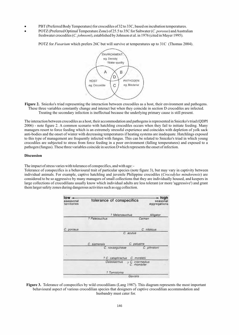
PBT (Preferred Body Temperature) for crocodiles of 32 to 33C, based on incubation temperatures. POTZ (Preferred Optimal Temperature Zone) of 25.5 to 35C for Saltwater (C. porosus) and Australian freshwater crocodiles (C. johnsoni), established by Johnson et al. in 1976 (cited in Mayer 1995).
POTZ for Fusarium which prefers 26C but will survive at temperatures up to 31C (Thomas 2004).
Figure 2. Sniezko's triad representing the interaction between crocodiles as a host, their environment and pathogens. These three variables constantly change and interact but when they coincide in section D crocodiles are infected.
Treating the secondary infection is ineffectual because the underlying primary cause is still present.
The interaction between crocodiles as a host, their accommodation and pathogens is represented in Sniezko's triad (QDPI 2006) - note figure 2. A common scenario with hatchling crocodiles occurs when they fail to initiate feeding. Many managers resort to force feeding which is an extremely stressful experience and coincides with depletion of yolk sack anti-bodies and the onset of winter with decreasing temperatures if heating systems are inadequate. Hatchlings exposed to this type of management are frequently infected with fungus. This can be related to Sniezko's triad in which young crocodiles are subjected to stress from force feeding in a poor environment (falling temperatures) and exposed to a pathogen (fungus). These three variables coincide in section D which represents the onset of infection.
Discussion
The impact of stress varies with tolerance of conspecifics, and with age: - Tolerance of conspecifics is a behavioural trait of particular species (note figure 3), but may vary in captivity between individual animals. For example, captive hatchling and juvenile Philippine crocodiles (Crocodylus mindorensis) are considered to be so aggressive by many managers of small collections that they are individually housed, and keepers in large collections of crocodilians usually know which individual adults are less tolerant (or more 'aggressive') and grant them larger safety zones during dangerous activities such as egg collection.
Figure 3. Tolerance of conspecifics by wild crocodilians (Lang 1987). This diagram represents the most important behavioural aspect of various crocodilian species that designers of captive crocodilian accommodation and
husbandry must cater for.
146
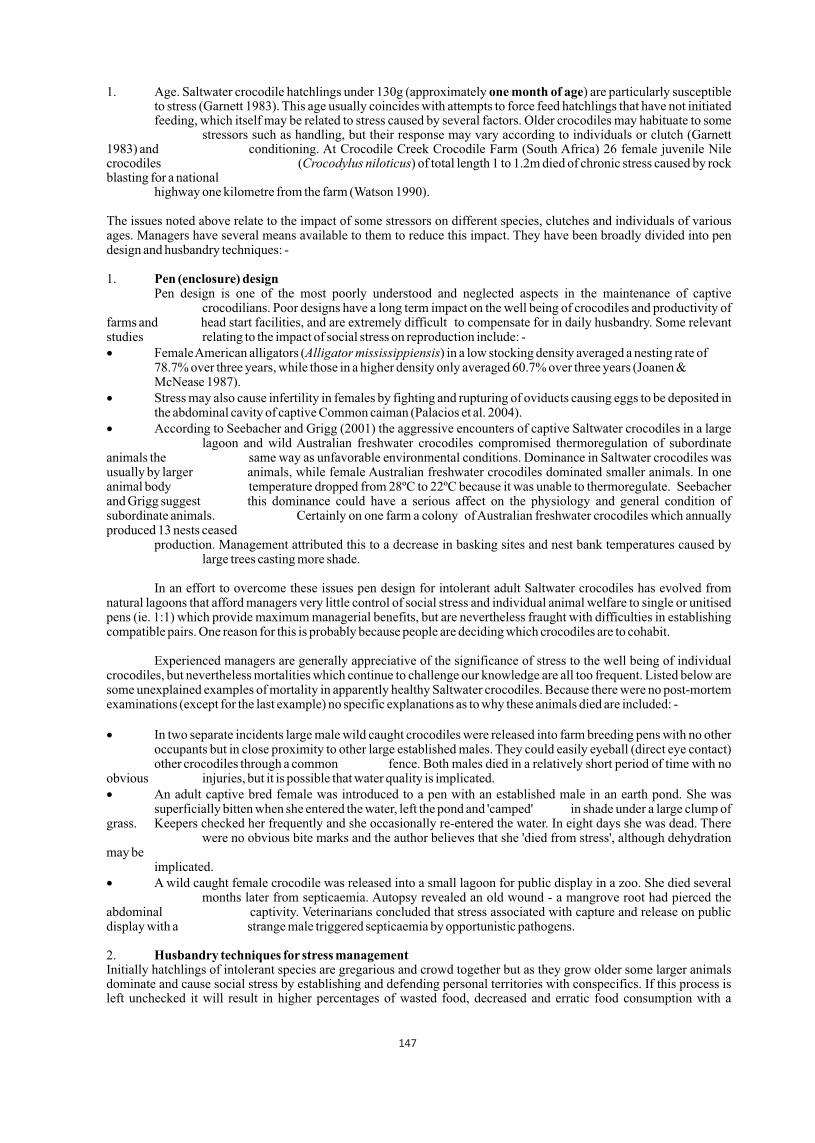
1. Age. Saltwater crocodile hatchlings under 130g (approximately one month of age) are particularly susceptible to stress (Garnett 1983). This age usually coincides with attempts to force feed hatchlings that have not initiated feeding, which itself may be related to stress caused by several factors. Older crocodiles may habituate to some stressors such as handling, but their response may vary according to individuals or clutch (Garnett 1983) and conditioning. At Crocodile Creek Crocodile Farm (South Africa) 26 female juvenile Nile crocodiles (Crocodylus niloticus) of total length 1 to 1.2m died of chronic stress caused by rock blasting for a national highway one kilometre from the farm (Watson 1990).
The issues noted above relate to the impact of some stressors on different species, clutches and individuals of various ages. Managers have several means available to them to reduce this impact. They have been broadly divided into pen design and husbandry techniques: -
1. Pen (enclosure) design Pen design is one of the most poorly understood and neglected aspects in the maintenance of captive crocodilians. Poor designs have a long term impact on the well being of crocodiles and productivity of farms and head start facilities, and are extremely difficult to compensate for in daily husbandry. Some relevant studies relating to the impact of social stress on reproduction include: - Female American alligators (Alligator mississippiensis) in a low stocking density averaged a nesting rate of 78.7% over three years, while those in a higher density only averaged 60.7% over three years (Joanen & McNease 1987). Stress may also cause infertility in females by fighting and rupturing of oviducts causing eggs to be deposited in the abdominal cavity of captive Common caiman (Palacios et al. 2004). According to Seebacher and Grigg (2001) the aggressive encounters of captive Saltwater crocodiles in a large lagoon and wild Australian freshwater crocodiles compromised thermoregulation of subordinate animals the same way as unfavorable environmental conditions. Dominance in Saltwater crocodiles was usually by larger animals, while female Australian freshwater crocodiles dominated smaller animals. In one animal body temperature dropped from 28ºC to 22ºC because it was unable to thermoregulate. Seebacher and Grigg suggest this dominance could have a serious affect on the physiology and general condition of subordinate animals. Certainly on one farm a colony of Australian freshwater crocodiles which annually produced 13 nests ceased production. Management attributed this to a decrease in basking sites and nest bank temperatures caused by large trees casting more shade.
In an effort to overcome these issues pen design for intolerant adult Saltwater crocodiles has evolved from natural lagoons that afford managers very little control of social stress and individual animal welfare to single or unitised pens (ie. 1:1) which provide maximum managerial benefits, but are nevertheless fraught with difficulties in establishing compatible pairs. One reason for this is probably because people are deciding which crocodiles are to cohabit.
Experienced managers are generally appreciative of the significance of stress to the well being of individual crocodiles, but nevertheless mortalities which continue to challenge our knowledge are all too frequent. Listed below are some unexplained examples of mortality in apparently healthy Saltwater crocodiles. Because there were no post-mortem examinations (except for the last example) no specific explanations as to why these animals died are included: -
In two separate incidents large male wild caught crocodiles were released into farm breeding pens with no other occupants but in close proximity to other large established males. They could easily eyeball (direct eye contact) other crocodiles through a common fence. Both males died in a relatively short period of time with no obvious injuries, but it is possible that water quality is implicated. An adult captive bred female was introduced to a pen with an established male in an earth pond. She was superficially bitten when she entered the water, left the pond and 'camped' in shade under a large clump of grass. Keepers checked her frequently and she occasionally re-entered the water. In eight days she was dead. There were no obvious bite marks and the author believes that she 'died from stress', although dehydration may be implicated. A wild caught female crocodile was released into a small lagoon for public display in a zoo. She died several months later from septicaemia. Autopsy revealed an old wound - a mangrove root had pierced the abdominal captivity. Veterinarians concluded that stress associated with capture and release on public display with a strange male triggered septicaemia by opportunistic pathogens.
2. Husbandry techniques for stress management Initially hatchlings of intolerant species are gregarious and crowd together but as they grow older some larger animals dominate and cause social stress by establishing and defending personal territories with conspecifics. If this process is left unchecked it will result in higher percentages of wasted food, decreased and erratic food consumption with a
147
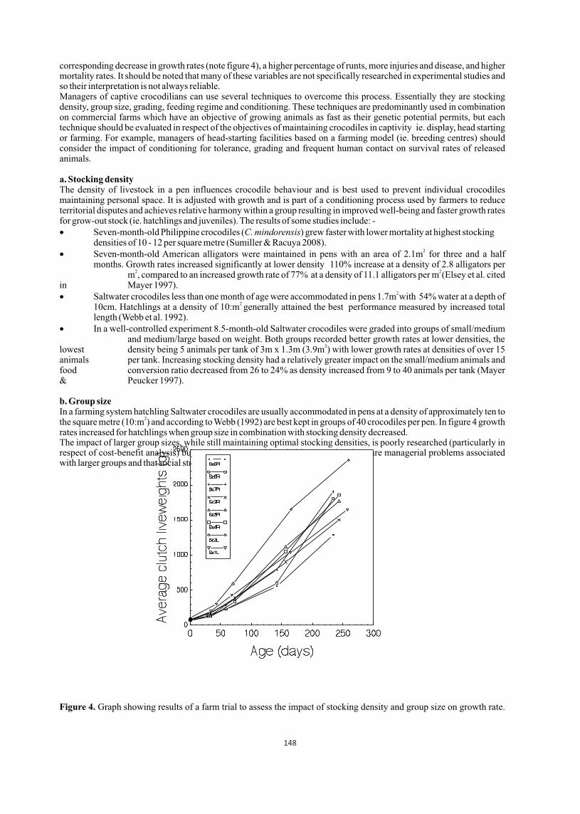
corresponding decrease in growth rates (note figure 4), a higher percentage of runts, more injuries and disease, and higher mortality rates. It should be noted that many of these variables are not specifically researched in experimental studies and so their interpretation is not always reliable.Managers of captive crocodilians can use several techniques to overcome this process. Essentially they are stocking density, group size, grading, feeding regime and conditioning. These techniques are predominantly used in combination on commercial farms which have an objective of growing animals as fast as their genetic potential permits, but each technique should be evaluated in respect of the objectives of maintaining crocodiles in captivity ie. display, head starting or farming. For example, managers of head-starting facilities based on a farming model (ie. breeding centres) should consider the impact of conditioning for tolerance, grading and frequent human contact on survival rates of released animals.
a. Stocking densityThe density of livestock in a pen influences crocodile behaviour and is best used to prevent individual crocodiles maintaining personal space. It is adjusted with growth and is part of a conditioning process used by farmers to reduce territorial disputes and achieves relative harmony within a group resulting in improved well-being and faster growth rates for grow-out stock (ie. hatchlings and juveniles). The results of some studies include: - Seven-month-old Philippine crocodiles (C. mindorensis) grew faster with lower mortality at highest stocking densities of 10 - 12 per square metre (Sumiller & Racuya 2008).
2 Seven-month-old American alligators were maintained in pens with an area of 2.1m for three and a half months. Growth rates increased significantly at lower density 110% increase at a density of 2.8 alligators per 2 2 m , compared to an increased growth rate of 77% at a density of 11.1 alligators per m (Elsey et al. cited
in Mayer 1997). 2 Saltwater crocodiles less than one month of age were accommodated in pens 1.7m with 54% water at a depth of
2 10cm. Hatchlings at a density of 10:m generally attained the best performance measured by increased total length (Webb et al. 1992). In a well-controlled experiment 8.5-month-old Saltwater crocodiles were graded into groups of small/medium and medium/large based on weight. Both groups recorded better growth rates at lower densities, the
2lowest density being 5 animals per tank of 3m x 1.3m (3.9m ) with lower growth rates at densities of over 15 animals per tank. Increasing stocking density had a relatively greater impact on the small/medium animals and
food conversion ratio decreased from 26 to 24% as density increased from 9 to 40 animals per tank (Mayer & Peucker 1997).
b. Group sizeIn a farming system hatchling Saltwater crocodiles are usually accommodated in pens at a density of approximately ten to
2the square metre (10:m ) and according to Webb (1992) are best kept in groups of 40 crocodiles per pen. In figure 4 growth rates increased for hatchlings when group size in combination with stocking density decreased.The impact of larger group sizes, while still maintaining optimal stocking densities, is poorly researched (particularly in respect of cost-benefit analysis) but most farmers would probably agree that there are managerial problems associated with larger groups and that social stress is an issue, particularly at feed time.
Figure 4. Graph showing results of a farm trial to assess the impact of stocking density and group size on growth rate.
148

Growth rate up to day 142 appears to be inhibited for clutches scute 5, 6 and 7 Right by the high stocking density of 116 animals to a pen with an area of 5m² (23:1m²) and then accelerates after 44 animals are removed, resulting in a stocking density for those remaining hatchlings (ie. clutches scute 5 and 6 Right) of 14:m², and to 8.8:m² for clutch scute 7 Right. c. Grading Grading is the removal of crocodiles from a particular pen with the objective of reducing dominance by size by separating small, medium and large animals into respective groups to maximise growth. Large hatchlings tend to grow well in graded or ungraded pens, but no grading results in greater size discrepancy within the group and associated stress with erratic feeding, lower growth rates of some animals, and higher percentages of runts and mortality. Some farmers grade all animals in a particular age cohort on a regular basis starting as early as six weeks of age, and managers of some head start facilities grade young animals to reduce physical injuries because they may impair the survival of individuals after release. However, one of the major causes of mortality in some released species is cannibalism, and grading to reduce the impact of dominance may impair the ability of released crocodiles to recognise larger conspecifics as a threat.
There are negative aspects to grading. It causes stress or even trauma during capture that will quite often increase susceptibility to infection or kill weaker crocodiles, particularly when associated with thermal stress in winter; and disrupts crocodiles accommodated in adjacent pens. It is possible that the whole hatchery (nursery) will go off normal feeding, and the growth of graded crocodiles will be compromised for up to three weeks while pecking orders are re-established in new accommodation. This can be interpreted as a loss of production. It should be noted that crocodiles in a hobbyist collection or research facility are often conditioned to frequent handling and are not as adversely affected by grading.
Stress associated with grading can be reduced by always leaving some water in the pond, or may consist of intermittently removing one or a few animals rather than a whole group. Removing one large dominant animal to reduce social stress is usually a temporary solution because another will soon take its place in the pecking order so consideration should be given to removing smaller animals first.
d. Feeding regimeA good feeding regime should involve a balanced diet fed out in sufficient quantities and frequency so all animals receive adequate food. In a healthy environment this will result in a continual rise in food consumption by grow-out stock. Conversely, insufficient frequency of feeding and small feed platforms will encourage more dominance and stress at feed time, and stress from overcrowding will cause erratic food consumption note figure 5. There is an obvious need for managers of head start programs to assess the relationship of captive feeding regimes to a released animal's ability to recognise and capture live prey.
e. Conditioning as part of daily activitiesConditioning is simple learning, and is integral to good husbandry. Essentially managers of captive crocodiles are conditioning their livestock to tolerate conspecifics by using a combination of the above techniques, but they also condition livestock to remain calm in the presence of keepers by working calmly and quietly. One of the wisest comments in relation to this subject comes from a sheep shearer whose daily quota may be up to 200 animals - if you want to be aggressive the sheep will retaliate, and at the end of the day the shearer (or keeper) will have suffered the most. It is generally accepted that animals intended for wild release should receive minimal human contact. One example of how this may be detrimental is an adult Philippine crocodile that had been maintained in captivity for many years. After release it frequently attacked farmer's poultry and had to be captured and returned to captivity (Willem van de Ven 2012, pers. com.).
149
Figure 5. Food consumption graph (developed by a farm keeper for hatchery tub number 3) used to monitor stress in eight-month-old Saltwater crocodiles. Erratic consumption is caused by hatchlings that have outgrown their
accommodation and need grading. Conclusion
Susceptibility to stress in captive crocodilians can vary between species, individuals and age; and its impact is related to severity and duration. It is often poorly understood and difficult to assess visually, but management to prevent stress is nonetheless extremely important as an animal welfare issue because of its effect on the well being of individual animals and its implications in the onset of infection which may impact on the wider group. It is thus incumbent upon managers of captive animals to use tools such as feed charts and daily observation by keepers to monitor the well being of individuals and groups.
Crocodiles are maintained in captivity for various reasons for display, farming or head starting. Each of these types of captivity has a unique system which should be determined by its own objectives. For example, farmers have developed a system to manage stress with an overall commercial objective of improving productivity based on the efficient production of a blemish free skin. They incorporate certain pen designs and several techniques researched on a species-specific basis - these are stocking density, grading, group size and conditioning.
Head starting has a different objective the efficient production of animals on a species specific basis that are suitable for release and will attain a high survival rate. Most importantly managers of head starting facilities, and particularly those structured as large breeding centres, need to assess those attributes necessary for the survival of released crocodiles on a species specific basis; and then design pens and adopt stress management techniques that will enhance the survivability of these animals.
Literature cited
01. Franklin, C.E., Davis, B.M., Peucker, S.K.J., Stephenson, H., Mayer, R., Whittier, J., Lever, J. & Grigg, G. 2003, Comparison of Stress Induced by Manual Restraint and Immobilisation in the Estuarine Crocodile, Crocodylus porosus, Journal of Experimental Zoology 298A:86-92.
02. Garnett, S. 1983, Nutrition and Farm Husbandry of the Green Sea Turtle and the Estuarine Crocodile, Ph.D. Thesis, James Cook University of North Queensland, Townsville.
03. Joanen, T. & McNease, L. 1987, Alligator Farming Research in Lousiana, USA, Wildlife Management, Crocodiles and Alligators, Surrey Beatty and Sons, Chipping Norton, NSW.
04. Lang, J.W. 1987, Crocodilian Behaviour: Implications for Management, Wildlife Management, Crocodiles and Alligators, Surrey Beatty and Sons, Chipping Norton, NSW.
05. Mayer, B. 1995, Temperature Trial on Two Month Old Juveniles, Crocodile Research and Development Bulletin, QDPI&F, Brisbane, volume 1, June, 1995.
06. Mayer, B. 1997, Pilot Light Trial, Crocodile Research and Development Bulletin, QDPI&F,Brisbane, volume 2, June, 1997.
07. Mayer, B. & Peucker, S. 1997, Effects of Rearing Juvenile Crocodiles at Different Densities, Crocodile Research and Development Bulletin, QDPI&F, Brisbane, volume 2, June, 1997.
08. Queensland Government Department of Primary Industries. 2006, Australian Prawn Farming Manual, Health Management for Profit. Ch3.p50. Retrieved May 2013 from:
http://aciar.gov.au/files/node/737/Australian%20prawnfarming%20manual%20final.pdf
09. Palacios, I.D. & Beltran, F.A. 2004, Ultrasound as a Tool to Evaluate Reproductive Structures of Female Caiman (Crocodilus fuscus) in th Closed Cycle Conditions, Crocodiles, Proceedings of the 17 Working Meeting of the Crocodile Specialist Group, Darwin.
10. Seebacher, F. & Grigg, G.C. 2001, Social Interactions Compromise Thermoregulation in Crocodiles Crocodylus johnstoni and Crocodylus porosus, Crocodilian Biology and Evolution eds G. C. Grigg, F. Seebacher and C.E. Franklin, Surrey Beatty and Sons, Sydney.
11. Sumiller, R.Q. & Racuya, F. 2008, Effects of Rearing Hatchling-Juvenile Crocodylus , 2008.mindorensis at Various Stocking Densities, Proceedings of Forum on Crocodiles in the Philippines, Manila.
12. Thomas, A. 2004, Observations on Bacterial and Fungal Contamination of Farmed Freshwater Crocodile Eggs, Crocodile Capers, QDPI&F, vol. 1, no. 9, pp 7-9.
th13. Watson, P.A.I. 1990, Effects of Blasting on Nile Crocodiles, Proceedings of the 10 Working Meeting of the Crocodile Specialist Group, Gainsville.
14. Webb, G. 1989, The Crocodile as a Production Unit, Proceedings of QDPI&F Intensive Tropical Animal Production Seminar, Townsville. 15. Webb, G.J.W., Manolis, S.C., Ottley, B. & Heyward, A. 1992, Crocodile Management and Research in the Northern Territory: 1990-92,
th Proceedings of the 11 Working Meeting of the CSG, Zimbabwe.
150
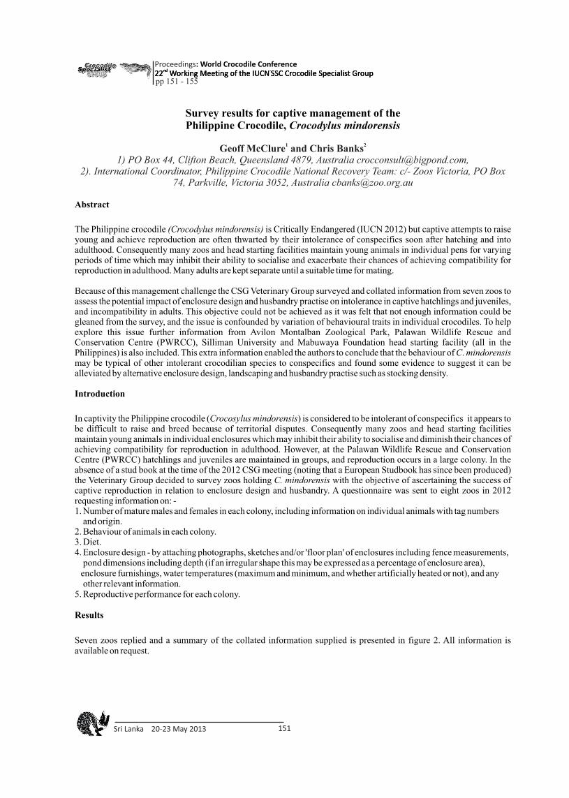
Survey results for captive management of the Philippine Crocodile, Crocodylus mindorensis
1 2 Geoff McClure and Chris Banks1) PO Box 44, Clifton Beach, Queensland 4879, Australia [email protected],
2). International Coordinator, Philippine Crocodile National Recovery Team: c/- Zoos Victoria, PO Box 74, Parkville, Victoria 3052, Australia [email protected]
Abstract
The Philippine crocodile (Crocodylus mindorensis) is Critically Endangered (IUCN 2012) but captive attempts to raise young and achieve reproduction are often thwarted by their intolerance of conspecifics soon after hatching and into adulthood. Consequently many zoos and head starting facilities maintain young animals in individual pens for varying periods of time which may inhibit their ability to socialise and exacerbate their chances of achieving compatibility for reproduction in adulthood. Many adults are kept separate until a suitable time for mating.
Because of this management challenge the CSG Veterinary Group surveyed and collated information from seven zoos to assess the potential impact of enclosure design and husbandry practise on intolerance in captive hatchlings and juveniles, and incompatibility in adults. This objective could not be achieved as it was felt that not enough information could be gleaned from the survey, and the issue is confounded by variation of behavioural traits in individual crocodiles. To help explore this issue further information from Avilon Montalban Zoological Park, Palawan Wildlife Rescue and Conservation Centre (PWRCC), Silliman University and Mabuwaya Foundation head starting facility (all in the Philippines) is also included. This extra information enabled the authors to conclude that the behaviour of C. mindorensis may be typical of other intolerant crocodilian species to conspecifics and found some evidence to suggest it can be alleviated by alternative enclosure design, landscaping and husbandry practise such as stocking density.
Introduction
In captivity the Philippine crocodile (Crocosylus mindorensis) is considered to be intolerant of conspecifics it appears to be difficult to raise and breed because of territorial disputes. Consequently many zoos and head starting facilities maintain young animals in individual enclosures which may inhibit their ability to socialise and diminish their chances of achieving compatibility for reproduction in adulthood. However, at the Palawan Wildlife Rescue and Conservation Centre (PWRCC) hatchlings and juveniles are maintained in groups, and reproduction occurs in a large colony. In the absence of a stud book at the time of the 2012 CSG meeting (noting that a European Studbook has since been produced) the Veterinary Group decided to survey zoos holding C. mindorensis with the objective of ascertaining the success of captive reproduction in relation to enclosure design and husbandry. A questionnaire was sent to eight zoos in 2012 requesting information on: -1. Number of mature males and females in each colony, including information on individual animals with tag numbers and origin.2. Behaviour of animals in each colony.3. Diet.4. Enclosure design - by attaching photographs, sketches and/or 'floor plan' of enclosures including fence measurements, pond dimensions including depth (if an irregular shape this may be expressed as a percentage of enclosure area), enclosure furnishings, water temperatures (maximum and minimum, and whether artificially heated or not), and any other relevant information.5. Reproductive performance for each colony.
Results
Seven zoos replied and a summary of the collated information supplied is presented in figure 2. All information is available on request.
Sri Lanka 20-23 May 2013 151
Proceedings : World Crocodile Conferencend -22 Working Meeting of the IUCN SSC Crocodile Specialist Groupnd -22 Working Meeting of the IUCN SSC Crocodile Specialist Groupnd -22 Working Meeting of the IUCN SSC Crocodile Specialist Group
pp 151 - 155
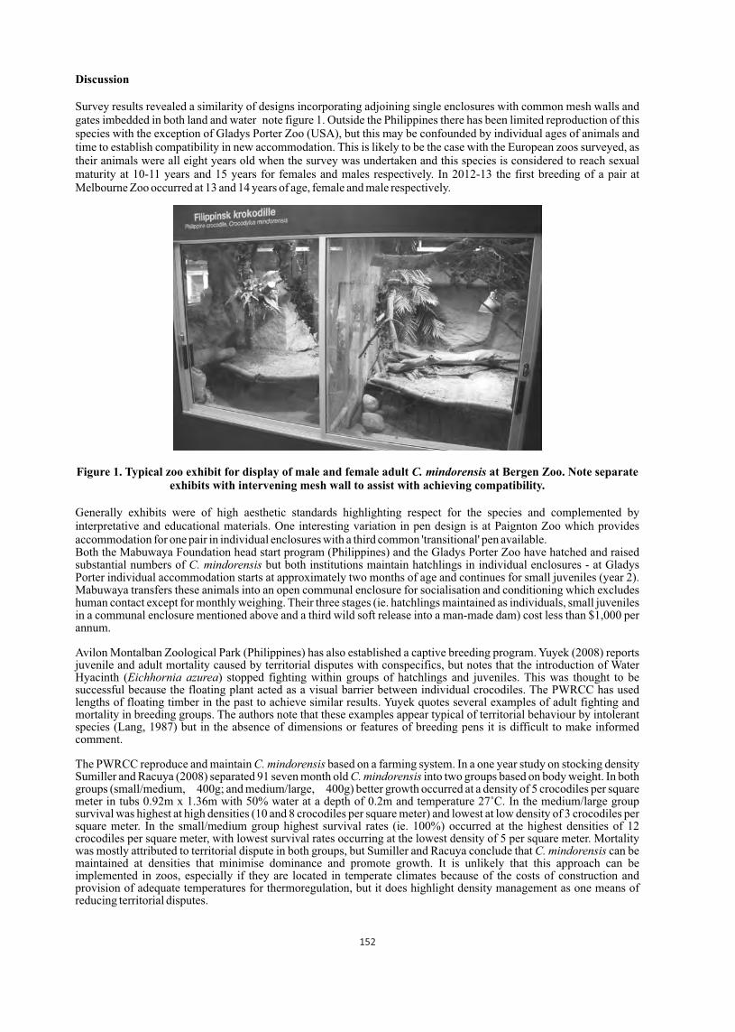
Discussion
Survey results revealed a similarity of designs incorporating adjoining single enclosures with common mesh walls and gates imbedded in both land and water note figure 1. Outside the Philippines there has been limited reproduction of this species with the exception of Gladys Porter Zoo (USA), but this may be confounded by individual ages of animals and time to establish compatibility in new accommodation. This is likely to be the case with the European zoos surveyed, as their animals were all eight years old when the survey was undertaken and this species is considered to reach sexual maturity at 10-11 years and 15 years for females and males respectively. In 2012-13 the first breeding of a pair at Melbourne Zoo occurred at 13 and 14 years of age, female and male respectively.
Figure 1. Typical zoo exhibit for display of male and female adult C. mindorensis at Bergen Zoo. Note separate exhibits with intervening mesh wall to assist with achieving compatibility.
Generally exhibits were of high aesthetic standards highlighting respect for the species and complemented by interpretative and educational materials. One interesting variation in pen design is at Paignton Zoo which provides accommodation for one pair in individual enclosures with a third common 'transitional' pen available.Both the Mabuwaya Foundation head start program (Philippines) and the Gladys Porter Zoo have hatched and raised substantial numbers of C. mindorensis but both institutions maintain hatchlings in individual enclosures - at Gladys Porter individual accommodation starts at approximately two months of age and continues for small juveniles (year 2). Mabuwaya transfers these animals into an open communal enclosure for socialisation and conditioning which excludes human contact except for monthly weighing. Their three stages (ie. hatchlings maintained as individuals, small juveniles in a communal enclosure mentioned above and a third wild soft release into a man-made dam) cost less than $1,000 per annum.
Avilon Montalban Zoological Park (Philippines) has also established a captive breeding program. Yuyek (2008) reports juvenile and adult mortality caused by territorial disputes with conspecifics, but notes that the introduction of Water Hyacinth (Eichhornia azurea) stopped fighting within groups of hatchlings and juveniles. This was thought to be successful because the floating plant acted as a visual barrier between individual crocodiles. The PWRCC has used lengths of floating timber in the past to achieve similar results. Yuyek quotes several examples of adult fighting and mortality in breeding groups. The authors note that these examples appear typical of territorial behaviour by intolerant species (Lang, 1987) but in the absence of dimensions or features of breeding pens it is difficult to make informed comment.
The PWRCC reproduce and maintain C. mindorensis based on a farming system. In a one year study on stocking density Sumiller and Racuya (2008) separated 91 seven month old C. mindorensis into two groups based on body weight. In both groups (small/medium, 400g; and medium/large, 400g) better growth occurred at a density of 5 crocodiles per square meter in tubs 0.92m x 1.36m with 50% water at a depth of 0.2m and temperature 27˚C. In the medium/large group survival was highest at high densities (10 and 8 crocodiles per square meter) and lowest at low density of 3 crocodiles per square meter. In the small/medium group highest survival rates (ie. 100%) occurred at the highest densities of 12 crocodiles per square meter, with lowest survival rates occurring at the lowest density of 5 per square meter. Mortality was mostly attributed to territorial dispute in both groups, but Sumiller and Racuya conclude that C. mindorensis can be maintained at densities that minimise dominance and promote growth. It is unlikely that this approach can be implemented in zoos, especially if they are located in temperate climates because of the costs of construction and provision of adequate temperatures for thermoregulation, but it does highlight density management as one means of reducing territorial disputes.
152
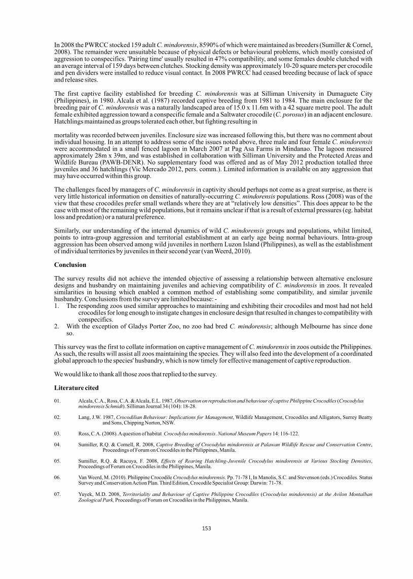
In 2008 the PWRCC stocked 159 adult C. mindorensis, 8590% of which were maintained as breeders (Sumiller & Cornel, 2008). The remainder were unsuitable because of physical defects or behavioural problems, which mostly consisted of aggression to conspecifics. 'Pairing time' usually resulted in 47% compatibility, and some females double clutched with an average interval of 159 days between clutches. Stocking density was approximately 10-20 square meters per crocodile and pen dividers were installed to reduce visual contact. In 2008 PWRCC had ceased breeding because of lack of space and release sites.
The first captive facility established for breeding C. mindorensis was at Silliman University in Dumaguete City (Philippines), in 1980. Alcala et al. (1987) recorded captive breeding from 1981 to 1984. The main enclosure for the breeding pair of C. mindorensis was a naturally landscaped area of 15.0 x 11.6m with a 42 square metre pool. The adult female exhibited aggression toward a conspecific female and a Saltwater crocodile (C. porosus) in an adjacent enclosure. Hatchlings maintained as groups tolerated each other, but fighting resulting in
mortality was recorded between juveniles. Enclosure size was increased following this, but there was no comment about individual housing. In an attempt to address some of the issues noted above, three male and four female C. mindorensis were accommodated in a small fenced lagoon in March 2007 at Pag Asa Farms in Mindanao. The lagoon measured approximately 28m x 39m, and was established in collaboration with Silliman University and the Protected Areas and Wildlife Bureau (PAWB-DENR). No supplementary food was offered and as of May 2012 production totalled three juveniles and 36 hatchlings (Vic Mercado 2012, pers. comm.). Limited information is available on any aggression that may have occurred within this group.
The challenges faced by managers of C. mindorensis in captivity should perhaps not come as a great surprise, as there is very little historical information on densities of naturally-occurring C. mindorensis populations. Ross (2008) was of the view that these crocodiles prefer small wetlands where they are at “relatively low densities”. This does appear to be the case with most of the remaining wild populations, but it remains unclear if that is a result of external pressures (eg. habitat loss and predation) or a natural preference.
Similarly, our understanding of the internal dynamics of wild C. mindorensis groups and populations, whilst limited, points to intra-group aggression and territorial establishment at an early age being normal behaviours. Intra-group aggression has been observed among wild juveniles in northern Luzon Island (Philippines), as well as the establishment of individual territories by juveniles in their second year (van Weerd, 2010).
Conclusion
The survey results did not achieve the intended objective of assessing a relationship between alternative enclosure designs and husbandry on maintaining juveniles and achieving compatibility of C. mindorensis in zoos. It revealed similarities in housing which enabled a common method of establishing some compatibility, and similar juvenile husbandry. Conclusions from the survey are limited because: -1. The responding zoos used similar approaches to maintaining and exhibiting their crocodiles and most had not held crocodiles for long enough to instigate changes in enclosure design that resulted in changes to compatibility with conspecifics. 2. With the exception of Gladys Porter Zoo, no zoo had bred C. mindorensis; although Melbourne has since done so.
This survey was the first to collate information on captive management of C. mindorensis in zoos outside the Philippines. As such, the results will assist all zoos maintaining the species. They will also feed into the development of a coordinated global approach to the species' husbandry, which is now timely for effective management of captive reproduction.
We would like to thank all those zoos that replied to the survey.
Literature cited
01. Alcala, C.A., Ross, C.A. & Alcala, E.L. 1987, Observation on reproduction and behaviour of captive Philippine Crocodiles (Crocodylus mindorensis Schmidt). Silliman Journal 34 (104): 18-28.
02. Lang, J.W. 1987, Crocodilian Behaviour: Implications for Management, Wildlife Management, Crocodiles and Alligators, Surrey Beatty and Sons, Chipping Norton, NSW.
03. Ross, C.A. (2008). A question of habitat Crocodylus mindorensis. National Museum Papers 14: 116-122.
04. Sumiller, R.Q. & Cornell, R. 2008, Captive Breeding of Crocodylus mindorensis at Palawan Wildlife Rescue and Conservation Centre, Proceedings of Forum on Crocodiles in the Philippines, Manila.
05. Sumiller, R.Q. & Racuya, F. 2008, Effects of Rearing Hatchling-Juvenile Crocodylus mindorensis at Various Stocking Densities, Proceedings of Forum on Crocodiles in the Philippines, Manila.
06. Van Weerd, M. (2010). Philippine Crocodile Crocodylus mindorensis. Pp. 71-78 I, In Manolis, S.C. and Stevenson (eds.) Crocodiles. Status Survey and Conservation Action Plan. Third Edition, Crocodile Specialist Group: Darwin: 71-78.
07. Yuyek, M.D. 2008, Territoriality and Behaviour of Captive Philippine Crocodiles (Crocodylus mindorensis) at the Avilon Montalban Zoological Park, Proceedings of Forum on Crocodiles in the Philippines, Manila.
153
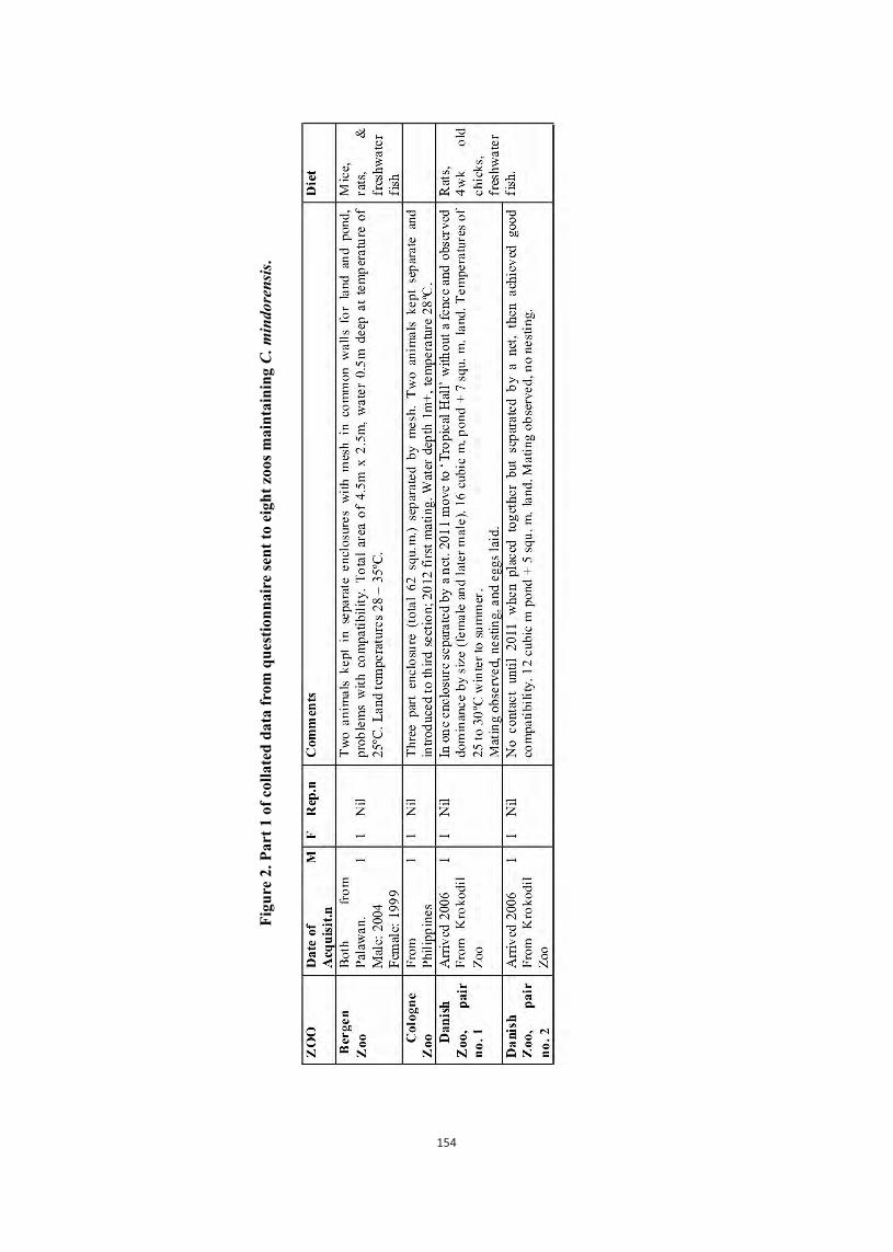
Fig
ure
2. P
art
1 of
col
late
d d
ata
from
qu
esti
onn
aire
sen
t to
eig
ht
zoos
mai
nta
inin
g C
. min
dore
nsi
s.
154
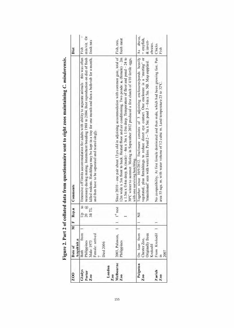
Fig
ure
2. P
art
2 of
col
late
d d
ata
from
qu
esti
onn
aire
sen
t to
eig
ht
zoos
main
tain
ing C
. m
indore
nsi
s.
155
An introduction to reproductive study of the mugger crocodile(Crocodylus palustris) in Iran
Asghar Mobaraki and Elham AbtinDepartment of Environment, Biodiversity and Wildlife Bureau, PO.Box 14155-7383,Tehran, Iran
([email protected]) | Department of Environment office in Sistan and Baluchestan Province, PO.Box 477, Zahedan, Iran ([email protected])
A small population of Mugger crocodiles is distributed in the southeastern part of Iran, in Sistan & Baluchestan Province, the westernmost global range of the species. The nesting season for Mugger crocodiles in Iran has been reported to be in May and consequently, the eggs hatch in July; but based on the observations made in the surveys it seems that the nesting season could be extended. From four nests found in the wild, 24, 26, 21 and 13 eggs were counted. A 2.2 m female Mugger, who had fallen into the Pishin Dam overflow pond, laid 21 eggs exposed to air on the concrete. The female crocodile was removed and relocated to a captive situation in Dargas where she laid 18 and 25 eggs in two consecutive years. The mean size for the eggs in the last three nests was calculated 75.66 mm in length, 47.72 mm in width and 91.27 g in weight. The main habitats with adult crocodiles were visited in the early hatching season to catch the hatchlings using the "spot light" method. During the studies 19 hatchlings from 7 different nests were captured from four main sites. The mean total length and weight for 19 captured hatchlings was 30.47 cm (range 29.24 to 33.90 cm) and 84.29 g (range 66.19 to 90.30 g) respectively Based on the observed and counted hatchlings in each site and comparing the clutch size, it seems that hatchlings have a high mortality rate in the early stages after hatching due to natural threats. Guarding and defense of the nests and hatchlings was evident in most studied cases. anon one occasion, a hatchling was found in a burrow, which the authors believe indicates parental care or intention of using burrows by hatchlings as a refuge in harsh situations.
Hematology and biochemistry of juvenile and yearling gharials
Gharials (Gavialis gangeticus) belong to schedule I of Indian Wildlife Protection Act (1972) and are critically endangered (IUCN, 2009). But, there are critical gaps in database for normal health parameters for clinically evaluating the health status of gharial. During the study period at, Gharial Rearing Centre, National Chambal Sanctuary, Dewari, Morena, Madhya Pradesh gharials were physically restrained for detailed examination and collection of different biological samples. A concise systematic schedule was prepared for physical restraining and blood samples were collected aseptically from 8 restrained juvenile and 9 yearling gharials to analyze hemato-biochemical parameters and to establish a reference range for various parameters. The mean values of TEC (millions/µl), TLC (thousands/µl), Hb (g/dl), PCV (%), MCV(fl), MCH (pg), MCHC (g/dl), Heterophils (%), Eosinophils (%), Basophils (%), Lymphocytes (%) and Monocytes (%) for juveniles and yearlings are 0.72, 12.13, 6.65, 22.88, 319.96, 92.87, 28.77, 27.88, 5.50, 3.88, 52.63 and 11.50 and 1.18, 21.78, 7.96, 25.33, 244.00, 70.16, 31.57, 31.11, 8.44, 6.56, 43.00 and 10.67 respectively. The mean values of AST (IU/L), ALT (IU/L), ALP (IU/L), Total Protein (g/dl), Albumin (g/dl), Total Bilirubin (mg/dl), Direct Bilirubin (mg/dl), Uric acid (mg/dl), Creatinine (mg/dl), Calcium (mg/dl), Phosphorous (mg/dl), Sodium (mmol/L), Chloride (mmol/L) and Potassium (mmol/L) for juvenile and yearlings are 43.25, 74.35,43.25, 4.95, 1.93, 1.47, 0.64, 4.14, 0.55, 6.79, 2.20, 159.66, 6.67 and 104.74 and 55.73, 77.00, 64.00, 4.52, 1.91, 1.02, 0.59, 4.13, 0.53, 10.39, 3.29, 150.75, 5.77 and 101.58 respectively. Comparison of the values were made between juvenile and yearling gharials and also to the known values of crocodilians.
1 1 Avadh Bihari Shrivastav and Himanshu R. Joshi
1 Centre for Wildlife Forensic and Health, Jabalpur, Madhya Pradesh, India | 1 Nanaji Deshmukh Veterinary Science University, Jabalpur - 482001, Madhya Pradesh, India
Proceedings : World Crocodile Conferencend -22 Working Meeting of the IUCN SSC Crocodile Specialist Groupnd -22 Working Meeting of the IUCN SSC Crocodile Specialist Groupnd -22 Working Meeting of the IUCN SSC Crocodile Specialist Group
Sri Lanka 20-23 May 2013 156
p 156
Host-Parasite interactions of Osteolaemus tetraspis, the African Dwarf Crocodile, and its gastric parasites
1 2 1 1Marisa Tellez *, Matt Shirley , Naada Azeemuddin and Anum Qureshi1 UCLA, Department of Ecology and Evolutionary Biology, 612 Charles E. Young Drive East, Los Angeles,
2 CA, USA, 90095-7246 | University of Florida, Department of Wildlife Ecology and Conservation, 110
Newins-Ziegler Hall, Gainesville, FL 32611-0430 |* [email protected]
Knowledge of the trophic position of O. tetraspis in the local foodweb network can assist in conservation management, particularly if environmental perturbations negatively affect food web connectivity. Additionally, parasites can be utilized as bioindicators to identify the strength of foodweb topology interactions, and to monitor environmental alterations. Nematode parasites were collected from Osteolaemus tetraspis from the Abanda cave system off the Fernan Vaz Lagoon in Gabon. A total of 95 helminths were collected from 11 infected hosts (50%) of the 22 crocodiles examined. Parasite prevalence, intensity, and abundance were analyzed to evaluate variation between size, location (inside or outside of cave), and sex of host crocodiles. No significant difference was found between the prevalence of infection inside and outside the cave system (W=280.5, P =0.3586), between sex (W=277.5, P =0.3891), or between size (W= 288.5, P=0.2703). However, sex significantly differed in mean parasite abundance (Bootstrap t-test, p=0.039), and size was significantly different in median intensity (Mood's median, p=0.01). Location and size were also shown not to affect parasite intensity (MANOVA, p=0.644). Parasitism among O. tetraspis was found to be homogenous and not aggregated among sub-categories of size, sex, and location (Total nematode overdispersion coefficient: 6.88619e+16). This is an interesting finding as aggregation is predicted among a population of hosts, i.e, there should be a significant difference among hosts that are infected and not infected.
Based on diet data of crocodile specimens from this study, and previous documentation of infections of Dujardinascaris sp. infections in other reptiles in the region, fish are hypothesized to be the intermediate hosts for this crocodilian nematode. These data provide insight of preferred prey, and the role of O. tetraspis in the food web.
Genetic analysis on these nematodes in correlation with genetic data of the host O. tetraspis will be discussed. In brief, by comparing genetic data of parasites and crocodiles (hosts), we can further clarify questions on crocodilian dispersal and population structure which can have implications for conservation. This concept can be expanded for a comparative phylogeographic study that includes specimens of Dujardinascaris sp. collected from every crocodilian species. An upcoming study on nematode crocodile coevolution will be discussed.
Sri Lanka 20-23 May 2013 157
Proceedings : World Crocodile Conferencend -22 Working Meeting of the IUCN SSC Crocodile Specialist Groupnd -22 Working Meeting of the IUCN SSC Crocodile Specialist Groupnd -22 Working Meeting of the IUCN SSC Crocodile Specialist Group
p 157

13 15Stable isotope ratios (δ C and δ N) predict gastric parasite dynamics in the American
Alligator (Alligator mississippiensis)
1 2Marisa Tellez * and James Nifong1 UCLA, Department of Ecology and Evolutionary Biology, 612 Charles E. Young Drive East, Los Angeles,
2 CA, USA, 90095-7246 | University of Florida, Department of Biology, Carr Hall 409, PO Box 118525,
Gainesville, FL, USA, 32611-0430 |* [email protected]
In this study, we tested various hypotheses regarding patterns of gastric nematode parasitism in Alligator mississippiensis 13 15from estaurine and inland freshwater habitats using measurements of stable isotope ratios (δ C and δ N) from epidermal
keratin and stomach contents. Alligators inhabit a wide geographic range in the southeastern US in which prey availability and abundance vary, influencing the hosts' parasite infracommunity. Interestingly enough, American Alligators have similar parasite infracommunities across their distribution. It is possible that the range of alligator parasite intermediate hosts is sympatric with the reptilian definite host, distinguishing the preferred prey of alligators as a result of a long co-evolutionary history between predator, prey, and parasite. Although stomach content analysis provides insight on alligator diet in the short-term, it provides only a limited account of the full spectrum of the alligator trophic interactions. Stable isotope ratios, however, provide insights into long-term trophic interactions, as turnover rates for keratinized epidermis is ~ 2 years. Additionally, parasites that are trophically transmitted provide evidence of prey consumed over an extensive period of time, providing a more accurate picture of the predator-prey dynamics.
Parasite intensity and species richness was significantly different between estuarine and inland freshwater populations 13 15(both P<0.01). A MANOVA performed on the combined data of δ C, δ N, and location significantly predicted parasite
15intensity (P=0.01178), the most significant predictors being δ N and location. Pearson correlation test showed that 15 15parasite intensity increased as δ N increased (P=0.03933). These data suggests variation of habitat and levels of δ N can
assist in predicting alligator parasite intensity. This information can assist in identifying alligator parasite intermediate hosts.
Proceedings : World Crocodile Conferencend -22 Working Meeting of the IUCN SSC Crocodile Specialist Groupnd -22 Working Meeting of the IUCN SSC Crocodile Specialist Groupnd -22 Working Meeting of the IUCN SSC Crocodile Specialist Group
Sri Lanka 20-23 May 2013 158
p 158

Necropsy report of the largest Indo-pacific Crocodile, "Lolong', in captivity at Bunawan, Agusan Del Sur, Philippines
1Stephen Toledo , 2 3 4 5Emilia Lastica , Ma. Theresa Aquino , Glenn Rebong and Rainier Manalo1). d Area and Wildlife Bureau (PAWB) Department of Environment and Natural [email protected] 2). College of Veterinary Medicine University of the Philippines Los Baños
[email protected] 3). Marine Wildlife Watch of the Philippines (MWWP) [email protected] 4). Palawan Wildlife Rescue and Conservation Center (PWRCC)
5). CrocodylusPorosus Philippines, Inc. (CPPI) [email protected]
Abstract
“Lolong”, a 6.11 meterlong Crocodylusporosus, was caught in Bunawan, Agusan del Sur, Philippines on 3 September 2011 and kept in captivity until his death on 10 February 2013. Necropsy and histopathology revealed that the cause of death was congestive heart failure complicated by late stage mycoticpneumonia and multiple organ failure. Other lesions also indicated problems in management and husbandry. Liver and kidney samples also indicated high levels of mercury at 55 and 18 ppm, respectively.
The high levels of mercury found in the liver and kidney suggested that contamination may have been present even prior to captivity. As such, Lolong's condition indicated that the Agusan Marsh is already contaminated and thus compromised as a habitat. It was strongly recommended that water quality testing be conducted throughout the marsh and steps be taken to identify the source, stop and, if possible, remove the contamination especially given its public health significance.
Introduction
1 Consultant, Crocodylus Porosus Philippines, Inc. (CPPI)A 6.11 meterlong Indo-Pacific crocodile, christened “Lolong”, was caught from Magsagansang Creek, a tributary of Agusan Marsh in Nueva Era, Bunawan, Agusan del Sur, Philippines on 3 September 2011. The animal was kept in an8 x 10 meter pen with a pond at the center about 1.5 meters deep in Bunawan. It started feeding a month after its capture and was thereafter fedabout 10 kgs of pork alternated by chicken meat every 10-20 daysusually between 7 to 11 pm. White spotsperceived as “rope pressure” marks were noted on the left front limb, tail base, and on tail end and were treated with povidone iodine (Sumiller, 2012a).Three months later, the white spots, which were eventually suspected to be fungal infection,were said to have been controlled with a combination of guava leaves extract, povidone iodine, and anti-fungal ointment (Sumiller, 2012b).
Case HistoryOn 9 February 2013, the crocodile was observed floating in the pond, listing to its right. The on-call veterinarian visited the following day and noted a slight distension in the right chest area. He further noted that it was less active. Although it could not put weight on its legs, tail movement was observed and the animal growled when approached. Body temperature registered at 22°C. Multivitamins (20 mL) were given intramuscularly with no signs of improvement. The condition of the animal deteriorated further as the day progressed. The crocodile ceased to breathe and respond to any stimuli and was pronounced deadat 8:12 pm of 10 February 2013 (Collantes, 2013).
It should probably be noted that the animal supposedly regurgitated food a week prior to its death (Lastica and Masangkay, 2013). Apart from this matter being part of the clinical history on the histopathology report, there was no mention of the incident in any other records related to the crocodile. This was only verbally imparted by the caretaker and was never recorded in either the monitoring or clinical report prior to death.
Methods
Necropsy was conducted on 11 February 2013 from 1:50 to 7 am. Measurements were taken and the animal was examined for external lesions. Swabs were taken from the nares, ear, and cloaca for bacterial and fungal isolationand these were sent to the municipal hospital (results not received as of writing). The crocodile was then placed on its back
Sri Lanka 20-23 May 2013 159
Proceedings : World Crocodile Conferencend -22 Working Meeting of the IUCN SSC Crocodile Specialist Groupnd -22 Working Meeting of the IUCN SSC Crocodile Specialist Groupnd -22 Working Meeting of the IUCN SSC Crocodile Specialist Group
pp 159 - 163

Figure 2.Broken lines show the path taken by the long
trachea. Solid arrow points to distended pleural lining.
and the skin deflected laterally to expose the ventral musculature. The ventral wall of the body was removed to expose the internal organs by cutting the soft tissues from the mandibular symphysis going lateral and caudal towards the cloaca while both clavicles were detached at the shoulder joints.
The organs were first observed in situ before the respiratory tract and heart were removed and examined separately from the rest of the internal organs. Tissue samples were collected for histopathologic and toxicologic examination. The skin and skull were then removed from the body and preserved for taxidermy at a later date. The bones were buried with muscles attached for future retrieval. Because of plans for taxidermy, the brain, brain stem,and spinal cord were not examined.
Results and Discussion
Necropsy FindingsExternal Examination: Old scars and ulcerations were observed on the palmar and plantar surfaces of the appendages(Figure 1). These lesions suggested constant, chronic pressure on the said surfaces. Other similar crocodilian cases in the past have proven fatal because of the ensuing systemic infection (Ross, pers. comm.)Sides of exposed teeth were apparently scraped and a 3.9 cm-long bruise on the left jaw was also observed. Clear, stringy mucus fluidconstantly dripped out of the nostrils when the animal was placed on its back. The right side of the torso appeared distended.
Respiratory Tract: The lungs were adhered to the liver, diaphragm, and pericardial sac. White mucoid exudate partially occluded the tracheal opening. The trachea measured over1 meter long and veered to the left side of the chest before looping up towards the right side then down on the same side and finally towards the midline of the body before bifurcating into the bronchi (Figure 2). Tracheal rings near the folded portions were larger than the rest,apparentlyan adaptation to the bends which suggests a development occurred over several years at least. According to Dr. Perran Ross (pers. comm.), extremely long tracheas are often seen in exceptionally large crocodiles, likely to accommodate the downward pull of the lungs and diaphragm when inhaling. The fold on the right bronchi, however, was collapsed and folded inward. Trapped clear gas presumed to be air distended the pleural lining on the right side of the thoracic cavity. About 5 milliliters of yellow-brown liquid pooled at the bottom of the right side of the chest cavity. The right lung lobe exhibited a large bruised area on its dorsal aspect.These lesions alluded to a rupture in the right lung lobe. Both lung lobes were large, occupying a little over two thirds of the whole body cavity.
Petecchial and paintbrush hemorrhages were scattered throughout the length of the trachea. The white mucoidexudate extended to the bronchioles and lung tissue in increasing concentrations. Crepitation (crackling) was felt interspersed with hard masses all over the lung surface. Upon incision, the lung tissue revealedyellow, caseous nodulesscattered over dark red and almost solid (red hepatization) lung parenchyma. These nodules varied in size and solidity (Figure 3) and signified decay of dead tissues. Some nodules coalesced in several parts of the two lobes to form larger solid masses.
Heart: The pericardial sac was thick and almost opaque. Blood-tinged fluid was observed within the sac upon incision. White granules adhered to the sac and muscle surfaces while coronary vessels were severely injected (Figure 4).The atria appeared grayish externally. Heart muscles showed pale areas in between darker ones.About 15 mL of bloody fluid filled the right ventricle. Chicken fat clot was also present, extending to the right atrium. The right atrium was also severely enlarged (about five times the size of the left), suggestive ofvery poor circulation to the lungs over a prolonged period prior to death. The left ventricle was thick, very muscular and filled with blood. Clotted blood filled the opening to the aorta.
Liver: Both left and right lobes of the liver were pale. The left lobe had rounded posterior edges with bruising on its lateral margin. Cut surface apposed well enough suggesting that the edema, if present at all, was minimal. It also revealed concentric pinhead pale areas proximal to the diaphragm. All the edges of the right lobe were rounded. Cut surface, again, apposed satisfactorily but showed blackish-brown parenchyma with pale areas near the surface (Figure 5).
Kidneys: Both kidneys had a gelatinous consistency with loss of normal architecture resulting in grossly indistinct lobulation. The left kidney was slightly larger than the right.
Fat body: Isolating the fat body proved a bit difficult due to adhesions with the mesentery. The fat body was relatively
Figure 1. Scarring on plantar and palmar surfaceswere noted.
Figure 3. Incised surface of the lung lobe showed red hepatized
parenchyma stippled with caseous nodules.
160
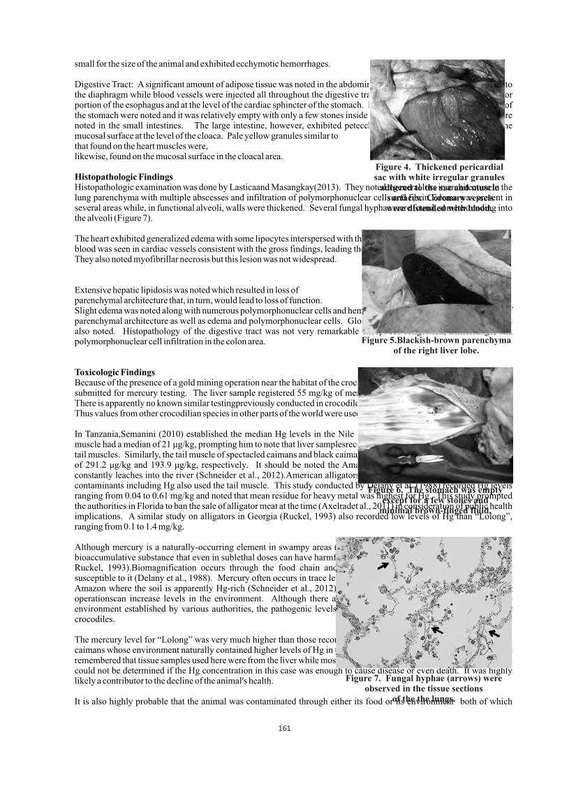
small for the size of the animal and exhibited ecchymotic hemorrhages.
Digestive Tract: A significant amount of adipose tissue was noted in the abdominal area. The mesentery was adhered to the diaphragm while blood vessels were injected all throughout the digestive tract. There was bruising on the anterior portion of the esophagus and at the level of the cardiac sphincter of the stomach. Hemorrhages on the mucosal surface of the stomach were noted and it was relatively empty with only a few stones inside (Figure 6). No significant lesions were noted in the small intestines. The large intestine, however, exhibited petecchial to paintbrush hemorrhages on the mucosal surface at the level of the cloaca. Pale yellow granules similar to that found on the heart muscles were, likewise, found on the mucosal surface in the cloacal area.
Histopathologic FindingsHistopathologic examination was done by Lasticaand Masangkay(2013). They noted a general loss in architecture in the lung parenchyma with multiple abscesses and infiltration of polymorphonuclear cells and fibrin.Edema was present in several areas while, in functional alveoli, walls were thickened. Several fungal hyphae were found, some extending into the alveoli (Figure 7).
The heart exhibited generalized edema with some lipocytes interspersed with the musculature. Coagulated and separated blood was seen in cardiac vessels consistent with the gross findings, leading them to conclude that death was protracted. They also noted myofibrillar necrosis but this lesion was not widespread.
Extensive hepatic lipidosis was noted which resulted in loss of parenchymal architecture that, in turn, would lead to loss of function. Slight edema was noted along with numerous polymorphonuclear cells and hemosiderin. The kidney also showed loss of parenchymal architecture as well as edema and polymorphonuclear cells. Glomerular and marked tubular necrosis was also noted. Histopathology of the digestive tract was not very remarkable except for congestion, hemorrhage, and polymorphonuclear cell infiltration in the colon area.
Toxicologic FindingsBecause of the presence of a gold mining operation near the habitat of the crocodile, liver and kidney tissue samples were submitted for mercury testing. The liver sample registered 55 mg/kg of mercury content and the kidney at 18 mg/kg. There is apparently no known similar testingpreviously conducted in crocodilesin the country to compare these results to. Thus values from other crocodilian species in other parts of the world were used as basis for comparison.
In Tanzania,Semanini (2010) established the median Hg levels in the Nile crocodile liver at 146 μg/kg while its tail muscle had a median of 21 μg/kg, prompting him to note that liver samplesrecorded higher concentrations of Hg than the tail muscles. Similarly, the tail muscle of spectacled caimans and black caimans in the Amazon recorded mean Hg levels of 291.2 μg/kg and 193.9 μg/kg, respectively. It should be noted the Amazon soil is also naturally rich in Hg that constantly leaches into the river (Schneider et al., 2012).American alligators in Florida tested in1988 for heavy metal contaminants including Hg also used the tail muscle. This study conducted by Delany et al. (1988) recorded Hg levels ranging from 0.04 to 0.61 mg/kg and noted that mean residue for heavy metal was highest for Hg. This study prompted the authorities in Florida to ban the sale of alligator meat at the time (Axelradet al., 2011) in consideration of public health implications. A similar study on alligators in Georgia (Ruckel, 1993) also recorded low levels of Hg than “Lolong”, ranging from 0.1 to 1.4 mg/kg.
Although mercury is a naturally-occurring element in swampy areas (Ruckel, 1993), it is also recognized as a toxic, bioaccumulative substance that even in sublethal doses can have harmful effects to animals imbibing it (Eisler, 1987 in Ruckel, 1993).Biomagnification occurs through the food chain and top predators such as crocodiles are highly susceptible to it (Delany et al., 1988). Mercury often occurs in trace levels in the environment except in places like the Amazon where the soil is apparently Hg-rich (Schneider et al., 2012). Anthropogenic activities such as gold mining operationscan increase levels in the environment. Although there are acceptable levels of Hg in animals and the environment established by various authorities, the pathogenic levels have not been determined,most especially for crocodiles.
The mercury level for “Lolong” was very much higher than those recorded by other studies even those of the Amazon caimans whose environment naturally contained higher levels of Hg in their soil (Schneider et al., 2012) but it should be remembered that tissue samples used here were from the liver while most of the other studies used the tail muscle. Thus it could not be determined if the Hg concentration in this case was enough to cause disease or even death. It was highly likely a contributor to the decline of the animal's health.
It is also highly probable that the animal was contaminated through either its food or its environment both of which
Figure 4. Thickened pericardial sac with white irregular granules
adhered to the sac and muscle surfaces. Coronary vessels were distended with blood.
Figure 5.Blackish-brown parenchyma of the right liver lobe.
Figure 6. The stomach was empty except for a few stones and
minimal brown-tinged fluid.
Figure 7. Fungal hyphae (arrows) were observed in the tissue sections
of the the lungs.
161
originated from Agusan Marsh. This led the necropsy team to believe that Agusan Marsh may be contaminated as well. (Severalrepresentatives of various towns covering Agusan Marsh confirmed this when the necropsy results were presented to the Agusan Marsh Protected Area Management Board (PAMB). Some even stated that they have had reports declaring their waters contaminated with mercury as far back as 10 years.)
Summary and Conclusion
Based on the gross and histopathology results, the cause of death was determined to be congestive heart failure complicated by late stage mycotic pneumonia and multiple organ failure. Other lesions point to underlying management and husbandry problems that may have likely contributed to the decline in the animal's health condition. For one, the increased number of lipocytes in the heart as well as the extensive hepatic lipidosis could be a consequence of the high fat diet during captivity. That combined with inadequate pen space and depth of pond for proper movement/exercise could have contributed to the excessive deposition of fat as well the ulcerations in the appendages.
How much of the general deterioration of the health of the animal could be attributed to Hg poisoning was not determined. Given the level of Hg found in the crocodile's body, it is highly likely that the animal was already exposed to contamination even prior to his captivity. This led to the conclusion that Agusan Marsh as a crocodilian habitat is already compromised.
RecommendationsIt is highly recommended that water and soil quality assays be done throughout the length of Agusan Marsh, including its tributaries and the source of the Hg contamination be identified. In addition, immediate steps need to be taken to stop further contamination and possibly reverse the condition. As a corollary to this, the communities within and surrounding the marsh need to be informed of the potential (or likely current) threat to their health and livelihood and plans for the proposed sanctuary in Agusan Marsh need to be reviewed given the possible level and extent of Hg contamination in the area.It would be a good idea to establish a set of baseline values for heavy metals especially mercury in view of the proliferation of mining operations near crocodile habitats. It might also be worthwhile to investigate the possible relationship between the age of a crocodile and the number of tracheal rings or length of the tracheaas an alternative way to determining the age of the crocodiles in the wild.
Literature Cited
01. Delany, M.F., Bell, J.U. and Sundlof, S. F. 1988. Concentration of Contaminants in Muscle of the American Alligator in Florida. J. Wild. Dis. 24(1): 62-66.
02. Ruckel, S.W. 1993. Mercury Concentrations in Alligator Meat in Georgia.Proc. Annu. Conf. SEAFWA. 47: 287-292.03. Semanini, F.M. 2010. Assessment of Mercury and Cadmium Concentrations in Fish and Crocodiles of Mtera Dam, Tanzania. Research report submitted to the University of the Witwatersrand.
04. Axelrad, D.M., Lange, T., and Gabriel, M.C. 2011. Chapter 3B: Mercury and Sulfur Monitoring, Research and Environmental Assessment in South Florida. Pp. 3B1-53 in 2011 South Florida Environmental Report.
05. Schneider, L., Peleja, R.P., Kluczkovski A., Freire, G.M., Marioni, B., Vogt, R.C., and Da Silveira, R. 2012. Mercury Concentration in the Spectacled Caiman and Black Caiman (Alligatoridae) of the Amazon: Implicationsfor Human Health.Arch. Environ.Contam.Toxicol.DOI 10.1007/s00244-012-9768-1.
06. Sumiller, R. 2012a. Monitoring Report for Lolong the Crocodile, April 2012. Report submitted to the Protected Areas and Wildlife Bureau, Department of Environment and Natural Resources.
07. Sumiller, R. 2012b. Monitoring Report for Lolong the Crocodile, July 2012. Report submitted to the Protected Areas and Wildlife Bureau, Department of Environment and Natural Resources.
08. Collantes, A. 2013. Lolong Report, 10 February 2013. Report submitted to the Protected Areas and Wildlife Bureau, Department of Environment and Natural Resources.
09. Lastica, E. and Masangkay, J.S. 2013. Histopathology Report, 27 February 2013. Report submitted to the Protected Areas and Wildlife Bureau, Department of Environment and Natural Resources.
10. Ross, P.J., pers. comm.2013. CSG IUCN Red List Authority.
162

163
Hematological response of siamese crocodiles (Crocodylus siamensis) after large volume blood collection
** *Sirilak Yamkong , Win Chaeychomsri and Jindawan SiruntawinetiDepartment of Zoology, Faculty of Science, Kasetsart University,
Bangkok 10900, Thailand (*[email protected] ; **[email protected])
Abstract
Crocodile blood used in the supplementary-fooded production has mostly received from the healthy crocodiles gone in to the slaughterhouse and died. In the present study, the crocodile blood collection process on animal life maintains of captive Siamese crocodile (Crocodylus siamensis) was developed from 20 crocodiles which average weight was 27 kilograms, 191 centimeters in length and 4-5 years of age. The samples were divided into 3 experimental groups and a control group according to the frequency and volume crocodile blood withdrawn as following: Experimental group 1: the
st th crocodiles were withdrawn 150 ml of blood on the 1 week and the 12 week with 10 ml of blood collection every week nd ththat starting from the 2 till the 11 week. Experimental group 2: withdrawn 150 ml of the crocodile blood at week- 1, - 12
st ndand collected blood 10 ml at week- 4 - 8. Experimental group 3: 150 ml of the blood was collected at the 1 and the 12 weeks. In control group, blood was collected every week in amount of 10 milliters for 12 weeks. The results were evaluated in crocodile blood donor behavior i.e. food feeding and social behaviors, determination of hematological values. These data revealed that although some significant difference of some hematological variables between a control group and experimental groups (p< 0.05) were found in some weeks, the variation occurred within reference ranges and did not represent a significant biological change. Therefore, the crocodile blood collection might be taken at least 150 ml in volume (approximately 25% of blood volume) and the duration time for each collection should not less than 12 weeks. However, the optimal blood volume has remained to be elucidated.
Introduction
Crocodile blood consumption as supplementary-fooded product is famous for Asian people especially Chinese, Hong Kong, Taiwan, and Thai because of its therapeutic properties as well as health and immunity promotion (Siruntawineti et al., 2006; Chaeychomsri et al., 2009). The blood used in the production has mostly received from healthy crocodiles which have gone into the slaughterhouse. However, the blood received from dead crocodiles is not sufficient to keep up with production. Therefore, the crocodile blood collection process on animal life maintain of captive crocodiles has been developed. (Pitimol et al., 2008) However, large volume blood collection from animals may have adverse effect on them (Voigt, 2000; Stockham and Scott, 2008; Turgeon, 2012). The symptoms depends on the blood volume lost from their body. In addition, the clinical effects of more extreme blood loss are related to the health of the blood donors, time that is adequate, nutrition in an animal's body that is essential for rebuildting its blood supply and the rate at which the blood is lost (Voigt, 2000). The purpose of this study is to determine the effects on hematological variables, after the withdrawal of 150 ml of the blood from healthy crocodiles over a period of 12 weeks. It was intended that these data will provide guidelines for crocodile blood collection, increase the crocodile blood production and promote a sustainable use.
Materials And Methods
Crocodile samples: The twenty captive Siamese crocodile (Crocodylus siamensis) used in this study were obtained from Rungtaweechai Farm, Nakhon Pathom, Thailand. They were about 27 kilograms in weight, 191 centimeters in total length, 4-5 years old and consisted of both genders. All of the samples were tagged and divided into a control group and 3 experimental groups according to frequency of hematological investigation: Experimental group-1 (E-1): investigated blood every week; Experimental group-2 (E-2): investigated blood every month and Experimental group-3 (E-3):
thinvestigated blood in the 12 weeks. Each group contained 5 crocodiles (n= 5) and was retained in single pond. Blood collection: The crocodiles were immobilized by 220 volts of electricity and snared with catapult. Blood samples were collected from the supravertebral vein. Different equipment was used depending on the blood volume collected from crocodiles: - 1) A 21 gauge needle and 10 ml syringe were used for 10 ml of blood collection and 2) A needle and peristaltic pump were used for 150 ml of blood collection (Kasetsart University, Thailand Patent application No. 0601001179,
th2006). A total volume of 150 ml of blood was collected from the 3 experimental groups in the first time and the 12 week while 10 ml of blood was collected from a control group in every week for 12 weeks. To evaluate the hematological
Proceedings : World Crocodile Conferencend -22 Working Meeting of the IUCN SSC Crocodile Specialist Groupnd -22 Working Meeting of the IUCN SSC Crocodile Specialist Groupnd -22 Working Meeting of the IUCN SSC Crocodile Specialist Group
Sri Lanka 20-23 May 2013 164
pp 164 - 169
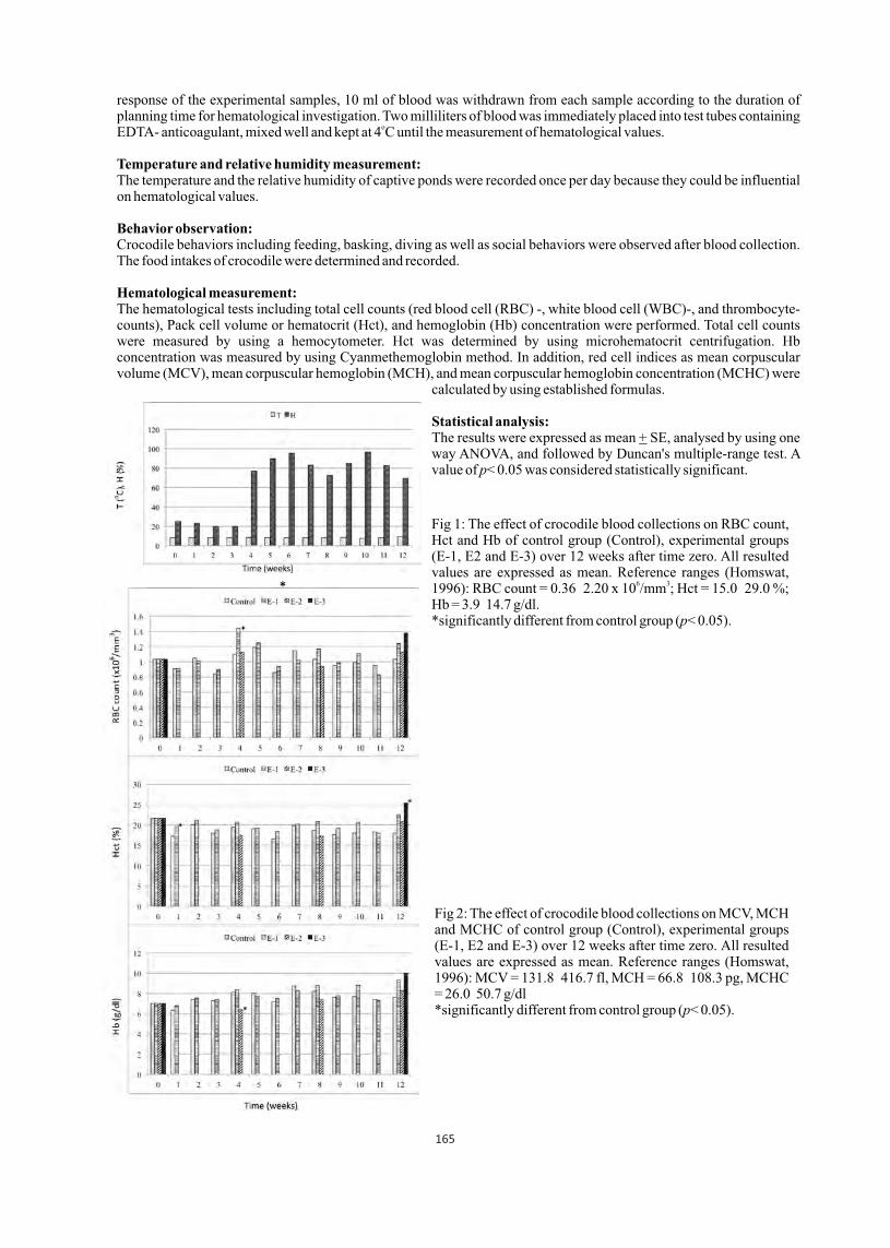
response of the experimental samples, 10 ml of blood was withdrawn from each sample according to the duration of planning time for hematological investigation. Two milliliters of blood was immediately placed into test tubes containing
oEDTA- anticoagulant, mixed well and kept at 4 C until the measurement of hematological values.
Temperature and relative humidity measurement: The temperature and the relative humidity of captive ponds were recorded once per day because they could be influential on hematological values.
Behavior observation: Crocodile behaviors including feeding, basking, diving as well as social behaviors were observed after blood collection. The food intakes of crocodile were determined and recorded.
Hematological measurement: The hematological tests including total cell counts (red blood cell (RBC) -, white blood cell (WBC)-, and thrombocyte- counts), Pack cell volume or hematocrit (Hct), and hemoglobin (Hb) concentration were performed. Total cell counts were measured by using a hemocytometer. Hct was determined by using microhematocrit centrifugation. Hb concentration was measured by using Cyanmethemoglobin method. In addition, red cell indices as mean corpuscular volume (MCV), mean corpuscular hemoglobin (MCH), and mean corpuscular hemoglobin concentration (MCHC) were
calculated by using established formulas.
Statistical analysis:The results were expressed as mean + SE, analysed by using one way ANOVA, and followed by Duncan's multiple-range test. A value of p< 0.05 was considered statistically significant.
Results
*
Fig 1: The effect of crocodile blood collections on RBC count, Hct and Hb of control group (Control), experimental groups (E-1, E2 and E-3) over 12 weeks after time zero. All resulted values are expressed as mean. Reference ranges (Homswat,
6 31996): RBC count = 0.36 2.20 x 10 /mm ; Hct = 15.0 29.0 %; Hb = 3.9 14.7 g/dl. *significantly different from control group (p< 0.05).
Fig 2: The effect of crocodile blood collections on MCV, MCH and MCHC of control group (Control), experimental groups (E-1, E2 and E-3) over 12 weeks after time zero. All resulted values are expressed as mean. Reference ranges (Homswat, 1996): MCV = 131.8 416.7 fl, MCH = 66.8 108.3 pg, MCHC = 26.0 50.7 g/dl*significantly different from control group (p< 0.05).
165
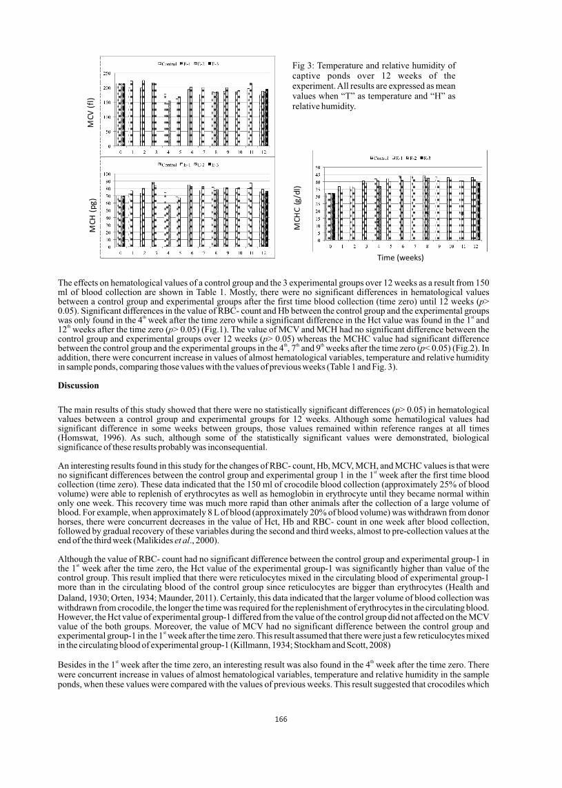
The effects on hematological values of a control group and the 3 experimental groups over 12 weeks as a result from 150 ml of blood collection are shown in Table 1. Mostly, there were no significant differences in hematological values between a control group and experimental groups after the first time blood collection (time zero) until 12 weeks (p> 0.05). Significant differences in the value of RBC- count and Hb between the control group and the experimental groups
th stwas only found in the 4 week after the time zero while a significant difference in the Hct value was found in the 1 and th12 weeks after the time zero (p> 0.05) (Fig.1). The value of MCV and MCH had no significant difference between the
control group and experimental groups over 12 weeks (p> 0.05) whereas the MCHC value had significant difference th th thbetween the control group and the experimental groups in the 4 , 7 and 9 weeks after the time zero (p< 0.05) (Fig.2). In
addition, there were concurrent increase in values of almost hematological variables, temperature and relative humidity in sample ponds, comparing those values with the values of previous weeks (Table 1 and Fig. 3).
Discussion
The main results of this study showed that there were no statistically significant differences (p> 0.05) in hematological values between a control group and experimental groups for 12 weeks. Although some hematilogical values had significant difference in some weeks between groups, those values remained within reference ranges at all times (Homswat, 1996). As such, although some of the statistically significant values were demonstrated, biological significance of these results probably was inconsequential.
An interesting results found in this study for the changes of RBC- count, Hb, MCV, MCH, and MCHC values is that were stno significant differences between the control group and experimental group 1 in the 1 week after the first time blood
collection (time zero). These data indicated that the 150 ml of crocodile blood collection (approximately 25% of blood volume) were able to replenish of erythrocytes as well as hemoglobin in erythrocyte until they became normal within only one week. This recovery time was much more rapid than other animals after the collection of a large volume of blood. For example, when approximately 8 L of blood (approximately 20% of blood volume) was withdrawn from donor horses, there were concurrent decreases in the value of Hct, Hb and RBC- count in one week after blood collection, followed by gradual recovery of these variables during the second and third weeks, almost to pre-collection values at the end of the third week (Malikides et al., 2000).
Although the value of RBC- count had no significant difference between the control group and experimental group-1 in stthe 1 week after the time zero, the Hct value of the experimental group-1 was significantly higher than value of the
control group. This result implied that there were reticulocytes mixed in the circulating blood of experimental group-1 more than in the circulating blood of the control group since reticulocytes are bigger than erythrocytes (Health and Daland, 1930; Orten, 1934; Maunder, 2011). Certainly, this data indicated that the larger volume of blood collection was withdrawn from crocodile, the longer the time was required for the replenishment of erythrocytes in the circulating blood. However, the Hct value of experimental group-1 differed from the value of the control group did not affected on the MCV value of the both groups. Moreover, the value of MCV had no significant difference between the control group and
stexperimental group-1 in the 1 week after the time zero. This result assumed that there were just a few reticulocytes mixed in the circulating blood of experimental group-1 (Killmann, 1934; Stockham and Scott, 2008)
st thBesides in the 1 week after the time zero, an interesting result was also found in the 4 week after the time zero. There were concurrent increase in values of almost hematological variables, temperature and relative humidity in the sample ponds, when these values were compared with the values of previous weeks. This result suggested that crocodiles which
Fig 3: Temperature and relative humidity of captive ponds over 12 weeks of the experiment. All results are expressed as mean values when “T” as temperature and “H” as relative humidity.
MC
V(f
l)M
CH
(pg
)
Time (weeks)
MCH
C(g
/dl)
*
*
*
166
are exothermic reptiles and unable to maintain a constant internal body temperature independently of the environment, adapted to their surroundings (Huchzermeyer, 2003). The increase of values of almost hematological variables simultaneously occurred with the sample pond's temperature increase which is consistent with Lang (1987) who reported that the growth rate of juvenile crocodiles is the fastest in maximum temperature where they are kept. The growth rate increased harmoniously with raising temperature probably suggests that the raise of temperature might be involved in the enhancement of a variety of compensatory neuroendocrine homeostatic mechanisms, facilitating rapid growth rate as well as hematopoiesis and other processes implicated with the hematology of crocodiles.
thIn addition, in the 4 week after the time zero, all samples in this study including the control group and experimental group-1 ate food increasingly more than previous weeks. Being held capture, disturbances and/or low temperature
st rdpossibly caused the crocodiles to reduce feeding behavior in the 1 3 weeks after the time zero (Huchzermeyer, 2003). thTherefore, the increase in feeding behavior in the 4 week after the time zero might imply that the activation of
hematopoiesis and other processes involved with the hematology of crocodiles related to nutritions in an animal's body because nutrients are essential for rebuilting its blood supply (Voigt, 2000).
thIn the 4 week after the time zero, the value of RBC and WBC counts of the experimental group-1 were significantly higher than those values of the control group and experimental group-2 whereas the Hb value of the experimental group-2 was significantly lower than the values of the control group and experimental group-1. These implied that the crocodiles are capable of adapting very well to circumstances as a result of being the sole survivors from the ruling age of reptiles, whose ancestry dates back to the Mesozoic Era, about 265 million years ago. Moreover, the lineage of crocodilians retaining the characteristic features of the group has continued through to the present day (Alderton, 1991). Even though there were significant differences in the values of some hematological variables between sample groups in
th thsome weeks during the 5 12 weeks after the time zero, those values remained within reference ranges (Homswat, 1996) at all times.
In conclusion, this study demonstrated that there was no adverse effects on donor crocodiles when 150 ml of blood (approximately 25% of blood volume) was withdrawn from them. Although significant differences of some hematological variables between a control group and experimental groups (p<0.05) were found in some weeks, the variations occurred within reference ranges and did not represent a significant biological change. Therefore, a crocodile could be donated their blood in volume of 150 ml every 12 weeks with no change in behavior and no adverse effect on hematological values. We suggest that the repeated 150 ml of blood collection is not detrimental to donor crocodile health and welfare. This information may provide important guidelines for regular large volume of blood collection in crocodiles. However, the optimal blood volume remains to be elucidated.
Acknowledgements
This study was supported by National Research Council of Thailand (NRCT), Department of Zoology, Faculty of Science, Kasetsart University, Thailand. The co-operation and assistance of the owner and manager of Rungtaweechai Farm, and the farm staff were acknowledged.
Literature Cited
01. Alderton, D. (1991). Crocodiles and Alligators of the World. Papirografika, Yugoslavia.02. Chaeychomsri, W., Chaeychomsri, S., Siruntawineti, J., Hengsawadi, D., and Cuptapan, Y. (2009). Freeze-dried crocodile blood production
as food supplement. J. Biosc. Bioeng. 08: S22.03. Health, C. W. and Daland, G. A. (1930). The Life of Reticulocytes. Arch. Int. Med. 46: 533.04. Homswat, S. (1996). Hematological and Sume Serum Chemistry Values of Siamese Crocodile (Crocodylus siamensis). M.S. Thesis,
Kasetsart University. 05. Huchzermeyer, F. W. (2003). Crocodiles Biology, Husbandry and Diseases. Biddles Ltd., Guildford and King's Lynn., UK.06. Kasetsart University. By: Chaeychomsri, W. and Siruntawineti, J. Thailand Patent application No. 0601001179, 16 March 2006.07. Killmann, S. A. (1934). Relation of Variation in Mean Corpuscular Volume to Number of Reticulocytes in Perniciuous Anemia. J. Clin.
Invest. 13: 669.08. Lang, J. W. (1987). Crocodilian Thermal Selection in Webb, G.J.W., Manolis, S.C. and Whitehead, P.J. (eds) Wildlife Management:
Crocodiles and Alligators. Surrey Beatty and Sons, Chipping Norton, New South Wales, Australia, 301 317. 09. Malikides, N., Mollison, P. J., Reid, S. W. J. and Murray, M. (2000). Haematological Responses of Repeated Large Volume Blood
Collection in the Horse. Res. Vet. Sci. 68: 275-278.10. Maunder, R.B. (2011). Understanding Laboratory Tests. Transcontinental, Canada.11. Orten, J.M. (1934). The Properties and Significance of the Reticulocyte. Yale J. Biol. & Med. 6: 519.12. Pitimol, N., Busayaplakorn, P., Siruntawineti, J. and Chaeychomsri, W. (2008). Development of Crocodile Blood Collection Process on
thAnimal Life Maintain for Innovation of Crocodile Blood Product in Proceeding of the 19 Working Meeting of the IUCN-SSC Crocodile Specialist Group. 2 7 June 2008, Republic of Bolivia.
13. Siruntawineti, J., Chaeychomsri, W., Sathsumpun, C., Tungsathian, P, Vajarasathira, B. and Temsiripong, Y. (2006). Development and thEvaluation of Dried Siamese Crocodile Blood Product as Supplemented Food in Proceeding of the 18 Working Meeting of the Crocodile
Specialist Group. 19 23 June 2006, Monte'limar, France.nd14. Stockham, S. T. and Scott, M. A. (2008). Fundamentals of Veterinary Clinlcal Pathology, 2 ed. Blackwell Publishing Ltd., USA.
th15. Turgeon, M. L. (2012). Clinical Hematology Theory and Procedures, 5 ed. Wolters Kluwer business, China.st16. Voigt, G. L. (2000). Hematology Techniques and Concepts for Veterinary Technicians, 1 ed. Iowa State University Press, USA.
167

Tab
le 1
. R
esu
lts
(mea
n +
SE
) of
th
e ef
fect
on
hem
atol
ogic
al v
aria
ble
s af
ter
wit
hd
raw
ing
150
ml
of b
lood
ove
r 12
wee
ks
168
Tab
le 1
(C
ont.
). R
esu
lts
(mea
n +
SE
) of
th
e ef
fect
on
hem
atol
ogic
al v
aria
ble
s af
ter
wit
hd
raw
ing
150
ml
of b
lood
ove
r 12
wee
ks
ND
= N
on-d
etec
ted,
Sm
all
lett
er s
igni
fica
nt d
iffe
renc
e in
col
umn
(p<
0.0
5).
169

Habitat suitability of mugger crocodile in Sarbaz River, Iran
Elham Abtin
Wildlife expert, Provincial office of Department of Environment in Sistan and Baluchestan province,
Zahedan, Iran, Email: [email protected],
Abstract
In order to study the habitat suitability and classification of Muggers in Iran, we used Habitat Evaluation Procedure
(HEP) to define the best and most suitable habitats for Muggers. A habitat Suitability Index (HSI), was used to compare
habitats. HSI values range between 0 (worst habitat) and 1 (best habitat).
e (HEP) to define the best and most suitable habitats for Muggers. A habitat Suitability Index (HSI), was used to compare
habitats. HSI values range between 0 (worst habitat) and 1 (best habitat).
Ten natural ponds along the Sarbaz River were selected for the study. Climatic, physical and chemical characteristics of
soil, water and vegetation cover, as well as some ecological characteristics, were collected during monthly surveys.
Information on fauna (fishes and amphibians) and flora diversity, water depth, habitat slope and the area of ponds and
number of crocodiles in habitats were also included in the study.
The significant variables were scored using Analytical Hierarchy Process (AHP) and IDRISI KLIMANJARO 8.4
software and the HSI for each habitat calculated. Cluster analysis was used to classify the habitats based on their main
characteristics. Based on AHP significant parameters classified into three main categories: cover (vegetation cover,
depth, slope); food (fish and amphibian species); and, chemical specification of water Like DO and Conductivity. Based
on the results, the HSI for Pishin Dam pond was 1 (highest suitability),
followed by other habitats with lower HSI and Kollani had the lowest HSI
(0.2). In general, Muggers prefer habitats with 2-4 m water depth, mean
vegetation cover of 35%, mean slope of 25-35% and high density of fish and
amphibians.
HSI, material and methods
Southwestern Iran is the western most limits for the Mugger crocodile
(Crocodylus palustris). The main habitats for crocodiles are along the main
rivers, of which the Sarbaz River is the most important. Local people of the area
call the crocodiles 'Gandou” and respect them, so they are not directly harmed
by people. The main threats for crocodiles are natural incidents such as floods
and drought (Mobaraki &abtin 2010) .No studies on habitat suitability and
classification have been undertaken for Muggers in Iran. Information on
habitat characteristics and requirements would provide considerable support to
species management and conservation and provide the basis for responses to
any deficiencies. We used Habitat Evaluation Procedure (HEP) to define and
identify the best and most suitable habitats for Muggers. A Habitat Suitability
Index (HSI), a nominal index representing habitat capacity for providing the
needs of the species, was used to compare habitats. HSI values range between 0
(worst habitat) and 1 (best habitat).
Ten natural ponds along the Sarbaz River were selected for the study: listed
from north to south - Firuz Abad, Shekar Jangal, Azadi, Pishin Dam, Dargas, Figure 1: Selected sites/habitats
along the Sarbaz River
Proceedings : World Crocodile Conferencend -22 Working Meeting of the IUCN SSC Crocodile Specialist Groupnd -22 Working Meeting of the IUCN SSC Crocodile Specialist Groupnd -22 Working Meeting of the IUCN SSC Crocodile Specialist Group
Sri Lanka 20-23 May 2013 170
pp 170 -172

Lower (Paien) Hutkat, upper (Bala) Hutkat, ShirGoovaz, Bahukalat, Kollani (near Govater Bay) (see Fig. 1) Climatic,
physical and chemical characteristics of soil, water and vegetation cover, as well as some ecological characteristics, were
collected during monthly surveys over the period of September 2008 to January 2010. Information on fauna (fishes and
amphibians) and flora diversity, water depth, habitat slope and area of ponds and number of crocodiles in habitats were
also included in the study.
The area of the habitat was considered as the initial criterion for selection of the habitats as study stations and comprised
the highest population density, number of nests and occurrence of juvenile and adult crocodiles, based on the direct
observation on crocodile occurrence in the habitats. As most sites had thick vegetation, sampling of the number and
variety of species was only conducted in 10% of the areas involved. Based on number and variety of species, vegetation
diversity was determined using the Ecological Methodology (software) program. Climatic variables (e.g. mean
temperature, humidity, evaporation, and rainfall) were compiled from existing meteorological stations at Bahu-kalat and
Sarbaz.
Monthly data were averaged for each parameter, and analyzed using SPSS (correlation between factors) .The significant
variables were scored using Analytical Hierarchy Process (AHP) and IDRISI KLIMANJARO 8.4 software and the HSI
for each habitat calculated. Cluster analysis was used to classify the habitats based on their main characteristics.
There were no significant differences between habitats with respect to variables such as air and soil temperature,
humidity, water and soil pH, and soil structure and elements of the habitats. Moreover, correlation test did not show any
significant relationship between parameters and crocodile population, with the exception of 6 variables for which
significance was recorded.
Based on AHP all data, significant parameters, could be classified into one of three main categories: cover (vegetation
cover, depth, slope); food (fish and amphibian species); and, chemical specification of water Like DO and Conductivity
(figure 2).
Figure 2: Analytical Hierarchy Process= AHP
Table 1: Pearson Correlation for significant parameters with respect to crocodile density
Results
The highest mean crocodile numbers were observed in the Pishin Dam pond, 10 crocodiles, and the lowest (zero) were
recorded in Kollani and Firuzabad ponds. The HSI for Pishin Dam pond was 1 (highest suitability) (Fig. 2), followed by
Parameter Pearson P
Correlation (2 tail)
Water depth 0.739 0.015
Vegetation cover 0.660 0.038
Slope 0.753 0.012
Amphibians 0.864 0.001
Fish 0.674 0.032
Conductivity EC 0.650 0.042
171

Shirgovaz, Dargas, Hutkat-e-bala, Hutkat-E-pain respectively (0.96, 0.84, 0.75 and 0.74). Azadi, Firuzabad and Shekar
Jangal had similar HSI ( 0.3-0.5) and Kollani had the lowest HSI (0.2), indicating that it was the most unsuitable habitat
(Fig. 3).
figure 3: Classification of the habitats based on the significant criteria.
Each index received a value and using the equation of ((Cb.Cnn or Cnt)1/2 the overall HSI Model prepared for the
determination of the habitat suitability value for each habitat:
HSI:24.37 X1+2.9 X2+5.52 X3+4.5 X4+2.54 X5+4.9 X6
In which the X values are the significant parameters. In general, Muggers prefer habitats with 2-4 m water depth, mean
vegetation cover of 35%, mean slope of 25-35% and high density of fish and amphibians (Behrouzi and etal 2010).
Refrences:
1. 1- - Newsom, J.D., T. Joanen and R.J. Howard. 1987, Habitat Suitability Index Model: American Alligator. U.S. Fish and wild life serv. Biol. Rep. 8x10.136). 14pp2. 4- Mobaraki, Asghar , Elham Abtin, 2010, Mugger crocodile study in Iran, wild life Middle east, Vol 5, Issue 2, ISSN 1990-8237, 6p.3. 5- Behroozi Rad. Behrooz, Elham Abtin, Asghar Mobaraki, 2010, MUGGER CROCODILE HABITAT SUITABILITY STUDY IN 4. SARBAZ RIVER, IRAN, Crocodile specialist group newsletter, Vol 29, No 2, 8-11pp
Figure 4: Pishin Dam Pond as one of the most important and suitable habitat for
Mugger crocodiles. Photo: Elham Abtin.
Figure 5 : Basking crocodile in Pishin Dam pond. Photo: Elham Abtin
172
Dendrogram
Molecular identification of Crocodylus siamensis using specific primers for reintroduction to Kangkrachan National Park in Phetchaburi, Thailand
1 2 1 Win Chaeychomsri , Sudawan Chaeychomsri , Jindawan Siruntawineti , Wisachinee
3 4 4Rungtaweechai , Jitraporn Chanrajakit and Panya Youngprapakorn
1 Department of Zoology, Faculty of Science, Kasetsart University, Bangkok 10900, Thailand.
2 Central Laboratory and Greenhouse Complex, Kasetsart University, Kamphaeng Saen,
3 Nakhon Pathom 73140, Thailand Rungtaweechai Crocodile Farm, DonToom,
. 4 Nakhon Pathom 73150, Thailand Golden Crocodile Agriculture (Thailand) Co., Ltd. Nakhon Pathom 73140, Thailand
Abstract
Crocodylus siamensis, the Siamese crocodile, is a critically endangered species of freshwater crocodile previously distributed throughout much of Southeast Asia. Recovery plans call for reintroductions to the wild using founder individuals currently in captivity, mostly in commercial crocodile farms. On many farms C. siamensis has been intentionally hybridized with the estuarine crocodile, C. porosus, and hybrids may be difficult to distinguish morphologically. The objective of this study was to develop a new molecular identification method capable of discriminating among the crocodilian species and to apply this tool in combination with reintroduction programs. Six specific microsatellite markers from C. siamensis were developed and used for characterization of closely related crocodiles. A total of 120 blood samples of C. siamensis (based on morphological characteristics) were collected from the crocodile farms in Thailand. These samples were derived from Golden Crocodile Agriculture (Thailand) Co., Ltd., (100 samples) and Rungtaweechai Crocodile Farm (20 samples). The samples were tested using the polymerase chain reaction (PCR) compared with 4 unknown crocodile samples from Crocodile Education and Conservation Centre, United Kingdom (UK) and 4 samples of C. porosus saltwater crocodile as the out group. The results showed that the markers CS-4, CS-5, CS-20, CS-25, CS-26 and CS-32 could identify all species tested. Three microsatellite markers CS-4, CS-5, and CS-26 showed polymorphic alleles in the same species that were considered as intra-specific markers. The number of alleles per locus ranged from 1-2, and the observed heterozygosities ranged from 0.48 to 0.89. The markers CS-20, CS-25 and CS-32 showed monomorphic allele in C. siamensis. They showed only 1 allele and the observed heterozygosities ranged from 0 to 0.03. These results revealed that the samples of C. siamensis from Golden Crocodile Agriculture (Thailand) Co., Ltd. and those from Rungtaweechai Crocodile Farm were pure C. siamensis and suitable for reintroduction to Kangkrachan National Park. Another 4 unknown crocodile samples from Crocodile Education and Conservation Centre, UK were also pure C. siamensis. These microsatellite markers can be used for species testing and also evaluated for assigning parentage in situations where maternity, paternity, or both. The use of these microsatellite markers has potential for application in captive breeding programs, evaluating hybridization, the effects of introductions of exotic species and to the identification of products in trade and reintroduction programs.Keywords: Siamese crocodile, microsatellite marker, crocodile reintroduction
Introduction
In the IUCN Action Plan for crocodiles (Ross, 1998), mention is made of several species where information was needed on their genetic diversity and lineage. For example, population genetics surveys were needed for the critically endangered Orinoco crocodile (C. intermedius), to assess their level of genetic diversity to inform restocking and reintroduction programs. The Siamese crocodile is an endangered species that formerly inhabited wetlands and waterways of Southeast Asia. Hunting and habitat loss have reduced its population in the wild of Thailand, and most of the remaining Siamese crocodiles have been maintained and bred in captivity. Captive Breeding and reintroduction programs of pure breed Siamese crocodile back to the wild are the simplest and most efficient ways to overcome these problems. There has been an attempt to release pure bred Siamese crocodiles back into the wild to support conservation strategies, there was an increasing need to develop genetic markers in Crocodylus sp., particularly in C. siamensis. Although the microsatellite loci used had some overlap of allele sizes between species, it was possible to use assignment tests for differentiation. However, within this genus, most tests of cross-species PCR amplification indicated the presence
Sri Lanka 20-23 May 2013 173
Proceedings : World Crocodile Conferencend -22 Working Meeting of the IUCN SSC Crocodile Specialist Groupnd -22 Working Meeting of the IUCN SSC Crocodile Specialist Groupnd -22 Working Meeting of the IUCN SSC Crocodile Specialist Group
pp 173 - 178

of homologous microsatellite loci that were variable. In previous studies (Chaeychomsri et al., 2008a, b) it has been shown that microsatellites from C. siamensis are useful marker for parentage analyses and individual identification.
Microsatellites (also called simple sequence repeats, SSRs) are a class of genetic polymorphism commonly used for mapping, linkage analysis (Dib et al., 1996), behavioral ecology (Schlötterer and Pemberton, 1998), population genetics (Goldstein and Schlötterer, 1999), and phylogeny reconstruction (Harr et al., 1998). Microsatellites are tandemly repeated sequences, where the repeating unit is 1 to 4 nucleotides long. The number of times the unit is repeated in a given microsatellite can be highly variable, a characteristic that makes them useful as genetic markers. Many reports have demonstrated several advantages of using microsatellite markers in crocodylian systematic and population genetics. Glenn et al. (1998) developed nuclear microsatellite markers to address questions on the genetic population structure and the mating system of American alligators (Alligator mississippiensis) and showed that microsatellite markers could differentiate between populations of Florida and American alligators and had a higher discriminatory power of differentiation than the allozyme isozyme markers. Davis et al. (2001) reported a high level of genetic variation for six populations of American alligators located throughout the southeastern United States. FitzSimmons et al. (2001) developed 26 new microsatellite markers from the DNA of C. acutus, C. porosus and C. johnstoni to compare the population genetic structure in crocodylian families and tested loci for Crocodylus spp., which represented the first microsatellite loci found in Crocodylus.
This paper describes the potential utilization of the microsatellites as molecular markers in C. siamensis and for identification of a closely related species. These markers will be a useful tool for a captive breeding program, developing conservation strategies and for understanding the genetic structure of the remnant wild populations of this species.
MATERIALS AND METHODS
Sample collectionBlood samples were taken from freshwater and saltwater crocodiles (based on morphological characteristics) by collecting from the anterior dorsal sinus and stored in the heparinized tubes as described by Chaeychomsri et al. (2008a, b). The samples of freshwater crocodile were derived from Golden Crocodile Agriculture (Thailand) Co., Ltd., (100 samples) and Rungtaweechai Crocodile Farm (20 samples) while the samples of saltwater crocodile were taken from Rungtaweechai Crocodile Farm (4 samples). Four unknown crocodile samples from Crocodile Education and Conservation Centre, UK were received as ethanol-preserved blood.
DNA extractionGenomic DNA of crocodiles was isolated from red blood cells by using the QIAamp DNA Blood Mini Kit (QIAGEN) according to the manufacturer's protocol. DNA concentration and purity were assayed by spectrophotometry and agarose gel electrophoresis (Sambrook et al., 1989).
Microsatellite analysisSix microsatellite primers previously described by Chaeychomsri et al. (2008a) were utilized for this study with primer sequences and PCR conditions shown in Table 1. PCR amplification was carried out in 50 μl reaction mixture containing 200 ng DNA as the template. Genomic DNA 10.0 μl (20 ng/l), 10x PCR buffer (+ MgCl ) 5.0 l, 2.5 mM 2
dNTP 4.0 l, Forward primer (F) (10 pmol/l) 2.0 l, Reverse primer (R) (10 pmol/l) 2.0 l, Taq DNA polymerase (2 unit/l) 0.5 l, sterile distilled water 26.5 l were added and PCR was performed in a DNA thermal cycler (GeneAmp PCR System 9700, PE Applied Biosystems) programmed for 3 min at 94°C for initial denaturation. Following the initial denaturation
othe thermal cycler was programmed for 35 cycles of 1 min at 94°C for denaturation, 1 min at 60-70 C depended on each primer pairs (Table 1) for annealing and 1 min at 72°C for extension. The PCR products were analyzed by electrophoresis in a 1% agarose gel in 1x TBE buffer (Sambrook et al., 1989) in parallel with 100-bp DNA ladder (Invitrogen) and bands were visualized by ethidium bromide staining. Images were analyzed using Gene Tools image analysis and quantification software version 4.01c (Syngene, UK) and sizes of microsatellite alleles were determined by comparing with 100-bp DNA ladder. Allele frequencies, number of alleles per locus, expected and observed heterozygocities (He and Ho) were calculated using GENEPOP software package (Raymond and Rousset, 2001).
Results
Six microsatellite markers were amplified by PCR to assess the level of polymorphism in C. siamensis (n=120), C.
porosus (n=4) and 4 unknown crocodile samples. The PCR conditions used were optimal for amplification of
microsatellite markers from C. siamensis and thus may not be optimal for the other species to test all taxa.
PCR products from loci CS-4 (Fig. 1A), CS-5 (Fig. 1B), CS 20 (Fig. 2A), CS-25 (Fig. 2B), CS-26 (Fig. 3A), and CS-32
(Fig. 3B) showed a clear polymorphic pattern in C. siamensis. The average observed heterozygosity (Table 1) was 0.23
(range 00.49) and average expected heterozygosity was 0.32 (range 00.89).
PCR products from loci CS-20 (Fig. 2A), CS-25 (Fig. 2B) and CS-32 (Fig. 3B) showed a clear pattern of single specific
174

band in C.siamensis. Additionally, all products scored were 249, 180 and 245 bp, respectively.
PCR products from loci CS-4 (Fig. 1A), CS-5 (Fig. 1B) and CS-26 (Fig. 3A) showed a clear polymorphic pattern in C.
siamensis. The average observed heterozygosity (Table 1) was 0.37 (range 0-0.49) and average expected heterozygosity
was 0.41 (range 0.12-0.68).
For heterozygocity, the results showed that Ho is very low in C. siamensis (0-0.89) and revealed that most of C. siamensis
were inbred in the same species, or have purebred in the farm. The PCR product size of loci CS-4, CS-5, CS-20, CS-25,
CS-26 and CS-32 can identified C. siamensis and C. porosus and hybrid (Fig. 4A and B). These results showed difference
in alleles size in C. siamensis and C. porosus.
Table 1 Microsatellite DNA primers from C. siamensis, 5'->3' PCR primer sequences, annealing temperature (Ta), size length, number of alleles, observed and expected heterozygosity (Ho and He)
Figure 1 Agarose gel electrophoresis of PCR products generated from crocodile DNA samples using C. siamensis microsatellite loci. (A) locus CS-4 and (B) locus CS-5. The numbers at the top of the figure indicate samples derived
from freshwater crocodile used in this study. M, 100-bp DNA ladder.
Figure 2 Agarose gel electrophoresis of PCR products generated from crocodile DNA samples using C. siamensis microsatellite loci. (A) locus CS-20 and (B) locus CS-25. The numbers at the top of the figure indicate samples
derived from freshwater crocodile used in this study. M, 100-bp DNA ladder; M1, 50-bp DNA ladder.
Locus Primer sequence:(5'->3') Ta (oC) Observed
allele size (bp) No. of allelles
Ho He
CS-4
F: CCATGCCCTACCACACAACTG R: CAAACACAAAGGCATTCAAAGATG
65 200, 240 2 0.53 0.40
CS-5
F: TCTCTCCTTCTTTCTGCTTGTGC R: GTGCAAGGTCTGTACCTGTGTGA
65 200, 240 2 0.89 0.49
CS-20
F: CCTTCCGCCCAAAATAATTGC R: CCCTGCACTAACAAGGAGCAG
60 249 1 0 0
CS-25
F: ATCCTCAGTGTCATCTGCTCACC R: TCTTCCCTGCTCACCTTTCTTTC
65 180 1 0.02 0.01
CS-26
F: GCCATGTGTACTAACTGGGAAGTC R: GCCATTTTTAGTCAGGTTGTTGC
65 240-280 2 0.48 0.42
CS-32
F: GTACCAAGCCCCTTTAACACCTG R: GGGGAGAAGGAAACTAGGAGAGG
68 245 1 0 0.03
600
100
200
300 400 500
bp
A M 13 14 15 16 17 18 19 bp
100
600
200
300 400 500
B M 61 62 63 64 65 66 67
M 92 93 94 95 96 97 98 A bp
600
100
200 300 400 500
B bp
350
150 250
M1 43 44 45 46 47 48 49
175
Figure 3 Agarose gel electrophoresis of PCR products generated from crocodile DNA samples using C. siamensis microsatellite loci. (A) locus CS-26 and (B) locus CS-32. The numbers at the top of the figure indicate samples
derived from freshwater crocodile used in this study. M, 100-bp DNA ladder.
Figure 4 Agarose gel electrophoresis of PCR products generated from crocodile DNA samples using C. siamensis microsatellite loci. (A) locus CS-25 and (B) locus CS-32. The numbers at the top of the figure indicate samples
derived from freshwater crocodile and the additional P letters refer to the samples from saltwater crocodile used in this study. M, 100-bp DNA ladder.
Table 2 Observed allele size from all loci and total number of crocodiles per locus
GCA, Golden Crocodile Agriculture (Thailand) Co., Ltd.RCF, Rungtaweechai Crocodile FarmCECC, Crocodile Education and Conservation Centre
A
bp
600
100
200
300 400 500
M 13 14 15 16 17 18 19 B
bp
100
600
200
300 400 500
M 1 2 3 4 5 6 7
A
bp
600
100
200
300 400 500
M 33 35 36 P1 P2 B
bp
100
600
200
300 400 500
M 33 35 36 P1 P2
Locus Allele size (bp) Sample
GCA (n=100) RCF (n=20) CECC (n=4)
CS-4 200 95 18 4
240 54 4 -
CS-5 200 94 18 4
240 84 4 -
CS-20 249 100 20 4
CS-25 180 100 20 4
CS-26 240 91 16 4
280 45 8 -
CS-32 245 100 20 4
176
Even with these results, the initial applications of these primers are promising. These data from populations of C. siamensis and C. porosus indicated that several loci would be quite useful for species identification in crocodile. In a captive population of C. siamensis, it appeared that there would be enough genetic variation to determine paternal exclusion in clutches. It is hoped that these primers will enable the application of genetic investigations in other studies of order Crocodylia. As in other species, these genetic markers offer the most powerful insights when they are combined with available demographic or behavioral data. In addition, using both mtDNA and microsatellite markers will be the most effective means of elucidating questions on gene flow and population history.Additionally, a combined approach using both mtDNA and microsatellite markers will be the most effective for elucidating questions of gene flow and population history. The availability of these microsatellite markers will allow comparative studies of mating systems and population structure among crocodilians.
For heterozygosity, the observed heterozygosity (Ho) of CS-20, CS-25 and CS-32 was very low in C. siamensis ranging from 0-0.02. This indicated that 100 samples of C. siamensis from Golden Crocodile Agriculture (Thailand) Co., Ltd. and 20 samples of C. siamensis from Rungtaweechai Crocodile Farm have been inbred in the same species, or have been purebred in the crocodile farm. Thus, selection for individuals with higher heterozygosity as is typically recommended (Moritz, 1999) is possible for the planned reintroduction into National Park. This strategy is indicated theoretically, to provide a greater range of options upon which selection can, if indeed variation at dinucleotide microsatellite loci provides a surrogate measure of variation in coding regions of the genomic DNA. Whether or not this assumption is met, selecting individuals with higher heterozygosity (if all other concerns are equal) remains a risk minimization strategy.
For the reintroduction effort, it is important to confirm that the captive population consists of mostly purebred C. siamensis. These results have been useful in identifying captive hybrids of the Siamese crocodile, C. siamensis. Of equal concern in reintroductions and augmentations is the possibility of breaking up locally adapted gene complexes through outbreeding among different source populations (Storfer, 1999). If this is true for C. siamensis, then it will remain an inherent problem for this and other crocodilian reintroductions, because crocodile farms typically have individuals originating from widely scattered populations. Records may not have been kept, individuals might not be marked for identification and breeders are typically put into large communal pens. One could argue that to avoid the problem of out breeding depression, only the most productive lineages should be selected for reintroduction (if indeed this information is known). However, this runs the risk of selecting individuals who are best suited to captive rather than wild situations. The extreme contrast between out breeding depression versus hybrid vigor is something that undoubtedly occur make comparisons.
Conclusion
In these studies we evaluated 6 microsatellite markers from Siamese crocodile (C. siamensis) and their transferability to other Crocodylus species. These microsatellite markers were evaluated in captive breeding crocodile farm for species testing. These markers composed of CS-4, CS-5, CS-20, CS-25, CS-26 and CS-32 can identify all species. Three microsatellite markers showed polymorphic alleles in the same species that were considered as intra-specific markers. These markers composed of CS-4, CS-5, and CS-26. CS-20, CS-25 and CS-32 showed monomorphic allele in C. siamensis. They showed only 1 allele and the observed heterozygosities ranged from 0 to 0.03. These microsatellite markers can be used for species testing and also evaluated for assigning parentage in situations where maternity, paternity, or both. It is recommended that more microsatellite markers should be used in these situations. Therefore, the use of these microsatellite markers will allow a fast screening, and have potential for application in captive breeding programs, evaluating hybridization, and reintroduction programs.
Acknowledgements
We would like to thank Rungtaweechai Crocodile Farm, DonToom, Nakhon Pathom, Thailand, Golden Crocodile Agriculture (Thailand) Co., Ltd. Thailand and Crocodile Education and Conservation Centre, UK for providing DNA samples of C. siamensis and C. porosus. This research was supported by a grant from Rungtaweechai Crocodile Farm, Thailand.
Discussion
177
Literature Cited
01. Chaeychomsri, W., Tabthipwon, P., Noparatnaraporn, N. and Siripholvat, V. (2008a). Development of microsatellite markers for Siamese crocodile (Crocodylus siamensis). Kasetsart J. (Nat. Sci.). 42: 256-262.
02. Chaeychomsri, W., Chaeychomsri, S., Tuntirungkij, M., Tabthipwon, P., Noparatnaraporn, N. and Siripholvat, V. (2008b). Characterization of microsatellite markers for the Siamese crocodile and amplification in the closely related genus Crocodylus. Kasetsart J. (Nat. Sci.). 42: 682-689.
03. Davis, L. M., Glenn, T. C., Elsey, R. M., Dessauer, H. C. and Sawyer, R. H. (2001). Multiple paternity and mating patterns in the American alligator, Alligator mississippiensis. Mol. Ecol. 10: 1011-1024.
04. Dib, C., Faure, S., Fizames, C., Samson, D., Drouot, N., Vignal, A., Millasseau, P., Marc, S., Kazan, J., Seboun, E., Lathrop, M., Gyapay, G., Morissette, J. and Weissenbach, J. (1996). A comprehensive genetic map of the human genome based on 5,264 microsatellites. Nature 380: 152-154.
05. FitzSimons, N. N., Tanksley, S., Forstner, M. R., Louis, E. E., Daglish, R., Gratten, J. and Davis, S. (2001). Microsatellite markers for Crocodylus: new genetic tools for population genetics, mating system studies and forensics, Pp. 51-57 In Crocodilian Biology and Evolution, ed. by G. Grigg, F. Seebacher and C.E. Franklin. Surey Beatty and Sons: Chipping Norton.
06. Glenn, T. C., Dessauer, H. C. and Braun, M. (1998). Characterization of microsatellite DNA loci in American Alligators. Copeia 1998: 591-602.
07. Goldstein, D. B. and SchlÖtterer, C. (1999). Microsatellites: Evolution and Applications. Oxford University Press: Oxford. 08. Harr, B., Weiss, S., David, J. R., Brem, G. and SchlÖtterer, C. (1998). A microsatellite-based multilocus phylogeny of the Drosophila
melanogaster species complex. Curr. Biol. 8:1183-1186.
09. Moritz, C. (1999). Conservation units and translocations: strategies for conserving evolutionary processes. Heriditas 13:217-228. Raymond, M. and Rousset, F. (2001). Genepop on the Web [Online]. Available at http://wbiomed.curtin.edu.au/genepop (cited 7 May 2001; modified 2 Feb. 2004; verified 19 Feb. 2004). Laboratiore de Genetique et Environment, Montpellier, France
10. Ross, J. P. (1998). Crocodiles, Status Survey and Conservation Action Plan (online). 2nd edition IUCN/SSC Crocodile Specialist Group. IUCN, Gland, Switzerland. 167pp. Available: http://www.flmnh.ufl.edu/natsci/herpetology/act-plan/plan1998a.htm.
nd11. Sambrook, J., Fritsch, E. F. and Maniatis, T. (1989). Molecular Cloning: A laboratory manual, 2 edition. Cold Spring Harbor Laboratory Press: New York.
12. SchlÖtterer, C. and Pemberton, J. (1998). The use of microsatellites for genetic analysis of natural populations-a critical review. Pp. 71-86 In Molecular approaches to ecology and evolution, ed. by R. Desalle and B. Schierwater. Birkhäuser: Basel.
13. Storfer, A. (1999). Gene flow and local adaptation in a sunfish-salamander system. Behav. Ecol. Sociobiol. 46: 273-279.
178

Crocodile blood capsule-Kasetsart university research product, the first registered as dietary supplement in Thailand: development and trends
1 1 2Win Chaeychomsri *, Jindawan Siruntawineti , Sudawan Chaeychomsri ,
3 3 4Duangchan Hengsawadi , Yaovadee Cuptapan and Wisachinee Rungtaweechai 1 .Department of Zoology, Faculty of Science, Kasetsart University, Chatuchak, Bangkok 10900, Thailand
2Central Laboratory and Greenhouse Complex, Kasetsart University, Kamphaeng Saen, Nakhon Pathom
373140, Thailan. Institute of Food Research and Product development, Kasetsart University, Chatuchak, 4Bangkok 10900, Thailand. WaniThai Limited Partnership ; Rungtaweechai Crocodile Farm, DonToom,
Nakhon Pathom 73150, Thailand * [email protected]
Abstract
Crocodile blood capsule, a dietary supplement, is the world and Thailand's first crocodile blood product based on research studies conducted by the Crocodile Research Unit, Department of Zoology, Faculty of Science, Kasetsart University (KU). This health supplement from Siamese crocodile (Crocodylus siamensis) blood contained 100% natural ingredients rich in protein, which are useful iron, minerals and vitamins. With KU innovative processes in the large blood volume blood collections and freeze drying, it presented no bacteria and heavy metal contaminations. In collaboration with private sector, KU has made an effort to enhance clean and safe production processes at the acute, sub-chronic, and chronic levels, and to study the efficiency of freeze dried crocodile blood product, which has subsequently approved by the Food and Drug Administration (FDA) of Thailand, Ministry of Public Health, as a dietary supplement since the year 2008. Then, KU exclusive permitted in intellectual properties related to crocodile blood capsule production and transferred the technology to WaniThai Limited Partnership under “WaniThai” brand that has received certificate from Thai FDA as No. 10-1-04752-1-0001 and No. 10-1-04752-1-0002. Moreover, it has been widely known among the general public for support good health in many consumers who suffer with anemia, cancer, AIDS and diabetes. Moreover, the research results in rats presented the correlation with their improvement of anemic condition and reduction of blood sugar level. Since, insulin like growth factor-I (IGF-I) was found in Siamese crocodile serum and good evidence in diabetes cases, recently, the crocodile blood product has been developed for diabetes supplement designed as crocodile serum capsule. In addition, the donation of crocodile blood was studied by development of blood collection process on animal life maintains in captive Siamese crocodile for sustainable supplying. Therefore, these data demonstrate a good case in value adding of the crocodile blood and its applications.
Background and Significance
Siamese crocodiles (Crocodylus siamensis) are an important economic animal of Thailand. They are easy to raise and grow fast. The amount of money and time spent on raising a crocodile are small. They are classified as a protected animal in accordance with the Wildlife Amnesty Act, B.E. 2535 (1992) that can be traded under the Convention on International Trade in Endangered Species of Wild Fauna and Flora (CITES). There are more than 25 certified crocodile farms. Currently, there are more than one million crocodiles in Thailand. Crocodile is an important medicinal animal. Asian people, especially in China, Hong Kong and Taiwan, believe that consumption of crocodile meat and blood will enhance immune response, which helps prevent allergies and asthma. Moreover, it is a kind of prehistoric animal that has survived until these days. It depends on its lung to breathe. A crocodile can stay underwater for a long time after each breath. When it is injured as a result of fighting with other crocodile or any other reasons and has a large and deep wound, it can still remain underwater while its immunity system can effectively prevent an attack of underwater bacteria and other micro-organisms. Its wound heals quickly. Crocodile's immunity system is very effective in fighting against bacterial infection. This ability to fight against infection of micro-organisms is an innate quality that can be found in many other animals, such as insects, shrimps, fish, frogs, snakes, cows, pigs, and human beings.
Crocodile blood that runs through and nourishes all parts of the body may contain important chemical compositions, both organic and inorganic, as well as various minerals serve as nutrients, immune system, and substances capable of fighting against undesired conditions. In rearing crocodiles, no medicines, vaccines or growth hormones are used. No infection of significant diseases, such as foot and mouth disease, anthrax, mad cow disease, bird flu, and parasite, is reported. Thus, this crocodile blood contains various components that are more valuable and essential than the blood of many other animals, but there are many problems that have to be solved, such as small amount of blood that can be collected, a high chance of contamination, production process that is not clean or efficient enough and improper form of product.
Sri Lanka 20-23 May 2013 179
Proceedings : World Crocodile Conferencend -22 Working Meeting of the IUCN SSC Crocodile Specialist Groupnd -22 Working Meeting of the IUCN SSC Crocodile Specialist Groupnd -22 Working Meeting of the IUCN SSC Crocodile Specialist Group
pp 179 - 183

Therefore, many research studies in Siamese crocodile blood have been done and led to creative ideas, inventions capable of solving technical problems, original inventions that have never been produced before and can be used for industrial, agricultural and commercial purposes.
Development of Crocodile Blood Capsule as Dietary SupplementThe research team in the Crocodile Research Unit, Department of Zoology, Faculty of Science, Kasetsart University (KU) conducted a research study on efficiency of crocodile plasma in inhibiting bacterial growth and found that it was capable of inhibiting the growth of many kinds of bacteria (Chaeychomsri et al., 2003; Siruntawineti et al., 2004 (b)). The research findings are in agreement with those of the research studies conducted in United States of America and Australia (Merchant et al., 2003). Later, research studies were conducted to determined feasibility of producing supplement food from crocodile blood. In Thailand, crocodiles are reared in farms and feed on chicken carcass from standard plants. However, for safety and confidential consumption of crocodile blood, the crocodile research started with a study of helminthes in the blood and intestines of crocodiles from all over the country (Siruntawineti et al., 2004 (a); Chaeychomsri et al., 2004). A total of 384 crocodiles were used as the research sample in accordance with distribution ratio of the crocodiles throughout the country. No helminthes were found in the crocodiles' blood and intestines. This shows that the blood of crocodiles from crocodile farms nationwide was free from helminthes and could be developed into a supplement food. As a result supporting in large amount of blood collection limited of bacteria contamination, apparatus for collecting a large volume of blood was invented and a patent application was filed in 2006 (Thai patent application No. 0601001179, Kasetsart University, 2006). In order to achieve a clean and high quality product free from bacterial contamination and made in a form of capsule for easy consumption, a process for freeze dried crocodile blood preparation was created (Thai Petty Patent No. 5074, Kasetsart University, 2006). After that, in collaboration with private sector, we made an effort to enhance clean and safe production process. To study the efficiency of freeze dried crocodile blood product, toxicity studies at acute, sub-chronic, and chronic levels in experiment rats were performed (Chaeychomsri et al., 2006; 2009). The result revealed that administration of 5000 mg/kg (body weight) per day or a dosage of 6 capsules/day for six months did not cause toxicity to the experiment rats or result in abnormalities of biochemical substances in the blood, such as blood sugar, blood urea nitrogen (BUN), creatinine, alkaline phosphatase, and alanine transaminase. Moreover, it did not cause abnormalities in tissues, kidney, liver, and intestine. Regarding a study of heavy metal contamination in crocodile blood capsules, the result shows that contamination of mercury and arsenic was not found. Contamination of bacteria capable of causing diseases in humans, such as Escericia coli, Salmonella, Staphylococcus aureus, and Clostridium, was also not found. These data support the clean and safety of the crocodile blood product (Figure 1). Since 2009, the crocodile blood product based on Kasetsart University - research studies have been approved by FDA - Thailand as a dietary supplement product. This is the first registered of dietary supplement originated from crocodile blood in Thailand and as the first world's record.
Kasetsart University has permitted for using intellectual properties and transferred production technology exclusively to “Wanithai Limited Partnership”, since 2008. Crocodile blood capsules under the trademark of “Wanithai” are available in the market and many consumers with anemia and diabetes have told the research team that their anemia conditions and blood sugar level are improved.
Then, our research team studied compositions of crocodile blood (Siruntawineti et al., 2010) and found that its main compositions included nutrients shown in Table 1. This means that crocodile has all nutrients that our body needs in a small amount per day. It also contains protein that carries many kinds of essential nutrients into and out of cells. Afterwards, there was a test to determine the effect of crocodile blood on iron deficiency anemia (IDA). The test was performed on IDA-induced rats with hemoglobin value of 5.58 gm% and normal rats with the hemoglobin value of 15.14 gm%. After feeding the IDA-induced rat freeze dried crocodile blood as supplement food for three week in an amount of 250 mg/day, their hemoglobin value was improved to 13.7% whereas the hemoglobin value of iron deficiency anemic rats was 8.8 gm% (Chaeychomsri et al., 2007, 2009 ; Siruntawineti et al., 2008).
Moreover, the IDA rats receiving crocodile blood as supplement food has normal value of hemoglobin. This is in line with information received from anemic persons who reported that after consuming freeze dried crocodile blood, they became less fatigue and were able to work better. The research team continued to keep experiment rats that recovered from anemia and breed them naturally. It was also found that the anemic rats receiving crocodile blood as supplement food had more offspring than the control group and the group with anemia throughout the experiment did. This showed that crocodile blood was able to strengthen the body and eating crocodile blood increased a chance for these experiment rats to have more offspring and easier than the control group.
180
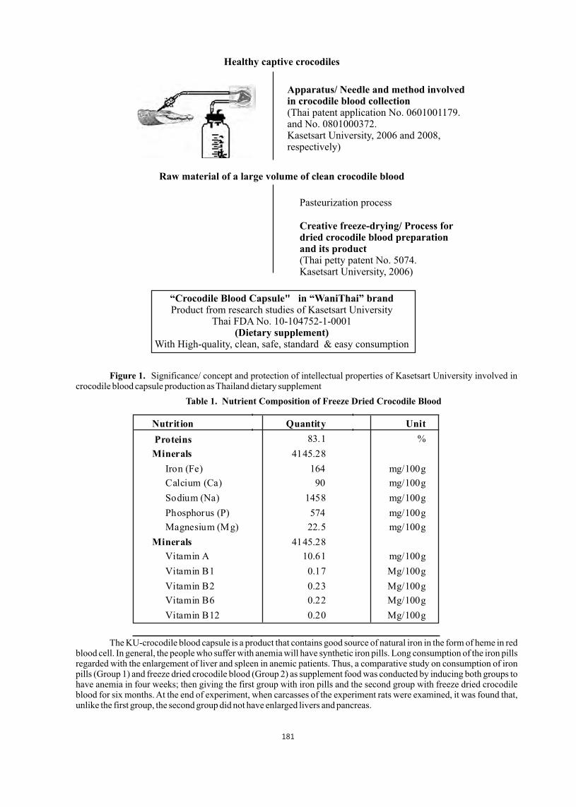
Figure 1. Significance/ concept and protection of intellectual properties of Kasetsart University involved in crocodile blood capsule production as Thailand dietary supplement
Table 1. Nutrient Composition of Freeze Dried Crocodile Blood
The KU-crocodile blood capsule is a product that contains good source of natural iron in the form of heme in red blood cell. In general, the people who suffer with anemia will have synthetic iron pills. Long consumption of the iron pills regarded with the enlargement of liver and spleen in anemic patients. Thus, a comparative study on consumption of iron pills (Group 1) and freeze dried crocodile blood (Group 2) as supplement food was conducted by inducing both groups to have anemia in four weeks; then giving the first group with iron pills and the second group with freeze dried crocodile blood for six months. At the end of experiment, when carcasses of the experiment rats were examined, it was found that, unlike the first group, the second group did not have enlarged livers and pancreas.
Healthy captive crocodiles
Apparatus/ Needle and method involved in crocodile blood collection (Thai patent application No. 0601001179.and No. 0801000372.Kasetsart University, 2006 and 2008, respectively)
Raw material of a large volume of clean crocodile blood
Pasteurization process
Creative freeze-drying/ Process for dried crocodile blood preparation and its product(Thai petty patent No. 5074.Kasetsart University, 2006)
“Crocodile Blood Capsule" in “WaniThai” brandProduct from research studies of Kasetsart University
Thai FDA No. 10-104752-1-0001(Dietary supplement)
With High-quality, clean, safe, standard & easy consumption
Nutrition Quantity Unit
Proteins 83.1 %
Minerals 4145.28
Iron (Fe) 164 mg/100g
Calcium (Ca) 90 mg/100g
Sodium (Na) 1458 mg/100g
Phosphorus (P) 574 mg/100g
Magnesium (Mg) 22.5 mg/100g
Minerals 4145.28
Vitamin A 10.61 mg/100g
Vitamin B1 0.17 Mg/100g
Vitamin B2 0.23 Mg/100g
Vitamin B6 0.22 Mg/100g
Vitamin B12 0.20 Mg/100g
181
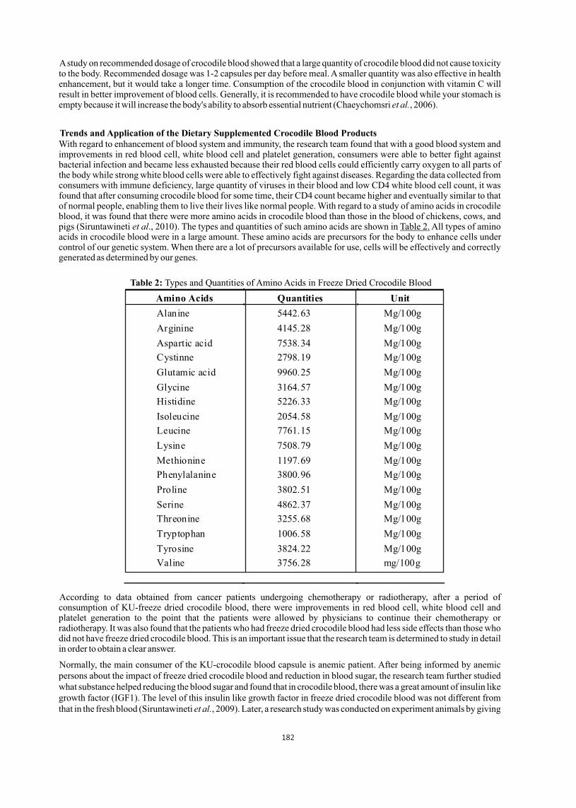
A study on recommended dosage of crocodile blood showed that a large quantity of crocodile blood did not cause toxicity to the body. Recommended dosage was 1-2 capsules per day before meal. A smaller quantity was also effective in health enhancement, but it would take a longer time. Consumption of the crocodile blood in conjunction with vitamin C will result in better improvement of blood cells. Generally, it is recommended to have crocodile blood while your stomach is empty because it will increase the body's ability to absorb essential nutrient (Chaeychomsri et al., 2006).
With regard to enhancement of blood system and immunity, the research team found that with a good blood system and improvements in red blood cell, white blood cell and platelet generation, consumers were able to better fight against bacterial infection and became less exhausted because their red blood cells could efficiently carry oxygen to all parts of the body while strong white blood cells were able to effectively fight against diseases. Regarding the data collected from consumers with immune deficiency, large quantity of viruses in their blood and low CD4 white blood cell count, it was found that after consuming crocodile blood for some time, their CD4 count became higher and eventually similar to that of normal people, enabling them to live their lives like normal people. With regard to a study of amino acids in crocodile blood, it was found that there were more amino acids in crocodile blood than those in the blood of chickens, cows, and pigs (Siruntawineti et al., 2010). The types and quantities of such amino acids are shown in Table 2. All types of amino acids in crocodile blood were in a large amount. These amino acids are precursors for the body to enhance cells under control of our genetic system. When there are a lot of precursors available for use, cells will be effectively and correctly generated as determined by our genes.
Table 2: Types and Quantities of Amino Acids in Freeze Dried Crocodile Blood
According to data obtained from cancer patients undergoing chemotherapy or radiotherapy, after a period of consumption of KU-freeze dried crocodile blood, there were improvements in red blood cell, white blood cell and platelet generation to the point that the patients were allowed by physicians to continue their chemotherapy or radiotherapy. It was also found that the patients who had freeze dried crocodile blood had less side effects than those who did not have freeze dried crocodile blood. This is an important issue that the research team is determined to study in detail in order to obtain a clear answer.
Normally, the main consumer of the KU-crocodile blood capsule is anemic patient. After being informed by anemic persons about the impact of freeze dried crocodile blood and reduction in blood sugar, the research team further studied what substance helped reducing the blood sugar and found that in crocodile blood, there was a great amount of insulin like growth factor (IGF1). The level of this insulin like growth factor in freeze dried crocodile blood was not different from that in the fresh blood (Siruntawineti et al., 2009). Later, a research study was conducted on experiment animals by giving
Amino Acids Quantities Unit
Alanine 5442.63 Mg/100g
Arginine 4145.28 Mg/100g
Aspartic acid 7538.34 Mg/100g
Cystinne 2798.19 Mg/100g
Glutamic acid 9960.25 Mg/100g
Glycine 3164.57 Mg/100g
Histidine 5226.33 Mg/100g
Isoleucine 2054.58 Mg/100g
Leucine 7761.15 Mg/100g
Lysine 7508.79 Mg/100g
Methionine 1197.69 Mg/100g
Phenylalanine 3800.96 Mg/100g
Proline 3802.51 Mg/100g
Serine 4862.37 Mg/100g
Threonine 3255.68 Mg/100g
Tryptophan 1006.58 Mg/100g
Tyrosine 3824.22 Mg/100g
Valine 3756.28 mg/100g
Trends and Application of the Dietary Supplemented Crocodile Blood Products
182

diabetes-induced animals freeze dried crocodile blood. It was found that the diabetic rats' blood sugar was restored to normal level in four weeks after receiving the freeze dried crocodile blood, but they still had diabetes. This showed that freeze dried crocodile blood and plasma could help reducing the blood sugar at a level. Recently, the crocodile blood product has been developed for diabetes supplement designed as “crocodile serum capsule” (Thai patent application No. 0901001229, Kasetsart University, 2009). It is the second food supplement product that may give the advantage for diabetic patients. However, more research in clinical trial should be performed. In industrial scale, crocodile blood used in the production has mostly received from healthy crocodiles gone into the slaughterhouse. So, the blood received from dead crocodiles has not enough plenty to produce the product. Therefore, crocodile blood collection process on animal life maintain of captive crocodiles has been created. (Pitimol et al. 2008). The donation of crocodile blood was studied by development of blood collection process on animal life maintains in captive Siamese crocodile for sustainable supplying (Thai petty patent No. 7468, Kasetsart University, 2009). This will be a new crocodile blood product with differ in process of blood collection to maintain live crocodiles. In the past, crocodile blood was waste. For these data of KU-WaniThai crocodile blood capsule demonstrated that the crocodile blood useful and has been added value with researches and scientific creations. It is a good case in value adding of the crocodile blood and its applications to support sustainable use.
References
01. Chaeychomsri, W., J. Siruntawineti, B. Vajarasathira, M. Tuntirungkij, S. Chaeychomsri, Y. Temsiripong, and R. Sappapan. 2003. Bactericidal Efficiency of the Thai Freshwater Crocodile Serum. J. Med. Tech. & Phys. Therapy, 15 supp 1: S120.
02. Chaeychomsri, W., J. Siruntawineti., B. Vajarasathira, K. Komoljindakul, T. Ong-aat, and Y. Temsiripong. 2004. Intestinal parasites of freshwater crocodile, Crocodylus siamensis, in Thailand. In: Proceeding of the 17th Working Meeting of the Crocodile Specialist Group. 2429 May 2004, Darwin Australia.
03. Chaeychomsri, W., J. Siruntawineti, B. Vajarasathira, and Y. Temsiripong. 2006. le blood diet. In: tion of crocodiProceeding of the 18th
Working Meeting of the Crocodile Specialist Group. 19 23 June 2006, Monte'limar, France.
04. Chaeychomsri, W., J. Siruntawineti, D. Hengsawadi, Y. Cuptapan, B. Vajarasathira and Y. Temsiripong. Efficiency of freeze-dried crocodile blood in iron deficiency anemia male rats. In: Proceeding of the 33rd Congress on Science and Technology of Thailand. 18-20 October 2007, Walailak University, Nakhon Si Thammarat, Thailand.
05. Chaeychomsri, W., S. Chaeychomsri, J. Siruntawineti, D. Hengsawadi, and Y. Cuptapan. 2009. Freeze-dried crocodile blood production as food supplement. J Biosci Bioeng 108: S22.
06. Siruntawineti, J., W. Chaeychomsri, B. Vajarasathira, A. Ritwichai T. Ong-aat, and Y. Temsiripong. 2004a. Blood parasites of Crocodylus siamensis in Thailand. In: Proceeding of the 17th Working Meeting of the Crocodile Specialist Group. 2429 May 2004, Darwin, Australia
07. Siruntawineti, J., W. Chaeychomsri, B. Vajarasathira, T. Ong-aat, and Y. Temsiripong. 2004b. Efficacy of Siamese crocodile (Crocodylus siamensis) serum on bacterial growth inhibition. In: Proceeding of the 17th Working Meeting of the Crocodile Specialist Group. 2429 May 2004, Darwin, Australia.
08. Siruntawineti, J., W. Chaeychomsri, D. Hengsawadi, Y. Cuptapan and B. Vajarasathira. 2008. Efficiency of freeze-dried crocodile blood in iron deficiency anemia male rats. In: Proceeding of the 19th Working Meeting of the Crocodile Specialist Group of the Species Survival Comission of IUCN-The World Conservation Union convened at Santa Cruz de la Sierra, Bolivia, 2-6 June 2008, 196-201.
09. Siruntawineti, J., W. Chaeychomsri and S. Ardom. 2009. Analysis of insulin like growth factor-I in crocodile serum for development of diabetes mellitus pateint's functional food. In: Proceeding of the 35th Congress on Science and Technology of Thailand. 15 - 17 October 2009, The Tide Resort (Bangsaen Beach), Chonburi, Thailand.
10. Siruntawineti, J., W. Chaeychomsri, D. Hengsawadi, and Y. Cuptapan. Chemical compositions of Siamese crocodile blood. In: Proceeding of the 36th Congress on Science and Technology of Thailand. 26 - 28 October 2010, Bangkok International Trade & Exhibition Centre (BITEC), Bangkok, Thailand.
11. Merchant, M., C. Roche, R.M. Elsey and J. Prudhomme. 2003. Antibacterial properties of serum from the American alligator (Alligator mississippiensis). Comp. Biochem. Physiol., B. 136: 505-513.
12. Thai patent application No. 0601001179. Apparatus and process for large volume of blood collection. Kasetsart University ; Chaeychomsri, W. and J. Siruntawineti on 16 March 2006.
13. Thai Petty Patent No. 5074. Process for preparation of crocodile blood powder and its product. Kasetsart University ; Chaeychomsri, W. and J. Siruntawineti on 5 April 2006.
14. Thai patent application No. 0901001229. Process for large volume of crocodile serum production and its product. Kasetsart University ; Chaeychomsri, W. and J. Siruntawineti on 19 March 2009.
15. Thai Petty Patent No. 7468. Process for crocodile blood collection by maintain life including its product. Kasetsart University ; Chaeychomsri, W. and J. Siruntawineti. on 20 Novemberl 2009.
183
Effects of fire Ant (Solenopsis invitica) predation on Alligator mississippiensis hatchlings
1 1 2 2 3Rodolfo Falconi , Mark Merchant , Virginia Parachu , Carlos Pina and Amos Cooper
1 Department of Chemistry, McNeese State University, Lake Charles, Louisiana, USA |2 CONICET, Diamante,Entre Rios, Argentina |3 JD Murphree Wildlife Management Area, Texas Parks and Wildlife Department,
Port Arthur, Texas, USA
Red fire ants are known to colonize mound nests of American alligators, and attack hatchlings at the time of hatching. These predatory attacks often results in consumption of the hatchling(s) by fire an colonies, and those that survive exhibit reduced growth rates and increased morbidity. We exposed newly hatched, wild-caught alligator hatchlings to red fire an colonies for different amounts of time. Increased exposure of alligator hatchlings to fire ants resulted in increased morbidity. In addition, longer exposure times resulted in higher plasma corticosteroid levels, and indicator of physiological stress. However, we observed a decrease in the total leukocyte counts, and a decrease in heterophil/leukocyte ratios, another indicator of stress. These data indicate that the red fire ant attacks may have an immunosuppressive effect of alligator hatchlings.
1 1 2 3Mark Merchant , Dusty Savage , Amos Cooper and Chris Murray
1 Department of Chemistry, McNeese State University, Lake Charles, Louisiana, USA | 2 JD Murphree WildlifeManagement Area, Texas Parks and Wildlife Department, Port Arthur, Texas, USA | 3 Biology Department,
Southeastern Louisiana University, Hammond, Louisiana, USA
Previous data from our laboratory has shown that motion-sensitive, infrared (IR)-based camera traps are not reliable for the quantitative capture of images of alligators. Therefore, we designed a camera trigger mechanism which included an electrical circuit board, coupled to a camera, which powered an IR Led light. The circuit was designed to turn the IR LED on for 2 seconds every five minutes. In the field, the IR LED was positioned such that the light was pointing directly into the IR detector of the camera. Therefore, the cameras were stimulated to take photos every five minutes, throughout the entire nesting period. The data revealed that alligators attend and maintain their nests more frequently during the first four to seven days after egg deposition, and then attendance is decreased. Nest attendance increased toward the end of the incubation period as eggs neared the hatching stage. In addition to nest visitation data, we also gained information concerning nest predation.
Proceedings : World Crocodile Conferencend -22 Working Meeting of the IUCN SSC Crocodile Specialist Groupnd -22 Working Meeting of the IUCN SSC Crocodile Specialist Groupnd -22 Working Meeting of the IUCN SSC Crocodile Specialist Group
Sri Lanka 20-23 May 2013 184
Assessment of nest attendance of the American Alligator (Alligator mississippiensis)using a modified motion sensitive camera trap
p 184
Salinity and alligator egg shape variation: A geometric morphometric analysis
1 1 2 2 3Mark Merchant , Dusty Savage , Amos Cooper , Monique Slaughter and Chris Murray1 Department of Chemistry, McNeese State University, Lake Charles, Louisiana, USA | 2 JD Murphree Wildlife
Management Area, Texas Parks and Wildlife Department, Port Arthur, Texas, USA | 3 Biology Department,Southeastern Louisiana University, Hammond, Louisiana, USA
Past studies have dismissed the importance of egg shape in crocodilians, Squamates, and turtles and deemed eggs shape in birds and other amniotes as similar, in relation to functionality. The complexity of crocodilian eggs has been examined and both turtle and Squamate eggs have recently been regarded as physiologically more intricate than bird eggs. This study introduces a physiological approach to monitor alligator populations in freshwater and low salinity environments by quantifying egg shape in correlation with varying salinity. We introduce a fractional semi landmark shape template method to quantify egg shape within a geometric morphometric framework. This approach is beneficial because it allows for the quantification of shape for curved structures, such as eggs, which lack homologous landmarks. The results from this study suggest that alligator egg shape is correlated with varying salinity levels, such that variation in alligator egg shape at low salinities changes in gradient like fashion while salinities high enough to be deemed stressful result in reversion back to a low salinity egg shape or desiccation. This study elucidates a correlation that can be implemented in management and breeding techniques and opens the door to in depth physiological examination of the system
Progress report on crocodilian genome sequencing
Chris MoranThe International Crocodylian Genomes Working Group, Faculty of Veterinary Science,
University of Sydney, NSW 2006, Australia
Genome and transcriptome sequences have been generated for the saltwater crocodile, Crocodylus porosus, the American alligator, Alligator mississipiensis, and the Indian gharial, Gavialis gangeticus. These sequences are being analysed and annotated by The Crocodylian Genomics Consortium, coordinated by Dr. David Ray of Mississippi State University. As well as automated gene identification and annotation of the sequences for alligator and crocodile, accessible via a GBROWSE viewer and freely available to all interested parties (go to http://gatorwiki. soe.ucsc.edu/doku.php?id=start for details on access), the genome sequences are available for direct interrogation via BLAST and manual analysis to authorized members of the consortium via CoGE or by using other more specialized bioinformatics tools. The genome sequences of all three species are already at a relatively advanced stage of assembly and annotation but further refinement is also underway. Large numbers of SNPs (single nucleotide polymorphisms) have been identified in both alligator and crocodile as part of the initial genome sequencing, with preliminary results suggesting lower levels of genetic variability in these crocodylians compared with other vertebrates. Currently a concerted effort of large scale SNP identification and genotyping via sequencing is being implemented in the saltwater crocodile. These genome sequences represent very valuable resources for all crocodylian researchers. Updated details and resources are available at http://crocgenomes.org
Sri Lanka 20-23 May 2013 185
Proceedings : World Crocodile Conferencend -22 Working Meeting of the IUCN SSC Crocodile Specialist Groupnd -22 Working Meeting of the IUCN SSC Crocodile Specialist Groupnd -22 Working Meeting of the IUCN SSC Crocodile Specialist Group
p 185

Disturbance effects on a South African river and the impact on the Mutale River Crocodylus niloticus population
1,2 1 2 2Ashley Pearcy , Myfannwyn Gibson , Jenny Balmagia , Julia Berkey ,
2 2Peter Flynn and Storme Viljoen1 Organization for Tropical Studies, PO Box 33, Skukuza, South Africa, 1350
2Animal Plant and Environmental Sciences, University of the Witwatersrand, South Africa 2001
Abstract
In 2000, major floods occured in several rivers within South Africa, resulting in alterations of river shape and biodiversity. The Mutale River was greatly affected by these floods and changed from a narrow, deep river often associated with a gorge and a clear riparian area, to the now flat, shallow river with a multitude of small pools. With these physical alterations in the river, the macrobiota in the river also changed. This river now hosts larger crocodiles in deeper pools, while crocodiles of varying size inhabit the runs between the pools. Currently, the Mutale River is under consideration to become a reserve for Nile crocodiles, Crocodylus niloticus. As of January 2013, heavy rains induced a large flood in Limpopo province, further flattening the river and delivered large amounts of sediment downstream. We found that the percent of suitable breeding habitat has doubled, the water quality has improved and the crocodile population is still present in a 15km stretch of river after the January 2013 floods. While these results indicate habitat improvement for breeding individuals, there has been a loss of habitat for larger crocodiles in the river. These results point towards the sensitivity of the Mutale River to disturbance by floods, which could lead to future degradation of Crocodylus niloticus habitat. This makes continued monitoring of this area necessary for a successful crocodile reserve and also highlights the sensitivity of South African rivers and vulnerability of Nile crocodiles to these disturbances.
Introduction
The availability of South Africa's freshwater resources has a poor outlook for the near future. According to the National Biodiversity Institute (2004), 84% of ecosystems associated with larger rivers are considered vulnerable (Colvin et al. 2011). The increase in human settlement and development around rivers, coupled with global climate change which is predicted to increase the occurrence of extreme weather events, the state of river ecosystems is increasingly vulernerable. With 82 % of rivers already threatened, 44% of which are also critically endangered, the conservation of these scarce water sources is of serious concern. South Africa is a dry region, receiving an average of only 490 mm of rainfall annually (Colvin et al. 2011). This aridity has direct implications for river flow and water availability, particularly for the rural communities that use rivers as primary water sources, as well as for the species that inhabit them. River conservation in South Africa is focussed on the catchment areas in the east. However, this leaves the majority of the river systems susceptible to anthropogenic influences before they reach protected areas. Monitoring of these highly valued and vulnerable systems is imperative for their conservation.
Management of crocodiles is closely linked to river conservation, as they form an important component of aquatic ecosystems (Davis and Day 1998). The only crocodile found in South Africa is the Nile crocodile, Crocodylus niloticus, whose range is restricted to the northern and eastern regions and shrinking further as a result of habitat destruction (Alexander and Marais 2007). Populations of C. niloticus are relatively high in protected areas (Fergusson 2010); however, they are still being heavily impacted by upstream activity in the unprotected areas. Crocodiles in South Africa are listed as 'vulnerable' by the IUCN. Threats to these include habitat loss, human-crocodile conflict, anthropogenic impacts from land use and active hunting for the crocodile skin trade (Ross 1998 from Botha 2005, Combrink et al. 2011).
Water quality is an important variable in assessing river health. Crocodiles, as apex predators, can serve as important indicators of a healthy river system (Mazzotti et al. 2009). Their health is closely linked to the health of food webs, diversity and productivity of other species and to freshwater flow. Crocodile population responses, including their distribution and abundance, are directly related to changes in environmental condition, thus these responses are valuable for assessment of disturbance impacts for both river and crocodile conservation.
A significant population of C. niloticus is found in Mutale River of the HaMakuya district, South Africa. The Mutale River is part of the Limpopo catchment, and has low levels of pollution and human-crocodile conflict despite being an important water resource for the surrounding villages (Celestino et al. 2012 cited by Correa et al. 2012). It contains all size classes of crocodiles, although mostly juveniles and mid-size individuals (Botha 2005). After extensive flooding in 2000, the Mutale River became more shallow and wide, and had significant impacts on the species composition of the
Proceedings : World Crocodile Conferencend -22 Working Meeting of the IUCN SSC Crocodile Specialist Groupnd -22 Working Meeting of the IUCN SSC Crocodile Specialist Groupnd -22 Working Meeting of the IUCN SSC Crocodile Specialist Group
Sri Lanka 20-23 May 2013 186
pp 186 - 191
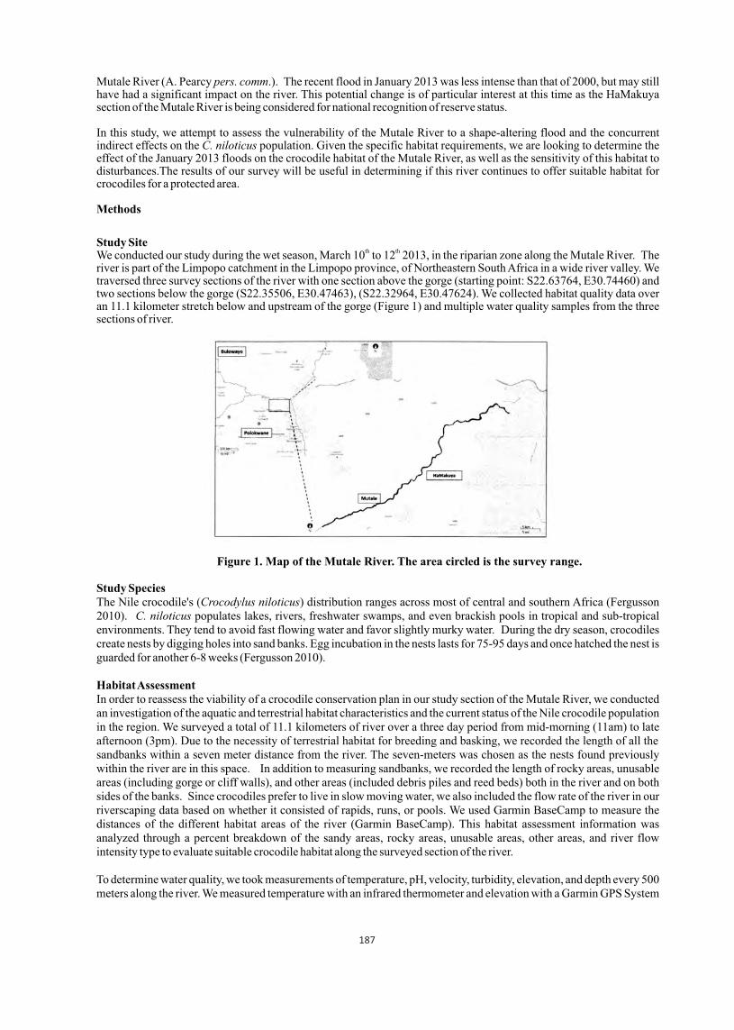
Mutale River (A. Pearcy pers. comm.). The recent flood in January 2013 was less intense than that of 2000, but may still have had a significant impact on the river. This potential change is of particular interest at this time as the HaMakuya section of the Mutale River is being considered for national recognition of reserve status.
In this study, we attempt to assess the vulnerability of the Mutale River to a shape-altering flood and the concurrent indirect effects on the C. niloticus population. Given the specific habitat requirements, we are looking to determine the effect of the January 2013 floods on the crocodile habitat of the Mutale River, as well as the sensitivity of this habitat to disturbances.The results of our survey will be useful in determining if this river continues to offer suitable habitat for crocodiles for a protected area.
Methods
Study Siteth thWe conducted our study during the wet season, March 10 to 12 2013, in the riparian zone along the Mutale River. The
river is part of the Limpopo catchment in the Limpopo province, of Northeastern South Africa in a wide river valley. We traversed three survey sections of the river with one section above the gorge (starting point: S22.63764, E30.74460) and two sections below the gorge (S22.35506, E30.47463), (S22.32964, E30.47624). We collected habitat quality data over an 11.1 kilometer stretch below and upstream of the gorge (Figure 1) and multiple water quality samples from the three sections of river.
Figure 1. Map of the Mutale River. The area circled is the survey range.
Study SpeciesThe Nile crocodile's (Crocodylus niloticus) distribution ranges across most of central and southern Africa (Fergusson 2010). C. niloticus populates lakes, rivers, freshwater swamps, and even brackish pools in tropical and sub-tropical environments. They tend to avoid fast flowing water and favor slightly murky water. During the dry season, crocodiles create nests by digging holes into sand banks. Egg incubation in the nests lasts for 75-95 days and once hatched the nest is guarded for another 6-8 weeks (Fergusson 2010).
Habitat Assessment In order to reassess the viability of a crocodile conservation plan in our study section of the Mutale River, we conducted an investigation of the aquatic and terrestrial habitat characteristics and the current status of the Nile crocodile population in the region. We surveyed a total of 11.1 kilometers of river over a three day period from mid-morning (11am) to late afternoon (3pm). Due to the necessity of terrestrial habitat for breeding and basking, we recorded the length of all the sandbanks within a seven meter distance from the river. The seven-meters was chosen as the nests found previously within the river are in this space. In addition to measuring sandbanks, we recorded the length of rocky areas, unusable areas (including gorge or cliff walls), and other areas (included debris piles and reed beds) both in the river and on both sides of the banks. Since crocodiles prefer to live in slow moving water, we also included the flow rate of the river in our riverscaping data based on whether it consisted of rapids, runs, or pools. We used Garmin BaseCamp to measure the distances of the different habitat areas of the river (Garmin BaseCamp). This habitat assessment information was analyzed through a percent breakdown of the sandy areas, rocky areas, unusable areas, other areas, and river flow intensity type to evaluate suitable crocodile habitat along the surveyed section of the river.
To determine water quality, we took measurements of temperature, pH, velocity, turbidity, elevation, and depth every 500 meters along the river. We measured temperature with an infrared thermometer and elevation with a Garmin GPS System
187

(GPSmap 60CSx). Turbidity was assessed by counting the number of visible dots of different sizes on a piece of paper, out of five, that could be seen through a clear plastic bottle filled with the sample water. Velocity was measured by determining the amount of time it took for a meter long string attached to a water-soaked apple to fully extend. Using a tape measure to find depth, we measured three randomly selected points at the sample site's edge. We collected three macroinvertebrate community samples every 1000 meters within each survey section of river. For safety, these samples were collected in rapids since crocodiles tend to avoid fast flowing water. The water was disturbed within the site for five minutes and we collected debris by holding a net directly downstream of the disturbance. Six people then performed active searching on the sample for fifteen minutes. The collected taxa were identified using an invertebrate identification guide (Gerber and Gabriel 2002).
The water quality data was assessed using the South African Scoring System (SASS) Version 5 Rapid Bioassessment Method (Dickens and Graham 2010). The average score per taxon (ASPT) was calculated for the macroinvertebrates found in each site. We used ASPT instead of SASS scores as a conservative estimate of river quality and as a comparison to previously conducted studies (Dickens and Graham 2010).
Disturbance Assessment To assess the broad scale changes and sensitivity of the Mutale River to a disturbance, we compared riverscaping data before and after the January 2013 floods. We accomplished this comparison using percentage breakdown of habitat type pre- and post- disturbance (Dzumaga 2012). We only compared above gorge data because that was the only section of the river where the pre- and post- disturbance data overlapped.
River quality pre- and post- disturbance were also compared in order to assess the sensitivity of the Mutale River to disturbance. Significant drivers of variation in river quality were identified using Principle Component Analysis (PCA). The PCA was calculated using JMP (2010) with the inputs including ASPT, pH, velocity, temperature, width, average depth, and elevation for all ten sample sites. ASPT scores were used as a comparison tool between pre- and post- disturbance river quality. Crocodile surveysWe conducted night spotlight surveys over a three-day period. Since crocodiles are most active after sunset, we walked back along the same stretch of river surveyed during the day and located crocodiles from 2000 to 2300. We detected crocodiles based on their distinctive red eye shine reflection. Life stages were confirmed through day sightings or full body visuals. This survey, along with signs of crocodile activities we observed during our daily data collection, gave us an indication of the current demographic distribution of the Nile crocodile in this portion of the river. It also allowed us to compare crocodile population distributions over several seasons.
Results
Habitat AssessmentAlong our survey section of the Mutale River, we surveyed sections covering bank both above and below the gorge and took measurements of water quality at 10 sites along this stretch. In this total length surveyed, we found a similar percent of the banks covered by sand (37%) and terrain unusable by C. niloticus (32%) (Figure 2). We found the majority of the river to be comprised of runs (41%) with relatively few pools (5%) (Figure 3). Water quality throughout the surveyed area was relatively high, with an average ASPT score of 8.99 for the 10 water quality sites.
Figure 2: Percentage breakdown of terrestrial habitat along surveyed section of the Mutale River.
188
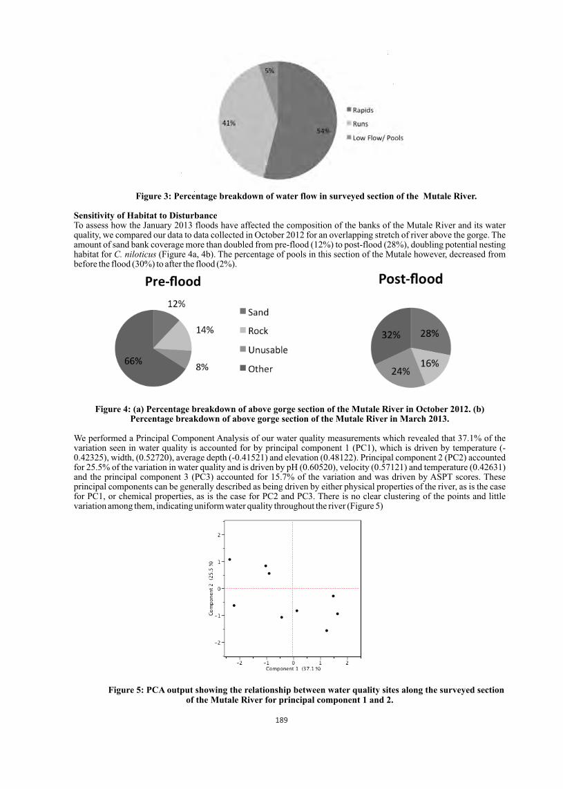
Figure 3: Percentage breakdown of water flow in surveyed section of the Mutale River.
Sensitivity of Habitat to DisturbanceTo assess how the January 2013 floods have affected the composition of the banks of the Mutale River and its water quality, we compared our data to data collected in October 2012 for an overlapping stretch of river above the gorge. The amount of sand bank coverage more than doubled from pre-flood (12%) to post-flood (28%), doubling potential nesting habitat for C. niloticus (Figure 4a, 4b). The percentage of pools in this section of the Mutale however, decreased from before the flood (30%) to after the flood (2%).
Figure 4: (a) Percentage breakdown of above gorge section of the Mutale River in October 2012. (b) Percentage breakdown of above gorge section of the Mutale River in March 2013.
We performed a Principal Component Analysis of our water quality measurements which revealed that 37.1% of the variation seen in water quality is accounted for by principal component 1 (PC1), which is driven by temperature (-0.42325), width, (0.52720), average depth (-0.41521) and elevation (0.48122). Principal component 2 (PC2) accounted for 25.5% of the variation in water quality and is driven by pH (0.60520), velocity (0.57121) and temperature (0.42631) and the principal component 3 (PC3) accounted for 15.7% of the variation and was driven by ASPT scores. These principal components can be generally described as being driven by either physical properties of the river, as is the case for PC1, or chemical properties, as is the case for PC2 and PC3. There is no clear clustering of the points and little variation among them, indicating uniform water quality throughout the river (Figure 5)
Figure 5: PCA output showing the relationship between water quality sites along the surveyed section of the Mutale River for principal component 1 and 2.
189
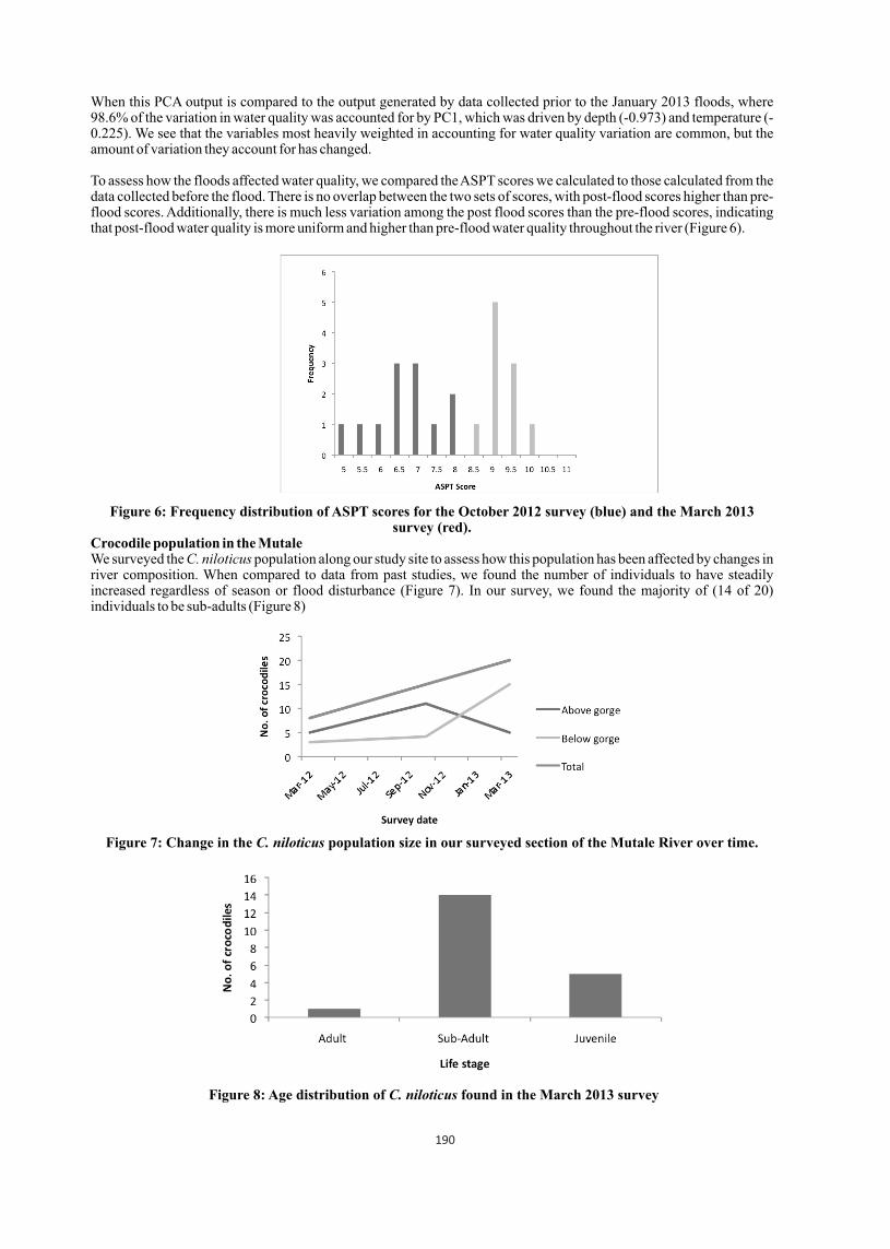
When this PCA output is compared to the output generated by data collected prior to the January 2013 floods, where 98.6% of the variation in water quality was accounted for by PC1, which was driven by depth (-0.973) and temperature (-0.225). We see that the variables most heavily weighted in accounting for water quality variation are common, but the amount of variation they account for has changed.
To assess how the floods affected water quality, we compared the ASPT scores we calculated to those calculated from the data collected before the flood. There is no overlap between the two sets of scores, with post-flood scores higher than pre-flood scores. Additionally, there is much less variation among the post flood scores than the pre-flood scores, indicating that post-flood water quality is more uniform and higher than pre-flood water quality throughout the river (Figure 6).
Figure 6: Frequency distribution of ASPT scores for the October 2012 survey (blue) and the March 2013 survey (red).
Crocodile population in the MutaleWe surveyed the C. niloticus population along our study site to assess how this population has been affected by changes in river composition. When compared to data from past studies, we found the number of individuals to have steadily increased regardless of season or flood disturbance (Figure 7). In our survey, we found the majority of (14 of 20) individuals to be sub-adults (Figure 8)
Figure 7: Change in the C. niloticus population size in our surveyed section of the Mutale River over time.
Figure 8: Age distribution of C. niloticus found in the March 2013 survey
190
Discussion
The protection of the vulnerable Nile crocodile presents an important issue for conservation in South Africa, especially since the crocodile is an apex predator and thus serves as an indicator of ecosystem health and water quality. In order to determine if the HaMakuya section of the Mutale River continues to offer suitable habitat for a protected area, we assessed the quality of crocodile habitat remaining after the January 2013 floods. We also examined the overall sensitivity of the system to such disturbances. In terms of post-disturbance habitat, the presence of a significant amount of sand banks along the crocodile buffer region of the river suggests that the terrestrial habitat remains favorable to crocodiles. Such sandbanks present optimal habitat for nesting sites (Fergusson 2010). Comparisons of our habitat assessment to the pre-flood data show that breeding habitat has actually increased post-disturbance, as a result of sediment deposition. Similarly, the 46% of the river that consists of runs or pools indicates that this section of the Mutale has remained deep enough to present suitable hunting and resting habitat for the crocodiles in the water. Furthermore, the overall quality of the water has increased post-flood. This could be because the recent flood had a cleansing effect, potentially removing pollutants and debris from the river.
However, not all the changes were as encouraging. As was the case with the major 2000 floods, this recent disturbance both widened and flattened out the river (A. Pearcy pers. comm.). As a result, the number of deep, slow moving pools, which represents ideal crocodile habitat within the river, decreased after the recent January floods. Furthermore, our PCA suggests that most of the variation observed along the river is attributable to properties such as width and depth; physical factors that are very sensitive to flooding effects. Since the relative importance of these physical factors in explaining variation has changed from before the floods, we can assume that these variables are sensitive to disturbance. Therefore, future floods may continue the flattening trend of the river, and potentially leave very little suitable pool habitat for the crocodiles.
The Mutale River's lack of deep pools may be attributing to the small number of adults found within our survey site. Only one fully grown adult was surveyed. The larger crocodiles may have moved downstream with the floods and have not yet returned or simply could be missing from this population. Either way, continued monitoring of the crocodile population will be necessary if the river is to serve as a protected area for this species.
Given the persistence of the crocodile population along the Mutale River after the recent floods, as well as the relatively favorable change in crocodile nesting habitat resulting from the flood, we have concluded that the HaMakuya region of this river continues to present a suitable conservation area for crocodiles. However, due to the flattening of the river as well as the sediment deposition associated with the recent flooding, the Mutale River now more closely resembles the Olifants River (A. Pearcy pers. comm.). Therefore, in order to avoid another crocodile die-off similar to the pansteatitis epidemic that the Olifants experienced from 2008-2009, conservation measures around the Mutale may be necessary to minimize the polluting effects of development (Ferreira and Pienaar 2001). As C. nilotocus numbers continue to decline in South Africa, the creation of such protected areas will be crucial in maintaining the species and offering a model for the rest of Africa in terms of balancing development with both crocodile and freshwater habitat conservation.
Acknowledgements
We would like to thank OTS students for helping with field work, the Tshulu Camp Staff, and Fhatuwani Makuya for aiding us in completing this study.
References
01. Alexander, G. and J. Marais. 2007. A guide to the reptiles of southern Africa. Struik, Cape Town, South Africa.02. Aust, P., B. Boyle, R. Fergusson, and T. Coulson. 2009. The impact of Nile crocodiles on rural livelihoods in northeastern Namibia. South African Journal of Wildlife Research 39(1): 57-69.03. Botha, P. J. 2005. The ecology and population dynamics of the Nile crocodile Crocodylus niloticus in the Flag Boshielo Dam, Mpumalanga province, South Africa. MSc thesis. University of Pretoria, Pretoria, South Africa.04. Colvin, C., A. Burns, K. Schachtschneider, A. Maherry, J. Charmier, and M. de Wit. 2011. Coal and water futures in South Africa: The case
for protecting headwaters in the Enkangala grassland. Report of WWF-World Wide Fund for Nature- South Africa, Cape Town, South Africa.
05. Combrink, X., J. L. Korrûbel, R. Kyle, R. Taylor, and P. Ross. 2011. Evidence of a declining Nile crocodile (Crocodylus niloticus) population at Lake Sibaya, South Africa. South African Journal of Wildlife Research 41(2): 145157.
06. Correa, M., Q. Gao, M. McClay. 2012. State of the Mutale River habitat for Crocodylus niloticus: assessing the feasibility of a river conservation plan. Organization for Tropical Studies report spring 2012. HaMakuya, Limpopo Province, South Africa.
07. Dickens, C. W. S., and P. M. Graham. 2010. The South African Scoring System (SASS) Version 5 Rapid Bioassessment Method for Rivers. African Journal of Aquatic Science 27(1): 110.
08. Dzumaga, Y., K. Li, C. Pratson, D. Prinsloo. 2012. Assessment of Nile crocodile (Crocodylus niloticus) habitat in the Mutale River. Organization for Tropical Studies report fall 2012. HaMakuya, Limpopo Province, South Africa.
09. Fergusson, R. A. 2010. Nile Crocodile Crocodylus niloticus. Pp. 84-89 in S. C. Manolis and C. Stevenson, editors. Crocodiles. Status Survey and Conservation Action Plan. Crocodile Specialist Group: Darwin. 10. Ferreira, S. M. and D. Pienaar. 2011. Degradation of the crocodile population in the Olifants River Gorge of Kruger National Part, South
Africa. Aquatic Conservation: Marine and Freshwater Ecosystems 21:155-164.11. Garmin BaseCamp. 2012. Version 4.0.1 Garmin Ltd.12. Gerber, A., and M. J. M. Gabriel. 2002. Aquatic Invertebrates of South African Rivers. Institute for Water Quality Studies. Version 2.
Department of Water Affairs and Forestry.13. JMP. 2010. Desktop Release version 9.0.0. SAS Institute Inc.14. Mazzotti, F. J., G. R. Best, L. A. Brandt, M. C. Cherkiss, B. M. Jeffery and K. G. Rice. 2009. Alligators and crocodiles as indicators for
restoration of Everglades ecosystems. Ecological Indicators 9: 137-149
191

Effects of freeze-dried crocodile serum supplementation on blood glucose level in diabetic Spraque dawley rats
1 1 2 Jindawan Siruntawineti *, Win Chaeychomsri and Sudawan Chaeychomsri1 Department of Zoology, Faculty of Science, Kasetsart University, Bangkok 10900, Thailand
2 Central Laboratory and Greenhouse Complex, Kasetsart University,
Kamphaeng Saen, Nakhon Pathom 73140, Thailand (* [email protected] )
Abstract
Previous studied showed that freeze-dried crocodile blood contain 677.2 ± 3.41 ng/gm of insulin-like growth factor-I (IGF-1). IGF-1 is synthesized in the liver upon stimulation by growth hormone and involved in cell replication, proliferation, protein synthesis, carbohydrate homeostasis, and bone metabolism. Recently, crocodile blood was developed as food supplement, dried crocodile blood capsule. According to preliminary study of the diabetic patient that consumed crocodile blood capsules showed the decreasing of blood sugar. Therefore, effects of freeze-dried crocodile serum supplementation on blood glucose level in Diabetic Spraque Dawley rats were performed by using 48 Spraque Dawley Rats. The experiments were divided in to 8 groups with 6 each and feeding for 8 weeks. Control group was fed on high fat corn starch based on AIN93G diet. Fourty two Spraque Dawley rats were induced to be diabetic which blood glucose level >400 ng/dl by streptozotocin 40 ng/kg single dose. They were performed to diabetic and experimental group 1-6. Diabetic group was fed on high fat corn starch based on AIN93G diet. The experiment groups 1-3 were fed on high fat corn starch based on AIN93G + freeze dried crocodile serum 310, 465 and 620 mg/kg/day respectively. The experiment groups 4-6 were fed on high fat corn starch based on AIN93G + freeze dried crocodile blood 1000, 1500 and 2000 mg/kg/day respectively. The results revealed that blood glucose level of experiment group 1-3 that supplemented with freeze-dried crocodile serum reduced to be normal in 4 week. In addition, blood glucose level of experimental group 4-6 that supplemented with freeze-dried crocodile blood still to be diabetic after 4 week. The present data demonstrated that freeze-dried crocodile serum have effects on reduction of blood glucose level better than freeze-dried crocodile blood.
Introduction
Freeze-dried crocodile blood in capsule was newly developed as a supplemented food product in Thailand (Chaeychomsri et al., 2006, 2009 ; Kasetsart University, patent application 2006 ; Siruntawineti et al., 2006, 2008) as KU (Kasetsart University) -crocodile blood capsule. It has been widely consumed not only for its nutritious composition, but also for its claimed medicinal value. Diabetes mellitus is an endocrine disorder characterized by hyperglycemia and glycosuria due to absolute or relative lack of insulin. Its incidence is increasing with alarming mortality and morbidity. There is an increasing demand for anti-diabetic products due to the drawbacks associated with insulin and oral hypoglycemic agents. From studying of pre- and post consumptions of KU-crocodile blood capsule in 2006, it demonstrates the good efficacy in a reduction of blood glucose level and good wound healing in consumers who suffer with diabetes (diabetic pateints). It has been hypothesized that there is IGF-1, a like substance that acts as insulin hormone (Rosen, 1999) in the serum part of crocodile blood. Then, the freeze-dried crocodile serum was studied (Siruntawineti et al., 2009) and presents that in Siamese crocodile serum has IGF-1 (67.28 + 4.79 ng/ml). Moreover, the level of IGF-1 in freeze-dried crocodile serum is not different from in the fresh serum. In 2009, Chaeychomsri and Siruntawineti were created process for large volume of crocodile serum production and its product (Thailand Patent application No. 0901001229, 2009). To clarify the beneficial effect of this freeze-dried crocodile serum capsule, the present study was performed to determine effects of freeze-dried crocodile serum supplementation on blood glucose level in diabetic Spraque Dawley rats.
Materials and Methods
Collection of crocodile blood: The blood in large volume was collected from Siamese crocodiles (Crocodylus siamensis) that captives at Rungtaweechai Crocodile Farm, Don Toom District, Nakhonpathum Province, Thailand according to a KU-patented method (Chaeychomsri and Siruntawineti, 2006).Freeze-dried crocodile serum/ blood preparation: Process for freeze-dried crocodile serum production of Thai patent application No. 0901001231, Kasetsart University (Chaeychomsri and Siruntawineti, 2009) was performed as following. After special separation with aseptic technique, the mass crocodile serum was pasteurized and prepared in tray and freeze-drying using freeze dryer (Lyomaster, USA). The freeze-dried serum was powdered by graining at sterile
ocondition and stored at 4 C until use. For freeze-dried crocodile blood preparation, the whole crocodile blood that descript in previous part was done as in freeze-dried crocodile serum.Laboratory animals: The Animal Ethics Committee of Kasetsart University, Thailand approved the use of laboratory animals in this study. Spraque Dawley male rats with weight 150-200 gm were used. Control group that the rat (n = 6)
Proceedings : World Crocodile Conferencend -22 Working Meeting of the IUCN SSC Crocodile Specialist Groupnd -22 Working Meeting of the IUCN SSC Crocodile Specialist Groupnd -22 Working Meeting of the IUCN SSC Crocodile Specialist Group
Sri Lanka 20-23 May 2013 192
pp 192 - 195
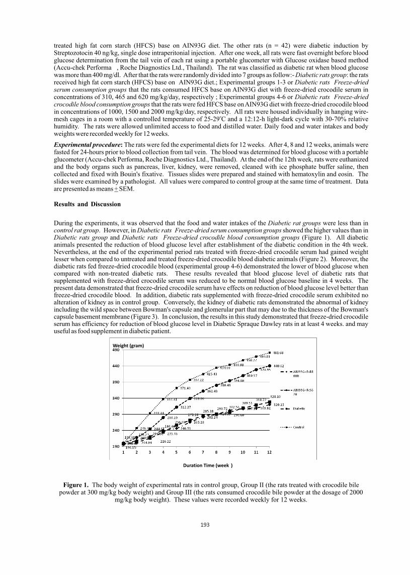
treated high fat corn starch (HFCS) base on AIN93G diet. The other rats (n = 42) were diabetic induction by Streptozotocin 40 ng/kg, single dose intraperitonial injection. After one week, all rats were fast overnight before blood glucose determination from the tail vein of each rat using a portable glucometer with Glucose oxidase based method (Accu-chek Performa , Roche Diagnostics Ltd., Thailand). The rat was classified as diabetic rat when blood glucose was more than 400 mg/dl. After that the rats were randomly divided into 7 groups as follow:- Diabetic rats group: the rats received high fat corn starch (HFCS) base on AIN93G diet.; Experimental groups 1-3 or Diabetic rats Freeze-dried serum consumption groups that the rats consumed HFCS base on AIN93G diet with freeze-dried crocodile serum in concentrations of 310, 465 and 620 mg/kg/day, respectively ; Experimental groups 4-6 or Diabetic rats Freeze-dried crocodile blood consumption groups that the rats were fed HFCS base on AIN93G diet with freeze-dried crocodile blood in concentrations of 1000, 1500 and 2000 mg/kg/day, respectively. All rats were housed individually in hanging wire-
omesh cages in a room with a controlled temperature of 25-29 C and a 12:12-h light-dark cycle with 30-70% relative humidity. The rats were allowed unlimited access to food and distilled water. Daily food and water intakes and body weights were recorded weekly for 12 weeks.
Experimental procedure: The rats were fed the experimental diets for 12 weeks. After 4, 8 and 12 weeks, animals were fasted for 24-hours prior to blood collection from tail vein. The blood was determined for blood glucose with a portable glucometer (Accu-chek Performa, Roche Diagnostics Ltd., Thailand). At the end of the 12th week, rats were euthanized and the body organs such as pancreas, liver, kidney, were removed, cleaned with ice phosphate buffer saline, then collected and fixed with Bouin's fixative. Tissues slides were prepared and stained with hematoxylin and eosin. The slides were examined by a pathologist. All values were compared to control group at the same time of treatment. Data are presented as means + SEM.
Results and Discussion
During the experiments, it was observed that the food and water intakes of the Diabetic rat groups were less than in control rat group. However, in Diabetic rats Freeze-dried serum consumption groups showed the higher values than in Diabetic rats group and Diabetic rats Freeze-dried crocodile blood consumption groups (Figure 1). All diabetic animals presented the reduction of blood glucose level after establishment of the diabetic condition in the 4th week. Nevertheless, at the end of the experimental period rats treated with freeze-dried crocodile serum had gained weight lesser when compared to untreated and treated freeze-dried crocodile blood diabetic animals (Figure 2). Moreover, the diabetic rats fed freeze-dried crocodile blood (experimental group 4-6) demonstrated the lower of blood glucose when compared with non-treated diabetic rats. These results revealed that blood glucose level of diabetic rats that supplemented with freeze-dried crocodile serum was reduced to be normal blood glucose baseline in 4 weeks. The present data demonstrated that freeze-dried crocodile serum have effects on reduction of blood glucose level better than freeze-dried crocodile blood. In addition, diabetic rats supplemented with freeze-dried crocodile serum exhibited no alteration of kidney as in control group. Conversely, the kidney of diabetic rats demonstrated the abnormal of kidney including the wild space between Bowman's capsule and glomerular part that may due to the thickness of the Bowman's capsule basement membrane (Figure 3). In conclusion, the results in this study demonstrated that freeze-dried crocodile serum has efficiency for reduction of blood glucose level in Diabetic Spraque Dawley rats in at least 4 weeks. and may useful as food supplement in diabetic patient.
Figure 1. The body weight of experimental rats in control group, Group II (the rats treated with crocodile bile powder at 300 mg/kg body weight) and Group III (the rats consumed crocodile bile powder at the dosage of 2000
mg/kg body weight). These values were recorded weekly for 12 weeks.
Weight (gram)
Duration Time (week )
193
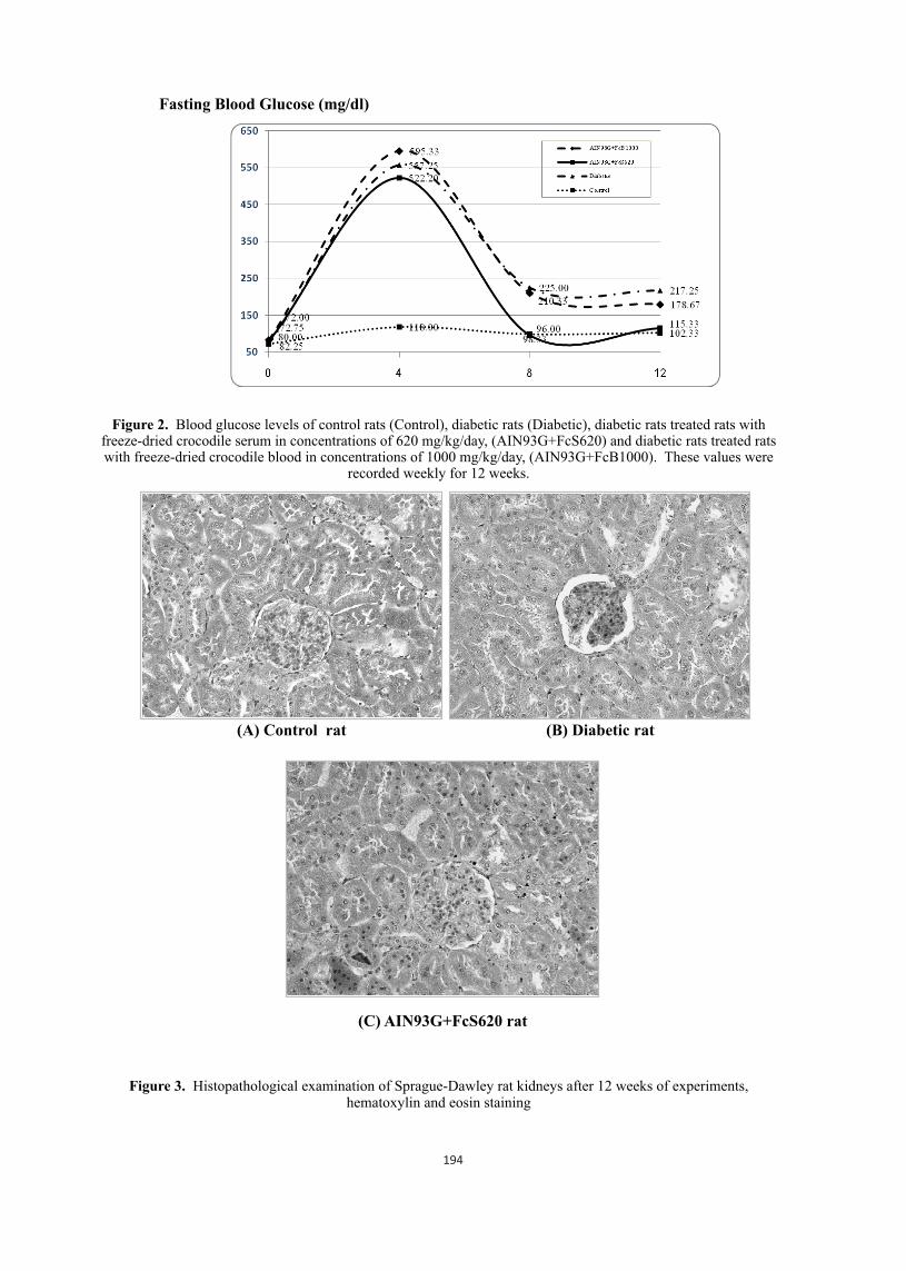
Figure 2. Blood glucose levels of control rats (Control), diabetic rats (Diabetic), diabetic rats treated rats with freeze-dried crocodile serum in concentrations of 620 mg/kg/day, (AIN93G+FcS620) and diabetic rats treated rats with freeze-dried crocodile blood in concentrations of 1000 mg/kg/day, (AIN93G+FcB1000). These values were
recorded weekly for 12 weeks.
Figure 3. Histopathological examination of Sprague-Dawley rat kidneys after 12 weeks of experiments, hematoxylin and eosin staining
Fasting Blood Glucose (mg/dl)
(A) Control rat (B) Diabetic rat
(C) AIN93G+FcS620 rat
194
(A) Kidney cell of control group (B) Abnormal sign of kidney cell from diabetic rat (C) Kidney cell of diabetic rat supplementary fed with freeze-dried crocodile serum, 620 mg/kg/day,
(AIN93G+FcS620 rat)
Acknowledgements
We appreciate Rungtaweechai Crocodile Farm and Wani Thai Limited Partnership for the crocodile blood samples and technical assistance. This study was supported by a Kasetsart University Research and Development Institute (KURDI) grant.
Literature Cited
01. Chaeychomsri, W., J. Siruntawineti, B. Vajarasathira, and Y. Temsiripong. 2006. Safety evaluation of crocodile blood diet. In: Proceeding of the 18th Working Meeting of the Crocodile Specialist Group. Monte'limar, France,19-23 June 2006.
02. Chaeychomsri, W., S. Chaeychomsri, J. Siruntawineti, D. Hengsawadi, and Y. Cuptapan. 2009. Freeze-dried crocodile blood production as food supplement. J Biosci Bioeng 108: S22.
03. Rosen, C.J. 1999. Serum insulin-like growth factors and insulin-like growth factor-binding proteins: clinical implications. Clin Chem 45: 1384-1390.
04. Siruntawineti, J., W. Chaeychomsri, C. Sathsumpun, P. Tungsathian, B. Vajarasathira and Y. Temsiripong. 2006. Development and evaluation of dried Siamese crocodile blood product as supplemented food. In; Proceeding of the 18th Working Meeting of the Crocodile Specialist Group. Monte'limar, France, 19 23 June 2006.
05. Siruntawineti, J., W. Chaeychomsri, D. Hengsawadi, Y. Cuptapan and B. Vajarasathira. 2008. Efficiency of freeze-dried crocodile blood in iron deficiency anemia male rats. In: Proceeding of the 19th Working Meeting of the Crocodile Specialist Group of the Species Survival Comission of IUCN-The World Conservation Union convened at Santa Cruz de la Sierra, Bolivia, 2-6 June 2008, 196-201.
06. Siruntawineti, J., W. Chaeychomsri and S. Ardom. 2009. Analysis of insulin like growth factor-I in crocodile serum for development of diabetes mellitus pateint's functional food. In: Proceeding of the 35th Congress on Science and Technology of Thailand. 15 - 17 October 2009, The Tide Resort (Bangsaen Beach), Chonburi, Thailand.
07. Thai patent application No. 0601001179. Apparatus and process for large volume of blood collection including the blood product. Kasetsart University ; Chaeychomsri, W. and Siruntawineti, J. on 16 March 2006.
08. Thai petty patent application No. 0901001229. Process for large volume of crocodile serum production and its product. Kasetsart University ; Chaeychomsri, W. and Siruntawineti, J. on 19 March 2009.
195
Three aliens and a predator: interactions between dingoes, passion vines, cane toads and freshwater crocodiles in arid Australia
1,2 2Ruchira Somaweera and Richard Shine
1 Biologic Environmental Survey, North Perth, WA, Australia ([email protected]) | 2 School of Biological Sciences, The University of Sydney, NSW, Australia ([email protected])
Human induced species introductions are one of the greatest threats to global biodiversity. Introduced species can have varying impacts on the viability of crocodilian populations but very limited information is available on these complex interactions, despite them providing excellent opportunities to understand the ways in which crocodilians deal with novel challenges. We studied the interactions between three introduced species and the Australian freshwater crocodile (Crocodylus johnstoni) at Lake Argyle in seasonally arid north western Australia, where dingoes predate on crocodile nests, passion vines modify nesting habitats and cane toads cause mortality of crocodiles through direct poisoning. We examined how the distinctive attributes of this relatively naïve lakeside landscape have affected predator guilds and fashioned unique predator prey interactions. Dingoes (Canis lupus dingo: introduced to Australia 3500 4000 yrs ago) were responsible for 98% of all predator visits to crocodile nests and the predation risk lasted throughout the incubation period and at hatching. Nest vulnerability was linked to abiotic features of the nest sites rather than to features of the nests.
Many nesting sites at the lake have been invaded by passion vine (Passiflora foetida var. hispida: introduced to Australia before 1900s) over the last few years and crocodiles actively avoided beaches with this species. Dingoes fed on the fruits of this plant and potentially spread them among nesting beaches when they defecated, further accelerating the spread of this vine. Studies are underway to investigate long term impacts of this plant on the crocodile population. In early 2009, toxic cane toads (Rhinella marina: introduced to Australia in 1935) arrived and have been spreading ever since. We monitored crocodile densities and body sizes, the spread and movement of cane toads and the diet of crocodiles to understand the interactions between the two species. Crocodiles commonly encountered toads, but contrary to expectation, crocodile mortality was rare, and crocodile numbers did not decrease through time, nor differ between toad infested versus toad free areas of the lake. In a broader study we investigated potential reasons for spatial heterogeneity of cane toad impacts (in some other areas, crocodile numbers have been decimated by toad arrival) and the potential mechanisms through which crocodile populations can recover.
Proceedings : World Crocodile Conferencend -22 Working Meeting of the IUCN SSC Crocodile Specialist Groupnd -22 Working Meeting of the IUCN SSC Crocodile Specialist Groupnd -22 Working Meeting of the IUCN SSC Crocodile Specialist Group
Sri Lanka 20-23 May 2013 196
p 196
Impact of a severe flood event on survivorship of hatchling Crocodylus johnstoni in northwestern Australia
In seasonal tropical environments, the magnitude and timing of monsoonal rainfall vary among years (Englehart and Douglas, 2001, Moron et al., 2003; Pittock et al., 2006), with strong effects both on native animals and plants (Collins et al., 1981; Pandey and Singh, 1992) as well as on the humans that depend upon those systems (Baddiley, 2003; Mahmudul et al., 2011). Rainfall averages have increased in the many parts of the world during the latter part of the last century (Diaz et al., 1989; IPCC 2007; Vinnikov et al., 1990). Additionally, higher than average or extreme rainfall events has also increased over the last century and projected to continue increasing under a variety of emissions scenarios (CCSP 2008; Ishak et al., 2010). For example, from 1950 to 2005, extreme daily rainfall intensity and frequency has increased in most parts of northwestern Australia (Kingsford et al., 2009; Steffen et al., 2009; CSIRO and BOM, 2011), and in recent years several tropical areas have experienced devastating floods as a result (e.g. Bennett et al., 2006; Done et al., 2007). Inter-annual variation in monsoonal flooding is natural, but because extreme weather events are predicted to occur more frequently and strongly in a changing climate (Knutson et al., 2010; Salvat and Wilkinson, 2011), we need to understand the pathways by which more frequent, longer and/or extreme flooding influences the biota.
For terrestrial species, severe flooding may result in mortality by drowning of individuals (Cornelius et al., 2007) or their prey, as well as reduced access to prey and critical habitats (Larriera and Piña, 2000). But are aquatic species also impacted by severe flooding? Here we present the results of a study which examined the impact of a severe flood on survivorship of hatchling Australian freshwater crocodiles (Crocodylus johnstoni) in a large anthropogenic lake in northwestern Australia.
Methods
Field sampling.- We conducted the study at Lake Argyle (16°29´S, 128°75´E), northeastern Western Australia, in the 2wet-dry tropics of Australia. Lake Argyle is the largest artificial lake in Australia (>880km at normal water level),
constructed in 1971 under the Ord River Irrigation Scheme, and is recognized as an important wetland under the Ramsar Convention. It contains one of the largest C. johnstoni populations in Australia (Webb and Manolis, 2010), but saltwater crocodiles (C. porosus) has been only sporadically recorded in the system (Somaweera, 2011). Hatchling C. johnstoni in this lake largely select shoreline vegetation mats (made of distinct clusters of submerged as well as emergent and floating-leaved macrophytes including Najas graminea, Hydrilla verticillata, Potamogeton tricarinatus, Valisneria spiralis, Myriophyllum verrucosum, Chara sp. and Nymphoides indica) as their living space, a selection driven both by higher prey availability and lower predation risk (Somaweera et al., 2011a). In order to monitor the amount of preferred habitat available for hatchlings, we delineated the area covered by shoreline vegetation at 16 sites within six sections at north-western portion Lake Argyle on a 1:20 000 topographical map during daytime surveys conducted in March 2009, November 2010 and August 2011. All 16 sites were bordered by steep hills and rocky alcoves, a common shoreline feature in the northern sections of the lake.
As a part of a multi-disciplinary study, between 2008 and 2010, annual spotlight surveys for crocodiles (Messel et al., 1981) were conducted in November, for 6-9 nights per month, from 1800-2300 h (15hp engine on a 3.7m boat). Hatchling C. johnstoni were caught and individually marked by tail-scute clipping (Jennings et al., 1991) within the 16 sites. The following August (2009-2011) we recaptured the crocodiles and measured them for lengths and weight.
We obtained data on daily water levels of the lake from the Kununurra Water Corporation and daily rainfall data from two weather monitoring stations (Lake Argyle resort 16.11°S, 128.74°E and Kununurra Aero 15.78°S, 128.71°E: Australian Bureau of Meteorology, 2011).
Statistical analysis.- One-way ANOVAs were conducted using JMP 5.01 software (SAS Institute, Cary, NC, USA) to compare among-survey variations in vegetation cover, recapture rates and growth of hatchlings; and used linear
1,2* 2 3Ruchira Somaweera , Jonathan Webb and Richard Shine1 2Biologic Environmental Survey, North Perth 6059, Australia. School of the Environment,
3 University of Technology, Sydney 2007, Australia. School of Biological Sciences, The University of Sydney, Sydney 2006, Australia. * [email protected]
Introduction
Sri Lanka 20-23 May 2013 197
Proceedings : World Crocodile Conferencend -22 Working Meeting of the IUCN SSC Crocodile Specialist Groupnd -22 Working Meeting of the IUCN SSC Crocodile Specialist Groupnd -22 Working Meeting of the IUCN SSC Crocodile Specialist Group
pp 197 - 202
regressions to analyze the effect of habitat loss on crocodile survival and size. Our comparisons of growth and recapture rates were based on nine-month old hatchings from 2008 (recaptured in 2009), 2009 (recaptured in 2010) and 2010 (recaptured in 2011), respectively.
Additionally, we used program MARK software (Version 6.1; Cooch and White, 2011) to estimate survival () and recapture probabilities (p) of C. johnstoni from Lake Argyle between February 2009 and August 2011 (total eight trapping occasions). We formulated a series of a priori candidate Cormack-Jolly-Seber models for and p, and implemented these in program MARK following White and Burnham (1999). Previous long-term mark-recapture studies have documented age-specific survival in C. johnstoni (Webb and Smith, 1984) so we included age structure in our models. Our null model was one in which survival and recapture rates were age-dependent, with three age classes (because our study spanned three years). We then formulated age-structured models to investigate whether survival rates were influenced by (1) the degree of vegetation cover; (2) flooding; or (3) interactions between vegetation and flooding.
We used an information-theoretic approach (AIC) for model selection (Burnham and Anderson 1998). We used the median ĉ goodness-of-fit procedure approach to estimate the over-dispersion parameter (ĉ) (Cooch and White, 2011). The over-dispersion quasi-likelihood parameter indicates the degree of inadequacy of the fit of the model to the data; that is, the relative strength of evidence of the model. Following estimation of ĉ, we adjusted the model output using the variance inflation factor (ĉ = 1.23). We determined the sensitivity of the model rankings to ĉ by incrementally decreasing ĉ (White et al., 2001). This showed that the order of models (i.e. ranked support) did not change with changes in ĉ. We ranked models by comparing ∆AIC (the difference between the lowest AIC and AIC from the set of models), and we used c
normalised AIC weights to evaluate the relative strength of evidence of models (Burnham and Anderson, 1998).
Results and DiscussionThe 2010-11 “wet-season” (December-March) in tropical northwestern Australia was one of the longest and wettest ever recorded in the area (1090.1 mm of rain compared with the 42-year mean for Lake Argyle of 650.5 mm: BOM 2011). Flooding of natural and regulated riverine systems was extensive (Department of Water, 2011): the volume of Lake Argyle rose to more than double the usual wet-season maximum (from 10,763,000 ML to 22,087,740 ML: Water Corporation, 2011; Fig. 1a).
This increase in volume (thus the water level) drowned about half of the shoreline vegetation mats (F = 7.14, p = 0.002; 2,45
post hoc Fisher's PLSD test, p < 0.05 for August 2011 vs both previous surveys; trend similar in all study sites F = 0.38, 8,27
p = 0.92; Fig. 1b), the preferred habitat of hatchling crocodiles. Coincident with the disappearance of the vegetation mats, recapture rates (F = 17.06, p < 0.001; Fig. 1d; also see below) and growth rates (F = 4.12, p = 0.02; Fig. 1e) of 1,224 2,159
hatchling crocodiles (at nine months old by August 2011) were much lower than recorded in previous years (compared with the nine month old hatchlings in 2009 and 2010).
Was the effect on crocodile viability a result of habitat degradation, or a more direct effect of other mechanisms? Hatching success of eggs was unaffected, because all eggs in this population generally hatch by late November (Somaweera et al. 2011b), a trend also observed in 2010. Dispersal into other areas is unlikely, because the steep banks surrounding the sites caused the lake to 'expand' vertically but not horizontally, and water did not spill over the banks of the lake in any of these sites. Predation is unlikely to have increased, because the deeper water presumably provided more opportunities for hatchling crocodiles to escape from predators. The lack or reduction of vegetation also enabled approaching the crocodiles more readily for size estimation and capture.
The putative role of shoreline vegetation mats in the crocodile decline can be tested with a spatial comparison. If shoreline vegetation mats are critical to hatchling survival, decreases in crocodile numbers should have been greatest in areas of the Lake that experienced the greatest reduction in vegetation mat coverage. Our analysis showed exactly that
2 pattern: areas that retained more vegetation also retained more hatchling crocodiles after the flood in August 2011 (r = 2 0.41, F = 9.65, p = 0.008; Fig. 2a) and in sites where vegetation was less affected, hatchlings had a higher mass (r = 1,14
0.32, F = 5.67, p = 0.03; Fig. 2b). No marked hatchlings were re-caught at two sites which experienced heavy loss of 1,4
floating vegetation.
Our MARK analysis supports these results and suggests that changes in habitat characteristics affected both survival and
recapture probabilities of freshwater crocodiles (Table 1). The best supported model was one in which survival rates
differed among age classes and were lower in the post-flooding capture periods, and were affected by the amount of
remaining vegetation. In all three “remaining vegetation” categories (low, medium and high), survival rates of three year
old crocodiles were higher than those of one and two year olds, but survival was lower in all age classes after the floods. In
this model, recapture probabilities were higher for hatchlings (p = 0.77, SE = 0.04) than two (p = 0.60, SE = 0.05) or three
year olds (p = 0.39, SE = 0.04), and recapture rates were age invariant during the two periods post-flooding (p = 0.55, SE
198
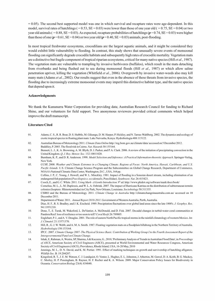
= 0.05). The second best supported model was one in which survival and recapture rates were age-dependent. In this
model, survival rates of hatchlings ( = 0.53, SE = 0.03) were lower than those of one year old ( = 0.75, SE = 0.04) or two
year old animals ( = 0.88, SE = 0.03). As expected, recapture probabilities of hatchlings (p = 0.74, SE = 0.03) were higher
than those of one (p = 0.61, SE = 0.04) or two year old (p = 0.40, SE = 0.03) animals, post-flooding.
In most tropical freshwater ecosystems, crocodilians are the largest aquatic animals, and it might be considered they
would exhibit little vulnerability to flooding. In contrast, this study shows that unusually severe events of monsoonal
flooding can significantly degrade crocodile habitats and subsequently high rates of crocodile mortality. Vegetation mats
are a distinctive but fragile component of tropical riparian ecosystems, critical for many native species (Hill et al., 1987).
The vegetation mats are vulnerable to trampling by invasive herbivores (buffalos), which result in the mats detaching
from riverbanks and being flushed out to sea during monsoonal floods (Hill et al., 1987) or which allow saline
penetration upriver, killing the vegetation (Whitfield et al., 2006). Overgrowth by invasive water-weeds also may kill
many mats (Adams et al., 2002). Our results suggest that even in the absence of these threats from invasive species, the
flooding due to increasingly extreme monsoonal events may imperil this distinctive habitat type, and the native species
that depend upon it.
Acknowledgments
We thank the Kununurra Water Corporation for providing data; Australian Research Council for funding to Richard
Shine, and our volunteers for field support. Two anonymous reviewers provided critical comments which helped
improve the draft manuscript.
Literature Cited
01. Adams, C. S., R. R. Boar, D. S. Hubble, M. Gikungu, D. M. Harper, P. Hickley, and N. Tarras-Wahlberg. 2002. The dynamics and ecology of
exotic tropical species in floating plant mats: Lake Naivasha, Kenya. Hydrobiologia 488:115122.
02. Australian Bureau of Meteorology 2011. Climate Data Online http://reg.bom.gov.au/climate/data/ accessed on 5 December 2012.
Baddiley, P. 2003. The flood risk in Cairns. Nat. Hazards 30:155164.
03. Bennett, L. J., K. A. Browning, A. M. Blyth, D. J. Parker, and P. A. Clark. 2006. A review of the initiation of precipitating convection in the
United Kingdom. Q. J. Roy. Meteor. Soc. 132:10011020.
04. Burnham, K. P., and D. R. Anderson. 1998. Model Selection and Inference: A Practical Information-theoretic Approach. Springer-Verlag,
New York.
05. CCSP, 2008: Weather and Climate Extremes in a Changing Climate. Regions of Focus: North America, Hawaii, Caribbean, and U.S.
Pacific Islands. U.S. Climate Change Science Program and the Subcommittee on Global Change Research, Department of Commerce,
NOAA's National Climatic Data Center, Washington, D.C., USA, 164 pp.
06. Collins, J. P., C. Young, J. Howell, and W. L. Minckley. 1981. Impact of flooding in a Sonoran desert stream, including elimination of an
endangered fish population (Poeciliopsis o. occidentalis, Poeciliidae). Southwest. Nat. 26:415423.th07. Cooch, E., and G. C. White. 2011. Using Mark - A Gentle Introduction. 9 ed. http://www.phidot.org/software/mark/docs/book/
Cornelius, M. L., L. M. Duplessis, and W. L. A. Osbrink. 2007. The impact of Hurricane Katrina on the distribution of subterranean termite
colonies (Isoptera : Rhinotermitidae) in City Park, New Orleans, Louisiana. Sociobiology 50:311335.
08. CSIRO and the Bureau of Meteorology. 2011. Climate Change in Australia http://climatechangeinaustralia.com.au/ accessed on 10
December 2011.
09. Department of Water. 2011. Annual Report 2010-2011. Government of Western Australia, Perth, Australia.
Diaz, H. F., R. S. Bradley, and J. K. Eischeid. 1989. Precipitation fluctuations over global land areas since the late 1800's. J. Geophys. Res.
94:11951210.
10. Done, T., E. Turak, M. Wakeford, L. DeVantier, A. McDonald, and D. Fisk. 2007. Decadal changes in turbid-water coral communities at
Pandora Reef: loss of resilience or too soon to tell? Coral Reefs 26:789805.
11. Englehart, P. J., and A. V. Douglas. 2001. The role of eastern North Pacific tropical storms in the rainfall climatology of western Mexico. Int.
J. Climatol. 21:13571370.
12. Hill, R., G. J. W. Webb, and A. M. A. Smith. 1987. Floating vegetation mats on a floodplain billabong in the Northern Territory of Australia.
Hydrobiologia 150:153164.
13. IPCC. 2007. Climate Change 2007- The Physical Science Basis: Contribution of Working Group I to the Fourth Assessment Report of the
Intergovernmental Panel on Climate Change.
14. Ishak, E, Rahman, A, Westra, SP, Sharma, A & Kuczera, G., 2010, 'Preliminary Analysis of Trends in Australian Flood Data', in Proceedings
of ASCE, American Society of Civil Engineers (ASCE), presented at World Environmental and Water Resources Congress, American
Society of Civil Engineers (ASCE), Providence, Rhode Island, USA, 16-20 May, 2010
15. Jennings, M. L., D. N. David, and K. M. Portier. 1991. Effect of marking techniques on growth and survivorship of hatchling alligators.
Wildlife Soc. B. 19:204207.16. Kingsford, R. T., J. E. M. Watson, C. J. Lundquist, O. Venter, L. Hughes, E. L. Johnston, J. Atherton, M. Gawel, D. A. Keith, B. G. Mackey,
C. Morley, H. P. Possingham, B. Raynor, H. F. Recher and K. A. Wilson. 2009. Major Conservation Policy Issues for Biodiversity in Oceania. Conservation Biology 23(4): 834840.
199

17. Knutson, T. R., J. L. McBride, J. Chan, K. Emanuel, G. Holland, C. Landsea, I. Held, J. P. Kossin, A. K. Srivastava, and M. Sugi. 2010.
Tropical cyclones and climate change. Nature Geoscience 3:157163.
18. Larriera, A., and C. Piña. 2000. Caiman latirostris (Broad-snouted caiman) nest predation: does low rainfall facilitate predator access?
Herpetol. Nat. Hist. 7:7377.
19. Mahmudul, A. Md., T. Mohd Ekhwan, S. Chamhuri, and T. Basri. 2011. Rainfall variation and changing pattern of agricultural cycle. Am. J.
Environ. Sci. 7:8289.
20. Messel, H., G. C. Vorlicek, A. G. Wells, W. J. Green, H. S. Curtis, C. R. R. Roff, C. M. Weaver, and A. Johnson. 1981. Surveys of Tidal
Waterways on Cape York Peninsula, Queensland, Australia, and Their Crocodile Populations. Monograph 16. Pergamon Press: Sydney.
21. Moron, V., M. N. Ward, and A. Navarra. 2001. Observed and SST-forced seasonal rainfall variability across tropical America. Int. J.
Climatol. 21:14671501
22. Pandey, C. B., and J. S. Singh. 1992. Rainfall and grazing effects on net primary productivity in a tropical savanna, India. Ecology
73:20072021.
23. Pittock, B., D. Abbs, R. Suppiah, and R. Jones. 2006. Climatic background to past and future floods in Australia. Adv. Ecol. Res. 39:1339.
Salvat, B., and C. Wilkinson. 2011. Cyclones and climate change in the South Pacific. Rev. Ecol-Terre. Vie. 66:105115.
Somaweera, R. 2011. A report of a probable unprovoked attack by an Australian freshwater crocodile at Lake Argyle in Western Australia.
Aust. Zoologist 35(4): 973-976.
24. Somaweera, R., J. Webb, and R. Shine. 2011a. Determinants of habitat selection by hatchling Australian freshwater crocodiles PLOS One
6(12):e28533.
25. Somaweera, R., J. Webb, and R. Shine. 2011b. It's a dog-eat-croc world: dingo predation on the nests of freshwater crocodiles in tropical
Australia. Ecol. Res. 26:957967.
26. Steffen, W., Burbidge, A.A., Hughes, L., Kitching, R., Lindenmayer, D., Musgrave, W., Stafford Smith, M. and Werner, P.A. 2009.
Australia's biodiversity and climate change. Technical Synthesis. Department of Climate Change, Canberra.
27. Vinnikov, K.Ya., P. Ya. Groisman, and K. M. Lugina. 1990. Empirical data on contemporary global climate change (temperature and
precipitation). J. Climate. 3:662677.
28. Water Corporation. 2011.
http://www.watercorporation.com.au/D/dams_storagedetail.cfm?id=22241 accessed on 29 September 2011.
29. Webb, G. J. W. and S. C. Manolis. 2010. Australian Freshwater Crocodile Crocodylus johnstoni. Pages 66-70 in S. C. Manolis and C.
Stevenson, editors. Crocodiles. Status Survey and Conservation Action Plan. Crocodile Specialist Group.
30. Webb, G. J. W., and A. M. A. Smith. 1984. Sex ratio and survivorship in the Australian freshwater crocodile Crocodylus johnstoni. Symp.
Zool. Soc. Lond. 52:319355.
31. White, G. C., K. P. Burnham, and D. R. Anderson. 2001. Advanced Features of Program Mark. In R. Field, R. J. Warren, H. Okarma, and P.
R. Sievert, P.R. (eds.), Wildlife, Land, and People: Priorities for the 21st Century, pp. 368377. The Wildlife Society, Bethesda, Maryland,
USA.
32. White, G. C., and K. P. Burnham. 1999. Program MARK: Survival Estimation from Populations of Marked Animals. Bird Study
(Supplement). 46:S120139.
33. Whitfield, A. K., R. H. Taylor, C. Fox, and D. P. Cyrus. 2006. Fishes and salinities in the St Lucia estuarine system - a review. Rev. Fish. Biol.
Fisher. 16:120.Table 1. Ranking of candidate crocodile survival and recapture models investigating the effects of age structure, vegetation,
and floods. The table shows number of parameters (k), change in QAICc compared to the best-ranked model (ΔQAICc) and model weights
(wi) (see Methods for explanation). The capture-mark-recapture data set included encounter histories of 712 individual crocodiles.
200
Table 1. Ranking of candidate crocodile survival and recapture models investigating the effects of age structure, vegetation, and floods. The table shows number of parameters (k), change in QAICc compared to the best-ranked model (ΔQAICc) and model weights (wi) (see Methods for explanation). The capture-mark-recapture data set included encounter histories of 712 individual crocodiles.
Survival Recapture K ?AICc w i
age 3 + vegetation + flood age + flood 16 0 0.546
age age 6 0.76 0.373 age age + flood 7 4.94 0.0463 age + vegetation age 12 6.00 0.027 age + vegetation + flood age 15 9.00 0.006 age + vegetation age + flood 12 13.60 0.000 age + vegetation + flood age + vegetation + flood 24 16.06 0.000
!
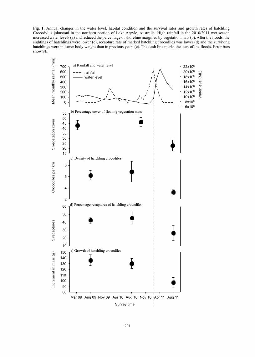
d) Percentage recaptures of hatchling crocodiles
10
20
30
40
50
60
a) Rainfall and water level
0
100
200
300
400
500
600
700
6x10⁶
8x10⁶
10x106
12x106
14x106
16x106
18x106
20x106
22x106
rainfall
water level
e) Growth of hatchling crocodiles
Survey time
Mar 09 Aug 09 Nov 09 Apr 10 Aug 10 Nov 10 Apr 11 Aug 11
80
90
100
110
120
130
140
150
b) Percentage cover of floating vegetation mats
15
20
25
30
35
40
45
50
55
c) Density of hatchling crocodiles
2
4
6
8
Incr
emen
t in
mas
s (g
) 5
re
ca
ptu
res
Cro
co
dile
s p
er
km
5
ve
ge
tatio
n c
ove
r M
ea
n m
on
thly
ra
infa
ll (m
m)
Wa
ter
leve
l (M
L)
Fig. 1. Annual changes in the water level, habitat condition and the survival rates and growth rates of hatchling Crocodylus johnstoni in the northern portion of Lake Argyle, Australia. High rainfall in the 2010/2011 wet season increased water levels (a) and reduced the percentage of shoreline margined by vegetation mats (b). After the floods, the sightings of hatchlings were lower (c), recapture rate of marked hatchling crocodiles was lower (d) and the surviving hatchlings were in lower body weight than in previous years (e). The dash line marks the start of the floods. Error bars show SE.
201

Fig. 2. Correlations between loss of floating vegetation, recapture rates, and body sizes of hatchling Crocodylus johnstoni at Lake Argyle, Western Australia. Areas that retained more vegetation also retained more hatchling crocodiles after the flood in August 2011 (a). In the sites where shoreline vegetation was less badly affected, young crocodiles were heavier (b). The “% loss of vegetation” refers to changes in the proportion of shoreline area with aquatic vegetation between November 2010 and August 2011.
a) Percentage recaptures of hatchling crocodiles
0
20
40
60
b) Weight gain in hatchling crocodiles
% loss of floating vegetation
20 40 60 80 10050
100
150
200
mea
n w
eigh
t ga
in (
g)%
rec
aptu
res
of m
ark
ed h
atcl
ings
202

Basking behaviour of saltwater crocodile in Bhitarkanika Wildlife Sanctuary, Odisha
1 2 1C. Venkatraman , K. Venkataraman and P. Padmanaban1 Marine Biology Regional Centre, Zoological Survey of India, 130, Santhome High Road, Chennai 600 028,
South India | 2 Zoological Survey of India, M.Block, New Alipore, Kolkata 700053
Crocodilians are a highly advanced group of reptiles. They are large, aquatic carnivores with skin covered with scales and bony scutes. Reptiles are cold blooded animals, meaning that they do not maintain a constant body temperature. But some reptiles exercise a degree of thermoregulation by either physiological or behavioural mechanisms. When temperature drops lizards, snakes and crocodiles bask in the sun to achieve the desired body temperature; once this thermal level is attained they will divide their time between sun and shade to maintain it. The direct effect of temperature influences the animal behavior to considerable extent. Basking is a regular and common habit of crocodiles. By following a strict daily pattern of alternating periods in the sun, in the shade and in water, they are able to maintain a body temperature of 250C varying by only a few degrees either way. The basking behavior of SaltwaterCrocodile and its functional significance is essential for the proper management of the crocodilesin India.
The basking behavior of wild population of Saltwater or Estuarine Crocodile (Crocodylus porosus) was carried out in Bhitarkanika Wildlife Sanctuary, Odisha from October 2012 and March 2013. Observations were made in crocodiles, basking along the river banks and creeks by boat. Hourly record of the temperature of bottom and surface water, soil and atmosphere along with the position of individual crocodile were made during the study period. Pearson's co efficient ofcorrelation was calculated to measure the relationships between various temperatures and various kind of basking patterns. The difference between the time spent for basking by adults and juveniles were discussed in detail.
Sri Lanka 20-23 May 2013 203
Proceedings : World Crocodile Conferencend -22 Working Meeting of the IUCN SSC Crocodile Specialist Groupnd -22 Working Meeting of the IUCN SSC Crocodile Specialist Groupnd -22 Working Meeting of the IUCN SSC Crocodile Specialist Group
p 203

Acute effects of freeze dried crocodile bile on male Sprague Dawley Rats
1 1 1Patcharavadee Wongspanich , Sakanjit Pathompota , Jindawan Siruntawineti ,
2 1Sudawan Chaeychomsri and Win Chaeychomsri * 1 .Department of Zoology, Faculty of Science, Kasetsart University, Bangkok 10900, Thailand
2 Central Laboratory and Greenhouse Complex, Kasetsart University, Kamphaeng Saen, Nakhon Pathom
73140, Thailand (*[email protected])
Abstract
Crocodile gall bladder is an important traditional medicine. Previous studied suitable processes in crocodile gall-bladder preservation for value added product was freeze-dried. The result showed that could sublimate to be gold finely powder which make it easy to capsule if combine with rice starch powder. However, freeze dried crocodile bile for healthful doesn't have science report about toxicity of crocodile bile. For high efficacy of crocodile bile product, acute effects of freeze-dried crocodile bile were performed on male Sprague Dawley rats. The result showed that morbidity and mortality have not found any in treatment 300 mg/kg group of rats. Because of recommendation dose in human is 25 mg/60kg/day, there is very less than in the rat treatment. These revealed that freeze dried crocodile bile is appropriate for product development to be supplementary food in the future.
Introduction
Freshwater or Siamese Crocodile, Crocodylus siamensis, is an economic animal in Thailand. The minor products of crocodile are internal organs such as bone liver, gall bladder, etc. They have more demand for improving human health found in the traditions of many Asian cultures. In traditional medicine, animal parts and derivatives have been used e.g. tiger bone, rhino horn, gland from musk deer, bear gall bladders (Picheinsutthorn and Jeerawong, 2003). For treatment of a range of complaints such as fever, hemorrhoids, conjunctivitis and liver diseases, bear gallbladders and bile products are use. Animal bile including bear bile and snake bile has high amounts of bile acids. The ursodeoxycholic acid (UDCA), an ingredient in bile bear, can be successfully used to cure certain liver problems and dissolution of gallstones (Feng et al., 2009). Crocodile bile pill, a form of crocodile bile product is used as an asthma traditional Chinese medicine combination product (Irwin, 2001). In Thailand, Siamese crocodile internal organs are waste material in slaughtering industry. Although their bile has advantage in traditional medicine, a lot of crocodile gall bladders are useless per year. Moreover, general style of the preservation is naturally drying such as sunlight exposure, natural wind drying, etc. With these drying methods, the un-safety of the product about bacterial and fungal contamination has been found. For adding value to the bile product, Chaeychomsri et al. (2009) was studied the suitable processes in captive Siamese crocodile galls preservation and its bile product. The proper preservation process and crocodile bile product development has been done and registered as a Kasetsart University's innovative property (Thai patent application No. 0901001231, 2009). Recently, Siamese crocodile bile provides significant insight into the anticarcinogenic action on human cholangiocarcinoma cells (Song et al., 2012; Kang et al., 2012). However, it has no report about toxicity of the crocodile bile. In this study, we prepared freeze-dried product of Siamese crocodile bile, investigated the acute effects on male Sprague Dawley rats. The out put of this study will provide basic data of sub-chronic and chronic study for the crocodile bile product development and may support efficacy and value-adding of this freeze-dried crocodile bile product.
Materials and Methods
Collection of gall bladders: One hundred of captive Siamese crocodile (Crocodylus siamensis) gall bladders were collected from Rungtaweechai Crocodile Farm, Don Toom District, Nakhonpathum Province, Thailand.Crocodile bile powder preparation: Process for dried crocodile bile production of Thai patent application No. 0901001231, Kasetsart University (Chaeychomsri and Siruntawineti, 2009) was performed as following. The gall bladders without fat tissue outside were surface sterile with 70% ethyl alcohol and the crocodile bile were collected in tray and freeze-drying using freeze dryer (Lyomaster, USA). The freeze-dried bile was powdered by graining at sterile
ocondition. The crocodile bile powder was kept at 4 c until use.
Laboratory animals: The Animal Ethics Committee of Kasetsart University, Thailand approved the use of laboratory animals in this study. Nine Spraque-Dawley males were purchased from The National Laboratory Animal Center, Mahidol University, Salaya, Thailand. The rats were randomly divided into 3 groups, each comprising three rats. Male
Proceedings : World Crocodile Conferencend -22 Working Meeting of the IUCN SSC Crocodile Specialist Groupnd -22 Working Meeting of the IUCN SSC Crocodile Specialist Groupnd -22 Working Meeting of the IUCN SSC Crocodile Specialist Group
Sri Lanka 20-23 May 2013 204
pp 204 - 206
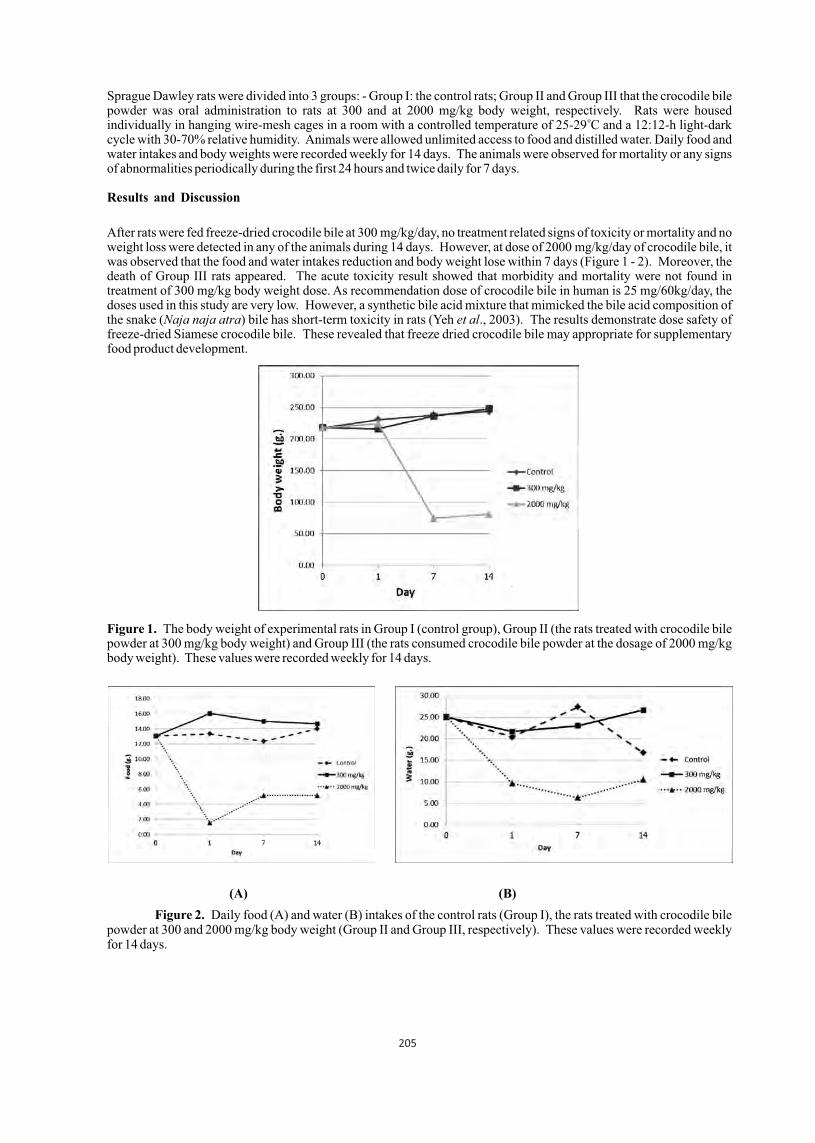
Sprague Dawley rats were divided into 3 groups: - Group I: the control rats; Group II and Group III that the crocodile bile powder was oral administration to rats at 300 and at 2000 mg/kg body weight, respectively. Rats were housed
oindividually in hanging wire-mesh cages in a room with a controlled temperature of 25-29 C and a 12:12-h light-dark cycle with 30-70% relative humidity. Animals were allowed unlimited access to food and distilled water. Daily food and water intakes and body weights were recorded weekly for 14 days. The animals were observed for mortality or any signs of abnormalities periodically during the first 24 hours and twice daily for 7 days.
Results and Discussion
After rats were fed freeze-dried crocodile bile at 300 mg/kg/day, no treatment related signs of toxicity or mortality and no weight loss were detected in any of the animals during 14 days. However, at dose of 2000 mg/kg/day of crocodile bile, it was observed that the food and water intakes reduction and body weight lose within 7 days (Figure 1 - 2). Moreover, the death of Group III rats appeared. The acute toxicity result showed that morbidity and mortality were not found in treatment of 300 mg/kg body weight dose. As recommendation dose of crocodile bile in human is 25 mg/60kg/day, the doses used in this study are very low. However, a synthetic bile acid mixture that mimicked the bile acid composition of the snake (Naja naja atra) bile has short-term toxicity in rats (Yeh et al., 2003). The results demonstrate dose safety of freeze-dried Siamese crocodile bile. These revealed that freeze dried crocodile bile may appropriate for supplementary food product development.
Figure 1. The body weight of experimental rats in Group I (control group), Group II (the rats treated with crocodile bile powder at 300 mg/kg body weight) and Group III (the rats consumed crocodile bile powder at the dosage of 2000 mg/kg body weight). These values were recorded weekly for 14 days.
(A) (B)
Figure 2. Daily food (A) and water (B) intakes of the control rats (Group I), the rats treated with crocodile bile powder at 300 and 2000 mg/kg body weight (Group II and Group III, respectively). These values were recorded weekly for 14 days.
205
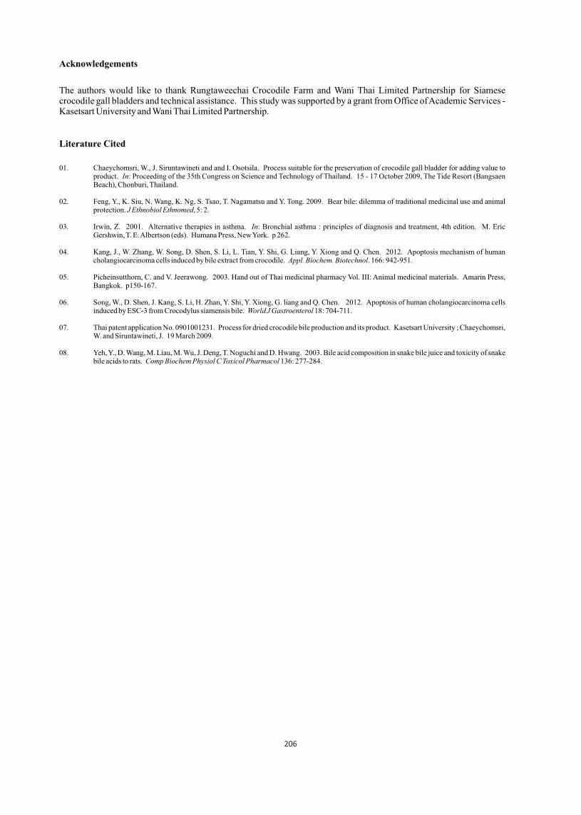
Acknowledgements
The authors would like to thank Rungtaweechai Crocodile Farm and Wani Thai Limited Partnership for Siamese crocodile gall bladders and technical assistance. This study was supported by a grant from Office of Academic Services - Kasetsart University and Wani Thai Limited Partnership.
Literature Cited
01. Chaeychomsri, W., J. Siruntawineti and and I. Osotsila. Process suitable for the preservation of crocodile gall bladder for adding value to product. In: Proceeding of the 35th Congress on Science and Technology of Thailand. 15 - 17 October 2009, The Tide Resort (Bangsaen Beach), Chonburi, Thailand.
02. Feng, Y., K. Siu, N. Wang, K. Ng, S. Tsao, T. Nagamatsu and Y. Tong. 2009. Bear bile: dilemma of traditional medicinal use and animal protection. J Ethnobiol Ethnomed, 5: 2.
03. Irwin, Z. 2001. Alternative therapies in asthma. In: Bronchial asthma : principles of diagnosis and treatment, 4th edition. M. Eric Gershwin, T. E. Albertson (eds). Humana Press, New York. p 262.
04. Kang, J., W. Zhang, W. Song, D. Shen, S. Li, L. Tian, Y. Shi, G. Liang, Y. Xiong and Q. Chen. 2012. Apoptosis mechanism of human cholangiocarcinoma cells induced by bile extract from crocodile. Appl. Biochem. Biotechnol. 166: 942-951.
05. Picheinsutthorn, C. and V. Jeerawong. 2003. Hand out of Thai medicinal pharmacy Vol. III: Animal medicinal materials. Amarin Press, Bangkok. p150-167.
06. Song, W., D. Shen, J. Kang, S. Li, H. Zhan, Y. Shi, Y. Xiong, G. liang and Q. Chen. 2012. Apoptosis of human cholangiocarcinoma cells induced by ESC-3 from Crocodylus siamensis bile. World J Gastroenterol 18: 704-711.
07. Thai patent application No. 0901001231. Process for dried crocodile bile production and its product. Kasetsart University ; Chaeychomsri, W. and Siruntawineti, J. 19 March 2009.
08. Yeh, Y., D. Wang, M. Liau, M. Wu, J. Deng, T. Noguchi and D. Hwang. 2003. Bile acid composition in snake bile juice and toxicity of snake bile acids to rats. Comp Biochem Physiol C Toxicol Pharmacol 136: 277-284.
206