Embed Size (px)
Citation preview

Carlsberg Res. Commun. Vol, 46, p. 259-278, 1981
COMMENTS ON THE STRUCTURE AND FUNCTION OF THE LARGE SUBUNIT OF THE ENZYME
RIBULOSE BISPHOSPHATE CARBOXYLASE-OXYGENASE
by
CARSTEN POULSEN
Department of Physiology, Carlsberg Laboratory, Gamle Carlsberg Vej 10, DK-2500 Copenhagen, Valby
Keywords: Fraction I protein, nucleotide sequence, amino acid sequence, maize, barley, spinach, catalytic site, activation site, modification of active groups, substrate affinity
analogues, transition state analogues, fructose bisphosphate, aldolase
The complete amino acid sequence of the large subunit (LS) of ribulose bisphosphate carboxylase has been determined for maize through nucleotide sequencing of the chloroplast gone encoding this polypeptide (34). Previously published amino acid sequences from the large subunits of barley and spinach as well as new evidence for the amino acid sequence of these two polypeptides are aligned with the complete maize sequence. The complete spinach amino acid sequence is also presented according to the sequence of the spinach gene (63). The number of amino acids in the 475-residue LS primary structure for which there is direct or circumstantial evidence from barley, is 278. Amino acid replacements are encountered in seven positions in comparison with the maize sequence and in 20 positions when compared to the spinach LS. From affinity labelling studies ( 13, 26) active site residues, nominally lysines and cysteines, have been deduced and located in the total maize and spinach LS sequences, thus being available for further study of structure-function relationships. The complete amino acid sequence and knowledge about catalytic site residues also allows direct comparison with primary structures of other enzymes of the same class, the lyases. Particular attention is devoted to aldolase, an enzyme also functioning in carbohydrate metabolism. Comparison with the 361-residue aldolase of glycolysis in rabbit muscle cells suggests homology in 67 positions centered around catalytic site lysine-175 of RuBPCase LS and catalytic site lysine-227 of aldolase. It is discussed how this relationship may throw light on the reaction mechanism of the RuBP carboxylation.
Abbreviations: BBBP = D-3-bromo-l,4-dihydroxy-butanone-l,4-bisphosphate; CA = carbonic anhydrase; CABP = D-2-carboxyarabinitoI-1,5-bisphosphate; DHAP = dihydroxyacetonephosphate; FBP = D- fructose-l,6-bisphosphate; FBPald = fructose-l,6-bisphosphate aldolase; 1,3GABP = glyceric acid-l,3- bisphosphate; G3P = glyceraldehyde-3-phosphate; G3PDH = (NADP)-glyceraldehyde-3-phosphate dehydro- genase; G3Piso = G3P ketol-isomerase; LS = large subunit; NBA-EAP = N-bromoacetyl-ethanolamine phosphate; 3PGA = 3-phosphoglycerate; 3PGAkin = 3-phosphoglycerate kinase; 6PGIuA = D-6- phosphogluconate; PLP = pyridoxal-5"-phosphate; RuBP = D-ribulose-l,5-bisphosphate; RuBPCase = RuBP carboxylase; SS = small subunit.
0105-1938/81 /0046/0259/$ 04.00

C. POULSEN: Structure and function of RUBPCase
1. INTRODUCTION
The major function of the enzyme D- ribulose- 1,5-bisphosphate carboxylase-oxyge- nase (3-phosphoglycerate: carboxylyase (dimerizing) E.C.4.1.1.39) is the photosynthetic fixation of CO2 into carbohydrate through the Calvin-cycle which functions in the chloroplasts of green plants and algae as well as in cyanobacteria and photosynthetic bacteria (15). The seemingly less important role is catalysis of the first step in photorespiratory glycolate production from RuBP and 02 (15). This dual function is thought to be important for regula- tion and protection of the carbon reduction cycle in chloroplasts in response to environmental factors such as light-dark regimes, CO2 concen- tration and concentration of intermediates of carbohydrate metabolism (28, 30). Due to the importance of CO2-fixation for production of biomass and in particular for crop productivity a full understanding of the enzyme structure and its modes of action, is desirable. This will allow to evaluate if improvements in agricultural plants can be obtained by changing the activity of this enzyme.
The purpose of the present paper is to relate the enzymological data obtained by active site modifications (13, 26, 27, 38, 42, 50, 52, 55)of the catalytically active large subunit of the enzyme to its primary structure as determined by amino acid sequencing (44, 45) and nucleotide sequencing of the gene for this polypeptide in maize (34) and spinach (63).
2. ENZYME STRUCTURE
In all reports on the isolation of RuBPCase from eukaryotic sources the enzyme has been estimated to have a molecular weight of 500,000-560,000 and to contain eight copies of two types of polypeptides, a large subunit (LS) of molecular weight 50,000-55,000 with which the catalytic activity is associated (18, 37), and a small subunit (SS) of 12,000-16,000 whose function is not well understood (18). Interpreta- tions of electron micrographs of negatively stained enzyme molecules place the eight large subunits at the corners of a cube (33), and two small subunit copies on each of four sides of this cube, leaving at the two remaining sides a central hole which is penetrated by stain. This hypothe-
tical quaternary structure is based on the assumption that both subunits must have close to spherical tertiary structures (4). However, X- ray crystallographic studies on the Nicotiana tabacum enzyme have revealed that this is not necessarily the case and leave the possibility for a different quaternary structure open (D. EmEN- BERG, pers. commun.). Complementary to the X- ray diffraction analysis, the amino acid sequen- ces for the two subunits of the N. tabacum enzyme are being established (36). The primary structure of the polypeptides from other sources are now characterized. The complete amino acid sequence of the small subunits from the pea and spinach enzymes have been determined by protein sequencing methods (31, 56) and cDNA clones containing pea SS mRNA specific sequen- ces have been sequenced (5) and there is full agreement between the primary structures of the pea small subunit as determined by the two different methods. Partial amino acid sequences comprising 92 and 278 residues have been established for the spinach and barley large subunits, respectively (13, 26, 45, 52, 55 and Figure 1). Complete amino acid sequences have been derived for the large subunit of maize and spinach by nucleotide sequencing of its gene after cloning of the appropriate chloroplast DNA restriction fragments in E.coli (34, 63).
3. DETERMINATION OF THE LARGE SUBUNIT PRIMARY STRUCTURE
Determination of nucleotide sequences of a cloned structural gene or cDNA sequence is an efficient way to obtain the complete primary structure of a protein, when partial amino acid sequences are available for translation of the reading frame of nucleotide sequences into the corresponding complete amino acid sequences.
When the colinearity of the maize LS structural gene and the maize LS mRNA had been established and the direction of transcrip- tion determined (25) the nucleotide sequence was determined for the maize LS gene by the Maxam-Gilbert procedure (32) and translated into the amino acid sequence (34). This used a comparison to the available amino acid sequence data from the barley and the spinach large subunit (44, 45). Only two long stretches of sequence (nucleotides 451-824 and 1190-1575)
260 Carlsberg Res. Commun. Vol. 46, p. 259-278, 1981

C. POULSEN: Structure and function of RUBPCase
without appropriate restriction endonuclease sites for 32p-labelling with 5' polynucleotide kinase or with the KLENOW fragment of DNA polymerase caused some difficulty. However, overlapping sequences were eventually obtained reproducibly with sequencing gels on which nucleotides more than 350 bases away from the labelling point could be read off. A detailed re- analysis of the autoradiograms of the sequencing gels revealed that a CTA codon for leucine was omitted in the published sequence (34) between Leu-290 and Leu-291. On the other hand an extra GCT codon for Ala was by mistake inserted in position 64. This has been corrected in Figure I. This brings the amino acid sequence into phase with that of spinach (63). The GCC- codon for Ala-452 in the maize LS sequence should be corrected to a CCG-codon for proline, which is also the corresponding amino acid in the spinach sequence.
From the amino acid sequence derived for the maize RuBPCase LS ambiguities in the barley LS amino acid sequence can be resolved and errors corrected. This will be done with the aid of Figure 1 which shows the complete maize LS primary structure as well as previously reported and new barley LS partial amino acid sequences. Techniques employed for obtaining the latter are listed in Table I.
The amino acid sequence of the maize LS extends for 14 amino acids beyond the previo- usly determined alanine at the N-terminus of barley LS. With the purpose of comparing the barley and the maize LS polypeptides and their CNBr-fragments new isolations of RuBP car- boxylase from maize and barley were carried out. For maize the procedure previously used for barley (44) was followed except that one additional Sepharose 6B step was employed and for barley a shortened procedure in which a step of DE52 ion exchange chromatography substitu- ted the Sepharose 6B. The LS was prepared in the usual way and cleaved with CNBr. The fragments were separated by sodium dodecyl sulfate polyacrylamide gel electrophoresis (44) and the N-terminal amino acid residues determi- ned by the dansylation procedure. The N- terminal fragments from both species had the same size and the dansylation procedure failed to yield an N-terminal amino acid, most likely due to N-terminal blocking. It can therefore at
present not be decided, whether the loss of the 14 N-terminal amino acids in the barley large subunit subjected to sequencing is due to accidental proteolytic break-down or to normal post-translational processing. Recently LANG- RmGE (23) has reported a 2000 molecular weight larger precursor for the LS of spinach after in vitro translation which thus could signify removal of an N-terminal sequence in vivo. This implies that the LS isolated from some enzyme preparations would have been subjected to N- terminal blocking after the cleavage.
The number of methionine residues in the maize and barley polypeptides are larger than determined by amino acid analysis. The maize LS contains one N-terminal and 11 internal methionines. In the work on barley LS 9 internal CNBr cleavage points have been found, namely at positions 116, 212,250, 266,297,320, 341, 371 and 387 (Table I). The methionine residue in position 341 is absent in maize and spinach, which on the other hand have a methionine in position 472 not present in barley (44, p. 178). The two remaining methionine residues (Met-155 and Met-309) in barley can be accounted for in the following way: The cleavage point Met-155 results most likely in the N- terminal blocked fragment reported in Table VI of (44) although its amino acid composition does not show perfect agreement with that postulated from the maize and the spinach LS. This could be due to contamination of this fragment with histidine containing fragments derived from the region 310-371.
The CNBr fragment His-310 to Met-320 resulting from cleavage between Met-309 and His-310 was only found in small quantities and therefore not reported previously. It could be cleaved into three peptides by chymotrypsin and by manual EDMAN degradation an amino acid sequence corresponding to that of the maize LS was verified.
In addition to the amino acid sequences obtained by automated analysis, Table I shows amino acid sequences obtained for barley LS peptides by manual EDMAN degradation and compositional analyses. This reveals agreement with the nucleotide sequence of maize in the following regions: 80-83, 286-297, 304-309, 310-318 ,341-357 ,446-449 ,463-466 . Ambi- guity is found for positions 298-303 in the His-
Carlsberg Res. Commun. Vol. 46, p. 259-278, 1981 261

C. POULSEN: Structure and function of RUBPCase
14 14
0 0 0 14 14 14
14 14 14
m
L5 L9
###
,..-t
014 ~ 14 0 ~ ~ 0 14 14 ~-I
i
,.-4 ,.-q ,-.-I
? ,-4
:>
o ~
r-,
::::, > >
; [ -il
I
�9 r~] -~ ,~ ,~ L?J >, >, >,
I I
L~,J H
m m
�9
>
�9 "~ 0 I �9 0 ~, eq.-q ,-~ ,-4 Lr~ .-~
r-
b~
1.4
I -~ 14 14
I !
m
14
L) r j ~ ~ I
14 14 ~4 r ~
14 I1)
~5
,--t
I
I
I 0 ~ 14 t.4 o
I
L<,J < I
I
r ~ 14
m m ,~
,.-I
! I
r'~ ~ N o ~::~
I
I
C9 ~ ~
14 14 ~ m m ~
m m ~
1,4
14
0
0 14,4
I o r II) �9 ~ ,-q ,-4
~-I 1.4
/
~ m m 1.4
/
I
~ ~ m
I I I
r k l I 14 I:> ~ < :.2.
,-~ 14
, �9 , , , 0) m
I I I 14 I1) I~
o ~ , = o~ t9 ,-I ,-I ,-I ~ ~.
, ~
I O O
~ ~ ~>
,.--I
/
~ 14 14 C~
i ] ~ ~ ~ U L)
0 ~ ~ to
r..9 (.9
0 0 O~
; e I
I
~ m
?
I !
m N ,~ ,-4 ~
14 ~ U
I
0 0 ~ I,~ ~-I ,--t
H I=.l=a
~5
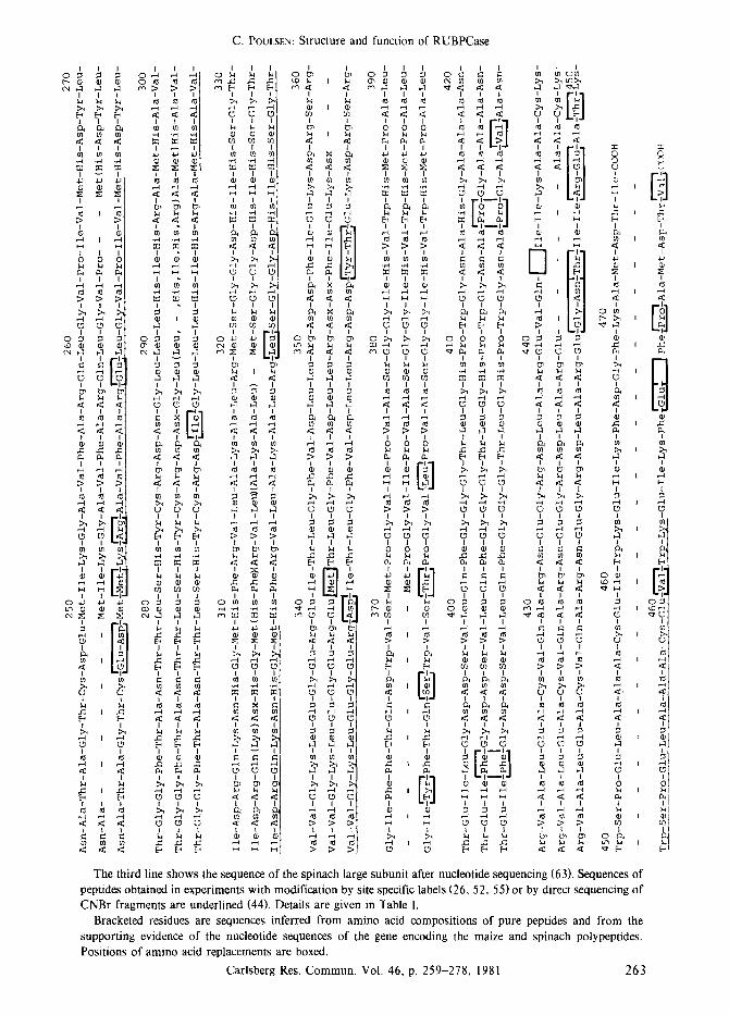
Figure 1. The amino acid sequence of the large subunit of RuBP carboxylase-oxygenase.
I o ~ ~ :~
E. E~ E~
~ ~5 L9
H H H
o ~I ~
~L,~j
I
�9 H L # J
I I
I /
? < < , <,
I_~ r.3 r.) L)
.C
[i] ] �9 I1~ I11 I11
& g g ~ , 14 N 14 ~ ,~ < ,r <
-
I I
I I
m ~
The uppermost line shows the sequence of the maize LS as derived by nucleotide sequencing of the LS gene (34). The second line shows previously published N-terminal sequences of peptide fragments isolated from the large subunit of the barley enzyme after CNBr-cleavage (45), as well as sequences obtained for peptides isolated after tryptic or chymotryptic cleavage of these fragments or from fractions containing mixtures of fragments (44). Details are given in Table I.
262 Carlsberg Res. Commun. Vol. 46, p. 259-278, 1981
C, POULSEN: Structure and function of RUBPCase
o , ~ o ~
7 to
I I
m tO
I I
,'-4
> >,
H
I I I ,-4 ,.-4
I
,-4 ,-4 < <
I
~J W
~I 1.4 ~4
.,-I .,"t .
m m m
o ~
m E-, E-,
2 2
~4
,--4
I ~ IN c,
I
I
I
m ~ m w I
0 ~ OJ �9 W
I I '
~ ,--4
�9 --4 ~
I I .
M I-t
0 ~ ~ 4J
o ~ ; ~ o-~ ~ 'r~
:.3, :._q
U L) U
I
U3 W ~ W �9
to
,...4
< I
to to
U
,-4 4"-4
.1~ X:
to
,.C ..~
I I I k~ k~
I I
I
, - t
I
r d ~ r~
I
> > >
I 0 ~ to to
u ~ u m m ~
~ to
g g ~
m m ~ < <
o ~ 5 o ~ ~ ~ o ~ ~ / I I
~ o o o ~
,
'
N ~
" ~ ; ~ ~ ~ ~ .~ .~ ~ " ~ ~ ~
~ t o
I
I ~ N 0 0 0 ~
,.-4 ,.-4 ~ < < <
,-4
<
I
,-4 I ,-4 ? <
~I I L " I J r r
m
IN
o,? ?L~j ~,?
,--t
I
> I IN
r , ,--t
?
I W
l
,--t
~ C9 I
o ~ ~
I I I
IN ~ IN
I
.~ L~..._~, l
,-4 ~ .-4
I
,C r" ..~
Us i~ U~ 0 a)
I I
O
I ,-4 ,-4 ,--I ~
> >
~ o ~ L~ k~
The third line shows the sequence of the spinach large subunit after nucleotide sequencing (63). Sequences of peptides obtained in experiments with modification by site specific labels (26, 52, 55) or by direct sequencing of CNBr fragments are underlined (44). Details are given in Table I.
Bracketed residues are sequences inferred from amino acid compositions of pure peptides and from the supporting evidence of the nucleotide sequences of the gene encoding the maize and spinach polypeptides. Positions of amino acid replacements are boxed.
Carlsberg Res. Commun. Vol. 46, p. 259-278, 1981 263
:E
~4
I
t~ ,--4 <
0)
H to
H
,..q t.9
I to
I IN ~4
O ~ '.D ;'-q m , r ~
I to
U
I
I
<
0

C. POULSEN: Structure and function of RUBPCase
Table I.
Assignment of amino acids by analysis of peptide fragment from the large sabunit of RuBPearboxylase- oxygenase of harley and spinach.
Method of determination Positions
BARLEY Automated EDMAN degradation of CNBr 15-60, I17-130, t32, 213-242, 251-263, 267-286, 321- fragments (34, 45) 340, 388-440
Manual EDMAN degradation of CNBr 298-304 (Figure 19 and Table XII in ref. 44) fragments 342-357 (sequence of fragment listed in Table X of ref. 44)
372-384 (Figure 18 and Table XII in ref. 44)
Tryptic or chymotryptic peptides of CNBr 306-309 (Figure 19 in ref. 44) fragments 310-311, 312-314, 315-318 (not previously reported)
386-387 (Figure 18 in ref. 44)
Micro-dansyl EDMAN degradation (59) of tryptic peptides from CNBr fragments (Tables refer to ref. 44)
15-18 (D in Table III), 22-26 (H in Table liD, 33-36 (I in Table liD, 42-44 (A in Table liD, 80-83 (B + G in Table III), 216-217 (P in Table IX), 228-230 (Q + R in Table IX), 251- 252 (N in Table XI), 286-289 (U in Table IX), 296-297 (S in Table IX), 351-356 (new analysis of X in Table XI), 432-435 (K in Table V), 436-439 (new analysis of J in Table V), 446- 449 (O in Table V), 463-465 (new analysis of M in Table V)
SPINACH Automated EMAN degradation of tryptic peptides Manual EDMAN degradation of CNBr fragments
165-177 (ref. 13, 52, 55), 195-211 (ref. 26), 320-339 (ref. 13, 55), 451-463 (ref. 13, 52), 252-259 (Figure 19 in ref. 44) 298-304 (Figure 19 in ref. 44)
fragment (298-309) reported originally in Table XII of reference (44). The amino acid composi- tion is correct but the N-terminal sequence deduced for this peptide (Lys-Ala-Val-Ile-Asx- His-Arg-Glx) was incorrect. The N-terminal lysine must have been incorrectly identified instead of a histidine, as no N-terminal lysine could be detected among the CNBr fragments in the cleavage experiment with barley LS mentio- ned by MCINTosn et al. (34). An explanation for the observation of a His-residue between Asp-302 and Arg-303 cannot be given. Thus no difference in the amino acid sequence between maize, spinach and barley appears to be present in positions 298 to 309. Manual sequencing with the aid of a-chymotrypsin and subtilisin (44, p. 184) had suggested the presence of an extra tryptophan residue between Trp-385 and His-386. There is no room for this tryptophan when the sequence of the Pro-fragment (372- 387) is aligned with the nucleotide sequences (34, 63).
For the lie-fragment (251-267) 13 amino acids were correctly identified (45, 60) while the 14th amino acid in position 264 was considered to be Glu. It can now be seen that this was due to contamination with the Thr-fragment (342-371) which has a Glu-residue in its 14th position (355). This contamination and the abnormal chromatographic behaviour of the Ile-fragment is the reason that its size was overestimated (45). In the meantime the Thr-fragment (342-371) could be purified sufficiently by repeated chro- matography in order to allow the amino acid sequence for positions 342 to 357 to be identified in full agreement with the nucleotide sequence.
The spinach LS sequences shown in Figure 1, in positions corresponding to 165-177, 195- 211, 320-339 and 451-463 of the spinach LS will be described in more detail in section 5. The complete amino acid sequence of spinach LS as derived by nucleotide sequencing (63) is also shown in Figure 1. Comparison of the amino acid sequence evidence for the barley and spinach
264 Carlsberg Res. Commun. Vol. 46, p. 259-278, t981

C. POULSEN: Structure and function of RUBPCase
G A>C C C+T
q- r A
A , C J
T . T J C . T J
. ,A T J
T T
T A A
G C
T A
C A c
A A G
T T
T C
G C
T A
C C
T A
T G
G T
A T
A 3"
T ....... :~ -T 3'
, : 5 , /
,
A T A lie T G G Trp
A A G Lys
G A G Glu A
T C lie A
A A Lys
T T C Phe
G A T Asp
G G
T Gly T
T C Phe
A A
A Lys G
C G Ala A
T G Met G
A T Asp
A C
C Thr A
T A lie
T A
A Ochre A
3'
Figure 2. Autoradiogram of a 6 % polyacrylamide sequencing gel. The [32p]-labeled fragment subjected to sequencing according to the Maxam-Gilbert procedure (32)corresponds to the Hhal-Bglll fragment covering the 3'-end of the mRNA for the maize LS (34). The LS-encoding sequence was labeled at the Hinfl- and Bg I II - sites with 3,-[32p]-ATP and polynucleotide kinase. Secondary restriction was performed with HhaI, whereafter the singly labeled HhaI-Bglll fragment was purified through polyacrylamide gel electrophoresis and elution from the appropriate slice of gel. The four chemical reactions giving cleavage at guanine (G), at adenine better than at cytosine (A>C), at cytosine (C) and at cytosine plus thymine (C+T) (32) were employed on aliquots of the labeled DNA. Subsequently, sequencing gels were run to cover most of the 272 basepair fragment. The shown sequence is 150-197 bases away from the labeling point, covering the coding for the carboxy-terminus of the LS.
large subunits described here, with the complete amino acid sequence of the maize and spinach large subunits can now be carried out for 320 residues. This include 278 barley LS residues
and 92 spinach LS residues. All spinach residues are in agreement with those determined by nucleotide sequencing of the spinach LS gene.
When the complete maize and spinach LS
Carlsberg Res. Commun. Vol. 46, p. 259-278, t981 265
C. POULSEN: Structure and function of RUBPCase
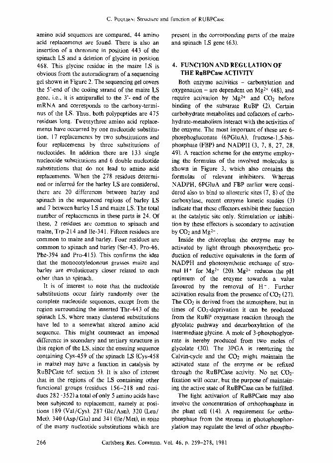
amino acid sequences are compared, 44 amino acid replacements are found. There is also an insertion of a threonine in position 443 of the spinach LS and a deletion of glycine in position 468. This glycine residue in the maize LS is obvious from the autoradiogram of a sequencing gel shown in Figure 2. The sequencing gel covers the 5'-end of the coding strand of the maize LS gene, i.e., it is antiparallel to the Y- end of the mRNA and corresponds to the carboxy-termi- nus of the LS. Thus, both polypeptides are 475 residues long. Twentythree amino acid replace- ments have occurred by one nucleotide substitu- tion, 17 replacements by two substitutions and four replacements by three substitutions of nucleotides. In addition there are 133 single nucleotide substitutions and 6 double nucleotide substitutions that do not lead to amino acid replacements. When the 278 residues determi- ned or inferred for the barley LS are considered, there are 20 differences between barley and spinach in the sequenced regions of barley LS and 7 between barley LS and maize LS. The total number of replacements in these parts is 24. Of these, 2 residues are common to spinach and maize, Trp-214 and I1e-341. Fifteen residues are common to maize and barley. Four residues are common to spinach and barley (Ser-43, Pro-46, Phe-394 and Pro-415). This confirms the idea that the monocotyledonous grasses maize and barley are evolutionary closer related to each other than to spinach.
It is of interest to note that the nucleotide substitutions occur fairly randomly over the complete nucleotide sequences, except from the region surrounding the inserted Thr-443 of the spinach LS, where many clustered substitutions have led to a somewhat altered amino acid sequence. This might counteract an imposed difference in secondary and tertiary structure in this region of the LS, since the ensuing sequence containing Cys-459 of the spinach LS (Cys-458 in maize) may have a function in catalysis by RuBPCase (cf. section 5). It is also of interest that in the regions of the LS containing other functional groups (residues 156-218 and resi- dues 282-352) a total of only 5 amino acids have been subjected to replacement, namely at posi- tions 189 (Val/Cys), 287 (Ile/Asn), 320 (Leu/ Met), 340 (Asp/Glu)and 341 (Ile/Met), in spite of the many nucleotide substitutions which are
present in the corresponding parts of the maize and spinach LS gene (63).
4. FUNCTION AND REGULATION OF T H E R u B P C a s e ACTIVITY
Both enzyme activities - carboxylation and oxygenation - are dependent on Mg 2§ (48), and require activation by Mg 2+ and CO2 before binding of the substrate RuBP (2). Certain carbohydrate metabolites and cofactors of carbo- hydrate-metabolism interact with the activities of the enzyme. The most important of these are 6- phosphogluconate (6PGIuA), fructose- 1,5-bis- phosphate (FBP) and NADPH (3, 7, 8, 27, 28, 49). A reaction scheme for the enzyme employ- ing the formulas of the involved molecules is shown in Figure 3, which also contains the formulas of relevant inhibitors. Whereas NADPH, 6PGluA and FBP earlier were consi- dered also to bind to atlosteric sites (7, 8) of the carboxylase, recent enzyme kinetic studies (3) indicate that these effectors exhibit their function at the catalytic site only. Stimulation or inhibi- tion by these effectors is secondary to activation by CO2 and Mg 2+ .
Inside the chloroplast the enzyme may be activated by light through photosynthetic pro- duction of reductive equivalents in the form of NADPH and photosynthetic exchange of stro- mal H + for Mg 2+ (20). Mg 2+ reduces the pH optimum of the enzyme towards a value favoured by the removal of H +. Further activation results from the presence of CO2 (27). The CO2 is derived from the atmosphere, but in times of CO2-deprivation it can be produced from the RuBP oxygenase reaction through the glycolate pathway and decarboxylation of the intermediate glycine. A mole of 3-phosphoglyce- rate is hereby produced from two moles of glycolate (30). The 3PGA is reentering the Calvin-cycle and the CO2 might maintain the activated state of the enzyme or be refixed through the RuBPCase activity. No net CO2- fixation will occur, but the purpose of maintain- ing the active state of RuBPCase can be fulfilled.
The light activation of RuBPCase may also involve the concentration of orthophosphate in the plant cell (14). A requirement for ortho- phosphate from the stroma in photophosphor- ylation may regulate the level of other phospho-
266 Carlsberg Res. Commun. Vol. 46, p. 259-278, 1981

C. POULSEN: Structure and function of RUBPCase
0-~ /0 - H ,.P~.
H..(~..O 0 I
c-o, [Mg2+-co2] H-C-OH
, CO 2 H-C-OH
i ~.C ,~
H I O.. ~..O H O< P..o_
D-Ribulose- 1,5- bisphosphate
RuBP
i
..O" "~O H"C O \ p . ~ O -
I HO-C - COO
I C = 0 - Mg 2+- CO 2
I H-C - OH
I
H "c " 0 . ~0 0 -" P'O-
D- 2 - Carboxy- 3- keto- arabinitol
-1,5- bisphosphate
3- keto - CABP
H 0-.. p .~0-
I C..
H ~ i COO- ~ OH
OH -OOC..(~ ,.. H
,~C~ H , 0 ~0
H O~ P..o_
D-3- Phosphoglycerate 3 PGA
H O\p.~O- H O-..p~O-
H,,(~..O" ~O H O-.p..O- H . ~ . O " "~0
I I I H-C-Br HO-C -CO0- HO-C- H
I I I ,C .. H- C - OH H-C- OH
H t 4 0 . p~..O I I O -/ "O- H-C-OHi H-C-OHI
..C.. ..C.. D-3-Bromo-l,4-dihydroxybutanone- H i O.. ~..O H t O.. #O 1,4-bisphosphate BBBP H o ~ P . o _ H O_~.P..o_
O \ ..O- D-2- Carboxy- arabinitol D - Fructose -1,6- bisphosphate H .. P~..
H.. I ..O O 1,5- bisphosphate 1,6 FBP
I CABP O..~c,.O- CH 2
i i NH H - C - O H
I i C=O HO-C-H
I I H-C-OH CH2 i
Br H -C-OH I
N - Bromoacet ylet hano]amine phosphate H"Ci "O... p~O H O_/ "O- NBA- EAP
D-6- Phosphogluconate
6PGluA
Figure 3. Reaction scheme for RuBP carboxylase, in the formation of two moles of D-3-phosphoglycerate (3FGA) from the substrates D-ribulose-1,5-bisphosphate and CO2. Also shown are analogues used to elucidate the functional sites of the enzyme.
The transition state analogue D-2-carboxyarabinitol-l,5-bisphosphate (CABP) mimics the transition state D-2-carboxy-3-ketoarabinitol-l,5-bisphosphate (3-keto-CABP) and has been used to find a polypeptide functional group in the Mg 2+ -CO2 activation of the enzyme. The compounds D-6-Phosphogluconate (6PG I uA) and D-Frutose- 1,6-bisphosphate (FBP) bind to the activated enzyme being stimulatory at low concentrationsand inhibitory at higher concentrations. The synthetic compounds D-3-bromo-l,4-dihydroxybutanone-l,4- bisphosphate (BBBP) and N-bromoacetyl-ethanolamine phosphate (NBA-EAP) were used to study the structure around functional groups at the RuBP binding site.
rylated carbohydrates in the stroma, through a
phosphate translocator (28). The concentration of metabolites such as FBP, which is an effector of RuBPCase may be decreased in this way. The enzyme FBP phosphatase which is also affected by light (17) may have a key role in regulation of the CO2 reduction by inducing or maintaining steady state levels of metabolites and thereby the
activation of the Calvin-cycle enzymes between
FBP phosphatase and RuBPCase. However, it is difficult to assign specific regulatory roles for these compounds due to the discrepancy between their physiologically active concentration and their binding constants in vitro. This could be an artefact of the enzyme assays in vitro.
From the chemical structures of substrate and
Carlsberg Res. Commun. Vol. 46, p. 259-278, 1981 267

C. POULSEN: Structure and function of RUBPCase
product, hypotheses about the structure of the intermediates in the RuBP carboxylation have been discussed. Transition state analogues have been synthesized and their binding to as well as inactivation of RuBPCase have been studied. Thus it was demonstrated that D-2-carboxyara- binitol-1,5-bisphosphate (CABP) is the strongest inhibitor and an almost irreversibly binding analogue when compared with its ~liastereomer D-2-carboxyribitol- 1,5-bisphosphate (42, 53). An intermediate of the reaction could then be D-2-carboxy-3-ketoarabinitol- 1,5-bisphosphate (3-keto-CABP). The structure of the intermedi- ate and its analogue is shown in Figure 3. After cleavage between the C-2 and C-3 of RuBP the D-3-phosphoglycerate (3PGA) derived from C-I, C-2 and CO2 probably undergoes an enzyme-mediated carbanion-rearrangement at C-2 (42) before a proton is added from H20 and the 3PGA released.
The CO2 reacting with C-2 of RuBP was found to be a different CO2 molecule than that activating the enzyme together with Mg > , since a stable complex of RuBPCase-CO2-Mg 2+ with CABP already containing a carboxyl group at the reactive carbon atom can be formed. After preincubation with Mg 2+ and CO2 the CABP is bound to the catalytic sites in two steps (42). The first step is probably the binding of the two phosphate groups at C-I and C-5 of RuBP. Mg 2+ or a polypeptide cationic group, e.g., a lysine e amino group is introduced into the catalytic site after an activation caused conforma- tional change. This might mediate the second step, the ionic binding of the carboxyl-group at C-2.
5. THE STRUCTURE OF RuBPCase SITES IMPORTANT IN CATALYSIS
The possibility of forming the stable quater- nary complex RuBPCase-CO>Mg > -CABP was exploited to find the z-amino group of the lysine on the large subunit of the spinach enzyme, which is essential for activation of the enzyme by CO2 in the presence of Mg 2+ .The experiments carried out by LORIMER and MIzIoRKo are reported in (29) and outlined in Figure 4 line III: After binding of the activator 14CO2 and Mg 2§ , CABP was added to form the complex and subsequently excess 14CO 2 was removed by
isotope dilution and gel filtration. The enzyme bound activator 14CO2 was then covalently linked to the polypeptide employing diazomet- hane as a methylating reagent. The resultant carbamate derivative was subsequently identified as N-e-methoxycarbonyllysine (29). After tryptic digestion a radioactive peptide containing the modified lysine was isolated and sequenced (26). The amino acid sequence obtained correspond to residues 195-211 in the LS sequence and has the modified lysine in position 201. As seen, there is exact sequence homology between the spinach LS and maize LS in this region, expressing the importance of this lysyl residue in the enzyme from higher plants (26, cf. section 3).
Identification of functional groups at the RuBP binding site(s) of the spinach enzyme has been achieved by affinity labelling. The most important substrate affinity analogues are 3- bromo- 1,4-dihydroxybutanone- 1,4-bisphosphate (BBBP) and N-bromoacetyl-ethanolamine phos- phate (NBA-EAP) (13, 38, 50, 52, 55). The structure of these compounds are shown in Figure 3, and a scheme of the procedures to modify the functional groups resulting in en- zyme inactivation is shown in Figure 4. It should be mentioned that these modifications are inhibited by other substrate analogues (13, 52). After covalent linkaging of the radioactive analogues to the enzyme the modified amino acid residues could be identified by isolating, purify- ing and sequencing specifically labeled peptides after tryptic digestion (13, 38, 50, 52, 55). The three sequences obtained correspond to positions 165-177, 320-339 and 451-463. Only two amino acid replacements occurred in these peptides, namely in positions 320 and 461.
As seen in Figure 4 (line Ia and Ib) the BBBP modifies the same two lysine residues (Lys-175 and Lys-334) irrespective of whether the enzyme is activated by Mg 2+ or not prior to modifica- tion. Compared to RuBP this analogue is one carbon atom shorter and therefore small enough to permit its binding to the same site even before Mg 2§ -activation has imposed a conformational limitation.
NBA-EAP which is missing the C 1 phosphate group binds to the enzyme both with and without Mg 2+ -activation. However, in this case binding occurs at different positions of the polypeptide chain (Figure 4, lines Ila and IIb).
268 Carlsberg Res. Commun. Vol. 46, p. 259-278, 1981

C. POULSEN: Structure and function of RUBPCase
BBBP ~ J Lys 172 Lys 334 T a ~ co2 ' ,.)
Mg2+CO 2 ~ co2 ) BBBP Tb ~ QMg2+ I ~ ' Lys-175, Lys-334
) ) ---....,
Tr a ( .J NBA-EAP , Q il 'J ' Cys 172 Cys 458 co~ ~Z)
" " 3 ,,2 oo2 c o;+ ) ,BA-EAp . . s-17, rr b
Tr[ ~ Mg2"14CO2 14c~ �9 2CABP 14c~ i CH2Nz , (Mg2+ , ~ 12002 ~ . Lys201
) ) 2 Figure 4. Scheme to the analysis of the catalytic functions in the carboxylation of RuBP.
The scheme is based on the irreversible modification with chemically reactive substrate affinity analogues shown in Figure 3. The catalytically active large subunit is depicted as the serpentine line shown in more detail in Figure 5. The first column shows the conformation of the large subunit in the inactive and relaxed enzyme. This is either modified directly by D-3-bromo- 1,4-dihydroxybutanone- 1,4-bisphosphate or by N-bromoacetylethano- lamine phosphate as shown in lines la and lla (control), or after binding of enzyme-activating CO2 and Mg 2+ , shown in lines Ib and Ilb. The results are presented in the second and third columns. At the end of each line are given the amino acid residues to which the substrate affinity analogue was bound.
Line II1 shows the approach to finding a site responsible for activation by CO2 and Mg 2+ . After activation of the enzyme with Mg 2§ and 14CO2, D-2-carboxy-arabinitol-1,5-bisphosphate was added to mimic the transition state and protect the RuBP/CO2 substrate site. Thereby, its modification with radioactive CO2 is avoided. Subsequently, excess 14CO l was removed by isotope dilution and gel filtration. The bound 14CO2 was then covalently linked to the polypeptide employing diazomethane as a methylating reagent of the enzyme-CO2 adduct.
Without Mg 2+ two cysteines (Cys-172 and Cys-459) are modified and with Mg 2+ added in the preincubation mixture, only Lys-175 is modified. This is one of the two lysine residues modified by BBBP. That the two reagents modify the enzyme differently, may also be due to the different positions of the substituted bromo-groups. The commonly modified Lys-175 is 26 residues away from the CO2- activation site lysine in position 201. These two lysines could be close together in the tertiary structure of the LS and thus Lys- 175 may have a central role in catalysis.
Binding of BBBP at Lys-334, and of NBA- EAP at Cys-172 and at Cys-459 indicate that these residues are also close to the catalytic site.
Interestingly, the Mg 2§ -induced conformational change shifts the binding of NBA-EAP from Cys-172 to Lys-175, i.e. three amino acids away. It is possible that one of the two thiol- groups or the amino group serve as nucleophiles in the splitting of the transition state. More detailed information will be obtainable upon elucidation of the LS tertiary structure.
Some information about groups functioning in catalysis has further been obtained through binding of pyridoxal-5'-phosphate (PLP). RuBP- Case from higher plants can bind 16 moles of PLP, eight at high affinity sites and eight at low affinity sites, the former indicative of the catalytic site(s) and the latter of the activation site(s) (39). There is no cooperative effect in PLP
Carlsberg Res. Commun. Vol, 46, p, 259-278, 1981 269

C. POVLSEN: Structure and function of RUBPCase
binding, complete inactivation being obtained only through binding of 16 moles of PLP per mole of enzyme. It is possible that the aldehyde group of PLP forms Schiff base derivatives of the e-amino groups of Lys-175 and Lys-201. However, if the catalytic sites of the dimeric enzyme from Rhodospirillum rubrum is modi- fied by PLP, cooperativity is observed. This enzyme is completely inactivated by the binding of one mole of PLP per mole of dimeric enzyme (47). After [3H]-NaBH4 reduction and tryptic digestion, a peptide containing a modified lysine was isolated from the dimeric enzyme and sequenced: Ala-Leu-Gly-Arg-Pro-Glu-Val-Asp- PLPLys-Gly-Thr-Leu-Val-lle-Lys. The only si-
milarity this sequence shows to the octameric higher plant LS is the tripeptide Gly-Arg-Pro corresponding to Gly- 166, Arg-167 and Pro- 168 of the higher plant large subunits. If the two sequences have similar functions in the bacterial and higher plant enzymes, it is surpri- sing that the more complex plant enzyme does not show any cooperative effect during PLP- modification. It is therefore possible that diffe- rent lysines are modified in the two enzymes. Modification of a higher plant catalytic site cysteine with para-chloro-mercuribenzoate inhi- bits the binding of 8 moles of PLP (39) indicative of cysteine at the catalytic sites.
PLP-binding analogous to that of RuBPCase
N Y . V V y . V ~ :
10 20 30 4O 50
' v . . �9 . v .~7~,~7,. 2604.,,,..'~" 0 " � 9 " (g / ~ 250 240 230 220 210
270 (
2 8 0 , ~ BBBP "o,,. 2 9 0 " ' ~ g : � 9 1 4 9 ~ �9
300 310 320 330 340
�9 Lys �9 His V Arg 0 Cys
- ( j ~ :
60 70 80 90
Act. C~2 BBBP],
�9 Lio~ v . ~ vT~z : l( .)"
NBA-EAP
i... ~ 130
150
NBA-EAP+ Mg 2+
350 360 . . � 9 1 4 9 : 00~.~. 410 370 380 390 4
, q f 430
C y y V "C.) .~'~00" 440 470 460 450
t NBA-EAP
Figure 5. Relationship between structure and function of the large subunit of RuBPCase. The complete primary structure of the maize polypeptide as shown in Figure 1 is represented by the serpentine
line with the N-terminus in the upper left corner (N) and the C-terminus in the lower right corner (C). Distribution of amino acid side chains on the polypeptide backbone are shown with triangles and circles as indicated. Some of these are thought to have functions in the binding of substrates, Mg 2+ or as nucleophiles in the carboxylation of RuBP. Sequences corresponding to peptides from the spinach RuBPCase isolated after tryptic cleavage of enzyme specifically modified with substrate affinity analogues (cf. Figure 2) are shown as heavy lines and a sequence modified by activator CO2, employing CH2N2 and CABP, is shown as a double line. See text and Figure 4 for further explanation.
270 Carlsberg Res. Commun. Vol. 46, p. 259-278, 1981
C. POta~SEN: Structure and function of RUBPCase
is found in fructose bisphosphate aldolase active in glycolysis in the rabbit muscle. Here, modifi- cation of lysine-107 by PLP leads to aldolase inactivation. Lys-107 is however not directly involved in catalysis by this enzyme (22). Similarly, enzyme-function of RuBPCase is not PLP-dependent. This binding may thus be an evolutionary relic, as other enzymes of the same class, e.g., some decarboxylases, are PLP- dependent, but these show no sequence homo- logy in the PLP-binding domain (58) to the PLP- modified sequence of the Rhodospirillum ru- brum bacterial enzyme or to any parts of the maize, barley and spinach RuBPCase LS sequen- ces.
Other modifications of RuBPCase leading to enzyme inactivation implicate arginines (24, 51) and tyrosines (10, 24, 46) as essential to the function of the enzyme. It is not known where such residues are placed in the primary structure of the LS. It has been suggested that the guanidinium-groups of arginines like the e- amino-groups of lysines play a central role in ionic binding of phosphategroups (40). Cationic sidechains of the polypeptide may also bind or activate substrate site CO2, if this role is not performed by Mg 2+ . These ideas are supported by the positions of basic amino acids in the primary structure of the maize LS as depicted in Figure 5. As evident from Figure 5 the basic arginine and lysine residues are clustered in the primary structure around the lysine and cysteine residues implicated in catalysis.
Additionally, cysteine and histidine residues are also prominent in the central and C-terminal regions of the LS sequence. The clustering of these side-chains are characteristic for metalloen- zymes and metalloproteins such as superoxide dismutases (16), carbonic anhydrases (9), and metaUo-thioneins (19). It was formerly thought that RuBPCase is a Cu 2+ -enzyme (62), a claim which could not be substantiated. The sequences permitting Mg 2+ binding, which to some extent can be substituted by Mn 2+ and other divalent cations (35, 62), could be evolutionary derived from sequences used in metalloproteins. It can be suggested that the many electropositive groups provided by histidine or cysteine are involved in chelation of Mg 2+ , whereby the tertiary struc- ture of LS, is restrained to the activated state. Alternatively, LOmMER (26) has suggested that
the four acidic residues close to the activator Lys-201 are involved in Mg 2+ binding.
6. ON THE EVOLUTION OF THE LARGE SUBUNIT OF RuBPCase
With the knowledge of the primary structure of LS as described in section 3 and the identification of certain residues involved in catalysis by RuBPCase as described in section 5, a comparison with functionally related enzymes may give hints on the evolution of these polypeptides.
It is possible that enzymes of glycolysis and enzymes for similar catalytic events in the Calvin-cycle are related. One of these pathways may have arisen from the other through reversal of catalysis in certain enzymes, and through the introduction of new enzymes in the evolutionary course of organeUar compartmentalization. If the Calvin-cycle arose from glycolysis, RuBPCase would be such a newly acquired enzyme. The evolution of the catalytic large subunit of RuBPCase may have taken place by several steps of duplication and modification of genes for enzymes already in existence.
Some of the enzymatic steps which are of interest in relation to the LS of RuBPCase are shown in Figure 6. The carbonic anhydrase step (HCO3 ~ CO2 + OH-) is shown, because this enzyme has been found in significant amounts in preparations of intact spinach chloroplasts (43) and may function in relation to the Calvin-cycle (6, 17) for instance by concentrating CO2 in the stroma and providing RuBPCase with its true substrate. The fact that carbonic anhydrase (E.C.4.2.1.1.) belongs to the same enzyme class as RuBPCase (R.C.4.1.1.39.), the lyases, makes it specially interesting. While the plant carbonic anhydrase is not characterized, structural and functional data are available for mammalian erythrocyte carbonic anhydrases (9).
Of the other enzymes shown in Figure 6, the best known corresponding enzymes of glycolysis are glyceraldehyde-3-phosphate (G3P) dehydro- genase and fructose bisphosphate (FBP) aldolase. Since FBP aldolase has a substrate which is an effector of RuBPCase, and since it also belongs to the lyase enzyme class, it attracts some attention here. In plants, the FBP aldolase functioning in cytoplasmic glycolysis is very
Carlsberg Res. Commun. Vol. 46, p. 259-278, 1981 271

C. POULSEN: Structure and function of RUBPCase
CA? RuBP HCO3~CO2 ~- ~I# ' HeO
OH- ~ RuBPCase
2• 3PGA
2 ATP ~ ~ 3PGAk'n
2 ADP --
2• GABP
2H++ 2 NADPH �9
I G3PDH 2Pi "- ~l - -
2 NADP+~----'s# 2xG3P
DHAP ~ r G3P
I FBP aldolase
FBP J Figure 6. Steps of the photosynthetic carbon reduc- tion cycle in eukaryotes.
similar to that operating in the chloroplast Calvin-cycle (1). The plant aldolases belong to the same class as the mammalian glycolytic aldolases, being Class I aldolases with four identical subunits in the quaternary structure, no metal requirement and related primary structu- res.
Functional groups have been analyzed in the rabbit muscle FBP aldolase (E.C.4.1.2.13.). Lys-227 of aldolase forms a Schiff base deriva- tive with FBP and a dihydroxyacetone phosphate (DHAP) (21,22). Modification of aldolase at two different pH's with NBA-EAP (cf. Figure 3) lead to enzyme inactivation and derivatization of Lys-146 and His-359, respectively. After the modification of Lys-146 a Schiff base can no longer be formed with substrate DHAP at Lys-227 (11), whereas this remains possible after modification of His-359 (12). These two residues must therefore be involved in catalysis in sterically different ways. The Lys-146 or the
neighbouring Arg- 148 may participate in elect- rostatic binding of one phosphate group whereas the His-359 rather functions in electron transfer for hydrolysis of the DHAP-Schiff base (22). The Tyr-361 seems to determine the selectivity of the enzyme towards FBP and fructose-l- phosphate as substrates. Therefore the second phosphate group of the substrate FBP may be bound to the phenol-group of the tyrosine, and hydrogen-bonding to the imidazole of His-359 cannot be excluded either (41). It is likely that this is the C-6 phosphate and therefore the C-I phosphate is bound at Lys-146 or at Arg-148. If this is the case, binding of NBA-EAP in the two experiments must have occurred with different orientations of this analogue at the catalytic site. Either Cys-72 or Cys-336 are essential for cleavage of the FBP into G3P and DHAP (22). Here the PLP-modified Lys-107 (cf. section 5) may also play a role.
RuBPCase and FBP aldolase are, thus, two enzymes of the same enzyme class, they are close to one another in the Calvin-cycle and substrate of one is stimulator-inhibitor of the other. They both form Schiff bases with pyridoxal-5'- phosphate and both are inactivated and modified by the same analogue, N-bromoacetyl ethanola- mine phosphate. Finally, the same types of amino acid sidechains are involved in catalysis by the two enzymes.
I have therefore compared the primary structure of the large subunit of RuBPCase with that of the FBP aldolase and the best fit is outlined in Figure 7. The comparison is centered around the putative important catalytic site lysines of the two enzymes, Lys-175 of the LS and Lys-227 of the aldolase. A major problem is the difference in the size of the two polypeptide chains, the subunit of FBP aldolase being only 361 amino acids long as compared to the 475 amino acids of the LS. Alignment was therefore done in the following way: The N-terminus of aldolase and residues 382-392 in the LS are analogous, especially considering the Pro-Ala- Leu-Thr sequence in both polypeptides. This eliminates residues 310-381 and the 28 N- terminal residues of the LS from the compari- son. A total of 67 residues (out of 361 analyzed) have been juxtaposed in the presented compari- son of aldolase with the spinach and the maize LS. This defines a number of deletions or
272 Carlsberg Res. Commun. Vol. 46, p. 259-278, 1981

C. POULSEN: Structure and function of RUBPCase
I0 E 20 3~K F 40 S P 50 ~-~s P Q T ~ T K A S V G r K A G V K D Y K L T Y Y T P E ~ DTD~LAAr Vr ~.~V~
80~IETInI_~ Q K A D O ~ P~ v ~ s
N 80 90~ E T 7O E N
102-G G V V G I D K G Y VP n A G T~JD G_ E T TQ_ GIL DIG L S E - A O KKD g FA K
i00 ii0 190 130 140 V
147-WRICIV L_K I G Q H TP SA L A I M E A N VL A R Y A S I C Q Q N O P I E VP P D G D H D §
I I V 150 H I 160 170 # # 180 V 190
146-S T GPP R GM Q ER D K KY G R
195-L_IKiR YVTQKVLAA YKA NHHIY Q TLL NMVTP TQK §
192-CLRGGLD
241-Y S H Q Q~A MAT A LRR_ P VTG VT _ G S Q E E G S I N L I N K
240 E D M R E 270 280 �9
287-C P L L_ WP_ K IAILITIF S Y G RA L Q A K WG G KK E N L KA A Q EE YVK RAL S _
I 290 300 310 L
336- Q G K Y TP S G Q_A G A A S E S L F I S _ Y-361 + + §
I D M 350 Y T 360 Y S 370 T L 330-TVVGKLEG E RE I TLGFVDLLRDDF I EKDRSRG IFFTQDWVS MPGVI PVAS
, ,oo n
I-PIHIS P Q_ KK SD IA H R I VA P[~ IGIKGIL DQ S T G SIIAIKR
L_IQIS I TEN TE E~N R_RFY Q L L L TA l) O RVN P GVI~L~-79 §
Figure 7. Suggestive homology between the maize large subunit of RuBPCase (top line) and Fructose-l,6- bisphosphate aldolase of rabbit muscle (bottom line).
Analogous residues are boxed. The polypeptides are aligned with respect to their catalytic site lysines, which are residue 175 for the LS and residue 227 for aldolase. Other amino acids which have been found to contribute essential groups in catalysis are also marked, with (~) in the primary structure of LS of RuBPCase and with ( + ) in the primary structure of atdolase. Amino acid residues which have been replaced in the spinach or in the barley LS are shown over the maize LS sequence in the indicated positions. Underlined residues in the FBP aldolase sequence can be changed into the corresponding LS residues by single nucleotide substitutions. See section 6 for further detail.
insertions and one gross transposition. Assum- ing that the LS evolved from aldolase, it must have acquired sites for the binding of the second substrate, CO2, and for binding o f Mg 2+ . The
CO2-activator lysine, Lys-201 of LS is found in a region where homology between the two poly- peptides is particularly poor, as evidenced by many deletions and insertions in the surrounding
Carlsberg Res. Commun. Vol. 46, p. 259-278, 1981 273

C. POULSEN: Structure and function of RUBPCase
parts. Another catalytic site residue, Lys-334 in the LS sequence is in the region without counterpart in the aldolase and could thus be involved with the CO2-substrate.
Since the masking of 2-3 cysteines per catalytic subunit of both enzymes with thiol- masking reagents inactivates RuBPCase as well as FBP aldolase (22,57) it is of interest to note that five of the eight cysteines in aldolase are found in corresponding positions in the LS (84/134, 99/149, 189/237, 284/336 and 459/72). With the exception of residue 189, both maize and spinach have cysteine in these positions. Three of the four ~surface,-cysteines in aldolase are among these (72, 237 and 336). Either Cys-72 or Cys-336 are thought to be involved in catalysis by aldolase. The LS-cysteine corresponding to Cys-72 of aldolase, Cys-459, has also been suggested to have a role in RuBPCase catalysis (cf. section 5). The Cys-172 of carboxylase has no corresponding cysteine in aldolase, but can be modified with NBA-EAP (Figure 5, IIa). Modification of the two cysteines of the LS could be a result of the orientation of the substrate analogue in the catalytic site. The only way for the enzyme to recognize the correct orientation of the analogue is via the amide- carbonyl, which is not comparable to a substrate keto-group. However, the lack of a correspond- ing cysteine at the PLP-binding site of the Rhodospirillum rubrum enzyme (cf. section 5) makes a specific function of Cys-172 in the higher plant carboxylase doubtful. This residue is close to the single lysine-residue (Lys-175), which was significantly modified by NBA-EAP after the Mg 2§ -induced conformational change. It would suggest that the Cys-172 was the only modifiable group available in the vicinity of the bromo-group of NBA-EAP, in one of the two orientations in the catalytic site. After the Mg 2§ - induced conformational change, these cysteines are removed from the catalytic site, and the Lys-175 is introduced. There is no evidence for any role of Cys-284 in the LS, this residue being homologous to catalytic site Cys-336 of aldolase.
Of the four cysteines burried inside the aldolase Cys-134 and Cys-149 line up with Cys-85 and Cys-99 in the LS. The Cys-149 of aldolase is in the vicinity of the catalytic site, since the close-by Lys-146 and Arg-148 are probably involved in binding of the C-1 phos-
phate of FBP. In the LS sequence a related sequence Lys-Gly-Arg-Cys is found in position 81 to 84 implying that Cys-84 in the LS may be functionally related to Cys-149 rather than to Cys-134 in aldolase. If Lys-81 and Arg-83 next to Cys-84 in the LS is involved in the C-1 phosphate-binding of RuBP this adjustment could have evolved to accomodate the one carbon atom shorter RuBP substrate, which may require a different folding.
As mentioned earlier His-359 and Tyr-361 of FBP aldolase are thought to be involved in C-6 phosphate binding. In the analogous region of the LS, the histidine is also present whereas the tyrosine is not. This region of the LS is, however, rich in other residues which could provide sidechains for C-5 phosphate-binding. Between residues 292 and 319 there are 5 histidines, 4 arginines and 2 lysines. It should also be noted that this is halfway between catalytic site Cys-336 of aldolase (and Cys-284 of the LS) and the putative catalytic site Lys-334 of the LS.
The large unique piece of sequence in the LS, is located between residues 310 and 381. In addition to the Lys-334, interest is attracted by the sequence His-Ile-His-Ser-Glv-Thr-Val-Val- Gly-Lys-334, which appears analogous to the sequence of the Zn2+-bind~ng site in human carbonic anhydrase C: His-Phe-His-Trp-Gly- Ser-Leu-Asp-Gly-Gln-103 (9). It is therefore possible that this sequence provides a function for binding of Mg 2+ or other divalent cations in the case of RuBPCase.
The analogy between RuBPCase and rabbit muscle FBP aldolase put forward here can be critisized in three ways: (a) FBP aldolase reacts through a Schiff base intermediate at Lys-227, whereas RuBPCase does not appear to do so at the corresponding Lys-175. (b) The affinity analogue NBA-EAP which was used to find essential groups at the catalytic site of both enzymes does not seem to modify groups with the same apparent function. In FBP aldolase groups involved with electrostatic binding or hydrogen-bonding of substrate phosphate groups were modified. In RuBPCase modification was observed at two cysteines of which at least one might be involved in electron transfer mecha- nisms and at the Lys-175 which could be centrally placed in catalysis. (c) Reaction with
274 Carlsberg Res. Commun. Vol. 46, p. 259-278, 1981

C. POULSEN: Structure and function of RUBPCase
pyridoxal-5"-phosphate modifies lysines of diffe- rent functions in the two enzymes. In aldolase Lys-107 was modified, whereas in the RuBP- Case of Rhodospirillum rubrum a lysine pro- bably corresponding to Lys-175 of the higher plant enzyme was modified. No evidence is yet available that Lys-175 of the higher plant enzyme is modified by PLP (cf. section 4).
FBP aldolases from fungi and some bacteria (54) are quite different from those discussed above. They represent FBP aldolases of class II, have a two subunit quaternary structure (re- miniscent of Rhodospirillum rubrum RuBP- Case), are dependent on Zn 2§ which can be replaced by other divalent cations and appear not to employ a Schiff base intermediate in catalysis (54). Thus, catalysis by the class II aldolases seem to share more features with the RuBPCase than the class I aldolases. Unfortunately, data on their primary structure are not yet available. It has been suggested that Me 2+ ions permit an intermediary enol-formation at the carbonyl possibly through interaction with a histidine- imidazole. This would substitute for the mecha- nism in which the Schiff base intermediate is formed with Lys-227 in the class I aldolases (22). If a similar mechanism of enol-formation operates at the catalytic site of RuBPCase, it might be expected that the Lys- 175 of the LS is not directly involved at the C-2 of RuBP. The modification of Lys-175 with BBBP neither supports nor invalidates this idea. It is conceiv- able that the small size of this analogue allows it to move around in the catalytic site, so that substitution of the bromo-group at C-3 of BBBP is possible. It does suggest, however, that this lysine is not involved in phosphate electrostatic binding, but is centrally placed in the catalysis at C-2 or C-3 of RuBP. The introduction of two sites for CO2 binding and one or two sites for Mg 2+ -binding has made the surface topography at the catalytic sites of the two enzymes quite different. It is not surprising that NBA-EAP and PLP modify the RuBPCase and aldolase in different ways, as different types of essential groups in the two enzymes may be within reach of the reactive bromogroup of NBA-EAP or the aldehyde-group of PLP.
When the amino acid sequence of the LS is compared with the sequence of NADH-depen- dent G3 P dehydrogenase of glycolysis, almost no
homology can be observed. This enzyme is analogous to the NADPH-dependent G3P dehy- drogenase of the Calvin-cycle and the latter is closer to RuBPCase in this biosynthetic pathway than the aldolase. In conclusion, the comparative analysis of the primary structure of the RuBP- Case from three species has revealed interesting relationships to the aldolases. Elucidation of the primary structure of other aldolases, e.g. plant aldolases and metalloaldolases may thus help to determine functionally significant amino acid configurations in RuBPCase.
ACKNOWLEDGEMENTS
Since this is the last part of my studies aimed at obtaining my Lic. Scient. degree from the University of Copenhagen, I would like to extend my gratitude towards the people who have been helpful, through the course of this work, in many discussions and with invaluable suggestions. This includes my friends and colleagues at the Carlsberg Laboratory and from the Institutes of Genetics and Biochemical Genetics at the University of Copenhagen. Also the people who have been directly involved in my work should be thanked. They are drs. A. A. HOLDER, B. MARTIN and I. SVENDSEN at the Carlsberg Laboratory and drs. L. MCINTOSH and Z. SCHWARTZ at the Biological Laboratories of Harvard University where I enjoyed a one year visit. Furthermore, I would like to thank the people who have been continuous sources of inspiration. They are professor L. BOGORAO, professor T. AKAZAWA, professor S. G. WILD- MAN, dr. N.-H. CHUA, dr. G. H. LORIMER, dr. F. C. HARTMAN, professor B. FOLTMANN, professor M. OTTESEN and dr. J. T. JOHANSEN. Dr. W. BOTTOmLEY should be thanked for communicat- ing the sequence of the spinach LS gene prior to publication. Dr. J. T. JOHANSEN should also be thanked for reading the manuscript for this publication, At last, my most sincere thoughts are given to professor D. YON WETTSTEIN who has been particularly helpful and understanding through my stay at the Department of Physio- logy of the Carlsberg Laboratory.
Carlsberg Res. Commun. Vol. 46, p. 259-278, 1981 275

C. POUt.SEN: Structure and function of RUBPCase
R E F E R E N C E S
1. ANDERSON, L. E., R. L. HEINRIKSON & C. NOYES: Chloroplast and cytoplasmic enzymes. Subunit structure of pea leaf aldolases. Arch. Biochem. Biophys. 169, 262-268 (1975)
2. BADGER, M. R. & G. H. LORIMER: Activation of ribulose- 1,5-bisphosphate oxygenase. The role of Mg 2+ and pH. Arch. Biochem. Biophys. 175, 723-729 (1976)
3. BADGER, M. R. & G. H. LORIMER: Interaction of sugar phosphates with the catalytic site of ribulose-1,5-bisphosphate carboxylase. Bioche- mistry 20, 2219-2225 (1981)
4. BAKER, T. S., D. EISENBERG & F. EISERLING: Ribulose bisphosphate carboxylase: A two- layered, square-shaped molecule of symmetry 422. Science 196, 293-295 (1977)
5. BEDBROOK, J. R., S. M. SMITH & R. J. ELLIS: Molecular cloning and sequencing of cDNA encoding the precursor to the small subunit of chloroplast ribulose-l,5-bisphosphate carboxy- lase. Nature 287, 629-697 (1980)
6. BUNDY, H. F. & S. COT~: Purification and properties of carbonic anhydrase from Chlamy- domonas reinhardii. Phytochemistry 19, 2531- 2534 (1981)
7. CHU, D. K. & J. A. BASSHAM: Activation of ribulose 1,5-diphosphate carboxylase by nicoti- namide adenine dinucleotide phosphate and other chloroplast metabolites. Plant Physiol. 54, 556- 559 (1974)
8. CHU, D. K. & J. A. BASSHAM: Regulation of ribulose-l,5-bisphosphate carboxylase by sub- strates and other metabolites. Further evidence for several types of binding sites. Plant Physiol. 55, 720-726 (1975)
9. DAYHOFF, M. O.: Atlas of protein sequence and structure, vol. 5, suppl. 3, pp. 108-109 (1978)
10. GRIPON, J.-C. & T. HOFMANN: Inactivation of aspartyl proteinases by butane-2,3-dione. BiD- chem. J. 193, 55-65 (1981)
I1. HARTMAN, F. C. & J. P. BROWN: Affinity labeling of a previously undetected essential lysyl residue in class I fructose bisphosphate aldolase. J. Biol. Chem. 251, 3057-3062 (1976)
12. HARTMAN, F. C. & M. H. WELCH: Identification of the histidyl residue of rabbit muscle aldolase alkylated by N-bormoacetylethanolamine phos- phate. Biochem. Biophys. Res. Commun. 57, 85-92 (1974)
13. HARTMAN, F. C., I. L. NORTON, C. D. STRINGER & J. V. SCHLOSS: Attempts to apply affinity label- ing techniques to ribulose bisphosphate carboxy- lase/oxygenase. In: Photosynthetic carbon assi- milation, H. W. Siegelman and G. Hind, eds.,
Plenum Press, New York and London, pp. 245- 269 (1978)
14. HELDT, H. H., C. J. CHON & G. H. LORIMER: Phosphate requirement for the light activation of ribulose-1,5-bisphosphate carboxylase in intact spinach chloroplasts. FEBS Lett. 92, 234-240 (1978)
15. JENSEN, R. H. & J. T. BAHR: Ribulose 1,5- bisphosphate carboxylase-oxygenase. Ann. Rev. Plant Physiol. 28, 379-400 (1977)
16. JOHANSEN, J. T., C. OVERBALLE-PETERSEN, B. MARTIN, V. HASEMANN & M. OTTESEN: The complete amino acid sequence of copper, zinc superoxide dismutase from Saccharomyces cere- visiae. Carlsberg Res. Commun. 44, 201-217 (1979)
17. KELLY, G. J., E. LATZKO & M. GIBBS: Regulatory aspects of photosynthetic carbon matabolism. Ann. Rev. Plant Physiol. 27, 181-205 (1976)
18. KOBAYASHI, H., T. TAKABE, M. NISHIMURA & T. AKAZAWA: Roles of the large and small subunit of ribulose-l,5-bisphosphate carboxylase in the activation by CO2 and Mg 2+ . J. Biochem. 85, 923-930 (1979)
19. KOJIMA, Y., C. BERGER, B. L. VALLEE & J. H. R, KAGI: Amino acid sequence of equine metallo- thionein-lB. Proc. Nat. Acad. Sci. USA. 73, 3413-3417 (1976)
20. KRAUSE, G. H., G. H. LORIMER, U. HEBER & M. R. KinK: Photorespiratory energy dissipation in leaves and chloroplasts. In: Proceedings of the fourth international congress on photosynthesis, D. O. Hall, J. Coombs and T. W. Goodwin eds., The Biochemical Society, London, pp. 299-310 (1977)
21. LAI, C. Y.: Studies on the structure of rabbit muscle aldolase. Determination of the primary structure of the COOH-terminal BrCN peptide, the complete sequence of the subunit polypeptide chain. Arch. Biochem. Biophys. 166, 358-368 (1974)
22. LAI, C. Y., N. NAKAI & D. CHANG: Amino acid sequence of rabbit muscle aldolase and the structure of the active center. Science 183, 1204- 1206 (1974)
23. LANGRIDGE, P.: Synthesis of the large subunit of spinach ribulose bisphosphate carboxylase may involve a precursor polypeptide. FEBS Lett. 123, 85-89 (1981)
24. LAWLIS,V. B. & B. A. MCFADDEN: Modification of ribulose bisphosphate carboxylase by 2,3- butanedione, Biochem. Biophys. Res. Commun. 80, 580-585 (1978)
25. LINK, G. & L. BOGORAD: Sizes, locations and directions of transcription of two genes on a
276 Carlsberg Res. Commun. Vol. 46, p. 259-278, 1981

C. POULSEN: Structure and function of RUBPCase
cloned maize chloroplast DNA sequence. Proc. Nat. Acad. Sci. USA 77, 1832-1836 (1980)
26. LORIMER, G. H.: Ribulosebisphosphate carboxy- lase: Amino acid sequence of a peptide bearing the activator carbon dioxide. Biochemistry 20, 1236-1240 (1981)
27. LORIMER, G. H., M. R, BADGER & T. J. ANDREWS: The activation of ribulose-l,5-bis- phosphate carboxylase by carbon dioxide and magnesium ions. Equilibria, kinetics, a suggested mechanism, and physiological implications. Bio- chemistry 15, 529-563 (1976)
28. LORIMER, G. H., M. R. BADGER & H. W. HELDT: The activation of ribulose 1,5-bisphosphate carboxylase. In: Photosynthetic carbon assimila- tion, H. W. Siegelman and G. Hind eds., Plenum Press, New York and London, pp. 283-306 (1978)
29. LORIMER, G. H. & M. MIZIORKO: Carbamate formation on the ~-amino group of a lysyl resi- due as the basis for the activation of ribulosebis- phosphate carboxylase by CO2 and Mg 2+ . Bio- chemistry 19, 5321-5328 (1980)
30. LORIMER, G. H., K. C. Woo, J. A. BERRY & C. B. OSMOND: The C2 photorespiratory carbon oxida- tion cycle in leaves of higher plants: Pathway and consequences. In: Proceedings of the fourth international congress on photosynthesis, D. O. Hall, J. Coombs and T. W. Goodwin eds., The Biochemical Society, London, pp. 311-322 (1977)
31. MARTIN, P. G.: Amino acid sequence of the small subunit of ribulose-l,5-bisphosphate car- boxylase from spinach. Aust. J. Plant Physiol. 6, 401-408 (1979)
32. MAXAM, A. & W. GILBERT: A new method for sequencing DNA. Proc. Nat. Acad. Sci. USA 74, 560-564 (1977)
33. MCFADDEN, B. A., J. M. LORD, A. ROWE & S. DILKS: Composition, quaternary structure, and catalytic properties of D-ribulose-l,5-bisphos- phate from Euglena gracilis. Eur. J. Biochem. 54, 195-206 (1975)
34. MCINTOSH, L., C. POULSEN & L. BOGORAD: Chloroplast gene sequence for the large subunit of ribulose bisphosphate carboxylase of maize. Nature 288, 556-560 (1980)
35. MIZlORKO, H. M. & A. S. MIEDVAN: Electron paramagnetic resonance, t H and 13C nuclear magnetic resonance studies of the interaction of manganese and bicarbonate with ribulose 1,5- diphosphate carboxylase. J. Biol. Chem. 249, 2743-2750,(1974)
36. M/.JLLER, K.-D., J. SALNIKOW, I. AMIRI & J. VATER: Ribulose-l,5-bisphosphate carboxylase/
oxygenase from tobacco: Partial amino acid sequence and comparison with the homologous spinach and barley enzymes. Eur. J. Cell Biol. 22, 278 (1980). Abstracts, Second International Congress on Cell Biology, September 1980, Berlin.
37. NISHIMURA, M,, T. TAKABE, T. SUGIYAMA & T, AKAZAWA: Structure and function of chloroplast proteins. XIX. Dissociation of spinach leaf ribulose-1,5-diphosphate carboxylase by p-mer- curibenzoate. J. Biochem. 74, 945-954 (1973)
38. NORTON, I. L., M. n . WELCH & F. C. HARTMAN: Evidence for essential lysyl residues in ribulose- bisphosphate carboxylase by use of the affinity label 3-bromo- 1,4-dihydroxy-2-butanone- 1,4- bisphosphate. J. Biol. Chem. 250, 8062-8068 (t975)
39. PAECH, C. & N. E. TOLBERT: Active site studies of ribulose- 1,5-bisphosphate carboxylase/oxyge- nase with pyridoxal 5'-phosphate. J. Biol. Chem. 253, 7864-7873 (1978)
40. PAECH, C., S. D. McCuRRY, J. PIERCE & N. E. TOLBERT: Active site of ribulose-l,5-bisphos- phate carboxylase/oxygenase. In: Photosynthetic carbon assimilation, H. W. Siegelman and G. Hind eds., Plenum Press, New York and London, pp. 227-243 (1978)
41. PERIANA, R. A., R. MOTIU-DEGROOD, Y. CHI- ANG & D. J. HUPE: Does substrate rather than protein provide the catalyst for a-proton abstrac- tion in aldolase. J. Am. Chem. Soc. 102, 3923- 3927 (1980)
42. PIERCE, J., N. E. TOLBERT & R. BARKER: Interaction of ribulosebisphosphate carboxylase/ oxygenase with transition state analogues. Bio- chemistry 19, 934-942 (I 980)
43. POINCELOT, R. P.: Intracellular distribution of carbonic anhydrase in spinach leaves. Biochim. Biophys. Acta 258, 637-642 (1972)
44. POULSEN, C.: The cyanogen bromide fragments of the large subunit of ribulose-bisphosphate carboxylase from barley. Carlsberg Res. Com- mun. 44, 163-189 (1979)
45. POULSEN, C., B. MARTIN & I. SVENDSEN: Partial amino acid sequence of the large subunit of ribulosebisphosphate carboxylase from barley. Carlsberg Res. Commun. 44, 191-199 (1979)
46. RoalSON, P. D. & F. R. TABITA: Modification of ribulose bisphosphate carboxylase from Rhodo- spirillum rubrum with tetranitromethane. BiD- chem. Biophys. Res. Commun. 88, 85-91 (1979)
47. ROBISON, P. D., W. B. WHITMAN, F. WADDILL, A. F. RIGGS & F. R. TABITA: Isolation and sequence of the pyridoxal 5'-phosphate active-
Carlsberg Res. Commun. Vol. 46, p. 259-278, 1981 277

C. POULSEN: Structure and function of RUBPCase
site peptide from Rhodospirillum rubrum ribu- lose- 1,5-bisphosphate carboxylase/oxygenase. Biochemistry 19, 4848-4853 (1980)
48. RYAN, F. J. & N. E. TOLBERT: Ribulose diphosphate carboxylase/oxygenase III. Isolation and properties. J. Biol. Chem. 250, 4229-4233 (1975)
49. RYAN, F. J. & N. E. TOLBERT: Ribulose diphosphate carboxylase/oxygenase IV. Regula- tion by phosphate esters. J. Biol. Chem. 250, 4234-4238 (I 975)
50. SCHLOSS, J. V. & F. C. HARTMAN: Inactivation of ribulosebisphosphate carboxylase/oxygenase from spinach with the affinity label N-bromo- acetylethanolamine phosphate. Biochem. Bio- phys. Res. Commun. 77, 230-236 (1977)
5 I. SCHLOSS, J. V., I. L. NORTON, C. D. STRINGER & F. C. HARTMAN: Inactivation of ribulosebis- phosphate carboxylase by modification of argi- nyl residues with phenylglyoxal. Biochemistry 17, 5626-5631 (1978)
52. SCHLOSS, J. V., C. D. STRINGER & F. C. HARTMAN: Identification of essential lysyl and cysteinyl residues in spinach ribulosebisphos- phate carboxylase/oxygenase modified by the affinity label N -bromoacetylethanolamine phosphate. J. Biol. Chem. 253, 5707-5711 (1978)
53. SIEGEL, M. I. & M. D. LANE: Chemical and enzymatic evidence for the participation of a 2- carboxy-3-ketoribitol-l,5-diphosphate interme- diate in the carboxylation of ribulose-l,5- diphosphate. J. Biol. Chem. 248, 5486-5498 (1973)
54. SMITH, G. M., A. S. MILDVAN & E. T. HARPER: Nuclear relaxation studies of the substrates with a metalloaldolase from yeast. Biochemistry 19~ 1248-1255 (1980)
55. STRINGER, C. D. & F. C. HARTMAN: Sequences of
two active site peptides from spinach ribulose- bisphosphate carboxylase/oxygenase. Biochem. Biophys. Res. Commun. 80, 1043-1048 (I 978)
56. TAKRURI, I. A. H., D. BOULTER & R. J. ELLIS: Amino acid sequence of the small subunit of ribulose-1,5-bisphosphate carboxylase of Pisum sativum. Phytochemistry 20, 413-415 ( 198 I)
57. TAKABE, T. & T. AKAZAWA: The role of sulfhydryl groups in the ribulose-l,5-bisphos- phate carboxylase and oxygenase reactions. Arch. Biochem. Biophys. 169, 686-694 (1975)
58. TANASE, S., H. KOJIMA & Y. MORINO: Pyridoxal 5'-phosphate binding site of pig heart alanine amino transferase. Biochemistry 18, 3002-3007 (1979)
59. VANDERKERCKHOVE, J. & M. VON MONTAGU: Sequence analysis of fluorescamine-stained pep- tides and proteins purified on a nanomole scale. Application to proteins of bacteriophage MS2. Eur. J. Biochem. 44, 279-288 (1974)
60. WETTSTEIN, D. VON, C. POULSEN & A. A. HOLDER: Ribulose- 1,5-bisphosphate carboxylase as a nuclear and chloroplast marker. Theor. Appl. Genet. 53, 193-197 (1978)
61. WHITMAN, W. B., M. N. MARTIN & F. R. TABITA: Activation and regulation of ribulose bisphosphate carboxylase-oxygenase in the ab- sence of small subunits. J. Biol. Chem. 254, 10184-10189 (1979)
62. WISHNICK, M., M. D. LANE & M. C. SCRUTTON: The interaction of metal ions with ribulose 1,5- diphosphate carboxylase from spinach. J. Biol. Chem. 245, 4939-4947 (1970)
63. ZURAWSKI, G., B. PERROT, W. BOTTOMLEY & P. R. WrnTEIELD: The structure of the gene for the large subunit of ribulose-l,5-bisphosphate car- boxylase from spinach chloroplast DNA. Nucl. Acid. Res., in press (1981)
278 Carlsberg Res. Commun. Vol. 46, p. 259-278, 1981