Embed Size (px)
Citation preview
Combination Prophylactic Therapy with Rifampin Increases Efficacyagainst an Experimental Staphylococcus epidermidis SubcutaneousImplant-Related Infection
Alexandra I. Stavrakis,a Jared A. Niska,a Jonathan H. Shahbazian,b Amanda H. Loftin,a Romela Irene Ramos,c Fabrizio Billi,a
Kevin P. Francis,d Michael Otto,e Nicholas M. Bernthal,a Daniel Z. Uslan,f Lloyd S. Millerb
Orthopaedic Hospital Research Center (OHRC), UCLA/Orthopaedic Hospital-Department of Orthopaedic Surgery,a Division of Dermatology,c Division of InfectiousDiseases,f David Geffen School of Medicine at University of California, Los Angeles (UCLA), Los Angeles, California, USA; Department of Dermatology, Johns HopkinsUniversity School of Medicine, Baltimore, Maryland, USAb; PerkinElmer, Inc., Hopkinton, Massachusetts, USAd; Pathogen Molecular Genetics Section, Laboratory of HumanBacterial Pathogenesis, National Institute of Allergy and Infectious Diseases, National Institutes of Health, Bethesda, Maryland, USAe
The incidence of infections related to cardiac devices (such as permanent pacemakers) has been increasing out of proportion toimplantation rates. As management of device infections typically requires explantation of the device, optimal prophylactic strat-egies are needed. Cefazolin and vancomycin are widely used as single agents for surgical prophylaxis against cardiac device-re-lated infections. However, combination antibiotic prophylaxis may further reduce infectious complications. To model a local-ized subcutaneous implant-related infection, a bioluminescent strain of Staphylococcus epidermidis was inoculated onto amedical-procedure-grade titanium disc, which was placed into a subcutaneous pocket in the backs of mice. In vivo biolumines-cence imaging, quantification of ex vivo CFU from the capsules and implants, variable-pressure scanning electron microscopy(VP-SEM), and neutrophil enhanced green fluorescent protein (EGFP) fluorescence in LysEGFP mice were employed to monitorthe infection. This model was used to evaluate the efficacies of low- and high-dose cefazolin (50 and 200 mg/kg of body weight)and vancomycin (10 and 110 mg/kg) intravenous prophylaxis with or without rifampin (25 mg/kg). High-dose cefazolin andhigh-dose vancomycin treatment resulted in almost complete bacterial clearance, whereas both low-dose cefazolin and low-dosevancomycin reduced the in vivo and ex vivo bacterial burden only moderately. The addition of rifampin to low-dose cefazolinand vancomycin was highly effective in further reducing the CFU harvested from the implants. However, vancomycin-rifampinwas more effective than cefazolin-rifampin in further reducing the CFU harvested from the surrounding tissue capsules. Futurestudies in humans will be required to determine whether the addition of rifampin has improved efficacy in preventing device-related infections in clinical practice.
Infection represents one of the most serious complications ofimplanted medical devices and remains a major impediment to
successful clinical outcomes (1). Infections associated with car-diac implantable electrophysiological devices (CIED), such aspermanent pacemakers (PPMs) or implantable cardioverters-defibrillators (ICDs), are especially problematic as they are partic-ularly difficult to treat and result in increased morbidity andmortality (2–4). Most CIED-related infections involve the sur-rounding tissues in the subcutaneous pocket, leading to pain, ery-thema, swelling, and, occasionally, purulent drainage and fistula for-mation (2–4). However, deep-seated pocket infections can presentwith nonspecific pain in the pocket without other local or systemicsigns of infection, making them difficult to diagnose (2–4). CIEDinfections can result in life-threatening complications since the bac-teria can follow the lead wires to the endocardial surface, leading toendocarditis, septic shock, and pulmonary septic emboli (2–4).
A hallmark of CIED infections (and surgical implant infectionsin general) is the development of bacterial biofilms on the im-planted foreign materials that prevent penetration of immunecells and antibiotics (5, 6). Biofilm infections are exceedingly dif-ficult to treat, and removal and replacement of the infected device,along with prolonged antibiotics, are often necessary to eradicatethe infection (1–4). Therefore, preventing an infection at the timeof surgical implantation is critical (1–4). Numerous strategies forprophylaxis have been attempted, including intraoperative lavageof the device pocket with an antiseptic or antibiotic solution, di-
rect antibiotic application to the device, and an antibiotic-impreg-nated mesh coating for the device (1–4, 7–10). However, periop-erative intravenous antibiotic prophylaxis has become themainstay of preventative therapy and has been proven to reduceCIED infections (4, 11–15). Currently, either a first-generationcephalosporin (e.g., cefazolin) or vancomycin is recommendedfor antibiotic prophylaxis because they provide coverage againststaphylococcal species, especially Staphylococcus epidermidis andS. aureus, which are the causative bacteria in the majority (60% to80%) of CIED infections (16–20). However, despite enhancedaseptic surgical techniques and the widespread use of antibioticprophylaxis, the numbers of infections associated with CIED overthe past decade have been rising faster than the rate of implanta-tion (21, 22). The reasons for this have been attributed to theincreased numbers of CIED implantations in elderly patients and
Received 7 September 2013 Returned for modification 5 December 2013Accepted 2 February 2014
Published ahead of print 10 February 2014
Address correspondence to Daniel Z. Uslan, [email protected], orLloyd S. Miller, [email protected].
A.I.S. and J.A.N. contributed equally to this article.
Copyright © 2014, American Society for Microbiology. All Rights Reserved.
doi:10.1128/AAC.01943-13
April 2014 Volume 58 Number 4 Antimicrobial Agents and Chemotherapy p. 2377–2386 aac.asm.org 2377
on May 30, 2018 by guest
http://aac.asm.org/
Dow
nloaded from
patients with comorbidities (18, 21, 22). As the demand for CIEDcontinues to grow, optimizing antibiotic prophylaxis strategiesmay help reduce infectious complications and improve clinicaloutcomes (2–4). With an infection rate of �1% for CIED (14, 18),a randomized prospective clinical trial designed to compare effi-cacies of different prophylactic regimens would take a large num-ber of patients and would thus be extremely costly to perform.Therefore, we set out in this study to develop a preclinical mousemodel of a localized CIED infection to evaluate the efficacies ofdifferent prophylactic antibiotic therapies before larger studies inhumans. This model involved the subcutaneous implantation of amedical-procedure-grade titanium disc inoculated with a S. epi-dermidis bioluminescent strain (23). In vivo bioluminescent im-aging was employed to monitor the bacterial burden noninva-sively and longitudinally over time. In addition, to evaluate thedegree of inflammation induced by the infection, LysEGFP mice,which represent a genetically engineered mouse strain that pos-sesses green-fluorescent myeloid cells (mostly neutrophils), wereused in combination with in vivo fluorescence imaging (24–26).Using this model, the efficacy of cefazolin and vancomycin intra-venous prophylaxis was evaluated. In addition, combination pro-phylactic therapy with rifampin was also investigated, since rifam-pin can penetrate biofilms (27–30) and rifampin combinationtherapy is recommended in the treatment regimens for certainsurgical implant infections (e.g., orthopedic implant infectionsand prosthetic valve infections [31, 32]). Moreover, rifampin isincluded (along with minocycline) in the FDA-approved antibi-otic-impregnated mesh to prevent CIED infections (10).
MATERIALS AND METHODSS. epidermidis bioluminescent strain. The Xen43 S. epidermidis strain(PerkinElmer, Hopkinton, MA) used in this study was previously derivedfrom S. epidermidis 1457, a clinical isolate from an infected central venouscatheter that has established biofilm-producing activity as previously de-scribed (23, 33). This strain possesses a stably integrated, modifiedluxABCDE operon from the bacterial insect pathogen Photorhabdus lumi-nescens which results in the natural emission of a blue-green light from liveand metabolically active bacteria. The construct is integrated into thebacterial chromosome and is thus maintained in all progeny. This strainhas been previously used to study biofilm formation in a subcutaneouscatheter infection in mice (23).
Preparation of bacteria. Xen43 was streaked onto tryptic soy agarplates (tryptic soy broth [TSB] plus 1.5% Bacto agar [BD Biosciences,Franklin Lakes, NJ]) and grown at 37°C overnight. Single bacterial colo-nies of Xen43 were cultured in TSB and grown overnight at 37°C in ashaking incubator (MaxQ 420 HP; Thermo Fisher Scientific, Waltham,MA) (240 rpm) in TSB. Mid-logarithmic-phase bacteria were obtainedafter a 2-h subculture of a 1/50 dilution of the overnight culture. Bacteriawere pelleted, resuspended, and washed three times in TSB. Bacterial in-ocula (1 � 106, 1 � 107, or 1 � 108 CFU/ml TSB) were estimated bymeasuring the absorbance at 600 nm (Biomate 3; Thermo Fisher Scien-tific) and verified after overnight culture on plates.
Bacterial inoculation of titanium discs. Medical-procedure-gradetitanium discs (Medtronic, Inc., Mounds View, MN) (5 mm in diameter,0.4 mm thick) (sterilized by autoclaving) were incubated for 30 min withmild shaking at 37°C in TSB containing 1 � 106, 1 � 107, or 1 � 108 CFUof Xen43 or broth alone with no bacteria (uninfected). The discs werethoroughly rinsed with sterile saline solution before being surgically im-planted into the mice.
Mice. Eight-week-old male C57BL/6 mice obtained from Jackson Lab-oratories (Bar Harbor, ME) were used. In some experiments, 8-week-oldLysEGFP mice, representing a genetically engineered mouse line on aC57BL/6 background that possesses green-fluorescent myeloid cells due
to a knock-in of enhanced green fluorescence protein (EGFP) into thelysozyme M gene, were used (24–26).
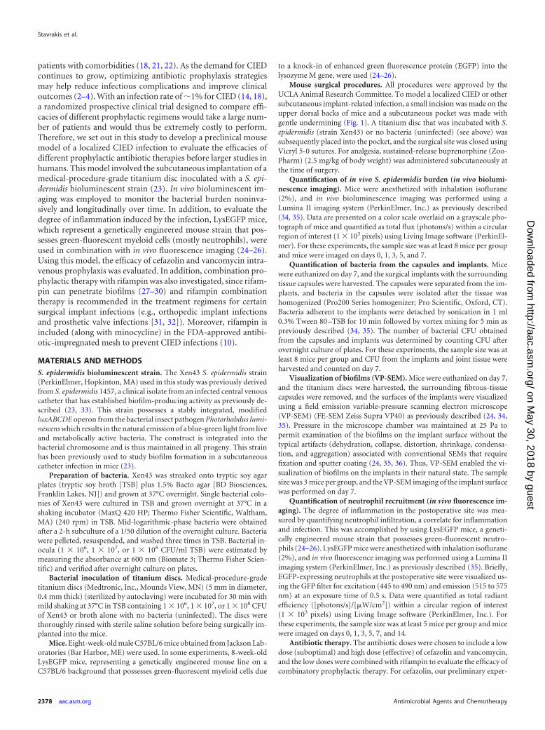
Mouse surgical procedures. All procedures were approved by theUCLA Animal Research Committee. To model a localized CIED or othersubcutaneous implant-related infection, a small incision was made on theupper dorsal backs of mice and a subcutaneous pocket was made withgentle undermining (Fig. 1). A titanium disc that was incubated with S.epidermidis (strain Xen43) or no bacteria (uninfected) (see above) wassubsequently placed into the pocket, and the surgical site was closed usingVicryl 5-0 sutures. For analgesia, sustained-release buprenorphine (Zoo-Pharm) (2.5 mg/kg of body weight) was administered subcutaneously atthe time of surgery.
Quantification of in vivo S. epidermidis burden (in vivo biolumi-nescence imaging). Mice were anesthetized with inhalation isoflurane(2%), and in vivo bioluminescence imaging was performed using aLumina II imaging system (PerkinElmer, Inc.) as previously described(34, 35). Data are presented on a color scale overlaid on a grayscale pho-tograph of mice and quantified as total flux (photons/s) within a circularregion of interest (1 � 103 pixels) using Living Image software (PerkinEl-mer). For these experiments, the sample size was at least 8 mice per groupand mice were imaged on days 0, 1, 3, 5, and 7.
Quantification of bacteria from the capsules and implants. Micewere euthanized on day 7, and the surgical implants with the surroundingtissue capsules were harvested. The capsules were separated from the im-plants, and bacteria in the capsules were isolated after the tissue washomogenized (Pro200 Series homogenizer; Pro Scientific, Oxford, CT).Bacteria adherent to the implants were detached by sonication in 1 ml0.3% Tween 80 –TSB for 10 min followed by vortex mixing for 5 min aspreviously described (34, 35). The number of bacterial CFU obtainedfrom the capsules and implants was determined by counting CFU afterovernight culture of plates. For these experiments, the sample size was atleast 8 mice per group and CFU from the implants and joint tissue wereharvested and counted on day 7.
Visualization of biofilms (VP-SEM). Mice were euthanized on day 7,and the titanium discs were harvested, the surrounding fibrous-tissuecapsules were removed, and the surfaces of the implants were visualizedusing a field emission variable-pressure scanning electron microscope(VP-SEM) (FE-SEM Zeiss Supra VP40) as previously described (24, 34,35). Pressure in the microscope chamber was maintained at 25 Pa topermit examination of the biofilms on the implant surface without thetypical artifacts (dehydration, collapse, distortion, shrinkage, condensa-tion, and aggregation) associated with conventional SEMs that requirefixation and sputter coating (24, 35, 36). Thus, VP-SEM enabled the vi-sualization of biofilms on the implants in their natural state. The samplesize was 3 mice per group, and the VP-SEM imaging of the implant surfacewas performed on day 7.
Quantification of neutrophil recruitment (in vivo fluorescence im-aging). The degree of inflammation in the postoperative site was mea-sured by quantifying neutrophil infiltration, a correlate for inflammationand infection. This was accomplished by using LysEGFP mice, a geneti-cally engineered mouse strain that possesses green-fluorescent neutro-phils (24–26). LysEGFP mice were anesthetized with inhalation isoflurane(2%), and in vivo fluorescence imaging was performed using a Lumina IIimaging system (PerkinElmer, Inc.) as previously described (35). Briefly,EGFP-expressing neutrophils at the postoperative site were visualized us-ing the GFP filter for excitation (445 to 490 nm) and emission (515 to 575nm) at an exposure time of 0.5 s. Data were quantified as total radiantefficiency ([photons/s]/[�W/cm2]) within a circular region of interest(1 � 103 pixels) using Living Image software (PerkinElmer, Inc.). Forthese experiments, the sample size was at least 5 mice per group and micewere imaged on days 0, 1, 3, 5, 7, and 14.
Antibiotic therapy. The antibiotic doses were chosen to include a lowdose (suboptimal) and high dose (effective) of cefazolin and vancomycin,and the low doses were combined with rifampin to evaluate the efficacy ofcombinatory prophylactic therapy. For cefazolin, our preliminary exper-
Stavrakis et al.
2378 aac.asm.org Antimicrobial Agents and Chemotherapy
on May 30, 2018 by guest
http://aac.asm.org/
Dow
nloaded from
iments found that the 50 mg/kg dose (37, 38), which approximated thearea under the concentration-time curve (AUC) of 670 mg · h/ml for thetypical human exposure of cefazolin (2 g) (39), had a suboptimal effect intreating the S. epidermidis implant infection and was thus used as the lowdose. For the high dose, 200 mg/kg of cefazolin was used, as doses above 50mg/kg have increased efficacy against S. aureus skin infections in mice (37).For vancomycin, the low dose of 10 mg/kg was used, which approximatedthe 50% effective dose (ED50) (10.6 mg/kg) in the neutropenic mousethigh S. aureus infection model (40). For the high dose, 110 mg/kg ofvancomycin was used (41), which approximated the AUC of 440 mg ·h/ml for typical human exposure for vancomycin (1 g) (42, 43). For ri-fampin, the dose of 25 mg/kg was used based on previous studies that haveused doses ranging from 20 to 25 mg/kg in various mouse models ofstaphylococcal infection in mice (44–48). There are important factors thatinfluence matching the mouse doses to typical human exposures for ce-fazolin, vancomycin, and especially rifampin, including differences ofhalf-life and serum protein binding between the species, and these aredescribed in detail in the Discussion. Prophylactic therapy with vancomy-cin or cefazolin (both from Hospira, Inc., Lake Forest, IL) or a shaminjection (saline solution) was administered intravenously via the retro-orbital vein 30 min preoperatively. To evaluate the efficacy of combina-tion therapy, rifampin (25 mg/kg administered subcutaneously) (Pfizer,Inc., New York, NY) was added to the low-dose cefazolin or vancomycinprophylactic therapy. The S. epidermidis strain (Xen43) in this study hadthe following MICs: cefazolin � 0.5 �g/ml, vancomycin � 2 �g/ml, and
rifampin � 0.5 �g/ml. For these experiments, the sample size was 5 to 10mice per group (all groups initially had an n � 5, and a second iterationwas performed to confirm results for certain groups) and the efficacies ofthe prophylactic antibiotics were determined using in vivo biolumines-cence imaging and ex vivo CFU counting (see above).
Statistical analysis. Data were compared using Student’s t test (twotailed). All data are expressed as mean � standard error of the mean.Values of P � 0.05 were considered statistically significant.
RESULTSMouse model of an implant-related infection. Using our in vivomodel of a subcutaneous S. epidermidis implant infection (Fig. 1),we found that the bacterial bioluminescent signals of the 1 � 106,1 � 107, and 1 � 108 CFU inocula differed on day 0 (2.5 � 106,2.0 � 105, and 7.3 � 104 photons/s, respectively), became similaron day 1 (�2.5 � 105 photons/s), and then decreased to back-ground levels (2 � 104 photons/s) by day 7 (Fig. 2A and B). Thedecreasing bioluminescent signals were likely a result of lowernumbers of bacteria present as well as lower metabolic activity ofbacteria as they began to form biofilms as previously describedwith this strain (23). Next, the CFU harvested from the capsulesand implants were enumerated. Of note, S. epidermidis-infectedmice formed larger fibrous-tissue capsules surrounding the im-
FIG 1 Mouse model of a subcutaneous-implant-related infection. Representative photographs of the surgical procedure are shown in the upper panels. A smallskin incision was made on the upper dorsal backs of mice, and undermining was performed to create a subcutaneous pocket. A medical-procedure-gradetitanium disc (5 mm in diameter and 0.4 mm thick) that had previously been incubated with S. epidermidis (infected) or no bacteria (uninfected) wassubsequently placed into the pocket, and the surgical site was sutured closed. Representative photographs of the surgical site and harvesting of the surroundingtissue capsules and implants on postoperative day 7 are shown in the lower panels. The sizes and thicknesses of capsules were much more substantial in theinfected implants than in the uninfected implants.
Combination Prophylaxis against Implant Infections
April 2014 Volume 58 Number 4 aac.asm.org 2379
on May 30, 2018 by guest
http://aac.asm.org/
Dow
nloaded from
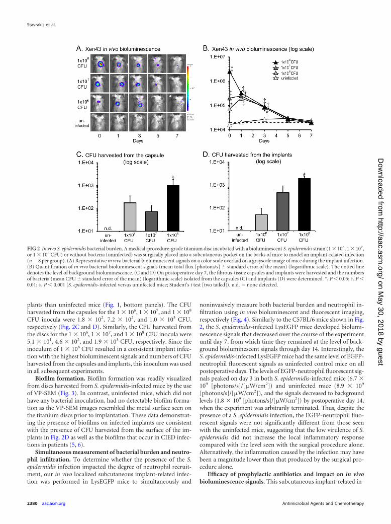
plants than uninfected mice (Fig. 1, bottom panels). The CFUharvested from the capsules for the 1 � 106, 1 � 107, and 1 � 108
CFU inocula were 1.8 � 102, 7.2 � 102, and 1.0 � 103 CFU,respectively (Fig. 2C and D). Similarly, the CFU harvested fromthe discs for the 1 � 106, 1 � 107, and 1 � 108 CFU inocula were5.1 � 101, 4.6 � 102, and 1.9 � 103 CFU, respectively. Since theinoculum of 1 � 108 CFU resulted in a consistent implant infec-tion with the highest bioluminescent signals and numbers of CFUharvested from the capsules and implants, this inoculum was usedin all subsequent experiments.
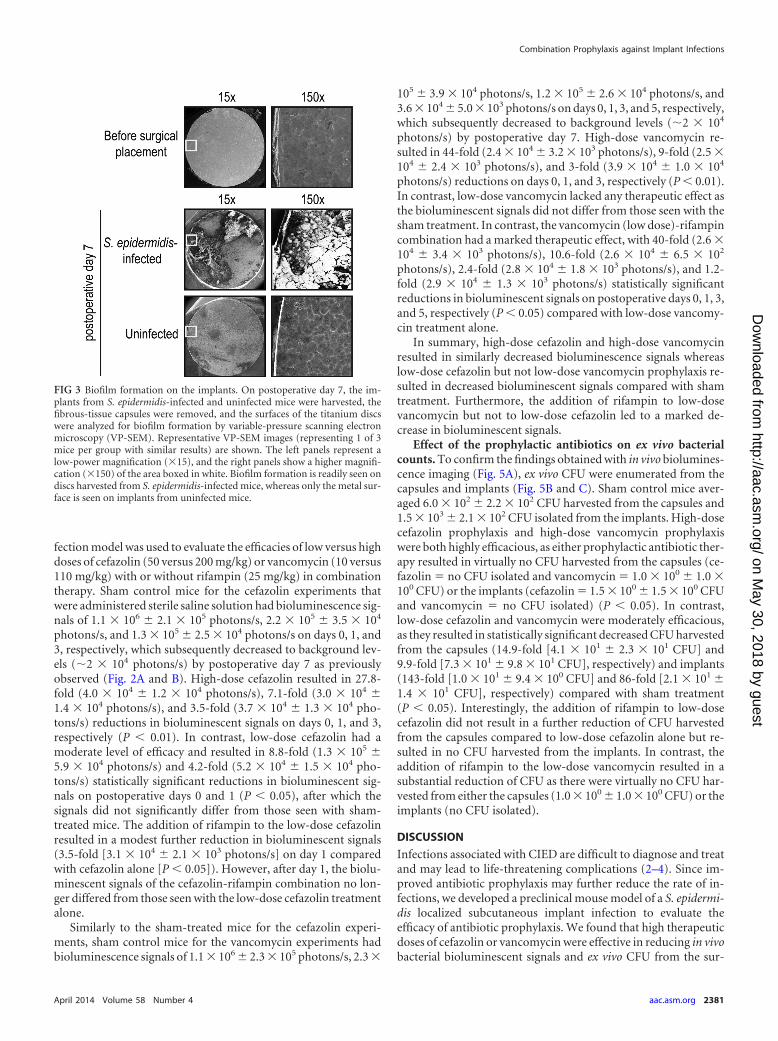
Biofilm formation. Biofilm formation was readily visualizedfrom discs harvested from S. epidermidis-infected mice by the useof VP-SEM (Fig. 3). In contrast, uninfected mice, which did nothave any bacterial inoculation, had no detectable biofilm forma-tion as the VP-SEM images resembled the metal surface seen onthe titanium discs prior to implantation. These data demonstrat-ing the presence of biofilms on infected implants are consistentwith the presence of CFU harvested from the surface of the im-plants in Fig. 2D as well as the biofilms that occur in CIED infec-tions in patients (5, 6).
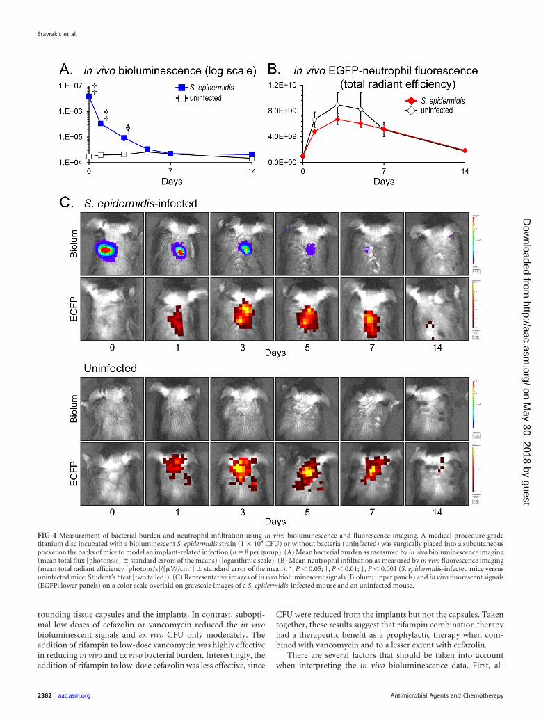
Simultaneous measurement of bacterial burden and neutro-phil infiltration. To determine whether the presence of the S.epidermidis infection impacted the degree of neutrophil recruit-ment, our in vivo localized subcutaneous implant-related infec-tion was performed in LysEGFP mice to simultaneously and
noninvasively measure both bacterial burden and neutrophil in-filtration using in vivo bioluminescent and fluorescent imaging,respectively (Fig. 4). Similarly to the C57BL/6 mice shown in Fig.2, the S. epidermidis-infected LysEGFP mice developed biolumi-nescence signals that decreased over the course of the experimentuntil day 7, from which time they remained at the level of back-ground bioluminescent signals through day 14. Interestingly, theS. epidermidis-infected LysEGFP mice had the same level of EGFP-neutrophil fluorescent signals as uninfected control mice on allpostoperative days. The levels of EGFP-neutrophil fluorescent sig-nals peaked on day 3 in both S. epidermidis-infected mice (6.7 �109 [photons/s]/[�W/cm2]) and uninfected mice (8.9 � 109
[photons/s]/[�W/cm2]), and the signals decreased to backgroundlevels (1.8 � 109 [photons/s]/[�W/cm2]) by postoperative day 14,when the experiment was arbitrarily terminated. Thus, despite thepresence of a S. epidermidis infection, the EGFP-neutrophil fluo-rescent signals were not significantly different from those seenwith the uninfected mice, suggesting that the low virulence of S.epidermidis did not increase the local inflammatory responsecompared with the level seen with the surgical procedure alone.Alternatively, the inflammation caused by the infection may havebeen a magnitude lower than that produced by the surgical pro-cedure alone.
Efficacy of prophylactic antibiotics and impact on in vivobioluminescence signals. This subcutaneous implant-related in-
FIG 2 In vivo S. epidermidis bacterial burden. A medical-procedure-grade titanium disc incubated with a bioluminescent S. epidermidis strain (1 � 106, 1 � 107,or 1 � 108 CFU) or without bacteria (uninfected) was surgically placed into a subcutaneous pocket on the backs of mice to model an implant-related infection(n � 8 per group). (A) Representative in vivo bacterial bioluminescent signals on a color scale overlaid on a grayscale image of mice during the implant infection.(B) Quantification of in vivo bacterial bioluminescent signals (mean total flux [photons/s] � standard error of the mean) (logarithmic scale). The dotted linedenotes the level of background bioluminescence. (C and D) On postoperative day 7, the fibrous-tissue capsules and implants were harvested and the numbersof bacteria (mean CFU � standard error of the mean) (logarithmic scale) isolated from the capsules (C) and implants (D) were determined. *, P � 0.05; †, P �0.01; ‡, P � 0.001 (S. epidermidis-infected versus uninfected mice; Student’s t test [two tailed]). n.d. � none detected.
Stavrakis et al.
2380 aac.asm.org Antimicrobial Agents and Chemotherapy
on May 30, 2018 by guest
http://aac.asm.org/
Dow
nloaded from
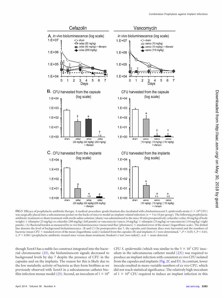
fection model was used to evaluate the efficacies of low versus highdoses of cefazolin (50 versus 200 mg/kg) or vancomycin (10 versus110 mg/kg) with or without rifampin (25 mg/kg) in combinationtherapy. Sham control mice for the cefazolin experiments thatwere administered sterile saline solution had bioluminescence sig-nals of 1.1 � 106 � 2.1 � 105 photons/s, 2.2 � 105 � 3.5 � 104
photons/s, and 1.3 � 105 � 2.5 � 104 photons/s on days 0, 1, and3, respectively, which subsequently decreased to background lev-els (�2 � 104 photons/s) by postoperative day 7 as previouslyobserved (Fig. 2A and B). High-dose cefazolin resulted in 27.8-fold (4.0 � 104 � 1.2 � 104 photons/s), 7.1-fold (3.0 � 104 �1.4 � 104 photons/s), and 3.5-fold (3.7 � 104 � 1.3 � 104 pho-tons/s) reductions in bioluminescent signals on days 0, 1, and 3,respectively (P � 0.01). In contrast, low-dose cefazolin had amoderate level of efficacy and resulted in 8.8-fold (1.3 � 105 �5.9 � 104 photons/s) and 4.2-fold (5.2 � 104 � 1.5 � 104 pho-tons/s) statistically significant reductions in bioluminescent sig-nals on postoperative days 0 and 1 (P � 0.05), after which thesignals did not significantly differ from those seen with sham-treated mice. The addition of rifampin to the low-dose cefazolinresulted in a modest further reduction in bioluminescent signals(3.5-fold [3.1 � 104 � 2.1 � 103 photons/s] on day 1 comparedwith cefazolin alone [P � 0.05]). However, after day 1, the biolu-minescent signals of the cefazolin-rifampin combination no lon-ger differed from those seen with the low-dose cefazolin treatmentalone.
Similarly to the sham-treated mice for the cefazolin experi-ments, sham control mice for the vancomycin experiments hadbioluminescence signals of 1.1 � 106 � 2.3 � 105 photons/s, 2.3 �
105 � 3.9 � 104 photons/s, 1.2 � 105 � 2.6 � 104 photons/s, and3.6 � 104 � 5.0 � 103 photons/s on days 0, 1, 3, and 5, respectively,which subsequently decreased to background levels (�2 � 104
photons/s) by postoperative day 7. High-dose vancomycin re-sulted in 44-fold (2.4 � 104 � 3.2 � 103 photons/s), 9-fold (2.5 �104 � 2.4 � 103 photons/s), and 3-fold (3.9 � 104 � 1.0 � 104
photons/s) reductions on days 0, 1, and 3, respectively (P � 0.01).In contrast, low-dose vancomycin lacked any therapeutic effect asthe bioluminescent signals did not differ from those seen with thesham treatment. In contrast, the vancomycin (low dose)-rifampincombination had a marked therapeutic effect, with 40-fold (2.6 �104 � 3.4 � 103 photons/s), 10.6-fold (2.6 � 104 � 6.5 � 102
photons/s), 2.4-fold (2.8 � 104 � 1.8 � 103 photons/s), and 1.2-fold (2.9 � 104 � 1.3 � 103 photons/s) statistically significantreductions in bioluminescent signals on postoperative days 0, 1, 3,and 5, respectively (P � 0.05) compared with low-dose vancomy-cin treatment alone.
In summary, high-dose cefazolin and high-dose vancomycinresulted in similarly decreased bioluminescence signals whereaslow-dose cefazolin but not low-dose vancomycin prophylaxis re-sulted in decreased bioluminescent signals compared with shamtreatment. Furthermore, the addition of rifampin to low-dosevancomycin but not to low-dose cefazolin led to a marked de-crease in bioluminescent signals.
Effect of the prophylactic antibiotics on ex vivo bacterialcounts. To confirm the findings obtained with in vivo biolumines-cence imaging (Fig. 5A), ex vivo CFU were enumerated from thecapsules and implants (Fig. 5B and C). Sham control mice aver-aged 6.0 � 102 � 2.2 � 102 CFU harvested from the capsules and1.5 � 103 � 2.1 � 102 CFU isolated from the implants. High-dosecefazolin prophylaxis and high-dose vancomycin prophylaxiswere both highly efficacious, as either prophylactic antibiotic ther-apy resulted in virtually no CFU harvested from the capsules (ce-fazolin � no CFU isolated and vancomycin � 1.0 � 100 � 1.0 �100 CFU) or the implants (cefazolin � 1.5 � 100 � 1.5 � 100 CFUand vancomycin � no CFU isolated) (P � 0.05). In contrast,low-dose cefazolin and vancomycin were moderately efficacious,as they resulted in statistically significant decreased CFU harvestedfrom the capsules (14.9-fold [4.1 � 101 � 2.3 � 101 CFU] and9.9-fold [7.3 � 101 � 9.8 � 101 CFU], respectively) and implants(143-fold [1.0 � 101 � 9.4 � 100 CFU] and 86-fold [2.1 � 101 �1.4 � 101 CFU], respectively) compared with sham treatment(P � 0.05). Interestingly, the addition of rifampin to low-dosecefazolin did not result in a further reduction of CFU harvestedfrom the capsules compared to low-dose cefazolin alone but re-sulted in no CFU harvested from the implants. In contrast, theaddition of rifampin to the low-dose vancomycin resulted in asubstantial reduction of CFU as there were virtually no CFU har-vested from either the capsules (1.0 � 100 � 1.0 � 100 CFU) or theimplants (no CFU isolated).
DISCUSSION
Infections associated with CIED are difficult to diagnose and treatand may lead to life-threatening complications (2–4). Since im-proved antibiotic prophylaxis may further reduce the rate of in-fections, we developed a preclinical mouse model of a S. epidermi-dis localized subcutaneous implant infection to evaluate theefficacy of antibiotic prophylaxis. We found that high therapeuticdoses of cefazolin or vancomycin were effective in reducing in vivobacterial bioluminescent signals and ex vivo CFU from the sur-
FIG 3 Biofilm formation on the implants. On postoperative day 7, the im-plants from S. epidermidis-infected and uninfected mice were harvested, thefibrous-tissue capsules were removed, and the surfaces of the titanium discswere analyzed for biofilm formation by variable-pressure scanning electronmicroscopy (VP-SEM). Representative VP-SEM images (representing 1 of 3mice per group with similar results) are shown. The left panels represent alow-power magnification (�15), and the right panels show a higher magnifi-cation (�150) of the area boxed in white. Biofilm formation is readily seen ondiscs harvested from S. epidermidis-infected mice, whereas only the metal sur-face is seen on implants from uninfected mice.
Combination Prophylaxis against Implant Infections
April 2014 Volume 58 Number 4 aac.asm.org 2381
on May 30, 2018 by guest
http://aac.asm.org/
Dow
nloaded from
rounding tissue capsules and the implants. In contrast, subopti-mal low doses of cefazolin or vancomycin reduced the in vivobioluminescent signals and ex vivo CFU only moderately. Theaddition of rifampin to low-dose vancomycin was highly effectivein reducing in vivo and ex vivo bacterial burden. Interestingly, theaddition of rifampin to low-dose cefazolin was less effective, since
CFU were reduced from the implants but not the capsules. Takentogether, these results suggest that rifampin combination therapyhad a therapeutic benefit as a prophylactic therapy when com-bined with vancomycin and to a lesser extent with cefazolin.
There are several factors that should be taken into accountwhen interpreting the in vivo bioluminescence data. First, al-
FIG 4 Measurement of bacterial burden and neutrophil infiltration using in vivo bioluminescence and fluorescence imaging. A medical-procedure-gradetitanium disc incubated with a bioluminescent S. epidermidis strain (1 � 108 CFU) or without bacteria (uninfected) was surgically placed into a subcutaneouspocket on the backs of mice to model an implant-related infection (n � 8 per group). (A) Mean bacterial burden as measured by in vivo bioluminescence imaging(mean total flux [photons/s] � standard errors of the means) (logarithmic scale). (B) Mean neutrophil infiltration as measured by in vivo fluorescence imaging(mean total radiant efficiency [photons/s]/[�W/cm2] � standard error of the mean). *, P � 0.05; †, P � 0.01; ‡, P � 0.001 (S. epidermidis-infected mice versusuninfected mice; Student’s t test [two tailed]). (C) Representative images of in vivo bioluminescent signals (Biolum; upper panels) and in vivo fluorescent signals(EGFP; lower panels) on a color scale overlaid on grayscale images of a S. epidermidis-infected mouse and an uninfected mouse.
Stavrakis et al.
2382 aac.asm.org Antimicrobial Agents and Chemotherapy
on May 30, 2018 by guest
http://aac.asm.org/
Dow
nloaded from
though Xen43 has a stable lux construct integrated into the bacte-rial chromosome (23), the bioluminescent signals decreased tobackground levels by day 7 despite the presence of CFU in thecapsules and on the implants. The reason for this is likely due tothe low metabolic activity of bacteria as they form biofilms as wepreviously observed with Xen43 in a subcutaneous catheter bio-film infection mouse model (23). Second, an inoculum of 1 � 108
CFU S. epidermidis (which was similar to the 5 � 107 CFU inoc-ulum in the subcutaneous catheter model [23]) was required toproduce an implant infection with consistent ex vivo CFU isolatedfrom the capsules and implants (Fig. 2C and D). In contrast, lowerinocula resulted in more-variable numbers of ex vivo CFU, whichdid not reach statistical significance. The relatively high inoculumof 1 � 108 CFU required to induce an implant infection in this
FIG 5 Efficacy of prophylactic antibiotic therapy. A medical-procedure-grade titanium disc incubated with a bioluminescent S. epidermidis strain (1 � 108 CFU)was surgically placed into a subcutaneous pocket on the backs of mice to model an implant-related infection (n � 5 to 10 per group). The following prophylacticantibiotic treatment or sham treatment with sterile saline solution (sham) was administered to the mice 30 min preoperatively: cefazolin (cefaz; 50 mg/kg of bodyweight) � rifampin (25 mg/kg) or cefazolin (200 mg/kg) (left panels) or vancomycin (vanco; 10 mg/kg) � rifampin (25 mg/kg) or vancomycin (110 mg/kg) (rightpanels). (A) Bacterial burden as measured by in vivo bioluminescence (mean total flux [photons/s] � standard error of the mean) (logarithmic scale). The dottedline denotes the level of background bioluminescence. (B and C) On postoperative day 7, the capsules and titanium discs were harvested and the numbers ofbacteria (mean CFU � standard error of the mean [logarithmic scale]) isolated from the capsules (B) and implants (C) were determined. *, P � 0.05; †, P � 0.01;‡, P � 0.001 (prophylactic antibiotic-treated mice versus sham treatment; Student’s t test [two tailed]). n.d. � none detected.
Combination Prophylaxis against Implant Infections
April 2014 Volume 58 Number 4 aac.asm.org 2383
on May 30, 2018 by guest
http://aac.asm.org/
Dow
nloaded from
model is likely due to the low virulence of S. epidermidis and thesubcutaneous site of infection, which induced robust postopera-tive neutrophil recruitment in LysEGFP mice that was the same inthe presence of the S. epidermidis infection and the surgical pro-cedure alone (Fig. 4).
There are also important factors pertaining to the approxima-tion of the human exposures of cefazolin, vancomycin, and rifam-pin in mice that should be taken into account in comparing expo-sures between the species. First, there are differences in serumprotein binding, and the amount of drug bound is higher in hu-mans versus mice for cefazolin (�85% versus �50% [49–51]),vancomycin (35% to 50% versus �25% [52, 53]), and rifampin(�97% versus �90% [54]). Thus, the amount of free and activedrug for these antibiotics is higher in mice than in humans. Sec-ond, the half-lives are much longer in humans versus mice forcefazolin (2 h versus 30 min [55, 56]) and vancomycin (7.7 hversus 2 h [42, 57]). Since the infected discs were implanted 30min after administering the prophylactic antibiotics, the increasedhalf-life of vancomycin may have contributed to its showing bettereffectiveness than cefazolin. Conversely, the half-life of rifampin is4 h in humans and 12 h in mice (58, 59), indicating that rifampinhas a prolonged effect in mice and providing an explanation forthe increased efficacy of rifampin in this study. It should be men-tioned that the typical human exposure of rifampin (10 mg/kg or600 mg daily) was originally based on a combination of pharma-cokinetics, toxicity, and cost (45, 60). At this dose, the AUC valuesare 48.5 �g · h/ml in humans and 132 �g · h/ml in mice (54).However, as mentioned above, the amount unbound drug is3-fold higher and the half-life is 3-fold shorter in humans than inmice, making it difficult to match the human exposure. Takentogether, these differences in the pharmacokinetics and pharma-codynamics (PK/PD) of the antibiotics between the species high-light the limitations of approximating human exposures in mice.Ideally, for evaluating a range of antibiotic doses, additional clin-ical S. epidermidis isolates as well as other bacterial strains thatcause CIED infections (with various antibiotic susceptibilities)would provide more-precise PK/PD data to better match the ex-posures in humans and provide greater evidence of efficacy. Thesestudies are to be the subject of our future investigations.
From a mechanistic standpoint, although the precise reasonfor the increased efficacy of rifampin combination therapy in ourmodel is unknown, previous studies have found that rifampin hasenhanced activity against bacteria in biofilms (27–30). This anti-biofilm activity is consistent with our findings since cefazolin-rifampin- and vancomycin-rifampin-treated mice had no CFUisolated from the implants. In addition, a prior study found thatbacteria in biofilms have increased cell wall thickness, renderingthem less sensitive to vancomycin (61). Since both vancomycinand cefazolin act by disrupting bacterial cell wall synthesis, theseantibiotics may be less active against bacteria in biofilms. In con-trast, rifampin acts by inhibiting bacterial DNA-dependent RNAsynthesis by binding to the bacterial RNA polymerase (62, 63).
From a clinical perspective, there are several factors that areimportant to take into account in considering rifampin combina-tion as prophylactic therapy. Rifampin can cause toxicity, includ-ing hepatitis and drug interactions (32). In addition, althoughearly reports found that rifampin combination prophylactic ther-apy had a therapeutic benefit in patients prior to cardiac valvereplacement and cardiac bypass surgery (64, 65), these patientsdeveloped rifampin-resistant S. epidermidis infections in their
normal human skin flora (66). However, certain combinations,such as daptomycin and rifampin, have been shown to decreasethe development of resistance in an experimental vascular graftbiofilm model (67).
Taken together, our findings suggest that antibiotic prophy-laxis against CIED and perhaps other surgical implant infectionscan be further optimized to prevent infectious complications andimprove clinical outcomes. In particular, a reexamination andreconsideration of rifampin combination as prophylactic therapymay be warranted in certain patient populations at highest risk forCIED infections as these infections can have deadly consequences.Alternatively, other antistaphylococcal drugs with longer half-lives such as tetracyclines (doxycycline, minocycline, or tigecy-cline) or newer glycopeptides (daptomycin, telavancin, dalbavan-cin, or oritavancin) could serve as additional candidates forcombination prophylactic therapy and our mouse model couldserve as a valuable preclinical system to evaluate these potentialantibiotic combinations before initiating clinical trials.
ACKNOWLEDGMENTS
This work was supported by the Medtronic, Inc., CRDM (CardiacRhythm Disease Management) External Research Program (to D.Z.U.and L.S.M.), an H & H Lee Surgical Resident Research Scholars Program(to A.I.S. and J.A.N.), the Intramural Program of the National Institute ofAllergy and Infectious Diseases (NIAID), the National Institutes of Health(to M. O.), and National Institutes of Health grants R24-CA92865 (to theUCLA Small Animal Imaging Resource Program), R01 HS021188 (toD.Z.U.), and R01-AI078910 (to L.S.M.).
REFERENCES1. Darouiche RO. 2004. Treatment of infections associated with surgical
implants. N. Engl. J. Med. 350:1422–1429. http://dx.doi.org/10.1056/NEJMra035415.
2. Uslan DZ, Baddour LM. 2006. Cardiac device infections: getting to theheart of the matter. Curr. Opin. Infect. Dis. 19:345–348. http://dx.doi.org/10.1097/01.qco.0000235160.78302.24.
3. Gould PA, Gula LJ, Yee R, Skanes AC, Klein GJ, Krahn AD. 2011.Cardiovascular implantable electrophysiological device-related infec-tions: a review. Curr. Opin. Cardiol. 26:6 –11. http://dx.doi.org/10.1097/HCO.0b013e328341384e.
4. Baddour LM, Epstein AE, Erickson CC, Knight BP, Levison ME, LockhartPB, Masoudi FA, Okum EJ, Wilson WR, Beerman LB, Bolger AF, EstesNA, III, Gewitz M, Newburger JW, Schron EB, Taubert KA, AmericanHeart Association Rheumatic Fever, Endocarditis, and Kawasaki DiseaseCommittee; Council on Cardiovascular Disease in Young; Council onCardiovascular Surgery and Anesthesia; Council on Cardiovascular Nurs-ing; Council on Clinical Cardiology; Interdisciplinary Council on Qualityof Care; American Heart Association. 2010. Update on cardiovascular im-plantable electronic device infections and their management: a scientific state-ment from the American Heart Association. Circulation 121:458–477. http://dx.doi.org/10.1161/CIRCULATIONAHA.109.192665.
5. Toba FA, Akashi H, Arrecubieta C, Lowy FD. 2011. Role of biofilm inStaphylococcus aureus and Staphylococcus epidermidis ventricular assistdevice driveline infections. J. Thorac. Cardiovasc. Surg. 141:1259 –1264.http://dx.doi.org/10.1016/j.jtcvs.2010.07.016.
6. Rohacek M, Weisser M, Kobza R, Schoenenberger AW, Pfyffer GE, Frei R,Erne P, Trampuz A. 2010. Bacterial colonization and infection of electro-physiological cardiac devices detected with sonication and swab culture.Circulation 121:1691–1697. http://dx.doi.org/10.1161/CIRCULATIONAHA.109.906461.
7. Kolker AR, Redstone JS, Tutela JP. 2007. Salvage of exposed implantablecardiac electrical devices and lead systems with pocket change and localflap coverage. Ann. Plast. Surg. 59:26 –29. http://dx.doi.org/10.1097/01.sap.0000261846.73531.2e.
8. Sohail MR, Uslan DZ, Khan AH, Friedman PA, Hayes DL, Wilson WR,Steckelberg JM, Stoner S, Baddour LM. 2007. Management and outcomeof permanent pacemaker and implantable cardioverter-defibrillator in-
Stavrakis et al.
2384 aac.asm.org Antimicrobial Agents and Chemotherapy
on May 30, 2018 by guest
http://aac.asm.org/
Dow
nloaded from
fections. J. Am. Coll. Cardiol. 49:1851–1859. http://dx.doi.org/10.1016/j.jacc.2007.01.072.
9. Tascini C, Bongiorni MG, Gemignani G, Soldati E, Leonildi A, Arena G,Doria R, Giannola G, La Pira F, Tagliaferri E, Caravelli P, Dell’Anna R,Menichetti F. 2006. Management of cardiac device infections: a retrospec-tive survey of a non-surgical approach combining antibiotic therapy withtransvenous removal. J. Chemother. 18:157–163. http://dx.doi.org/10.1179/joc.2006.18.2.157.
10. Bloom HL, Constantin L, Dan D, De Lurgio DB, El-Chami M, Ganz LI,Gleed KJ, Hackett FK, Kanuru NK, Lerner DJ, Rasekh A, Simons GR,Sogade FO, Sohail MR; COoperative Multicenter study Monitoring aCIED ANtimicrobial Device Investigators. 2011. Implantation successand infection in cardiovascular implantable electronic device proceduresutilizing an antibacterial envelope. Pacing Clin. Electrophysiol. 34:133–142. http://dx.doi.org/10.1111/j.1540-8159.2010.02931.x.
11. Sohail MR, Uslan DZ, Khan AH, Friedman PA, Hayes DL, Wilson WR,Steckelberg JM, Stoner SM, Baddour LM. 2007. Risk factor analysis ofpermanent pacemaker infection. Clin. Infect. Dis. 45:166 –173. http://dx.doi.org/10.1086/518889.
12. de Oliveira JC, Martinelli M, Nishioka SA, Varejao T, Uipe D, PedrosaAA, Costa R, D’Avila A, Danik SB. 2009. Efficacy of antibiotic prophy-laxis before the implantation of pacemakers and cardioverter-defibrillators: results of a large, prospective, randomized, double-blinded,placebo-controlled trial. Circ. Arrhythm. Electrophysiol. 2:29 –34. http://dx.doi.org/10.1161/CIRCEP.108.795906.
13. Da Costa A, Kirkorian G, Cucherat M, Delahaye F, Chevalier P, CerisierA, Isaaz K, Touboul P. 1998. Antibiotic prophylaxis for permanentpacemaker implantation: a meta-analysis. Circulation 97:1796 –1801.http://dx.doi.org/10.1161/01.CIR.97.18.1796.
14. Uslan DZ, Gleva MJ, Warren DK, Mela T, Chung MK, Gottipaty V,Borge R, Dan D, Shinn T, Mitchell K, Holcomb RG, Poole JE. 2012.Cardiovascular implantable electronic device replacement infections andprevention: results from the REPLACE Registry. Pacing Clin. Electro-physiol. 35:81– 87. http://dx.doi.org/10.1111/j.1540-8159.2011.03257.x.
15. Darouiche R, Mosier M, Voigt J. 2012. Antibiotics and antiseptics toprevent infection in cardiac rhythm management device implantationsurgery. Pacing Clin. Electrophysiol. 35:1348 –1360. http://dx.doi.org/10.1111/j.1540-8159.2012.03506.x.
16. Margey R, McCann H, Blake G, Keelan E, Galvin J, Lynch M, MahonN, Sugrue D, O’Neill J. 2010. Contemporary management of and out-comes from cardiac device related infections. Europace 12:64 –70. http://dx.doi.org/10.1093/europace/eup362.
17. Anselmino M, Vinci M, Comoglio C, Rinaldi M, Bongiorni MG, TreviGP, Golzio PG. 2009. Bacteriology of infected extracted pacemaker andICD leads. J. Cardiovasc. Med. (Hagerstown) 10:693– 698. http://dx.doi.org/10.2459/JCM.0b013e32832b3585.
18. Uslan DZ, Sohail MR, St Sauver JL, Friedman PA, Hayes DL, StonerSM, Wilson WR, Steckelberg JM, Baddour LM. 2007. Permanent pace-maker and implantable cardioverter defibrillator infection: a population-based study. Arch. Intern. Med. 167:669 – 675. http://dx.doi.org/10.1001/archinte.167.7.669.
19. Dy Chua J, Abdul-Karim A, Mawhorter S, Procop GW, Tchou P,Niebauer M, Saliba W, Schweikert R, Wilkoff BL. 2005. The role of swaband tissue culture in the diagnosis of implantable cardiac device infection.Pacing Clin. Electrophysiol. 28:1276 –1281. http://dx.doi.org/10.1111/j.1540-8159.2005.00268.x.
20. Chambers ST. 2005. Diagnosis and management of staphylococcal infec-tions of pacemakers and cardiac defibrillators. Intern. Med. J. 35(Suppl2):S63–S71. http://dx.doi.org/10.1111/j.1444-0903.2005.00980.x.
21. Greenspon AJ, Patel JD, Lau E, Ochoa JA, Frisch DR, Ho RT, Pavri BB,Kurtz SM. 2011. 16-Year trends in the infection burden for pacemakersand implantable cardioverter-defibrillators in the United States 1993 to2008. J. Am. Coll. Cardiol. 58:1001–1006. http://dx.doi.org/10.1016/j.jacc.2011.04.033.
22. Voigt A, Shalaby A, Saba S. 2010. Continued rise in rates of cardiovas-cular implantable electronic device infections in the United States: tem-poral trends and causative insights. Pacing Clin. Electrophysiol. 33:414 –419. http://dx.doi.org/10.1111/j.1540-8159.2009.02569.x.
23. Vuong C, Kocianova S, Yu J, Kadurugamuwa JL, Otto M. 2008. Devel-opment of real-time in vivo imaging of device-related Staphylococcusepidermidis infection in mice and influence of animal immune status onsusceptibility to infection. J. Infect. Dis. 198:258 –261. http://dx.doi.org/10.1086/589307.
24. Bernthal NM, Stavrakis AI, Billi F, Cho JS, Kremen TJ, Simon SI,Cheung AL, Finerman GA, Lieberman JR, Adams JS, Miller LS. 2010. Amouse model of post-arthroplasty Staphylococcus aureus joint infectionto evaluate in vivo the efficacy of antimicrobial implant coatings. PLoSOne 5:e12580. http://dx.doi.org/10.1371/journal.pone.0012580.
25. Faust N, Varas F, Kelly LM, Heck S, Graf T. 2000. Insertion of enhancedgreen fluorescent protein into the lysozyme gene creates mice with greenfluorescent granulocytes and macrophages. Blood 96:719 –726.
26. Niska JA, Meganck JA, Pribaz JR, Shahbazian JH, Lim E, Zhang N, RiceBW, Akin A, Ramos RI, Bernthal NM, Francis KP, Miller LS. 2012.Monitoring bacterial burden, inflammation and bone damage longitudi-nally using optical and micro-CT imaging in an orthopaedic implant in-fection in mice. PLoS One 7:e47397. http://dx.doi.org/10.1371/journal.pone.0047397.
27. John AK, Baldoni D, Haschke M, Rentsch K, Schaerli P, Zimmerli W,Trampuz A. 2009. Efficacy of daptomycin in implant-associated infectiondue to methicillin-resistant Staphylococcus aureus: importance of combi-nation with rifampin. Antimicrob. Agents Chemother. 53:2719 –2724.http://dx.doi.org/10.1128/AAC.00047-09.
28. Raad I, Hanna H, Jiang Y, Dvorak T, Reitzel R, Chaiban G, Sherertz R,Hachem R. 2007. Comparative activities of daptomycin, linezolid, andtigecycline against catheter-related methicillin-resistant Staphylococcusbacteremic isolates embedded in biofilm. Antimicrob. Agents Chemother.51:1656 –1660. http://dx.doi.org/10.1128/AAC.00350-06.
29. Saginur R, Stdenis M, Ferris W, Aaron SD, Chan F, Lee C, Ramotar K.2006. Multiple combination bactericidal testing of staphylococcal biofilmsfrom implant-associated infections. Antimicrob. Agents Chemother. 50:55– 61. http://dx.doi.org/10.1128/AAC.50.1.55-61.2006.
30. Parra-Ruiz J, Vidaillac C, Rose WE, Rybak MJ. 2010. Activities ofhigh-dose daptomycin, vancomycin, and moxifloxacin alone or in com-bination with clarithromycin or rifampin in a novel in vitro model ofStaphylococcus aureus biofilm. Antimicrob. Agents Chemother. 54:4329 – 4334. http://dx.doi.org/10.1128/AAC.00455-10.
31. Liu C, Bayer A, Cosgrove SE, Daum RS, Fridkin SK, Gorwitz RJ, KaplanSL, Karchmer AW, Levine DP, Murray BE, Rybak J, Talan DA, Cham-bers HF. 2011. Clinical practice guidelines by the Infectious DiseasesSociety of America for the treatment of methicillin-resistant Staphylococ-cus aureus infections in adults and children. Clin. Infect. Dis. 52:e18 – e55.http://dx.doi.org/10.1093/cid/ciq146.
32. Osmon DR, Berbari EF, Berendt AR, Lew D, Zimmerli W, SteckelbergJM, Rao N, Hanssen A, Wilson WR. 2013. Diagnosis and management ofprosthetic joint infection: clinical practice guidelines by the InfectiousDiseases Society of America. Clin. Infect. Dis. 56:e1– e25. http://dx.doi.org/10.1093/cid/cis803.
33. Mack D, Siemssen N, Laufs R. 1992. Parallel induction by glucose ofadherence and a polysaccharide antigen specific for plastic-adherentStaphylococcus epidermidis: evidence for functional relation to intercel-lular adhesion. Infect. Immun. 60:2048 –2057.
34. Niska JA, Shahbazian JH, Ramos RI, Pribaz JR, Billi F, Francis KP,Miller LS. 2012. Daptomycin and tigecycline have broader effective doseranges than vancomycin as prophylaxis against a Staphylococcus aureussurgical implant infection in mice. Antimicrob. Agents Chemother. 56:2590 –2597. http://dx.doi.org/10.1128/AAC.06291-11.
35. Pribaz JR, Bernthal NM, Billi F, Cho JS, Ramos RI, Guo Y, Cheung AL,Francis KP, Miller LS. 2012. Mouse model of chronic post-arthroplastyinfection: noninvasive in vivo bioluminescence imaging to monitor bac-terial burden for long-term study. J. Orthop. Res. 30:335–340. http://dx.doi.org/10.1002/jor.21519.
36. Bernthal NM, Pribaz JR, Stavrakis AI, Billi F, Cho JS, Ramos RI,Francis KP, Iwakura Y, Miller LS. 2011. Protective role of IL-1betaagainst post-arthroplasty Staphylococcus aureus infection. J. Orthop. Res.29:1621–1626. http://dx.doi.org/10.1002/jor.21414.
37. Fernandez J, Hilliard JJ, Abbanat D, Zhang W, Melton JL, Santoro CM,Flamm RK, Bush K. 2010. In vivo activity of ceftobiprole in murine skininfections due to Staphylococcus aureus and Pseudomonas aeruginosa.Antimicrob. Agents Chemother. 54:116 –125. http://dx.doi.org/10.1128/AAC.00642-09.
38. Xiong YQ, Willard J, Kadurugamuwa JL, Yu J, Francis KP, Bayer AS.2005. Real-time in vivo bioluminescent imaging for evaluating the efficacyof antibiotics in a rat Staphylococcus aureus endocarditis model. Antimi-crob. Agents Chemother. 49:380 –387. http://dx.doi.org/10.1128/AAC.49.1.380-387.2005.
39. Douglas A, Udy AA, Wallis SC, Jarrett P, Stuart J, Lassig-Smith M,
Combination Prophylaxis against Implant Infections
April 2014 Volume 58 Number 4 aac.asm.org 2385
on May 30, 2018 by guest
http://aac.asm.org/
Dow
nloaded from
Deans R, Roberts MS, Taraporewalla K, Jenkins J, Medley G, Lipman J,Roberts JA. 2011. Plasma and tissue pharmacokinetics of cefazolin inpatients undergoing elective and semielective abdominal aortic aneurysmopen repair surgery. Antimicrob. Agents Chemother. 55:5238 –5242. http://dx.doi.org/10.1128/AAC.05033-11.
40. Hegde SS, Reyes N, Wiens T, Vanasse N, Skinner R, McCullough J,Kaniga K, Pace J, Thomas R, Shaw JP, Obedencio G, Judice JK. 2004.Pharmacodynamics of telavancin (TD-6424), a novel bactericidal agent,against gram-positive bacteria. Antimicrob. Agents Chemother. 48:3043–3050. http://dx.doi.org/10.1128/AAC.48.8.3043-3050.2004.
41. Reyes N, Skinner R, Kaniga K, Krause KM, Shelton J, Obedencio GP,Gough A, Conner M, Hegde SS. 2005. Efficacy of telavancin (TD-6424),a rapidly bactericidal lipoglycopeptide with multiple mechanisms of ac-tion, in a murine model of pneumonia induced by methicillin-resistantStaphylococcus aureus. Antimicrob. Agents Chemother. 49:4344 – 4346.http://dx.doi.org/10.1128/AAC.49.10.4344-4346.2005.
42. Reyes N, Skinner R, Benton BM, Krause KM, Shelton J, Obedencio GP,Hegde SS. 2006. Efficacy of telavancin in a murine model of bacteraemiainduced by methicillin-resistant Staphylococcus aureus. J. Antimicrob.Chemother. 58:462– 465. http://dx.doi.org/10.1093/jac/dkl222.
43. Crandon JL, Kuti JL, Nicolau DP. 2010. Comparative efficacies of humansimulated exposures of telavancin and vancomycin against methicillin-resistant Staphylococcus aureus with a range of vancomycin MICs in amurine pneumonia model. Antimicrob. Agents Chemother. 54:5115–5119. http://dx.doi.org/10.1128/AAC.00062-10.
44. Sakoulas G, Eliopoulos GM, Alder J, Eliopoulos CT. 2003. Efficacy ofdaptomycin in experimental endocarditis due to methicillin-resistantStaphylococcus aureus. Antimicrob. Agents Chemother. 47:1714 –1718.http://dx.doi.org/10.1128/AAC.47.5.1714-1718.2003.
45. van Ingen J, Aarnoutse RE, Donald PR, Diacon AH, Dawson R, Plem-per van Balen G, Gillespie SH, Boeree MJ. 2011. Why do we use 600 mgof rifampicin in tuberculosis treatment? Clin. Infect. Dis. 52:e194 – e199.http://dx.doi.org/10.1093/cid/cir184.
46. Mandell GL, Moorman DR. 1980. Treatment of experimental staphylo-coccal infections: effect of rifampin alone and in combination on devel-opment of rifampin resistance. Antimicrob. Agents Chemother. 17:658 –662. http://dx.doi.org/10.1128/AAC.17.4.658.
47. Lobo MC, Mandell GL. 1972. Treatment of experimental staphylococcalinfection with rifampin. Antimicrob. Agents Chemother. 2:195–200. http://dx.doi.org/10.1128/AAC.2.3.195.
48. Mandell GL, Vest TK. 1972. Killing of intraleukocytic Staphylococcusaureus by rifampin: in-vitro and in-vivo studies. J. Infect. Dis. 125:486 –490. http://dx.doi.org/10.1093/infdis/125.5.486.
49. Kunst MW, Mattie H. 1978. Cefazolin and cephradine: relationship be-tween antibacterial activity in vitro and in mice experimentally infectedwith Escherichia coli. J. Infect. Dis. 137:391– 402. http://dx.doi.org/10.1093/infdis/137.4.391.
50. Vella-Brincat JW, Begg EJ, Kirkpatrick CM, Zhang M, Chambers ST,Gallagher K. 2007. Protein binding of cefazolin is saturable in vivo bothbetween and within patients. Br. J. Clin. Pharmacol. 63:753–757. http://dx.doi.org/10.1111/j.1365-2125.2006.02827.x.
51. Craig WA, Welling PG, Jackson TC, Kunin CM. 1973. Pharmacology ofcefazolin and other cephalosporins in patients with renal insufficiency. J.Infect. Dis. 128(Suppl):S347–S353. http://dx.doi.org/10.1093/infdis/128.Supplement_2.S347.
52. Ackerman BH, Taylor EH, Olsen KM, Abdel-Malak W, Pappas AA.1988. Vancomycin serum protein binding determination by ultrafiltra-tion. Drug Intell. Clin. Pharm. 22:300 –303.
53. Knudsen JD, Fuursted K, Espersen F, Frimodt-Moller N. 1997. Activi-
ties of vancomycin and teicoplanin against penicillin-resistant pneumo-cocci in vitro and in vivo and correlation to pharmacokinetic parametersin the mouse peritonitis model. Antimicrob. Agents Chemother. 41:1910 –1915.
54. de Steenwinkel JE, Aarnoutse RE, de Knegt GJ, ten Kate MT, Teulen M,Verbrugh HA, Boeree MJ, van Soolingen D, Bakker-Woudenberg IA.2013. Optimization of the rifampin dosage to improve the therapeuticefficacy in tuberculosis treatment using a murine model. Am. J. Respir.Crit. Care Med. 187:1127–1134. http://dx.doi.org/10.1164/rccm.201207-1210OC.
55. Lee FH, Pfeffer M, Van Harken DR, Smyth RD, Hottendorf GH. 1980.Comparative pharmacokinetics of ceforanide (BL-S786R) and cefazolinin laboratory animals and humans. Antimicrob. Agents Chemother. 17:188 –192. http://dx.doi.org/10.1128/AAC.17.2.188.
56. Chapman SW, Steigbigel RT. 1983. Staphylococcal beta-lactamase andefficacy of beta-lactam antibiotics: in vitro and in vivo evaluation. J. Infect.Dis. 147:1078 –1089. http://dx.doi.org/10.1093/infdis/147.6.1078.
57. Healy DP, Polk RE, Garson ML, Rock DT, Comstock TJ. 1987. Com-parison of steady-state pharmacokinetics of two dosage regimens of van-comycin in normal volunteers. Antimicrob. Agents Chemother. 31:393–397. http://dx.doi.org/10.1128/AAC.31.3.393.
58. Forrest GN, Tamura K. 2010. Rifampin combination therapy for non-mycobacterial infections. Clin. Microbiol. Rev. 23:14 –34. http://dx.doi.org/10.1128/CMR.00034-09.
59. Jayaram R, Gaonkar S, Kaur P, Suresh BL, Mahesh BN, Jayashree R,Nandi V, Bharat S, Shandil RK, Kantharaj E, Balasubramanian V. 2003.Pharmacokinetics-pharmacodynamics of rifampin in an aerosol infectionmodel of tuberculosis. Antimicrob. Agents Chemother. 47:2118 –2124.http://dx.doi.org/10.1128/AAC.47.7.2118-2124.2003.
60. Perlroth J, Kuo M, Tan J, Bayer AS, Miller LG. 2008. Adjunctive use ofrifampin for the treatment of Staphylococcus aureus infections: a system-atic review of the literature. Arch. Intern. Med. 168:805– 819. http://dx.doi.org/10.1001/archinte.168.8.805.
61. Reipert A, Ehlert K, Kast T, Bierbaum G. 2003. Morphological andgenetic differences in two isogenic Staphylococcus aureus strains withdecreased susceptibilities to vancomycin. Antimicrob. Agents Chemother.47:568 –576. http://dx.doi.org/10.1128/AAC.47.2.568-576.2003.
62. Calvori C, Frontali L, Leoni L, Tecce G. 1965. Effect of rifamycin on proteinsynthesis. Nature 207:417–418. http://dx.doi.org/10.1038/207417a0.
63. Campbell EA, Korzheva N, Mustaev A, Murakami K, Nair S, GoldfarbA, Darst SA. 2001. Structural mechanism for rifampicin inhibition ofbacterial RNA polymerase. Cell 104:901–912. http://dx.doi.org/10.1016/S0092-8674(01)00286-0.
64. Spelman D, Harrington G, Russo P, Wesselingh S. 2002. Clinical,microbiological, and economic benefit of a change in antibiotic prophy-laxis for cardiac surgery. Infect. Control Hosp. Epidemiol. 23:402– 404.http://dx.doi.org/10.1086/502074.
65. Archer GL, Armstrong BC, Kline BJ. 1982. Rifampin blood and tissuelevels in patients undergoing cardiac valve surgery. Antimicrob. AgentsChemother. 21:800 – 803. http://dx.doi.org/10.1128/AAC.21.5.800.
66. Archer GL, Armstrong BC. 1983. Alteration of staphylococcal flora incardiac surgery patients receiving antibiotic prophylaxis. J. Infect. Dis.147:642– 649. http://dx.doi.org/10.1093/infdis/147.4.642.
67. Cirioni O, Mocchegiani F, Ghiselli R, Silvestri C, Gabrielli E, Mar-chionni E, Orlando F, Nicolini D, Risaliti A, Giacometti A. 2010.Daptomycin and rifampin alone and in combination prevent vasculargraft biofilm formation and emergence of antibiotic resistance in a subcu-taneous rat pouch model of staphylococcal infection. Eur. J. Vasc. Endo-vasc. Surg. 40:817– 822. http://dx.doi.org/10.1016/j.ejvs.2010.08.009.
Stavrakis et al.
2386 aac.asm.org Antimicrobial Agents and Chemotherapy
on May 30, 2018 by guest
http://aac.asm.org/
Dow
nloaded from