Embed Size (px)
Citation preview

RESEARCH ARTICLE Open Access
Combination of diffusion tensor and functionalmagnetic resonance imaging during recoveryfrom the vegetative stateDavinia Fernández-Espejo1,2, Carme Junque1,2*, Damian Cruse3, Montserrat Bernabeu4, Teresa Roig-Rovira5,Neus Fábregas6, Eva Rivas6, Jose M Mercader7
Abstract
Background: The rate of recovery from the vegetative state (VS) is low. Currently, little is known of themechanisms and cerebral changes that accompany those relatively rare cases of good recovery. Here, wecombined functional magnetic resonance imaging (fMRI) and diffusion tensor imaging (DTI) to study the evolutionof one VS patient at one month post-ictus and again twelve months later when he had recovered consciousness.
Methods: fMRI was used to investigate cortical responses to passive language stimulation as well as task-induceddeactivations related to the default-mode network. DTI was used to assess the integrity of the global white matterand the arcuate fasciculus. We also performed a neuropsychological assessment at the time of the second MRIexamination in order to characterize the profile of cognitive deficits.
Results: fMRI analysis revealed anatomically appropriate activation to speech in both the first and the secondscans but a reduced pattern of task-induced deactivations in the first scan. In the second scan, following therecovery of consciousness, this pattern became more similar to that classically described for the default-modenetwork. DTI analysis revealed relative preservation of the arcuate fasciculus and of the global normal-appearingwhite matter at both time points. The neuropsychological assessment revealed recovery of receptive linguisticfunctioning by 12-months post-ictus.
Conclusions: These results suggest that the combination of different structural and functional imaging modalitiesmay provide a powerful means for assessing the mechanisms involved in the recovery from the VS.
BackgroundThe vegetative state (VS) is a disorder of consciousness(DOC) characterized by wakefulness in the absence ofawareness [1]. The recovery rate from the VS is low,with only 52% of adults who are in a VS one month fol-lowing a traumatic brain injury (TBI) recovering con-sciousness within one year [2]. Furthermore, such arecovery of consciousness is not always accompanied bya recovery of functional abilities, such as communica-tion, or the ability to learn and perform adaptive tasks.It has been estimated that TBI patients in a VS onemonth after injury have a 17% chance of recovering to a
level of moderate disability after 12 months, and only a7% chance of achieving a good recovery in the sametime period [3].Little is known about the neural changes associated
with recovery from the VS. Early positron emissiontomography (PET) studies identified metabolic dysfunc-tion and impaired functional connectivity in a largefronto-parietal network in a group of VS patients [4].The recovery of consciousness of one VS patient haspreviously been linked to an increase in the functionalconnectivity within this network, [5,6] encompassing theareas known to be most active in resting-state condi-tions [7]. A growing body of evidence from PET andfunctional magnetic resonance imaging (fMRI) studiesof healthy volunteers in a variety of altered statesof consciousness has emphasized the role of this
* Correspondence: [email protected] of Psychiatry and Clinical Psychobiology, University ofBarcelona, Barcelona, SpainFull list of author information is available at the end of the article
Fernández-Espejo et al. BMC Neurology 2010, 10:77http://www.biomedcentral.com/1471-2377/10/77
© 2010 Fernández-Espejo et al; licensee BioMed Central Ltd. This is an Open Access article distributed under the terms of the CreativeCommons Attribution License (http://creativecommons.org/licenses/by/2.0), which permits unrestricted use, distribution, andreproduction in any medium, provided the original work is properly cited.
‘default-mode’ network (DMN) in the genesis of aware-ness. In keeping with this, functional impairments tothis network have been observed during sleepwalking,absence seizures, deep sleep and anesthesia [8-12].fMRI has also proved its utility in identifying a num-
ber of cognitive functions which may be preserved inDOC patients, the results of which have, in some cases,proved prognostic of positive outcome [13]. In one suchfMRI study investigating language processing, Colemanet al. found evidence of speech processing in three outof seven behaviorally non-communicative VS patients[14]. Six months after the scan, each of these patientshad made a marked behavioral recovery relative to thosepatients who did not demonstrate comparable activa-tions. Similar findings have also been reported for theneural responses observed when patients hear their ownname [15].Diffusion tensor imaging (DTI) is an emerging techni-
que that is able to characterize brain tissue microstruc-ture and identify changes that may not be observablewith conventional MRI techniques. DTI has previouslybeen identified as providing what may be clinically rele-vant biomarkers that may serve a prognostic role in theassessment of patients with TBI [16]. DTI can also serveas a tool for tracking the changes in neural tissue whichaccompany recovery [16]. A recent study of a group ofacute TBI patients with a DOC has highlighted thepotential of DTI for predicting outcome [17].In the current study, we combined fMRI and DTI to
study the evolution of a patient in a VS from onemonth post-TBI through to the recovery of conscious-ness one year later. We hypothesized that the extrainformation gained from the analyses of the DMN andDTI data would complement that obtained from theanalysis of an fMRI language paradigm, and wouldtherefore contribute to a greater understanding of thepatient’s neuropsychological profile following recovery.
MethodsThis study was approved by the Ethics Committee ofthe Hospital Clínic, Barcelona. Informed written consentwas obtained from the patient’s legal representative andfrom all healthy volunteers. Healthy volunteers reportedno history of psychiatric or neurological disorders.
Patient description and behavioural testsThe patient was a right-handed, 48-year-old man whosuffered a severe closed head injury after falling from aladder 33 days prior to the first MRI examination. HisGlasgow Coma Scale score at the time of admission was5 (E:1; V:1; M:3). Initial computed tomography (CT)revealed a large right parenchymal insular hematomawith subdural collection, right subarachnoid hemor-rhage, intraventricular hemorrhage and a right-to-left
shift, as well as subfalcial and infratentorial herniationaffecting the brainstem. A right craniotomy was per-formed and the insular hematoma was evacuated. Post-surgical and control CT revealed an important reductionof the mass-effect without midline shift or herniationsigns. Sedation was progressively withdrawn and thepatient remained in a comatose state. An EEG examina-tion showed a diffuse bilateral pattern of slow-waveactivity and no response to stimuli.At the time of the first MRI evaluation (33 days post-
TBI) the patient presented with spontaneous ocularopening and flexion withdrawal in the upper extremities.The patient’s score on the Disability Rating Scale [18]was 24, with a level 2 (generalised response) score onthe Level of Cognitive Functioning Scale [19]. There wasno evidence of visual fixation, visual pursuit, or ofresponse to command, indicative of the VS [20]. In themonths that followed, the patient made a progressiveneurological recovery. At the time of discharge (threemonths post-ictus), he demonstrated evidence of visualpursuit and reproducible responses to simple com-mands, indicative of a recovery to a minimally consciousstate (MCS) at this time [21].Seven months from discharge (ten months post-ictus),
the patient was re-admitted to hospital, at which pointhe and his relatives were approached for a re-evaluation.The patient was aware although disoriented in time. Abrief bed-side neuropsychological assessment assessedthe patient’s language and praxis using subtests of theSpanish version of the Boston Diagnosis Aphasia Exami-nation: conservational and expository speech, auditorycomprehension, oral expression and praxis [22]. Visualneglect was assessed with Albert’s test [23]. The patientshowed a recovery of language comprehension and repe-tition skills, although his spontaneous speech and pho-netic and semantic fluencies were still impaired.Although his speech was dysprosodic and dysarthric, therecovery of the patient’s receptive language abilitiesallowed reliable communication. Normal ideomotorpraxis was observed with the right hand, although bilat-eral neglect with a left-sided predominance, along witha major attentional impairment precluded the assess-ment of other complex cognitive functions.Twelve months after the TBI, the patient was
admitted to a neurorehabilitation center where heunderwent a second MRI examination and a compre-hensive neuropsychological assessment including theTest Barcelona-Revised, [24] Digit span and Letter-Number Sequencing (LNS) subtests of the WAIS-III,[25] Rey’s Auditory Verbal Learning Test (RAVLT), [26]and a phonetic verbal fluency task [27]. Although hewas orientated in person, place and time, he showedimpairment in attention, working memory and visuo-perceptive functioning. All language functions
Fernández-Espejo et al. BMC Neurology 2010, 10:77http://www.biomedcentral.com/1471-2377/10/77
Page 2 of 10
(repetition, confrontation naming and comprehensiontasks) were in the normal range. A second neuropsycho-logical assessment performed two months later(14-months post-ictus) showed a significant improve-ment in all impaired functions including, most mark-edly, declarative memory (Table 1).
MRI acquisitionMRI-data were acquired in a 3 T scanner (MagnetomTrio Tim, Siemens, Germany) at the Center for ImageDiagnosis of the Hospital Clinic, Barcelona. fMRI wasacquired in a single experimental session using a gradi-ent-echo echo-planar imaging (EPI) sequence including240 volumes (TR = 2000 ms, TE = 29 ms, 36 slices,matrix size = 128 × 128, slice thickness = 3 mm). Foranatomical reference, a high-resolution (1 × 1 × 1 mm)T1-weighted magnetization-prepared rapid gradient-echo dataset was acquired (TR = 2300 ms, TE = 2.98ms, matrix size = 256 × 256). Diffusion-weighted imageswere sensitized in 30 non-collinear directions with ab-value = 1000 s/mm2, using an EPI sequence (TR =5600 ms, TE = 89 ms, 49 slices; slice thickness = 1 mm,gap = 0.6 mm, matrix size = 122 × 122). Total scanningtime was approximately 30 minutes per subject.
In order to compare DTI images obtained from thepatient with healthy controls, we also acquired DTI-datasets using the same sequence from nineteen, right-handed, neurologically-normal subjects (8 male, 11female) aged between 19-49 years. Right-handednesswas assessed with the Edinburgh Handedness Inven-tory [28].All collected data underwent quality control tests prior
to the analysis. D.F.E visually inspected the raw imagesfor the presence of motion related artifacts in all cases.No volumes were discarded as a result of this process.
fMRI paradigmThe stimuli used are described in detail in a previousstudy [29]. In brief, these consisted of eight, twenty-second long spoken narratives regarding everydayevents. Participants heard these narratives played bothnormally (forward narratives) and reversed (backwardnarratives). Reversed narratives match the originals interms of acoustic characteristics but violate severalproperties of human speech, thus allowing the isolationof those processes specifically related to the processingof language relative to those associated with the pro-cessing of simple sounds [30,31]. Eight blocks of abaseline silence condition of the same duration wereused to separate the narratives. Stimuli were presentedusing ‘Presentation’ (v.10.1, Neurobehavioural System),running on a Windows XP PC with an MRI-compati-ble high-quality digital sound system incorporatingnoise-attenuated headphones (VisuaStim Digital. Reso-nance Technology, Inc.).
fMRI-data analysisThe fMRI data were pre-processed and analyzed usingSPM5 http://www.fil.ion.ucl.ac.uk/spm running inMatlab 7.0 (MathWorks, MA).For the task-related activation and deactivation ana-
lysis, fMRI images were first realigned to the firstimage in the dataset in order to reduce movement arti-facts. A rigid-body transformation was applied to co-register structural and functional data. Spatial normali-zation parameters were calculated from the structuralimage and applied to the co-registered functional data(re-sampled voxel size of 3 mm) [32]. Normalizedimages were smoothed with an 8 mm Gaussian kernel.The analysis was based on the general linear modelusing the canonical hemodynamic response function[33]. Each scan was modeled to belong to the speech,non-speech or silence condition. Motion was below1.5 mm of translation and 2 degrees of rotation.Nevertheless, to control any subtle effect on the statis-tical analysis, parameters calculated from the realign-ment step were also included as covariates of nointerest. High-pass filtering using a cut-off period of
Table 1 Neuropsychological test results
Test Raw Score1
Raw Score2
MaximumScore
TBR
Orientation
To person 7 7 7
To space 4 5 5
To time 23 23 23
Language
Repetition 10 10 10
Naming 14 14 14
Comprehension 13 16 16
Visuo-perception
Superimposedimages
3 15 20
WAIS-III
Attention
Digits forwards 4 5 9
Memory
Digits backwards 3 5 8
LNS 6 7 21
RAVLT
Learning 21 35 75
Delayed recall 0 10 15
Recognition 0 11 15
Verbal fluency
/p/ n.a. 25
TBR: Test Barcelona-Revised; WAIS-III: Wechsler Adult Intelligence Scale III; LNS:Letter-number sequencing; RAVLT: Rey Auditory-Verbal Learning Test; n.a.: notassessed (due to severe dysarthria).
Fernández-Espejo et al. BMC Neurology 2010, 10:77http://www.biomedcentral.com/1471-2377/10/77
Page 3 of 10
128 seconds was implemented in order to removeslow-signal drifts from the time series.Low-level auditory processing was assessed in a com-
parison of the fMRI responses to both auditory condi-tions with those associated with the silence condition.Speech processing was assessed by a comparison of thefMRI responses to forward narratives with those tobackward narratives. Task-induced deactivations wereassessed by the contrast between silence and forwardnarratives. The analysis of task-related deactivationsallows the identification of regions considered to beassociated with the DMN [34]. The statistical thresholdwas set at p < 0.05 False Discovery Rate (FDR) corrected[35]. Only clusters containing more than 10 contiguousvoxels were considered. When no significant activationswere found at this level we reduced the statisticalthreshold to p < 0.001-uncorrected to exclude the possi-bility of failing to detect more subtle changes in theBOLD signal due to this conservative approach. To pre-vent the false positive risk, and taking into account thestrong a priori hypothesis alongside the fact that awhole-brain approach might be a rather conservativeanalysis considering the specificity of the cognitive func-tion we sought to assess, a post-hoc analysis was appliedusing small volume correction to reduce the number ofcomparisons to specific regions. 15 mm radius spherescentered on the most significant foci reported ina meta-analysis of speech processing task (MNI-coordi-nates: -57,-40,2) [36] and the three most significant focireported in a meta-analysis of task-induced deactiva-tions: i.e. BA 31/7 (MNI-coordinates: -5,-49,40), BA39(MNI-coordinates: -45,-67,36) and BA 10 (MNI-coordi-nates: -1,47,-4) were used for the ‘forward > backwardnarratives’ and ‘silence > forward narratives’ contrastsrespectively [37]. A sphere of this size was chosen inorder to effectively cover any region of interest as wellas provide sufficient sensitivity for small volume correc-tion purposes.A ‘resting state’ functional connectivity analysis was
also performed in order to further characterize theDMN in this patient. Following the method proposed byFair et al. [38] we analyzed the interleaved restingblocks from our fMRI task. These authors demonstratedthat such a method is well-suited for resting state func-tional connectivity analysis leading to similar results tothose obtained from resting state acquisitions. In orderto minimize the effect of the task blocks on each restingstate epoch, 7 volumes (14 seconds) were excluded fromthe beginning of every resting state block (at the end ofeach task block) and 3 volumes (6 seconds) wereincluded after the start of each task block, to allow forthe hemodynamic response to return to baseline and toaccount for the hemodynamic delay respectively. Thisprocedure provided 90 seconds of resting state data for
each session. Pre-processing steps included realignment,spatial normalization into MNI space and spatialsmoothing using a Gaussian kernel of 8 mm. Then weapplied a temporal band-pass filter (0.009 Hz <f<0.08 Hz) using Resting-State fMRI Data Analysis Tookitv1.4 http://www.restfmri.net/forum/REST_V1.4. Timecourse of voxels in a seed region of interest located inthe posterior cingulate cortex (PCC) (6-mm sphere cen-tered on coordinates -5,-49,40) [37,39] was extractedusing MarsBar [40]. Similar time course extractionswere performed for a white matter and a ventricularsphere ROIs as well as for a whole brain mask. Those,along with the six parameters obtained by rigid bodyhead motion correction, were included as regressors ofnon interest in the statistical model. Correlation mapswere produced by computing the correlation coefficientbetween the time course extracted from the PCC ROIand all voxels included in the three spherical ROIs usedfor small volume correction in the equivalent deactiva-tion analysis (see above). Results were thresholded atp < 0.05 FDR-corrected.
DTI-data analysisImages were processed using the FMRIB SoftwareLibrary (FSL, v4.1.0; http://www.fmrib.ox.ac.uk/fsl). Pre-processing steps included eddy-current correction andskull and non-brain tissue removal [41,42]. Fractionalanisotropy (FA) and mean diffusivity (MD) maps wereobtained using FDT. T1-weighted data were skull-stripped and segmented to obtain white matter binarymasks [43]. After segmentation, T1-weighted data andwhite matter masks were registered with theT2-weighted (b0) images using a mutual-informationcost function and 12 degrees of freedom [44]. Afterregistration, masks were visually inspected and misclas-sified voxels were removed using FSLView maskingtools. The large right parieto-temporal lesion was manu-ally outlined on the T2-weighted images and removedfrom the WM masks obtaining normal-appearing whitematter (NAWM) masks.An in-house script running on Matlab 7.0 was used to
generate histograms from the FA and MD maps. Thiscomprised 250 bins, which were normalized by the totalnumber of voxels contributing to the histogram in orderto compensate for brain size differences. Histogramswere characterized by mean, peak position and peakheight. An arcuate fasciculus mask, defined as the tem-poral projection of the superior longitudinal fasciculus,was obtained from the JHU white-matter tractographyatlas [45]. It was warped into native space using FLIRT[44]. Affine-registration (12 degrees of freedom) andmutual-information cost function were applied to regis-ter the T2-weighted image from each subject to theMNI template of the FSL (T1-weighted). The inverse of
Fernández-Espejo et al. BMC Neurology 2010, 10:77http://www.biomedcentral.com/1471-2377/10/77
Page 4 of 10
the transformation matrix was applied to the mask instandard space to un-warp it into native space for eachsubject. For the patient, we registered the secondT2-weighted image to the first one using rigid-bodytransformation (6 degrees of freedom) and normalizedcorrelation function, and used the mask warped to thefirst one for both sessions. To ensure that only whitematter fibers were included, we applied a threshold FAvalue of 0.2 [45]. Mean FA and MD values wereobtained from the thresholded regions of interest masksin native space for each subject.
ResultsAt the time of the initial MRI (1-month post-ictus), thewhole-brain analysis showed no significant activation forthe contrast ‘forward narratives > backward narratives’at a FDR-correction level. However, when we reducedthe statistical threshold to p < 0.001-uncorrected wefound anatomically appropriate activation comprising asingle cluster in the left superior and middle temporalgyrus. To prevent the false positive risk, we applied asmall volume correction to reduce the number of com-parisons. Post-hoc analysis using small volume correc-tion provided significant results after correction formultiple comparisons (FDR corrected p < 0.05). Datafrom the fMRI task have been reported elsewhere pre-viously [29] and were shown to demonstrate anatomi-cally appropriate speech recognition activations using amore sensitive region of interest procedure. In thatsame study, [29] a group of healthy volunteers showedcomparable activation in the left superior and middletemporal gyrus (Brodmann areas [BA] [21,22]) and, lesssignificantly, in the left inferior frontal gyrus in thiscontrast.The ‘narratives > silence’ contrast revealed two signifi-
cant clusters comprising the left superior and transversetemporal gyrus and the right parahippocampal gyrusrespectively (Table 2, Figure 1). The ‘silence > forwardnarratives’ contrast failed to reveal any significant deac-tivations at FDR-corrected level. However, when the sta-tistical threshold was reduced to an uncorrected p <0.001 it revealed significant deactivations in some of theareas related to the DMN such as the left BA 39 or theprecuneus, as well as other more unexpected areas:cuneus, fusiform gyrus, thalamus and lingual gyrus.Post-hoc analysis using small volume correction con-firmed significant deactivation only in BA39 (Table 2,Figure 2).The analysis of the second fMRI acquisition
(12-months post-ictus, when the patient had regainedconsciousness) revealed four significant clusters in corti-cal language regions for the contrast ‘forward > back-ward narratives’ (FDR-corrected p < 0.05): left superiorand middle temporal gyrus, left precentral gyrus, left
postcentral gyrus and transverse temporal gyrus. Thecontrast ‘narratives > silence’ revealed two significantclusters comprising the left superior temporal gyrus andparacentral lobule respectively (FDR-corrected p < 0.05).Finally, the contrast ‘silence > forward narratives’revealed significant deactivations (FDR-corrected p <0.05) in the inferior parietal lobule, left precuneus andleft superior frontal gyrus (Table 2, Figures 1, 2).The functional connectivity analysis revealed that in
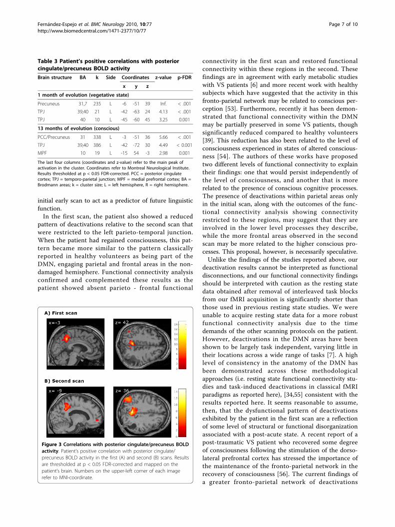
the first MRI scanning session the patient showed signif-icant correlations between PCC and two small clusterslocated in the parieto-temporal junction whereas in thesecond session he exhibited a more significant correla-tion pattern between the PCC and both the parieto-temporal junction and the medial prefrontal cortex(Table 3, Figure 3).In both sessions the mean, peak position, and peak
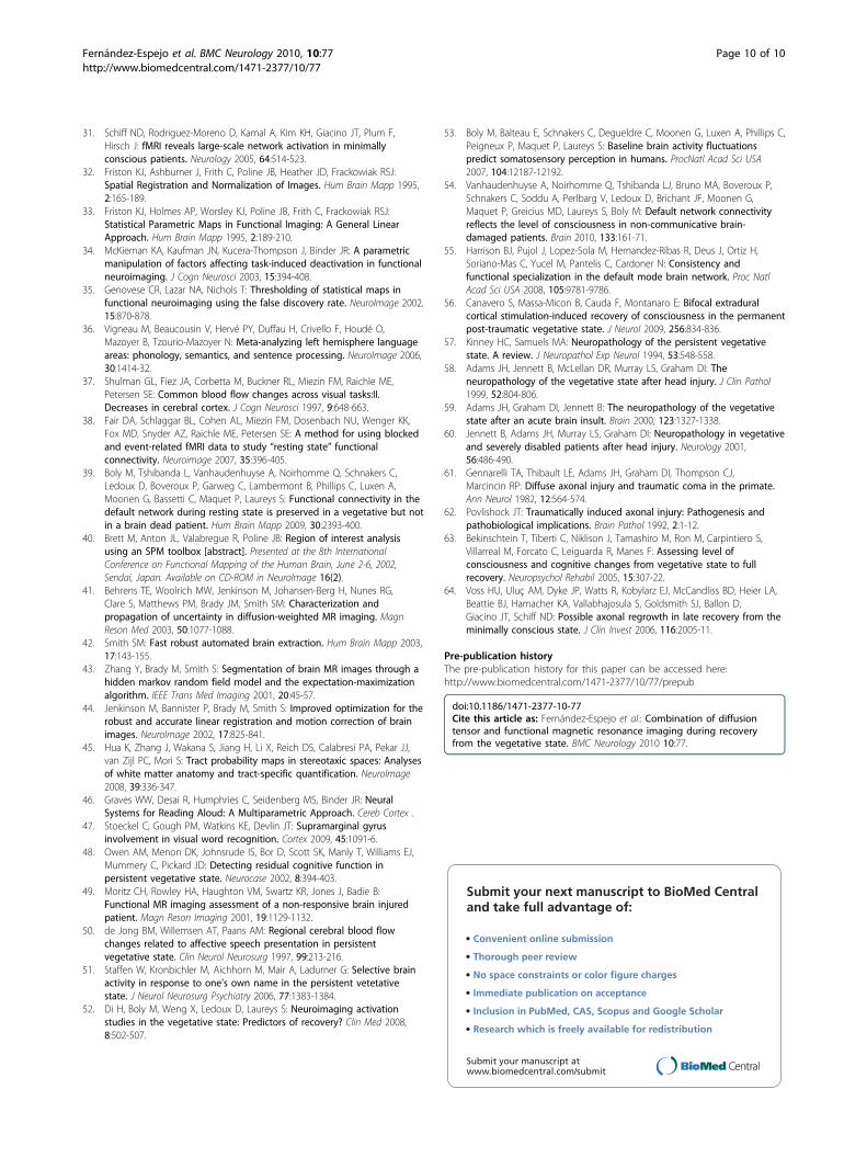
height from the NAWM FA histograms of the patientwere within the normal range. The patient’s MD histo-gram means were within the normal range in both scanswhile peak positions were within the normal range onlyin the first scan and increased in the second one. Peakheight was reduced relative to the normal range in boththe first and the second scan. Mean FA values from thearcuate fasciculus were also slightly reduced, while MDmean values were within the normal range in bothscans (Table 4, Figure 4).
DiscussionThe current study contrasted the fMRI and DTI scansof a TBI-patient at one-month post-ictus, when he wasin a VS, with those made one-year later when thepatient had recovered consciousness. The fMRI datashowed a pattern of cerebral activation in brain regionsassociated with language processing in both the firstscan, when he was unable to make an overt response tothese stimuli, and in the second scan, when he was ableto communicate verbally. At one-month post-ictus, thisactivation was restricted to a single locus in the poster-ior superior-middle temporal gyrus (Wernicke’s area)while in the second scan, 12-months later, it also com-prised more regions within these two gyri as well as,interestingly, areas of higher associative order, such asBrodmann area 40. This heteromodal area is known tobe involved in the association of visual and auditory lin-guistic information, and so activation may reflect arecovery of integrative functioning [46,47].The presence of anatomically appropriate activations
in response to speech in VS patients has been linkedwith a positive outcome [14,15,48,49]. However, othercases have been reported in which the presence of theseresponses were not associated with recovery [50,51]. Arecent review of eight PET studies and six fMRI studiesincluding a total of 48 VS patients estimated that the
Fernández-Espejo et al. BMC Neurology 2010, 10:77http://www.biomedcentral.com/1471-2377/10/77
Page 5 of 10

activation of high level associative cortical regions canpredict eventual recovery of consciousness with a speci-ficity of 93% and a sensitivity of 69% [52]. The presenceof similar fMRI markers in the patient reported in thecurrent study is consistent with this trend. It is clear,however, that a greater understanding of the prognostic
value of appropriate cerebral responses to language inthis population can be gained from future longitudinalstudies of large groups of these patients. This notwith-standing, the extent to which the linguistic functioningof the current patient improved upon his return to con-sciousness, points to the potential for the findings of an
Table 2 Patient’s cerebral activations and de-activations during the fMRI task
Contrast Brain structure BA k Side Coordinates z-value p-FDR
x y z
1 month of evolution (vegetative state)
Forward > Backward narratives* Superior temporal gyrus, Middle temporal gyrus 21, 22 17 L -57 -51 9 3.57 0.046
Narratives > Silence Trasverse temporal gyrus, Superior temporal gyrus 41,42 13 L -57 -18 9 4.81 0.017
Parahipocampal gyrus 19 14 R 39 -42 -6 4.73 0.017
Silence > Forward narratives* Superior Temporal Gyrus, 39 16 L -42 -57 27 4.33 0.003
13 months of evolution (conscious)
Forward > Backward narratives Precentral gyrus 6 42 L -51 0 48 7.77 < .001
Superior temporal gyrus, middle temporal gyrus 21, 22 54 L -51 -51 12 5.61 < .001
Superior temporal gyrus 22 15 L -63 -3 -3 5.19 < .001
Trasverse temporal gyrus, Postcentral gyrus 40, 42 33 L -66 -18 12 5.04 < .001
Narratives > Silence Superior temporal gyrus 41,42 461 L -63 -24 9 Inf. < .001
Paracentral lobule 4 24 L -6 -39 75 7 < .001
Silence > Forward narratives Inferior parietal lobule 7, 40 234 L -36 -66 48 5.35 0.001
Precuneus 7 17 L -15 -63 60 4.33 0.005
Superior frontal gyrus 6 17 L -18 30 60 4.12 0.01
The last four columns (coordinates and z-value) refer to the main peak of activation in the cluster. Coordinates refer to Montreal Neurological Institute. Resultsthresholded at p < 0.05 FDR-corrected. BA = Brodmann areas; k = cluster size; L = left hemisphere, R = right hemisphere. * = significant at p < 0.05 FDR smallvolume correction.
Figure 1 Task -related activations. Patient’s brain activation in thefirst (A) and second (B) scans for the contrast ‘forward > backwardnarratives’ (red) and the contrast ‘narratives > silence’ (green).Results are thresholded at p < 0.05 FDR-corrected and mapped onthe patient’s brain. Notice that the cluster displayed in the first scanfor the contrast ‘forward > backward narratives’ is thresholded at p< 0.05 FDR-small volume correction. Color-bars indicate t statisticvalues. Numbers on the left superior corner of each image refer tothe MNI-coordinate of the peak maxima in the x axis for eachcontrast.
Figure 2 Task- related deactivations. Patient’s task-induceddeactivations (contrast ‘silence > forward narratives’) in the first (A)and second (B) scans (medial and lateral views). Results arethresholded at p < 0.001 uncorrected and mapped on the patient’sbrain. Color-bars indicate t statistic values. Numbers on the upper-left corner of each image refer to MNI-coordinate in the x axis. Theimages on the right correspond to the peak-maxima of the maincluster for each contrast.
Fernández-Espejo et al. BMC Neurology 2010, 10:77http://www.biomedcentral.com/1471-2377/10/77
Page 6 of 10
initial early scan to act as a predictor of future linguisticfunction.In the first scan, the patient also showed a reduced
pattern of deactivations relative to the second scan thatwere restricted to the left parieto-temporal junction.When the patient had regained consciousness, this pat-tern became more similar to the pattern classicallyreported in healthy volunteers as being part of theDMN, engaging parietal and frontal areas in the non-damaged hemisphere. Functional connectivity analysisconfirmed and complemented these results as thepatient showed absent parieto - frontal functional
connectivity in the first scan and restored functionalconnectivity within these regions in the second. Thesefindings are in agreement with early metabolic studieswith VS patients [6] and more recent work with healthysubjects which have suggested that the activity in thisfronto-parietal network may be related to conscious per-ception [53]. Furthermore, recently it has been demon-strated that functional connectivity within the DMNmay be partially preserved in some VS patients, thoughsignificantly reduced compared to healthy volunteers[39]. This reduction has also been related to the level ofconsciousness experienced in states of altered conscious-ness [54]. The authors of these works have proposedtwo different levels of functional connectivity to explaintheir findings: one that would persist independently ofthe level of consciousness, and another that is morerelated to the presence of conscious cognitive processes.The presence of deactivations within parietal areas onlyin the initial scan, along with the outcomes of the func-tional connectivity analysis showing connectivityrestricted to these regions, may suggest that they areinvolved in the lower level processes they describe,while the more frontal areas observed in the secondscan may be more related to the higher conscious pro-cesses. This proposal, however, is necessarily speculative.Unlike the findings of the studies reported above, our
deactivation results cannot be interpreted as functionaldisconnections, and our functional connectivity findingsshould be interpreted with caution as the resting statedata obtained after removal of interleaved task blocksfrom our fMRI acquisition is significantly shorter thanthose used in previous resting state studies. We wereunable to acquire resting state data for a more robustfunctional connectivity analysis due to the timedemands of the other scanning protocols on the patient.However, deactivations in the DMN areas have beenshown to be largely task independent, varying little intheir locations across a wide range of tasks [7]. A highlevel of consistency in the anatomy of the DMN hasbeen demonstrated across these methodologicalapproaches (i.e. resting state functional connectivity stu-dies and task-induced deactivations in classical fMRIparadigms as reported here), [34,55] consistent with theresults reported here. It seems reasonable to assume,then, that the dysfunctional pattern of deactivationsexhibited by the patient in the first scan are a reflectionof some level of structural or functional disorganizationassociated with a post-acute state. A recent report of apost-traumatic VS patient who recovered some degreeof consciousness following the stimulation of the dorso-lateral prefrontal cortex has stressed the importance ofthe maintenance of the fronto-parietal network in therecovery of consciousness [56]. The current findings ofa greater fronto-parietal network of deactivations
Table 3 Patient’s positive correlations with posteriorcingulate/precuneus BOLD activity
Brain structure BA k Side Coordinates z-value p-FDR
x y z
1 month of evolution (vegetative state)
Precuneus 31,7 235 L -6 -51 39 Inf. < .001
TPJ 39,40 21 L -42 -63 24 4.13 < .001
TPJ 40 10 L -45 -60 45 3.25 0.001
13 months of evolution (conscious)
PCC/Precuneus 31 338 L -3 -51 36 5.66 < .001
TPJ 39,40 386 L -42 -72 30 4.49 < 0.001
MPF 10 19 L -15 54 -3 2.98 0.001
The last four columns (coordinates and z-value) refer to the main peak ofactivation in the cluster. Coordinates refer to Montreal Neurological Institute.Results thresholded at p < 0.05 FDR-corrected. PCC = posterior cingulatecortex; TPJ = temporo-parietal junction; MPF = medial prefrontal cortex; BA =Brodmann areas; k = cluster size; L = left hemisphere, R = right hemisphere.
Figure 3 Correlations with posterior cingulate/precuneus BOLDactivity. Patient’s positive correlation with posterior cingulate/precuneus BOLD activity in the first (A) and second (B) scans. Resultsare thresholded at p < 0.05 FDR-corrected and mapped on thepatient’s brain. Numbers on the upper-left corner of each imagerefer to MNI-coordinate.
Fernández-Espejo et al. BMC Neurology 2010, 10:77http://www.biomedcentral.com/1471-2377/10/77
Page 7 of 10
observed when consciousness had been regained relativeto those observed when the patient was in the VS, areconsistent with this notion. Future studies of this sort,however, may benefit from the information gained froma more robust functional connectivity analysis obtainedfrom a resting state acquisition, over and above thatobtained from the analysis of deactivations alone.The DTI data support and complement those results
obtained from the fMRI. The relative structural integrityof the arcuate fasciculus observed when the patient wasin the VS reinforces the predictions that can be madeon the basis of the preserved fMRI language activations.The global analysis of NAWM revealed an absence ofany major impairment in the WM tissue, without anyoutstanding changes between the first and second scans.One of the most commonly reported consequences ofTBI observed in post-mortem studies of TBI patientswho remained in a VS until death, is grade II/III diffuseaxonal injury (DAI) [57-60]. However, the mechanismscommonly involved in DAI, i.e. stretching and shearingof white matter fibers due to rotational forces normallyrelated to acceleration-deceleration effects,[61] did not
come into play in the current case, as the cause of theTBI was a fall. The accident caused an extensive concus-sive lesion restricted to the right parieto-temporal areabut it did not seem to affect areas far away from thelesion boundaries. DAI can disrupt critical cortical-subcortical pathways, leading to severe cognitive dys-function and precluding an effective reorganization ofthe preserved structures [62]. The absence of structuralevidence of DAI in our patient revealed by the DTI ana-lysis may highlight the potential for this as a measure ofthe extent to which recovery made on the basis of func-tional re-organization can take place, once the largehemorrhage and post-evacuation complications havesubsided.As discussed earlier, the current results are taken from
a single VS patient, and therefore must be interpretedwith care in relation to the wider VS population. Cur-rently, there are three other published longitudinal neu-roimaging studies of recovery from DOC, following twoVS patients [6,63] and one MCS patient, [64] whichhave provided insights into the mechanisms involved issuch recovery. These studies, however, focused on eitherfunctional or structural data, or contrasted a number ofscans performed after recovery. The current study,therefore, is the first time that data from fMRI, DTI andneuropsychological assessment have been combined inthe study of the cerebral and clinical changes of a VSpatient from the time of VS through to recovery. Thelow incidence of a good recovery in VS patients hastherefore made this case an exceptional opportunity inwhich to study the mechanisms associated with thisprocess.
ConclusionsThe current findings provide evidence that structural andfunctional preservation of linguistic cerebral networksmay have prognostic value for the language abilities ofVS patients following the recovery of consciousness. Inaddition, dysfunction in areas related to the DMN mayhave a role in explaining disorders of consciousness. Inthe absence of significant structural damage to
Table 4 FA and MD parameters for NAMW and arcuate fasciculus
Patient 1st scan Patient 2nd scan Controls range
NAWM FA Mean 3.5 × 10⁻¹ 3.4 × 10⁻¹ 3.1-3.6 × 10⁻¹
Peak position 3.3 × 10⁻¹ 2.9 × 10⁻¹ 1.3-3.6 × 10⁻¹
Peak height 1.0 × 10⁻² 1.0 × 10⁻² 1.0-1.1 × 10⁻²
MD Mean 8.5 × 10⁻⁴ 8.4 × 10⁻⁴ 7.7-8.6 × 10⁻⁴
Peak position 7.6 × 10⁻⁴ 8.2 × 10⁻⁴ 7.2-7.9 × 10⁻⁴
Peak height 2.7 × 10⁻² 2.5 × 10⁻² 2.9-4.3 × 10⁻²
Arcuate fascicle FA Mean 3.6 × 10⁻¹ 3.7 × 10⁻¹ 3.8-4.3 × 10⁻¹
MD Mean 8.1 × 10⁻⁴ 8.0 × 10⁻⁴ 7.0-8.1 × 10⁻⁴
NAWM = normal-appearing white matter; FA = fractional anisotropy values; MD = mean diffusivity values (mm2/s).
Figure 4 Histograms of normal-appearing white matter.Fractional anisotropy (A) and mean diffusivity (B) histograms ofnormal-appearing white matter of the patient in the first (blue line)and second (green line) scans and the 19 normal control subjects(black lines) over all voxels in the brain (top of the panel). Thebottoms of each panel display the correspondent fractionalanisotropy and mean diffusivity maps. Color-bars indicate fractionalanisotropy (arbitrary units) and mean diffusivity (mm2/sec) values.
Fernández-Espejo et al. BMC Neurology 2010, 10:77http://www.biomedcentral.com/1471-2377/10/77
Page 8 of 10
long-range connections, the functional recovery of thisnetwork may accompany the restoration ofconsciousness.Taken together, our findings suggest that a multi-
modal imaging approach can provide a powerful tool forassessing the mechanisms involved in the recovery ofconsciousness in DOC patients. Further longitudinalstudies with large cohorts will prove useful in assessingits full value in predicting outcome. Such insights maythen provide guidance for decisions relating to rehabili-tation programs by orientating these towards the effec-tive stimulation of those cognitive functions that appearpreserved, in order to maintain their functional andstructural integrity.
AknowledgementsThis work was supported by grants SAF2007-66077 from the SpanishMinistry of Science and Innovation and 2009SGR0941 from the Generalitatde Catalunya to the Neuropsychology Research Group. D. Fernández-Espejowas supported by a fellowship from the Spanish Ministry for Education(AP2006-00862). D. Cruse was supported by a Medical Research CouncilTranslational Grant (U.1055.01.002.00007.01).
Author details1Department of Psychiatry and Clinical Psychobiology, University ofBarcelona, Barcelona, Spain. 2Institute of Biomedical Research August Pi iSunyer (IDIBAPS), Barcelona, Spain. 3MRC Cognition and Brain Sciences Unit,Cambridge, UK. 4Head Injury Unit, Institut Universitari de NeurorehabilitacióGuttmann, Badalona, Spain. 5Department of Neuropsychology, InstitutUniversitari de Neurorehabilitació Guttmann, Badalona, Spain.6Anesthesiology Department, Hospital Clinic, University of Barcelona,Barcelona, Spain. 7Centre de Diagnòstic per la Imatge Hospital Clinic deBarcelona (CDIC), Hospital Clínic de Barcelona, Spain.
Authors’ contributionsDFE and CJ made substantial contribution to conception and design,interpretation of data as well as in the preparation of the first draft andfurther revisions of the manuscript. Neuroimaging data were collected andanalyzed by DFE. DC contributed heavily to the final revision, providingcomments and perspectives. MB and TR participated in the collection ofneuropsychological data. ER and NF were involved in the collection of acuteclinical data. MB, TR, ER, NF and JMM made a critical revision of themanuscript for important intellectual content providing additionalcomments and contributions. CJ supervised the study. All authors read andapproved the final manuscript.
Competing interestsThe authors declare that they have no competing interests.
Received: 9 December 2009 Accepted: 3 September 2010Published: 3 September 2010
References1. Jennett B, Plum F: Persistent vegetative state after brain damage. A
syndrome in search of a name. Lancet 1972, 1:734-737.2. Royal College of Physicians: The permanent vegetative state [Report of a
working party]. J R Coll Physicians Lond 1996, 30:119-121.3. Multi-Society Task Force on PVS: Medical aspects of the persistent
vegetative state - II. N Engl J Med 1994, 330:1572-1579.4. Laureys S, Goldman S, Phillips C, Van Bogaert P, Aerts J, Luxen A, Franck G,
Maquet P: Impaired effective cortical connectivity in vegetative state:Preliminary investigation using PET. NeuroImage 1999, 9:377-382.
5. Laureys S, Lemaire C, Maquet P, Phillips C, Franck G: Cerebral metabolismduring vegetative state and after recovery to consciousness. J NeurolNeurosurg Psychiatry 1999, 67:121.
6. Laureys S, Faymonville ME, Luxen A, Lamy M, Franck G, Maquet P:Restoration of thalamocortical connectivity after recovery frompersistent vegetative state. Lancet 2000, 355:1790-1791.
7. Gusnard DA, Raichle ME: Searching for a baseline: Functional imagingand the resting human brain. Nat Rev Neuros 2001, 2:685-694.
8. Bassetti C, Vella S, Donati F, Wielepp P, Weder B: SPECT duringsleepwalking. Lancet 2000, 356:484-485.
9. Salek-Haddadi A, Lemieux L, Merschhemke M, Friston KJ, Duncan JS,Fish DR: Functional magnetic resonance imaging of human absenceseizures. Ann Neurol 2003, 53:663-667.
10. Maquet P: Functional neuroimaging of normal human sleep by positronemission tomography. J Sleep Res 2000, 9:207-231.
11. Steriade M: Active neocortical processes during quiescent sleep. Arch ItalBiol 2001, 139:37-51.
12. Kaisti KK, Metsahonkala L, Teras M, Oikonen V, Aalto S, Jaaskelainen S,Hinkka S, Scheinin H: Effects of surgical levels of propofol andsevoflurane anesthesia on cerebral blood flow in healthy subjectsstudied with positron emission tomography. Anesthesiology 2002,96:1358-1370.
13. Owen AM, Coleman MR: Functional neuroimaging of the vegetativestate. Nat Rev Neurosci 2008, 9:235-243.
14. Coleman MR, Rodd JM, Davis MH, Johnsrude IS, Menon DK, Pickard JD,Owen AM: Do vegetative patients retain aspects of languagecomprehension? evidence from fMRI. Brain 2007, 130:2494-2507.
15. Di HB, Yu S, Weng X, Laureys S, Yu D, Li JQ, Zhu PM, Zhang S, Chen Y:Cerebral response to patient’s own name in the vegetative andminimally conscious state. Neurology 2007, 68:895-899.
16. Sidaros A, Engberg AW, Sidaros K, Liptrot MG, Herning M, Petersen P,Paulson OB, Jernigan TL, Rostrup E: Diffusion tensor imaging duringrecovery from severe traumatic brain injury and relation to clinicaloutcome: A longitudinal study. Brain 2008, 131:559-572.
17. Tollard E, Galanaud D, Perlbarg V, Shanchez-Pena P, Le Fur Y, Abdennour L,Cozzone P, Lehericy S, Chiras J, Puybasset L: Experience of diffusion tensorimaging and 1 H spectroscopy for outcome prediction in severetraumatic brain injury: Preliminary results. Crit Care Med 2009,37:1448-1455.
18. Rappaport M, Hall KM, Hopkins K, Belleza T, Cope DN: Disability ratingscale for severe head trauma: Coma to community. Arch Phys MedRehabil 1982, 63:118-123.
19. Hagen C, Malkmuss D, Durham P, Bowman K: Levels of cognitivefunctioning. In Rehabilitation of the head injured adult. Comprehensivephysical management. Edited by: Hagen C, Malkmuss D, Durham P, DowneyCA. Professional Staff Association of Rancho Los Amigos Hospital; 1979:.
20. Royal College of Physicians: The vegetative state: Guidance on diagnosisand management [Report of a working party]. Clin Med 2003, 3:249-254.
21. Giacino JT, Ashwal S, Childs N, Cranford R, Jennett B, Katz DI, Kelly JP,Rosenberg JH, Whyte J, Zafonte RD, et al: The minimally conscious state:Definition and diagnostic criteria. Neurology 2002, 58:349-353.
22. Goodglass H, Kaplan E: La evaluación de la afasia y de trastornosrelacionados. Madrid: Editorial Medica Panamericana, 2 1986.
23. Albert ML: A simple test of visual neglect. Neurology 1973, 23:658-64.24. Peña-Casanova J: Programa Integrado de Exploración Neuropsicológica
‘Test Barcelona’ [manual]. Barcelona: Masson 1991.25. Wechsler D: Escala de Inteligencia de Wechsler para Adultos (WAIS-III).
Madrid: TEA Ediciones 1999.26. Lezak MD, Howieson DB, Loring DW, Hannay HJ, Fischer JS:
Neuropsychological assessment. New York: Oxford University Press Inc, 42004.
27. Artiola-i-Fortuny L, Hermosillo Romo DH, Heaton RK, Pardee RE: Manual denormas y procedimientos para la batería neuropsicológica en español.Tucson, AZ: mPress 1999.
28. Oldfield RC: The assessment and analysis of handedness: the Edinburghinventory. Neuropsychologia 1971, 9:97-113.
29. Fernandez-Espejo D, Junque C, Vendrell P, Bernabeu M, Roig T, Bargallo N,Mercader JM: Cerebral response to speech in vegetative and minimallyconscious states after traumatic brain injury. Brain Inj 2008, 22:882-890.
30. Dehaene-Lambertz G, Dehaene S, Hertz-Pannier L: Functionalneuroimaging of speech perception in infants. Science 2002,298:2013-2015.
Fernández-Espejo et al. BMC Neurology 2010, 10:77http://www.biomedcentral.com/1471-2377/10/77
Page 9 of 10
31. Schiff ND, Rodriguez-Moreno D, Kamal A, Kim KH, Giacino JT, Plum F,Hirsch J: fMRI reveals large-scale network activation in minimallyconscious patients. Neurology 2005, 64:514-523.
32. Friston KJ, Ashburner J, Frith C, Poline JB, Heather JD, Frackowiak RSJ:Spatial Registration and Normalization of Images. Hum Brain Mapp 1995,2:165-189.
33. Friston KJ, Holmes AP, Worsley KJ, Poline JB, Frith C, Frackowiak RSJ:Statistical Parametric Maps in Functional Imaging: A General LinearApproach. Hum Brain Mapp 1995, 2:189-210.
34. McKiernan KA, Kaufman JN, Kucera-Thompson J, Binder JR: A parametricmanipulation of factors affecting task-induced deactivation in functionalneuroimaging. J Cogn Neurosci 2003, 15:394-408.
35. Genovese CR, Lazar NA, Nichols T: Thresholding of statistical maps infunctional neuroimaging using the false discovery rate. NeuroImage 2002,15:870-878.
36. Vigneau M, Beaucousin V, Hervé PY, Duffau H, Crivello F, Houdé O,Mazoyer B, Tzourio-Mazoyer N: Meta-analyzing left hemisphere languageareas: phonology, semantics, and sentence processing. NeuroImage 2006,30:1414-32.
37. Shulman GL, Fiez JA, Corbetta M, Buckner RL, Miezin FM, Raichle ME,Petersen SE: Common blood flow changes across visual tasks:II.Decreases in cerebral cortex. J Cogn Neurosci 1997, 9:648-663.
38. Fair DA, Schlaggar BL, Cohen AL, Miezin FM, Dosenbach NU, Wenger KK,Fox MD, Snyder AZ, Raichle ME, Petersen SE: A method for using blockedand event-related fMRI data to study “resting state” functionalconnectivity. Neuroimage 2007, 35:396-405.
39. Boly M, Tshibanda L, Vanhaudenhuyse A, Noirhomme Q, Schnakers C,Ledoux D, Boveroux P, Garweg C, Lambermont B, Phillips C, Luxen A,Moonen G, Bassetti C, Maquet P, Laureys S: Functional connectivity in thedefault network during resting state is preserved in a vegetative but notin a brain dead patient. Hum Brain Mapp 2009, 30:2393-400.
40. Brett M, Anton JL, Valabregue R, Poline JB: Region of interest analysisusing an SPM toolbox [abstract]. Presented at the 8th InternationalConference on Functional Mapping of the Human Brain, June 2-6, 2002,Sendai, Japan. Available on CD-ROM in NeuroImage 16(2).
41. Behrens TE, Woolrich MW, Jenkinson M, Johansen-Berg H, Nunes RG,Clare S, Matthews PM, Brady JM, Smith SM: Characterization andpropagation of uncertainty in diffusion-weighted MR imaging. MagnReson Med 2003, 50:1077-1088.
42. Smith SM: Fast robust automated brain extraction. Hum Brain Mapp 2003,17:143-155.
43. Zhang Y, Brady M, Smith S: Segmentation of brain MR images through ahidden markov random field model and the expectation-maximizationalgorithm. IEEE Trans Med Imaging 2001, 20:45-57.
44. Jenkinson M, Bannister P, Brady M, Smith S: Improved optimization for therobust and accurate linear registration and motion correction of brainimages. NeuroImage 2002, 17:825-841.
45. Hua K, Zhang J, Wakana S, Jiang H, Li X, Reich DS, Calabresi PA, Pekar JJ,van Zijl PC, Mori S: Tract probability maps in stereotaxic spaces: Analysesof white matter anatomy and tract-specific quantification. NeuroImage2008, 39:336-347.
46. Graves WW, Desai R, Humphries C, Seidenberg MS, Binder JR: NeuralSystems for Reading Aloud: A Multiparametric Approach. Cereb Cortex .
47. Stoeckel C, Gough PM, Watkins KE, Devlin JT: Supramarginal gyrusinvolvement in visual word recognition. Cortex 2009, 45:1091-6.
48. Owen AM, Menon DK, Johnsrude IS, Bor D, Scott SK, Manly T, Williams EJ,Mummery C, Pickard JD: Detecting residual cognitive function inpersistent vegetative state. Neurocase 2002, 8:394-403.
49. Moritz CH, Rowley HA, Haughton VM, Swartz KR, Jones J, Badie B:Functional MR imaging assessment of a non-responsive brain injuredpatient. Magn Reson Imaging 2001, 19:1129-1132.
50. de Jong BM, Willemsen AT, Paans AM: Regional cerebral blood flowchanges related to affective speech presentation in persistentvegetative state. Clin Neurol Neurosurg 1997, 99:213-216.
51. Staffen W, Kronbichler M, Aichhorn M, Mair A, Ladurner G: Selective brainactivity in response to one’s own name in the persistent vetetativestate. J Neurol Neurosurg Psychiatry 2006, 77:1383-1384.
52. Di H, Boly M, Weng X, Ledoux D, Laureys S: Neuroimaging activationstudies in the vegetative state: Predictors of recovery? Clin Med 2008,8:502-507.
53. Boly M, Balteau E, Schnakers C, Degueldre C, Moonen G, Luxen A, Phillips C,Peigneux P, Maquet P, Laureys S: Baseline brain activity fluctuationspredict somatosensory perception in humans. ProcNatl Acad Sci USA2007, 104:12187-12192.
54. Vanhaudenhuyse A, Noirhomme Q, Tshibanda LJ, Bruno MA, Boveroux P,Schnakers C, Soddu A, Perlbarg V, Ledoux D, Brichant JF, Moonen G,Maquet P, Greicius MD, Laureys S, Boly M: Default network connectivityreflects the level of consciousness in non-communicative brain-damaged patients. Brain 2010, 133:161-71.
55. Harrison BJ, Pujol J, Lopez-Sola M, Hernandez-Ribas R, Deus J, Ortiz H,Soriano-Mas C, Yucel M, Pantelis C, Cardoner N: Consistency andfunctional specialization in the default mode brain network. Proc NatlAcad Sci USA 2008, 105:9781-9786.
56. Canavero S, Massa-Micon B, Cauda F, Montanaro E: Bifocal extraduralcortical stimulation-induced recovery of consciousness in the permanentpost-traumatic vegetative state. J Neurol 2009, 256:834-836.
57. Kinney HC, Samuels MA: Neuropathology of the persistent vegetativestate. A review. J Neuropathol Exp Neurol 1994, 53:548-558.
58. Adams JH, Jennett B, McLellan DR, Murray LS, Graham DI: Theneuropathology of the vegetative state after head injury. J Clin Pathol1999, 52:804-806.
59. Adams JH, Graham DI, Jennett B: The neuropathology of the vegetativestate after an acute brain insult. Brain 2000, 123:1327-1338.
60. Jennett B, Adams JH, Murray LS, Graham DI: Neuropathology in vegetativeand severely disabled patients after head injury. Neurology 2001,56:486-490.
61. Gennarelli TA, Thibault LE, Adams JH, Graham DI, Thompson CJ,Marcincin RP: Diffuse axonal injury and traumatic coma in the primate.Ann Neurol 1982, 12:564-574.
62. Povlishock JT: Traumatically induced axonal injury: Pathogenesis andpathobiological implications. Brain Pathol 1992, 2:1-12.
63. Bekinschtein T, Tiberti C, Niklison J, Tamashiro M, Ron M, Carpintiero S,Villarreal M, Forcato C, Leiguarda R, Manes F: Assessing level ofconsciousness and cognitive changes from vegetative state to fullrecovery. Neuropsychol Rehabil 2005, 15:307-22.
64. Voss HU, Uluç AM, Dyke JP, Watts R, Kobylarz EJ, McCandliss BD, Heier LA,Beattie BJ, Hamacher KA, Vallabhajosula S, Goldsmith SJ, Ballon D,Giacino JT, Schiff ND: Possible axonal regrowth in late recovery from theminimally conscious state. J Clin Invest 2006, 116:2005-11.
Pre-publication historyThe pre-publication history for this paper can be accessed here:http://www.biomedcentral.com/1471-2377/10/77/prepub
doi:10.1186/1471-2377-10-77Cite this article as: Fernández-Espejo et al.: Combination of diffusiontensor and functional magnetic resonance imaging during recoveryfrom the vegetative state. BMC Neurology 2010 10:77.
Submit your next manuscript to BioMed Centraland take full advantage of:
• Convenient online submission
• Thorough peer review
• No space constraints or color figure charges
• Immediate publication on acceptance
• Inclusion in PubMed, CAS, Scopus and Google Scholar
• Research which is freely available for redistribution
Submit your manuscript at www.biomedcentral.com/submit
Fernández-Espejo et al. BMC Neurology 2010, 10:77http://www.biomedcentral.com/1471-2377/10/77
Page 10 of 10