Embed Size (px)
Citation preview

Patient Photographs in the
Electronic Health Record
Michael FlynnRadiology ResearchHenry Ford Health SystemDetroit, MI
Outline: pg.1.
Introduction
(2)
2.Acquisition
(9)3.Storage
(16 )
4.Viewing (20
)
Summit on Color in Medical ImagingSilver Spring, MD, USAMay 8-9, 2013

EHR2011Images, Electronic Health Records, and
Meaningful UseNIH NIBIB Workshop: Jan 10-11, 2011
Introduction
Electronic Medical Record (EMR) vs Electronic Health Record (EHR)•EMRs are episode and enterprise focused •EHRs are longitudinal, comprehensive, and patient-centered. •EHRs can also serve as a lifetime record for the patient.
R. Greenes, Arizona State Univ.
http://www.nibib.nih.gov/NewsEvents/MeetingsEvents/MeetingSummaries/EHR2011
The goal of this workshop was to understand the value of access to images as part of the
meaningful use of electronic medical records.
2
EHR2011Images, Electronic Health Records, and Meaningful Use
NIH NIBIB Workshop: Jan 10-11, 2011
Introduction
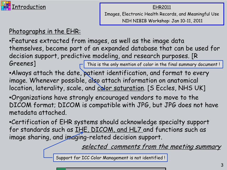
Photographs in the EHR:•Features extracted from images, as well as the image data themselves, become part of an expanded database that can be used
for
decision support, predictive modeling, and research purposes. [R Greenes]
•Always attach the date, patient identification, and format to every image. Whenever possible, also attach information on anatomical location, laterality, scale, and color saturation. [S Eccles, NHS UK]•Organizations have strongly encouraged vendors to move to the DICOM format; DICOM is compatible with JPG, but JPG does not have metadata attached.•Certification of EHR systems should acknowledge specialty support for standards such as IHE, DICOM, and HL7 and functions such as image sharing, and imaging-related decision support.
selected comments from the meeting summarySupport for ICC Color Management is not identified !
3
This is the only mention of color in the final summary document !

Clinical Photography Examples•Dermatology•Reconstructive Surgery•ER Abuse documentation•Wound Management(Trauma Surgery)
Introduction
Lyme DiseaseDelayed Rash
US CDC4

Clinical Photography Examples•Dermatology•Reconstructive Surgery•ER Abuse documentation•Wound Management(Trauma Surgery)
Introduction
Ear Reconstruction following Ca Surgery
E. Funk, MD
5

Clinical Photography Examples•Dermatology•Reconstructive Surgery•ER Abuse documentation•Wound Management(Trauma Surgery)
Introduction
5 YO Child AbuseL. Ricci MD, Medscape Ref
6

Clinical Photography Examples•Dermatology•Reconstructive Surgery•ER Abuse documentation•Wound Management(Trauma Surgery)
Introduction
Triathlon Bike FallXAVIER UNIVERSITY
WOUND CARE CASE STUDY
7

We consider here only clinical photographs.Color images in medical specialties such as Ophthamology and Pathology use specialized equipment and analysis methods for which industry PACS solutions are available.
Introduction
Retinal fundus image showing age- related macular degeneration.
National Eye Institute, NIHRef#: EDA22
Liver -
Masson Stain.
Paxcam image gallery
8

•
Flash illumination with low ambient illuminance is essential for consistent color.
•
ICC color profiles can easily be generated for specific flash camera systems used with established firmware parameters
9
Acquisition

US HIPPA regulations•Photographs should be immediately uploaded to the EMR/EHR and deleted from the recording device memory.•Policies should prohibit the use of cameras with ad hoc storage on non-compliant devices.
10
Acquisition
KR Silas, Louisiana Bar Journal, Oct 2011Among emerging technology, “no single device has produced as much health privacy angst as cell phones that double as cameras.”
Information Privacy and SecurityPatient Photography
May I take a picture of the patient for treatment purposes?Yes, you may take a picture but you must upload the picture into the patient's medical record immediately then delete the picture from the device on which the picture was taken.Pl
P
li
P i Ph
h d
VANDERBILT UNIVERSITY MEDICAL CENTER

Acquisition Software•Information associated with each photo study.
•
Patient Name, ID, DOB, Sex .. (EMR consistent)•
Attending Physician (associated with EMR encounter)
•
EMR Accession number (unique for study in PACS)
•Information associated with each image.•
Anatomic body part (consistent with protocol)
•
Photographic conditions (ICC profile, distance, ..)
•Camera image upload.•
Import from camera and delete. (WiFi, tethered, ..)
•
Export to EMR (PACS, DICOM, JPEG, ..)
11
Acquisition

PHOTO VIEWINGePhoto workstation
EMR (EPIC)•Open patient•Chart review / imaging•Photo filter
URL link to images
PacsGEAR viewer•
PACS Study•
Calibrated color•
Basic measurements
Health System IT
EMR( EPIC )
Primary Services
DICOM Server•Enterprise Srvs•ePhoto Database•ePhoto viewing
NASDICOM File Storage
Secondary StorageDICOM File StorageLong term storage[Disaster recovery]
HL7
DIC
OM
12
Acquisition Acquisition Software
•Practitioner initiates photo acquisition application from the EMR at the patient level.•API integration communicates demographic data.•API assigns a unique encounter number used as the study accession number.•Images are selected, identified for body part, annotated, and uploaded in DICOM format.

Epic integration with acquisition software Kaiser, San Francisco
13
Acquisition
14
Acquisition
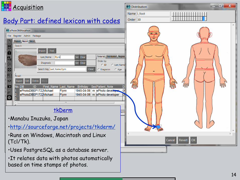
tkDerm•Manabu Inuzuka, Japan•http://sourceforge.net/projects/tkderm/•Runs on Windows, Macintosh and Linux (Tcl/Tk).•Uses PostgreSQL as a database server.•It relates data with photos automatically based on time stamps of photos.
Body Part: defined lexicon with codes

Acquisition Software -
Issues•Very few available applications.•None provided all needed functions.
•
Image deletion after upload.•
EMR integration for demographics, diagnosis et. al.
•
Coded body part assignment.•
ICC profile association
15
Acquisition
• Ideally, all camera & flash systems used with specific camera settings would have ICC profiles
• Acquisition software would identify the camera & flash system used and send the ICC profile.

Photo Storage•Documents in EMR are inadequate
•
No study organization or body part codes•
No analytics support for image search
•
Inadequate viewers
•DICOM Storage services apply nicely•
Well developed industry with numerous suppliers.
•
Recognized standard for object structure and communication.•
HIPPA compliant with support for patient media.
•
Advanced viewer functionality.
16
Storage

PHOTO VIEWINGePhoto workstation
EMR (EPIC)•Open patient•Chart review / imaging•Photo filter
URL link to images
PacsGEAR viewer•
PACS Study•
Calibrated color•
Basic measurements
Health System IT
EMR( EPIC )
Primary Services
DICOM Server•Enterprise Srvs•ePhoto Database•ePhoto viewing
NASDICOM File Storage
Secondary StorageDICOM File StorageLong term storage[Disaster recovery]
HL7
DIC
OM
17
Storage DICOM Storage Server
•Storage Class Provider (SCP) service for photo object in DICOM standard format.•Query/Retrieve services for specialized applications.•Export for interchange in using an IHE profile.•Support for viewer applications with audit trails showing who has viewed a study.

Audit Record
18
Storage

Storage Servers -
Issues•Numerous available DICOM servers.•Most provide all needed functions.
•
DICOM Storage Class Provider (SCP).•
Full function viewers (Web accessible)
•
Query/Retrieve Service.•
Storage of ICC profile in image objects
•
However,•
Viewers do not support color management
•
Currently no EXIF tags in DICOM image objects
19
Storage
• An IHE profile could address color management
• New DICOM tags could be considered by the DSC.

Image viewers•Authenticated access to view patient images•Organize all images in a study by body part.•Provide comparison of current and prior images for the same body part.•Mouse controlled zoom and pan with robust interpolation.
20
Viewers

PHOTO VIEWINGePhoto workstation
EMR (EPIC)•Open patient•Chart review / imaging•Photo filter
URL link to images
Photo viewer•
PACS Study•
Calibrated color•
Basic measurements
Health System IT
EMR( EPIC )
Primary Services
DICOM Server•Enterprise Srvs•ePhoto Database•ePhoto viewing
NASDICOM File Storage
Secondary StorageDICOM File StorageLong term storage[Disaster recovery]
HL7
DIC
OM
21
Viewers
Photo Viewer•Practitioner accesses (with authentication) patient information in the EMR.•Photo study is identified as having been acquired on a particular date.•Using a URL link, a photo viewer is opened with the identified study•Prior studies are available for comparison.

Epic integration with photo viewerKaiser, San Francisco
22
Viewers

Web based DICOM study viewer.
23
Viewers

All present DICOM viewers Lack Color Managment
24
Viewers
aRGB
sRGB

DICOM Photo viewers -
Issues•Excellent organization of images and studies.•Mature viewing functions.
•
Mouse controlled Pan & zoom.•
Robust iterpolation algorithms
•
Hanging protocols to compare current with prior.•
However,
•
NO COLOR MANAGEMENT SUPPORT
25
Viewers
• While IHE profiles would define the functions needed, adoption within the industry is likely to take some time.
• ICC software toolkits for common OSs would reduce the investment required for color management support.

Comparison of Color and Monochrome Displays in 2003
SCAR Univ. 404Boston -
2003
Questions?