Embed Size (px)
Citation preview
i
Colloquium III, August 11, 2017
This colloquium is invitation-only.
Presentation of research results here does not constitute public disclosure.
ii
The Smalley-Curl Institute
3rd Annual Summer Research Colloquium Sponsors
1
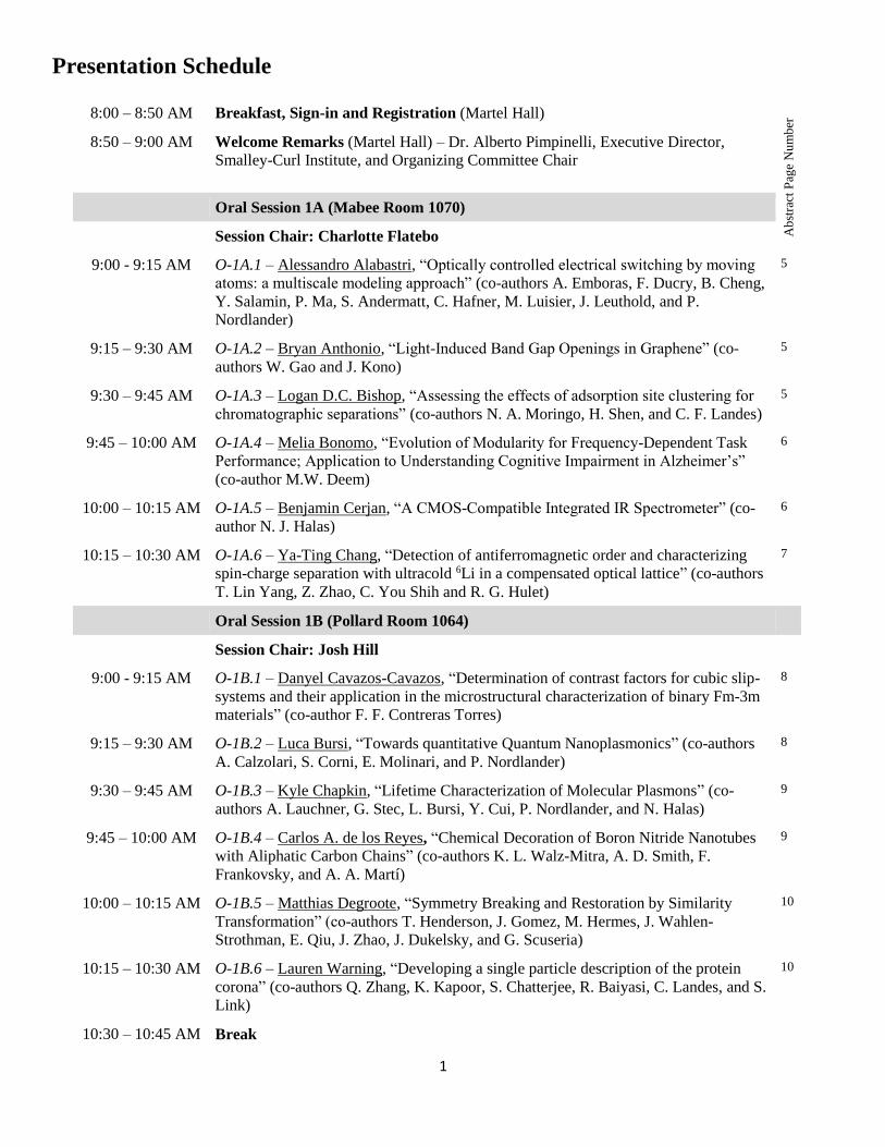
Presentation Schedule
8:00 – 8:50 AM Breakfast, Sign-in and Registration (Martel Hall)
Ab
stra
ct P
age
Nu
mb
er
8:50 – 9:00 AM Welcome Remarks (Martel Hall) – Dr. Alberto Pimpinelli, Executive Director,
Smalley-Curl Institute, and Organizing Committee Chair
Oral Session 1A (Mabee Room 1070)
Session Chair: Charlotte Flatebo
9:00 - 9:15 AM
O-1A.1 – Alessandro Alabastri, “Optically controlled electrical switching by moving
atoms: a multiscale modeling approach” (co-authors A. Emboras, F. Ducry, B. Cheng,
Y. Salamin, P. Ma, S. Andermatt, C. Hafner, M. Luisier, J. Leuthold, and P.
Nordlander)
5
9:15 – 9:30 AM
O-1A.2 – Bryan Anthonio, “Light-Induced Band Gap Openings in Graphene” (co-
authors W. Gao and J. Kono)
5
9:30 – 9:45 AM
O-1A.3 – Logan D.C. Bishop, “Assessing the effects of adsorption site clustering for
chromatographic separations” (co-authors N. A. Moringo, H. Shen, and C. F. Landes)
5
9:45 – 10:00 AM
O-1A.4 – Melia Bonomo, “Evolution of Modularity for Frequency-Dependent Task
Performance; Application to Understanding Cognitive Impairment in Alzheimer’s”
(co-author M.W. Deem)
6
10:00 – 10:15 AM
O-1A.5 – Benjamin Cerjan, “A CMOS-Compatible Integrated IR Spectrometer” (co-
author N. J. Halas)
6
10:15 – 10:30 AM
O-1A.6 – Ya-Ting Chang, “Detection of antiferromagnetic order and characterizing
spin-charge separation with ultracold 6Li in a compensated optical lattice” (co-authors
T. Lin Yang, Z. Zhao, C. You Shih and R. G. Hulet)
7
Oral Session 1B (Pollard Room 1064)
Session Chair: Josh Hill
9:00 - 9:15 AM
O-1B.1 – Danyel Cavazos-Cavazos, “Determination of contrast factors for cubic slip-
systems and their application in the microstructural characterization of binary Fm-3m
materials” (co-author F. F. Contreras Torres)
8
9:15 – 9:30 AM
O-1B.2 – Luca Bursi, “Towards quantitative Quantum Nanoplasmonics” (co-authors
A. Calzolari, S. Corni, E. Molinari, and P. Nordlander)
8
9:30 – 9:45 AM
O-1B.3 – Kyle Chapkin, “Lifetime Characterization of Molecular Plasmons” (co-
authors A. Lauchner, G. Stec, L. Bursi, Y. Cui, P. Nordlander, and N. Halas)
9
9:45 – 10:00 AM
O-1B.4 – Carlos A. de los Reyes, “Chemical Decoration of Boron Nitride Nanotubes
with Aliphatic Carbon Chains” (co-authors K. L. Walz-Mitra, A. D. Smith, F.
Frankovsky, and A. A. Martí)
9
10:00 – 10:15 AM
O-1B.5 – Matthias Degroote, “Symmetry Breaking and Restoration by Similarity
Transformation” (co-authors T. Henderson, J. Gomez, M. Hermes, J. Wahlen-
Strothman, E. Qiu, J. Zhao, J. Dukelsky, and G. Scuseria)
10
10:15 – 10:30 AM
O-1B.6 – Lauren Warning, “Developing a single particle description of the protein
corona” (co-authors Q. Zhang, K. Kapoor, S. Chatterjee, R. Baiyasi, C. Landes, and S.
Link)
10
10:30 – 10:45 AM Break

2
Oral Session 2A (Mabee Room 1070)
Session Chair: Pelham Keahey
10:45 – 11:00 AM
O-2A.1 – Didier Devaurs, “Studying Protein Structure through Hydrogen Exchange
and Conformational Sampling” (co-authors D. Antunes, J. Abella, M. Moll, and L.
Kavraki)
11
11:00 – 11:15 AM
O-2A.2 – Charlotte I. Evans, “Quantifying Remote Heating from Propagating Surface
Plasmon Polaritons” (co-authors P. Zolotavin, A. Alabastri, J. Yang, P. Nordlander,
and D. Natelson)
11
11:15 – 11:30 AM
O-2A.3 – Fumiya Katsutani, “Direct Observation of Cross-Polarized Excitons in
Aligned Single-Wall Carbon Nanotubes” (co-authors W. Gao, K. Yanagi, and J.
Kono)
12
11:30 – 11:45 AM
O-2A.4 – Varun Shenoy Gangoli, “Understanding Catalyst Residue Inhibition of the
Functionalization of Single Wall Carbon Nanotubes via Billups-Birch Reduction” (co-
authors K. Zhang, D. Pham, O. Lawal, S. Ghosh, R. H. Hauge, W. W. Adams, and A.
R. Barron)
12
11:45 – 12:00 PM
O-2A.5 – Benjamin Clark, “Catalytic Role of Titanium in the Synthesis of Aluminum
Nanocrystals” (co-authors C. DeSantis, G. Wu, A. Tsai, and N. Halas)
13
Oral Session 2B (Pollard Room 1064)
Session Chair: Charlotte Flatebo
10:45 – 11:00 AM
O-2B.1 – Sangheon Han, “Ultra-small Plasmonic Nanosensors for Cancer detection”
(co-authors S. Emelianov, R. Bouchard and K. Sokolov)
14
11:00 – 11:15 AM
O-2B.2 – Thomas Heiderscheit, “Spectral Response of Plasmonic Gold Nanoparticles
to Capacitive Charging: Morphology Effects” (co-authors B. Hoener, W. Chang, S.
Link)
14
11:15 – 11:30 AM
O-2B.3 – Zhiqi Hu, “Synthesis and Optoelectronic Properties for P3HT Based
Conjugated Block Copolymer with Flexible Linking Groups” (co-authors J. Jakowskij,
B. G. Sumpter, C. Zheng, C. J. Collison, J. W. Strzalka,and R. Verduzco)
15
11:30 – 11:45 AM
O-2B.4 – Yuefei Huang, “Borophene polymorphs for visible range plasmonics – a
first-principles exploration” (co-authors S. N. Shirodkar, and B. I. Yakobson)
15
11:45 – 12:00 PM
O-2B.5 – Sung Hoon Hwang, “Biomimetic, Strong, Tough and Self-healing Materials
from Universal Sealant-Loaded, Porous Building Blocks” (co-authors J. B. Miller and
R. Shahsavari)
15
12:00 – 12:15 PM Break (Lunch for Afternoon Poster Presenters and Judges)
12:15 – 1:45 PM Lunch and Afternoon Poster Sessions (Martel Hall)
Undergraduates and Nakatani RIES Fellowship Students (UG)
RSTEM / NEWT Research Experience for Undergraduates (REU, NEWT-REU)
RSTEM-NEWT Research Experience for Young Scholars (NEWT-YS)
RSTEM NEWT Research Experience for Teachers (NEWT-RET)
17
30
37
39
Oral Session 3A (Mabee Room 1070)
Session Chair: Pelham Keahey
1:45 – 2:00 PM
O-3A.1 – Seyyed Ali Hosseini Jebeli, “Interactions between a palsmonic particle and
transition metal coatings” (co-authors A. Joplin, E. Sung, W. Chang, and S. Link)
40
3
2:00 – 2:15 PM
O-3A.2 – Yi Jin, “Detecting the FFLO phase in an spin-imbalanced Fermi gas” (co-
authors J. A. Fry, A. L. Marchant, M. C. Revelle, and R. G. Hulet)
40
2:15 – 2:30 PM
O-3A.3 – Runmin Zhang, “How to Identify Plasmons from the Optical Response of
Nanostructures” (co-authors L. Bursi, J. D. Cox, Y. Cui, C. M. Krauter, A. Alabastri,
A. Manjavacas, A. Calzolari, S. Corni, E. Molinari, E. A. Carter, F. J. García de
Abajo, H. Zhang, and P. Nordlander)
40
2:30 – 2:45 PM
O-3A.4 – Thejaswi Tumkur, “Photoinduced force mapping of plasmonic
nanostructures” (co-authors X. Yang, C. Zhang, B. Cerjan, I. Thomann, P. Nordlander,
and N. J. Halas)
41
2:45 – 3:00 PM
O-3A.5 – Nicholas A. Moringo, “Protein transport dynamics at polymeric interfaces: A
single molecule study” (co-authors H. Shen, L. J. Tauzin, W. Wang, L. D.C. Bishop,
and C. F. Landes)
41
3:00 – 3:15 PM
O-3A.6 – Patricia Bilbao Ergueta, “Unified Spin Model for Magnetic Excitations in
Iron Chalcogenides” (co-authors W. Hu and A. H. Nevidomskyy)
42
Oral Session 3B (Pollard Room 1064)
Session Chair: Josh Hill
1:45 – 2:00 PM
O-3B.1 – Sruthi Radhakrishnan, “Synthesis and properties of fluorinated h-BN” (co-
authors D. Das, A. K. Singh, and P. M. Ajayan)
43
2:00 – 2:15 PM
O-3B.2 – Hossein Robatjazi, “Aluminum nanocrystals @ metal-organic frameworks
core-shell structure: a new addition for sustainable plasmonics” (co-authors D.
Weinberg, P. Nordlander, and Naomi J. Halas)
43
2:15 – 2:30 PM
O-3B.3 – Man-Nung Su, “Ultrafast Dynamics of Single Aluminum Nanodisks” (co-
authors P. Dongare, D. Chakraborty, Y. Zhang, C. Yi, F. Wen, W. Chang, P. J.
Nordlander, J. E. Sader, N. J. Halas, and S. Link)
44
2:30 – 2:45 PM
O-3B.4 – Bhuvanesh Sundar, “Synthetic dimensions in ultracold polar molecules:
Quantum strings and membranes” (co-authors B. Gadway, and K. R. A. Hazzard)
44
2:45 – 3:00 PM
O-3B.5 – Henry Yu, “Tilt Grain Boundary Topology Induced by Substrate
Topography” (co-authors N. Gupta, Z. Hu, K. Wang, B. Srijanto, K. Xiao, D.
Geohegan, and B. I. Yakobson)
45
3:00 – 3:15 PM
O-3B.6 – Yu Zheng, “Quenching of Single-Walled Carbon Nanotube Fluorescence by
Dissolved Oxygen Reveals Selective Single-Stranded DNA Affinities” (co-authors S.
M. Bachilo, and R. B. Weisman)
45
3:15 – 3:30 PM Break
Oral Session 4A (Mabee Room 1070)
Session Chair: Melia Bonomo
3:30 – 3:45 PM
O-4A.1 – Qingfeng Zhang, “Mechanistic Understanding of the Formation and
Evolution of the Nanoparticle Protein Corona: A Single-Particle and Single-Molecule
Spectroscopic Study” (co-authors C. Landes and S. Link)
45
3:45 – 4:00 PM
O-4A.2 – Nicholas G. Zaibaq, “Gadolinium-Filled Boron Nitride Nanotubes as MRI
Contrast Agents” (co-authors S. E. Moghaddam, R. A. Rivera, and L. J. Wilson)
46
4:00 – 4:15 PM
O-4A.3 – Jian Yang, “Plasmonic Nanoparticles Accelerate Laser Curing of Thermoset
Epoxies” (co-authors A. Alabastri, A. T. Roberts, M. E. Reish, H. O. Everitt, and P.
Nordlander)
46

4
4:15 – 4:30 PM
O-4A.4 – Michael Semmlinger, “A Full-Spectrum Stretchable Plasmonic Pixel” (co-
authors M. L. Tseng, J. Yang, C. Zhang, P. Nordlander, and N. J. Halas)
47
4:30 – 4:45 PM
O-4A.5 – Steven M. E. Demers, “Structural Analysis by Enhanced Raman Scattering”
(co-authors J. R. Matthews, C. R. Shirazinejad, G. A. Isakson and J. H. Hafner)
47
Oral Session 4B (Pollard Room 1064)
Session Chair: Carol Lively
3:30 – 3:45 PM
O-4B.1 – Chongyue Yi, “Damping of Acoustic Vibrations of Gold Nanostructures”
(co-authors M. Su, P. D. Dongare, D. Chakraborty, W. Chang, J. E. Sader, P.
Nordlander, N. J. Halas, and S. Link)
49
3:45 – 4:00 PM
O-4B.2 – Lauren A. McCarthy, “Unraveling the Structure-Function Relationship of
Semi-Hollow Nanorods” (co-authors J. R. Daniel, A. Kumar, M. S. Chagnot, D.
Boudreau, and E. Ringe)
49
4:00 – 4:15 PM
O-4B.3 – Pingfeng Yu, “Polyvalent Bacteriophages: Emerging Opportunities to
Address the Growing Challenges of Antibiotic Resistant Bacteria” (co-authors J.
Mathieu, and P. J.J. Alvarez)
50
4:15 – 4:30 PM
O-4B.4 – Shu Tian, “Aluminum Nanocrystals: a sustainable substrate for quantitative
SERS-based DNA detection” (co-authors X. Yang, O. Neumann, P. Nordlander, and
N. J. Halas)
50
4:30 – 4:45 PM
O-4B.5 – Xinwei Li, “Magnon-Crystal-Field-Transition Hybridization in ErFeO3” (co-
authors N. Yuan, Q. Zhang, S. Cao, Z. Jin, W. Ren, G. Ma, D. Turchinovich, and J.
Kono)
50
4:45 – 5:00 PM Break
5:00 – 5:15 PM Graduate Student and Postdoctoral Researcher Poster Sessions (Martel Hall)
Poster Session A (GP-A) Judging
5:15 – 5:30 PM Graduate Student and Postdoctoral Researcher Poster Sessions (Martel Hall)
Poster Session B (GP-B) Judging
52
5:30 – 7:00 PM Reception 60
5
Oral Session 1A: 9:00 am – 10:30 am
O-1A.1 - Optically controlled electrical switching by moving atoms: a multiscale modeling
approach
A. Alabastri,1 A. Emboras,2 F. Ducry,3 B. Cheng,2 Y. Salamin,2 P. Ma,2 S. Andermatt,3 C. Hafner,2 M.
Luisier,3 J. Leuthold,2 P. Nordlander1
1Department of Physics and Astronomy, Rice University, Houston, TX, USA 2 Institute of Electromagnetic Fields (IEF), ETH, Zurich, Switzerland
3 Computational Nanoelectronics Group, ETH, Zurich, Switzerland
Shrinking devices dimension is one of the most important topics in the photonics and electronics industry.
Smaller electrical and optical systems translate in energy savings and in larger densities of components that can
be assembled together. Processors employing the latest 10-nanometer chip-manufacturing technology will
probably start shipping during 2017. Every transistor update step leads chip makers closer to the fundamental
limits where manipulation at the atomic level starts to emerge.
In this context, employing a platform which combines photonics and electronics at the atomic scale, we show
how it is possible to optically induce an electronic switch by relocating one/few atoms. The device features a Si
optical waveguide which carries an electromagnetic (EM) signal at 1.55μm. The EM signal is then squeezed, by
means of an Ag covered tapered region, in a 20 nm thick and 100 nm wide SiO2 layer which separates two
metallic electrodes where a thin cone-shaped Ag filament was previously electrically formed. The light-to-heat
conversion within the filament region induces the thin conductive element to dissolve, thus increasing by orders
of magnitude the electrical resistance between the electrodes. Upon reduction of the input EM signal amplitude,
the filament is re-built and the initial resistance conditions are restored. The filament rupture and reconstruction
processes can be explained by the interplay of thermal diffusion, electrical forces and optical forces.
In particular here we show how a multiscale and classical modeling approach is capable to replicate the entire
switching dynamics on a relatively long time scale of tens of seconds.
O-1A.2 - Light-Induced Band Gap Openings in Graphene
Bryan Anthonio,1 Weilu Gao,1 and Junichiro Kono1,2,3
1Department of Electrical and Computer Engineering, Rice University, Houston, TX, USA
2Department of Physics and Astronomy, Rice University, Houston, TX, USA 3Department of Materials Science and Nanoengineering, Rice University, Houston, TX, USA
Graphene has recently emerged as one of the most studied materials in condensed matter physics due to
its extraordinary electrical and mechanical properties that may be useful for many technological innovations.
However, the fact that it lacks a bandgap renders it unfeasible for certain applications including optoelectronics
and other semiconducting technologies such as fast-switching transistors.
Recent theoretical calculations have elucidated the possibility of opening a sizable bandgap by
irradiating graphene with intense pulses of circularly polarized mid-infrared or far-infrared radiation through a
coherent modification of the topological properties of electronic states. These studies predict that the size of the
induced bandgaps varies with the wavelength and intensity of light excitations, which can be utilized to develop
tunable and switchable optoelectronic devices. In this talk, we will describe our current efforts to
experimentally demonstrate laser-induced bandgaps in graphene.
O-1A.3 - Assessing the effects of adsorption site clustering for chromatographic
6
separations
Logan D.C. Bishop,1 Nicholas A. Moringo,1 Hao Shen,1and Christy F. Landes2
1Department of Chemistry, William Marsh Rice University, Houston, TX, USA
Chromatographic separations represent a several billion-dollar expense in the pharmaceutical industry.
While it has proven to be suitable for splitting a simple mixture into its constituent components it often lacks
separation resolution in more complex systems. Overlapping of elution profiles, called tailing, reflects this loss
of separability of the mixture components. Formation of elution profile tails is attributed to the presence of
infrequent adsorption events that extend the elution time of a small subset of the eluent population. Single
molecule studies have been employed as an empirical methodology for studying these events under the
assumption that there are a variety of different adsorption sites with distinct adsorption/desorption times.
Though this heterogeneity is reflective of the nature of the stationary phase, the possibility of longer binding
times because of non-uniform site distribution has not been fully investigated. We assess the origins of tailing in
a single site system where the local density of sites is the source of elongated elution times. This hypothesis is
tested by Monte Carlo simulations of a single particle’s 1D path through a chromatographic column where
adsorption density of sites is variable along the trajectory. Organization of the stationary state is modeled by
varying the placement of regions with many sites. Identifying the source of these elution effects, primarily if the
heterogeneity of adsorption times is chemical or structural in nature, has applications in stationary phase
engineering and eventually improvement in the overall separability of mixtures.
O-1A.4 - Evolution of Modularity for Frequency-Dependent Task Performance;
Application to Understanding Cognitive Impairment in Alzheimer’s
Melia E. Bonomo,1,3 Michael W. Deem,1,2,3
1 Department of Physics and Astronomy, Rice University, Houston, TX 77005, USA
2 Department of Bioengineering, Rice University, Houston, TX 77005, USA 3 Center for Theoretical Biological Physics, Rice University, Houston, TX 77005, USA
Alzheimer’s disease (AD) is an increasingly prevalent condition worldwide characterized by progressive
neural deterioration, and no definitive cause has been identified. Neuroimaging techniques that measure
changes in neuron potentials, e.g., magnetoencephalogram (MEG), have revolutionized our ability to study
brain activity at high temporal resolution. Brain regions that exhibit synchronized functional activity within a
particular frequency band form a subnetwork. We analyze modularity as an informative network measure, the
degree to which neural activity within a group of brain regions is more highly correlated than is activity
between such groups. Interestingly, we analyze MEG data and find statistically significant higher modularity
for AD patients compared to healthy controls in the low frequency bands and lower modularity for AD patients
in the high frequency bands.
Previously we have theoretically derived and experimentally shown that healthy individuals with high
whole-brain modularity perform better when completing tasks on shorter timescales, whereas on longer
timescales, individuals with lower whole-brain modularity have higher performance. Here we adapt our
theoretical model to consider how modularity varies within each frequency subnetwork of an individual to
interpret the above mentioned MEG results for AD patients. I will present results from a model of evolving
populations that shows modularity emerges on a frequency dependent spectrum, optimized for task
performance, and effected by cognitive impairment.
O-1A.5 - A CMOS-Compatible Integrated IR Spectrometer
7
Benjamin Cerjan1 and Naomi J. Halas1,2,3,4
1Departmen of Physics and Astronomy, Rice University, Houston, TX, USA
2Department of Chemistry, Rice University, Houston, TX, USA 3Department of Electrical and Computer Engineering, Rice University, Houston, TX, USA
4Laboratory for Nanophotonics, Rice University, Houston, TX, USA
We demonstrate an integrated room temperature infrared (IR) spectrometer, fabricated using
complementary metal-oxide-semiconductor (CMOS) compatible processes. By using aluminum plasmonic
gratings as combined filter elements and electrical contacts on a doped silicon substrate, we show that a low-
cost IR spectrometer can be readily fabricated. As this spectrometer functions based on intra-band, or free
carrier, absorption in silicon, it works well in to mid-IR “fingerprinting” region and has broad applications in
chemical monitoring, e.g. environmental monitoring.
O-1A.6 - Detection of antiferromagnetic order1 and characterizing spin-charge separation2
with ultracold 6Li in a compensated optical lattice
Ya-Ting Chang1, Tsung-Lin Yang1, Zhenghao Zhao 1, Chung-You Shih1 and Randall G. Hulet1
1Department of Physics and Astronomy, Rice University, Houston, Texas, United States
We explore the physics of fermions in both 1D and 3D using 6Li atoms in an optical lattice. We have
realized the 3D Fermi-Hubbard model and detected short-range antiferromagnetic (AFM) spin correlations via
Bragg scattering3. We must cool the atoms to lower temperature to realize the long-range ordering. We are
setting up a low noise laser and servo to reduce the rate of heating by the lattice intensity fluctuation.
In addition, we are also studying 1D system with two lattice beams. Luttinger liquid theory predicts that
fermions have different speeds of sound for spin and charge excitations, an effect known as spin-charge
separation. Evidence of spin-charge separation has been obtained in quantum wire tunneling experiments4,5.
However, spin and charge dispersion have not been measured independently. Ultracold atoms provide a highly
tunable system for which we may directly observe this phenomenon using Bragg spectroscopy6.
Reference:
[1] Work supported by NSF and The Welch Foundation.
[2] Work supported by ARO MURI grant, NSF and The Welch Foundation.
[3] R. A. Hart, P. M. Duarte et al., Nature 519, 211-214 (2015).
[4]O. M. Auslaender et al., Science 308, 88 (2005).
[5] Y. Jompol et al., Science 325, 597 (2009).
[6] S. Hoinka et al., Phys. Rev. Lett. 109 , 050403 (2012)
8
Oral Session 1B: 9:00am – 10:30 am
O-1B.1 - Determination of contrast factors for cubic slip-systems and their application in
the microstructural characterization of binary Fm-3m materials
Danyel Cavazos-Cavazos,1 and Flavio F. Contreras Torres2
1Department of Physics and Astronomy, Rice University, Houston, Texas, USA
2Centro del Agua para América Latina y el Caribe y Departamento de Física, Tecnológico de Monterrey, Monterrey,
Nuevo León, Mexico
Theoretical attempts to rationalize the strain anisotropy of crystalline systems in terms of dislocations
often include the calculation of contrast factors. However, the evaluation of such parameters can be cumbersome
because both elastic properties and symmetry restraints must be considered simultaneously, especially when
calculating the distortion tensor and the elastic contributions in slip coordinate systems. In this study, a
dislocation-dependent coordinate system is introduced to obtain straightforward expressions for the evaluation of
individual contrast factors by a first principles approach. Herein, we report for the first time the contrast factors
for KCl and NaCl regarding edge and screw dislocations; a further analysis of their microstructure was also carried
out through the modified Williamson-Hall method.
https://doi.org/10.1016/j.jpcs.2017.05.027
O-1B.2 - Towards quantitative Quantum Nanoplasmonics
L. Bursi,1 A. Calzolari,2 S. Corni,2,3 E. Molinari2,4 and P. Nordlander1
1Department of Physics and Astronomy, Rice University, Houston, TX, USA
2CNR Institute of Nanoscience, Modena, Italy 3Department of Chemical Sciences, University of Padova, Padova, Italy
4Department of Physics, Informatics and Mathematics, University of Modena and Reggio Emilia, Modena, Italy
A recent trend in nanoplasmonics involves shrinking the size of plasmon-supporting nanostructures
down to a few nanometers, thus enabling control over light-matter interaction at molecular scales.
On the theoretical side, the microscopic definition of plasmons in such molecular-sized nanostructures is a
tremendous challenge.[1] Any sharp classification of the excitation nature (nonplasmonic vs plasmonic)
becomes blurred in this limit, where quantum effects, such as nonlocal screening and size quantization, strongly
affect the electronic excitation properties.
We recently introduced original microscopic approaches that ultimately provide a universal quantitative metric
for the plasmonic character of optical excitations in ultrasmall nanostructures, providing physically sound tools
to sort such excitations on the base of their “plasmonicity”. This implied both the reformulation of existing
concepts, such as the plasmonic electric field enhancement,[2] and the introduction of new descriptors, based on
rigorous theoretical derivations, called plasmonicity indexes.[3]
Their application, starting from first-principles simulations based on (TD)DFT, allows us to quantify the
plasmonic behaviour of metallic and semiconductor nanoclusters, prototypical C-based molecules, paradigmatic
hybrid systems; and more generally of nanospheres described within the jellium model and larger nanoparticles
characterized through classical electrodynamics, thus shedding new light on the nature of plasmonic excitations
at the molecular scale.
[1] S. Bernadotte, et al., J.Phys.Chem.C 117,1863 (2013); E.B. Guidez, et al., Nanoscale 6,11512 (2014);
E. Townsend, et al., J.Mater.Res. 30,2389 (2015).
[2] L. Bursi, et al., ACS Photonics 1,1049 (2014).
9
[3] L. Bursi, et al., ACS Photonics 3,520 (2016); R. Zhang, L. Bursi, P. Nordlander, et al., ACS Nano
10.1021/acsnano.7b03421 (2017).
O-1B.3 - Lifetime Characterization of Molecular Plasmons
Kyle Chapkin,1,5 Adam Lauchner,1,5 Grant Stec,2,5 Luca Borsi,1,5 Yao Cui,1,5 Peter Nordlander,1,3,4,5
Naomi Halas1,2,3,5
1ECE Department, Rice University, Houston, TX, United States
2Chemistry Department, Rice University, Houston, TX, United States 3Department of Physics and Astronomy, Rice University, Houston, TX, United States
4MSNE Department, Rice University, TX, United States 5Laboratory for Nanophotonics, Rice University, Houston, TX, United States
Graphene has been shown to be an exceptional material for supporting plasmons, with a tunability
ranging from the mid-infrared to terahertz frequencies by electrostatic or chemical doping. However, by
limiting the scale of the system to only a few dozen carbon atoms it becomes possible to access visible
resonances; this is the realm of polycyclic aromatic hydrocarbons (PAHs), which have many promising
applications including electrochromics and photocatalysis. PAHs are commercially available in high purity,
defect-free forms and can be regarded as molecular-scale, hydrogen-passivated graphene. Recent theoretical
work has shown that molecular-scale systems are capable of supporting collective excitation modes indicative
of plasmonic behavior, rather than just single electron transitions (SET). In particular, for PAHs, the
addition/removal of even a single electron changes the electronic structure of the molecule such that low-energy
collective excitations may arise. Following trends found in graphene on prolonged plasmon lifetimes, we have
more directly probed the plasmonic nature of these systems by taking excited state lifetime measurements, via
degenerate transient absorption spectroscopy, of three molecular plasmon systems: the anion states of
anthanthrene, benzo[ghi]perylene, and perylene. Our studies into the ultrafast dynamics of these charged PAH
systems has yielded exciting results and given further insight into their characterization. This investigation
explores the ultrafast dynamics of the molecular plasmon system and illuminates the distinction of short-lived
molecular plasmon excitations from long-lived single-electron excitation.
O-1B.4 - Chemical Decoration of Boron Nitride Nanotubes with Aliphatic Carbon Chains
Carlos A. de los Reyes,1 Kendahl L. Walz-Mitra,1 Ashleigh D. Smith,1 Frank Frankovsky,1 and Angel
A. Martí2
1Department of Chemistry, Rice University, Houston, TX, USA
Boron nitride nanotubes (BNNTs) present a unique set of properties such as high mechanical strength,
high thermal conductivity and a uniform wide band gap. They also possess high thermal and chemical stability,
property which also sets back a wide range of applications envisioned for this material. Due to their chemical
inertness, functionalization has been challenging and scarce. Moreover, to the best of our knowledge, only one
example on alkyl grafting has been published so far. Therefore, in this work, boron nitride nanotubes have been
functionalized with dodecyl chains (f-BNNTs) using the Billups-Birch reaction, a very straight-forward method.
The infrared spectrum of f-BNNTs show a new set of peaks in the 2820-3000 cm-1 region assigned to C-H
stretching and evidence of sp3 boron environment, aside from their typical vibrations. The TGA shows a 6.6%
weight loss, indicating approximately 1 chain per 95 BN units. A new property of the modified material is its
ability to disperse in more non-polar solvents, demonstrating that the alkyl chains help stabilize the tubes in such
environments. Additional to these studies, by correlating microscopy with photoluminescence, we have found
10
how to tune the centrifugation force of dispersions to obtain individual tubes and hexagonal boron nitride-free
material.
O-1B.5 - Symmetry Breaking and Restoration by Similarity Transformation
Matthias Degroote1, Thomas Henderson1,2, John Gomez2, Matthew Hermes1, Jacob Wahlen-
Strothman2, Ethan Qiu1, Jinmo Zhao1, Jorge Dukelsky3, Gustavo Scuseria1,2
1Department of Chemistry, Rice University, Houston, Texas
2Department of Physics and Astronomy, Rice University, Houston, Texas 3Instituto de Estructura de la Materia, CSIC, Madrid, Spain
Symmetry conserving correlation methods perform well for weak correlation. In the strong correlation
regime increasingly high levels of theory are needed to keep the same level of accuracy. This problem can
seemingly be overcome by letting the reference state break symmetry. While giving accurate ground state
energies at mean field cost, this sacrifices accuracy on most other properties in finite systems. Symmetry
restoration can be performed by either increasing the level of theory or projection of the broken-symmetry
ground state into a multi-determinant wave function. We have recently developed a method that expresses the
symmetry projected wave function as a similarity transformation on the symmetry adapted reference state,
which links traditional coupled cluster style correlation methods to symmetry projection. I will present the key
concepts, examples of performance for different hamiltonians and the challenges we still face.
O-1B.6 – Developing a single particle description of the protein corona
Lauren Warning,1 Q. Zhang, K. Kapoor, S. Chatterjee, R. Baiyasi, C. Landes, and
Stephan Link1
1Chemistry Department, Rice University, Houston, TX, USA
Nanomedicine is a promising field, but interactions between nanoparticles (NPs) and the human body
are not yet entirely understood. Heightened understanding of interactions like NP-protein (NPP) corona
formation—in which blood serum proteins bind to NP surfaces—could lead to improvements in cellular
targeting and other nanomedical procedures. Ex situ methods frequently used to study the NPP corona are
useful, but they often lack important in situ information about the system. In the current work, in situ ensemble
techniques are employed to investigate interactions between gold nanorods (AuNRs) and the three most
abundant blood serum proteins (serum albumin, fibrinogen, and immunoglobulin G). Specifically, effects of
relative protein concentration and competitive binding in the NPP corona system are explored by altering
protein concentration, composition, and order of exposure with protein mixtures. Additionally, the validity of
using a three-protein mixture as a model for blood serum is tested by comparing measurements of both
scenarios. Overall, we hope to create an in situ ensemble description of individual and competitive serum
protein binding in the NPP corona and to critique our model protein system.
11
Oral Session 2A: 10:45 am – 12:00 PM
O-2A.1 – Studying Protein Structure through Hydrogen Exchange and Conformational
Sampling
Didier Devaurs,1 Dinler Antunes,1 Jayvee Abella,1 Mark Moll,1 and Lydia Kavraki1
1Department of Computer Science, Rice University, Houston, TX, USA
A protein's function is known to be modulated by changes in its three-dimensional structure. Studying this
structure-function relationship requires gathering information about the protein's conformational space, i.e., the
space of all possible states of the protein. Some information can be obtained experimentally, using techniques
such as X-ray crystallography. Various computational methods, such as molecular dynamics, are also used to
obtain structural information. However, experimentally observing and computationally modeling large proteins
remain critical challenges for structural biology. Our work addresses these challenges by combining experimental
and computational techniques to overcome their respective shortcomings. At one end of the experimental
spectrum, X-ray crystallography yields atomic-resolution models, but is limited by high cost and low
applicability. At the other end of this spectrum, hydrogen-exchange monitoring is cheap and easier to implement,
but cannot produce structural models because of its low resolution. One side of our approach consists of
developing computational methods to complement such low-resolution experimental techniques. As these
computational methods suffer from the curse of dimensionality when applied to large proteins, the other side of
our approach consists of guiding them with experimental data. Our group leverages robotics-inspired techniques
to model and analyze protein structure, by implementing coarse-grained conformational sampling methods to
explore a protein's conformational space. We have developed a computational framework, named Structured
Intuitive Move Selector (SIMS) integrating sampling-based path-planning algorithms with the well-established
Rosetta library for protein modeling. Here, we present three outcomes of our coupled approach combining SIMS
on the computational side and hydrogen-exchange on the experimental side.
O-2A.2 – Quantifying Remote Heating from Propagating Surface Plasmon Polaritons
Charlotte I. Evans, 1 Pavlo Zolotavin, 1 Alessandro Alabastri, 1 Jian Yang, 1 Peter Nordlander, 1,2,3 and
Douglas Natelson1,2,3
1Department of Physics and Astronomy, Rice University, Houston, Texas, United States
2Department of Electrical and Computer Engineering, Rice University, Houston, Texas, United States 3Department of Materials Science and NanoEngineering, Rice University, Houston, Texas, United States
We report a method to electronically detect remote heating from the excitation of propagating surface
plasmon polaritons (SPP). The coupling between SPP and a continuous wave laser beam is realized using
lithographically defined gratings in the electrodes of gold thin film “bow tie” nanodevices. The propagating
SPPs couple optical energy into the nanowire constriction. This coupling causes the nanowire constriction to
heat, which is detectable through changes in the device conductance. This heating has contributions from both
thermal diffusion of heat generated at the grating and heat generated locally at the constriction by plasmon
dissipation. Through computational modeling, it is determined that the main contribution to the nanowire
constriction is due to the propagation of SPPs. Coupling optical energy into the constriction via propagating
SPPs in this geometry produces an inferred temperature rise of the constriction a factor of 60 smaller than
would take place if optical energy were introduced via directly illuminating the constriction. Remotely exciting
the constriction using the grating approach provides a path for remote excitation of nanoconstrictions using
12
SPPs for measurements that usually require direct laser illumination, such as surface-enhanced Raman
spectroscopy. This research was funded by NSF GRFP DGE-1450681 and ARO award W911 NF-13-1-0476.
O-2A.3 – Direct Observation of Cross-Polarized Excitons in Aligned Single-Wall Carbon
Nanotubes
Fumiya Katsutani,1 Weilu Gao,1 Kazuhiro Yanagi, 2 and Junchiro Kono1
1Department of Electrical and Computer Engineering, Rice University, Houston, TX, USA
2Department of Physics, Tokyo Metropolitan University, Hachiouji, Tokyo, Japan
Semiconducting single-wall carbon nanotubes (SWCNTs) possess rich optical properties arising from
one-dimensional excitons with extremely large binding energies. Although much has been understood about the
properties of excitons that are active for parallel-polarized light, excitons excited by perpendicular-polarized light
have not been explored experimentally. Such “cross-polarized” excitons are predicted to exhibit strong many-
body effects due to a combination of quantum confinement and Coulomb interactions. Here, we have directly
observed cross-polarized excitons by investigating the polarization dependence of optical absorption in a film of
highly aligned, single-chirality SWCNTs. As the angle between the polarization of the incident beam and the
nanotube alignment direction was increased from 0 to 90, a new peak (E12) appeared and grew in intensity at the
expense of the usual parallel-polarized excitons (E11 and E22). The photon energy of the E12 peak was found to be
between those of the E11 and E22 peaks. Together with the nematic order parameter of the aligned SWCNT film
known from electron microscopy, these polarization-dependent absorption measurements allow us to determine
the importance of the depolarizing dynamic screening effects on the electron-hole Coulomb interaction
quantitatively. In addition, we will consider possible relaxation of selection rules due to tube-tube coupling in this
densely packed SWCNT film.
O-2A.4 – Understanding Catalyst Residue Inhibition of the Functionalization of Single
Wall Carbon Nanotubes via Billups-Birch Reduction
Varun Shenoy Gangoli,1 Kevin Zhang,2 David Pham,2 Olawale Lawal,3 Saunab Ghosh,1 Robert H.
Hauge,1 W. Wade Adams,3 and Andrew R. Barron1,4
1Department of Chemistry, Rice University, Houston, Texas, USA
2Smalley-Curl NanoCarbon Center, Rice University, Houston, Texas, USA 3Department of Materials Science and NanoEngineering, Rice University, Houston, Texas, USA
4Energy Safety Research Institute, Swansea University Bay Campus, Swansea, Wales, UK
Note: Abstract withheld per request of presenter.
O-2A.5 – Catalytic Role of Titanium in the Synthesis of Aluminum Nanocrystals
Benjamin Clark,1 Chris DeSantis,2 Gang Wu,3 Ah-Lim Tsai,3 and Naomi Halas1,2
1Department of Chemistry, Rice University, Houston, Texas, USA
2Dept. of Electrical & Computer Engineering, Rice University, Houston, Texas, USA 3Division of Hematology, Internal Medicine & Biochemistry and
Molecular Biology Program, University of Texas Medical School, Houston, Texas, USA
Since Haber and Buhro’s seminal work [1], the synthesis of aluminum nanoparticles by titanium(IV)
isopropoxide catalyzed thermal decomposition of alane adducts has become the standard for high purity nanoscale
13
aluminum. Recently, monodisperse single crystal Al nanoparticles (nanocrystals) with controlled sizes have been
synthesized by this approach for sustainable plasmonics, giving rise to applications including selective
photocatalysis with antenna-reactor nanostructures [2-5]. To further tune and enhance the plasmonic response of
Al nanocrystals, precise control of nanocrystal shape is desired. However, the underlying mechanisms for the
synthesis of Al nanocrystals, particularly the catalytic role of the titanium(IV) isopropoxide, remain unknown.
Here, we capitalize on reduction of Ti4+ compounds into d1 Ti3+ species by complexation with AlL3 (L = H, CH3, iBu), to investigate the mechanism of Al nanocrystal synthesis using electron paramagnetic resonance
spectroscopy. Preliminary results indicate for Ti3+-AlH3 complexes the unpaired electron is delocalized between
Ti and Al atoms bridged by two hydrides. Reductive elimination of H2 is hypothesized to produce low valent Al+
compounds that rapidly nucleate into Al seeds, while simultaneously regenerating the Ti4+ species, completing
the catalytic cycle of titanium in the synthesis of Al nanocrystals.
1. Haber, J. A.; Buhro, W. E. Kinetic Instability of Nanocrystalline Aluminum Prepared by Chemical
Synthesis; Facile Room-Temperature Grain Growth. J. Am. Chem. Soc. 1998, 120 (42), 10847–10855.
2. McClain, M. J.; Schlather, A. E.; Ringe, E.; King, N. S.; Liu, L.; Manjavacas, A.; Knight, M. W.;
Kumar, I.; Whitmire, K. H.; Everitt, H. O.; et al. Aluminum Nanocrystals. Nano Lett. 2015, 15 (4),
2751–2755.
3. Swearer, D. F.; Zhao, H.; Zhou, L.; Zhang, C.; Robatjazi, H.; Martirez, J. M. P.; Krauter, C. M.; Yazdi,
S.; McClain, M. J.; Ringe, E.; et al. Heterometallic Antenna−reactor Complexes for Photocatalysis.
Proc. Natl. Acad. Sci. 2016, 113 (32), 201609769.
4. Robatjazi, H.; Zhao, H.; Swearer, D. F.; Hogan, N. J.; Zhou, L.; Alabastri, A.; Mcclain, M. J.;
Nordlander, P.; Halas, N. J. Plasmon-Induced Selective Carbon Dioxide Conversion on Earth-Abundant
Aluminum-Cuprous Oxide Antenna-Reactor Nanoparticles. Nat. Commun. 2017, 8 (27), 1–10.
5. Clark, B. D.; Jacobson, C.; DeSantis, C. J.; Renard, D.; Gottheim, S.; Zhang, R.; Yang, J.; Zhang, Y.;
McClain, M. J.; Nordlander, P.; Halas, N. J. Ultraviolet Light-Absorbing Aluminum Nanocrystals. In
preparation 2017
14
Oral Session 2B: 10:45 am – 12:00 PM
O-2B.1 – Ultra-small Plasmonic Nanosensors for Cancer detection
Sangheon Han,1,2 Stanislav Emelianov,3 Richard Bouchard2 and Konstantin Sokolov1,2
1Bioengineering, Rice University, Houston, TX
2Imaging Physics, The University of Texas MD Anderson Cancer Center, Houston, TX 3Electrical & Computer Engineering and Biomedical Engineering, Georgia Institute of Technology and Emory University
School of Medicine, Atlanta, GA
Surface plasmon resonance (SPR) is a non-radiative process that results in scattering and absorbance and
highly appears in noble metallic nanoparticles such as gold. This leads to explore biomedical applications
especially in diagnostic imaging. The efficacy of SPR is highly dependent on the size of the metallic nanoparticles.
Big nanoparticles tend to create high SPR but are not clinically translatable due to accumulation of the body. On
the other hand, the use of ultra-small nanoparticles (< 10 nm) can achieve favorable bodily excretion but may
have very low SPR effect for detection. Here, I present molecularly targeted plasmonic nanosensors (MAPS) of
two different sizes for sensitive detection of cancer cell in combination with darkfield (DF) and photoacoustic
(PA) imaging. MAPS consists of either 5 or 40 nm gold nanoparticle cores conjugated with anti-epidermal growth
factor receptor antibodies for molecular targeting of cancer cells; they are co-coated with PEG molecules for in
vivo applications. DF imaging is commonly used in a laboratory bench to detect scattering portion of SPR from
nanoparticles in a black background where the incident light is placed at an angle away from detection. PA
imaging is a clinically translatable diagnostic imaging method that uses a near-infrared light to stimulate thermally
tissues/cells or particles by absorption, which is detected as ultrasound.
The results show that 5 nm MAPS can generate comparable PA signals by absorption to 40 nm MAPS
but not DF signals by scattering. In addition, PA imaging with 5 nm MAPS can allow detection of cancer cells
with high sensitivity. 5 nm MAPS is likely to meet the threshold required for bodily excretion as well as highly
sensitive cancer detection.
O-2B.2 – Spectral Response of Plasmonic Gold Nanoparticles to Capacitive Charging:
Morphology Effects
Thomas Heiderscheit1, Ben Hoener1, Wei-Shun Chang1, Stephan Link1
1Department of Chemistry, Rice University, Houston, Texas
Techniques used to study the catalytic activity of plasmonic nanomaterials on the single particle level
typically utilize light through methods like dark-field scattering or fluorescence microscopy. These techniques
lead to measured catalytic activity arising from both the molecule’s affinity for certain facets of the nanoparticle
crystal and the contributions from the plasmonic generation of hot carriers. One method for separately
comparing the catalytic contributions of the molecules affinity for the surface and the generation of hot carriers
is through electrogenerated chemiluminescence (ECL) microscopy. This method entails generating light
through the redox reaction of Ru(bpy)32+ with coreactant tripropylamine. Measuring the generated ECL
intensity with and without laser excitation will allow for the comparison of catalytic activity arising from the
molecular affinity and hot carrier generation respectively. The work presented shows experimental optimization
of ECL conditions without light excitation and preliminary data with light excitation.
15
O-2B.3 – Synthesis and Optoelectronic Properties for P3HT Based Conjugated Block
Copolymer with Flexible Linking Groups
Zhiqi Hu,1 Jacek Jakowskij,2 Bobby G. Sumpter,2 Chenyu Zheng,3 Christopher J. Collison,3 Joseph W.
Strzalka,4and Rafael Verduzco1
1Department of Chemical and Biomolecular Engineering, Rice University, Houston, TX, USA
2Computer Science and Mathematics Division, Oak Ridge Natinal Laboratory, Oak Ridge, TN, USA 3School of Chemistry and Materials Sciences, Rochester Institute of Technology, Rochester, NY, USA
4Division of X-ray Science, Argonne National Laboratory, Lemont, IL, USA
State-of-the-art organic photovoltaics (OPVs) are prepared by depositing a disordered, co-continuous donor and
acceptor blend. While optimization of material processing has produced significant improvements in
performance, a fundamental understanding of charge separation and recombination at the donor/acceptor
interface is lacking. Block copolymers with donor and acceptor polymer blocks provide an opportunity for
controlling the donor-accepter interfacial structure and understanding its relationship to charge separation and
photovoltaic performance. Here, we report the synthesis and characterization of donor-linker-acceptor block
copolymers for use in OPVs. A series of poly(3-hexylthiophene)-block-poly((9,9-dioctylfluorene)-2,7-diyl-alt-
[4,7-bis(thiophen-5-yl)-2,1,3-benzothiadiazole]-2ʹ,2ʺ-diyl) (P3HT-PEG-PFTBT) are synthesized with flexible
oligo-ethylene glycol (PEG) linkers. Photoluminescence measurements and density functional simulations
demonstrate that the insertion of a non-conjugated linker has an insulation impact on energy transfer between
the two blocks. Same result can also be verified through device test using block copolymers as additives for
bulk heterojunction OPVs. This work provides insight into the charge separation process and demonstrates a
technique for tailoring the donor-accepter interface in OPVs.
O-2B.4 – Borophene polymorphs for visible range plasmonics – a first-principles
exploration
Yuefei Huang,1 Sharmila N. Shirodkar,1 and Boris I. Yakobson1
1Department of Materials Science and NanoEngineering, Rice University, Houston, TX, USA
Recently discovered two-dimensional (2D) boron polymorphs, collectively tagged borophene, are all
metallic with high free charge carrier concentration, pointing towards possibility of supporting plasmons. Ab-
initio linear response computations of the dielectric function allow one to calculate the plasmon frequencies (ω)
in the selected example-structures of boron layers. The results show that the electrons in these sheets indeed
mimic a 2D electron gas, and their plasmon dispersion in the small wavevector (q) limit accurately follows the
signature-dependence q. The plasmon frequencies that are not damped by single particle excitations do
reach to the near infrared and even visible regions, making borophene the first material with 2D plasmons at
such high frequencies, notably with no necessity for doping. The existence of several phases-polymorphs, with
varying degree of metallicity and anisotropy, can further permit to fine tune plasmon behaviors in borophene—
potentially a tantalizing material utility in nanophotonics.
O-2B.5 – Biomimetic, Strong, Tough and Self-healing Materials from Universal Sealant-
Loaded, Porous Building Blocks
Sung Hoon Hwang1, Joseph B. Miller2 and Rouzbeh Shahsavari1,2
1Materials Science & Nanoengineering, Rice University, Houston, Texas, United States 2Civil and Environmental Engineering, Rice University, Houston, Texas, Unites States

16
Calcium-silicate based materials are applied in diverse industries due to their excellent strength, thermal stability
and biodegradability. However, they are prone to various forms of mechanical damage due to inherent brittleness.
Consequently, they would largely benefit from enhanced mechanical properties and also, self-healing capability.
Herein, we present for the first time a unique bottom-up fabrication of biomimetic calcium-silicate composite,
comprising uniformly-sized calcium-silicate porous nanoparticles (CPNPs) loaded with organic sealant. Similar
to organic adhesives existing between nacre platelets in natural abalone shells, the organic sealant here serves as
glue between the particles, directly leading to the enhanced micromechanical properties. Control over CPNP
morphology, monodispersity and pore size is achieved through a refined solution-based synthesis, allowing ‘direct
impregnation’ loading of sealants. The sealant-loaded nanoparticles are assembled under external pressure to
generate a bioinspired structure, where the gluing effect of organic sealant induces 258% and 307% increase in
indentation hardness and elastic modulus respectively compared to a reference sample produced without organic
sealant. Furthermore, heating the damaged sample triggers further release of nanoconfined sealant to the
surrounding areas and full curing therein. This results in 16.6% increase in compressive strength and 36.3%
increase in toughness compared to a reference sample, confirming the complete structural and mechanical
recovery. Overall, the positive results demonstrate a unique, bottom-up pathway towards developing a
mechanically-enhanced calcium-silicate based material with self-healing capability to heat stimulus. Keywords: Self-healing, biomimetic, calcium-silicate porous nanoparticles
17
Undergraduate (UG) Poster Session Abstracts
UG-01 Phase Behavior of CNT Liquid Crystalline Solutions and Solution Processing
into Capacitive Pressure Sensors
Evan Biggers,1 Vida Jamali,1 Francesca Mirri,1 and Matteo Pasquali1,2 1Department of Chemical and Biomolecular Engineering, Rice University, Houston, TX 77005
2Department of Chemistry, Rice University, Houston, TX 77005
Carbon nanotubes (CNTs) are 100 times stronger than steel, as conductive as copper, and have a thermal
conductivity 3 times that of diamond, all while being extremely lightweight. These spectacular properties make
CNTs the perfect material for producing durable, flexible pressure sensors, which can be used in applications
ranging from wearable electronics to human-like robotics. To translate the molecular properties of CNTs to the
macro scale, it is important to use long, defect-free CNTs. By dissolving CNTs in chlorosulfonic acid, we are able
to effectively process them into macro materials. We use extensional rheology and polarized light microscopy to
determine CNT aspect ratio and solution morphology. Once characterized, the solution can be sheared to create
highly aligned, flexible CNT films. By separating two films with an equally flexible dielectric material, we can
create a durable, reversibly flexible capacitor that can be used as a pressure sensor.
UG-02 A Convolutional Neural Network-based Algorithm for Targeting Relevant
Diagnostic Sites in High-Resolution Microendoscope Images
David Brenes,1 Eric Yang,2 Nadarajah Vigneswaran,3 Ann M. Gillenwater,4 and Rebecca Richards-
Kortum2 1Biomedical Engineering, Duke University, Durham, NC, USA
2Bioengineering, Rice University, Houston, TX, USA 3School of Dentistry, University of Texas, Houston, TX, USA
4Head and Neck Surgery, MD Anderson Cancer Center, Houston, TX, USA
While treatable in early stages, oral cancer mortality rate remains high due to late diagnosis. The high-
resolution microendoscope (HRME) is an optical diagnostic capable of imaging proflavine stained nuclei that are
analyzed by automated algorithms for early diagnosis at the point-of-care. However, nuclei in these images are
often obscured by debris and keratin, impeding accurate analysis. To address this issue, we developed a
convolutional neural network (CNN)-based algorithm to identify regions within HRME images where nuclei are
obscured and must not be analyzed.
HRME images are processed in three steps. First, the CNN processes overlapping sub-images and outputs
a probability that nuclei are visible in the sub-image. Then, a probability map for the entire HRME image is
generated. Finally, the probability map is thresholded to exclude nuclei obscured regions. The CNN was generated
by fine-tuning a pre-trained AlexNet. Using the AUC performance metric, 5-fold cross validation was used to
optimize the learning rate, select an epoch, and predict the generalization performance of the CNN. The heat map
binarization threshold was set as the threshold generating the maximum balanced accuracy relative to the ground
truth.
Epochs peaked at a validation AUC of 0.9568 when training at the optimized learning rate of 0.001. The
average balanced accuracy across validations at the optimal thresholds was 0.90668. These metrics highlight the
success of the CNN-based algorithm to identify regions of HRME images with obscured nuclei. This technique
will allow existing automated algorithms to only process nuclei visible regions, thereby potentially deliver more
accurate diagnoses.
18
UG-03 Patterning Vertically Aligned Single Walled Carbon Nanotubes
Savannah Cofer,1,2 An Hua,3 Clement Delacou,3 Keigo Otsuka,3 Rong Xiang,3 and Shigeo Maruyama3,4
1Department of Mechanical Engineering, Rice University, Houston, TX, USA 2Nakatani RIES: Research & International Experiences for Students Fellowship in Japan, Rice University, Houston, TX,
USA 3Department of Mechanical Engineering, The University of Tokyo, Tokyo, Japan
4Energy NanoEngineering Laboratory, National Institute of Advanced Science and Technology (AIST), Tsukuba, Japan
Single-walled carbon nanotubes (SWNTs) are considered one of the most promising materials for next
generation optical and electronic devices, but their potential is currently limited by the gap between their high-
performance nanoscale properties and their less impressive macroscale performance [1]. Previously, using water
vapor treatment to form vertically aggregated SWNT walls on a buckypaper bottom in a micro-honeycomb
network (-HN) has been successful in creating a material with lower sheet resistance and higher optical
transmittance than buckypaper [2]. However, it is very difficult to control the size and uniformity of SWNT
walls using only the evaporation and condensation of water as a building tool. In order to provide a template for
this naturally occurring vertical aggregation, we investigated using micro-scale patterns created by
photolithography to mechanically stamp forests of VA-SWNTs into the desired form, prior to water treatment.
We found that these patterned materials exhibit greater transparency and lower sheet resistance than both
unstamped and untreated VA-SWNTs, which is promising for applications as a transparent conductor and in Si-
SWNT solar cells. Since parameters such as cell size and shape, force applied, and cell depth can be better
controlled with patterning, perhaps technique is promising for further optimization of -HN morphology for a
wide variety of applications.
[1] M.F. De Volder et. al. Science 535-539 (2013).
[2] K. Cui et. al. J. Phys. Chem. Lett. 2571 (2013).
UG-04 Analyzing the Response of the Scintillation Attenuation Spectrometer for
Short-Pulse Ultra-Intense Gamma-Ray Sources
Andriy Dashko,2 Edison Liang,1 Kelly Yao,1 Yingchao Lu,1 Aileen Zhang,3 Gary Wong,4 Allen Mao,1
Omar Garcia,1 Peter Buckman,3 and Kevin Jung3 1Department of Physics and Astronomy, Rice University, Houston, TX, U.S.A.
2Department of Electrical and Computer Engineering, University of Texas at Austin, Austin, TX, U.S.A. 3St. John’s School, Houston, TX, U.S.A.
4Department of Cancer Systems Imaging, The University of Texas MD Anderson Cancer Center, Houston, TX, U.S.A.
Many gamma ray detectors exist to obtain gamma-ray spectra from sources that individually count the
photons emitted by scintillators, but the experiment at Texas Petawatt Laser (TPW) demanded a spectrometer
that can detect the gamma-ray spectrum created by the short-pulse ultra-intense laser irradiating solid high-Z
targets. The scintillation attenuation spectrometer (SAS) was created to solve this challenge because even under
the TPW experiment conditions, it produces a usable response. The SAS consists of an array of 36 x 48 LYSO
scintillator crystal pixels partitioned with reflective foils to isolate the optical photons in each pixel, which is
coupled to a CCD camera. The gamma rays from the experiment are collimated to produce a 2D light pattern in
the pixels that is recorded by the camera, and this light pattern response corresponds to the gamma-ray spectrum
produced by the experiment. The goal of the analysis on the SAS is to accurately recreate the gamma-ray
spectrum based on the produced light pattern. Our analysis made use of GEANT4, a Monte Carlo simulation
code for high-energy physics, to simulate the detector’s response to monoenergetic gamma ray sources to create
19
a detector response matrix (DRM). The DRM was then used on experimental and simulation signals from the
SAS to reconstruct the gamma-ray spectrum using the techniques of unfolding and forward folding.
UG-05 ARPES Investigation of Pseudogap in Bi2212
William Funkenbusch1,2, R. Sobota3, and T. Takeuchi3 1Chemical Engineering Department, University of Rochester, Rochester, NY, USA
2Nakatani Research and International Experience for Students Fellowship in Japan, Rice University, Houston, TX, USA 3Energy Materials Laboratory, Toyota Technological Institute, Toyota, Japan
Cuprate superconductors are characterized by high critical temperatures exceeding liquid nitrogen temperature (77K),
giving them a strong potential for industrial applications. However, a dip in the density of electronic states near the Fermi
energy, named the pseudogap, was found to reduce the number of electrons contributing to the superconducting state, and,
as a result, decrease the critical temperature. A better understanding of the pseudogap and superconducting states may allow
the critical temperature of these superconductors to be further increased. The current study employed Angle-Resolved
Photoemission Spectroscopy (ARPES) to characterize the superconducting gap and pseudogap of Bi2Sr2CaCu2O8+δ
(Bi2212), a high critical temperature cuprate superconductor. A high-quality single crystal sample of optimally doped
Bi2212 (Pb = 0.4, Y = 0.05) was prepared by the Traveling Solvent Floating Zone (TSFZ) technique. Its orientation and
crystallinity were confirmed via X-Ray Diffraction (XRD). The sample’s electron transport and thermodynamic properties
(electrical resistivity, magnetic susceptibility, and Seebeck coefficient) were measured over a wide temperature range from
5 to 300 K. Finally, ARPES measurements were performed to investigate the energy-momentum dispersion of conduction
electrons in close vicinity to the Fermi energy. These measurements allowed us to study the evolution of the pseudogap and
superconducting gap as a function of temperature and Fermi vector on the 2D Fermi surface. Ultimately, it is hoped that
this work will lead to a better understanding of cuprate superconductivity in the optimally doped regime.
UG-06 Observing and Modelling Synchronization Phenomena in Oscillatory Systems
Jakob Grzesik,1,2 M. Shoufie Ukharty,3 and Riichiro Saito3 1Department of Electrical and Computer Engineering, Rice University, Houston, Texas, U.S.A
2Nakatani RIES: Research & International Experience for Students Fellowship in Japan, Rice University, Houston, Texas,
U.S.A. 3Department of Physics, Tohoku University, Sendai, Miyagi, Japan
Synchronization, a phenomenon in which two or more objects in a system act in unison, is prevalent
throughout nature. Examples include an audience’s applause, which synchronizes after a short period to create a
single, large, regular rhythm of clapping1; the illumination provided by many fireflies2, which may eventually
begin emitting light in unison. Interest in the synchronization phenomenon in physics began with observations
of pendulum synchronization on a ship by Dutch physicist Christiaan Huygens in the 17th century3.
Synchronization is also prevalent in solid state physics, and is an important component of studies in plasmons4,
and the coherent phonon phenomenon5 in a system of carbon nanotubes with synchronized radial breathing
modes. In order to understand synchronization in these types of systems, we consider a model which consists of
a group of oscillators interacting with each other through an oscillating substrate. Each oscillator is modelled by
a mass 𝑚 attached to a spring with a spring constant of 𝑘 and a damping factor of 𝛾. We expect that these
parameters affect the synchronization. Using analytical mechanics, we determine the equations of motion for
each of the small particles’ positions as a function of time through numerical calculations by solving coupled
differential equations using the Runge-Kutta approximation method, implemented through Python
programming. Defining synchronization time to be the time it takes for a system of small oscillators to have the
same displacement from equilibrium, we’ve found a link between k, m, and 𝛾 and the synchronization time.
References
[1] Z. Néda, E. Ravasz, Y. Brechet, T. Vicsek, & A.-L. Barabási. “Self-organizing processes: The sound of many hands
20
clapping.” Nature. 403, 849-850 (24 February 2000)
[2] Buck, John. “Synchronous Rhythmic Flashing of Fireflies. II.” The Quarterly Review of Biology 63.3, 265-289
(1988)
[3] M.Bennett, M. F. Schatz, H. Rockwood, & K. Wiesenfeld. “Huygens’s clocks.” Proceedings of the Royal Society A.
458. 563-579 (2002)
[4] D. Pines, D. Bohm, “A collective description of electron interactions: II. Collective vs individual particle aspects of
the interactions.” Phys. Rev. 85.2. 338 (15 January 1952)
[5] G. D. Sanders, A. R. T. Nugraha, K. Sato, J-H Kim, J. Kono, R. Saito, & C. J. Stanton. “Theory of coherent
phonons in carbon nanotubes and graphene nanoribbons.” J. Phys: Condens. Matter. 25 (2013)
UG-07 Mitral valve co-culture model in 3-Dimensional hydrogels
Mina Hemmati, 1 Amadeus Zhu, 1 Jane Grande-Allen1 1Bioengineering, Rice University, Houston, Texas, United States
Mitral regurgitation is a disease that affects the normal function of the mitral valve by causing blood to
leak backward. For severe mitral regurgitation, surgery to repair or replace the valve is often needed. Therefore,
we would like to find new treatments using drugs instead of surgery. Three-dimensional hydrogels allow us to
mimic the ECM and examine different drugs in order to find a suitable treatment.
We isolated primary cells from pig hearts. There are two types of cells in the heart valve: valvular
endothelial cells (VECs) and valvular interstitial cells (VICs). VICs are the fibroblast-like cells that populate the
interior of the heart valve, while VECs are endothelial cells that line both surfaces of the valve.
To prepare a model that mimics the structure of valves with these layers, we grew the cells on 3-
dimensional (PEG) hydrogels. We used PEG because it allows us to attach different peptides to the scaffold.
Overall, we isolated cells, sorted cells using beads, counted cells, passaged cells, and prepared the
hydrogel co-culture model. We successfully attached VICs to the interior of the gel. For the next step, we are
doing this experiment in hydrogels of different stiffness. However, we did not successfully attach VECs on the
surface of the hydrogel, because we had problems with the cell morphology. We tried to optimize the protocols
in order to improve cell attachment. Eventually, we will include both VECs and VICs in our 3-dimensional co-
culture, like they are in the valve.
UG-08 Terahertz Spectroscopy of High-Temperature Superconductor Yttrium
Barium Copper Oxide in High Magnetic Fields up to 30 T
Jeffrey Horowitz,1 G. Timothy Noe1, and Junichiro Kono1 1Department of Electrical and Computer Engineering, Rice University, Houston, Texas, USA
Although discovered 30 years ago, high-temperature superconductors are still a subject of ongoing
research, with the physics not yet fully understood. More recently they have been studied for use in
metamaterials and terahertz (THz) generation. With our setup for single-shot THz time-domain spectroscopy
capable of performing optical-pump/THz-probe experiments in high magnetic fields, we measured the THz
response of the high-temperature superconductor yttrium barium copper oxide (YBCO) in magnetic fields up to
30 T. In the setup, the output beam from a chirped pulse amplifier is split into two parts, one which is used to
generate THz and another which is used to probe THz radiation. THz radiation is generated using a ZnTe
crystal to achieve a bandwidth of ~2.5 THz. To increase the amplitude of the higher-frequency components of
the THz generated, we designed a cryostat to lower the temperature of the ZnTe crystal. Once the THz radiation
is generated, the THz passes through a sample placed in a magnet, and the results are imaged onto a camera.
Using liquid nitrogen, we cooled the YBCO sample to about 77 K, which is below the critical temperature,
therefore causing the sample to be superconducting. As a type-II superconductor, YBCO has two critical
21
magnetic fields between which the superconductor is in an intermediate state where some magnetic flux can
pass through the sample. Because the second critical field of YBCO is below 30 T at 77 K, we tuned the
magnetic field through all three states of the superconductor.
UG-09 Design of Microwave Antenna for Orbital Angular Momentum Transfer
Research Using Electron Spins in Diamond
Rose Huang,1,2,3 Kento Sasaki,3 Eisuke Abe,3 Yasuaki Monnai,3 and Kohei M. Itoh3 1Department of Applied Physics and Applied Mathematics, Columbia University, New York, New York, U.S.A.
2Nakatani RIES: Research and International Experience for Students Fellowship in Japan, Rice University, Houston,
Texas, U.S.A. 3School of Fundamental Science and Technology, Keio University, Yokohama, Japan
Nitrogen vacancy (NV) defects in diamond have promising applications in quantum information
processing and quantum sensing. Electrons of a NV center form a spin-1 system, and can be excited from ms = 0
to ms = ±1 using circularly polarized microwaves. We can readout the final spin state with photoluminescence
(ODMR: optically detected magnetic resonance). The transfer of orbital angular momentum to spin angular
momentum in a NV center will enable larger transitions between spin states. We report on a microwave antenna
that generates a twisted magnetic field. The design consists of 8 copper loops on a square FR-4 substrate with a
layer of copper behind it. There is a linear phase delay between each excitation port attached to the end of the
copper loops. Simulated on CST MICROWAVE STUDIO®, this antenna emits 2 GHz twisted microwave with
a 4π rotation. We expect that the antenna will excite electrons from ms = –1 to ms = +1 for a (111)-oriented
diamond. The excitation of the NV center will be assessed with twisted microwave light by an ODMR setup.
The electrons of the NV center will be excited from ms = 0 to ms = –1 with linearly polarized microwaves, and
then further excited from ms = –1 to ms = +1 using the twisted light microwave antenna. This additional
transition between the NV spin states will allow for increased sensitivity in NV-based sensors.
UG-10 Exciton Linewidth Effects on Valley Relaxation in 2D TMDCs
Alexander Hwang1,2,3, Wenjin Zhang4, Kazunari Matsuda4, and Yuhei Miyauchi4 1Department of Electrical and Computer Engineering, Rice University, Houston, TX, U.S.A.
2Department of Physics, Rice University, Houston, TX, U.S.A. 3Nakatani RIES: Research and International Experience for Students Fellowship in Japan, Rice University, Houston, TX,
U.S.A. 4Institute of Advanced Energy, Kyoto University, Kyoto, Japan
Monolayer transition metal dichalcogenides (TMDCs) have shown exceptional promise as valleytronic
materials. Their strongly coupled spin-valley physics allow selective valley population using circularly
polarized light. Understanding the physical mechanisms behind valley relaxation (loss of binary valley
information) in these materials is an important, ongoing research topic. We have accumulated experimental
evidence for a comprehensive theory [1] of two-dimensional screened, electron-hole exchange-interaction-
mediated valley relaxation processes in TMDCs [2]. Our results can also explain temperature-dependent and
excitation-density-dependent valley relaxation phenomena in a variety of previous studies. According to our
theory, valley relaxation times should show strong dependence on exciton homogeneous linewidth. Through
changing excitation density with a pulsed laser, we recently showed an inverse relationship between steady-
state valley polarization and exciton homogeneous linewidth at low temperature, consistent with our theory. We
also showcase recent efforts to enhance valley physics by encapsulating TMDCs in thin layers of hexagonal
boron nitride (hBN). This is a common practice for enhancing the optical and electronic properties of graphene,
22
but has been only recently been utilized for TMDCs. We demonstrate the effects of hBN encapsulation on
improving low-temperature excitonic spectra of monolayer MoS2, and report on how this affects valley
relaxation physics within the context of our current theoretical understanding. Our results help gain insight into
the fundamental valley physics of monolayer TMDCs, a class of exciting valleytronic materials. [1] S. Konabe, Appl. Phys. Lett. 109, 073104 (2016).
[2] Y. Miyauchi, S. Konabe, F. Wang, L. Zhou, S. Mouri, M. Toh, G. Eda, and K. Matsuda, submitted.
UG-11 Functional Architectures of Aligned Carbon Nanotubes
Natsumi Komatsu,1 Weilu Gao,1 Peiyu Chen, 1 Cheng Guo, 2 Aydin Babakhani, 1 and Junichiro Kono1 1Department of Electrical and Computer Engineering, Rice University, Houston, TX, USA
2Department of Applied Physics, Stanford University, Stanford, CA, USA
We have fabricated a novel structure consisting of multiple thin (≈20 nm) layers of aligned single-wall
carbon nanotubes (SWCNTs) with dopants inserted between the layers. The individual carbon nanotube films—
highly aligned, densely packed, and large (2 in. in diameter)—were produced using vacuum filtration [1] and
then stacked together in the presence of dopants [2]. Dopants were incorporated into the structure in such a way
that they reside between the conducting layers, effectively increasing the conductivity of the layers. This unique
3D architecture of doped SWCNTs exhibited excellent performance as a terahertz (THz) polarizer with an
ultrabroadband working frequency range (from 0.2 to 200 THz), a high extinction ratio (20 dB from 0.2 to 1
THz), and a low insertion loss (<2.5 dB from 0.2 to 200 THz), exceeding the performance of previously
reported THz polarizers using SWCNTs. Our basic fabrication procedure is general and can in principle be
extended with no limitations in size and complexity to achieve more complicated and larger structures
containing aligned SWCNTs. Furthermore, we have fabricated an aligned film of dye-inserted SWCNTs, in
order to realize a giant polarization by taking advantage of recently reported ultrahigh hyperpolarizability of
individual SWCNTs containing DANS molecules. We conducted oxygen plasma treatment to the filter
membrane to change and optimize the surface potential for DANS-inserted SWCNTs and obtained a reduced
linear dichroism of 0.57. 1. X. He, W. Gao, L. Xie, B. Li, Q. Zhang, S. Lei, J. M. Robinson, E. H. Hároz, S. K. Doorn, W. Wang, R. Vajtai, P. M. Ajayan, W. W.
Adams, R. H. Hauge, and J. Kono, Nature Nanotechnology 11, 633 (2016). 2. N. Komatsu, W. Gao, P. Chen, C. Guo, A. Babakhani, and J. Kono, Advanced Functional Materials 27, 1606022 (2017).
UG-12 Fluorescence-enhanced silver on gold nanoclusters for optical sensing
applications
Fernando Lejarza,1 Christian L. Conrood,1 Yinyuan “Ben” Yin1, Michael S. Wong*1,2,3,4 1Department of Chemical and Biomolecular Engineering, Rice University, Houston, TX, United States
2Department of Chemistry, Rice University, Houston, TX, United States 3Department of Civil and Environmental Engineering, Rice University, Houston, TX, United States
4Department of Materials Science and Nanoengineering, Rice University, Houston, TX, United States
Gold nanoparticles (NPs) larger than ~2 nm have been utilized for electronics and microscopy due to
their plasmonic nature - exhibiting a semi-continuous electronic structure. Atomically precise metal NPs with
sizes less than ~2 nm, however, were found to contain discrete electronic states due to strong quantum
confinement [1]. These noble metal nanoclusters (NCs) with core sizes < 2 nm display strong
photoluminescence properties and hold incredible promise for optical sensor, bioimaging and catalysis
applications [2] [3]. Unfortunately, their low quantum yield (QY) (~8%) has limited their implementation. To
overcome this, thiol groups (SR-), such as glutathione (GHS), are used to cap NCs (SR-NCs) resulting in
greater stability, enhanced QY (~15%) and new structural properties [4] [5]. Our early work utilized gold (Au)
23
NCs to detect the highly carcinogenic contaminant chromium(VI) (Cr(VI)) in water. In this work, the QY of the
Au NCs has been substantially enhanced by depositing silver (Ag) atoms in the Au core: resulting in a 2-fold
increase in fluorescence intensity and enhanced Cr(VI) sensitivity. It is estimated that the silver deposition on
core of the original Au NCs (Au29-43(GHS)27-37) yields a bimetallic NC (Au29-43Ag3-38(GHS)27-37). Not only does
this work demonstrate a greatly improved sensor at the mere cost of one additional synthesis step, but the
bimetallic core results in a peak emission shift from 610 nm (orange/yellow) to 622 nm (red). This observation
indicates that bimetallic NC hold great promise for the design of sensors with tunable emission and
optoelectronic properties. [1] Jie Zheng, Philip R. Nicovich, and Robert M. Dickson. Highly fluorescent noble-metal quantum dots. Annu. Rev. Phys. Chem.,
58:409–431, 2007.
[2] Indranath Chakraborty and Pradeep Thalappil. Atomically precise clusters of noble metals: Emerging link between atoms and
nanoparticles. Chem. Rev., 26(12):8208– 8271, 2017.
[3] Rongchao Jin. Atomically precise metal nanoclusters: stable sizes and optical properties. Nanoscale, 7:1549–1565, 2015.
[4] Zhentao Luo, Xun Yuan, Yue Yu, Qingbo Zhang, David Tai Leong, Jim Yang Lee, and Jianping Xie. From Aggregation-Induced
Emission of Au(I) - Thiolate Complexes to Ultrabright Au(0)@Au(I) - Thiolate Core - Shell Nanoclusters. J. Am. Chem. Soc.,
134(40):16662–16670, 2012.
[5] Xun Yuan, Magdiel Inggrid Setyawati, Audrey Shu Tan, Choon Nam Ong, David Tai Leong, and Jianping Xie. Highly
luminescent silver nanoclusters with tunable emissions: cyclic reduction–decomposition synthesis and antimicrobial properties. NPG
Asia Materials, 5:e39, 2012.
UG-13 Exploration of Charge Dynamics of Well-Aligned CNT (6,5) Through THz
Generation
Aaron Ludvigsen,1,2 Bagsican Filchito Renee G,3 Weilu Gao,4 Iwao Kawayama, 3
Junichoro Kono,4 Masayoshi Tonouchi,3 and Robert Vajtai5
1Department of Physics, Bethel University, St Paul, MM, USA 2Nakatani RIES: Research and International Experience for Students Fellowship in Japan, Rice University, Houston, TX,
USA 3Institute of Laser Engineering, Osaka University, Suita, Osaka, Japan
4Department of Physics and Astronomy, Rice University, Houston, TX, USA 5Department of Materials Science & NanoEngineering, Rice University, Houston, TX, USA
Homogenously aligned carbon nanotube (CNT) films are able to accentuate the unique anisotropic
properties of individual CNT and allow for greater control of these qualities. Knowing these characteristics will
help us understand more about the applications of homogenously aligned CNT films. This research will explore
the charge dynamics of CNT. Using CNT in a photo-antenna, THz radiation was produced and detected through
terahertz time-domain spectroscopy (THz-TDS). Variations included the polarization of the light falling on the
sample, the alignment of the photoconductive antenna (PCA) with respect to the aligned CNT, and the voltage
bias applied to the PCA. Further variation of the pump wavelength is expected to cause changes in the
excitation and relaxation times of the CNT. This research allows for the exploration of the anisotropy of CNT,
the efficiency of photon generation and generation of THz radiation, and the ease of breaking the electron-hole
pairs in CNT.
UG-14 Micron-scale Structure in Carbon Nanotube Liquid Crystal Droplets
Miranda Mittleman1 Vida Jamali1 Vicki Colvin2 and Matteo Pasquali1
1Department of Chemical and Biomolecular Engineering, Rice University, Houston TX 2Department of Chemistry, Brown University, Providence RI
24
Carbon nanotubes (CNT) are a type of nanomaterial with unique properties, which give them a potential
to be useful in a variety of applications. Solution processing of CNTs is a versatile method to make CNT-based
macromaterials including fibers, films, sponges. In order to improve the properties of these macromaterials,
however, we must understand the behavior of CNTs in solution.CNTs can be dissolved in chlorosulfonic acid
(CSA) and create a liquid crystal phase at high concentrations. At intermediate CNT concentrations, the liquid
crystal phase forms droplets known as tactoids which float freely in the more dilute isotropic phase. The alignment
of CNTs inside tactoids, classifies the type of liquid crystal phase as nematic or smectic. So far, tactoids formed
in CNT-CSA solutions are characterized as nematic. Here, we show that tactoids formed in solutions of purified
CNTs display a rippled texture on the surface with a spacing of about 2 µm.
UG-15 Visible-light two-photon excitation for subtractive SAX imaging
Katelyn Miyasaki,1,2 Ryosuke Oketani,3 Toshiki Kubo,3 and Katsumasa Fujita3 1Department of Biomedical Engineering, Washington University in St. Louis, St. Louis, MO, USA
2Nakatani RIES: Research and International Experiences for Students Fellowship in Japan, Rice University, Houston, TX,
USA 3Department of Applied Physics, Osaka University, Suita, Osaka, Japan
Techniques for imaging beyond the diffraction limit, especially those that do not damage cells and can
image multiple targets simultaneously, are particularly relevant to viewing biological processes. Since no
perfect imaging technique exists, scientists continue to optimize imaging processes for different situations.
Many of today’s super-resolution imaging techniques rely upon nonlinear relationships in fluorescence
emission. We are combining two techniques for super-resolution fluorescence imaging, subtractive saturated
excitation (SAX) microscopy and visible-light two-photon excitation (2PE), in order to ascertain the level of
detail that can be obtained. To this end, we developed an optical system to acquire scanning fluorescence
images and obtained fluorescence curves for fluorescent proteins to confirm 2PE. Then, we imaged a number of
different samples, including fluorescent beads and cells containing fluorescent protein tags. Using these images,
we produced subtractive images in search of a nonlinear response that would confirm the effectiveness of
subtractive SAX. We expect to be able to surpass the resolution achievable with either subtractive SAX or
visible-light 2PE alone through combining them, thereby improving biological microscopy studies.
UG-16 Exploring Zn-based Compounds for Visible Light Induced Photocatalysts
Emily Nishiwaki,1,2,3 Daichi Kato,4 and Hiroshi Kageyama4
1College of Arts and Sciences, Cornell University, Ithaca, NY, USA 2Department of Materials Science and Engineering, Case Western Reserve University, Cleveland, OH, USA
3Nakatani RIES: Research and International Experience for Students
Fellowship in Japan, Rice University, Houston, Texas, USA 4Department of Energy and Hydrocarbon Chemistry, Kyoto University, Kyoto, Japan
The implications of climate change urge the need for efficient alternative energy sources such as H2 gas.
H2 gas can be produced by reducing and oxidizing (splitting) of water; however, this process is
thermodynamically unfavorable. Many catalysts already exist that can utilize UV light to assist in the splitting
of water, but compounds that can utilize visible light, which have a band gap of less than 3.0eV, are still being
developed. Mixed anion compounds, which often have small band gaps, have great potential to split water with
visible light. In 2005, a ZnO-GaN solid solution adopting the wurzite structure was shown to have a band gap
well below 3.0eV and exhibit a remarkable photocatalytic activity. This discovery led to extensive search for
oxynitrides, but it is largely focused on perovskite type oxynitrides with octahedral coordination. We presume
25
that a polar structure is crucial to suppress undesirable electron-hole recombination. This research project aims
to explore new oxynitrides that have a tetrahedral network, which lack ‘local’ inversion symmetry. Using high
temperature ammonolysis reaction and high pressure reactions, we attempted to nitridize a SrZnO2 oxide by Ga
substitution to finally yield Sr(Zn1–xGax)O1–xNx. Other target compounds such as LaZnON and SrTiONF were
also investigated.
UG-17 Gold Nanoparticles for the Ultrafast Control of Light
Andrea Schirato,1,2 Alessandro Alabastri,2 Peter Nordlander,2 and Bruno Palpant1
1Laboratoire de Photonique Quantique et Moléculaire, Ecole Centrale Paris, Paris, France 2Department of Physics and Astronomy, Rice University, Houston, Texas, United States
A photonic crystal cavity represents an optical system that, owing to periodic sub-wavelength structures
and induced defects, makes a strong localization of photonic modes possible. High electric field enhancements
can be reached and an unusual concentration of energy is observed. In parallel, metal nanoparticles exhibit a
resonant optical response sensitive to sub-picosecond time scale perturbations and are able to modify the optical
properties of the effective medium. Furthermore, owing to their ultrafast responsivity, plasmonic nanostructures
can absorb femtosecond light pulses, experiencing remarkable photo-induced modifications of their optical and
thermal properties, provided that source electric fields are large enough. Thus, by coupling photonic crystal
cavities and plasmonic systems, photo-induced media properties modifications become possible.
This study investigates the coupling in a hybrid plasmonic-photonic structure, in which gold ellipsoidal-
shaped nanoparticles are embedded within a one-dimensional photonic crystal. The intrinsic geometric
anisotropy of these nano-ellipses leads to a double resonance according to the polarisation of light. This
property is considered as a promising degree of freedom with potential applications such as optical
interconnects or switches.
Simulations show the possibility to achieve an ultrafast control on the transient optical behaviour of such
a hybrid structure via the coupling of the two resonances. The system offers moreover a plethora of parameters
that can be easily modified in the sample, leading to a relevant tunability of the optical response.
UG-18 Relation Between Phase Transition and Laser Irradiation Strength and Time
Duration through Phase Patterning of MoTe2
Trevor Shimokusu,1,2 Kota Kamiya,3 Nobuyuki Aoki,3 and Jonathan Bird4
1Department of Mechanical Engineering, The University of Hawai’i at Mānoa, Honolulu, Hawai’i, U.S.A 2Naktatani RIES: Research & International Experience for Students Fellowship in
Japan, Rice University, Houston, Texas, U.S.A 3Graduate School of Advanced Integration Science, Chiba University, Inage-ku, Chiba, Japan
4Department of Electrical Engineering, University at Buffalo, The State University of New York, Buffalo, New
York, U.S.A
An intriguing property displayed by transition metal dichalcogenides (TMDCs) known as
polymorphism, is attractive for its application in future electronic devices. Molybdenum ditelluride (MoTe2) is a
TMDC with an energy difference of ~35 meV between its semiconducting 2H and metallic 1T’ phases, which is
considerably less than the homologous energy differences of other TMDC materials [1,2]. By taking advantage
of this smaller energy difference, we were able to feasibly observe polymorphism within MoTe2. In this study,
we fabricated MoTe2 field effect transistors, and used laser-driven phase patterning to induce property changes
in desired areas of the semiconducting channel [3]. While previous studies have confirmed successful 2H-1T’
phase transition at laser irradiated sites, we investigate the possibility of controlling semiconducting properties
26
by controlling laser irradiation strength and time. We are particularly interested in p-n polarity and threshold
voltage shifts (from normally-off to normally-on) of MoTe2 upon phase patterning, as establishment of such
relationships is essential for configuring electrical circuits within crystals of MoTe2. Furthermore, we attempt to
describe the mechanism behind the phase patterning induced phase transition. Although it is known that the
formation of the Te-atom vacancy serves as the key origin for phase transition [3], the specific cause of this
vacancy and other related causes of phase transition are still not confirmed. Realizing the previously stated
relationships along with the driving mechanism of phase patterning opens the potential of fabricating circuit
components, such as logic gates and transistors, with a uniform material. [1] K. -A. N Duerloo, Y. Li, E. J. Reed, Nat. Commun. 5, 4212 (2014)
[2] D. H. Keum et al., Nat. Phys. 11, 482-486 (2015)
[3] S. Cho et al., Science 349, 625 (2015)
UG-19 Encapsulation of MoTe2 nanoribbons inside carbon nanotubes
Shivani Shukla,1,2 Yusuke Nakanishi,2 and Hisanori Shinohara3
1Department of Materials Science and Engineering, Carnegie Mellon University, Pittsburgh, Pennsylvania, USA 2Nakatani RIES: Research and International Experience for Students Fellowship in Japan, USA
3Department of Chemistry & Institute for Advanced Research, Nagoya University, Nagoya, Japan
Previous research has shown that the physical properties of bulk crystalline materials can vary greatly as
the material is transformed from 3D-bulk to 2D-sheets or nanotubes, and finally to 1D-nanoribbons.
Specifically, bulk graphite is conductive, as is single-layer graphene, but carbon nanotubes conduct electricity
differently depending on chirality. Finally, graphene nanoribbons are semiconducting. A different pattern has
been observed in transition metal dichalcogenide (TMD) nanoribbons, such as molybdenum disulfide. It is
important to further study this interesting physical phenomenon in other TMDs and crystals, to uncover novel
physics and eventually apply each nanomaterial optimally in future devices. Research has shown that high-yield
synthesis of MoS2 nanoribbons via encapsulation inside CNTs is difficult to achieve. In this study, we have
successfully encapsulated MoTe2 nanoribbons and tellurium nanowires inside CNTs via sublimation.
UG-20 Small Compounds and Microfluidic Culture for Inducing Maturation of
hiPSC-derived Hepatocyte-Like Cells
Kaylene Stocking,1,2 Nicolas Minier,3 and Ken-Ichiro Kamei4
1Bioengineering and Computer Engineering, University of Pittsburgh, Pittsburgh, PA, USA
2Nakatani RIES: Research and International Experience for Students in Japan Fellowship, Rice University,
Houston, TX, USA 3Biomedical Engineering, Université Paris Descartes, Paris, Île-de-France, France
4Institute for Integrated Cell-Material Sciences (WPI-iCeMS), Kyoto University, Sakyo-ku, Kyoto, 606-8501
Japan
Hepatocytes play a critical role in drug metabolism in the human body, and thus culturing these cells in
vitro would allow for humane safety testing of new drugs. Instead of using human primary hepatocytes, causing
human induced pluripotent stem cells (hiPSCs) to differentiate into hepatocyte-like cells (HLCs) is one method
of obtaining cells for culture. Additionally, it is a promising treatment for liver disease. However, current
methods have produced cells resembling fetal hepatocytes more closely than adult ones, and do not express
enzymes critical for drug metabolism. We hypothesize that using 3D cell culture as well as incorporating tensile
stress and small-molecule compounds into the culture environment might better mimic in vivo conditions and
thus produce more mature HLCs from hiPSCs. To study 3D cell culture and chemical compounds, we introduce
27
six compounds to a microfluidic culture environment. These compounds are thought to play a role in signaling
pathways related to hepatocyte differentiation and maturation. Of them, five have been used in other types of
culture, while one is entirely new to differentiation research. Hepatocyte-committed hiPSCs at 16 days of
growth are cultured in microfluidic wells with different combinations of the compounds added to a hepatocyte-
growth basal medium. At day 22, cells are harvested and gene expression will be evaluated using
immunocytochemistry and quantitative reverse transcription polymerase chain reaction (RT-PCR) to determine
which compounds produce HLCs most similar to adult hepatocytes. We expect that the final results will help in
further refinement of HLC maturation procedures.
UG-21 Investigation of carbon nanotube fiber strength after post-spinning fiber
twisting
Xirui Wang1, Oliver Dewey1, Lauren Taylor1, Matteo Pasquali1 2
1. Department of Chemical and Biomolecular Engineering, Rice University, Houston, Texas, United States 2. Department of Chemistry, Rice University, Houston, Texas, United States
The unique properties of carbon nanotubes (CNTs) mean they have a good-combination of low density and high
flexibility, as well as outstanding mechanical strength and electrical conductivity. Fibers made of CNTs bring
these properties to the macroscopic level, making them an ideal multifunction material. In wet spinning fiber
processing, CNTs are dispersed in chlorosulfonic acid (CSA) to produce the spinning solution (dope). The dope
is extruded through a small orifice, forming a fiber filament to be collected on spinning-drums as CNT fibers.
The tensile strength of fiber samples is then measured by their breaking force under tension. Previous experiments
have improved tensile strength mainly by varying spinning parameters and CNT types but did not fully consider
post-spinning treatments as an alternative. The twisting of fibers likely packs more nanotubes in the same cross-
sectional area, increasing the density which as shown by past research, improves CNT fiber strength. This study
applies this hypothesis to investigate the correlation between the tensile strength and post-spinning twisting of
CNT fibers at specific lengths. Initial tests showed that fibers twisted between-50 and 125 turns had electrical
properties similar to the untwisted control samples, which is evidence of no additional misalignments after
twisting. Further tests were performed to confirm the density changes of twisted samples by examining fiber
diameter variations. Finally, the twisting of the samples was tested as a function of the breaking force to establish
a correlation. This would present future CNT fiber production with an available post-spinning treatment for
increasing fiber tensile strength.
UG-22 Encapsulation of Aluminum Nanocrystals @ Metal-Organic Frameworks
Daniel Weinberg1,5 *, Hossein Robatjazi2,5 *, Peter Nordlander1,3,4,5, Naomi J. Halas1,2,3,4,5
1 Department of Chemistry, Rice University, Houston, Texas, USA 2 Department of Electrical and Computer Engineering, Rice University, Houston, Texas, USA
3 Department of Physics and Astronomy, Rice University, Houston, Texas, USA 4 Department of Material Science and Nanoengineering, Rice University, Houston, Texas, USA
5 Laboratory for Nanophotonics, Rice University, Houston, Texas, USA
The incorporation of plasmonic metal nanoparticles into metal-organic frameworks (MOFs) has shown
promise across a range of applications including photocatalysis, drug delivery, and gas storage. The large
uniform pores in a MOF are particularly fascinating as the flexibility of MOF synthesis allows for rational
design of pore size, shape, and chemical environment. Plasmonic @ MOF hybrid structures combine the optical
properties of plasmonic nanoparticles with the selectivity gains due to molecular sieving in MOF pores. Thus
far, plasmonic @ MOF structures have incorporated only noble/coinage metal nanoparticles. Aluminum
28
nanocrystals (Al NCs) display excellent optical properties using cheaper and more abundant materials, but the
potential for plasmonic aluminum @ MOF structures has not yet been explored. The challenge posed by these
Al NCs is the oxide layer that quickly forms upon exposure to air. The amorphous nature of this layer makes it
poorly suited to traditional MOF nucleation and growth. In this work, we describe a strategy for the
encapsulation of Al NCs in MOF coatings that adapts to the unique task of Al NC functionalization by turning
this oxide layer to our advantage. Using a dissolution-regrowth mechanism we utilize the Al(III) centers within
the oxide layer to form the backbone of the growing MOF to afford a uniform MOF coating around every Al
NC while maintaining the shape and plasmonic properties of the particles. The encapsulated particles show
increased N2 absorption and surface area as compared to pristine Al NCs.
UG-23 WSe2 Thin-Film Growth by Molecular Beam Epitaxy and Electric Double-
Layer Transistor Implementation
Joshua Yang,1,2,3 Yuta Kashiwabara,3 Yue Wang,3 Masaki Nakano,3,4 and Yoshihiro Iwasa3,4 1Department of Electrical and Computer Engineering, The University of Texas at Austin, Austin, Texas, U.S.A.
2Nakatani RIES: Research and International Experience for Students Fellowship in Japan, Rice University, Houston,
Texas, U.S.A. 3Department of Applied Physics, The University of Tokyo, Tokyo, Japan
4Quantum-Phase Electronics Center, The University of Tokyo, Tokyo, Japan
In the past, transition metal dichalcogenides (TMDCs) have been widely studied for their bulk-form
applications as semiconductors, with an indirect bandgap in the near-infrared spectral range. More recently,
however, there has been a surge in interest towards TMDCs, with a new focus on thin films and monolayers
instead. In monolayer form, TMDCs (like WSe2) have direct bandgaps in the visible spectral range and display
photoluminescence of up to four orders of magnitude greater than in their bulk counterparts.1 This allows for
many novel applications in devices like FETs, LEDs, sensors, and even photovoltaic solar cells. While there are
many different methods to synthesize and fabricate TMDC films, including, but not limited to, mechanical
exfoliation, liquid exfoliation, and chemical vapor deposition, molecular beam epitaxy (MBE) is a bottom-up
method that has greater potential for scalability in industry and is capable of producing large uniform crystals
on the millimeter scale. To demonstrate the viability of MBE-grown films, we grew both multilayer and
monolayer WSe2 films by MBE on various substrates. Sapphire, SiC, and mica were chosen for their atomically
smooth surfaces, and SiO2 and SrTiO3 were chosen for their potential uses as back-gate dielectrics in
electronics. Characterizing the film with x-ray diffraction and Raman spectroscopy, we then selected the highest
quality films for fabrication in an electric double-layer transistor, measuring the resulting transport
characteristics using a Physical Property Measurement System (PPMS®). By examining substrate-dependent
MBE film growth quality, we hope to spur on and expand the field of future 2D TMDC research. 1Wang, Q. H., Kalantar-Zadeh, K., Kis, A., Coleman, J. N., & Strano, M. S., Nature Nanotech. 7, 699–712 (2012).
UG-24 Polarized Photoluminescence Excitation Spectroscopy of Aligned (6,5) Carbon
Nanotubes
Jorge Zepeda O1,2, Fumiya Katsutani1 and Junichiro Kono1,2,3
1Department of Electrical and Computer Engineering, Rice University, Houston, Texas 77005, USA 2Department of Physics and Astronomy, Rice University, Houston, Texas 77005, USA
3Department of Materials Science and NanoEngineering, Rice University, Houston, Texas 77005, USA
Photoluminescence excitation (PLE) spectroscopy is a strong tool to optically explore many of the
properties of one-dimensional materials. Due to the van Hove singularities present in single-wall carbon
29
nanotubes (SWCNTs), only photons with certain energies can excite the SWCNTs and cause emission.
Employing PLE, we are able to identify these peaks in excitation, and hence identify the species of SWCNTs by
this spectral signature. Further, by creating a film of aligned SWCNTs, we can use polarized PLE in order to
observe angular dependence allowing us to deduce the alignment quality of the film. Here, after optimizing the
PLE setup, we analyzed polarized PLE data collected from (6,5) SWCNTs in solution and partially aligned on a
film. The solution demonstrated a sharp peak of excitation near 2.18 eV with emission near 1.26 eV. Further, a
second emission peak was observed near 1.11 eV with similar excitation energy – likely caused by defects in the
sample. In contrast, the film demonstrated a peak of excitation near 2.14 eV with emission starting near 1.22 eV
and slowly decaying until 0.92 eV. The fact that we did not observe a strong peak as expected, but a peak with a
long tail is largely attributed to disorder and tube-tube interaction in the highly packed SWCNT film. Still,
polarized PLE data showed that an angular dependence exists, which allows us to estimate the degree of alignment
in the film as well as any modification to the polarization selection rules due to intertube coupling
UG-25 Insulation of Doped Carbon Nanotube Fibers at Elevated Temperatures
David Zhou1, Oliver Dewey1, Lauren Taylor1, and Matteo Pasquali1,2
1Department of Chemical and Biomolecular Engineering, Rice University, Houston, Texas, USA 2 Department of Chemistry, Rice University, Houston, Texas, USA
Carbon Nanotubes (CNT) have long been known for their impressive thermal conductivity, mechanical
strength, and electrical conductivity. Using a solution spinning process, CNTs are formed into neat,
macroscopic fibers with excellent properties similar to the single molecule in a more useful form for
application-based work. However, CNT fibers exhibit lower electrical conductivity than single molecules and
need to be improved for industry use. Electrical conductivity can be enhanced through the use of dopants such
as iodine and other halogens, acids such as chlorosulfonic acid, and organics such as tetracyano-p-
quinodimethane. However, when put under high temperatures doped CNT fibers are susceptible to decreased
performance due to dopants diffusing out of the fiber. One method to minimize dopant loss is by coating with
an impermeable layer to trap the dopant in the CNT fiber and allow for high electrical conductivity at elevated
temperatures. Critical variables to consider are the electrical resistance of the coating, thickness of this insulator,
the type of insulator used, and the dopant used. This research presents a method to test electrical conductivity as
a function of temperature. This method uses a CNT fiber strung along an insulating substrate with 4 small metal
leads soldered onto the apparatus in the usual positions of a 4-point probe. The device is then clamped down at
the location of the leads for improved electrical contact with the fiber and placed in an oven. Preliminary results
showing the performance of parylene as a coating will be presented, with future experiments focused on PEEK,
Kevlar, and Nomex coatings.
UG-26 Determination of Aromatics and Resins in Crude Oils
David Valdes1, Rocio Doherty1
1Chemical and Biomolecular Engineering Department, Rice University, Houston, Texas
Accurately quantifying and separating components of crude oils has shown to be cumbersome due to the four
different fractions of oil: saturates, aromatics, resins, and asphaltenes. Each component is more complex and
varied than the next, which results in accurate analysis being skewed. Asphaltene precipitation leads to
detrimental safety issues and thousands of dollars lost in the removal of the solids. The biggest safety concern
that follows asphaltene precipitation is the possibility of plugging safety devices/valves that, when fail to open
or close, lead to adverse effects (Sunil L. Kokal and Sunil L. Kokal 1995). Acquiring a proper technique to
30
analyze crude oil fractions, especially resins, aromatics, and asphaltenes (Leontaritis, K.J. and Mansoori, G.A
1988), is essential to comprehending how each crude oil component affects precipitation and how we can
prevent it. Resins and aromatics are neither volatile nor able to aggregate through precipitation, therefore a
consistent method to fractionate and quantify them is difficult to obtain. Through separation procedures, such as
evaporation, precipitation, and high performance liquid chromatography (HPLC), we can accurately separate
and quantify each component of crude oil samples. Using the HPLC as our main method of separating
aromatics and resins, we are able to sum the components masses to then quantify weight percentages of each
component. We have been able to accurately quantify asphaltenes and volatile saturates through deposition and
evaporation respectively. In this work, we propose a methodology to separate and quantify the amount of resins
and aromatics by HPLC.
RSTEM UG Poster Session Participants
RSTEM-REU-01 Microfluidics in the study of Hydra vulgaris’ nervous system
Darielys Prado Hernandez1, Krishna N. Badhiwala2, Daniel L. Gonzales 3,4, Jacob T. Robinson2-5
1Research Experience for Undergraduates Program, Rice University, Houston, TX, USA 2Bioengineering, Rice University, Houston, TX, USA
3Applied Physics Program, Rice University, Houston, TX, USA 4Electrical and Computer Engineering, Rice University, Houston, TX, USA
5Neuroscience, Baylor College of Medicine, Houston, TX, USA
Microfluidic technologies are a powerful tool to study the nervous system of small model organisms.
The cnidarian Hydra is a great model for neuroscience because of its distributed nerve net with reduced number
of spiking neurons. Despite lack of complex sensory organs, it has shown to be able to perceive a change in its
environment. In fact, it has been revealed before that Hydra is more responsive to blue light by increasing its
periodic activity. To better study the behaviors of the freshwater jellyfish, Hydra vulgaris, we are using
microfluidic technologies to precisely manipulate its environment. Here we show how changes in factors like
lighting conditions, temperature, and the presence of chemicals can alter rate of body contractions. The
anticipated outcome of this investigation, respecting the change in temperature, is that Hydra is likely to exhibit
normal contraction activity at the temperature it was raised at and prefer to occur the space at that temperature.
RSTEM-REU-02 Optimizing the Characterization of Carbon Nanotubes
Jamie John Mana-ay,1 Francesca Mirri,1 Lauren Taylor,1 and Matteo Pasquali1
1Chemical Engineering, Rice University, Houston, Texas, United States
Optimizing the purification process has always been a fundamental challenge to obtaining high quality carbon
nanotubes (CNTs). This problem stems from the fact that when CNTs are initially produced, impurities such as
amorphous carbon and catalyst elements are present in the reaction products. To remove the impurities CNTs
must be subjected to thermal oxidation; however, this process can shorten CNTs. Because both purity and length
are essential to make high quality materials, the purification temperature and time must be optimized to obtain
relativity long and pure CNTs. To determine the optimal combination, a control sample was prepared, different
thermal oxidation and exposure parameters were explored and a series of qualitative and quantitative
measurements to assess CNT length and purity were performed. The nematic transition concentration (onset of
liquid crystalline phase) was found by dissolving CNTs in Chlorosulfonic acid. This measurement can give an
31
estimate of the CNT length. In addition, the weight lost after oxidation was determined to assess the CNT purity.
Finally, extensional rheology was used to estimate the aspect ratio (length over diameter of CNTs), and Raman
spectroscopy to obtain the G to D ratio which is correlated to sample purity.
RSTEM-REU-03 Expanding EsaR N-Acyl Homoserine Lactone Signal Recognition
Capability
Nancy Licona,1 Joanne Ho,2 and Matthew Bennett2 3 1Office of STEM Engagement, Rice University, Houston, TX, USA
2Department of Biosciences, Rice University, Houston, TX, USA 3Department of Bioengineering, Rice University, Houston, TX, USA
Quorum sensing (QS) is used by bacteria as a communication signaling mechanism to modulate bacterial
behavior in proportion to cell density. The transcriptional regulatory protein EsaR binds to QS signaling
molecules to induce gene expression. However, EsaR only recognizes the signaling molecule N-hexanoyl-L-
homoserine lactone (C6HSL). The purpose of this study is to engineer EsaR to recognize an expanded set of N-
acyl homoserine lactone signals, i.e. C4HSL, C6HSL, C8HSL, C10HSL, C12HSL, and C14HSL. First, a fusion
protein SdiA-EsaR was prepared through site-directed mutagenesis to generate a library of chimeras. Next, HSL
sensitivity was tested and reporter gene expression was measured when the protein acts as a transcriptional
activator, which occurs when the esa box, or operator, sequence is at the -20 position relative to the
transcription start site. We used a non-continuous directed evolution technique called PANCE to generate and
select for evolved SdiA-EsaR variants that are specific to each of the signaling molecules. Results for this study
are yet to be determined. By expanding EsaR signaling sensitivity, the unique transcriptional characteristic of
EsaR as a dual activator-repressor is retained and this tool can be used to engineer genetic circuits inside living
cells.
RSTEM-REU-04 Image Dehazing with Dark Channel Prior
P. Griggs Hutaff,1 Manoj Kumar Sharma,2 and Ashok Veeraraghavan2
1Office of STEM Engagement, Rice University, Houston, TX 77005, USA 2Department of Electrical and Computer Engineering, Rice University, Houston, TX 77005, USA
Capturing high quality images in the presence of scattering media poses a challenge for designers of
imaging systems. Image hazing is the result of backscatter, which is ambient light scattered towards the imaging
device. Atmospheric conditions such as rain and fog are common factors of hazing. Multiple techniques have
been created to remove haze from a captured image. Most of these techniques exploit the fact that the measured
intensity at each pixel is a function of the medium transmission/scattering properties, object radiance and global
atmospheric light. Using an algorithm to solve for the unknown values, it is possible to then remove the effect
of haze and compute a de-hazed image that has higher resolution and contrast. The goal of our project is to
study, implement and demonstrate the performance characteristics of existing de-hazing algorithms.
RSTEM-REU-05 Study of the Physical Properties of Multidomain Peptide Hydrogels as
Biomaterials for Tissue Regeneration
Lei Gonzalez1, Tania López Silva1, and Jeffery D. Hartgerink1, 2 1Department of Chemistry, 2Department of Bioengineering, Rice University, Houston, Texas, United States of America
32
Self-assembling multidomain peptide hydrogels (MDP) are promising materials for tissue regeneration.
Several peptide sequences have been developed for specific applications. The physical properties and formation
of each peptide sequence are affected by its structure and other factors such as concentration, temperature, strain,
and pH. The purpose of this study is to determine the critical sol-gel concentration, and to study the properties
and limitations of the hydrogels in different environmental conditions, as well as the uniqueness of each variant.
In this work, six different peptide sequences were studied: K2(SL)6K2 (SL), K2(SL)6K2DWIVA (DWIVA),
K2(SL)6K2GESPLKRQ (ESP), K2(SL)6K2GIKVAV (GIKVAV), K(SL)6KGVFDNFLK (TenC 1K), and
K(SL)3RG(SL)3KG-RGDS (SLac). To find the physical properties of each gel, several experiments were
conducted to change the solution-gelation critical point, which is found using table-top rheology and oscillatory
rheology. First, we studied the effect of the concentration of peptide within the hydrogel, ranging from 2wt% to
0.01wt%. Second, the temperature of the environment, and third, the pH of the hydrogel. Oscillatory rheology
allowed for the quantification of the results from table top rheology and the determination of the storage and loss
moduli of the gel at each concentration. Finally, we found the lowest concentration at which the peptide in the
solution forms a beta-sheet structure using Circular Dichroism Spectroscopy. From these experiments, we found
that each gel is unique, and has different limitations and properties that affect their solution-gelation critical point.
Some are very strong, like DWIVA and GIKVAV, while others are very weak, like TenC 1K and SLac.
RSTEM-REU-06 Constraints on Temperature of Diamond-Carrying Magmas
Michael Dean, Chenguang Sun and Rajdeep Dasgupta Department of Earth, Environmental, and Planetary Sciences,
Rice University, Houston, Texas, United States
A major host of diamonds on the Earth is kimberlite, a CO2-rich ultramafic igneous rock. Temperatures
of kimberlitic magmas are important for understanding diamond formation in the deep mantle and consequent
transportation to the Earth’s surface. However, these temperatures remain unclear due to the lack of proper
geothermometers for CO2-rich magmatic systems. It has been well known that the equilibrium of Fe-Mg
between the early-crystalized phase, olivine, and its coexisting melt can be used as a thermometer to measure
the magmatic temperature of igneous rocks, but the effect of CO2 has not been systematically examined. To
calibrate this effect we ran piston-cylinder experiments at 1 GPa and 1250-1350 °C and varied the concentration
CO2 in our starting compositions. Olivine was added into a synthetic carbonatite to aid the saturation of olivine
crystals in the CO2-rich melt. Graphite capsules were used to maintain the reduced condition during the run.
The recovered samples were analyzed for major elements in olivine and quenched glasses using an electron
microprobe. Utilizing our new experimental data together with those in the literature, we plan to calibrate the
general expression for Fe-Mg exchange coefficient (KD)
logKD = log(XFeO
Ol )
(XFeOLiq )
(XMgOLiq )
(XMgOOl )
= A+B
T+ f (x)
where X denotes the cation (Fe or Mg) content in olivine or melt; A and B are coefficients to be determined; T
is temperature; and f(x) is a function of melt composition including CO2. This expression will then be applied to
natural kimberlite samples to determine their magmatic temperatures.
RSTEM-REU-07 Determining Posterior Signaling Pathways that Induce Neural Fates in
Human Embryonic Stem Cells
Lauren Crain,1 George Britton,2 and Aryeh Warmflash2
33
1Office of Stem Engagement, Rice University, Houston, Texas, United States 2Biosciences Department, Rice University, Houston, Texas, United States
The ectoderm differentiates into posterior neural fates under specific signaling conditions, however little
is known on how the signals are spatiotemporally organized to direct posterior ectodermal patterning. For the
differentiation of anterior ectodermal patterning, inhibition of TGFβ signaling form Sox2+ intermediate fates
that are later subpatterned into anterior fates of neural, neural crest, placode, and epidermal tissue. Interestingly,
in the formation of posterior fates, future neural ectoderm must first form neuromesodermal progenitor (NMP)
intermediate fates before differentiating into neural fates. This project is oriented to better understand whether
published signaling factors that form neuromesodermal intermediate fates are competent to be subsequently
patterned into fates composed within the posterior ectoderm. Alternatively, a separate set of signals may be
required to direct posterior ectodermal patterning that doesn’t promote a neuromesoderm intermediate fate.
To determine the signaling conditions needed to upregulate GBX2 expression correlated with posterior
fates, combinations of agonist and antagonists for RA, SB, Wnt, and FGF pathways were introduced. Currently,
no GBX2 expression was detected in experimental or control groups. Varying levels of OTX2 and Brachyury
expression were observed, leading to an assumption of a dysfunctional GBX2 antibody. Preliminary results
suggest the activation of RA induces neural fates, but the conditions have yet to be determined.
Subsequently, we will determine the window to introduce BMP as a signaling factor to initiate gene
expressions associated with various ectodermal fates. Lastly, this knowledge will be translated to
micropatterned surfaces to observe GBX2 expression associated with posterior neural induction.
RSTEM-REU-08 The Innovation of a Novel Self-Healing Composite Material
Jacob Braden1, Xinbo Wang2, and Enrique Barrera2
1Office of STEM Engagement, Rice University, Houston, Texas, United States
2Materials Science and Nanoengineering, Rice University, Houston, Texas, United States
Even though spacecraft are able to detect and avoid larger objects in space, they can have issues doing
so with the relatively smaller objects. These small objects move at very high speeds, so they have the ability to
cause significant damage to the spacecraft. When damage is done to the spacecraft’s shielding, it will usually
need to be repaired; in order to provide a safe repair system, which can cover the entire pressure vessel, a self-
healing, composite shielding is created. A carbon fiber weave is used in a Vacuum Assisted Resin Transfer
Molding (VARTM) procedure to create composite panels. Then, Nomex aramid honeycomb structures are
incorporated between the layers of carbon fiber reinforced composites; the purpose of the honeycomb structures
is to provide the shielding more strength and to hold the self-healing material. A thixotropic gel, with relatively
high strength and the ability to self-heal, is the chosen self-healing material for this project. It is theorized that
the gel will improve the strength of the structure of the shielding, absorb some of the energy from projectile
impacts, and most importantly, act as a self-healing agent for the shielding. Also, because gels have been
shown to self-heal in a very short period of time, it is theorized that the gel will quickly seal any damage done
to the shielding, which is very beneficial when working with pressure vessels. Characterization and testing
results of the prototype design are forthcoming.
RSTEM-REU-09 Species Difference in Exploratory Behavior
Ogechukwu Anwaegbu1, Madeline Burns2, and Julia Saltz2
1Office of Stem Engagement, Rice University, Houston, Texas, United States 2Biosciences Department, Rice University, Houston, Texas, United States
34
Often times, it is assumed that higher learning ability within species should be ideal, but if higher learning
ability is ideal and corresponded to better fitness, why is there a high proportion of variation between species
and individuals within species? Learning is defined as a relatively lasting change in behavior that is as the result
of experience. Current hypotheses have suggested that naive prior distribution, information from distant
ancestors play an important role in species expression of exploratory behavior; further suggesting that genes
may influence how an individual explores and processes information in the environment. There have been
works that have associated exploratory behavior and learning, but more remains to be done to determine how
the two are involved in a behavioral syndrome We will be studying how differences in habitat breadth influence
exploratory behaviors within and between species. As well as, how differences in exploratory behaviors
influence the learning process and lead to observable differences in learning. Drosophila sechellia and
Drosophila simulans serve as a good model for this study because they are closely related and crossable, but
they have significant differences in habitat breath. Through this, we will be able to begin to determine the
association between personality and its influence on learning.
RSTEM-NEWT-REU-01 Membrane Capacitive Deionization (MCDI) Optimization
Sandra Gonzalez,1 Oluwaseye Owoseni,1 and Dr. Shane Walker2 1 Civil Engineering Department, University of Texas at El Paso, El Paso, Texas, USA
2 Rice University, Houston, Texas, USA
Membrane Capacitive Deionization (MCDI) is an existing desalination technology that uses potential difference
across two porous electrodes to remove ions from solution. MCDI applications promises less energy
consumption than conventional desalination methods such as reverse osmosis (RO). Tests were performed to
show energy requirements, rejection, recovery for differential total dissolved solids (TDS) concentration for
brackish water ranges. Other parameters varied for product and concentrate flow, and energy consumption
include: detention time, voltage, and voltage applied. Results show low energy consumption but improvement is
required for recovery and rejection
RSTEM-NEWT-REU-02 3-D Printed Nanoparticle-templated Porous Nanomaterials for
Water Treatment
Carmen A. Chavez1; Andrew Pardo1.; Juan C. Noveron 1
1 Department of Chemistry, University of Texas at El Paso, El Paso, TX, United States
Metal ion contamination of natural water sources is a major concern. Heavy metals such as Uranium pose
threats to all living organisms exposed due to its chemical and radioactive properties. About 6 million residents
in the United States are exposed to Uranium contamination in the water, warranting selective high-surface area
absorbents of dangerous material. A 3D printable structure with integrated flow control incorporates
nanocavities with selective binding of Uranium salts. Synthesis of copper-nanoparticles coated with acrylate
molecules are photopolymerized into 3D structures using stereolithography to engineer the scaffold. Removal of
the Cu NPs yields organic zeolites with nanoscale cavities capable of absorbing heavy metallic ions. Adsorbtion
experiments have been done testing different ppm ratios of Sodium arsenate salt (Na2AsO4), comparing the
efficiency of copper nanoparticles with the polymer and the imprinted cavities inside the actual polymer. The
3D design provides a tunable absorption capacity capable of handling different water flow rates without the
application of an external force required.
35
RSTEM-NEWT-REU-03 Graphene Oxide Coupled ZIF-67 in Polluted Fresh Water
Solution Edgar Aldama1, Karen Ventura1, Dino Villagran2
Department of Chemistry, University of Texas at El Paso, El Paso, Texas 79968, United States
An efficient organic pollutant and common ions nano-adsorbent was developed using a Zeolitic
Imidazolate Framework (ZIF) containing Co and Zn metals (ZIF-67). The prepared ZIF was used as a building
block for three layer nanocomposites. Synthesis of the composites were developed by the successful preparation
of the ZIF@GO and ZIF hybrid nanocomposites. The prepared adsorbents were used to remove methylene blue
(MB) as a model for common organic pollutants and modeled fresh water was used for the removal of common
ions. The adsorption capacity for each sample was measured using ultraviolet visible (UV-Vis) spectroscopy.
These findings show that the newly developed adsorbents can remove organic pollutants, and simple ions better
than MOF and GO nano-composites which makes them suitable candidates for water and wastewater
treatments.
RSTEM-NEWT-REU-04 Two-dimensional nanomaterial: Hexagonal Boron Nitride (h-BN)
for anti-corrosive coating
LaBree Dotson,1 Weibing Chen,2 and Jun Lou2
1 Nanosystems Engineering Research Center for Nanotechnology-Enabled Water Treatment REU Program,
Rice University, Houston, TX, USA 2Department of Material Science and Nanoengineering, Rice University, Houston, TX, USA
Flint’s water crisis is evidence that corrosive water in pipelines without effective coating can cause
severe corrosion, leading to toxic levels in drinking water of heavy metals such as nickel and chromium,
dissolve in the water and could severely disturb the lives of residents and precipitate a social crisis. A common
practice is to coat pipelines with chromium-containing materials to protect against corrosion, but this harmful to
the environment because carcinogenic particles in chromium cause negative health impacts on aquatic animals.
Hexagonal boron nitride (h-BN) is a single-layer material that has attracted researchers’ attention due to its
superior thermal conductivity, chemically inertness, hydrophobicity, and relative low cost in comparison to
other two-dimensional materials. The objective of this research is to grow and functionalize h-BN on silicon
wafer to facilitate large-scale growth and industrial applications. Chemical Vapor Deposition (CVD) was
used to grow thin films of h-BN on a silicon wafer with a small amount of boron precursor. Furthermore, by
crosslinking h-BN with other compounds via the functionalization of h-BN with a wet chemistry method, we
obtained a material with a thinner physical structure and the formation of larger continuous flakes, which leads
to a denser film after deposition, thus providing a more efficient anti-corrosive additive. Our data show growth
of a substantial amount of raw h-BN and functionalization of exfoliated h-BN. We further characterized h-BN
by Fourier transform infrared spectroscopy, Raman spectroscopy, Brunauer–Emmett–Teller analysis and
scanning electron microscopy.
RSTEM-NEWT-REU-05 Ion-Exchange Polymers Laced with Functional Solutes for the
Improvement of Membrane Capacitive Deionization Performance and Selectivity
Daniel Caña,1 Amit Jain,2 Jun Kim3, Rafael Verduzco2, and Qilin Li3
1NEWT Research Experience for Undergraduates, Rice University, Houston, Texas, US 2Dpt. of Chemical & Biomolecular Engr., Rice University, Houston, Texas, US
36
3Dpt. of Civil and Environmental Engr., Rice University, Houston, Texas, US
Interest and demand for membrane capacitive deionization (MCDI) research and development has
increased steadily and substantially in recent years as a notably energy efficient method of water softening and
desalination. However, controlled selective removal within mixed ion solutions in MCDI systems is challenging
to achieve due to the behavioral similarity of hardness ions in the presence of co-ionic functional groups. Here
we pursue an enhanced ion selectivity, reduced energy consumption, and improved adsorption efficiency of
MCDI activated carbon electrodes through the addition of sodium alginate (Na-Alg) or chitosan solutes to the
ion exchange polymer membrane. Certain poly(vinyl alcohol) (PVA) polymer ion exchange membranes laced
with chitosan or Na-Alg solute were both deemed structurally suitable for MCDI applications and performance
testing. The impacts of the Na-Alg and chitosan electrodes were examined via standard CDI testing to
determine energy efficiency in addition to ICP-OES analysis to determine ion selectivity. Preliminary data for
the chitosan coated electrodes taken during MCDI testing may indicate comparable salt removal performance to
PVA coated electrodes that are absent of the additional solute.
RSTEM-NEWT-REU-06 High-Strength Interconnected Three-Dimensional Biocomposite
of Yeast and Two-Dimensional Materials
Andy Bui,1 Sruthi Radhakrishnan,2 and Pulickel M. Ajayan 2 1NEWT REU Program, Rice University, Houston, Texas, USA
2Department of Materials Science and Nanoengineering, Rice University, Houston, Texas, USA
Due to a variety of physical and chemical properties, unique, two-dimensional (2D) materials like
hexagonal boron nitride (h-BN) and graphene show potential in practical applications in nano-electronics,
optoelectronics, sensors, batteries and supercapacitors. However, translation of these properties to three-
dimensional (3D) architecture is still challenging, particularly in creating a hard and firm structure. We
demonstrate a new approach to produce 3D structures of layered materials utilizing yeast as structural support.
During the process, yeast intercalates between the layers of materials to successfully exfoliate h-BN, and
graphite. The resulting 3D exfoliated hBN/yeast and exfoliated graphite/yeast form clay-like, highly malleable
solids with interconnected architecture. The clay can be heated to harden into high-strength composites. This is
a simple, low-cost, scalable method that might be applicable to a wide range of 2D materials. These
biocomposites show promise for transforming layered materials into new functional 3D solids, as well as the
possibility of 3D-printing 2D materials into highly complex and efficient structures.
RSTEM-NEWT-REU-07 Needleless Electrospun Membranes of Layered Double Hydroxide
for Removal of Selenium in Wastewater
Tanner Rosenthal1, Mark Weller2, Candace Chan2
1NEWT REU Program, Arizona State University, Tempe, Arizona, United States 2Materials Science and Engineering, School for Engineering of Matter, Transport and Energy, Arizona State
University, Tempe, Arizona, United States
High surface area to volume membranes are ideal for the removal of contaminants in water. An
emerging method to form such membranes from polymers is via electrospinning. Electrospinning is known
generally to have a slow production rate; to overcome this, a needleless electrospinning setup was used to
increase productivity. For this work, poly(vinylpyrrolidone) (PVP) was utilized to form nanofibers, with
diameters ranging between 200-400 nm. Layered double hydroxides (LDH) have been shown to be a promising
37
sorbent for removing both oxoanionic forms of selenium (Se4+ and Se6+). Herein, Mg-Al-CO3 based LDH
nanofiber composites were synthesized via electrospinning, both by incorporating pre-made LDH into the
polymer solution before spinning, and by precipitation and growth from Mg and Al salts included in the
polymer solution followed by in situ formation of LDH from exposure to sodium hydroxide. Due to the
solubility of PVP in aqueous solutions, a facile ultraviolet crosslinking step was incorporated before immersion
of as-spun nanowires in solutions, resulting in an insoluble LDH-incorporated membrane. The membranes have
shown promising results after conducting a batch test. This novel method has potential for easily forming non-
toxic, inexpensive membranes for future water treatment applications for a variety of contaminants.
RSTEM-NEWT-REU-08 Recovery Limit Identification and Inorganic Fouling
Characterization in Direct Contact Membrane Distillation
Skyler Henry,1 Charles Dahill,2 Ana Barrios,2,3 and François Perreault2,3 1NEWT REU Program, Arizona State University, Tempe, AZ 2NEWT RET Program, Arizona State University, Tempe, AZ
3Nanosystems Engineering Research Center for Nanotechnology-Enabled Water Treatment, Arizona State University,
Tempe, AZ 4School of Sustainable Engineering and the Built Environment, Arizona State University, Tempe, AZ
Membrane distillation (MD) is a desalination technique that uses a temperature gradient across a hydrophobic,
microporous membrane that allows water to evaporate through its pores and condense on the other side. This
research seeks to determine how the water recovery by an MD process is limited by the formation of inorganic
species that block the pores of the membrane and increase the feed water’s resistance to flow. This information
will give better insight into the scaling process so that future scaling prevention and reversal procedures can be
developed. The general procedure includes identifying the recovery limit for a brackish oil and gas produced
water by monitoring permeate flux during recovery, characterizing the membrane fouling using scanning electron
microscopy coupled with energy dispersive X-ray spectroscopy, optical coherence tomography, and ion
chromatography, and modelling the change in bulk solution chemistry through the equilibrium modeling program
Visual Minteq. We found that permeate flux decline is dependent on both the initial ion concentration of the feed
water and recovery time. In addition, equilibrium modeling has shown that the majority of foulants nucleated on
the membrane are expected to be comprised of carbonate, sulfate, and calcium, and the elemental analyses support
this expectation. Future research should address whether the threshold for recovery is dependent on membrane
properties or temperature difference. These findings can also be used to compare inorganic fouling in traditional
MD to that of solar MD.
RSTEM-NEWT-REU-09 Pilot Scale Evaluation of Chitosan-Copper-Titanium Dioxide
Media for Arsenic Removal from Water
Chris De La O1, Ariel Atkinson2, Lauren Pincus3, Julie Zimmerman3, Paul Westerhoff2 1NEWT REU Program, Arizona State University, Tempe, Arizona, USA
2School of Sustainable Engineering and the Built Environment, Arizona State University, Tempe, Arizona, USA 3School of Forestry and Environmental Studies, Yale University, New Haven, Connecticut, USA
Arsenic is a carcinogen found in drinking water that must be removed before consumption. Zimmerman
et. al. developed an innovative medium to selectively remove arsenic, nicknamed Zimmermedia. This medium
is synthesized from titanium dioxide, copper oxide, and chitosan, which is a byproduct of shellfish
manufacturing. The overall objective of this study is to evaluate the removal capacity of arsenic by
Zimmermedia, in comparison to Bayoxide® E33, the industry standard for removal of arsenic. NSF53 standard
38
arsenic challenge water, was run through a column containing media for 7 days, and water constituents were
measured daily in the influent and effluent. Separate column tests were performed for arsenic(V) and
arsenic(III). During the arsenic(III) test, the column bed was under continuous UV light exposure, and no UV
light was used in the arsenic(V) test. Constituent analysis of the effluent in comparison to the feed tank showed
that Zimmermedia breakthrough occurred instantaneously under tested conditions.
RSTEM-NEWT-YS-01 Improving Membrane Capacitive Deionization Using Composite
Electrodes
Cierra Weathers,1 Amit Jain,2 Jun Kim,3 Qilin Li,3 and Rafael Verduzco2
1NEWT Young Scholars Program, Rice University, Houston, Texas, United States 2Dpt. of Chemical and Biomolecular Engr., Rice University, Houston, Texas, United States
3Dpt. of Civil and Environmental Engr., Rice University, Houston, Texas, United States.
MCDI, or Membrane Capacitive Deionization, is a promising method developed to desalinate water through the
removal of ions using a pair of activated carbon electrodes, each coated with an ion exchange membrane.
Currently, activated carbon electrodes are created by preparing sheets with a slurry of powdered activated
carbon and polyvinylidene difluoride binder. The desalination performance of such electrodes could be
improved by using a hydrophilic binder (polyvinyl alcohol) and incorporating ion exchange polymers
(sulfonated and aminated) into the activated carbon matrix. A composite slurry of these polymers and the
activated carbon were prepared and flow-coated onto graphite sheets producing electrodes in a simple and
scalable manner. These electrodes were then tested in the MCDI cell using NaCl as a feed solution, and their
performance was evaluated based on four parameters: average salt absorption rate, salt absorption capacity, salt
removal efficiency, and charge efficiency. The performance of the composite electrodes was superior to the
standard flow-coated electrodes. Uniform surface morphology was observed using a scanning electron
microscope FE-SEM, while the functional group structure was analyzed using a FTIR. Additionally, mixed salt
tests were conducted using a mixture of NaCl and CaCl2 as a feed solution. Slight ion selectivity was found for
Na+ (effluent samples were analyzed through ICP-OES testing). The lack of ion selectivity was due to a lack of
functional groups present in the slurry (analyzed through FTIR). Composite electrodes with increased functional
groups could optimize salt removal efficiency and ion selectivity, therefore greatly improving the performance
in the future.
RSTEM-NEWT-YS-02 Degradation of 1,4-dioxane at Low Concentrations using Gram-
Positive and Gram-Negative Mycobacteria From Two Bacterial Consortia Enriched In
Uncontaminated Environments
Aurian R. Maleki1, Ya He2, Pedro J. Alvarez Ph.D2
1NEWT Young Scholars Program, Rice University, Houston, TX, USA 2Civil and Environmental Engineering, Rice University, Houston, TX, USA
In the world today, manufacturing and polishing leads to extensive use of chemicals that pose a risk to
global water supplies. One of these volatile chemicals, 1,4-dioxane, is a highly miscible groundwater
contaminant proven to be a probable human carcinogen. Although dioxane can be removed through bacterial
biodegradation, the process is partially ineffective at low concentrations of dioxane. In this study, we evaluated
the potential of dioxane removal at low concentrations by two consortia enriched and sampled from
uncontaminated garden soil samples with the abundance of bacteria, and we monitored the change in
39
concentrations over time. In the process of our research, we also used groundwater from a dioxane-
contaminated site in Seattle, WA to mimic the contaminated environmental conditions and conducted
biodegradation experiments by amending 20 mg/L bacterial biomass from two enriched consortia. We found
that the two consortia can degrade dioxane to a non-detectable level (<5 𝜇g/L). In later experiments, we
discovered that the 16S rRNA gene sequencing shows that the dominant genus in the two consortia is
Mycobacterium. Soluble diiron monooxygenase (SDIMO) gene sequencing also shows that group 5 and 6
SDIMO genes are involved in the degradation of dioxane. This study bolstered our understanding of dioxane
degradation at low concentrations and the corresponding genetic study would facilitate us to develop
biomarkers to assess the degradation potentials in impacted sites and create future innovations that can degrade
large volumes of water.
RSTEM-NEWT-RET-01 Scaffolding Collaboration of Middle School and High school
teachers and students of water filtration with the use of nanotechnology.
Claire Cook1, Adam Carberry, Ph.D.2, Meredith Morrissey 3
1NEWT Research Experience for Teachers, Arizona State University, Tempe, AZ, USA
2Engineering and Manufacture Engineering Program, Arizona State University, Tempe, AZ, USA 3
Mountain Pointe High School Environmental Science teacher and RET NEWT 2016 intern, Phoenix, AZ
Opportunities to build on content from one grade level to another is almost impossible. Rarely is there
collaboration within a school among same subject teachers much less from one school to another. This
opportunity allows for such an experience and the goal is to educate about water importance and
nanotechnology. Centennial Middle School is located in Phoenix, AZ and its feeder high school is a mere 2
miles away where environmental science teacher Meredith Morrissey works and who couldn’t wait to jump on
the opportunity to collaborate. Using a water filtration activity in sixth grade where students engineer a filtration
system that filters “bacterium”, colored water, the high school students are doing something similar, but the
difference is an enhanced filtering system using nanotechnology, specifically silver nitrate. The project is three
fold: understanding clean water scarcity globally, understanding engineering and physically creating and testing
their own filtration system, and lastly understanding nanotechnology and its importance within water filtration.
Separately the students will learn these three aspects but together the collaboration and scaffolding occurs when
the high school students come to the middle school to teach and explain the importance of the enhanced filter
and in turn the middle schoolers can share their filters and results and together, they can compare and contrast
both types of filters and which did a better job of eliminating the “bacterium.” Lastly, the young students gain a
greater perspective of science beyond just their grade level. They get to make connections and see future
possibilities.
40
Oral Session 3A: 1:45 PM – 3:15 PM
O-3A.1 – Interactions between a palsmonic particle and transition metal coatings
Seyyed Ali Hosseini Jebeli,1 Anneli Joplin2, Eric Sung2, Wei-Shun Chang, and Stephan Link
1ECE Department, Rice University, Houston, Texas, USA 2Department of Chemistry, Rice University, Houston, Texas, USA
Hybrid nanostructures made from a noble and a transition metal have promising potential as
photocatalaysts because transition metals have oxidation potentials in a suitable range for important reduction
reactions and noble metals can absorb light strongly and act as an optical antenna. Au-Pt nanostructures are a
well-characterized example of noble-transition metal hybrids, providing an ideal model for study of their
fundamental optical properties. While the applications are vast for these types of nanostructures, the reason behind
their increased photocatalytic activity is not yet fully understood. The proposed mechanisms range from near field
enhancement by Au that results in increased absorption in Pt to hot carrier injection from Au to Pt that promotes
reduction reactions on the Pt surface. In this work, we present correlated dark-field scattering, photoluminescence,
and absorption measurements of single Au-Pt nanorods to evaluate the likelihood of these mechanisms based on
their detailed optical properties. FDTD simulations were utilized to explain features observed in the experimental
spectra. These simulations suggest that Au increases the absorption in Pt through field enhancement, which is
amplified by the large imaginary part of the permittivity of Pt. Despite the increased absorption rate in Pt,
however, absorption in Au itself is damped resulting in reduced radiative relaxation of hot carriers in the Au
nanorod, and consequently lower photoluminescence signal. These findings will make it possible to design better
structures for field enhancement and absorption and also to choose the best material for efficient photocatalysis.
O-3A.2 – Detecting the FFLO phase in an spin-imbalanced Fermi gas
Yi Jin, Jake A. Fry, Anna L. Marchant, Melissa C. Revelle, Randall G. Hulet
1Department of Physics and Astronomy, Rice University, Houston, Texas 77005 2Rice Center for Quantum Materials, Rice University, Houston Texas 77005
The exotic Fulde-Ferrell-Larkin-Ovchinnikov (FFLO) magnetized superconductor exhibits both
superconducting and magnetic order. It occupies a large region of the one-dimensional (1D) phase diagram.
However, the FFLO phase is more robust against quantum and thermal fluctuations in higher dimensions. This
motivated us to map the dimensional crossover between 1D and 3D, as it is predicted to be the optimal regime
to search for FFLO. We prepare a spin-imbalanced Fermi gas of lithium-6, analogous to creating a magnetized
atomic cloud. By using a 2D optical lattice, we confine the atoms to 1D tubes and bring the system to the
dimensional crossover regime by tuning the inter-tube tunneling rate and interaction strength. We take 1D time
of flight measurements using a blue-detuned anti-trapping beam to cancel the axial confinement of the optical
lattice. We look for signatures of FFLO in the time of flight spin densities.
O-3A.3 – How to Identify Plasmons from the Optical Response of Nanostructures
Runmin Zhang,1 Luca Bursi,2,3 Joel D. Cox,4 Yao Cui,1,5 Caroline M. Krauter,6 Alessandro Alabastri,1
Alejandro Manjavacas,7 Arrigo Calzolari,3 Stefano Corni,3,8* Elisa Molinari,2,3 Emily A. Carter,9 F.
Javier García de Abajo,4,10 Hui Zhang,1 and Peter Nordlander1
1Laboratory for Nanophotonics and the Department of Physics and Astronomy, MS61, Rice University, Houston TX
77005, USA
41
2Dipartimento di Fisica, Informatica e Matematica-FIM, Università di Modena e Reggio Emilia,
I-41125 Modena Italy
3Istituto Nanoscienze, Consiglio Nazionale delle Ricerche CNR-NANO, I-41125 Modena, Italy 4ICFO-Institut de Ciencies Fotoniques, The Barcelona Institute of Science and Technology, 08860 Castelldefels
(Barcelona), Spain 5Department of Chemistry, Rice University, Houston TX 77005, USA
6Department of Mechanical and Aerospace Engineering, Princeton University, Princeton, NJ 08544-5263, USA
7Department of Physics and Astronomy, University of New Mexico, Albuquerque, NM 87131, USA 8Dipartimento di Scienze Chimiche, Università di Padova, I-35131 Padova, Italy.
9School of Engineering and Applied Science, Princeton University, Princeton, NJ 08544-5263 USA 10ICREA-Institució Catalana de Reserca i Estudis Avançats, Passeig Lluís Companys 23, 08010 Barcelona, Spain
In the past few years, plasmonics with the size of a few nanometers raised much interest, which enables
the control over light-matter interaction at extreme-subwavelength scales. However, in this limit, quantum-
mechanical effects strongly influence the plasmonic response, making it different from classical predictions. For
small systems, collective plasmonic modes are hard to distinguish from other optical responses such as single-
electron transitions. In this work, with the Random Phase Approximation method, we describe the basic
properties of plasmonic reponses. Moreover, we define a universal metric for such classification, the
generalized plasmonicity index (GPI), which can be easily implemented in any computational electronic-
structure method or classical electromagnetic approach to discriminate plasmons from single-particle
excitations and photonic modes. Our study offers a rigorous foundation for the further design of ultra-small
nanostructures based on the molecular plasmonics.
O-3A.4 – Photoinduced force mapping of plasmonic nanostructures
Thejaswi Tumkur,*1 Xiao Yang*1,2, Chao Zhang,*2 Ben Cerjan2, Isabell Thomann1,3,4,5, P.
Nordlander2,4, N. J. Halas1,2,3,4,5
1Department of Electrical and Computer Engineering,2Department of Physics and Astronomy, 3Department of
Chemistry,4Laboratory for Nanophotonics,and 5Rice Quantum Institute, Rice University, Houston, TX 77005 USA
*Equal contribution
Nanophotonic systems such as plasmonic structures, metamaterials and 2-D materials enable the control
of several optical and chemical phenomena such as spontaneous emission, Raman scattering and catalysis, paving
the way for a wide range of applications. The optical properties of such materials depend on the morphology of
the constituents that make up the bulk structures. Consequently, it is imperative to understand the correlation
between morphology and near-field optical properties with a high spatiotemporal resolution. In this work, we
demonstrate the ability to map the forces between an optically excited sample and a tip, using a tool that combines
atomic force and scanning probe microscopies. We show that the enhancement of near-field optical forces in gold
disk dimers and nanorods are polarization sensitive and correlate well with expected plasmonic field
enhancements. In a different set of experiments, we image the mutual interactions (hybridization) of individual
gold-aluminum plasmonic heterodimers by mapping the spectral response of photoinduced forces and compare
them to far-field extinction measurements. All our experimental results are complemented by calculations which
estimate the forces between an optically excited sample and a nanoscale tip (in a realistic geometry). We believe
that photoinduced force mapping will be a valuable addition to the toolbox of existing techniques for near-field
imaging and characterization, enabling the study of fundamental physical and chemical phenomena.
O-3A.5 – Protein transport dynamics at polymeric interfaces: A single molecule study
42
Nicholas A. Moringo,1 Hao Shen,1 Lawrence J. Tauzin,1 Wenxiao Wang,2 Logan D.C. Bishop,1 and
Christy F. Landes1
1Department of Chemistry, Rice University, Houston, Texas, U.S.A. 2Department of Electrical and Computer Engineering, Rice University, Houston, Texas, U.S.A.
Tuning protein interactions at polymeric interfaces is essential to the efficacy of biomedical implants, food
packing materials, and industrial protein purification processes. Functionalization is often utilized to introduce
application specific surface chemistries to the outermost layer of a polymer, keeping the bulk polymer properties
unchanged. Studying the adsorption of a protein at a polymeric interface with traditional ensemble based
techniques provides little insight into the highly dynamic processes contained in protein-polymer interactions.
Single molecule microscopy is a powerful in situ technique which provides high spatiotemporal movies of
individual proteins interacting with polymeric interfaces. In this work single molecule microscopy is utilized to
track the real time adsorption dynamics of lysozyme, a well-studied antibacterial protein, at the interface of
polystyrene oxidized via UV exposure, oxygen plasma, and ligand grafting to produce varying degrees of surface
hydrophilicity, surface roughness, and induced oxygen content. Tracking results indicate that lysozyme
adsorption and surface mobility at the polymer interface is hindered as a result of all functionalization techniques.
However, lysozyme adsorption dynamics depend on the extent of functionalization and the specificity of the
oxygen moieties introduced to the polystyrene surface. Hindered adsorption and mobility is hypothesized to be
dominated by hydrophobic effects attributed to water hydration layer formation at the functionalized polystyrene
surfaces. These findings provide insight into determining application specific polymer functionalization
techniques utilized in a wide array of polymer film applications.
O-3A.6 – Unified Spin Model for Magnetic Excitations in Iron Chalcogenides
Patricia Bilbao Ergueta,1 Wen-Jun Hu,1 and Andriy H. Nevidomskyy1
1Rice University, Houston, Texas, USA
Recent inelastic neutron scattering (INS) measurements on FeSe and Fe(Te1-xSex), have sparked intense
debate over the nature of the ground state in these materials. Here we propose an effective bilinear-biquadratic
spin model which is shown to consistently describe the evolution of low-energy spin excitations in FeSe, both
under applied pressure and upon Se/Te substitution.
The phase diagram, studied using a combination of variational mean-field, flavor-wave calculations,
and density-matrix renormalization group (DMRG), exhibits a sequence of transitions between the columnar
antiferromagnet common to the iron pnictides, the non-magnetic ferroquadrupolar phase attributed to FeSe, and
the double-stripe antiferromagnetic order known to exist in Fe1+yTe. The calculated spin structure factor in these
phases mimics closely that observed with INS in the Fe(Te1-xSex), series. In addition to the experimentally
established phases, the possibility of incommensurate magnetic order is also predicted.
43
Oral Session 3B: 1:45 PM – 3:15 PM
O-3B.1 – Synthesis and properties of fluorinated h-BN
Sruthi Radhakrishnan1, Deya Das2, Abhishek K. Singh2, Pulickel M. Ajayan1
1Department of Materials Science and NanoEngineering, Rice University, Houston, Texas, USA-77005
2 Materials Research Center, Indian Institute of Science, Bangalore, India-560012.
In the midst of a number of ferromagnetic semiconductors such as tellurides and oxides, we explore the
possibility of a stable two-dimensional (2D) wide bandgap insulator to be chemically and structurally modified
into a magnetic semiconductor. The realm of 2D materials has emerged as promising new set of low-dimensional
materials for semiconductor electronic devices. Chemical functionalization has been a known strategy to
modulate and change the electronic properties of 2D materials. Here, we demonstrate a new approach to modify
a 2D layered material, namely hexagonal boron nitride (h-BN), known for its structural, chemical and thermal
stability, to create an unconventional semiconductor magnet, fluorinated h-BN (F-BN).
h-BN, a layered ceramic is an electrical insulator which on fluorination transforms into a wide bandgap
semiconductor via introduction of defect levels. The electrophilic nature of fluorine cause changes in the charge
distribution around neighboring nitrogen atoms in h-BN leading to room temperature weak ferromagnetism. The
observations are supported by theoretical calculations considering various possible configurations of fluorinated
h-BN structure and their energy states. This unconventional magnetic semiconductor material could spur studies
of stable 2D magnetic semiconductors. While the high thermal and chemical stability of h-BN have found a
variety of uses, this chemical functionalization approach expands its functionality to electronic and magnetic
devices.
O-3B.2 – Aluminum nanocrystals @ metal-organic frameworks core-shell structure: a
new addition for sustainable plasmonics
Hossein Robatjazi 1,5, Daniel Weinberg 2,5, Peter Nordlander1,3,4,5, Naomi J. Halas1,2,3,4,5
1 Department of Electrical and Computer Engineering, Rice University, Houston, Texas, USA
2 Department of Chemistry, Rice University, Houston, Texas, USA 3 Department of Physics and Astronomy, Rice University, Houston, Texas, USA
4 Department of Material Science and Nanoengineering, Rice University, Houston, Texas, USA 5 Laboratory for Nanophotonics, Rice University, Houston, Texas, USA
Metal-organic frameworks (MOFs) are a class of materials with exceptionally high porosity and surface
area consisting of a crystalline lattice of metal cations centers connected by anionic organic linker molecules
which create complex networks of regular pores and channels. The synthesis and characterization of MOFs is one
of the most rapidly developing areas of both chemistry and chemical engineering due to their enormous potential
for practical applications such as heterogeneous catalysis. In this regard, incorporation of MOFs around plasmonic
metal nanoparticles is a promising approach to enhance the efficiency in plasmon-driven photocatalysis due to
improving the reactant uptake near the catalytic centers. In this work, we describe a facile strategy for the synthesis
of aluminum nanocrystals (Al NCs) @ MOFs core-shell structure. Unlike noble/coinage metal nanoparticles, Al
naturally forms a few-nm protective oxide surface layer that protects Al NCs from further oxidation. However,
its amorphous nature is a barrier for further modification of the Al surface. The key for formation of an Al@MOF
hybrid lies in turning the native oxide layer to our advantage by adopting a dissolution-regrowth mechanism of
MOF formation. This approach utilizes Al(III) centers in the native oxide to form the backbone of the MOF,
obviating the need to introduce an additional metal precursor to the reaction mixture. The flexibility of this present
44
strategy was demonstrated for uniform coating of Al NC cores with MOFs of differing pore size and structural
geometry. The hybrid has shown drastically increased N2 uptake and surface area compared to pristine Al NCs.
The introduction of plasmonic@MOF structure based on Al NCs reaffirms the potential of Aluminum as
alternative material to plasmonic noble/coinage metals for sustainable plasmonics.
O-3B.3 – Ultrafast Dynamics of Single Aluminum Nanodisks
Man-Nung Su1, Pratiksha Dongare2, Debadi Chakraborty5, Yue Zhang2,3,
Chongyue Yi1, Fangfang Wen1, Wei-Shun Chang1, Peter J. Nordlander3,4,
John E. Sader5, Naomi J. Halas1,3,4 and Stephan Link1,4
1Department of Chemistry, 2Applied Physics Graduate Program, 3Department of Physics, 4Department of Electrical and
Computer Engineering, Laboratory for Nanophotonics, Rice University, Houston, TX 77005, USA 5Department of Mathematics and Statistics, The University of Melbourne, Victoria 3010, Australia
Aluminum nanostructures support tunable surface plasmon resonances and have become an alternative
to gold nanoparticles. Whereas gold is the most-studied plasmonic material, aluminum has the advantage of
high earth abundance and hence low cost. In addition to understanding the size and shape tunability of the
plasmon resonance, the fundamental relaxation processes in aluminum nanostructures after photoexcitation
must be understood to take full advantage of applications such as photocatalysis and photodetection. In this
work, we investigate the relaxation following ultrafast pulsed excitation and the launching of acoustic vibrations
in individual aluminum nanodisks, using single-particle transient extinction spectroscopy. We find that the
transient extinction signal can be assigned to a thermal relaxation of the photoexcited electrons and phonons.
The ultrafast heating-induced launching of in-plane acoustic vibrations reveals moderate binding to the glass
substrate and is affected by the native aluminum oxide layer. In addition, we found an additional 30-ps
relaxation corresponding to events happening at the core/shell metal/oxide interface. Finally, we increased the
time resolution to sub-picosecond to study the initial dynamics of aluminum nanodisks. The sub-picosecond
transient transmission is more responsive to lattice temperature than electron temperature.
O-3B.4 – Synthetic dimensions in ultracold polar molecules: Quantum strings and
membranes
Bhuvanesh Sundar,1,2 Bryce Gadway,3 and Kaden R A Hazzard1,2
1Department of Physics and Astronomy, Rice University, Houston TX 77005, USA
2Rice Center for Quantum Materials, Rice University, Houston TX 77005, USA 3Department of Physics, University of Illinois at Urbana Champaign, Urbana IL 61801, USA
We show that rotational states of ultracold molecules can be used as a large synthetic dimension. Microwaves
drive tunnelings between the synthetic lattice sites with a fully controllable magnitude and phase. For uniform
synthetic tunnelings, we find that two molecules that are frozen in space and interact via a dipole interaction
undergo two distinct binding-unbinding transitions in the synthetic dimension at critical interaction strengths. In
a system with molecules frozen in a periodic array, the system undergoes a spontaneous dimensional reduction
at the binding transitions, and collapses to a narrow strip in the synthetic direction. The resulting system is a
quantum string (for a one-dimensional chain of molecules) or a membrane (for a two-dimensional array). At
large interactions, an emergent strongly interacting condensate lives on the string or membrane. In a two-
dimensional array of molecules, we also find evidence of a metastable non-Abelian Ising anyon phase. We
show that these phases can be detected using measurements of rotational state populations.
45
O-3B.5 – Tilt Grain Boundary Topology Induced by Substrate Topography
Henry Yu,1,2 Nitant Gupta,2 Zhili Hu,2 Kai Wang,3 Bernadeta Srijanto,3 Kai Xiao,3 David Geohegan,3
and Boris I. Yakobson1,2
1Applied Physics Program, Rice University, Houston, TX, USA
2Materials Science & Nanoengineering, Rice University, Houston, TX, USA 3Center for Nanophase Materials Sciences, Oak Ridge National Laboratory, Oak Ridge, TN, USA
Synthesis of two-dimensional (2D) crystals is a topic of great current interest, since their chemical
makeup, electronic, mechanical, catalytic, and optical properties are so diverse. A universal challenge, however,
is the generally random formation of defects caused by various growth factors on flat surfaces. Here we show
through theoretical analysis and experimental demonstration that nonplanar, curved-topography substrates
permit the intentional and controllable creation of topological defects within 2D materials. We augment a
common phase-field method by adding a geometric phase to track the crystal misorientation on a curved surface
and to detect the formation of grain boundaries, especially when a growing monocrystal “catches its own tail”
on a nontrivial topographical feature. It is specifically illustrated by simulated growth of a trigonal symmetry
crystal on a conical-planar substrate, to match the experimental synthesis of WS2 on silicon template, with
satisfactory and in some cases remarkable agreement of theory predictions and experimental evidence.
O-3B.6 – Quenching of Single-Walled Carbon Nanotube Fluorescence by Dissolved
Oxygen Reveals Selective Single-Stranded DNA Affinities
Yu Zheng,1 Sergei M. Bachilo,1 and R. Bruce Weisman1, 2
1Department of Chemistry and the Smalley-Curl Institute and 2Department of Materials Science and NanoEngineering,
Rice University, Houston, TX, 77025
Surface coatings on single-walled carbon nanotubes (SWCNTs) play key roles in their properties and
applications. One of the most intriguing coatings is single-stranded DNA (ssDNA), which is known to helically
wrap individual nanotubes with affinities that depend on oligonucleotide sequence and SWCNT structure.
Based on the specific affinity, different nanotube species can be sorted by using specific ssDNA oligos through
aqueous two-phase or chromatographic separation methods. However, identifying the selective affinity ssDNA
sequences from an extremely large number of possible oligomeric compositions is time and money consuming.
Here we report a fast, reliable, and nondestructive way to test the wrapping affinity for ssDNA on SWCNTs.
We have discovered that fluorescence of ssDNA-wrapped SWCNTs is partially quenched by physisorbed O2,
and the extent of quenching measures affinities between ssDNA oligos and specific (n,m) SWCNT structures.
We have investigated 10 different ssDNA sequences, and found the following specific recognition sequences:
(ATT)4 for (7,5), TTA(TAT)2ATT for (6,5), and (TCG)4TC for (8,6). We will present experimental results and
a proposed mechanistic interpretation.
Oral Session 4A: 3:30 PM – 4:45 PM
O-4A.1 – Mechanistic Understanding of the Formation and Evolution of the Nanoparticle
Protein Corona: A Single-Particle and Single-Molecule Spectroscopic Study
Qingfeng Zhang,1 Christy Landes,1 and Stephan Link1
1Department of Chemistry, Rice University, Houston, TX, USA
Nanotechnology holds great promise for many biomedical applications. However, the interactions of
46
nanoparticles with living systems is extremely complex and still poorly understood. If widespread application of
nanomedicine is to be achieved, we must understand and control the complex and dynamic “protein corona” that
forms upon nanoparticle incorporation by a living organism. The protein corona greatly affects nanoparticles’
interactions with the physiological environment. My goal is to resolve the dynamics of the formation of the
nanoparticle protein corona and follow its evolution over time using state-of-the-art single-particle and single-
molecule spectroscopy, and subsequently to elucidate the underlying pathways in which the protein corona
influences cellular uptake both in situ and in vivo. The knowledge gained through the proposed research would
add significant new insights to the rational design of nanoparticles and their coatings including the ex situ rational
design of a protein corona with a desired structure, composition, and surface functionality to enhance the
nanoparticles’ intended biological fate.
O-4A.2 – Gadolinium-Filled Boron Nitride Nanotubes as MRI Contrast Agents
Nicholas G. Zaibaq,1 Sakineh E. Moghaddam,1 Ricardo A. Rivera,1 and Lon J. Wilson1
1Department of Chemistry, Rice University, Houston, TX, USA
Boron nitride nanotubes (BNNTs) are structural counterparts of carbon nanotubes (CNTs), with distinct
chemical and physical properties. BNNTs are a relatively unexplored material, with significant promising
findings that they can be used in biomedical applications. In this work, we have explored the gadolinium
loading capacity of BNNTs for use as contrast agents (CAs) for magnetic resonance imaging (MRI). We have
established a method that increases the Gd loading capacity 2.5 times that of current reports without the need
for prior covalent functionalization or oxidation. We were able to achieve a higher loading capacity with first
chelating the Gd3+ ions and then loading them into BNNTs, increasing the possible intermolecular forces that
keep the Gd sequestered in the tube. This new material with a higher Gd content without the need for oxidation
prior to loading could serve as a means for enhanced contrast for magnetic resonance imaging (MRI) and as a
vector for both imaging and therapeutic agents (termed “theranostic agent”) in the same material for image-
guided therapy applications. Further, if MRI can be used to track BNNTs in vivo, this will allow their
biodistribution and biological effects to be studied in a non-invasive manner with high resolution. Future
directions include challenging the material in biological media and measuring relaxivity values. We plan then to
incubate the material with stem cells to determine uptake, cyctocompatability, and imaging efficacy of the Gd-
loaded BNNTs.
O-4A.3 – Plasmonic Nanoparticles Accelerate Laser Curing of Thermoset Epoxies
Jian Yang,1 Alessandro Alabastri,1 Adam T. Roberts,2 Matt E. Reish,3 Henry O. Everitt,4 and Peter
Nordlander1
1Laboratory for Nanophotonics and the Department of Physics and Astronomy, Houston, TX, USA
2Northrop Grumman, Redondo Beach, TX, USA 3Oak Ridge Institute for Science and Education, Oak Ridge, TN, USA
4 Army Aviation and Missile Research, DeveloPMent, and Engineering Center, Redstone Arsenal, AL, USA
The benefit of using nanoparticles to concentrate heat and accelerate curing of high temperature epoxies is
demonstrated for laser-bonded composites. By shining nanoparticles at their localized plasmon resonance with a
laser source matching the transparency window of an epoxy resin, light power can be converted into heat at the
welding interface. This approach limits the required heating time due to a fast plasmon-induced temperature
increase which locally promotes polymer crosslinking. It is shown that, by employing plasmonic nanoparticles,
the curing time for a thermosetting epoxy is reduced by a factor of ten. A theoretical model which combines
plasmonic heating, optical absorption, curing reaction and heat transfer is built, and quantitative agreement
47
between measurements and calculations is obtained by fitting experimental parameters to reasonable values. This
model explains the underlying physics of the whole curing process, and provides useful guidelines to optimize
the system. The nanoparticles assisted laser curing approach avoids heating the whole composite structure,
serving as a versatile and efficient method of bonding materials in devices, vehicles and aircraft applications.
O-4A.4 – A Full-Spectrum Stretchable Plasmonic Pixel
Michael Semmlinger,1,4 Ming Lun Tseng,1,4 Jian Yang,2,4 Chao Zhang1,4, Peter Nordlander1,2,4, and
Naomi J. Halas1,2,3,4
1Department of Electrical and Computer Engineering
2Department of Physics and Astronomy 3Department of Chemistry
4Laboratory for Nanophotonics
Rice University, Houston, Texas, United States
Color pixels composed of plasmonic nanostructures provide a highly promising approach for new
display technologies, capable of vivid, robust coloration and incorporating the use of low-cost plasmonic
materials, such as Aluminum. However, current approaches are still quite limited in dynamical tuning range; a
full color tunable plasmonic pixel is an important unmet challenge. Here we report a plasmonic pixel that can be
tuned continuously across the entire visible spectrum, based on integrating Al nanostructures into an
elastomeric substrate. Mechanical deformation of the flexible polydimethylsiloxane (PDMS) substrate allows
for simple, quick, and reproducible tuning of the nanophotonic device. The nanostructures are rectangular
shaped and assembled in a two-dimensional periodic array. By exploiting the tunability in both dimensions, this
unique design enables active mechanical color tuning with unprecedented monochromaticity, while keeping
elastic deformation to a minimum.
Furthermore, due to the strong sensitivity of the scattering spectrum to substrate deformation, this design can
also be used to actively switch on and off pixels of different periods. In this way, this design strategy has the
potential to open the door for next-generation flexible photonic devices for a wide variety of visible-light
applications.
O-4A.5 – Structural Analysis by Enhanced Raman Scattering
Steven M. E. Demers,1 James R. Matthews1, Cyna R. Shirazinejad1, Grace A. Isakson1and Jason H.
Hafner1,2
1Department of Physics, Rice University, Houston, TX
2Department of Chemistry, Rice University, Houston, TX
Gold nanostructures focus light to a molecular length scale at their surface, creating the possibility to
visualize molecular structure. The high optical intensity leads to surface enhanced Raman scattering (SERS)
from nearby molecules. SERS spectra contain information on molecular position and orientation relative to the
surface, but are difficult to interpret quantitatively. Here we describe a ratiometric analysis method that
combines SERS and unenhanced Raman spectra with theoretical calculations of the optical field and molecular
polarizability. When applied to the surfactant layer on gold nanorods, the alkane chain is found to be tilted 25°
to the surface normal, which matches previous reports of the layer thickness. The analysis was also applied to
fluid phase phospholipid bilayers that contain tryptophan on the surface of gold nanorods. The lipid double
bond was found to be oriented normal to the bilayer and 13 Å from the nitrogen atom. Tryptophan was found to
sit near the glycerol headgroup region with its indole ring 43° from the bilayer normal. Current research focuses
on the orientation of various lipids and the orientation of solvents (such as DMSO) within the lipid bilayer. This

48
method can determine specific interfacial structure under ambient conditions, with microscopic quantities of
material, and without molecular labels.
49
Oral Session 4B: 3:30 PM – 4:45 PM
O-4B.1 – Damping of Acoustic Vibrations of Gold Nanostructures
Chongyue Yi1, Man-Nung Su1,Pratiksha D. Dongare2,3 , Debadi Chakraborty5, Wei-Shun Chang1, John
E. Sader5, Peter Nordlander2,4,6, Naomi J. Halas1,3,4,6, and Stephan Link1,4,6*
1 Department of Chemistry, Rice University, Houston, Texas, 77005, USA 2 Applied Physics Graduate Program, Rice University, Houston, TX 77005, USA.
3 Department of Physics and Astronomy, Rice University, Houston, Texas, 77005, USA. 4 Department of Electrical and Computer Engineering, Rice University, Houston, Texas 77005, USA.
5 School of Mathematics and Statistics, The University of Melbourne, Melbourne, Victoria, 3010, Australia. 6 Laboratory for Nanophotonics, Rice University, Houston, Texas, 77005, USA.
We present measurements of acoustic vibrations of single gold nanostructures prepared by e-beam lithography
by performing ultrafast transient extinction spectroscopy. After removing the inhomogeneous broadening effect
by carrying the measurement at single-particle level, accurate determination of acoustic vibration damping time
can be achieved. Although mechanical coupling between metal structures and glass substrate was observed, the
acoustic energy dissipation, which is quantitatively described by Q factor, was found to be dominated by
intrinsic damping channels instead of supporting substrate-induced damping mechanism. Further control on
morphology of nanorods allows us to rule out the surface defect contribution on intrinsic damping. By
comparing the acoustic vibration damping process in lithographic sample with chemical prepared single crystal
nanorods, we concluded that the internal crystal defect due to poor crystallinity of gold nanostructure is
responsible for the heavily damped acoustic vibrations. We anticipate these results on mechanical properties of
lithographic metal structures provided sufficient information about the photoacoustic imaging, highly precise
sensors, high-frequency resonators and ultrafast optical switching.
O-4B.2 – Unraveling the Structure-Function Relationship of Semi-Hollow Nanorods
Lauren A. McCarthy,1 Josée R. Daniel,2 Anjli Kumar,1 Matthew S. Chagnot,3 Denis Boudreau,2 and
Emilie Ringe1,3
1Department of Chemistry, Rice University, Houston, Texas, USA
2Département de Chimie et Centre D'optique, Photonique et Laser, Université Laval, Québec City, Québec, Canada 3Department of Materials Science and Nanoengineering, Rice University, Houston, Texas, USA
Plasmonic metal nanoparticles have far-reaching applications to the fields of catalysis, molecular
sensing, medicine, and fluorescent imaging due to their enhanced interaction with light.1 This enhancement can
be tuned by particle shape, size, local environment, and material composition. The galvanic replacement
reaction (GRR) provides a robust strategy for the synthesis of semi-hollow bimetallic particles, allowing
efficient manipulation of particle composition.2 In the GRR, a more noble metal (Au, Pd, Pt) replaces a less
noble metal template (Ag), and a semi-hollow bimetallic structure results. Previous work has indicated that in
semi-hollow AgAu nanorods, the location of the void has an impact on the localization of the electric field
around the nanoparticle.3 In this work, we build upon these results to explore the effect of void location on the
resonant energy of the nanoparticle with light. Utilizing optical single particle spectroscopy and transmission
electron microscopy, we have correlated void location, void size, and particle size with the spectral properties of
the semi-hollow plasmonic nanoparticles. This work unravels new ways to shape plasmon energy and near-field
properties in bimetallic nanostructures, with potential application to catalysis, sensing, and memory units.
50
O-4B.3 – Polyvalent Bacteriophages: Emerging Opportunities to Address the Growing
Challenges of Antibiotic Resistant Bacteria
Pingfeng Yu, Jacques Mathieu, and Pedro J.J. Alvarez
Department of Civil and Environmental Engineering, Rice University, Houston, Texas, 77005
Bacteriophages are virus specifically infecting and replicating within bacterial hosts. The widespread and
increasing occurrence of antibiotic resistant bacteria has become a global health concern and renewed interest in
phage therapy. However, the efficacy of phage therapy is limited by phage narrow host range, inefficient phage
delivery and develoPMent of phage-resistance. While there is strong evidence that polyvalent (broad host-range
phages) are present in natural systems, the use of enrichment isolation techniques biased selects for narrow
host-range phages. Sequential multi-host isolation approaches maintained the diversity of phage library and
ensured that specialist phages were diluted and that the isolated generalist phages had comparable efficiency of
infection to the previous hosts. The novel isolated polyvalent phages can be produced using safe hosts, and
therefore may circumvent the need to utilize pathogenic or hard-to-culture hosts during production. The phage
efficacy to suppress target bacteria in the biofilm is greatly improved by phage polyvalence since certain
bacteria served as a physical barrier for monovalent phage diffusion, while they might enhance polyvalent
phage proliferation and facilitate their propagation through the biofilm. Overall, this research developed
polyvalent phage therapy to control antibiotic resistant bacteria and may serve as alternative or supplement to
antibiotics.
Supported by NSF PIRE grant (Fund number OISE-1545756).
O-4B.4 – Aluminum Nanocrystals: a sustainable substrate for quantitative SERS-based
DNA detection
Shu Tian,1 Xiao Yang,2 Oara Neumann3, Peter Nordlander2,3 and Naomi J. Halas1,2,3
1Department of Chemistry, Rice University, Houston, TX, 77005
2 Department of Physics and Astronomy, Rice University, Houston, TX, 77005 3 Department of Electrical and Computer Engineering, Rice University, Houston, TX, 77005
Since its discovery in the 1970’s, surface-enhanced Raman scattering (SERS) has been primarily
associated with substrates composed of nanostructured noble metals. Here we investigate chemically
synthesized nanocrystal aggregates of Aluminum, an inexpensive, highly abundant, and sustainable metal, as
SERS substrates. Al nanocrystal aggregates are capable of substantial near-infrared SERS enhancements,
similar to Au nanoparticles. The intrinsic nanoscale surface oxide of Al nanocrystals supports molecule-
substrate interactions that differ dramatically from noble metal substrates. Al nanocrystal aggregates, as
synthesized, are SERS substrates that enable the first label-free detection of ssDNA with no modification to
either the ssDNA or the substrate surface. The preferential affinity of the ssDNA phosphate backbone for the Al
oxide surface preserves both the spectral features and nucleic acid cross sections relative to conventional Raman
spectroscopy, enabling quantitative ssDNA detection and analysis.
O-4B.5 – Magnon-Crystal-Field-Transition Hybridization in ErFeO3
Xinwei Li,1 Ning Yuan,2 Qi Zhang,3 Shixun Cao,2 Zuanming Jin,2 Wei Ren,2 Guohong Ma,2 Dimitry
Turchinovich,4 and Junichiro Kono1,5,6
1Department of Electrical and Computer Engineering,
Rice University, Houston, Texas, USA
51
2Department of Physics, Shanghai University, Shanghai, China 3Argonne National Laboratories, Lemont, Illinois, USA
4Max Plank Institute for Polymer Research, Ackermannweg, Mainz, Germany 5Department of Physics and Astronomy, Rice University, Houston, Texas, USA
6Department of Material Science and NanoEngineering,
Rice University, Houston, Texas USA
Rare-earth orthoferrite compounds RFeO3 (where ‘R’ = a rare-earth element) exhibit various temperature-
and magnetic-field-driven phase transitions while they are promising for applications in ultrafast spintronic
devices. It has been suggested that strong coupling between polarized R3+ ions and antiferromagnetically ordered
Fe3+ spins is behind these phase transitions and controllable ultra-fast spin dynamics, but the coupling mechanism
still remains unexplained despite much previous effort on both theoretical and experimental sides. Here, as
opposed to using traditional static measurement techniques, we adopted a novel optical technique to tackle this
problem. We used pulsed terahertz radiation, in combination with a high magnetic field and low temperature
environment, to probe optical excitations in single-crystal ErFeO3. We spectroscopically resolved magnetic
excitation lines related to Fe3+ magnons and electrical excitation lines corresponding to Er3+ crystal field
transitions (CFTs) simultaneously. With magnetic field tuning of the resonance frequencies of both types of lines,
we observed clear and consistent magnon-CFT hybridization behavior, from which the Er3+-Fe3+ coupling
strength was quantitatively determined. By changing both the temperature and sample composition, we were able
to tune the coupling strength at will and determine the governing physical quantity for this hybridization behavior.
52
Graduate Student and Postdoctoral Researcher Poster Sessions
GP-A Poster Session
GP-A-01 Spectroscopic Studies of Intense Deep Blue Exciplex in NPB/TPBi:PPh3O-
Based OLEDs
Ujjal Bhattacharjee,1, 2Chamika Hippola,2 Dusan Danilovic,2 Jacob W. Petrich,2 Joseph Shinar,2 and
Ruth Shinar2
1Supervisor: Stephan Link, Department of Chemistry, Rice University, Houston, TX, USA 2Ames Laboratory and Iowa State University, Ames, IA, USA
We describe a spectroscopic study of an intense deep blue (430 – 440 nm) exciplex emission from
NPB/TPBi:PPh3O OLEDs which demonstrated a maximal brightness as high as 22,000 Cd/m2. Time-resolved
and steady-state PL measurements confirm the exciplex emission from NPB/TPBi:PPh3O (5:1)/PPh3O. In
addition to earlier study by Monkman and coworkers [Adv. Mater. 25, 1455 (2013)], the inclusion of PPh3O
improves the OLED performance significantly. Further studies elucidates the triplet nature of the exciplex and
the emission observed has been shown to be phosphorescence in contrast to the other possibility of delayed
fluorescence by triplet-triplet annihilation. Keywords: OLEDs, Time-resolved emission spectroscopy, photoluminescence, electroluminescence, PPh3O.
GP-A-02 Metal-Semiconductor Hybrid Plasmonic-Photonic Nanostructures for High
Q-Factor Selective Thermal Emitters
Chloe F. Doiron1 and Gururaj V. Naik2
1Applied Physics Graduate Program, Smalley-Curl Institute, Rice University, Houston, Texas, USA
2Department of Electrical and Computer Engineering, Rice University, Houston, Texas, USA
Broadband thermal radiation may be squeezed into a narrowband by using selective thermal emitters.
Unlike blackbody emitters, selective thermal emitters radiate thermal photons in a narrow spectral band and are
useful in many thermal applications including thermophotovoltaic energy conversion. The narrow emission
bandwidth of selective emitters is accomplished by some resonant phenomenon, and often built using refractory
metals because of their thermal stability. However, refractory metals, have high optical losses and limit the
quality factor of the resonance or the spectral selectivity of the emitter. On the other hand, nearly lossless
semiconductors provide high spectral selectivity but with very small absorption or thermal emission
(Kirchhoff’s law). The limitations of both these materials may be overcome by designing resonators that include
both metal and semiconductor nanostructures. The high Q photonic mode excited in the semiconductor
hybridizes with the high loss plasmonic mode supported by the metal thereby providing high spectral selectivity
and high thermal emissivity. Here, we experimentally demonstrate a thermal emitter based on tungsten-silicon
hybrid plasmonic-photonic nanoresonator achieving a narrowband selective thermal emission with a selectivity
of 1:14 and peak emissivity of 0.65 while operating in the temperature range 600-1200 K. We further show that
the spectral selectivity of the thermal emitter of the hybrid plasmonic-photonic resonator may be tuned by its
geometry without sacrificing peak emissivity. This demonstration provides a way to relax the trade-off between
peak emissivity and spectral selectivity of a thermal emitter by using hybrid plasmonic-photonic resonances.
53
GP-A-03 Copper-Carbon Nanotube Composites for Electrical Applications
Pavan M. V. Raja,1 Gibran L. Esquenazi,1 Kourtney D. Wright,1 Cathren E. Gowenlock,2 and Andrew
R. Barron1, 2, 3
1Department of Chemistry, Rice University, Houston, Texas, USA
2Energy Safety Research Institute, College of Engineering, Swansea University, Swansea, Wales, UK 3Center for NanoHealth, Swansea University, Swansea, Wales, UK
Recent developments related to the development of copper-carbon nanotube composites for enhanced
electrical conduction and current density capacity have necessitated systematic research into how such composites
can be optimally prepared. Copper, the traditional material used for electrical wiring has high conductivity, but
may be limited in terms of its ampacity (or the ability to carry large current densities). Metallic, or armchair
singlewalled carbon nanotubes (SWCNTs), by virtue of their hollow, high-aspect ratio structure, are conducting
and have the capacity to channel large amounts of electrons through their cavities through ballistic transport. A
combination of both copper and nanotubes can thus potentially lead to the creation of excellent conductors with
high ampacity. While this is an exciting prospect, several challenges exist in synthesizing such composites: the
lack of adhesivity between copper and carbon, quality of SWCNTs, need for optimization of the various
fabrication techniques, and subsequent processing of the composites into wires and cables for use in actual
electrical applications, to name a few. This poster reviews progress to date in this field of research, and attempts
to provide insights into possible directions for future research and development. The learning from this study can
be used to optimize the manufacture of high-quality copper-nanotube constructs in particular, and superior metal-
nanomaterial composites in general.
GP-A-04 Inhibition of electrolytic dissolution pathways of gold nanorods in common
physiological buffers
Charlotte Flatebo,1,2 Sean Collins,2 Christy Landes2,3 1Applied Physics Graduate Program, Rice University, Houston, Texas, USA
2Department of Chemistry, Rice University, Houston, Texas, USA 3Department of Electrical and Computer Engineering, Rice University, Houston, Texas, USA
Modifying electrodes with metal nanocrystals can significantly increase their Faradaic efficiency and
sensitivity. These enhancements are due to the increased surface-to-volume ratios, higher surface energy of
certain exposed crystal facets, and the significant increase of metal-support interactions. Gold nanocrystals are
surprisingly effective electrooxidation catalysts and electrochemical sensors of biological and organic
molecules. In order to retain high Faradaic efficiency and sensitivity in electrochemical systems over time, it is
important to study how different electrolyte conditions affect the chemistry and morphology of gold
nanocrystals under applied bias. In this study, we demonstrate that the ionic composition of complex electrolyte
solutions can significantly influence the morphological stability of gold nanorod (AuNR) modified indium tin
oxide anodes. Hyperspectral dark field analysis of single AuNRs and correlated scanning electron microscopy
shows the introduction of small concentrations of bicarbonate and phosphate ions inhibit chloride-mediated
gold dissolution pathways. At high concentrations of chloride ions, both bicarbonate and phosphate ions
prevents the AuNRs from dissolving until applying highly anodic potentials (>1.7 V vs Pt/PPy quasi-reference
electrode). Subjecting single AuNRs to anodic bias in sodium chloride causes plasmon resonance energy red
shifts in the scattering spectrum of single AuNRs corresponding to increased aspect ratios. In contrast,
subjecting bicarbonate- and phosphate-buffered saline to sufficient anodic bias causes plasmon resonance
energy blue shifts corresponding to decreased aspect ratios. This work demonstrates the importance of the
electrolyte composition on the stability of metal nanocrystal modified electrodes for electrocatalysis and
electrochemical sensing in complex physiological environments.
54
GP-A-05 Modelling the Dynamical Properties of the Vestibular Hair Cell Synapse
Aravind Chenrayan G.1, Imran Quraishi2, Ruth Anne Eatock3, Robert Raphael1 1Department of Bioengineering, Rice University, Houston, Texas, USA
2Department of Neurology, Yale University, New Haven, Connecticut, USA 3Department of Neurobiology, University of Chicago, Chicago, Illinois, USA
Over the past decades much has been unraveled about the physiology and function of ion channels, pumps
and transporters within the vestibular system. Calyceal synapses are a cup like structure formed by the afferent
nerve around certain sensory haircells within the utricle. However, calyceal synapses are fragile and remain hard
to access; this hinders the measurement of ion concentrations and potentials within them. In our work we bring
together the experimentally obtained values for the open probabilities, surface density, conductance and activation
times for channels such as KNCC, HCN, Sodium-Potassium ATPase, KCC and others to form a model of the
vestibular hair cell synapse.
The induction of potentials in the afferent’s calyx caused by release of neurotransmitters into the synaptic
cleft by discrete vesicles from the haircell is termed quantal transmission. The induction of potentials by the ionic
milieu, and other cellular process is termed non-quantal transmission. Our model aims to predict and account for
both modes. The main benefit of such a model is the ability to see the effects of changing parameters (eg. Voltage,
ion concentration, conductance change of channels via introduction of cAMPs) on the entire gamut of channels
as opposed to the investigation of single channel types by gene knockout and various voltage clamp methods that
are difficult to perform on the calyceal synapse. Consolidation of our current knowledge into a representative
model will help foster a conclusive explanation of the mechanotransduction processes that lead to neural encoding
in the vestibular system.
GP-A-06 Laser Mediated Heterogeneous Dissolution of Single Gold Nanorods
Benjamin S. Hoener,1 Sean S.E. Collins,1 Silke R. Kirchner,2 Wei-Shun Chang,1 Stephan Link,1,3 and
Christy F. Landes,1,3 1Department of Chemistry, Rice University, Houston, Texas, United States
2Department of Applied Physics and Materials Science, Caltech, Pasadena, California, United States 3Department of Electrical Engineering, Rice University, Houston, Texas, United States
Plasmonic nanoparticles have been proposed as active and selective electrocatalysts due to local heating
and hot carrier generation through local surface plasmon resonance (LSPR) excitation. The electrochemical
stability of gold nanoparticles based on size and crystal structure has been extensively studied in halide
solutions. However, under light excitation in addition to electrochemical potential the same plasmonic effects
that enhance catalytic activity may make the nanoparticles less stable and dissolve at lower electrochemical
potentials. In this work we study single plasmonic gold nanorods (AuNRs) at various electrochemical potentials
with and without white light laser (WLL) excitation in an aqueous chloride electrolyte solution. As
electrochemical potential was increased, AuNR dissolution was tracked by changes in the LSPR scattering
spectra and confirmed with correlated SEM. WLL excitation produced heterogeneous dissolution, some AuNRs
dissolved at much lower potential under WLL excitation and at a greater rate. Ongoing work will test the
wavelength dependence of the WLL to determine whether the laser mediated dissolution is due to local
plasmonic heating, plasmonic hot-carrier generation, or local heating of the aqueous electrolyte solution by the
WLL.
GP-A-07 Time-resolved spectroelectrochemical studies of nanomaterials
55
Anjli Kumar,1 Eduardo Villarreal,2 Vivian Huang,3 Saptarshi Das,3 and Emilie Ringe1,2
1Chemistry, Rice University, Houston, TX, USA
2Materials Science and Nanoengineering, Rice University, Houston, TX, USA 3Engineering Sciences and Mechanics, Penn State University, PA, USA
Nanomaterial properties are studied using a variety of techniques (e.g., electron microscopy, optical
microscopy, Raman spectroscopy, etc). Spectroelectrochemical studies enable time-resolved exploration of
electrochemical reactions on material surfaces. In this study, dark-field (DF) optical microscopy and reflectance
spectroscopy are used to monitor material events. DF scattering provides information regarding modifications
of plasmonic nanoparticles at the single-particle level. Reflectance spectral measurements enable the
exploration of the corrosion of 2D materials. These studies provide real-time information regarding modulating
properties of interest for nanomaterials.
GP-A-08 The Effect on Non-Flat Interfaces on System Dynamics
Iyabo G. Lawal1, Shrinil Shah2, Gary Hu3, Mario G. Madrid4, C.W. Schwingshackl5, M.R.W. Brake6 1Mechanical Engineering, Rice University, Houston, Texas, USA
2Arizona State University, Tempe, Arizona, USA 3Washington University in St. Louis, St. Louis, Missouri, USA
4ISAE SUPAERO, Toulouse, France 5Imperial College London, London, United Kingdom
6Mechanical Engineering, Rice University, Houston, TX, USA
Manufactured surfaces are never completely flat due to variability in manufacturing operations, material
behavior, achievable geometric tolerances and other reasons. The curvature of surfaces is a local geometric
effect that affects part-to-part variability of jointed surfaces. Joints with different interface geometries behave
in unpredictable ways. Among the factors that drive the uncertainty in joint performance are frictional micro
and macro sliding events, surface tribology effects, residual stress from manufacturing and assembly, loss of
bolt pre-load, changes in contact area and the resulting pressure field variation around the joint. The goal of this
research is to identify key variables that account for the measured uncertainty in the dynamics of jointed
structures, which may have local regions of conformal and non-conformal contact due to variability inherent in
the manufacturing process. Using the standard benchmark system of the Brake-Reuβ beam (BRB),
recommendations are made for which design parameters require higher tolerances than others to minimize
variability in a cost-effective manner. Conformal beams with strong and weak curvature are studied as well as
non-conformal (flat vs. curved) beams. Experimental and numerical approaches model and validate the
physical behavior of beams to understand primary causes of non-linearity in joints with different interface
geometries.
GP-A-09 Engineering MultiDomain Peptides for Controlled Release and Anisotropic
Applications in Bioregeneration
I-Che Li,1 and Jeffrey D. Hartgerink1, 2 1Chemistry Department, Rice University, Houston, Texas, USA
2Bioengineering Department, Rice University, Houston, Texas, USA
A variety of polymeric hydrogels that mimic the native extracellular matrix have been used in tissue
engineering strategies. Among all the materials, self-assembling hydrogels have shown unique physical
56
properties, including injectability and shear recovery, as promising candidates for biomedical applications. Due
to its highly compliant criteria for sequence selection, peptide hydrogels have been widely developed for
controlled small molecule release, protein delivery, and cell encapsulation. As a type of peptide hydrogel,
MultiDomain Peptides (MDPs) respond to external shearing forces and have reversible self-assembly under mild
conditions. In addition, their high biocompatibility make them important candidates for regenerative strategies.
In our recent works, we modify the MDP sequence to achieve controlled drug release and macroscopic anisotropy.
In the first part, we design a novel “missing tooth” self-assembling material to deliver small molecules
with low water solubility controllably. Our characterization demonstrates that the gels exhibit intrafibrillar
encapsulation and controlled release of hydrophobic drugs. With this design, the MDP hydrogel can be used as
an effective carrier of small molecules with low water solubility. In the second part, we modify the hydrophilic
exterior of the MDP to achieve organized self-assembly into parallel aligned fiber bundles. With the help of
shearing forces of syringe extrusion and the incorporation of the amino acid DOPA (3,4-dihydroxyphenylalanine),
the self-assembled nanofibers form an anisotropic hydrogel string under modest shear stress.
GP-A-10 Imaging Stem Cells Using MRI with Next-Generation, Surfactant-Free
Gadonanotubes
Sakineh E Moghaddam,1,2 Mayra Hernandez-Rivera,1,2 Nicholas J Zaibaq,1 Maria da Graça Cabreira-
Hansen,2 Lon J Wilson,1
1 Department of Chemistry, Rice University, Houston, TX, USA 2 Stem Cell Center, Texas Heart Institute at St. Luke's Episcopal Hospital, Houston, TX, USA
Among the many applications for carbon nanotubes, their use in medicine has drawn special attention due to their
potential for a variety of therapeutic and diagnostic applications. However, dispersibility of these nano-carriers
in a biological medium is a challenge for in-vivo studies. They can be functionalized by either covalent or non-
covalent chemistry to produce water friendly functional bioconjugates for in-vivo applications. In this work, we
report new functionalized and high-performance Gadonanotubes (GNTs), ultra-short carbon nanotubes containing
clusters of Gd3+ ions, MRI contrast agent (CA) materials which are highly-water-dispersible without the need of
a surfactant. The new GNT materials were obtained by an in-situ polymerization of acrylic acid and have
extremely high T1-weighted relaxivities of 144 (mM s)−1 per Gd3+ ion at 1.5 T at room temperature. They have
been used to safely label porcine bone-marrow-derived mesenchymal stem cells for MR imaging. The labeled
cells display excellent image contrast in phantom imaging experiments, and TEM images of the labeled cells, in
general, reveal presence of the CA material located within the cytoplasm with ~1014 Gd3+ ions per cell.
GP-A-11 Width dependent study of gold nanorods photoluminescence
Behnaz Ostovar 1 Yi-Yu Cai ,2 Lawrence J. Tauzin, 2 and Stephan Link1,2
1Department of Electrical and Computer Engineering, Rice University, Houston, TX, USA 2Department of Chemistry, Rice University, Houston, TX, USA
In this research, we provide a systematic study on photoluminescence (PL) in gold nanorods (AuNRs)
with different sizes. We investigated the physical origin of shortband peak in PL spectra by measuring PL
spectra of different sizes of AuNR. In order to better understand the underlying mechanism for this peak, we
also conducted power dependence and polarization dependence experiments. The expected outcome of this
project is the better understanding of the underlying process of electronic decay pathways in plasmonic
nanostructures.
57
GP-A-12 Water Stable Aluminum Nanocrystals
David Renard,1 Shu Tian,1 Christopher J. DeSantis,2 Benjamin D. Clark,1 and Naomi Halas1,2
1Department of Chemistry, Rice University, Houston, Texas, United States 2Department of Electrical and Computer Engineering, Rice University, Houston, Texas, United States
Aluminum is a promising plasmonic material capable of supporting plasmons in the ultraviolet and
visible regimes. This feature, paired with its low cost, makes Al a sustainable, cheap, and powerful plasmonic
metal. However, Al nanocrystals have poor stability in aqueous systems, limiting their use in many traditional
plasmonic applications. Here, we present a simple method for surface functionalization of Al nanocrystals with
the bio-inspired polymer, polydopamine. This treatment dramatically improves the water stability of Al
nanocrystals, rendering them stable in water for over two weeks where bare Al nanocrystals degrade after only
one day in water. Our polymer-functionalized Al nanocrystals have also demonstrated improved resistance to
water-induced oxidation in higher temperature conditions compared to bare nanocrystals. These aqueous Al
nanocrystals are well-suited to electrophoretic separation based on nanocrystal size and shape, yielding truly
monodisperse populations necessary for plasmonic sensing applications. These results expand the possible
implementation of Al plasmonics to a wide range of aqueous applications traditionally monopolized by Au and
Ag such as bioassays and other chip-based sensors.
GP-A-13 Polymer-assisted seedless synthesis and surface modification of gold nanorods
Katherinne Requejo,1 Anton Liopo,1 and Eugene Zubarev1,2 1Department of Chemistry, Rice University, Houston, Texas, USA
2Department of Materials Science and NanoEngineering, Rice University, Houston, Texas, USA
Gold nanorods (AuNRs) are anisotropic nanoparticles that have attracted the most attention because of
their unique optical properties and different size that enable applications in sensing, imaging and therapy.
Although several protocols have reported the broad LSPR band tunability, few of them have focused on the
synthesis of small particles in high yield and their scalability. Here, we report a novel protocol for the seedless
synthesis of AuNRs in high yield with hydroquinone as weak reducing agent and poly(vinylpyrrolidone) (PVP,
10 kDa) as additive. The polymer is introduced to growth solutions during synthesis for distinct concentrations
of surfactant cetyltrimethylammonium bromide (CTAB): 25, 50 or 100 mM. In addition to tuning the LSPR band
from 700 to 1050 nm, the synthesis is reproducible, scalable and produces AuNRs with dimensions from 17 x 4.8
nm to 43 x 7 nm for 50 and 100 mM and 12 x 4.5 nm for 25 mM CTAB. By adding PVP, the original aspect ratio
(length/width) is reduced at different extents for each surfactant concentration. In order to determine the
interaction of the polymer to the nanorod surface, ATR and XPS analysis was conducted. It is suggested that PVP
interacts with AuNRs through the oxygen atom of the carbonyl group. Then, surface modification with PEG-SH
and MUTAB was performed for three distinct aspect ratio AuNRs without and with additive. The results indicate
that the presence of PVP in MUTAB coated AuNRs increases the nanoparticle stability as compared to PEGylated
or CTAB protected nanorods.
GP-A-14 Nanomachines Destroy Cancer Cells
Brian E. Troutman,1 Robert Pal, Ph.D.2, James M. Tour, Ph.D3,4,5 and Richard S. Gunasekera, Ph.D.3,6,7 1 Department of Physics and Astronomy, Rice University, Houston, TX, 77005, USA
2Department of Chemistry, Durham University, Durham, UK 3 Department of Chemistry, Rice University, Houston, TX, 77005, USA
4 Smalley-Curl Institute and NanoCarbon Center, Rice University, Houston, TX, 77005, USA
58
5Department of Materials Science and NanoEngineering, Rice University, Houston, TX, 77005, USA 6Department of BioSciences Rice University, 6100 Main St. Houston, TX 77005
7Department of Biological Sciences, University of Houston-Victoria, 3007 N. Ben Wilson Drive, Victoria, TX 77901
Prostate cancer and breast cancer are the second-highest incidence of cancer deaths among men and women in the
United States, respectively. Unimolecular submersible nanomolecules consist of synthetic light-driven molecular motors
(nanomachines) that utilize peptides to attach to target cells and fluorophores for their tracking. Effects of two-photon-
activated nanomachines (NM) were studied in PC3 (prostate) and MCF-7 (breast) cancer cell lines. The cells were
incubated with the nanomachines for 10 minutes and subsequently exposed to two-photon excitation (2PE) and imaging
using propidium iodide (PI) to indicate cell death (necrosis) during imaging. Results showed that nanomachines were
incorporated into cells during incubation by endocytosis and when activated by 2PE, necrosis was indicated by PI dye
entering the cell cytoplasm. When compared to the control, where cells were exposed to 2PE without the
nanomachines, the nanomachines in media accelerated necrosis by 50%. After incubation of nanomachines in cells,
necrosis was accelerated up to 60 -75% when nanomachines entered nuclear membranes and endoplasmic reticula
within the cells. It is concluded that these nanomachines opened cell membranes externally and internally causing
necrosis. The use of 2PE for nanomachine activation is considered to be better for future biological in-vivo studies, due
to the lack of detrimental effects caused in ultraviolet light exposure. This technology has great promise for utilization
as a new cancer treatment that can destroy cancer cells without harming healthy cells and tissue.
GP-A-15 A finite element model for magnetohydrodynamic squeeze-film flows between
fractal surfaces of complex geometries
Jordan R. Wagner1 and C. Fred Higgs III1
1Department of Mechanical Engineering, Rice University, 6100 Main St., Houston,
TX 77005, USA
(Dated: 28 July 2017)
A computational model is developed to analyze squeeze-film flows of an electrically conducting fluid in the
presence of a quasi-static magnetic field; the constraining surfaces of which are fractally rough and may be of
any user-defined geometry. The model is employed to analyze the performance of realistic squeeze-film
bearings that appear ubiquitously in a wide range of mechanical, aerospace, and nuclear engineering systems. In
particular, the potential increase in load carrying capacity attained from magnetohydrodynamic (MHD) effects
and surface roughness is investigated. The MHD Reynold’s equation, which contains a Lorentz body force, is
derived by reduction of the coupled governing equations for electro- and fluid dynamics, namely the Maxwell
and Navier-Stokes equations. Surface roughness is incorporated via a generalized Weierstrass-Mandelbrot
fractal for which, when truncating the series to a finite number, yields a deterministic surface. The weak form is
derived for the dimensionless MHD Reynold’s equation and solved with the Galerkin finite element method
(GFEM). We show that the GFEM is an advantageous solution method for problems requiring gradients from
computer generated surfaces. Finally, it is shown that significant improvement in the load carrying capacity
may be achieved with increased MHD effects and surface roughness.
GP-A-16 Detection of antiferromagnetic order1 and characterizing spin-charge
separation2 with ultracold 6Li in a compensated optical lattice
Zhenghao Zhao1, Tsung-Lin Yang1, Ya-Ting Chang 1, Chung-You Shih1 and Randall G. Hulet1
1Department of Physics and Astronomy, Rice University, Houston, Texas, United States

59
We explore the physics of fermions in both 1D and 3D using 6Li atoms in an optical lattice. We have
detected the short-range antiferromagnetic (AFM) spin correlations in 3D Fermi-Hubbard model via Bragg
scattering3. Our next goal is to cool the atoms to lower temperature and realize the long-range ordering. We are
setting up a low noise laser and servo to reduce the heating by the lattice intensity fluctuation.
We are also studying the spin-charge separation effect in 1D system with two lattice beams. Due to the
Luttinger liquid theory, fermions have different speeds of sound for spin and charge excitations. Evidence of
spin-charge separation has been obtained in quantum wire tunneling experiments4,5. However, spin and charge
dispersion have not been measured independently. Ultracold atoms provide a highly tunable system for which
we may directly observe this phenomenon using Bragg spectroscopy6. Reference:
[1] Work supported by NSF and The Welch Foundation.
[2] Work supported by ARO MURI grant, NSF and The Welch Foundation.
[3] R. A. Hart, P. M. Duarte et al., Nature 519, 211-214 (2015).
[4]O. M. Auslaender et al., Science 308, 88 (2005).
[5] Y. Jompol et al., Science 325, 597 (2009).
[6] S. Hoinka et al., Phys. Rev. Lett. 109 , 050403 (2012)
60
GP-B Poster Session
GP-B-01 Multi-lobed point spread functions reveal orientation-dependent coupling of
fluorescent emitters to large Ag nanowires
Rashad Baiyasi,1 Christy Landes,1,2 and Stephan Link1,2 1Department of Electrical & Computer Engineering, Rice University, Houston, TX, USA
2Department of Chemistry, Rice University, Houston, TX, USA
It has been demonstrated that fluorophores close to large metallic nanowires generate non-standard point spread
functions based on the position and orientation of the transition dipole. These complex point spread functions
possess between one and four lobes – here we have focused our analysis on two-lobed point spread functions
due to the information loss in single lobes and the relative infrequency of four-lobed point spread functions.
These two-lobed point spread functions are observed in experimental data wherein stationary fluorophores are
excited by surface plasmon polarition propagation along Ag nanowires, and compared to point spread functions
from finite-difference time-domain simulations. This comparison allows for a general sense of preferential
excitation of fluorophores based on their transition dipole orientation, providing insight into the exited surface
plasmon modes supported by large Ag nanowires. In the future, this extraction of transition dipole orientation
from multi-lobed point spread functions can provide further insight into heterogenous surface interactions for
such applications as the development of nanocatalysts.
GP-B-02 Anti-Stokes photoluminescence of single gold nanorods
Yi-Yu Cai,1 Eric Sung,1 Lawrence J. Tauzin,1 Behnaz Ostovar,2 Yue Zhang,3 Peter Nordlander2,3 and
Stephan Link1,3 1Department of Chemistry, Rice University, Houston, TX, USA
2Department of Electrical and Computer Engineering, Rice University, Houston, TX, USA 3Department of Physics and Astronomy, Rice University, Houston, TX, USA
In this research, we studied the anti-Stokes photoluminescence (PL) in gold nanorods (AuNRs). The
single particle PL spectra and quantum yields are collected with different excitation wavelengths and power.
We then applied the theory of Purcell effect enhanced radiative relaxation of plasmon-induced hot carriers to
explain the anti-Stokes PL in AuNRs. The theory agrees with the excitation wavelength dependent and power
dependent experimental results. The effect of hot carrier energy distribution and local density of photonic states
on PL are addressed in this research. The results can help clarify and form a unified mechanism for both Stokes
and anti-Stokes PL of AuNRs.
GP-B-03 Understanding effects of phosphorylation on C-terminal domain of AMPA
receptors using single molecule FRET
Sudeshna Chatterjee,1 Carina Ade,1 Caitlin E. Nurik,4 Drew M. Dolino,4 Sana A Shaikh,4 Charlotte
Flatebo,1 Taylor M Hernandez,1 Vasanthi Jayaraman4 Stephan Link1,2,3 and Christy F. Landes1,2,3
1Department of Chemistry, Rice University, Houston, Texas, USA
2Department of Electrical and Computer Engineering, Rice University, Houston, Texas, USA 3Smalley-Curl Institute, Rice University, Houston, Texas, USA
4Center for Membrane Biology, Department of Biochemistry and Molecular Biology, Graduate School of Biomedical
Sciences, University of Texas Health Science Center, Houston, Texas, USA
61
Ionotropic glutamate receptors serve as ion channels in the neuronal membrane and are responsible for
fast excitatory neurotransmission across neuronal synapses in our central nervous system. Among the four
different domains of glutamate receptors, the intracellular C-terminal domain (CTD) is the most difficult one to
crystallize because of its lack of secondary structural elements. Thus structural and dynamical properties of CTD
is least investigated. However, being the only intracellular domain of the whole protein, CTD potentially holds a
significant role in the activation-deactivation mechanism of the receptor ion channel. Phosphorylation at CTD
has been known to enhance channel conductance and hypothesized to transcend channel activation-deactivation
signal through structural changes. However, the exact mode of conformational changes upon phosphorylation is
still not understood. Single molecule Fӧrster resonance energy transfer (smFRET) has been used as a
spectroscopic technique to probe conformational changes of a section of CTD upon phosphorylation. The results
from this study are expected to provide a deeper understanding of phosphorylation induced ion channel gating
mechanism and therefore contribute to the ever expanding research of targeted neuro-therapy.
GP-B-04 Nanogapped Au antennas for ultrasensitive SEIRA Spectroscopy
Liangliang Dong1,4, Xiao Yang2,4, Chao Zhang3,4, Benjamin Cerjan2,4, Linan Zhou1,4, Yu Zhang2,4,
Alessandro Alabastri3,4, Peter Nordlander2,3,4 and Naomi J. Halas1,2,3,4
1Department of Chemistry, 2Department of Physics and Astronomy, 3Department of Electrical and Computer Engineering, 4Laboratory for Nanophotonics and the Smalley-Curl Institute, Rice University, 6100 Main Street, Houston, Texas USA
Surface-enhanced infrared absorption (SEIRA) spectroscopy has outstanding potential in chemical
detection as a complement to surface-enhanced Raman spectroscopy (SERS), yet it has historically lagged well
behind SERS in detection sensitivity. Here we report a new ultrasensitive infrared antenna designed to bring
SEIRA spectroscopy into the few-molecule detection range. Our antenna consists of a bowtie-shaped Au
structure with a sub-3 nm gap, positioned to create a cavity above a reflective substrate. This 3D geometry
tightly confines incident mid-infrared radiation into its ultrasmall junction, yielding a hot spot with a theoretical
SEIRA enhancement factor of more than 107, which can be designed to span a range of frequencies useful for
SEIRA. We quantitatively evaluated the IR detection limit of this antenna design using mixed monolayers of 4-
nitrothiophenol (4-NTP) and 4-methoxythiolphenol (4-MTP). The optimized antenna structure results in the
detection of ~500 molecules of 4-NTP and ~600 molecules of 4-MTP with a standard commercial FTIR
spectrometer. This strategy offers a new platform for analyzing the IR vibrations of minute quantities of
molecules, and lends insight into the ultimate limit of single-molecule SEIRA detection.
GP-B-05 Engineering a 3D migration model of lung adenocarcinoma tumor cells in
response to cancer associated fibroblasts
Sydney M. Gibson,1 and Mary E. Dickinson1,2 1Department of Bioengineering, Rice University Houston, TX, USA
2Department of Molecular Physiology and Biophysics, Baylor College of Medicine, Houston, TX, USA
Lung adenocarcinoma cells modulate their migration and metastasis in response to cancer associated
fibroblasts (CAFs). Previous research suggests that CAFs support tumor’s invasive behavior by altering the
stromal matrix structure and secreting pro-tumorigenic factors. However, how CAFs affect lung adenocarcinoma
tumor cell migration and invasiveness is still poorly understood, and these complex behaviors are difficult to
observe in real time. I have developed an in vitro model that allows easy visualization and tracking of tumor and
CAF cell interactions in 3D. Tumor spheroids made of mouse lung adenocarcinoma tumor cells (344SQs) are
embedded in collagen hydrogels to replicate the 3D tumor microenvironment. Spheroids are embedded with or
without CAFs. A tungsten rod is casted in the gels adjacent to the spheroid and removed after gel polymerization
62
to leave a 50 μm tunnel. Over 72 hours, I am able to successfully live-image 344SQs as they collectively migrate
into the tunnels and not the surrounding collagen matrix. In the presence of CAFs, 344SQs follow the CAFs in a
leader-follower fashion into both the tunnels and the surrounding matrix. It was also found that CAF migration
speed into the tunnels is significantly enhanced in CAF-344SQ cell spheroids compared to CAF spheroids alone.
This model allows easy visualization and quantification of tumor cell migration and CAF-tumor cell behaviors in
a 3D microenvironment. This model can be used to understand these complex interactions in real time, providing
a better understanding of lung adenocarcinoma metastasis.
GP-B-06 Multifunctional Core-Shell Nanomatryoshkas: A Nano-complex for Cancer
Therapy
Luke Henderson,1,4 Oara Neumann,3,4 and Naomi J. Halas1,2,3,4
1Department of Chemistry, 2Department of Material Science and Nanoengineering, 3Department of Electrical and
Computer Engineering, 4Laboratory for Nanophotonics, 6100 Main St., Rice University, Houston, TX 77005
Multifunctional plasmonic nanostructures have demonstrated great promise for their potential in the
treatment of solid tumors; however, the crucial ability to efficiently track the uptake of these particles within
tumors is still lacking. We have addressed this challenge through the design and creation of theranostic core
shell particles that are near-IR resonant and traceable via fluorescence and magnetic resonance imaging (MRI).
This is achieved through the encapsulation of chelating agents and fluorescent dyes in an interstitial silica layer
between two layers of gold known as Au nanomatryoshkas (NM). This multifunctional nanocomplex increases
MRI sensitivity by concentrating Fe(III) ions into the Fe-NMs and reduces the potential toxicity of Fe(III) ions
by preventing their release in vivo through the outer Au shell protection. A fluorescent dye was simultaneously
loaded within the nanostructure and demonstrated significantly reduced photobleaching compared to the free
dye, a finding that could potentially overcome a major drawback in fluorescence imaging. These novel systems
could allow for not only the diagnosis and treatment of solid tumors, but also the tracking of the particles in
vivo, a step essential to improve their application and safety.
GP-B-07 Circular Differential Scattering Studies of Bovine Serum Albumin Coated
Nanorod Aggregates
Taylor Hernandez,1 Kyle Smith,1 and Qingfeng Zhang1
1Chemistry, Rice University, Houston, TX, USA
The biological fate of plasmonic nanoparticles in living systems is largely dependent on the interaction
between the particle and the proteins in the system. A protein corona is formed, which alters the recognition of
the particle by the cell and ultimately determines the fate of the particle. The chiral structures of proteins
interact preferentially with circularly polarized light of the same handedness. When circularly polarized light is
incident on a plasmonic structure that is chiral, the helical pitch of the light is reduced in size from the
wavelength of the light to a scale which is more comparable with the size of tertiary and quaternary chiral
structures in proteins. This size match up greatly increases the chiral signal from the chiral protein structure.
When gold nanorods are placed in a low concentration solution of bovine serum albumin (BSA), the rods will
aggregate. The unperturbed aggregates in solution give both the expected circular dichroism (CD) signal from
BSA in the UV, and another very strong plasmon coupled signal in the visible region. In order to determine
whether the aggregates themselves are chiral and contributing to the CD signal, which is likely, it is proposed
that single particle circular dichroism scattering (CDS) measurements be correlated with 3D TEM tomography.
In this way, the contribution to the CDS signal from the chiral plasmonic aggregate structure can be determined,
63
and theoretically the signal from the protein can be separated.
GP-B-08 Imaging Protein Binding onto Gold Nanoparticle Surface: A Model System
for Studying Protein Corona
Kshipra Kapoor, 1 Christy Landes, 1,2
1Department of Electrical and Computer Engineering, Rice University, Houston, Texas, United States 2Department of Chemistry, Rice University, Houston, Texas, United States
Nanoparticles(NPs), once introduced into a biological system, become coated with several biomolecules
present in the medium. This coating is designated as the protein corona. I use single-molecule fluorescence
microscopy to probe the binding of low concentration (nanomolar range) model serum protein (i.e. bovine
serum albumin (BSA)) on gold NP(Au) surfaces. Previous work by the Landes group showed that, in low
concentrations the proteins irreversibly attach and unfold on the NP surface resulting in irreversible
aggregation. To explore the extent of unfolding of the proteins, I concentrated on the use of Au
nanowires(NWs) because of their higher aspect ratio, which leads to binding of multiple BSA molecules onto a
single wire’s surface. Single molecule fluorescence microscopy is utilized as a spectroscopic tool to monitor the
interaction of protein molecules with the NWs in real time. The single molecule data confirms multiple BSA
molecules were adsorbed onto the NW surface. The trend in separation suggests the most likely smallest
separation of about 400nm between multiple adsorbed BSA molecules on the nanowire and these separations
seem to be anomalously large and suggest that proteins unfold upon adsorption to the NW surface. My
preliminary results also suggest that there is no preferential binding of protein molecules at the ends or middle
of the wires. This is the first step towards understanding the binding and extent of unfolding of the protein on
nanoparticles in-situ. This deeper understanding of the nanoparticle-protein binding will result in significant
contributions toward engineering nanoparticles for biomedical applications.
GP-B-09 Evaluation of the host response to Multidomain Peptide Hydrogels
Tania Lopez Silva1 and Jeffrey D. Hartgerink1,2
1Department of Chemistry, Rice University, Houston, Texas, USA
2Department of Bioengineering, Rice University, Houston, Texas, USA
A current challenge for regenerative medicine and tissue engineering is the effective regeneration of vital
organs into a functional and healthy state. A strategy to overcome this challenge is the use of biomaterials that
provide support for in situ regeneration. The success of this approach depends on the properties and
biocompatibility of the material, which needs to work as a scaffold for cell growth, attachment, and differentiation,
be biodegradable, allow oxygen and nutrient exchange, and be accepted by the host without inducing immune
and chronic inflammatory responses. The Hartgerink Lab developed the Multidomain Peptide hydrogels (MDP),
which have demonstrated to be promising biomaterials for tissue engineering applications, including drug
delivery, cellular encapsulation, angiogenesis, among others. An area of opportunity is the evaluation of the
immune and inflammatory responses to MDP hydrogels, which are relevant parameters to determine the
effectiveness of this material. In this project, we evaluated and characterized the immune and inflammatory
responses to MDP hydrogels using the subcutaneous injection in vivo model. The characterization of the host
response was achieved by histological analysis of the implanted material and the quantification and
immunophenotyping of the cellular infiltrate using Fluorescence-Activated cell sorting (FACS). The studied MDP
hydrogel showed to be rapidly infiltrated by immune cells, mostly granulocytes, monocytes, and macrophages.
Also, blood vessels and nerves were found to be present within the implants. The outcomes of the project will
64
provide a better understanding of hydrogel interactions with the body, which will facilitate the translation of MDP
hydrogels to clinical use.
GP-B-10 Thermal Model of Gas-nanocatalyst Colloid for Plasmon-induced
Photocatalysis
Minhan Lou,1 Linan Zhou,2 Alessandro Alabastri,3 Peter Nordlander,1,3 and Naomi Halas1,2,3 1Department of Electrical and Computer Engineering, Rice University, Houston, TX, United States
2 Department of Chemistry, Rice University, Houston, TX, United States 3 Department of Physics and Astronomy, University or Rice University, Houston, TX, United States
Plasmon-induced photocatalysis in nanoparticles can be attributed to an increased temperature or to a
large hot carriers generation. To quantify the hot-carrier contribution, it is important to obtain the volume
temperature distribution of the porous antenna-reactor sample filled with gases. The temperature distribution is
simulated by using COMSOL Multiphysics steady-state 2D axisymmetric heat transfer model. Maxwell-
Garnett’s model and Wassiljewa model is employed to determine the thermal conductivity of the gas-antenna-
reactor composite. During the experiments, a thermal camera captures the surface temperature distribution of
the sample. We achieve good agreement between experimental data and theoretical data using different laser
power, laser wavelength and reaction gases. We find the temperature contribution to be less than 1% of the total
photocatalystic reaction.
GP-B-11 Design and Evaluation of Peptide Analogues Targeting Human Melanocortin
1 Receptor for Skin Cancer Prevention
Saghar Mowlazadeh Haghighi,1 Yang Zhou,1 Jonathan R. Sawyer,1 Victor J Hruby,1 and Minying Cai 1
1Department of Chemistry and Biochemistry, University of Arizona, Tucson, Arizona 85721, United States
Melanoma is a lethal form skin cancer which causes more than 10,000 deaths in the U. S. annually. Skin
pigmentation, which is regulated by the melanocortin 1 receptor (MC1R), is an effective protection against
melanoma. However, the endogenous MC1R agonists and some of its analogues lack selectivity to MC1R and
can have side effects through other melanocortin receptors. Even though many MC1R selective agonists were
developed, the use of non-canonical amino acids raises safety concerns on the pharmacokinetics. Here we report
the development of potent and selective hMC1R agonist with canonical amino acids. Using γ-MSH as a template,
we developed the peptide Tyr-Val-Leu-Pro-His-Phe-Leu-Phe-Asp-Arg-Trp-Gly-NH2 with at least 16-fold
selectivity to hMC1R (EC50= 4.5 nM). The serum stability was determined to be 17.5 min, which is ideal for
inducing short term skin pigmentation. Key interactions between this peptide and hMC1R were identified through
our molecular docking studies. Compound Tyr-Val-Leu-Pro-His-Phe-Leu-Phe-Asp-Arg-Trp-Gly-NH2 is MC1R
selective without any concerns on pharmacokinetics. Thus, this novel peptide is the ideal peptide ligand that fit
into the niche as a skin care product for melanoma prevention.
GP-B-12 Extending the AgAu Galvanic Replacement Reaction to Non-Aqueous Media
Sarah M. Rehn,1 Josée R. Daniel,2 Denis Boudreau,2 and Emilie Ringe1,3
1Department of Chemistry, Rice University, Houston, Texas, USA 2Département de Chimie et Centre D’optique, Photonique et Laser, Université Laval, Québec City, Québec, Canada
3Department of Materials Science and Nanoengineering, Rice University, Houston, Texas, USA
65
The galvanic replacement reaction (GRR) has been previously demonstrated as a valuable and resilient
tool in the highly controlled synthesis of hollow and semi-hollow bimetallic nanostructures,1 which have
significant applications in fields such as catalysis, sensing, and imaging.2 The GRR is a spontaneous redox
reaction resulting in atoms of a sacrificial nanoparticle template being replaced with more noble metal ions in
solution. In this case, a silver nanoparticle sacrificial template is replaced with gold (III) ions, creating semi-
hollow plasmonic structures. Traditionally, the GRR has been a synthetic strategy appropriate only for aqueous
environments1 with few exceptions in organic media.3-5 Here we discuss a new strategy for GRR in alcohols as
well as organic media, investigating various reaction parameters such as temperature and the presence of
reducing agents. Additionally, a synthetic method for pinhole closure in the resultant replaced nanostructures is
explored, to allow for the investigation of beam-driven electrochemistry inspired by previous results on water-
filled structures.6 This work opens new avenues for effectively controlling the GRR under new conditions and
paves the way for further elucidation of the novel properties of reconfigurable, semi-hollow, plasmonic
structures. 1 Xia, X.; Wang, Y.; Ruditskiy, A.; Xia, Y. Adv. Mater. 2013, 25, 6313-6333. 2 Genc, A.; Patarroyo, J.; Sancho-Parramon, J.; Bastus, N.; Puntes, V.; Arbiol, J. Nanophotonics 2017, 6, 193-213. 3 Yin, Y.; Erdonmez, C.; Aloni, S.; Alivisatos, A. P. JACS 2006, 128, 12671-12673. 4 Lu, X.; Tuan, H.; Chen, J.; Li, Z.; Korgel, B. A.; Xia, Y. JACS 2007, 129, 1733-1742. 5 Collins, G.; McCarty, E. K.; Holmes, J. D. CrystEngComm 2015, 17, 6999-7005. 6 Yazdi, S.; Daniel, J. R.; Large, N.; Schatz, G. C.; Boudreau, D.; Ringe, E. Nano. Lett. 2016, 16, 6939-6945.
GP-A-13 Photoinduced force mapping of Au-Al heterodimer
Xiao Yang,1,4 Thejaswi U. Tumkur,3,4 Chao Zhang,3,4Peter Nordlander,1,3,4and Naomi J.Halas1,2,3,4
1Department of Physics and Astronomy, Rice University, Houston, TX, USA
2Department of Chemistry, Rice University, Houston, TX, USA 3Department of Electrical and Computer Engineering, Rice University, Houston, TX, USA
4Laboratory for Nanophotonics, Rice University, Houston, TX, USA
Photoinduced force microscopy (PiFM) has been attracting a growing attention due to the ability to image the
optical near-fields of nanoscale structures with high spatiotemporal resolution. Here we examine the feasibility
of PiFM for tracking the wavelength dependence of near field, by comparing the photoinduced force mapping
of Au-Al heterodimer and the corresponding monomer. We also demonstrate the difference between a gold tip
and a silicon tip by showing the difference between the force spectra on an Al monomer both experimentally
and theoretically. Finally, we reprove the advantage of Maxwell stress tensor method over the regular dipole
approximation method by comparing their results with the corresponding experimental result. This study proves
that PiFM is a promising technique in characterizing properties of nanostructures.
GP-B-14 Selective Sensing and Simultaneous Removal Chromium(VI) in Water Using
Fluorescent Nanogold
Yiyuan “Ben” Yin1, 2, Christian L. Coonrod1, 2 and Kimberly N. Heck1, 2 and Michael S. Wong1, 2,3,4,5
2Department, University or Research Institute, City, State, Country1Department of Chemical and Biomolecular
Engineering, Rice University, Houston, TX 2Nanosystem Engineering Research Center for Nanotechnology-Enabled Water Treatment, Rice University, Houston, TX
3Department of Civil and Environmental Engineering, Rice University, Houston, TX 4Department of Chemistry, Rice University, Houston, TX
5Department of Material Science & Nanoengineering, Rice University, Houston, TX
66
Fluorescent gold nanoclusters (Au NCs) are a promising material for selective chemical sensing and
biomedical imaging. [1] However, low quantum yields (QY) and a limited understanding of the mechanistic
origin of the fluorescence limit their practical implementation. [2] To overcome this we have sought to restrict
the intramolecular motions of the surface ligands on the Au NCs, serving to increase the likelihood of photo-
excitation leading to emission, known as the aggregation-induced emission (AIE) phenomenon. Through the
physical mixing of glutathione-protected Au NCs, cationic polymer, multivalent anion precursors, and an outer
silica nanoparticle shell [3,4] we have synthesized highly fluorescent, stable microcapsules (Au-NACs) that
exhibit a QY of 29%: twice that of unencapsulated Au NCs with a five-fold enhancement in fluorescence
emission intensity at 600 nm. Further, we demonstrated that chromium(VI) (Cr(VI)) ions can be detected in
model drinking water through fluorescent quenching of the Au-NACs at concentrations as low as 0.05 μM (0.01
ppm) - 10× lower than the US regulation for total chromium in drinking water. As a practical example of this
material, we incorporated these fluorescent NACs into filter paper and showed its possible use for rapid, at-
home detection of Cr(VI) – analogous to a pH test strip.
GP-B-15 Multi-layer Nanoparticles with scattering/Absorption@1200nm
Ming Zhang,1 Runmin Zhang,1 and Peter Nordlander1
1Laboratory for Nanophotonics and the Department of Physics and Astronomy, Houston, TX, USA
Multiple nanoparticles with different materials and two different shapes (sphere and rod) are simulated through
finite-difference time-domain method and Mie theory. The plasmon modes of different nanoparticles are plotted
and the relation between peak position and particle parameters is analyzed. It is shown that by changing the
parameters of the nanoparticles within experimental limit, the dipole modes with strong absorption can be tuned
to 1200nm. Clear hybridization of multipole modes of metallic shell and core is also observed in rod-like
nanoparticles. With strong resonance in the second near-infrared window of biological tissue, these particles
provide a new insight for applications in medical imaging.
GP-B-16 Plasmon-induced Selective Carbon Dioxide Conversion on Earth-abundant
Aluminum-cuprous Oxide Antenna-reactor Nanoparticles
Hangqi Zhao1,2, Hossein Robatjazi1,2, Dayne F. Swearer2,3, Nathaniel J. Hogan2,4, Linan Zhou2,3,
Alessandro Alabastri2,4, Michael J. McClain2,3, Peter Nordlander1,2,4,5 and Naomi Halas1,2,3,4,5
1Department of Electrical and Computer Engineering, Rice University, Houston, TX, USA
2Laboratory for Nanophotonics, Rice University, Houston, TX, USA 3Department of Chemistry, Rice University, Houston, TX, USA
4Department of Physics and Astronomy, Rice University, Houston, TX, USA 5Department of Material Science and Nanoengineering, Rice University, Houston, TX, USA
The rational combination of plasmonic nanoantennas with active transition metal-based catalysts, known
as ‘antenna-reactor’ nanostructures, holds promise to expand the scope of chemical reactions possible with
plasmonic photocatalysis. Here, we report earth-abundant embedded aluminum in cuprous oxide antenna-reactor
heterostructures that operate more effectively and selectively for the reverse water-gas shift reaction under milder
illumination than in conventional thermal conditions. Through rigorous comparison of the spatial temperature
profile, optical absorption, and integrated electric field enhancement of the catalyst, we have been able to
distinguish between competing photothermal and hot-carrier driven mechanistic pathways. The antenna-reactor
geometry efficiently harnesses the plasmon resonance of aluminum to supply energetic hot-carriers and increases
67
optical absorption in cuprous oxide for selective carbon dioxide conversion to carbon monoxide with visible light.
The transition from noble metals to aluminum based antenna-reactor heterostructures in plasmonic photocatalysis
provides a sustainable route to high-value chemicals and reaffirms the practical potential of plasmon-
mediated chemical transformations.
GP-ALUM Elucidating the Fluid-Particle Interaction in Binder Jet Additive
Manufacturing
Joshua J. Wagner,1 Christian R. Renovato,1 and C. Fred Higgs III2 1Department of Mechanical Engineering, Rice University, Houston, Texas, USA
In binder jet additive manufacturing (AM), strongly coupled fluid-particle interaction is a primary factor
governing the final quality of a 3D printed part; thus, better understanding of this complex physics will allow for
the optimization and advancement of binder jet technology. Fundamentally, binder jet AM is comprised of many
repetitive cycles, in which each cycle is made up of two key stages: 1) spreading of a powder material into a thin
layer over a substrate, and 2) selectively joining particles by means of a fluid binder. Each cycle produces a solid
cross section of the desired part in accordance to a CAD model by binding powder particles to one another to
form solid points, lines, curves, and surfaces. At the end of each cycle, the build platform drops slightly and a
new cross section is built on top of the preceding one forming a complete part layer-by-layer. The fluid is delivered
to the powder layer in small droplets due to liquid jet breakup – the binder jet is administered from a printhead
that is precisely controlled by computer software. At the instant a single droplet impinges the powder layer, a
wealth of complex physics ensues. This work seeks to elucidate the predominant physics governing the fluid-
particle interaction in binder jet additive manufacturing. This is a necessary precursor step in future and ongoing
computational and experimental studies that will provide predictive capability of the process.
GP-STAFF Observations on interactions between seeds and nanomaterials
Seung Mook Lee1, Pavan M. V. Raja2, Gibran L. Esquanazi2, and Andrew R. Barron2, 3, 4
1Memorial High School, Houston, TX, USA
2Deptartment of Chemistry, Rice University, Houston, TX, USA 3Deptartment of Materials Science and Nanoengineering, Rice University, Houston, TX USA
4Energy Safety Research Institute; College of Engineering; Center for NanoHealth, Swansea University,
Swansea, UK
Prior studies have addressed concerns pertaining to the potential impact of nanomaterials on human/animal
health and the environment. However, the question of how these modern materials can affect plant life is still
being addressed, and not many reports exist in this regard, that address this question in a systematic manner.
The current study focuses on the impact of pristine and purified carbon nanotubes, and iron oxide nanoparticles
on the germination and growth of wheatgrass seeds in a hydroponic environment, from a mechanistic
perspective. Parameters such as germination rate, mean germination time, and shoot growth rate, as specified
by the International Seed Testing Association, in addition to a semi-quantitative composite score developed in-
house, were evaluated and recorded over a two-week interaction period between the nanomaterials (with or
without added solvents for better dispersion) and seeds/seedlings. Overall, the results revealed enhanced
germination rates in the case of some of the nanotube scenarios, but the opposite effect was observed when
purified SWNTs in association with the solvent, THF were used. The results of this study highlight the
importance of understanding the effect of nanomaterials on the environment, especially in conjunction with
adsorbed organic species, which is typically the case in a stochastic, natural interaction setting.

68
Please Note: To encourage open communication, each participant to the SCI Colloquium agrees that any
information presented at the Colloquium, whether in a formal talk, poster session, or discussion, is a private
communication from the individual making the contribution and is presented with the restriction that such
information is not for public use. The recording by any means, the photography of slide or poster material, and
printed reference to SCI Colloquium papers and discussion is prohibited.