Embed Size (px)
Citation preview

TESTICULAR CANCER
College voor Oncologie Nationale Richtlijnen
V2.2010 © 2010 College of Oncology
College voor Oncologie Nationale Richtlijnen
College voor Oncologie Nationale Richtlijnen College of Oncology National Guidelines
T
CCOOLLLLEEGGEE OOFF OONNCCOOLLOOGGYY
NNaattiioonnaall CClliinniiccaall PPrraaccttiiccee GGuuiiddeelliinneess
RReeccttuumm CCaanncceerr
VVeerrssiioonn 11..22000044
Teessttiiccuullaarr CCaanncceerr
VVeerrssiioonn 22..22001100
Continue

TESTICULAR CANCER College voor Oncologie Nationale Richtlijnen
College voor Oncologie Nationale Richtlijnen
College voor Oncologie Nationale Richtlijnen College of Oncology National Guidelines
College of Oncology National Guidelines
Expert panel
Testicular Cancer Guidelines Expert Panel Prof. dr. Bertrand Tombal Coordinator National Guidelines Testicular Cancer Cliniques Universitaires Saint-Luc
Prof. dr. Gert De Meerleer University Hospital Ghent
Prof. dr. Thierry Gil Bordet Institute Brussels
Dr. Laurette Renard Cliniques Universitaires Saint-Luc
Dr. Sandrine Rorive Hôpital Erasme Brussels
Prof. dr. Sylvie Rottey University Hospital Ghent
Prof. dr. Isabelle Salmon Hôpital Erasme Brussels
Dr. Dirk Schrijvers Middelheim Antwerp
Geert Villeirs University Hospital Ghent
Dr. Sabine Stordeur Belgian Health Care Knowledge Centre
Dr. Joan Vlayen Belgian Health Care Knowledge Centre
Prof. dr. Marc Peeters Chairman College of Oncology University Hospital Antwerp
Prof. dr. Jacques De Grève Chairman Working Party Manuals, College of Oncology Universitair Ziekenhuis Brussel
This report was supported by the Belgian Healthcare Knowledge Centre. The full scientific report can be consulted at the KCE website (www.kce.fgov.be). B. Tombal, J. Vlayen, S. Stordeur, G. De Meerleer, T. Gil, L. Renard, S. Rorive, S. Rottey, I. Salmon, D. Schrijvers, G. Villeirs. Wetenschappelijke ondersteuning van het College voor Oncologie: een update van de nationale richtlijn voor testiskanker. Good Clinical Practice (GCP). Brussel: Federaal Kenniscentrum voor de Gezondheidszorg (KCE). KCE Reports 142A. D/2010/10.273/72 or B. Tombal, J. Vlayen, S. Stordeur, G. De Meerleer, T. Gil, L. Renard, S. Rorive, S. Rottey, I. Salmon, D. Schrijvers, G. Villeirs. Soutien scientifique au Collège d’Oncologie: mise à jour des recommandations de bonne pratique pour la prise en charge du cancer du testicule. Good Clinical Practice (GCP). Bruxelles: Centre fédéral d’expertise des soins de santé (KCE). KCE Reports 142B. D/2010/10.273/73
V2.2010 © 2010 College of Oncology

TESTICULAR CANCER College voor Oncologie Nationale Richtlijnen
College voor Oncologie Nationale Richtlijnen
College voor Oncologie Nationale Richtlijnen College of Oncology National Guidelines
College of Oncology National Guidelines
External reviewers External validators
External reviewers
Prof. dr. Herlinde Dumez University Hospital Leuven Dr Joseph Kerger CHU Mont-Godinne
Belgian Society of Medical Oncology Belgian Society of Medical Oncology
Dr. Thierry Puttemans Clinique Saint-Pierre Ottignies
Royal Belgian Radiological Society - Koninklijke Belgische vereniging voor Radiologie - Société Royale Belge de Radiologie (RBRS)
Prof. dr. Guy Soete Universitair Ziekenhuis Brussel Belgische Vereniging voor Radiotherapie–Oncologie / Association Belge de Radiothérapie-Oncologie
External validators Prof. dr. Martine Piccart Institut Jules Bordet, Brussel (medical oncology)
Prof. dr.Hans Wildiers University Hospital Leuven (medical oncology)
Prof. Dr. Bert Aertgeerts Academic Center for General Practice, KULeuven; Belgian Centre For Evidence-Based Medicine
V2.2010 © 2010 College of Oncology

TESTICULAR CANCER College voor Oncologie Nationale Richtlijnen
College voor Oncologie Nationale Richtlijnen
College voor Oncologie Nationale Richtlijnen College of Oncology National Guidelines
College of Oncology National Guidelines
Table of contents
• Testicular cancer guidelines expert panel
• External reviewers and validators
• General algorithm
• Algorithm: Treatment of stage 1 disease
• Algorithm: Treatment of metastatic disease
• t icular cancer (Full text) Int
• Se
f recommendation iew
gy
management stis
• Staging • Fertility issues
Appendix 2: TNM staging
Na ional guidelines test• roduction
arch for evidence Sources Grade o
• External rev• Epidemiolo• Definitions • Diagnosis • Primary • Contralateral te
• Histological examination
Classification Macroscopic examination Microscopic examination
• Treatment of stage I disease Stage I seminoma Stage I non seminoma
• Treatment of metastatic disease Stage II and III seminoma Stage II, III and IV non-seminoma
• Residual disease Imaging Treatment of residual NSGCT Treatment of residual SGCT
• Follow-up Primary surveillance post-orchidectomy Follow-up after systemic treatment or radiotherapy Follow-up of the contralateral testis Follow-up for late toxicity
• Treatment of relapsing or refractory disease
• References
• Appendix 1: Grade system
•
V2.2010 © 2010 College of Oncology

TESTICULAR CANCER
College voor Oncologie Nationale Richtlijnen
V2.2010 © 2010 College of Oncology
College voor Oncologie Nationale Richtlijnen
College voor Oncologie Nationale Richtlijnen College of Oncology National Guidelines
College of Oncology National Guidelines
General algorithm
Table of contents
Clinical presentation
Urgent urological assessment
Radical inguinal orchidectomy
Clinical staging
MDT meeting
Clinical exam Bilateral testicular US AF, HCG, LDH
Consider biopsy of contralateral testis
CT thorax/ abdomen/pelvis (MRI abdomen/pelvis + CT thorax) Targeted diagnostic interventions on
indication
Consider pre-treatment sperm storage

TESTICULAR CANCER College voor Oncologie Nationale Richtlijnen
College voor Oncologie Nationale Richtlijnen
College voor Oncologie Nationale Richtlijnen College of Oncology National Guidelines
College of Oncology National Guidelines
Treatment of stage I disease
Table of contents
Stage I disease
Seminoma Non-seminoma
Active surveillance OR
Radiotherapy OR
Single-dose carboplatin
High-risk Low-risk
Active surveillance 1-2-courses BEP
V2.2010 © 2010 College of Oncology

TESTICULAR CANCER College voor Oncologie Nationale Richtlijnen
College voor Oncologie Nationale Richtlijnen
College voor Oncologie Nationale Richtlijnen College of Oncology National Guidelines
College of Oncology National Guidelines
Treatment of metastatic disease
Table of contents
Metastatic Disease
Seminoma Non-seminoma
Stage IIA/B Stage IIC Stage III
Chemotherapy Cisplatin-based chemotherapy
Intermediate/poor prognosis
Good prognosis
Chemotherapy or radiotherapy
3 courses BEP or 4 courses of EP
4 courses BEP
V2.2010 © 2010 College of Oncology

TESTICULAR CANCER
College voor Oncologie Nationale Richtlijnen
V2.2010 © 2010 College of Oncology
College voor Oncologie Nationale Richtlijnen
College voor Oncologie Nationale Richtlijnen College of Oncology National Guidelines
College of Oncology National Guidelines
Full Text
Table of contents
National Guidelines Testicular Cancer
INTRODUCTION This document presents the updated clinical practice guidelines on testicular cancer which was first published in 2006 [1]. It covers a broad range of topics: diagnosis, staging, treatment, and follow-up. The guidelines primarily concern men presenting with testicular germ cell tumours and does not address primary extragonadal germ cell cancer or non-germ cell testicular cancers (e.g. Leydig cell tumours, lymphoma, sarcoma, metastatic disease). For more in-depth information and the scientific background, we would like to ask the readers to consult the full scientific report at www.kce.fgov.be. The guidelines are developed by a panel of experts (see 'expert panel') comprising clinicians of different specialties and were reviewed by relevant professional associations (see and validators')
'external reviewers.
SEARCH FOR EVIDENCE Sources The present guidelines were developed by adapting (inter)national clinical practice guidelines to the Belgian context using the ADAPTE methodology [2]. To identify published clinical practice guidelines on testicular cancer, OVID Medline, the National Guideline Clearinghouse and specific
websites were searched. Both national and international clinical practice guidelines were searched. A language (English, Dutch, French) and date restriction (2000–2009) were used. Clinical practice guidelines without references were excluded, as were clinical practice guidelines without clear recommendations. The search for peer-reviewed articles included a search in OVID Medline and the Cochrane Database of Systematic Reviews. The search was limited to articles published in English, French and Dutch. No date limit was set. For therapeutic questions, only systematic reviews and randomized controlled trials were included. For diagnostic questions, the search was limited to systematic reviews, randomized controlled trials and diagnostic accuracy studies. Finally, for prognostic questions, systematic reviews and cohort studies were included. The methodological quality of the identified clinical practice guidelines was assessed using the AGREE instrument [3]. The quality of the systematic reviews, randomized controlled trials and prognostic studies was critically appraised using the checklists of the Dutch Cochrane Centre. The methodological quality of the diagnostic accuracy studies was assessed using the Quality Assessment of Diagnostic Accuracy Studies checklist [4].
Grade of recommendation A grade of recommendation was assigned to each recommendation using the GRADE system (appendix 1).
1

TESTICULAR CANCER
College of Oncology National Guidelines
Full Text
Table of contents
EXTERNAL REVIEW The guidelines prepared by the expert panel were circulated to the relevant professional associations (see 'external reviewers'). Each association was asked to assign two key persons to discuss the recommendations during an open meeting. As a preparation of the meeting all invited experts were asked to score each recommendation on a 5-point Likert-scale to indicate their agreement with the recommendation, with a score of ‘1’ indicating ‘completely disagree’, ‘2’ indicating ‘somewhat disagree’, ‘3’ indicating ‘unsure’, ‘4’ indicating ‘somewhat agree’, and ‘5’ indicating ‘completely agree’ (the experts were also able to answer ‘not applicable’ in case they were not familiar with the underlying evidence). In case an expert disagreed with the recommendation (score ‘1’ or ‘2’), (s)he was asked to provide appropriate evidence. All scores were then anonymized and summarized into a median score, minimum score, maximum score and % of ‘agree’-scores (score ‘4’ and ‘5’) to allow a targeted discussion. The recommendations were then discussed during a face-to-face meeting on September 14th 2010. Based on this discussion a final draft of the recommendations was prepared.
EPIDEMIOLOGY [5-10] In Belgium, 269 new testicular cancers were diagnosed in 2006, with a crude incidence rate of 5.2/100 000 person years (source: Belgian Cancer Registry). Since 2003, the crude incidence rate slightly increased (4.7/100 000 person years), although it should be noted that the coverage of the cancer registration markedly improved since then. Testicular cancer typically is a cancer of young men, with a peak age-standardised
incidence rate of 20.9/100 000 person years in the age category 25-30 years in 2006. In males aged 15-44 years, testicular cancer was the most frequent cancer in the period 2004-2005. No published mortality or survival data specifically for testicular cancer are available for Belgium. However, in the period 2000-2001, the relative 5-year survival for testicular cancer was 95% in Flanders [2]. These data are in line with those reported in the literature for other countries and regions.
DEFINITIONS Germ cell tumours are classified as seminomas and non-seminomas. Seminomas develop from the sperm-producing germ cells of the testicle. The 2 main subtypes of these tumours are classical (or typical) seminomas and spermatocytic seminomas. The latter is a rare type of seminoma that tends to occur in older men. Spermatocytic tumours tend to grow more slowly and are less likely to spread to other parts of the body than classical seminomas. Non-seminomas include multiple cell types, such as embryonal cell carcinoma, choriocarcinoma, yolk sac tumour and teratoma. Teratomas are considered to be either mature or immature, depending on whether adult-type differential cell types or partial somatic differentiation is found. When both elements of a seminoma and non-seminoma are present (including an increased alpha-fetoprotein, a serum tumour marker produced by non-seminomatous cells and not by seminomatous cells), management follows that for a non-seminoma, since this is the more clinically aggressive tumour. Accepted histological precursors of testicular germ cell cancers include carcinoma in situ or intratubular germ cell neoplasia.
V1.2004 © 2007 College of Oncology 2

TESTICULAR CANCER
College of Oncology National Guidelines
Full Text
Table of contents
DIAGNOSIS [11-17] • Patients with a clinical suspicion of testicular malignancy should
undergo urgent urological assessment, including clinical exam and bilateral testicular ultrasonography (1C recommendation).
PRIMARY MANAGEMENT [18-22] • Preoperative assessment of tumour markers (AFP, HCG, LDH) is
recommended for postoperative management of patients with testicular cancer (expert opinion).
• In patients with a high suspicion of testicular malignancy after urological assessment, radical orchidectomy through inguinal approach is indicated (expert opinion).
CONTRALATERAL TESTIS [23-34] • Patients with the highest risk of contralateral testicular carcinoma in situ
are those with known infertility, an atrophic testis (i.e. < 12 ml) and a history of cryptorchidis. In these patients, a biopsy of the contralateral testis at the time of primary orchidectomy should be considered.
STAGING [11,18,35-43] • Contrast-enhanced CT of the thorax, abdomen and pelvis is
recommended in patients with confirmed testicular cancer for the
detection of (nodal and extranodal) metastatic disease (2C recommendation).
• In patients with confirmed testicular cancer, magnetic resonance imaging is an alternative for the detection of abdominal metastatic disease if contrast-enhanced CT is contraindicated (expert opinion).
• The evidence supporting other staging techniques is too weak to recommend their routine use for the staging of testicular cancer (1C recommendation).
• In selected patients, targeted diagnostic interventions are indicated (expert opinion).
• Treatment options for patients with testicular cancer should be discusses at the multidisciplinary team meeting (expert opinion).
FERTILITY ISSUES [11,44] • Pre-treatment sperm storage should be offered to men who may require
chemotherapy or radiotherapy (expert opinion).
HISTOPATHOLOGICAL EXAMINATION [23,45-66] Classification • The recommended histological classification of testicular tumours is that
of the World Health Organization (WHO) Classification of Tumours.
V1.2004 © 2007 College of Oncology 3

TESTICULAR CANCER
College of Oncology National Guidelines
Full Text
Table of contents
• The pathological staging of testicular tumours follows the International Union Against Cancer Classification (UICC) TNM classification (appendix 2).
• For metastatic germ cell tumours, the International Germ Cell Consensus Classification (IGCCC) prognostic grouping is now widely used (Table 1).
Table 1: IGCCC prognostic grouping Non-seminoma Seminoma
Good prognosis All of the following criteria: • Testis/retroperitoneal primary • No non-pulmonary visceral metastases • AFP < 1000 ng/mL • HCG < 5000 IU/L (1000 ng/mL) • LDH < 1.5 x upper limit of normal
All of the following criteria: • Any primary site • No non-pulmonary visceral
metastases • Normal AFP • Any HCG • Any LDH
Intermediate prognosis All of the following criteria: • Testis/retroperitoneal primary • No non-pulmonary visceral metastases • AFP > 1000 and < 10000 ng/mL or • HCG > 5000 and < 50000 IU/L or • LDH > 1.5 and < 10 x upper limit of
normal
All of the following criteria: • Any primary site • Non-pulmonary visceral
metastases • Normal AFP • Any HCG • Any LDH
Poor prognosis Any of the following criteria: • Mediastinal primary • Non-pulmonary visceral metastases • AFP > 10000 ng/mL or • HCG > 50000 IU/L (10000 ng/mL) or • LDH > 10 x upper limit of normal
No patients classified as poor prognosis
Macroscopic examination • The macroscopic description of the surgical resection specimen should
include the following items: o Radical orchidectomy vs. tumorectomy o Side of tumour o Testis size o Tumour size (3 measures) and description o Size (3 measures) and description of: o Epididymis o Spermatic cord o Tunica vaginalis (note the presence of intratunical fluid) o Albuginea
• A sample of the following structures needs to be taken: o Tumour: 1 cm2 section for each cm of maximum tumour diameter; o Normal macroscopic testis tissue: scar area if present; o Albuginea nearby the tumour; o Epididymis; o Proximal and distal (surgical margin) sections of spermatic cord.
The distal margin has to be cut prior to incision of the testis to avoid tumour cell contamination of the spermatic cord (expert opinion);
o If any suspected area is found, extensive sampling has to be done.
Microscopic examination • If the tumour is classified as a mixed type germ cell tumour, the
pathologist has to estimate the amount of each component (as a percentage) (1C recommendation).
V1.2004 © 2007 College of Oncology 4

TESTICULAR CANCER
College of Oncology National Guidelines
Full Text
Table of contents
• The presence or absence of IGCN in non-tumoural parenchyma needs to be described.
• The pathological TNM Staging needs to be done with specific attention to:
o Presence or absence of vascular and/or lymphatic invasion; o Presence or absence of invasion or extension through tunica
albuginea, tunica vaginalis, rete testis, epididymis or spermatic cord invasion.
TREATMENT OF STAGE 1 DISEASE Stage I seminoma [67-71] • In patients with stage I seminoma post-orchidectomy, active
surveillance can be considered as a management option (2B recommendation).
• In patients with stage I seminoma post-orchidectomy, radiotherapy can be considered as a management option (2B recommendation).
• In patients with stage I seminoma post-orchidectomy, single-dose carboplatin can be considered as a management option (2B recommendation).
Stage I non-seminoma [18,71-75] • Primary surveillance is recommended for patients with stage I non-
seminoma (without vascular or lymphatic invasion and without predominant embryonal component) post-orchidectomy, with treatment
at relapse (2B recommendation).
TREATMENT OF METASTATIC DISEASE Stage II and III seminoma [11,76-82] • Patients with stage IIA or IIB seminoma should be treated with
chemotherapy or radiotherapy (2C recommendation). • In patients with stage IIC seminoma chemotherapy is the treatment of
choice (2C recommendation). • In patients with stage III seminoma cisplatin-based chemotherapy is
recommended (1B recommendation).
Stage II, III en IV non-seminoma [18,76,83-99] • Patients with good prognosis metastatic NSGCT should be treated with
3 cycles of first-line BEP chemotherapy or 4 cycles of first-line EP chemotherapy (1A recommendation).
• Patients with intermediate prognosis metastatic NSGCT should receive first-line BEP chemotherapy in 4 cycles (2A recommendation).
• Patients with poor prognosis metastatic NSGCT should be treated with first-line BEP chemotherapy in 4 cycles (2A recommendation).
• Patients with intermediate and poor prognosis metastatic NSGCT should be enrolled in clinical trials when available (expert opinion).
V1.2004 © 2007 College of Oncology 5

TESTICULAR CANCER
College of Oncology National Guidelines
Full Text
Table of contents
RESIDUAL DISEASE Imaging [100-102] • CE-CT scan is recommended for the imaging of residual masses after
systemic treatment of testicular cancer (expert opinion). • PET-scan is not routinely recommended for the evaluation of residual
masses, but may be useful in metastatic seminoma (2C recommendation).
Treatment of residual residual NSGCT [103-107] • In patients with NSGCT who have residual retroperitoneal masses after
chemotherapy and whose markers have normalised, the residual masses should be removed (expert opinion).
• In patients with NSGCT and non-retroperitoneal masses after chemotherapy, metastatectomy is recommended if feasible (expert opinion).
• If the primary testicular tumour has not already been removed, an orchidectomy should be performed at the same time as excision of the residual mass (expert opinion).
Treatment of residual SGCT [75,101,103,108,109] • In patients with seminoma who have residual masses ≤ 3 cm,
surveillance is recommended (expert opinion). • In patients with seminoma previously treated with chemotherapy, and
who have a residual mass > 3 cm and/or positive PET findings, radiotherapy can be considered (expert opinion).
• In patients with seminoma relapsing after first-line radiotherapy or whose tumour markers become positive, salvage chemotherapy is indicated (expert opinion).
• In patients with seminoma who have residual masses following chemotherapy or radiotherapy, extirpative surgery is not recommended (expert opinion).
FOLLOW UP Primary surveillance post-orchidectomy [11,18,40, 110,111] • In patients with stage I seminoma under primary surveillance, physical
examination and blood serum marker tests (AFP, HCG, LDH) should be conducted every 3 months in the first and second years, and every six months in the third, fourth and fifth years (expert opinion).
• Although the evidence is insufficient to propose a standard scheme for CT follow-up in patients with stage I seminoma under primary surveilance, at least an abdomino-pelvic CT every 6 months during the 2 first years post-orchidectomy is desirable (expert opinion).
• In patients with stage I non-seminoma under primary surveillance, physical examination and blood serum marker tests (AFP, HCG, LDH) should be conducted every month in the first year, every two months in the second year, every three months in the third year, and every six months in the fourth and fifth years (expert opinion).
• Although the evidence is insufficient to propose a standard scheme for CT follow-up in patients with stage I non-seminoma under primary
V1.2004 © 2007 College of Oncology 6

TESTICULAR CANCER
College of Oncology National Guidelines
Full Text
Table of contents
TREATMENT OF RELAPSING OR REFRACTORY DISEASE [11,113-114]
surveilance, at least an abdomino-pelvic CT at 3 and 12 months is recommended (2B recommendation).
• Patients with relapsing or refractory GCT should be enrolled in clinical
trials when available (expert opinion). Follow-up after systemic treatment or radiotherapy [11]
• In patients with testicular GCT relapsing after cisplatin-based first-line chemotherapy, high-dose chemotherapy with autologous bone marrow support is not recommended outside a clinical trial (1A recommendation).
• In patients treated with chemotherapy or radiotherapy post-orchidectomy or as primary treatment, physical examination and blood serum marker tests (AFP, HCG, LDH) should be conducted every 3 months in the first and second years, and every six months in the third, fourth and fifth years (expert opinion).
• There is insufficient evidence to define a standard scheme for CT follow-up in patients with advanced stage testicular germ cell cancer (expert opinion).
Follow-up of the contralateral testis [112] • Ultrasonography of the contralateral testis can be considered during the
follow-up of patients with testicular germ cell cancer (expert opinion).
Follow-up for late toxicity [11] • For the present guideline, no specific systematic search was done
addressing late toxicity after treatment for testicular cancer. Therefore, no separate recommendations were formulated addressing this issue.
V1.2004 © 2007 College of Oncology 7
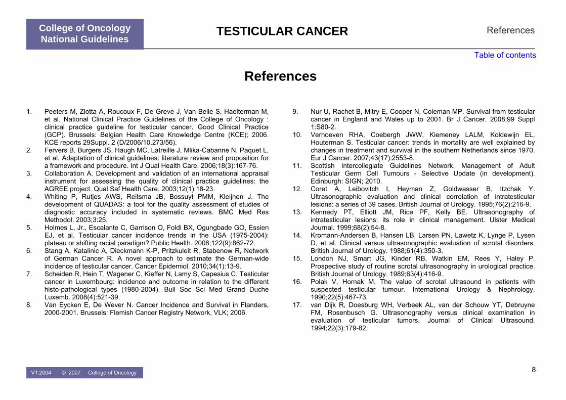
TESTICULAR CANCER
College of Oncology National Guidelines
References
Table of contents
1. Peeters M, Zlotta A, Roucoux F, De Greve J, Van Belle S, Haelterman M,
et al. National Clinical Practice Guidelines of the College of Oncology : clinical practice guideline for testicular cancer. Good Clinical Practice (GCP). Brussels: Belgian Health Care Knowledge Centre (KCE); 2006. KCE reports 29Suppl. 2 (D/2006/10.273/56).
2. Fervers B, Burgers JS, Haugh MC, Latreille J, Mlika-Cabanne N, Paquet L, et al. Adaptation of clinical guidelines: literature review and proposition for a framework and procedure. Int J Qual Health Care. 2006;18(3):167-76.
3. Collaboration A. Development and validation of an international appraisal instrument for assessing the quality of clinical practice guidelines: the AGREE project. Qual Saf Health Care. 2003;12(1):18-23.
4. Whiting P, Rutjes AWS, Reitsma JB, Bossuyt PMM, Kleijnen J. The development of QUADAS: a tool for the quality assessment of studies of diagnostic accuracy included in systematic reviews. BMC Med Res Methodol. 2003;3:25.
5. Holmes L, Jr., Escalante C, Garrison O, Foldi BX, Ogungbade GO, Essien EJ, et al. Testicular cancer incidence trends in the USA (1975-2004): plateau or shifting racial paradigm? Public Health. 2008;122(9):862-72.
6. Stang A, Katalinic A, Dieckmann K-P, Pritzkuleit R, Stabenow R, Network of German Cancer R. A novel approach to estimate the German-wide incidence of testicular cancer. Cancer Epidemiol. 2010;34(1):13-9.
7. Scheiden R, Hein T, Wagener C, Kieffer N, Lamy S, Capesius C. Testicular cancer in Luxembourg: incidence and outcome in relation to the different histo-pathological types (1980-2004). Bull Soc Sci Med Grand Duche Luxemb. 2008(4):521-39.
8. Van Eycken E, De Wever N. Cancer Incidence and Survival in Flanders, 2000-2001. Brussels: Flemish Cancer Registry Network, VLK; 2006.
References 9. Nur U, Rachet B, Mitry E, Cooper N, Coleman MP. Survival from testicular
cancer in England and Wales up to 2001. Br J Cancer. 2008;99 Suppl 1:S80-2.
10. Verhoeven RHA, Coebergh JWW, Kiemeney LALM, Koldewijn EL, Houterman S. Testicular cancer: trends in mortality are well explained by changes in treatment and survival in the southern Netherlands since 1970. Eur J Cancer. 2007;43(17):2553-8.
11. Scottish Intercollegiate Guidelines Network. Management of Adult Testicular Germ Cell Tumours - Selective Update (in development). Edinburgh: SIGN; 2010.
12. Coret A, Leibovitch I, Heyman Z, Goldwasser B, Itzchak Y. Ultrasonographic evaluation and clinical correlation of intratesticular lesions: a series of 39 cases. British Journal of Urology. 1995;76(2):216-9.
13. Kennedy PT, Elliott JM, Rice PF, Kelly BE. Ultrasonography of intratesticular lesions: its role in clinical management. Ulster Medical Journal. 1999;68(2):54-8.
14. Kromann-Andersen B, Hansen LB, Larsen PN, Lawetz K, Lynge P, Lysen D, et al. Clinical versus ultrasonographic evaluation of scrotal disorders. British Journal of Urology. 1988;61(4):350-3.
15. London NJ, Smart JG, Kinder RB, Watkin EM, Rees Y, Haley P. Prospective study of routine scrotal ultrasonography in urological practice. British Journal of Urology. 1989;63(4):416-9.
16. Polak V, Hornak M. The value of scrotal ultrasound in patients with suspected testicular tumour. International Urology & Nephrology. 1990;22(5):467-73.
17. van Dijk R, Doesburg WH, Verbeek AL, van der Schouw YT, Debruyne FM, Rosenbusch G. Ultrasonography versus clinical examination in evaluation of testicular tumors. Journal of Clinical Ultrasound. 1994;22(3):179-82.
V1.2004 © 2007 College of Oncology 8

TESTICULAR CANCER
College of Oncology National Guidelines
References
Table of contents
18. Mottet N, Culine S, Iborra F, Avances C, Bastide C, Lesourd A, et al. [Testicular tumors]. Prog Urol. 2007;17(6):1035-45.
19. Gilligan TD, Seidenfeld J, Basch EM, Einhorn LH, Fancher T, Smith DC, et al. American Society of Clinical Oncology Clinical Practice Guideline on uses of serum tumor markers in adult males with germ cell tumors. J Clin Oncol. 2010;28(20):3388-404.
20. Connolly SS, D'Arcy FT, Bredin HC, Callaghan J, Corcoran MO. Value of frozen section analysis with suspected testicular malignancy. Urology. 2006;67(1):162-5.
21. Elert A, Olbert P, Hegele A, Barth P, Hofmann R, Heidenreich A. Accuracy of frozen section examination of testicular tumors of uncertain origin. Eur Urol. 2002;41(3):290-3.
22. Heidenreich A, Weissbach L, Holtl W, Albers P, Kliesch S, Kohrmann KU, et al. Organ sparing surgery for malignant germ cell tumor of the testis. J Urol. 2001;166(6):2161-5.
23. Fossa SD, Chen J, Schonfeld SJ, McGlynn KA, McMaster ML, Gail MH, et al. Risk of contralateral testicular cancer: a population-based study of 29,515 U.S. men. J Natl Cancer Inst. 2005;97(14):1056-66.
24. Dieckmann K-P, Kulejewski M, Pichlmeier U, Loy V. Diagnosis of contralateral testicular intraepithelial neoplasia (TIN) in patients with testicular germ cell cancer: systematic two-site biopsies are more sensitive than a single random biopsy. European Urology. 2007;51(1):175-83; discussion 83-5.
25. Harland SJ, Cook PA, Fossa SD, Horwich A, Mead GM, Parkinson MC, et al. Intratubular germ cell neoplasia of the contralateral testis in testicular cancer: defining a high risk group. J Urol. 1998;160(4):1353-7.
26. Akre O, Pettersson A, Richiardi L. Risk of contralateral testicular cancer among men with unilaterally undescended testis: a meta analysis. Int J Cancer. 2009;124(3):687-9.
27. Peng X, Zeng X, Peng S, Deng D, Zhang J. The association risk of male subfertility and testicular cancer: a systematic review. PLoS ONE [Electronic Resource]. 2009;4(5):e5591.
28. Walsh TJ, Dall'Era MA, Croughan MS, Carroll PR, Turek PJ. Prepubertal orchiopexy for cryptorchidism may be associated with lower risk of testicular cancer. J Urol. 2007;178(4 Pt 1):1440-6; discussion 6.
29. Wood HM, Elder JS. Cryptorchidism and testicular cancer: separating fact from fiction. J Urol. 2009;181(2):452-61.
30. Dieckmann K-P, Classen J, Loy V. Diagnosis and management of testicular intraepithelial neoplasia (carcinoma in situ)--surgical aspects. APMIS. 2003;111(1):64-8; discussion 8-9.
31. Classen J, Dieckmann K, Bamberg M, Souchon R, Kliesch S, Kuehn M, et al. Radiotherapy with 16 Gy may fail to eradicate testicular intraepithelial neoplasia: preliminary communication of a dose-reduction trial of the German Testicular Cancer Study Group. Br J Cancer. 2003;88(6):828-31.
32. Petersen PM, Giwercman A, Daugaard G, Rorth M, Petersen JH, Skakkeaek NE, et al. Effect of graded testicular doses of radiotherapy in patients treated for carcinoma-in-situ in the testis. J Clin Oncol. 2002;20(6):1537-43.
33. Sedlmayer F, Holtl W, Kozak W, Hawliczek R, Gebhart F, Gerber E, et al. Radiotherapy of testicular intraepithelial neoplasia (TIN): a novel treatment regimen for a rare disease. Int J Radiat Oncol Biol Phys. 2001;50(4):909-13.
34. Kleinschmidt K, Dieckmann K-P, Georgiew A, Loy V, Weissbach L. Chemotherapy is of limited efficacy in the control of contralateral testicular intraepithelial neoplasia in patients with testicular germ cell cancer. Oncology. 2009;77(1):33-9.
35. Cremerius U, Wildberger JE, Borchers H, Zimny M, Jakse G, Gunther RW, et al. Does positron emission tomography using 18-fluoro-2-deoxyglucose improve clinical staging of testicular cancer?--Results of a study in 50 patients. Urology. 1999;54(5):900-4.
36. de Wit M, Brenner W, Hartmann M, Kotzerke J, Hellwig D, Lehmann J, et al. [18F]-FDG-PET in clinical stage I/II non-seminomatous germ cell tumours: results of the German multicentre trial. Annals of Oncology. 2008;19(9):1619-23.
V1.2004 © 2007 College of Oncology 9

TESTICULAR CANCER
College of Oncology National Guidelines
References
Table of contents
37. Frick MP, Feinberg SE, Knight LC. Evaluation of retroperitoneum with computerized tomography and ultrasonography in patients with testicular tumors. Computerized Tomography. 1979;3(3):181-4.
38. Richie JP, Garnick MB, Finberg H. Computerized tomography: how accurate for abdominal staging of testis tumors? Journal of Urology. 1982;127(4):715-7.
39. Thomas JL, Bernardino ME, Bracken RB. Staging of testicular carcinoma: comparison of CT and lymphangiography. AJR. 1981;American Journal of Roentgenology. 137(5):991-6.
40. Tarin TV, Sonn G, Shinghal R. Estimating the risk of cancer associated with imaging related radiation during surveillance for stage I testicular cancer using computerized tomography. J Urol. 2009;181(2):627-32; discussion 32-3.
41. Hansen J, Jurik AG. Diagnostic value of multislice computed tomography and magnetic resonance imaging in the diagnosis of retroperitoneal spread of testicular cancer: a literature review. Acta Radiol. 2009;50(9):1064-70.
42. Schwerk WB, Schwerk WN, Rodeck G. Testicular tumors: prospective analysis of real-time US patterns and abdominal staging. Radiology. 1987;164(2):369-74.
43. Vlayen J, Stordeur S, Van den Bruel A, Mambourg F, Eyssen M. Positron Emission Tomography (PET) in Belgium: an update. Health Technology Assessment (HTA). Brussels: Belgian Health Care Knowledge Centre (KCE); 2009. KCE reports 110 (D/2009/10.273/24)
44. Lee SJ, Schover LR, Partridge AH, Patrizio P, Wallace WH, Hagerty K, et al. American Society of Clinical Oncology recommendations on fertility preservation in cancer patients. J Clin Oncol. 2006;24(18):2917-31.
45. World Health Organisation. WHO Classification of Tumours: Pathology and Genetics of Tumours of the Urinary System and Male Genital Organs. Third ed. Eble JN, Sauter G, Epstein JL, Sesterhenn IA, editor.: WHO/IARC; 2008.
46. UICC International Union Against Cancer. TNM Classification of Malignant Tumours. Seventh ed. Sobin L, Gospodarowicz M, Wittekind C, editor. New York: Wiley- Blackwell; 2009.
47. International Germ Cell Consensus Classification: a prognostic factor-based staging system for metastatic germ cell cancers. International Germ Cell Cancer Collaborative Group. J Clin Oncol. 1997;15(2):594-603.
48. Albers P, Siener R, Kliesch S, Weissbach L, Krege S, Sparwasser C, et al. Risk factors for relapse in clinical stage I nonseminomatous testicular germ cell tumors: results of the German Testicular Cancer Study Group Trial. J Clin Oncol. 2003;21(8):1505-12.
49. Parker C, Milosevic M, Panzarella T, Banerjee D, Jewett M, Catton C, et al. The prognostic significance of the tumour infiltrating lymphocyte count in stage I testicular seminoma managed by surveillance. Eur J Cancer. 2002;38(15):2014-9.
50. Warde P, Gospodarowicz MK, Banerjee D, Panzarella T, Sugar L, Catton CN, et al. Prognostic factors for relapse in stage I testicular seminoma treated with surveillance. J Urol. 1997;157(5):1705-9; discussion 9-10.
51. Warde P, Specht L, Horwich A, Oliver T, Panzarella T, Gospodarowicz M, et al. Prognostic factors for relapse in stage I seminoma managed by surveillance: a pooled analysis.[see comment]. J Clin Oncol. 2002;20(22):4448-52.
52. Nazeer T, Ro JY, Kee KH, Ayala AG. Spermatic cord contamination in testicular cancer. Mod Pathol. 1996;9(7):762-6.
53. Klepp O, Olsson AM, Henrikson H, Aass N, Dahl O, Stenwig AE, et al. Prognostic factors in clinical stage I nonseminomatous germ cell tumors of the testis: multivariate analysis of a prospective multicenter study. Swedish-Norwegian Testicular Cancer Group. J Clin Oncol. 1990;8(3):509-18.
54. Moul JW, McCarthy WF, Fernandez EB, Sesterhenn IA. Percentage of embryonal carcinoma and of vascular invasion predicts pathological stage in clinical stage I nonseminomatous testicular cancer. Cancer Res. 1994;54(2):362-4.
55. Mead GM, Stenning SP, Parkinson MC, Horwich A, Fossa SD, Wilkinson PM, et al. The Second Medical Research Council study of prognostic factors in nonseminomatous germ cell tumors. Medical Research Council Testicular Tumour Working Party.[erratum appears in J Clin Oncol 1992 May;10(5):867]. J Clin Oncol. 1992;10(1):85-94.
V1.2004 © 2007 College of Oncology 10

TESTICULAR CANCER
College of Oncology National Guidelines
References
Table of contents
56. Steyerberg EW, Keizer HJ, Fossa SD, Sleijfer DT, Toner GC, Schraffordt Koops H, et al. Prediction of residual retroperitoneal mass histology after chemotherapy for metastatic nonseminomatous germ cell tumor: multivariate analysis of individual patient data from six study groups. Journal of Clinical Oncology. 1995;13(5):1177-87.
57. Freedman LS, Parkinson MC, Jones WG, Oliver RT, Peckham MJ, Read G, et al. Histopathology in the prediction of relapse of patients with stage I testicular teratoma treated by orchidectomy alone. Lancet. 1987;2(8554):294-8.
58. Tavolini IM, Bettella A, Boscolo Berto R, Bassi PF, Longo R, Menegazzo M, et al. Immunostaining for placental alkaline phosphatase on fine-needle aspiration specimens to detect noninvasive testicular cancer: a prospective evaluation in cryptorchid men.[see comment]. BJU International. 2006;97(5):950-4.
59. Izquierdo MA, Van der Valk P, Van Ark-Otte J, Rubio G, Germa-Lluch JR, Ueda R, et al. Differential expression of the c-kit proto-oncogene in germ cell tumours. J Pathol. 1995;177(3):253-8.
60. de Jong J, Stoop H, Dohle GR, Bangma CH, Kliffen M, van Esser JWJ, et al. Diagnostic value of OCT3/4 for pre-invasive and invasive testicular germ cell tumours. J Pathol. 2005;206(2):242-9.
61. Heidenreich A, Sesterhenn IA, Mostofi FK, Moul JW. Immunohistochemical expression of monoclonal antibody 43-9F in testicular germ cell tumours. Int J Androl. 1998;21(5):283-8.
62. Cheville JC, Rao S, Iczkowski KA, Lohse CM, Pankratz VS. Cytokeratin expression in seminoma of the human testis. Am J Clin Pathol. 2000;113(4):583-8.
63. Leroy X, Augusto D, Leteurtre E, Gosselin B. CD30 and CD117 (c-kit) used in combination are useful for distinguishing embryonal carcinoma from seminoma. J Histochem Cytochem. 2002;50(2):283-5.
64. Bosman FT, Giard RW, Nieuwenhuijen Kruseman AC, Knijnenburg G, Spaander PJ. Human chorionic gonadotrophin and alpha-fetoprotein in testicular germ cell tumours: a retrospective immunohistochemical study. Histopathology. 1980;4(6):673-84.
65. Santagata S, Ligon KL, Hornick JL. Embryonic stem cell transcription factor signatures in the diagnosis of primary and metastatic germ cell tumors. Am J Surg Pathol. 2007;31(6):836-45.
66. Jones TD, Ulbright TM, Eble JN, Baldridge LA, Cheng L. OCT4 staining in testicular tumors: a sensitive and specific marker for seminoma and embryonal carcinoma.[see comment]. American Journal of Surgical Pathology. 2004;28(7):935-40.
67. Chung P, Mayhew LA, Warde P, Winquist E, Lukka H, and Members of the Genitourinary Cancer Disease Site Group. Management of Stage I Seminoma: Guideline Recommendations. Toronto: CCO; 2008. (Evidence-Based Series #3-18)
68. Oliver RTD, Mason MD, Mead GM, von der Maase H, Rustin GJS, Joffe JK, et al. Radiotherapy versus single-dose carboplatin in adjuvant treatment of stage I seminoma: a randomised trial.[see comment]. Lancet. 2005;366(9482):293-300.
69. Jones WG, Fossa SD, Mead GM, Roberts JT, Sokal M, Horwich A, et al. Randomized trial of 30 versus 20 Gy in the adjuvant treatment of stage I Testicular Seminoma: a report on Medical Research Council Trial TE18, European Organisation for the Research and Treatment of Cancer Trial 30942 (ISRCTN18525328).[see comment]. J Clin Oncol. 2005;23(6):1200-8.
70. Fossa SD, Horwich A, Russell JM, Roberts JT, Cullen MH, Hodson NJ, et al. Optimal planning target volume for stage I testicular seminoma: A Medical Research Council randomized trial. Medical Research Council Testicular Tumor Working Group.[see comment]. J Clin Oncol. 1999;17(4):1146.
71. Groll RJ, Warde P, Jewett MAS. A comprehensive systematic review of testicular germ cell tumor surveillance. Critical Reviews in Oncology-Hematology. 2007;64(3):182-97.
72. Hotte S, Mayhew LA, Jewett M, Chin J, Winquist E, and Members of the Genitourinary Cancer Disease Site Group. Management of Stage I Nonseminomatous Testicular Cancer: Guideline Recommendations. Toronto: CCO; 2008. (Evidence-Based Series #3-19)
V1.2004 © 2007 College of Oncology 11

TESTICULAR CANCER
College of Oncology National Guidelines
References
Table of contents
73. Rorth M, Jacobsen GK, von der Maase H, Madsen EL, Nielsen OS, Pedersen M, et al. Surveillance alone versus radiotherapy after orchiectomy for clinical stage I nonseminomatous testicular cancer. Danish Testicular Cancer Study Group. J Clin Oncol. 1991;9(9):1543-8.
74. Albers P, Siener R, Krege S, Schmelz H-U, Dieckmann K-P, Heidenreich A, et al. Randomized phase III trial comparing retroperitoneal lymph node dissection with one course of bleomycin and etoposide plus cisplatin chemotherapy in the adjuvant treatment of clinical stage I Nonseminomatous testicular germ cell tumors: AUO trial AH 01/94 by the German Testicular Cancer Study Group.[see comment]. J Clin Oncol. 2008;26(18):2966-72.
75. Albers P, Albrecht W, Algaba F, Bokemeyer C, Cohn-Cedermark G, Horwich A, et al. Guidelines on Testicular Cancer. European Association of Urology; 2008.
76. Feldman DR, Bosl GJ, Sheinfeld J, Motzer RJ. Medical treatment of advanced testicular cancer. JAMA. 2008;299(6):672-84.
77. Shelley MD, Burgon K, Mason MD. Treatment of testicular germ-cell cancer: a cochrane evidence-based systematic review. Cancer Treatment Reviews. 2002;28(5):237-53.
78. Patterson H, Norman AR, Mitra SS, Nicholls J, Fisher C, Dearnaley DP, et al. Combination carboplatin and radiotherapy in the management of stage II testicular seminoma: comparison with radiotherapy treatment alone. Radiother Oncol. 2001;59(1):5-11.
79. Chung PWM, Gospodarowicz MK, Panzarella T, Jewett MAS, Sturgeon JFG, Tew-George B, et al. Stage II testicular seminoma: patterns of recurrence and outcome of treatment. Eur Urol. 2004;45(6):754-59; discussion 9-60.
80. Clemm C, Bokemeyer C, Gerl A, Holzel D, Weissbach L, Mamberg M, et al. Randomized Trial Comparing Cisplatin/Etoposide/ Ifosfamide with Carboplatin Monochemotherapy in Patients with Advanced Metastatic Seminoma. Proc Am Soc Clin Oncol. 2000;19 (abstr 1283).
81. Horwich A, Oliver RT, Wilkinson PM, Mead GM, Harland SJ, Cullen MH, et al. A medical research council randomized trial of single agent carboplatin
versus etoposide and cisplatin for advanced metastatic seminoma. MRC Testicular Tumour Working Party. Br J Cancer. 2000;83(12):1623-9.
82. Bokemeyer C, Kollmannsberger C, Stenning S, Hartmann JT, Horwich A, Clemm C, et al. Metastatic seminoma treated with either single agent carboplatin or cisplatin-based combination chemotherapy: a pooled analysis of two randomised trials. Br J Cancer. 2004;91(4):683-7.
83. Culine S, Kerbrat P, Kramar A, Theodore C, Chevreau C, Geoffrois L, et al. Refining the optimal chemotherapy regimen for good-risk metastatic nonseminomatous germ-cell tumors: a randomized trial of the Genito-Urinary Group of the French Federation of Cancer Centers (GETUG T93BP). Ann Oncol. 2007;18(5):917-24.
84. Horwich A, Sleijfer DT, Fossa SD, Kaye SB, Oliver RT, Cullen MH, et al. Randomized trial of bleomycin, etoposide, and cisplatin compared with bleomycin, etoposide, and carboplatin in good-prognosis metastatic nonseminomatous germ cell cancer: a Multiinstitutional Medical Research Council/European Organization for Research and Treatment of Cancer Trial. J Clin Oncol. 1997;15(5):1844-52.
85. de Wit R, Stoter G, Kaye SB, Sleijfer DT, Jones WG, ten Bokkel Huinink WW, et al. Importance of bleomycin in combination chemotherapy for good-prognosis testicular nonseminoma: a randomized study of the European Organization for Research and Treatment of Cancer Genitourinary Tract Cancer Cooperative Group.[see comment]. J Clin Oncol. 1997;15(5):1837-43.
86. Bokemeyer C, Kohrmann O, Tischler J, Weissbach L, Rath U, Haupt A, et al. A randomized trial of cisplatin, etoposide and bleomycin (PEB) versus carboplatin, etoposide and bleomycin (CEB) for patients with 'good-risk' metastatic non-seminomatous germ cell tumors. Ann Oncol. 1996;7(10):1015-21.
87. Einhorn LH, Williams SD, Loehrer PJ, Birch R, Drasga R, Omura G, et al. Evaluation of optimal duration of chemotherapy in favorable-prognosis disseminated germ cell tumors: a Southeastern Cancer Study Group protocol. J Clin Oncol. 1989;7(3):387-91.
88. Toner GC, Stockler MR, Boyer MJ, Jones M, Thomson DB, Harvey VJ, et al. Comparison of two standard chemotherapy regimens for good-
V1.2004 © 2007 College of Oncology 12

TESTICULAR CANCER
College of Oncology National Guidelines
References
Table of contents
prognosis germ-cell tumours: a randomised trial. Australian and New Zealand Germ Cell Trial Group. Lancet. 2001;357(9258):739-45.
89. Loehrer PJ, Sr., Johnson D, Elson P, Einhorn LH, Trump D. Importance of bleomycin in favorable-prognosis disseminated germ cell tumors: an Eastern Cooperative Oncology Group trial. J Clin Oncol. 1995;13(2):470-6.
90. Droz J-P, Kramar A, Biron P, Pico J-L, Kerbrat P, Peny J, et al. Failure of high-dose cyclophosphamide and etoposide combined with double-dose cisplatin and bone marrow support in patients with high-volume metastatic nonseminomatous germ-cell tumours: mature results of a randomised trial. Eur Urol. 2007;51(3):739-46; discussion 47-8.
91. Kaye SB, Mead GM, Fossa S, Cullen M, deWit R, Bodrogi I, et al. Intensive induction-sequential chemotherapy with BOP/VIP-B compared with treatment with BEP/EP for poor-prognosis metastatic nonseminomatous germ cell tumor: a Randomized Medical Research Council/European Organization for Research and Treatment of Cancer study. J Clin Oncol. 1998;16(2):692-701.
92. de Wit R, Stoter G, Sleijfer DT, Neijt JP, ten Bokkel Huinink WW, de Prijck L, et al. Four cycles of BEP vs four cycles of VIP in patients with intermediate-prognosis metastatic testicular non-seminoma: a randomized study of the EORTC Genitourinary Tract Cancer Cooperative Group. European Organization for Research and Treatment of Cancer. Br J Cancer. 1998;78(6):828-32.
93. de Wit R, Stoter G, Sleijfer DT, Kaye SB, de Mulder PH, ten Bokkel Huinink WW, et al. Four cycles of BEP versus an alternating regime of PVB and BEP in patients with poor-prognosis metastatic testicular non-seminoma; a randomised study of the EORTC Genitourinary Tract Cancer Cooperative Group. Br J Cancer. 1995;71(6):1311-4.
94. Williams SD, Stablein DM, Einhorn LH, Muggia FM, Weiss RB, Donohue JP, et al. Immediate adjuvant chemotherapy versus observation with treatment at relapse in pathological stage II testicular cancer. N Engl J Med. 1987;317(23):1433-8.
95. Nichols CR, Williams SD, Loehrer PJ, Greco FA, Crawford ED, Weetlaufer J, et al. Randomized study of cisplatin dose intensity in poor-risk germ cell
tumors: a Southeastern Cancer Study Group and Southwest Oncology Group protocol. J Clin Oncol. 1991;9(7):1163-72.
96. Nichols CR, Catalano PJ, Crawford ED, Vogelzang NJ, Einhorn LH, Loehrer PJ. Randomized comparison of cisplatin and etoposide and either bleomycin or ifosfamide in treatment of advanced disseminated germ cell tumors: an Eastern Cooperative Oncology Group, Southwest Oncology Group, and Cancer and Leukemia Group B Study. J Clin Oncol. 1998;16(4):1287-93.
97. Motzer RJ, Nichols CJ, Margolin KA, Bacik J, Richardson PG, Vogelzang NJ, et al. Phase III randomized trial of conventional-dose chemotherapy with or without high-dose chemotherapy and autologous hematopoietic stem-cell rescue as first-line treatment for patients with poor-prognosis metastatic germ cell tumors. J Clin Oncol. 2007;25(3):247-56.
98. Culine S, Kramar A, Theodore C, Geoffrois L, Chevreau C, Biron P, et al. Randomized trial comparing bleomycin/etoposide/cisplatin with alternating cisplatin/cyclophosphamide/doxorubicin and vinblastine/bleomycin regimens of chemotherapy for patients with intermediate- and poor-risk metastatic nonseminomatous germ cell tumors: Genito-Urinary Group of the French Federation of Cancer Centers Trial T93MP. J Clin Oncol. 2008;26(3):421-7.
99. Daugaard G, Skoneczna IA, Aass N, De Wit R, De Santis M, Dumez H, et al. A randomized phase III study comparing standard dose BEP with sequential high-dose cisplatin, etoposide, ifosfamide (VIP) plus stem cell support in males with poor prognosis germ cell cancer (GCC): An intergroup study of EORTC, GTCSG, and Grupo Germinal (EORTC 30974). J Clin Oncol (Meeting Abstracts). 2010;28(15_suppl):4512.
100. Pfannenberg AC, Oechsle K, Kollmannsberger C, Dohmen BM, Bokemeyer C, Bares R, et al. Early prediction of treatment response to high-dose chemotherapy in patients with relapsed germ cell tumors using [18F]FDG-PET, CT or MRI, and tumor marker. ROFO Fortschr Geb Rontgenstr Nuklearmed. 2004;176(1):76-84.
101. De Santis M, Becherer A, Bokemeyer C, Stoiber F, Oechsle K, Sellner F, et al. 2-18fluoro-deoxy-D-glucose positron emission tomography is a reliable predictor for viable tumor in postchemotherapy seminoma: an
V1.2004 © 2007 College of Oncology 13

TESTICULAR CANCER
College of Oncology National Guidelines
References
Table of contents
update of the prospective multicentric SEMPET trial. Journal of Clinical Oncology. 2004;22(6):1034-9.
109. Puc HS, Heelan R, Mazumdar M, Herr H, Scheinfeld J, Vlamis V, et al. Management of residual mass in advanced seminoma: results and recommendations from the Memorial Sloan-Kettering Cancer Center. J Clin Oncol. 1996;14(2):454-60.
102. Oechsle K, Hartmann M, Brenner W, Venz S, Weissbach L, Franzius C, et al. [18F]Fluorodeoxyglucose positron emission tomography in nonseminomatous germ cell tumors after chemotherapy: the German multicenter positron emission tomography study group. J Clin Oncol. 2008;26(36):5930-5.
110. Rustin GJ, Mead GM, Stenning SP, Vasey PA, Aass N, Huddart RA, et al. Randomized trial of two or five computed tomography scans in the surveillance of patients with stage I nonseminomatous germ cell tumors of the testis: Medical Research Council Trial TE08, ISRCTN56475197--the National Cancer Research Institute Testis Cancer Clinical Studies Group.[see comment]. J Clin Oncol. 2007;25(11):1310-5.
103. Heidenreich A, Thuer D, Polyakov S. Postchemotherapy retroperitoneal lymph node dissection in advanced germ cell tumours of the testis. Eur Urol. 2008;53(2):260-72.
104. Liu D, Abolhoda A, Burt ME, Martini N, Bains MS, Downey RJ, et al. Pulmonary metastasectomy for testicular germ cell tumors: a 28-year experience. Ann Thorac Surg. 1998;66(5):1709-14.
111. Segal R, Lukka H, Klotz L, Eady A, Bestic N, Johnston M, et al. Surveillance Programs for Early Stage Non-Seminomatous Testicular Cancer. Toronto: CCO; 2001. (Practice Guideline Report No. 3-5)
105. You YN, Leibovitch BC, Que FG. Hepatic metastasectomy for testicular germ cell tumors: is it worth it? J Gastrointest Surg. 2009;13(4):595-601.
112. Stoehr B, Zangerl F, Steiner E, Leonhartsberger N, Fritzer A, Bartsch G, et al. Routine scrotal ultrasonography during the follow-up of patients with testicular cancer leads to earlier detection of asynchronous tumours and a high rate of organ preservation. BJU Int. 2009.
106. Pfannschmidt J, Zabeck H, Muley T, Dienemann H, Hoffmann H. Pulmonary metastasectomy following chemotherapy in patients with testicular tumors: experience in 52 patients. Thorac Cardiovasc Surg. 2006;54(7):484-8.
113. Pico JL, Rosti G, Kramar A, Wandt H, Koza V, Salvioni R, et al. A randomised trial of high-dose chemotherapy in the salvage treatment of patients failing first-line platinum chemotherapy for advanced germ cell tumours. Ann Oncol. 2005;16(7):1152-9.
107. Ondrus D, Hornak M, Breza J, Mat'oska J, Schnorrer M, Belan V, et al. Delayed orchiectomy after chemotherapy in patients with advanced testicular cancer. Int Urol Nephrol. 2001;32(4):665-7. 114. Lorch A, Kollmannsberger C, Hartmann JT, Metzner B, Schmidt-Wolf IGH,
Berdel WE, et al. Single versus sequential high-dose chemotherapy in patients with relapsed or refractory germ cell tumors: a prospective randomized multicenter trial of the German Testicular Cancer Study Group. J Clin Oncol. 2007;25(19):2778-84.
108. Krege S, Beyer J, Souchon R, Albers P, Albrecht W, Algaba F, et al. European consensus conference on diagnosis and treatment of germ cell cancer: a report of the second meeting of the European Germ Cell Cancer Consensus group (EGCCCG): part I. European Urology. 2008;53(3):478-96.
V1.2004 © 2007 College of Oncology 14

TESTICULAR CANCER
College of Oncology National Guidelines
Appendix 1 Grade system
Table of contents
Grade of Recommendation/ Description Benefit vs. Risk and Burdens Methodological Quality of Supporting
Evidence Implications
1A/ Strong recommendation, high quality evidence
Benefits clearly outweigh risk and burdens, or vice versa
RCTs without important limitations or overwhelming evidence fromobservational studies
Strong recommendation, can apply to most patients in most circumstances without reservation
1B/ Strong recommendation, moderate quality evidence
Benefits clearly outweigh risk and burdens, or vice versa
RCTs with important limitations(inconsistent results, methodological flaws, indirect, or imprecise) or exceptionally strong evidence from observational studies
Strong recommendation, can apply to most patients in most circumstances without reservation
1C/ Strong recommendation, low quality evidence
Benefits clearly outweigh risk and burdens, or vice versa
Observational studies or case series Strong recommendation, but may change when higher quality evidence becomes available
2A/ Weak recommendation, high quality evidence
Benefits closely balanced with risks and burden
RCTs without important limitations or overwhelming evidence fromobservational studies
Weak recommendation, best action may differ depending on circumstances or patients’ or societal values
2B/ Weak recommendation, moderate quality evidence
Benefits closely balanced with risks and burden
RCTs with important limitations(inconsistent results, methodological flaws, indirect, or imprecise) or exceptionally strong evidence from observational studies
Weak recommendation, best action may differ depending on circumstances or patients’ or societal values
2C/ Weak recommendation, low quality evidence
Benefits closely balanced with risks and burden
Observational studies or case series Very weak recommendation, other alternatives may be equally reasonable
V1.2010 © 2010 College of Oncology

TESTICULAR CANCER
College of Oncology National Guidelines
Appendix 2 cTNM Staging
Table of contents
TNM classification for testicular cancer (UICC, 2009 Seventh Edition) cT Primary Tumour
Except for pT4, where radical orchidectomy is not always necessary for classification purposes, the extent of the primary tumour is classified after radical orchidectomy; see pT. In other circumstances, TX is used if no radical orchidectomy has been performed.
cN Regional Lymph Nodes
NX Regional lymph nodes cannot be assessed
N0 No regional lymph node metastasis
N1 Metastasis with a lymph node mass 2 cm or less in greatest dimension or multiple lymph nodes, none more than 2 cm in greatest dimension
N2 Metastasis with a lymph node mass >2cm but <5cm in greatest dimension; or multiple lymph nodes, any one mass >2cm but <5cm in greatest dimension
N3 Metastasis with a lymph node mass more than 5 cm in greatest dimension
cM Distant Metastasis
M0 No distant metastasis
M1a Non-regional lymph node(s) or lungmetastasis
M1b Distant metastasis other than non-regional lymph nodes and lung
9
V1.2010 © 2010 College of Oncology

TESTICULAR CANCER
College of Oncology National Guidelines
Appendix 2 pTNM Staging
Table of contents
pT Primary Tumour pTX Primary tumour cannot be assessed (see T - primary tumour, above)
pT0 No evidence of primary tumour (e.g., histologic scar in testis)
pTis Intratubular germ cell neoplasia (carcinoma in situ)
pT1 Tumour limited to testis and epididymis without vascular/lymphatic invasion:tumour may invade tunica albuginea but not tunica vaginalis
pT2 Tumour limited to testis and epididymis with vascular/lymphatic invasion, or tumour extending through tunica albuginea with involvement of tunica vaginalis
pT3 Tumour invades spermatic cord with or without vascular/lymphatic invasion
pT4 Tumour invades scrotum with or without vascular/lymphatic invasion
pN Regional Lymph Nodes
pNX Regional lymph nodes cannot be assessed
pN0 No regional lymph node metastasis
pN1 Metastasis with a lymph node mass 2 cm or less in greatest dimension and 5 or fewer positive nodes, none more than 2 cm in greatest dimension
pN2 Metastasis with a lymph node mass >2cm but <5cm in greatest dimension; or >5 nodes positive, none >5cm; or evidence or extranodal extension of tumour
pN3 Metastasis with a lymph node mass >5 cm in greatest dimension
pM Distant Metastasis
pM1 Distant metastasis microscopically confirmed
S Serum tumour markers
SX Serum marker studies not available
S0 Serum marker study levels within normal limits
S1 LDH <1.5 x N and betaHCG < 5000 mIU/ml and AFP < 1000 ng/ml S2 LDH 1.5-10 x N or betaHCG 5000-50000 mIU/ml or AFP 1000-10000 ng/ml
V1.2010 © 2010 College of Oncology

TESTICULAR CANCER
College of Oncology National Guidelines
Appendix 2 Stage grouping Table of contents
TNM Stage grouping
Stage 0 pTis N0 M0 S0Stage I pT1-4 N0 M0 SXStage IA pT1 N0 M0 S0Stage IB pT2 N0 M0 S0 pT3 N0 M0 S0 pT4 N0 M0 S0Stage IS Any pT/TX N0 M0 S1-3 Stage II Any pT/TX N1-3 M0 SX Stage IIA Any pT/TX N1 M0 S0 Any pT/TX N1 M0 S1 Stage IIB Any pT/TX N2 M0 S0 Any pT/TX N2 M0 S1 Stage IIC Any pT/TX N3 M0 S0 Any pT/TX N3 M0 S1 Stage III Any pT/TX Any N M1a SX Stage IIIA Any pT/TX Any N M1a S0 Any pT/TX Any N M1a S1 Stage IIIB Any pT/TX N1-3 M0 S2 Any pT/TX Any N M1a S2 Stage IIIC Any pT/TX N1-3 M1a S3 Any pT/TX Any N M1b S3
V1.2010 © 2010 College of Oncology