Embed Size (px)
Citation preview

Kingdom of Bahrain
Arabian Gulf University
College of Medicine and Medical Sciences
Cardiology
- Congestive Heart Failure (CHF):
It is defined as inability of the heart to pump blood normally thus leading to
hypoperfusion and reduced oxygen delivery to tissues. Hypoperfusion will result in
the following:
Increase in hear rate and contractility (trying to increase cardiac output).
It will be sensed by kidneys as reduced blood volume thus activating renin-
angiotensin system which leads to retention of salts and water.
Release of catecholamines from SNS will also increase heart rate and
contractility.
Causes of CHF:
Congenital
Increased pulmonary blood flow: large VSD, transposition of great
arteries, truncus arteriosus and total anomalous pulmonary venous
connection
Obstructive lesion: aortic, pulmonary or mitral stenosis; corarctation
of aorta
Others lesions: mitral or tricuspid regurgitation
Acquired
Viral myocarditis (most common), infections (endocarditis,
pericarditis), hypothyroidism, IHD, cardiomyopathies, dysarrhythmias
and drugs (chemotherapeutic agents)
Others Severe anemia
Clinical features:
Pulmonary congestion Tachypnea, cough, wheezing, rales and pulmonary
edema on CXR
Impaired myocardial
performance
Tachycardia, sweating, reduced urine output, pale
skin and enlarged cardiac shadow on CXR
Systemic venous congestion Hepatomegaly and peripheral edema
Late manifestations Cyanosis and shock
Management:
Cardiac glycosides Digoxin
Loop diuretics Furosemide
Surgical repair CHD secondary to congenital heart diseases

- Innocent cardiac murmurs:
Those which are caused by turbulent blood flow with no structural abnormalities of
the heart. This occurs in 50% of children at some point during childhood.
Grading of heart murmurs:
Grade-I Soft; heard under quiet conditions
Grade-II Soft; heard under noisy conditions
Grade-III Easily heard prominent murmur
Grade-IV Loud murmur with thrill
Grade-V Loud murmur with edge of stethoscope tilted against the chest plus a
thrill
Grade-VI Very loud murmur heard 5-10 mm from the chest plus a thrill
Clinical features of innocent heart murmurs:
Murmur Age Location Characteristics
Still’s murmur 2-7 years Mid-left sternal
border
Grade I-III; systolic, vibratory,
loudest when supine; louder
with exercise
Pulmonic systolic
murmur Any age
Upper-left
sternal border
Grade I-II; early systole;
blowing; loudest when supine;
louder with exercise
Venous hum Any age Neck and below
clavicles
Continuous murmur, heard
when sitting or standing
- Acyanotic congenital heart diseases:
Atrial Septal Defect (ASD):
It is a left-to-right shunt in which blood is flowing from left atrium (area of
high resistance) to right atrium (area of low resistance). Increased blood flow
to right atrium and right ventricle through this septal defect will result in
increased pulmonary blood flow.
Classification
Septum primum
Dfect in lower part of septum; common with
Down syndrome
Septum secondum
Defect in middle part of septum; most common type
Sinus venosus
defect near junction of RA and SVC; right pulmonary
veins draining in RA instead of LA
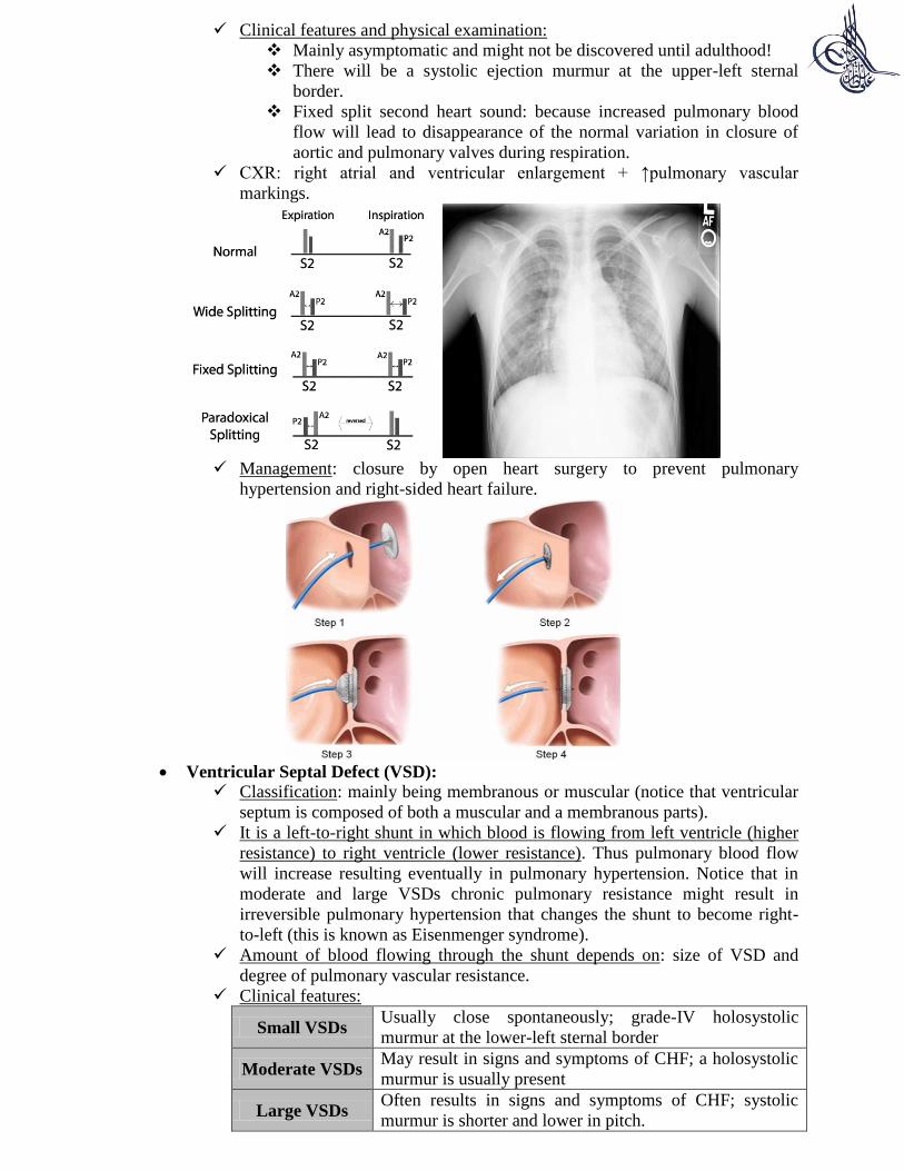
Clinical features and physical examination:
Mainly asymptomatic and might not be discovered until adulthood!
There will be a systolic ejection murmur at the upper-left sternal
border.
Fixed split second heart sound: because increased pulmonary blood
flow will lead to disappearance of the normal variation in closure of
aortic and pulmonary valves during respiration.
CXR: right atrial and ventricular enlargement + ↑pulmonary vascular
markings.
Management: closure by open heart surgery to prevent pulmonary
hypertension and right-sided heart failure.
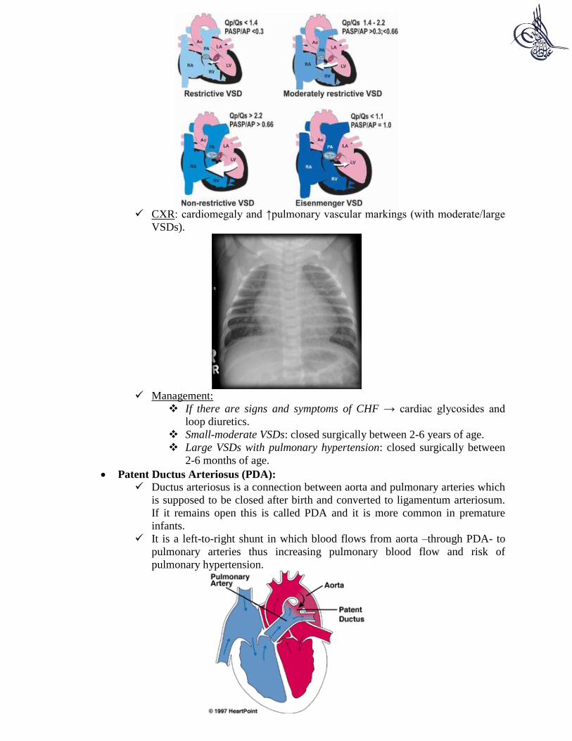
Ventricular Septal Defect (VSD):
Classification: mainly being membranous or muscular (notice that ventricular
septum is composed of both a muscular and a membranous parts).
It is a left-to-right shunt in which blood is flowing from left ventricle (higher
resistance) to right ventricle (lower resistance). Thus pulmonary blood flow
will increase resulting eventually in pulmonary hypertension. Notice that in
moderate and large VSDs chronic pulmonary resistance might result in
irreversible pulmonary hypertension that changes the shunt to become right-
to-left (this is known as Eisenmenger syndrome).
Amount of blood flowing through the shunt depends on: size of VSD and
degree of pulmonary vascular resistance.
Clinical features:
Small VSDs Usually close spontaneously; grade-IV holosystolic
murmur at the lower-left sternal border
Moderate VSDs May result in signs and symptoms of CHF; a holosystolic
murmur is usually present
Large VSDs Often results in signs and symptoms of CHF; systolic
murmur is shorter and lower in pitch.
CXR: cardiomegaly and ↑pulmonary vascular markings (with moderate/large
VSDs).
Management:
If there are signs and symptoms of CHF → cardiac glycosides and
loop diuretics.
Small-moderate VSDs: closed surgically between 2-6 years of age.
Large VSDs with pulmonary hypertension: closed surgically between
2-6 months of age.
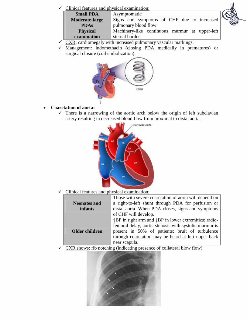
Patent Ductus Arteriosus (PDA):
Ductus arteriosus is a connection between aorta and pulmonary arteries which
is supposed to be closed after birth and converted to ligamentum arteriosum.
If it remains open this is called PDA and it is more common in premature
infants.
It is a left-to-right shunt in which blood flows from aorta –through PDA- to
pulmonary arteries thus increasing pulmonary blood flow and risk of
pulmonary hypertension.
Clinical features and physical examination:
Small PDA Asymptomatic
Moderate-large
PDAs
Signs and symptoms of CHF due to increased
pulmonary blood flow
Physical
examination
Machinery-like continuous murmur at upper-left
sternal border
CXR: cardiomegaly with increased pulmonary vascular markings.
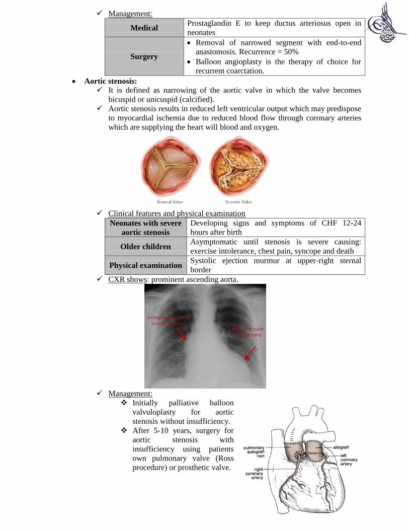
Management: indomethacin (closing PDA medically in prematures) or
surgical closure (coil embolization).
Coarctation of aorta:
There is a narrowing of the aortic arch below the origin of left subclavian
artery resulting in decreased blood flow from proximal to distal aorta.
Clinical features and physical examination:
Neonates and
infants
Those with severe coarctation of aorta will depend on
a right-to-left shunt through PDA for perfusion or
distal aorta. When PDA closes, signs and symptoms
of CHF will develop.
Older children
↑BP in right arm and ↓BP in lower extremities; radio-
femoral delay, aortic stenosis with systolic murmur is
present in 50% of patients; bruit of turbulence
through coarctation may be heard at left upper back
near scapula.
CXR shows: rib notching (indicating presence of collateral blow flow).
Management:
Medical Prostaglandin E to keep ductus arteriosus open in
neonates
Surgery
Removal of narrowed segment with end-to-end
anastomosis. Recurrence = 50%
Balloon angioplasty is the therapy of choice for
recurrent coarctation.
Aortic stenosis:
It is defined as narrowing of the aortic valve in which the valve becomes
bicuspid or unicuspid (calcified).
Aortic stenosis results in reduced left ventricular output which may predispose
to myocardial ischemia due to reduced blood flow through coronary arteries
which are supplying the heart will blood and oxygen.
Clinical features and physical examination
Neonates with severe
aortic stenosis
Developing signs and symptoms of CHF 12-24
hours after birth
Older children Asymptomatic until stenosis is severe causing:
exercise intolerance, chest pain, syncope and death
Physical examination Systolic ejection murmur at upper-right sternal
border
CXR shows: prominent ascending aorta.
Management:
Initially palliative balloon
valvuloplasty for aortic
stenosis without insufficiency.
After 5-10 years, surgery for
aortic stenosis with
insufficiency using patients
own pulmonary valve (Ross
procedure) or prosthetic valve.

Pulmonary stenosis:
It is defined as narrowing of the pulmonary valve which results in decreased
right ventricular output.
Clinical features: asymptomatic unless severe in neonates where there will be
cyanosis due to right-to-left shunt through from right atrium to left atrium
through patent foramen ovale. On physical examination, you will hear a
systolic ejection murmur at the upper-left sternal border.
CXR shows: enlarged pulmonary trunk.
Management: balloon valvuloplasty when symptomatic.
- Cyanotic congenital heart diseases:
Tetraology of Fallot (TOF):
It is the most common cause of central cyanosis after neonatal period and is
characterized by:
Pulmonary stenosis.
Right ventricular
hypertrophy.
Overriding aorta.
VSD.
Due to pulmonary stenosis, there
is a right-to-left shunt in which
blood is flowing from right
ventricle –through the VSD- to
left ventricle (mixing with
oxygenated blood) and being
pumped through the overriding
aorta.
Clinical features and physical
examination:
Systolic ejection murmur at the upper-left sternal border; increased
right ventricular impulse and cyanosis.
Cyanosis results with conditions that decrease systemic vascular
resistance (e.g. exercise, vasodilation and volume depletion) but
Congenital cyanotic heart diseases
↑Pulmonary blood flow
Truncus arteriosus, transposition of great
arteries and total anomalous pulmonary
venous connection
↓Pulmonary blood flow
Tetraology of Fallot; tricuspid atresia
decrease with conditions that increase systemic vascular resistance
(e.g. volume infusion, hypertension and valsalva maneuver).
TOF hypercyanotic or “tit” spells: this condition is characterized by
sudden onset of cyanosis and decreased murmur intensity. It occurs
when there is ↓oxygen saturation → baby cries and becomes irritable
→ ↑ right-to-left shunt. Baby will compensate by squatting (knee-chest
position) which is increasing systemic vascular resistance thus
decreasing the shunt.
CXR shows: a boot-shaped heart with right aortic notch.
Management: complete surgical repair at 4-8 months of age.
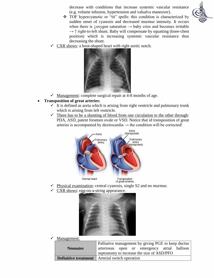
Transposition of great arteries:
It is defined as aorta which is arising from right ventricle and pulmonary trunk
which is arising from left ventricle.
There has to be a shunting of blood from one circulation to the other through:
PDA, ASD, patent foramen ovale or VSD. Notice that id transposition of great
arteries is accompanied by dextrocardia → the condition will be corrected!
Physical examination: central cyanosis, single S2 and no murmur.
CXR shows: egg-on-a-string appearance.
Management:
Neonates
Palliative management by giving PGE to keep ductus
arteriosus open or emergency atrial balloon
septostomy to increase the size of ASD/PFO
Definitive treatment Arterial switch operation

Tricuspid atresia:
It is characterized by the presence of a plate of tissue in the floor of the right
atrium instead of tricuspid valve. An ASD or Patent Foramen Ovale (PFO) is
always present.
Pathophysiology:
VSD NOT present
Pulmonary atresia will occur because there is no
blood reaching the right ventricle and flowing
through the pulmonary trunk.
A PDA must be present to allow flow of blood
through pulmonary arteries to the lung. As it
closes after birth, visible cyanosis will occur
Patients have single S2 and no murmur
VSD PRESENT
Blood will flow from left ventricle to the right
ventricle through the VSD and subsequently to the
lungs through pulmonary trunk. Therefore,
providing adequate saturation.
Patients have holosystolic murmur at lower-left
sterna borders.
Management: Fontan procedure (3-6 years) in which blood flow from IVC
will be directed directly into pulmonary trunk.
Truncus arteriosus:
It is a congenital cyanotic heart disease in
which there is no septum separating the
aorta from pulmonary trunk and they both
appear as one large vessel (the truncus).
Therefore, oxygenated and deoxygenated
blood will mix together and there will be
excessive blood flow to the lungs
eventually resulting in signs and
symptoms of CHF.
Physical examination: systolic ejection murmur along the left sternal border;
single S2; diastolic murmur at the apex (resulting from excessive pulmonary
blood flow going back to the left atrium through the mitral valve).
CXR shows: enlarged heart, right aortic notch and increased pulmonary
vascular markings.
Management: treatment of CHF with cardiac glycosides and loop diuretics;
closure of VSD and homograft placement between right ventricle and
pulmonary trunk.
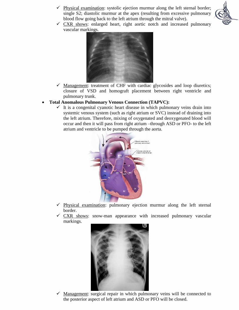
Total Anomalous Pulmonary Venous Connection (TAPVC):
It is a congenital cyanotic heart disease in which pulmonary veins drain into
systemic venous system (such as right atrium or SVC) instead of draining into
the left atrium. Therefore, mixing of oxygenated and deoxygenated blood will
occur and then it will pass from right atrium –through ASD or PFO- to the left
atrium and ventricle to be pumped through the aorta.
Physical examination: pulmonary ejection murmur along the left sternal
border.
CXR shows: snow-man appearance with increased pulmonary vascular
markings.
Management: surgical repair in which pulmonary veins will be connected to
the posterior aspect of left atrium and ASD or PFO will be closed.

- Acquired heart disease:
Infective endocarditis:
It is a microbial infection of the internal surface of the heart.
In 80% of cases → there is a structural abnormality of the heart.
In 50% of cases → it occurs after cardiac surgery.
Causative organisms: α-hemolytic streptococcus (Streptococcus viridians) and
Staphylococcus species. The organism will be introduced to the blood and
then affect the injured cardiac endothelium. Subsequently, fibrin and platelets
will adhere creating vegetations that might cause valve incompetency and
embolic phenomena.
Clinical features:
Fever The most common symptom
Splinter
hemorrhages
Linear hemorrhages beneath the nails
Osler’s nodes Small, pink, swollen, tender lesions on palms or soles
Janewy lesions Small, erythematous hemorrhagic lesions on palms or soles
Roth’s spots Round white spots seen in the retina
Investigations: blood culture (most important); ↑ESR; ↑RF (50% of patients);
transesophageal echocardiography (more sensitive in detecting vegetations).
Management: IV antibiotics directed against the identified organism (for 4-6
weeks). Antibiotic prophylaxis is given for:
All patients with structural heart abnormalities (except those with
secundum ASD).
All patients after having a cardiac surgery (for 6 months after the
repair).
Pericarditis:
It is inflammation of pericardial space.
Causes:
Infection
Viral (most common): coxsackievirus or EBV
Purulent pericarditis = bacterial infection:
S.aureus or S.pneumoniae
Collagen vascular disease SLE
Uremia -
Postpericardiotomy
syndrome Inflammatory response after cardiac surgery
Pathophysiology: inflammation of
pericardial layers (visceral and parietal)
will result in transudation/exudation of
fluid into the pericardial space that
leads to reduced venous return and
filling of the heart. Cardiac temponade
may occur (fluid building up in
pericardium resulting in cardiac
compression).

Clinical features:
Symptoms Fever, dyspnea and chest pain (which becomes more severe in
supine position and relieved when sitting upright)
Physical
examination
Pericardial friction rub, distal heart sounds (if effusion is
large), pulses paradoxus (< 10 mmHg reduction in systolic
blood pressure with deep inspiration) and hepatomegaly
Diagnosis:
Pericardiocentesis A needle is inserted into the pericardial space and fluid
is aspirated (this is both diagnostic and therapeutic)
ECG Low-voltage QRS complex in patients with large
pericardial effusion
CXR Enlarged cardiac shadow with large effusion
ECHO Demonstrates extent and quality of pericardial effusion
Management:
Bacterial pericarditis Appropriate antibiotics
Viral pericarditis Anti-inflammatory agents (e.g. aspirin)
Myocarditis:
It is inflammation of myocardium which is characterized by: cellular infiltrate
and myocardial cell death. It is one of several common causes of sudden death
in young athletes.
Causes:
Viruses Coxsackievirus
Bacteria S.aureus or S.pyogens
Fungi Candida or Cryptococcus
Protozoa T.cruzi (Chagas’ disease)
Autoimmune SLE
Kawasaki disease -
Clinical features:
It is usually preceded by viral or flu-like illness.
There is dyspnea and malaise.
Physical examination shows: resting tachycardia, muffled heart
sounds, gallop heart rhythm and hepatomegaly.
Investigation:
↑ESR, ↑CRP, ↑creatinine kinase MB fraction.
Endomyocardial biopsy can be obtained with PCR to detect the virus.
ECHO: global ventricular dynsfunction.
Management: supportive with treatment of CHF.
- Cardiomyopathies:
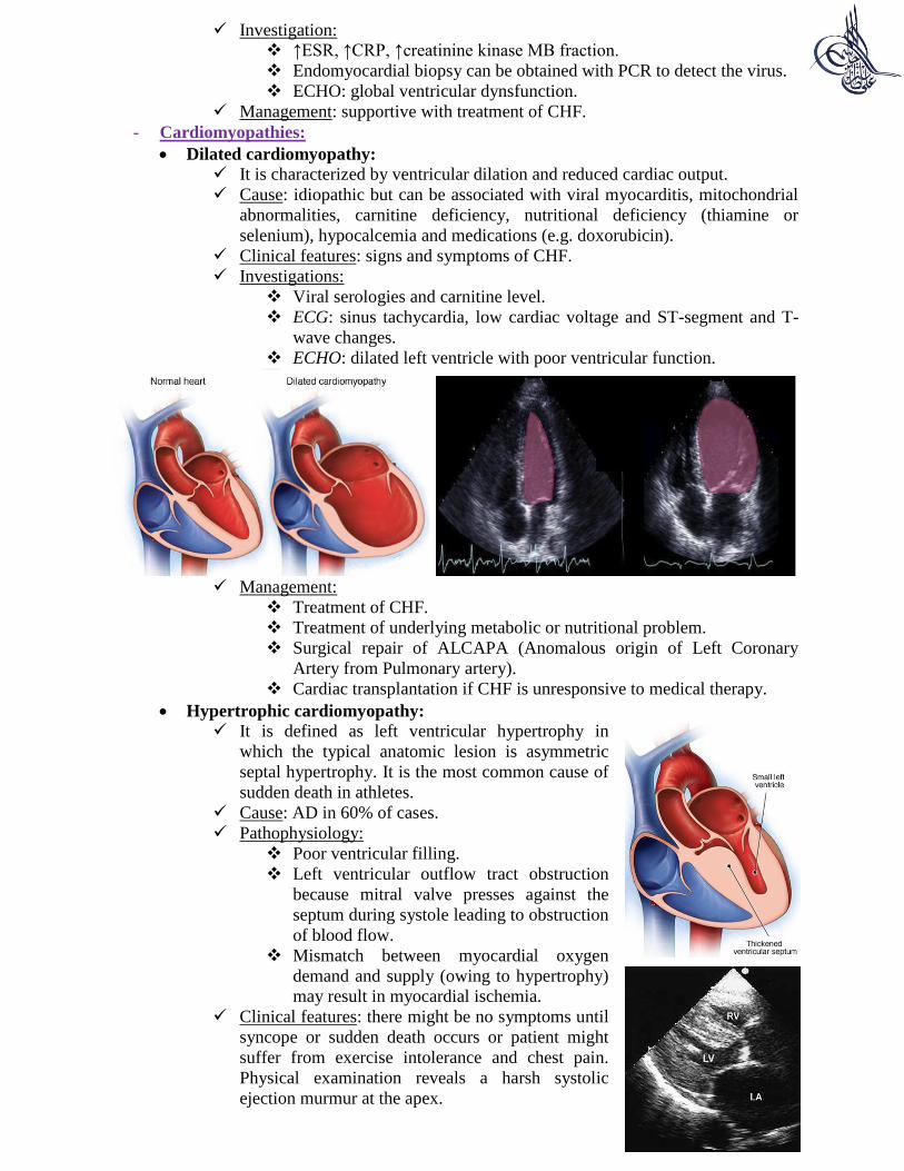
Dilated cardiomyopathy:
It is characterized by ventricular dilation and reduced cardiac output.
Cause: idiopathic but can be associated with viral myocarditis, mitochondrial
abnormalities, carnitine deficiency, nutritional deficiency (thiamine or
selenium), hypocalcemia and medications (e.g. doxorubicin).
Clinical features: signs and symptoms of CHF.
Investigations:
Viral serologies and carnitine level.
ECG: sinus tachycardia, low cardiac voltage and ST-segment and T-
wave changes.
ECHO: dilated left ventricle with poor ventricular function.
Management:
Treatment of CHF.
Treatment of underlying metabolic or nutritional problem.
Surgical repair of ALCAPA (Anomalous origin of Left Coronary
Artery from Pulmonary artery).
Cardiac transplantation if CHF is unresponsive to medical therapy.
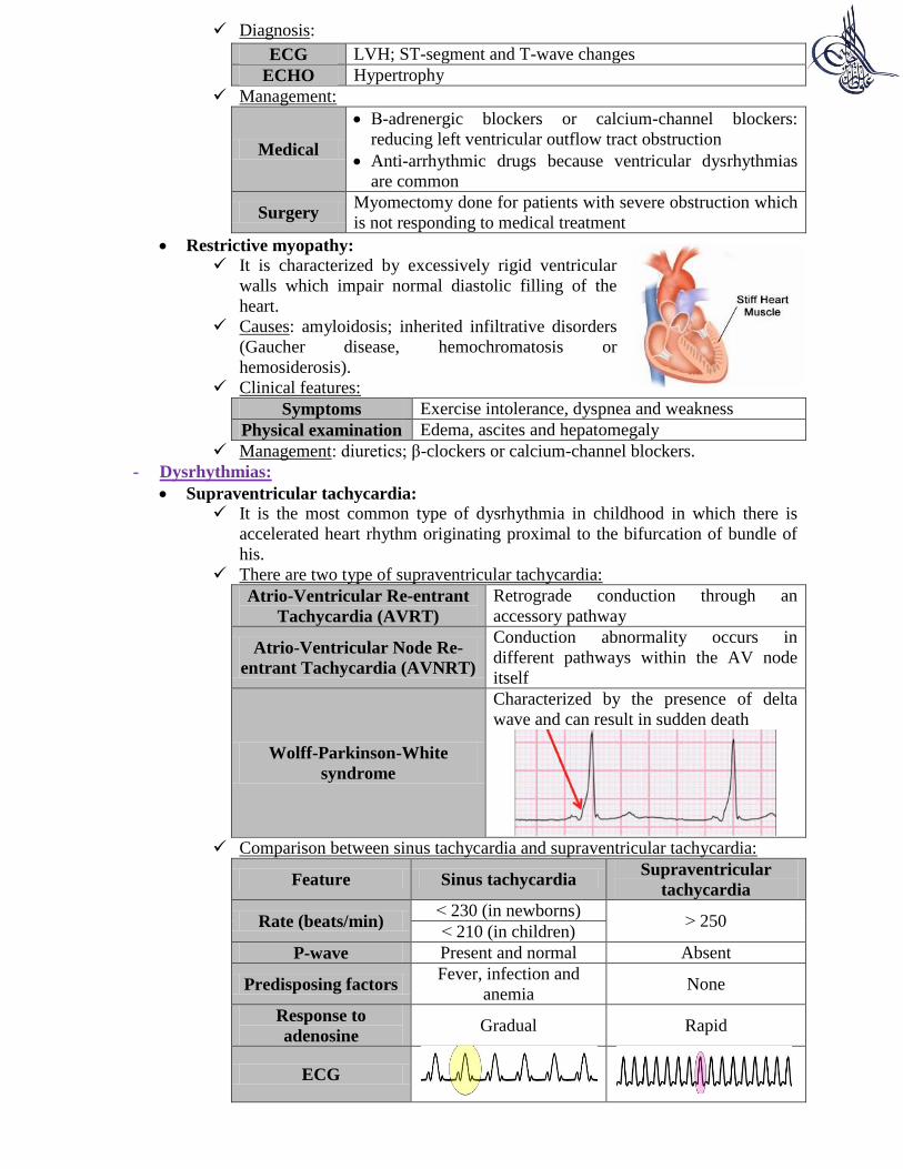
Hypertrophic cardiomyopathy:
It is defined as left ventricular hypertrophy in
which the typical anatomic lesion is asymmetric
septal hypertrophy. It is the most common cause of
sudden death in athletes.
Cause: AD in 60% of cases.
Pathophysiology: Poor ventricular filling.
Left ventricular outflow tract obstruction
because mitral valve presses against the
septum during systole leading to obstruction
of blood flow.
Mismatch between myocardial oxygen
demand and supply (owing to hypertrophy)
may result in myocardial ischemia.
Clinical features: there might be no symptoms until
syncope or sudden death occurs or patient might
suffer from exercise intolerance and chest pain.
Physical examination reveals a harsh systolic
ejection murmur at the apex.
Diagnosis:
ECG LVH; ST-segment and T-wave changes
ECHO Hypertrophy
Management:
Medical
Β-adrenergic blockers or calcium-channel blockers:
reducing left ventricular outflow tract obstruction
Anti-arrhythmic drugs because ventricular dysrhythmias
are common
Surgery Myomectomy done for patients with severe obstruction which
is not responding to medical treatment
Restrictive myopathy:
It is characterized by excessively rigid ventricular
walls which impair normal diastolic filling of the
heart. Causes: amyloidosis; inherited infiltrative disorders
(Gaucher disease, hemochromatosis or
hemosiderosis).
Clinical features:
Symptoms Exercise intolerance, dyspnea and weakness
Physical examination Edema, ascites and hepatomegaly
Management: diuretics; β-clockers or calcium-channel blockers.
- Dysrhythmias:
Supraventricular tachycardia:
It is the most common type of dysrhythmia in childhood in which there is
accelerated heart rhythm originating proximal to the bifurcation of bundle of
his.
There are two type of supraventricular tachycardia:
Atrio-Ventricular Re-entrant
Tachycardia (AVRT)
Retrograde conduction through an
accessory pathway
Atrio-Ventricular Node Re-
entrant Tachycardia (AVNRT)
Conduction abnormality occurs in
different pathways within the AV node
itself
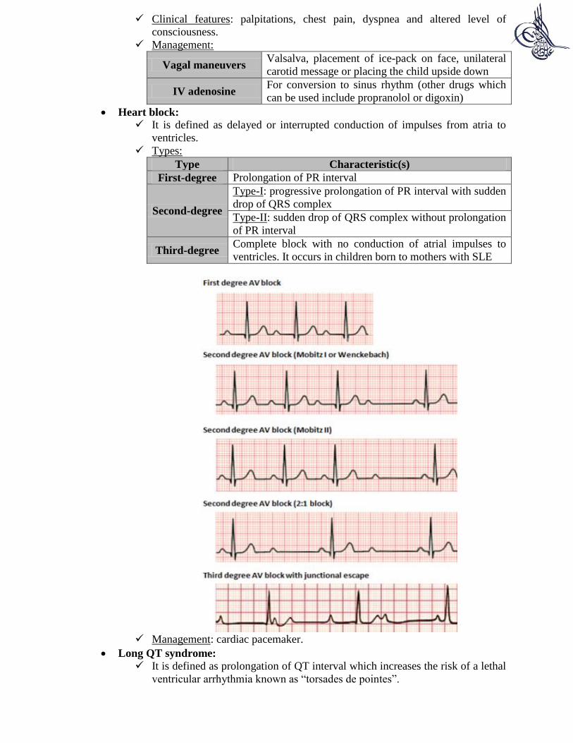
Wolff-Parkinson-White
syndrome
Characterized by the presence of delta
wave and can result in sudden death
Comparison between sinus tachycardia and supraventricular tachycardia:
Feature Sinus tachycardia Supraventricular
tachycardia
Rate (beats/min) > 230 (in newborns)
< 250 > 210 (in children)
P-wave Present and normal Absent
Predisposing factors Fever, infection and
anemia None
Response to
adenosine Gradual Rapid
ECG
Clinical features: palpitations, chest pain, dyspnea and altered level of
consciousness.
Management:
Vagal maneuvers Valsalva, placement of ice-pack on face, unilateral
carotid message or placing the child upside down
IV adenosine For conversion to sinus rhythm (other drugs which
can be used include propranolol or digoxin)
Heart block:
It is defined as delayed or interrupted conduction of impulses from atria to
ventricles.
Types:
Type Characteristic(s)
First-degree Prolongation of PR interval
Second-degree
Type-I: progressive prolongation of PR interval with sudden
drop of QRS complex
Type-II: sudden drop of QRS complex without prolongation
of PR interval
Third-degree Complete block with no conduction of atrial impulses to
ventricles. It occurs in children born to mothers with SLE
Management: cardiac pacemaker.
Long QT syndrome:
It is defined as prolongation of QT interval which increases the risk of a lethal
ventricular arrhythmia known as “torsades de pointes”.

Causes:
Inheritance
(50% of
cases)
AD (Romano-Ward syndrome): not associated with deafness
AR (Jervell-Lange-Nielsen syndrome): associated with
deafness
Drugs Tricyclic antidepressants or erythromycin
Idiopathic
Clinical features: syncope (most common) or sudden cardiac arrest.
Management: β-blockers