Embed Size (px)
Citation preview
Hematopoietic and Lymphoid Neoplasms 8/2/2012
NAACCR 2011‐2012 Webinar Series 1
Collecting Cancer Data: Hematopoietic & Lymphoid Neoplasms
NAACCR 2011‐2012 Webinar SeriesAugust 2, 2012
1
Q&A• Please submit all questions concerning webinar content through the Q&A panel.
Reminder:• If you have participants watching this webinar at your site, please collect their names and emails.– We will be distributing a Q&A document in about one week. This document will fully answer questions asked during the webinar and will contain any corrections that we may discover after the webinar.
2
Fabulous Prizes
3
Hematopoietic and Lymphoid Neoplasms 8/2/2012
NAACCR 2011‐2012 Webinar Series 2
Agenda• Overview• Hematopoietic Database and Manual• Collaborative Stage Collection System V02.04• Treatment
4
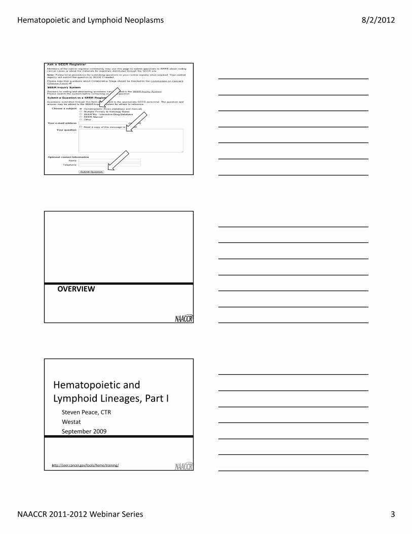
Ask a SEER Registrar• All questions about hematopoietic and lymphoid neoplasms concerning:– Multiple primaries– Primary site– Reportability– Grade– Any other issues related to the Hematopoietic and Lymphoid neoplasms Database and Manual
• Should go to Ask a SEER Registrar– http://seer.cancer.gov/seerinquiry/index.php?page=search
Hematopoietic and Lymphoid Neoplasms 8/2/2012
NAACCR 2011‐2012 Webinar Series 3
OVERVIEW
Hematopoietic and Lymphoid Lineages, Part I
Steven Peace, CTRWestatSeptember 2009
9http://seer.cancer.gov/tools/heme/training/
Hematopoietic and Lymphoid Neoplasms 8/2/2012
NAACCR 2011‐2012 Webinar Series 4
10
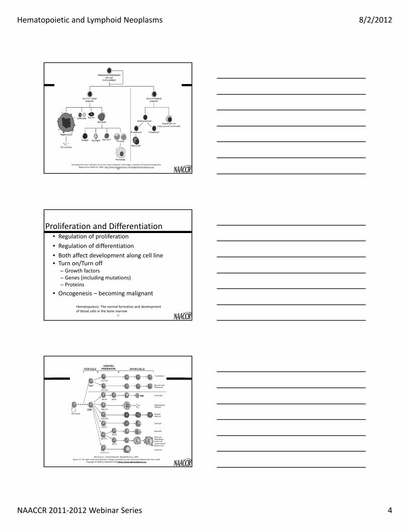
Hematopoietic stem cells give rise to two major progenitor cell lineages, myeloid and lymphoid progenitors Regenerative Medicine, 2006. http://www.dentalarticles.com/images/hematopoiesis.png
Proliferation and Differentiation• Regulation of proliferation• Regulation of differentiation• Both affect development along cell line• Turn on/Turn off
– Growth factors– Genes (including mutations)– Proteins
• Oncogenesis – becoming malignant
11
Hematopoiesis: The normal formation and development of blood cells in the bone marrow
12
Blood Lines – Donald Metcalf, AlphaMED Press, 2005Figure 3.2 The eight major hematopoietic lineages generated by self‐renewing multipotential stem cellsB
Copyright © 2008 by AlphaMed Press http://www.alphamedpress.org
Hematopoietic and Lymphoid Neoplasms 8/2/2012
NAACCR 2011‐2012 Webinar Series 5
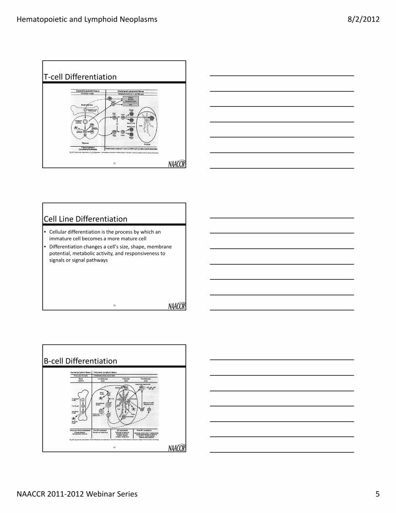
T‐cell Differentiation
13
Cell Line Differentiation• Cellular differentiation is the process by which an immature cell becomes a more mature cell
• Differentiation changes a cell's size, shape, membrane potential, metabolic activity, and responsiveness to signals or signal pathways
14
B‐cell Differentiation
15
Hematopoietic and Lymphoid Neoplasms 8/2/2012
NAACCR 2011‐2012 Webinar Series 6
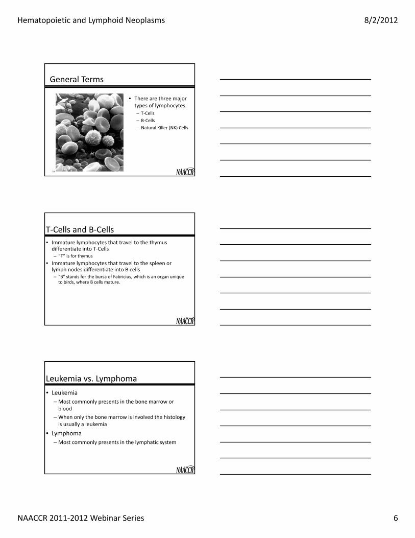
General Terms
• There are three major types of lymphocytes.– T‐Cells– B‐Cells– Natural Killer (NK) Cells
16
T‐Cells and B‐Cells• Immature lymphocytes that travel to the thymus differentiate into T‐Cells– “T” is for thymus
• Immature lymphocytes that travel to the spleen or lymph nodes differentiate into B cells– "B" stands for the bursa of Fabricius, which is an organ unique to birds, where B cells mature.
Leukemia vs. Lymphoma• Leukemia
– Most commonly presents in the bone marrow or blood
– When only the bone marrow is involved the histology is usually a leukemia
• Lymphoma– Most commonly presents in the lymphatic system
Hematopoietic and Lymphoid Neoplasms 8/2/2012
NAACCR 2011‐2012 Webinar Series 7
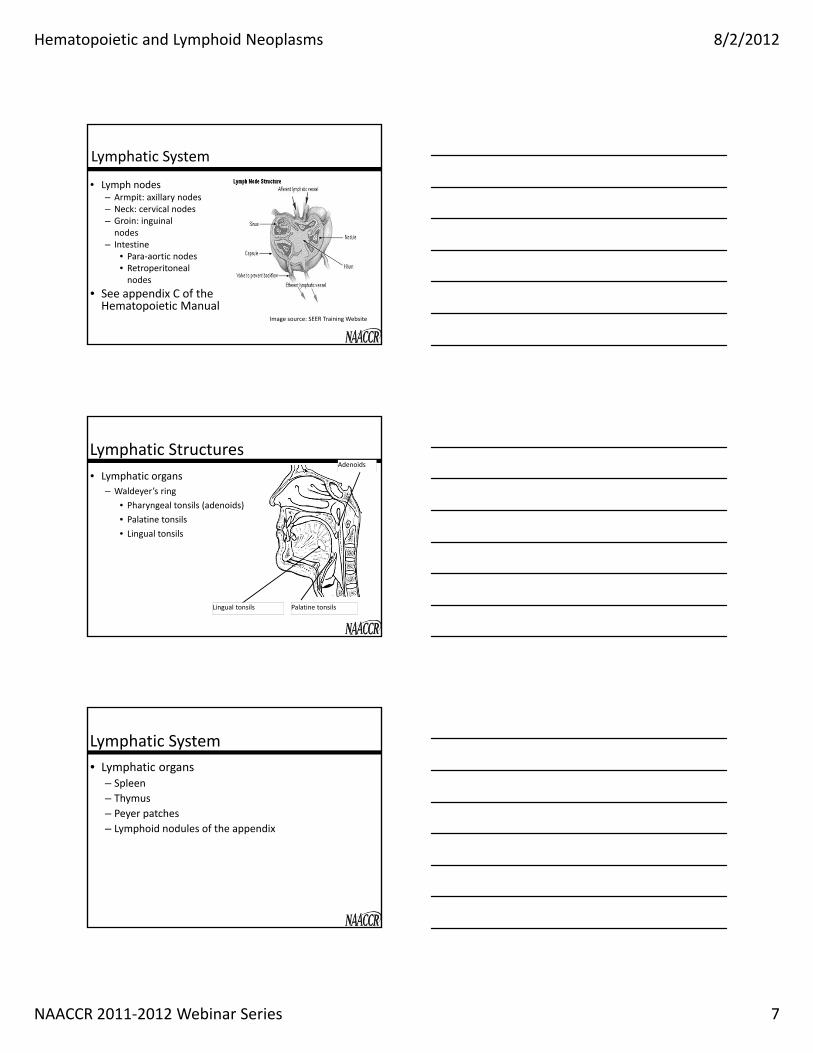
Lymphatic System
• Lymph nodes– Armpit: axillary nodes– Neck: cervical nodes– Groin: inguinal nodes
– Intestine• Para‐aortic nodes• Retroperitoneal nodes
• See appendix C of the Hematopoietic Manual
Image source: SEER Training Website
Lymphatic Structures• Lymphatic organs
– Waldeyer’s ring• Pharyngeal tonsils (adenoids)• Palatine tonsils • Lingual tonsils
Lingual tonsils
Adenoids
Palatine tonsils
Lymphatic System• Lymphatic organs
– Spleen– Thymus– Peyer patches– Lymphoid nodules of the appendix
Hematopoietic and Lymphoid Neoplasms 8/2/2012
NAACCR 2011‐2012 Webinar Series 8
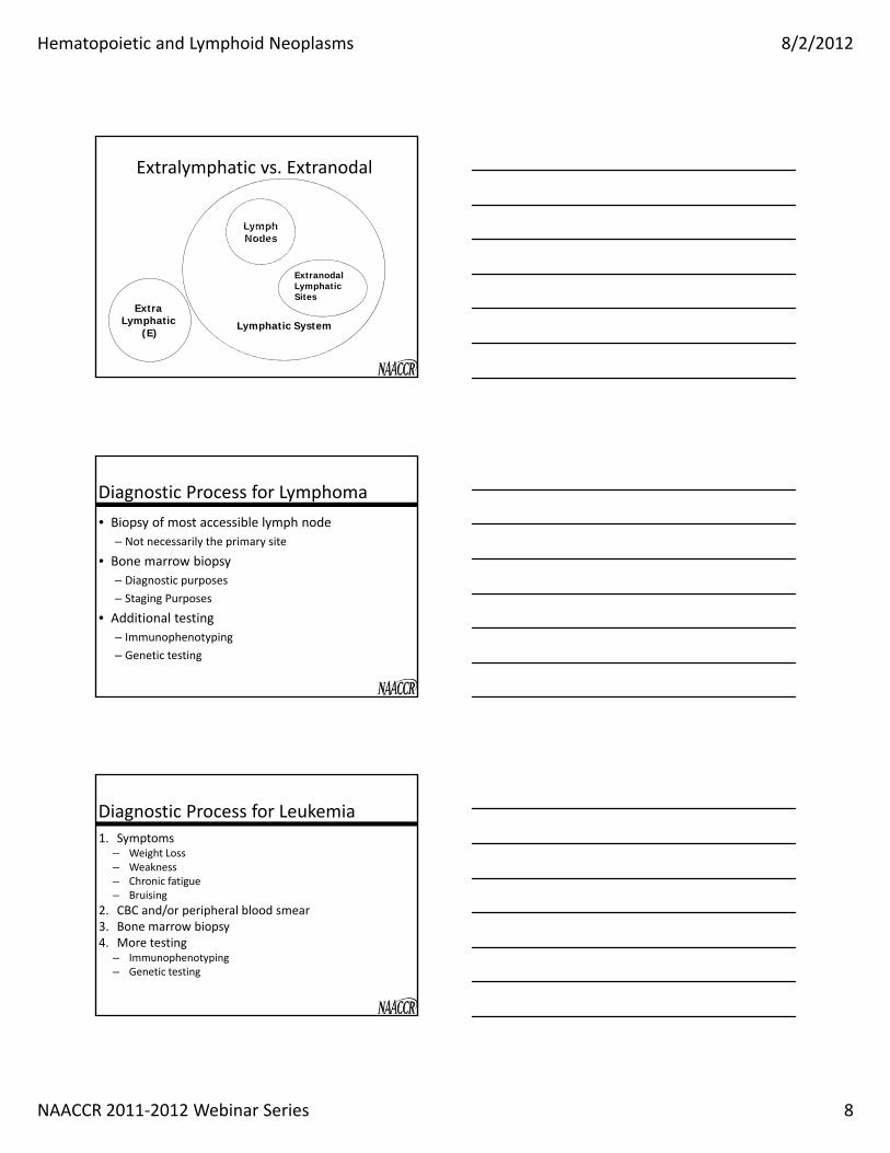
Extralymphatic vs. Extranodal
LymphNodes
Lymphatic System
Extra Lymphatic
(E)
ExtranodalLymphaticSites
Diagnostic Process for Lymphoma• Biopsy of most accessible lymph node
– Not necessarily the primary site
• Bone marrow biopsy – Diagnostic purposes – Staging Purposes
• Additional testing– Immunophenotyping– Genetic testing
Diagnostic Process for Leukemia1. Symptoms
– Weight Loss– Weakness– Chronic fatigue– Bruising
2. CBC and/or peripheral blood smear3. Bone marrow biopsy4. More testing
– Immunophenotyping– Genetic testing
Hematopoietic and Lymphoid Neoplasms 8/2/2012
NAACCR 2011‐2012 Webinar Series 9
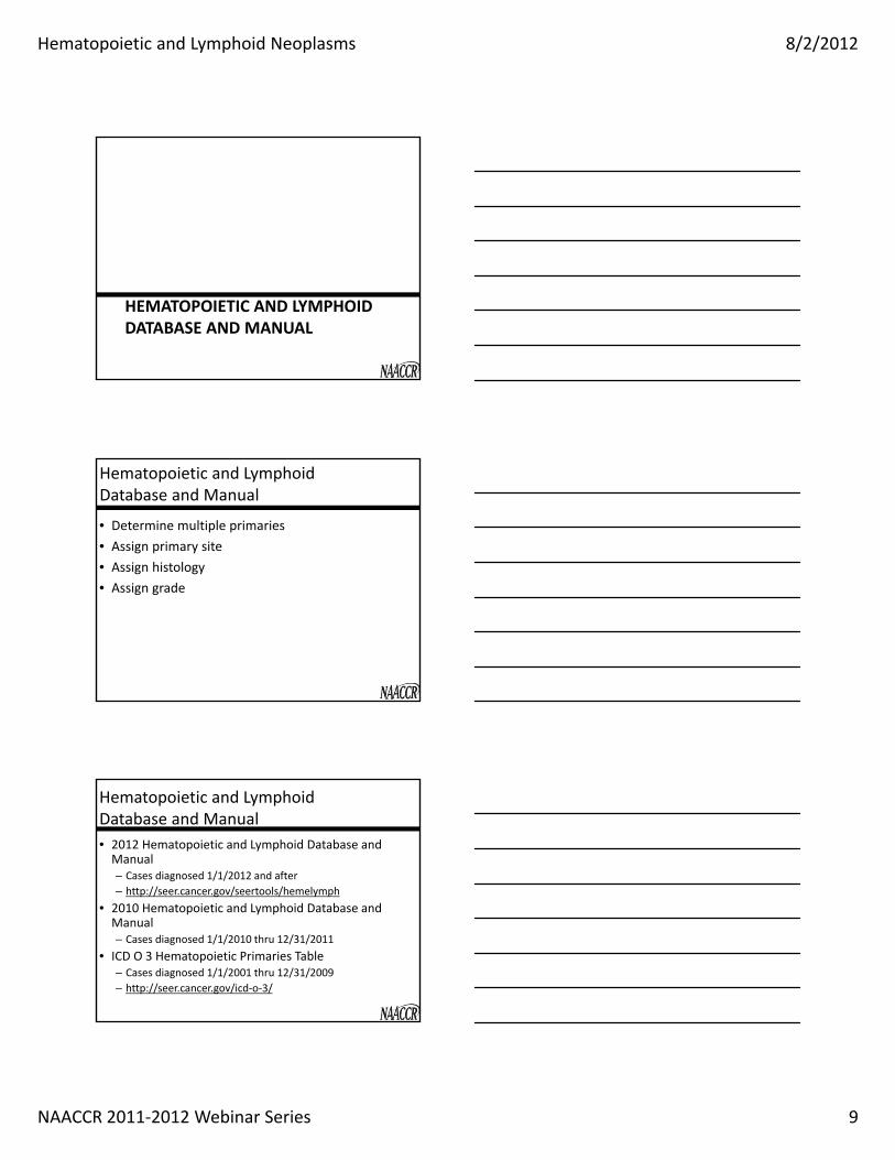
HEMATOPOIETIC AND LYMPHOID DATABASE AND MANUAL
Hematopoietic and Lymphoid Database and Manual
• Determine multiple primaries• Assign primary site• Assign histology• Assign grade
Hematopoietic and Lymphoid Database and Manual• 2012 Hematopoietic and Lymphoid Database and Manual– Cases diagnosed 1/1/2012 and after– http://seer.cancer.gov/seertools/hemelymph
• 2010 Hematopoietic and Lymphoid Database and Manual– Cases diagnosed 1/1/2010 thru 12/31/2011
• ICD O 3 Hematopoietic Primaries Table– Cases diagnosed 1/1/2001 thru 12/31/2009– http://seer.cancer.gov/icd‐o‐3/
Hematopoietic and Lymphoid Neoplasms 8/2/2012
NAACCR 2011‐2012 Webinar Series 10
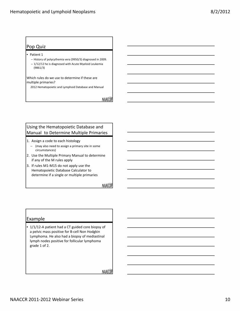
Pop Quiz• Patient 1
– History of polycythemia vera (9950/3) diagnosed in 2009. – 1/12/12 he is diagnosed with Acute Myeloid Leukemia (9861/3)
Which rules do we use to determine if these are multiple primaries?
2012 Hematopoietic and Lymphoid Database and Manual
Using the Hematopoietic Database and Manual to Determine Multiple Primaries
1. Assign a code to each histology – (may also need to assign a primary site in some
circumstances)
2. Use the Multiple Primary Manual to determine if any of the M rules apply
3. If rules M1‐M15 do not apply use the Hematopoietic Database Calculator to determine if a single or multiple primaries
Example• 1/1/12‐A patient had a CT guided core biopsy of a pelvic mass positive for B‐cell Non Hodgkin Lymphoma. He also had a biopsy of mediastinal lymph nodes positive for follicular lymphoma grade 1 of 2.
Hematopoietic and Lymphoid Neoplasms 8/2/2012
NAACCR 2011‐2012 Webinar Series 11
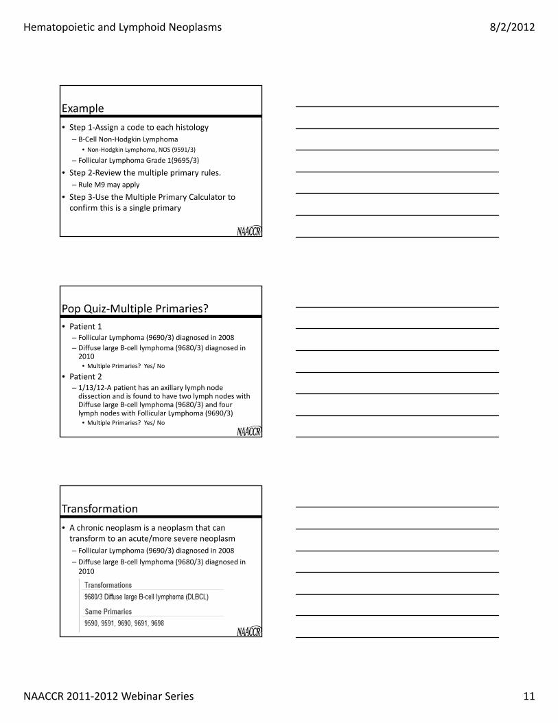
Example• Step 1‐Assign a code to each histology
– B‐Cell Non‐Hodgkin Lymphoma • Non‐Hodgkin Lymphoma, NOS (9591/3)
– Follicular Lymphoma Grade 1(9695/3)
• Step 2‐Review the multiple primary rules. – Rule M9 may apply
• Step 3‐Use the Multiple Primary Calculator to confirm this is a single primary
Pop Quiz‐Multiple Primaries?• Patient 1
– Follicular Lymphoma (9690/3) diagnosed in 2008– Diffuse large B‐cell lymphoma (9680/3) diagnosed in 2010
• Multiple Primaries? Yes/ No
• Patient 2– 1/13/12‐A patient has an axillary lymph node dissection and is found to have two lymph nodes withDiffuse large B‐cell lymphoma (9680/3) and four lymph nodes with Follicular Lymphoma (9690/3)
• Multiple Primaries? Yes/ No
Transformation• A chronic neoplasm is a neoplasm that can transform to an acute/more severe neoplasm– Follicular Lymphoma (9690/3) diagnosed in 2008– Diffuse large B‐cell lymphoma (9680/3) diagnosed in 2010
Hematopoietic and Lymphoid Neoplasms 8/2/2012
NAACCR 2011‐2012 Webinar Series 12
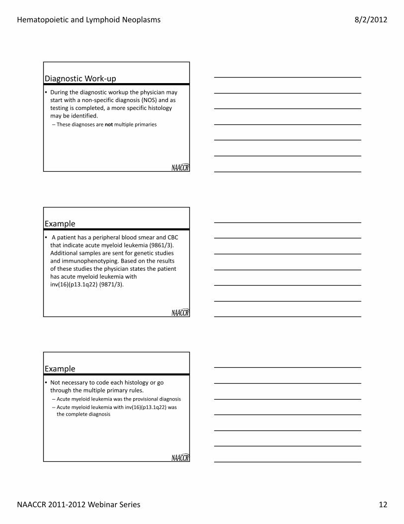
Diagnostic Work‐up• During the diagnostic workup the physician may start with a non‐specific diagnosis (NOS) and as testing is completed, a more specific histology may be identified. – These diagnoses are notmultiple primaries
Example• A patient has a peripheral blood smear and CBC that indicate acute myeloid leukemia (9861/3). Additional samples are sent for genetic studies and immunophenotyping. Based on the results of these studies the physician states the patient has acute myeloid leukemia with inv(16)(p13.1q22) (9871/3).
Example• Not necessary to code each histology or go through the multiple primary rules.– Acute myeloid leukemia was the provisional diagnosis– Acute myeloid leukemia with inv(16)(p13.1q22) was the complete diagnosis
Hematopoietic and Lymphoid Neoplasms 8/2/2012
NAACCR 2011‐2012 Webinar Series 13
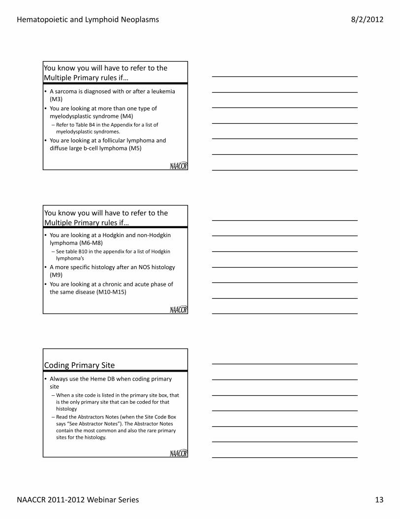
You know you will have to refer to the Multiple Primary rules if…
• A sarcoma is diagnosed with or after a leukemia (M3)
• You are looking at more than one type of myelodysplastic syndrome (M4)– Refer to Table B4 in the Appendix for a list of myelodysplastic syndromes.
• You are looking at a follicular lymphoma and diffuse large b‐cell lymphoma (M5)
You know you will have to refer to the Multiple Primary rules if…• You are looking at a Hodgkin and non‐Hodgkin lymphoma (M6‐M8)– See table B10 in the appendix for a list of Hodgkin lymphoma’s
• A more specific histology after an NOS histology (M9)
• You are looking at a chronic and acute phase of the same disease (M10‐M15)
Coding Primary Site• Always use the Heme DB when coding primary site– When a site code is listed in the primary site box, that is the only primary site that can be coded for that histology
– Read the Abstractors Notes (when the Site Code Box says “See Abstractor Notes”). The Abstractor Notes contain the most common and also the rare primary sites for the histology.
Hematopoietic and Lymphoid Neoplasms 8/2/2012
NAACCR 2011‐2012 Webinar Series 14
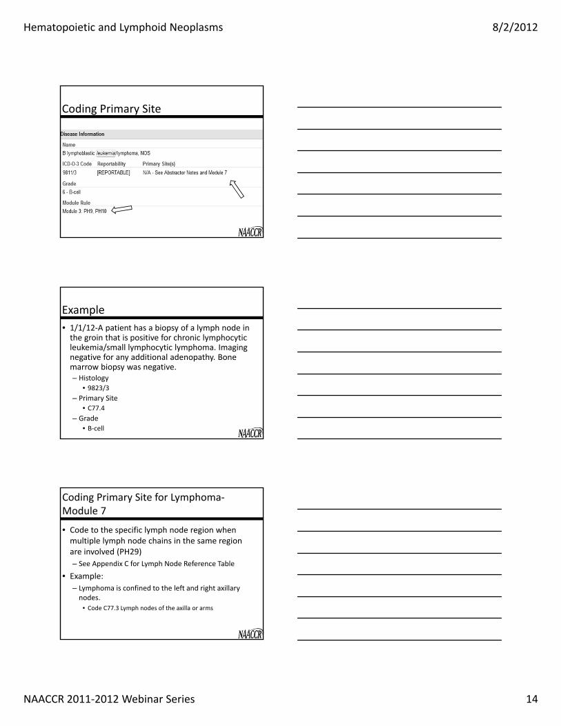
Coding Primary Site
Example• 1/1/12‐A patient has a biopsy of a lymph node in the groin that is positive for chronic lymphocytic leukemia/small lymphocytic lymphoma. Imaging negative for any additional adenopathy. Bone marrow biopsy was negative.– Histology
• 9823/3– Primary Site
• C77.4– Grade
• B‐cell
Coding Primary Site for Lymphoma‐Module 7
• Code to the specific lymph node region when multiple lymph node chains in the same region are involved (PH29)– See Appendix C for Lymph Node Reference Table
• Example:– Lymphoma is confined to the left and right axillary nodes.
• Code C77.3 Lymph nodes of the axilla or arms
Hematopoietic and Lymphoid Neoplasms 8/2/2012
NAACCR 2011‐2012 Webinar Series 15
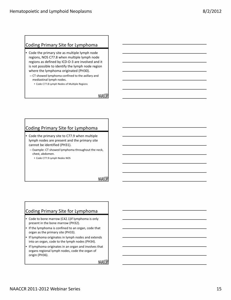
Coding Primary Site for Lymphoma• Code the primary site as multiple lymph node regions, NOS C77.8 when multiple lymph node regions as defined by ICD‐O‐3 are involved and it is not possible to identify the lymph node region where the lymphoma originated (PH30).– CT showed lymphoma confined to the axillary and mediastinal lymph nodes.
• Code C77.8 Lymph Nodes of Multiple Regions
Coding Primary Site for Lymphoma• Code the primary site to C77.9 when multiple lymph nodes are present and the primary site cannot be identified (PH31).– Example: CT showed lymphoma throughout the neck, chest, abdomen.
• Code C77.9 Lymph Nodes NOS
Coding Primary Site for Lymphoma• Code to bone marrow (C42.1)if lymphoma is only present in the bone marrow (PH32).
• If the lymphoma is confined to an organ, code that organ as the primary site (PH33).
• If lymphoma originates in lymph nodes and extends into an organ, code to the lymph nodes (PH34).
• If lymphoma originates in an organ and involves that organs regional lymph nodes, code the organ of origin (PH36).
Hematopoietic and Lymphoid Neoplasms 8/2/2012
NAACCR 2011‐2012 Webinar Series 16
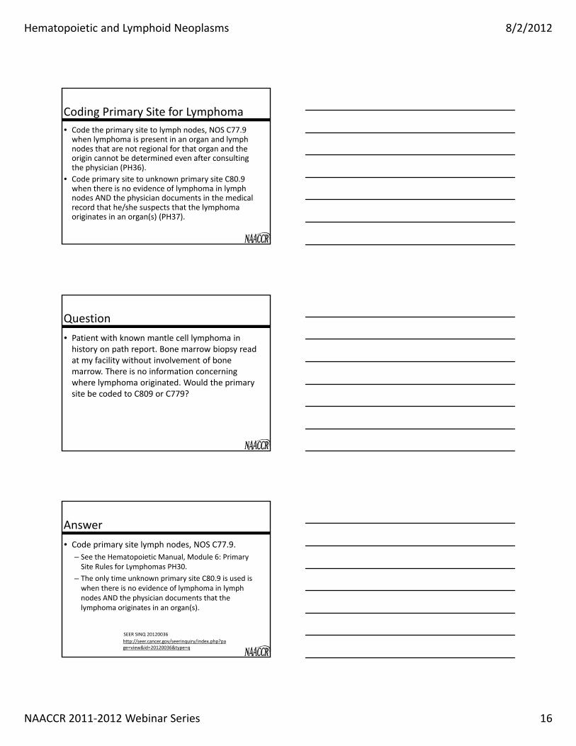
Coding Primary Site for Lymphoma• Code the primary site to lymph nodes, NOS C77.9 when lymphoma is present in an organ and lymph nodes that are not regional for that organ and the origin cannot be determined even after consulting the physician (PH36).
• Code primary site to unknown primary site C80.9 when there is no evidence of lymphoma in lymph nodes AND the physician documents in the medical record that he/she suspects that the lymphoma originates in an organ(s) (PH37).
Question• Patient with known mantle cell lymphoma in history on path report. Bone marrow biopsy read at my facility without involvement of bone marrow. There is no information concerning where lymphoma originated. Would the primary site be coded to C809 or C779?
Answer• Code primary site lymph nodes, NOS C77.9.
– See the Hematopoietic Manual, Module 6: Primary Site Rules for Lymphomas PH30.
– The only time unknown primary site C80.9 is used is when there is no evidence of lymphoma in lymph nodes AND the physician documents that the lymphoma originates in an organ(s).
http://seer.cancer.gov/seerinquiry/index.php?page=view&id=20120036&type=q
SEER SINQ 20120036
Hematopoietic and Lymphoid Neoplasms 8/2/2012
NAACCR 2011‐2012 Webinar Series 17
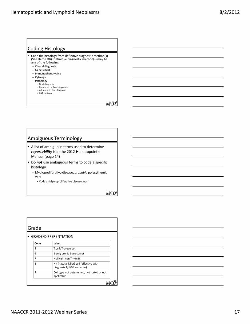
Coding Histology• Code the histology from definitive diagnostic method(s) (See Heme DB). Definitive diagnostic method(s) may be any of the following– Clinical diagnosis– Genetic test– Immunophenotyping– Cytology– Pathology
• Final diagnosis• Comment on final diagnosis• Addenda to final diagnosis• CAP protocol
Ambiguous Terminology• A list of ambiguous terms used to determine reportability is in the 2012 Hematopoietic Manual (page 14)
• Do not use ambiguous terms to code a specific histology.– Myeloproliferative disease, probably polycythemia vera
• Code as Myeloproliferative disease, nos
Grade• GRADE/DIFFERENTIATION
Code Label
5 T cell; T‐precursor
6 B cell; pre‐B; B‐precursor
7 Null cell; non T‐non B
8 NK (natural killer) cell (effective with diagnosis 1/1/95 and after)
9 Cell type not determined, not stated or not applicable
Hematopoietic and Lymphoid Neoplasms 8/2/2012
NAACCR 2011‐2012 Webinar Series 18
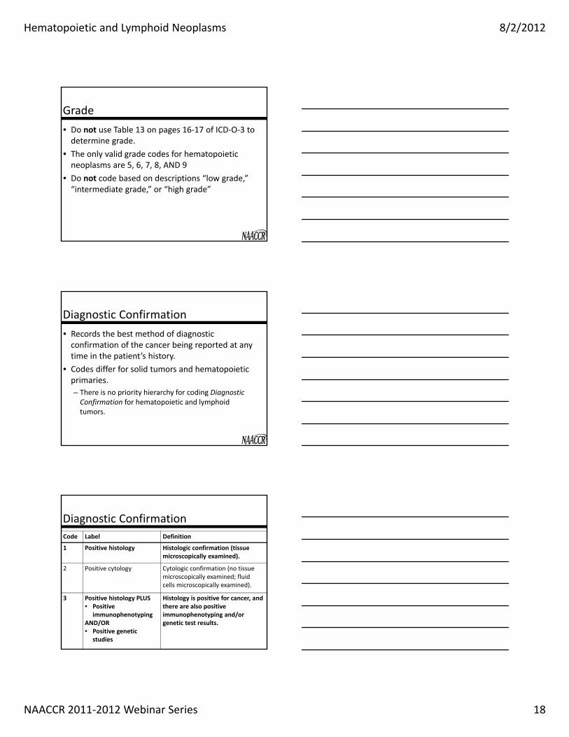
Grade• Do not use Table 13 on pages 16‐17 of ICD‐O‐3 to determine grade.
• The only valid grade codes for hematopoietic neoplasms are 5, 6, 7, 8, AND 9
• Do not code based on descriptions “low grade,” “intermediate grade,” or “high grade”
Diagnostic Confirmation• Records the best method of diagnostic confirmation of the cancer being reported at any time in the patient’s history.
• Codes differ for solid tumors and hematopoietic primaries.– There is no priority hierarchy for coding Diagnostic Confirmation for hematopoietic and lymphoid tumors.
Diagnostic ConfirmationCode Label Definition
1 Positive histology Histologic confirmation (tissue microscopically examined).
2 Positive cytology Cytologic confirmation (no tissue microscopically examined; fluidcells microscopically examined).
3 Positive histology PLUS• Positive
immunophenotypingAND/OR• Positive genetic
studies
Histology is positive for cancer, and there are also positiveimmunophenotyping and/or genetic test results.
Hematopoietic and Lymphoid Neoplasms 8/2/2012
NAACCR 2011‐2012 Webinar Series 19
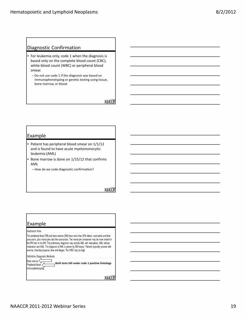
Diagnostic Confirmation• For leukemia only, code 1 when the diagnosis is based only on the complete blood count (CBC), white blood count (WBC) or peripheral blood smear. – Do not use code 1 if the diagnosis was based on immunophenotyping or genetic testing using tissue, bone marrow, or blood.
Example• Patient has peripheral blood smear on 1/1/12 and is found to have acute myelomonocytic leukemia (AML)
• Bone marrow is done on 1/15/12 that confirms AML– How do we code diagnostic confirmation?
Example
Both tests fall under code 1 positive histology
Hematopoietic and Lymphoid Neoplasms 8/2/2012
NAACCR 2011‐2012 Webinar Series 20
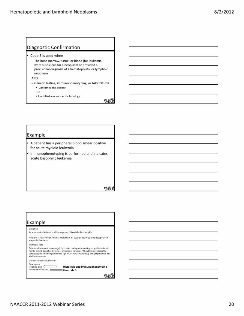
Diagnostic Confirmation• Code 3 is used when
– The bone marrow, tissue, or blood (for leukemia) were suspicious for a neoplasm or provided a provisional diagnosis of a hematopoietic or lymphoid neoplasm
AND– Genetic testing, immunophenotyping, or JAK2 EITHER
• Confirmed the diseaseOR• Identified a more specific histology
Example• A patient has a peripheral blood smear positive for acute myeloid leukemia
• Immunophenotyping is performed and indicates acute basophilic leukemia
Example
Histologic and immunophenotypingUse code 3
Hematopoietic and Lymphoid Neoplasms 8/2/2012
NAACCR 2011‐2012 Webinar Series 21
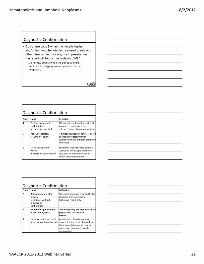
Diagnostic Confirmation• Do not use code 3 when the genetic testing and/or immunophenotyping are used to rule out other diseases. In this case, the impression on the report will be such as “rule out CML.”– Do not use code 3 when the genetics and/or immunophenotyping are not positive for the neoplasm.
Diagnostic ConfirmationCode Label Definition
4 Positive microscopic confirmation,method not specified
Microscopic confirmation is all that isknown. It is unknown if thecells were from histology or cytology.
5 Positive laboratory test/marker study
A clinical diagnosis of cancer is basedon laboratory tests/markerstudies which are clinically diagnosticfor cancer.
6 Direct visualization withoutmicroscopic confirmation
The tumor was visualized during a surgical or endoscopic procedureonly with no tissue resected for microscopic examination.
Diagnostic ConfirmationCode Label Definition7 Radiography and other
imagingtechniques without microscopicconfirmation
The malignancy was reported by the physician from an imagingtechnique report only.
8 8 Clinical diagnosis only, other than 5, 6 or 7
The malignancy was reported by thephysician in the medicalrecord.
9 Unknown whether or notmicroscopically confirmed
A statement of malignancy was reported in the medical record, butthere is no statement of how the cancer was diagnosed (usuallynonanalytic).
Hematopoietic and Lymphoid Neoplasms 8/2/2012
NAACCR 2011‐2012 Webinar Series 22
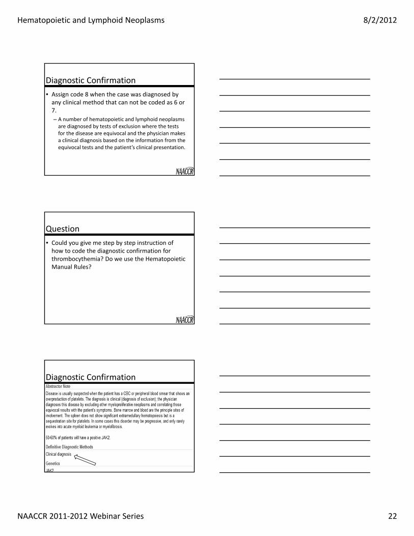
Diagnostic Confirmation• Assign code 8 when the case was diagnosed by any clinical method that can not be coded as 6 or 7. – A number of hematopoietic and lymphoid neoplasms are diagnosed by tests of exclusion where the tests for the disease are equivocal and the physician makes a clinical diagnosis based on the information from the equivocal tests and the patient’s clinical presentation.
Question• Could you give me step by step instruction of how to code the diagnostic confirmation for thrombocythemia? Do we use the Hematopoietic Manual Rules?
Diagnostic Confirmation
Hematopoietic and Lymphoid Neoplasms 8/2/2012
NAACCR 2011‐2012 Webinar Series 23
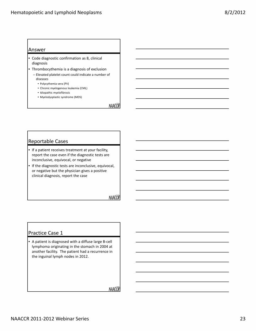
Answer• Code diagnostic confirmation as 8, clinical diagnosis
• Thrombocythemia is a diagnosis of exclusion– Elevated platelet count could indicate a number of diseases
• Polycythemia vera (PV)• Chronic myelogenous leukemia (CML)• Idiopathic myelofibrosis• Myelodysplastic syndrome (MDS)
Reportable Cases• If a patient receives treatment at your facility, report the case even if the diagnostic tests are inconclusive, equivocal, or negative
• If the diagnostic tests are inconclusive, equivocal, or negative but the physician gives a positive clinical diagnosis, report the case
Practice Case 1• A patient is diagnosed with a diffuse large B‐cell lymphoma originating in the stomach in 2004 at another facility. The patient had a recurrence in the inguinal lymph nodes in 2012.
Hematopoietic and Lymphoid Neoplasms 8/2/2012
NAACCR 2011‐2012 Webinar Series 24
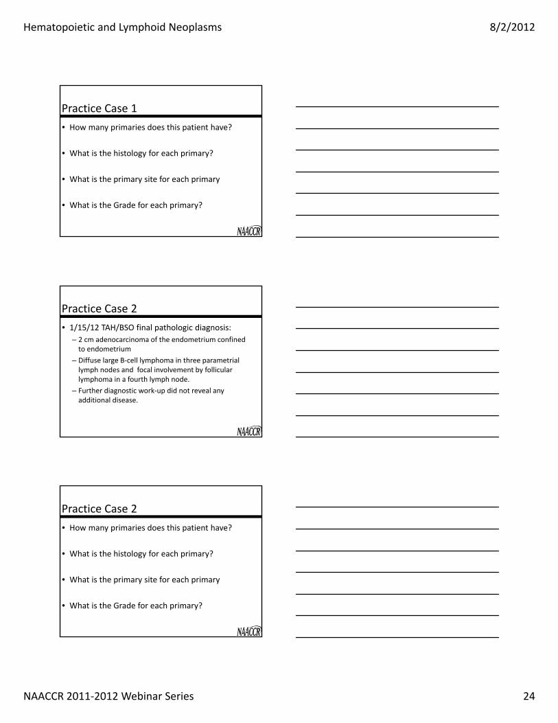
Practice Case 1• How many primaries does this patient have?
• What is the histology for each primary?
• What is the primary site for each primary
• What is the Grade for each primary?
Practice Case 2• 1/15/12 TAH/BSO final pathologic diagnosis:
– 2 cm adenocarcinoma of the endometrium confined to endometrium
– Diffuse large B‐cell lymphoma in three parametriallymph nodes and focal involvement by follicular lymphoma in a fourth lymph node.
– Further diagnostic work‐up did not reveal any additional disease.
Practice Case 2• How many primaries does this patient have?
• What is the histology for each primary?
• What is the primary site for each primary
• What is the Grade for each primary?
Hematopoietic and Lymphoid Neoplasms 8/2/2012
NAACCR 2011‐2012 Webinar Series 25
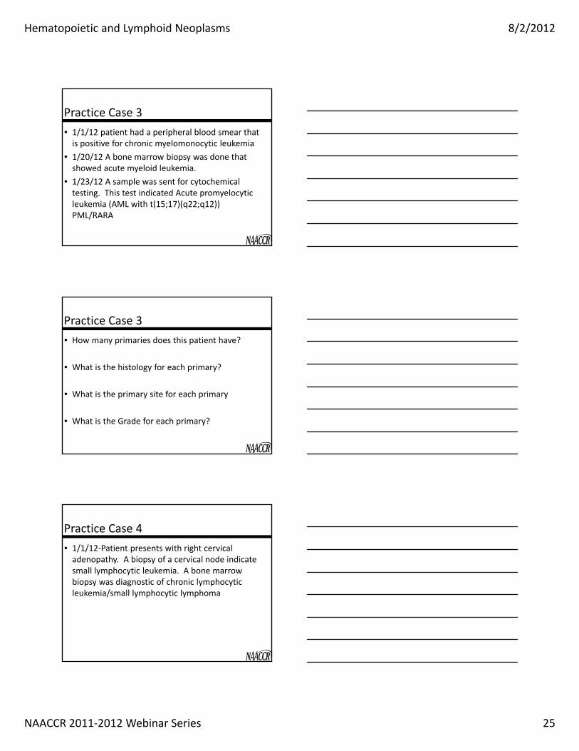
Practice Case 3• 1/1/12 patient had a peripheral blood smear that is positive for chronic myelomonocytic leukemia
• 1/20/12 A bone marrow biopsy was done that showed acute myeloid leukemia.
• 1/23/12 A sample was sent for cytochemical testing. This test indicated Acute promyelocytic leukemia (AML with t(15;17)(q22;q12)) PML/RARA
Practice Case 3• How many primaries does this patient have?
• What is the histology for each primary?
• What is the primary site for each primary
• What is the Grade for each primary?
Practice Case 4• 1/1/12‐Patient presents with right cervical adenopathy. A biopsy of a cervical node indicate small lymphocytic leukemia. A bone marrow biopsy was diagnostic of chronic lymphocytic leukemia/small lymphocytic lymphoma
Hematopoietic and Lymphoid Neoplasms 8/2/2012
NAACCR 2011‐2012 Webinar Series 26
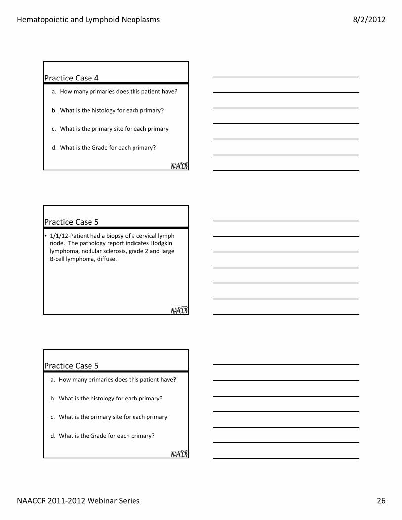
Practice Case 4a. How many primaries does this patient have?
b. What is the histology for each primary?
c. What is the primary site for each primary
d. What is the Grade for each primary?
Practice Case 5• 1/1/12‐Patient had a biopsy of a cervical lymph node. The pathology report indicates Hodgkin lymphoma, nodular sclerosis, grade 2 and large B‐cell lymphoma, diffuse.
Practice Case 5a. How many primaries does this patient have?
b. What is the histology for each primary?
c. What is the primary site for each primary
d. What is the Grade for each primary?
Hematopoietic and Lymphoid Neoplasms 8/2/2012
NAACCR 2011‐2012 Webinar Series 27
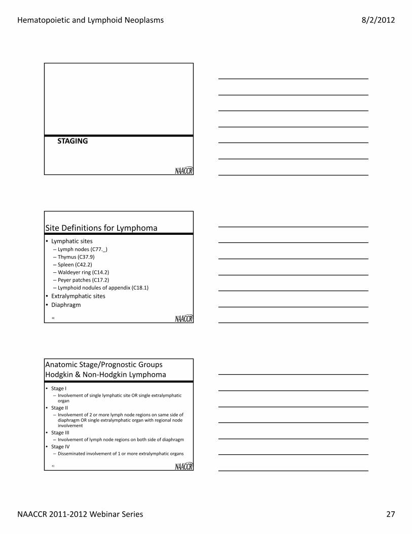
STAGING
Site Definitions for Lymphoma• Lymphatic sites
– Lymph nodes (C77._)– Thymus (C37.9)– Spleen (C42.2)– Waldeyer ring (C14.2)– Peyer patches (C17.2)– Lymphoid nodules of appendix (C18.1)
• Extralymphatic sites• Diaphragm
80
Anatomic Stage/Prognostic GroupsHodgkin & Non‐Hodgkin Lymphoma
• Stage I– Involvement of single lymphatic site OR single extralymphatic organ
• Stage II– Involvement of 2 or more lymph node regions on same side of diaphragm OR single extralymphatic organ with regional node involvement
• Stage III– Involvement of lymph node regions on both side of diaphragm
• Stage IV– Disseminated involvement of 1 or more extralymphatic organs
81
Hematopoietic and Lymphoid Neoplasms 8/2/2012
NAACCR 2011‐2012 Webinar Series 28
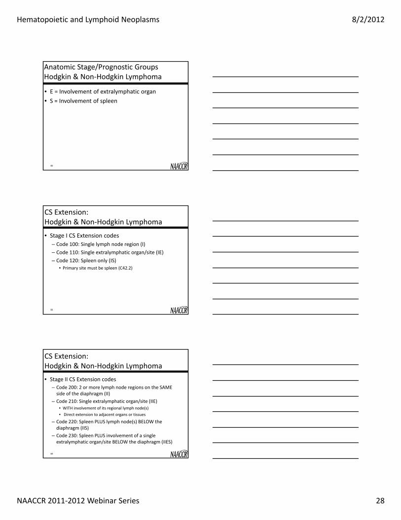
Anatomic Stage/Prognostic GroupsHodgkin & Non‐Hodgkin Lymphoma
• E = Involvement of extralymphatic organ• S = Involvement of spleen
82
CS Extension:Hodgkin & Non‐Hodgkin Lymphoma
• Stage I CS Extension codes– Code 100: Single lymph node region (I)– Code 110: Single extralymphatic organ/site (IE)– Code 120: Spleen only (IS)
• Primary site must be spleen (C42.2)
83
CS Extension:Hodgkin & Non‐Hodgkin Lymphoma
• Stage II CS Extension codes– Code 200: 2 or more lymph node regions on the SAME side of the diaphragm (II)
– Code 210: Single extralymphatic organ/site (IIE)• WITH involvement of its regional lymph node(s)• Direct extension to adjacent organs or tissues
– Code 220: Spleen PLUS lymph node(s) BELOW the diaphragm (IIS)
– Code 230: Spleen PLUS involvement of a single extralymphatic organ/site BELOW the diaphragm (IIES)
84
Hematopoietic and Lymphoid Neoplasms 8/2/2012
NAACCR 2011‐2012 Webinar Series 29
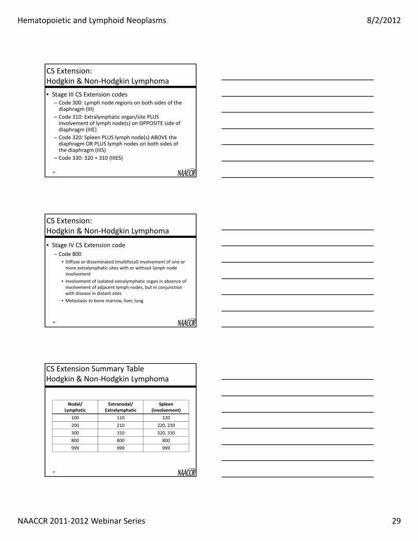
CS Extension:Hodgkin & Non‐Hodgkin Lymphoma• Stage III CS Extension codes
– Code 300: Lymph node regions on both sides of the diaphragm (III)
– Code 310: Extralymphatic organ/site PLUS involvement of lymph node(s) on OPPOSITE side of diaphragm (IIIE)
– Code 320: Spleen PLUS lymph node(s) ABOVE the diaphragm OR PLUS lymph nodes on both sides of the diaphragm (IIIS)
– Code 330: 320 + 310 (IIIES)
85
CS Extension:Hodgkin & Non‐Hodgkin Lymphoma
• Stage IV CS Extension code– Code 800
• Diffuse or disseminated (multifocal) involvement of one or more extralymphatic sites with or without lymph node involvement
• Involvement of isolated extralymphatic organ in absence of involvement of adjacent lymph nodes, but in conjunction with disease in distant sites
• Metastasis to bone marrow, liver, lung
86
CS Extension Summary TableHodgkin & Non‐Hodgkin Lymphoma
Nodal/Lymphatic
Extranodal/Extralymphatic
Spleen (involvement)
100 110 120200 210 220, 230300 310 320, 330800 800 800999 999 999
87
Hematopoietic and Lymphoid Neoplasms 8/2/2012
NAACCR 2011‐2012 Webinar Series 30
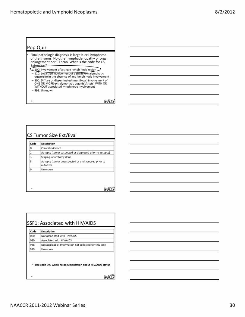
Pop Quiz• Final pathologic diagnosis is large b‐cell lymphoma of the thymus. No other lymphadenopathy or organ enlargement per CT scan. What is the code for CS Extension?– 100: Involvement of a single lymph node region– 110: Localized involvement of a single extralymphatic organ/site in the absence of any lymph node involvement
– 800: Diffuse or disseminated (multifocal) involvement of ONE OR MORE extralymphatic organ(s)/site(s) WITH OR WITHOUT associated lymph node involvement
– 999: Unknown
88
CS Tumor Size Ext/EvalCode Description0 Clinical evidence2 Autopsy (tumor suspected or diagnosed prior to autopsy)3 Staging laparotomy done8 Autopsy (tumor unsuspected or undiagnosed prior to
autopsy)9 Unknown
89
SSF1: Associated with HIV/AIDSCode Description000 Not associated with HIV/AIDS010 Associated with HIV/AIDS988 Not applicable: Information not collected for this case999 Unknown
• Use code 999 when no documentation about HIV/AIDS status
90
Hematopoietic and Lymphoid Neoplasms 8/2/2012
NAACCR 2011‐2012 Webinar Series 31
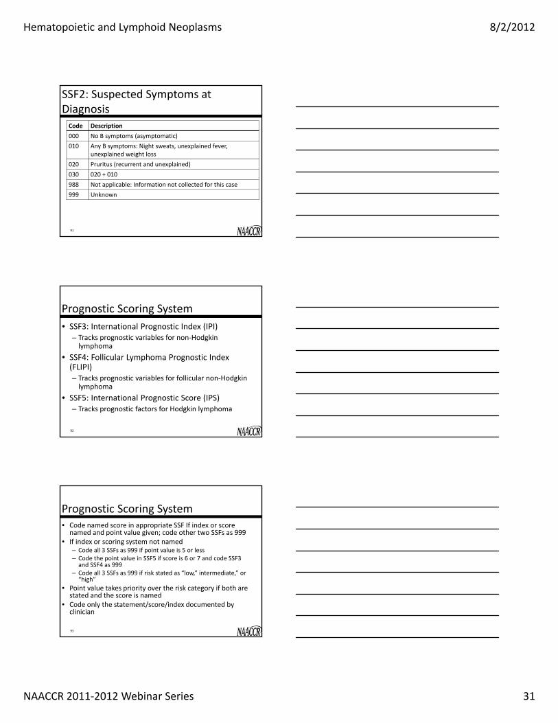
SSF2: Suspected Symptoms at DiagnosisCode Description000 No B symptoms (asymptomatic)010 Any B symptoms: Night sweats, unexplained fever,
unexplained weight loss020 Pruritus (recurrent and unexplained)030 020 + 010988 Not applicable: Information not collected for this case999 Unknown
91
Prognostic Scoring System• SSF3: International Prognostic Index (IPI)
– Tracks prognostic variables for non‐Hodgkin lymphoma
• SSF4: Follicular Lymphoma Prognostic Index (FLIPI)– Tracks prognostic variables for follicular non‐Hodgkin lymphoma
• SSF5: International Prognostic Score (IPS)– Tracks prognostic factors for Hodgkin lymphoma
92
Prognostic Scoring System• Code named score in appropriate SSF If index or score named and point value given; code other two SSFs as 999
• If index or scoring system not named– Code all 3 SSFs as 999 if point value is 5 or less– Code the point value in SSF5 if score is 6 or 7 and code SSF3 and SSF4 as 999
– Code all 3 SSFs as 999 if risk stated as “low,” intermediate,” or “high”
• Point value takes priority over the risk category if both are stated and the score is named
• Code only the statement/score/index documented by clinician
93
Hematopoietic and Lymphoid Neoplasms 8/2/2012
NAACCR 2011‐2012 Webinar Series 32
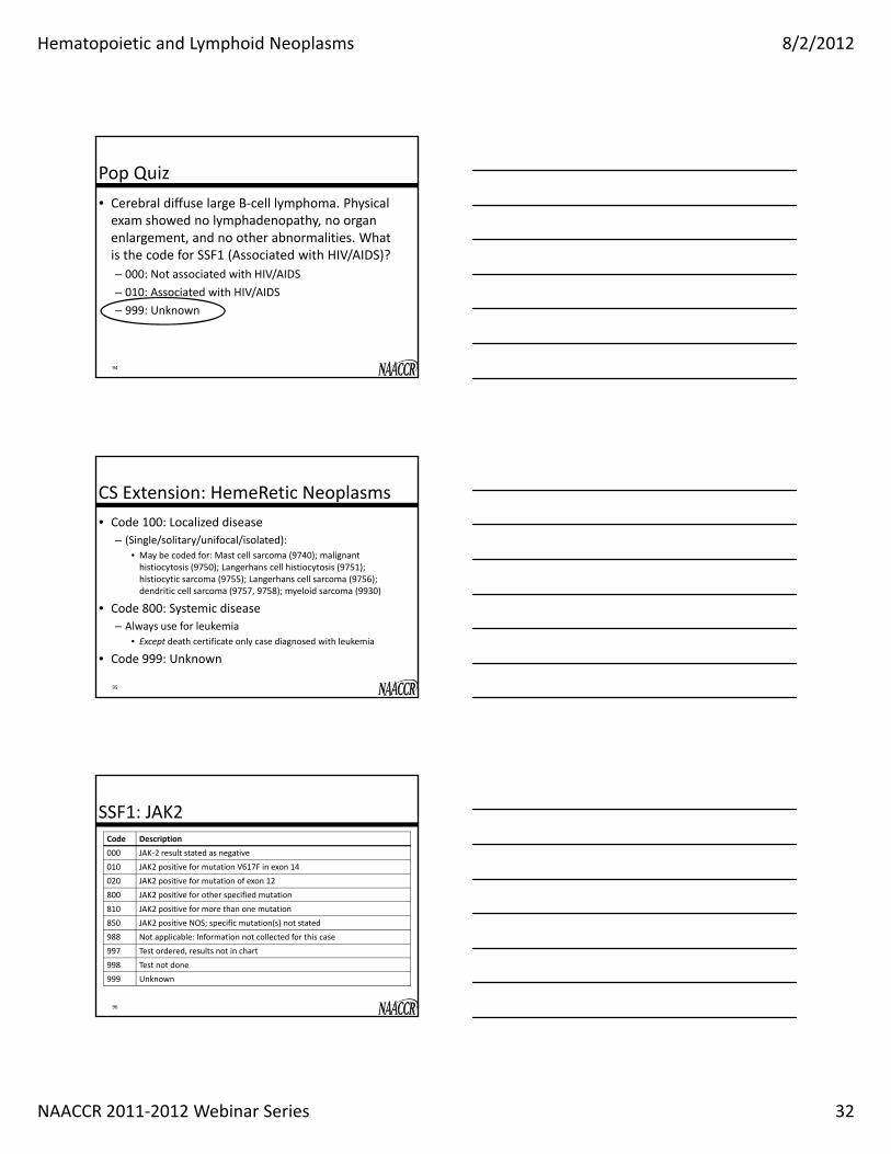
Pop Quiz• Cerebral diffuse large B‐cell lymphoma. Physical exam showed no lymphadenopathy, no organ enlargement, and no other abnormalities. What is the code for SSF1 (Associated with HIV/AIDS)?– 000: Not associated with HIV/AIDS– 010: Associated with HIV/AIDS– 999: Unknown
94
CS Extension: HemeRetic Neoplasms• Code 100: Localized disease
– (Single/solitary/unifocal/isolated):• May be coded for: Mast cell sarcoma (9740); malignant histiocytosis (9750); Langerhans cell histiocytosis (9751); histiocytic sarcoma (9755); Langerhans cell sarcoma (9756); dendritic cell sarcoma (9757, 9758); myeloid sarcoma (9930)
• Code 800: Systemic disease– Always use for leukemia
• Except death certificate only case diagnosed with leukemia
• Code 999: Unknown
95
SSF1: JAK2Code Description000 JAK‐2 result stated as negative010 JAK2 positive for mutation V617F in exon 14020 JAK2 positive for mutation of exon 12800 JAK2 positive for other specified mutation
810 JAK2 positive for more than one mutation850 JAK2 positive NOS; specific mutation(s) not stated988 Not applicable: Information not collected for this case997 Test ordered, results not in chart
998 Test not done999 Unknown
96
Hematopoietic and Lymphoid Neoplasms 8/2/2012
NAACCR 2011‐2012 Webinar Series 33
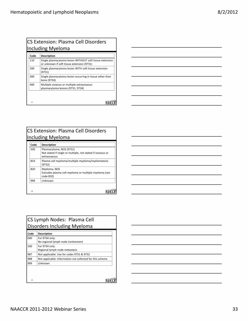
CS Extension: Plasma Cell Disorders Including MyelomaCode Description110 Single plasmacytoma lesion WITHOUT soft tissue extension
or unknown if soft tissue extension (9731)200 Single plasmacytoma lesion WITH soft tissue extension
(9731)300 Single plasmacytoma lesion occurring in tissue other than
bone (9734)400 Multiple osseous or multiple extraosseous
plasmacytoma lesions (9731, 9734)
97
CS Extension: Plasma Cell Disorders Including Myeloma
Code Description500 Plasmacytoma, NOS (9731)
Not stated if single or multiple, not stated if osseous or extraosseous
810 Plasma cell myeloma/multiple myeloma/myelomatosis (9732)
820 Myeloma, NOSExcludes plasma cell myeloma or multiple myeloma (see code 810)
999 Unknown
98
CS Lymph Nodes: Plasma Cell Disorders Including MyelomaCode Description000 For 9734 only:
No regional lymph node involvement100 For 9734 only:
Regional lymph node metastasis987 Not applicable: Use for codes 9731 & 9732 988 Not applicable: Information not collected for this schema999 Unknown
99
Hematopoietic and Lymphoid Neoplasms 8/2/2012
NAACCR 2011‐2012 Webinar Series 34
SSF2: Durie‐Salmon Staging SystemCode Description010 Durie Salmon Stage IA
020 Durie Salmon Stage IB030 Durie Salmon Stage I NOS040 Durie Salmon Stage IIA050 Durie Salmon Stage IIB060 Durie Salmon Stage II NOS
070 Durie Salmon Stage IIIA080 Durie Salmon Stage IIIB090 Durie Salmon Stage III NOS987 Not applicable: Use for codes 9731 & 9734988 Not applicable: Information not collected for this case
999 Unknown100
SSF3: Multiple Myeloma TerminologyCode Description000 Multiple myeloma/Plasma cell myeloma with no other modifiers
Multiple myeloma, NOS; Myeloma, NOS010 Asymptomatic myeloma020 Early or evolving myeloma030 Inactive, indolent, or smoldering myeloma080 Other terminology describing myeloma100 Any combination of terms in codes 010‐080
987 Not applicable: Use for codes 9731 & 9734988 Not applicable: Information not collected for this case999 Unknown
101
Pop Quiz• 3/1/2012: Based on CT scan and lab work, plasmacytoma of right rib.
• 3/15/2012: Bone marrow biopsy diagnoses multiple myeloma.
• What is the code for CS Extension?– 110: Single plasmacytoma lesion WITHOUT soft tissue extension or unknown if soft tissue extension
– 200: Single plasmacytoma lesion WITH soft tissue extension – 300: Single plasmacytoma lesion occurring in tissue other than bone
– 810: Plasma cell myeloma/multiple myeloma/myelomatosis
102
Hematopoietic and Lymphoid Neoplasms 8/2/2012
NAACCR 2011‐2012 Webinar Series 35
TREATMENT
103
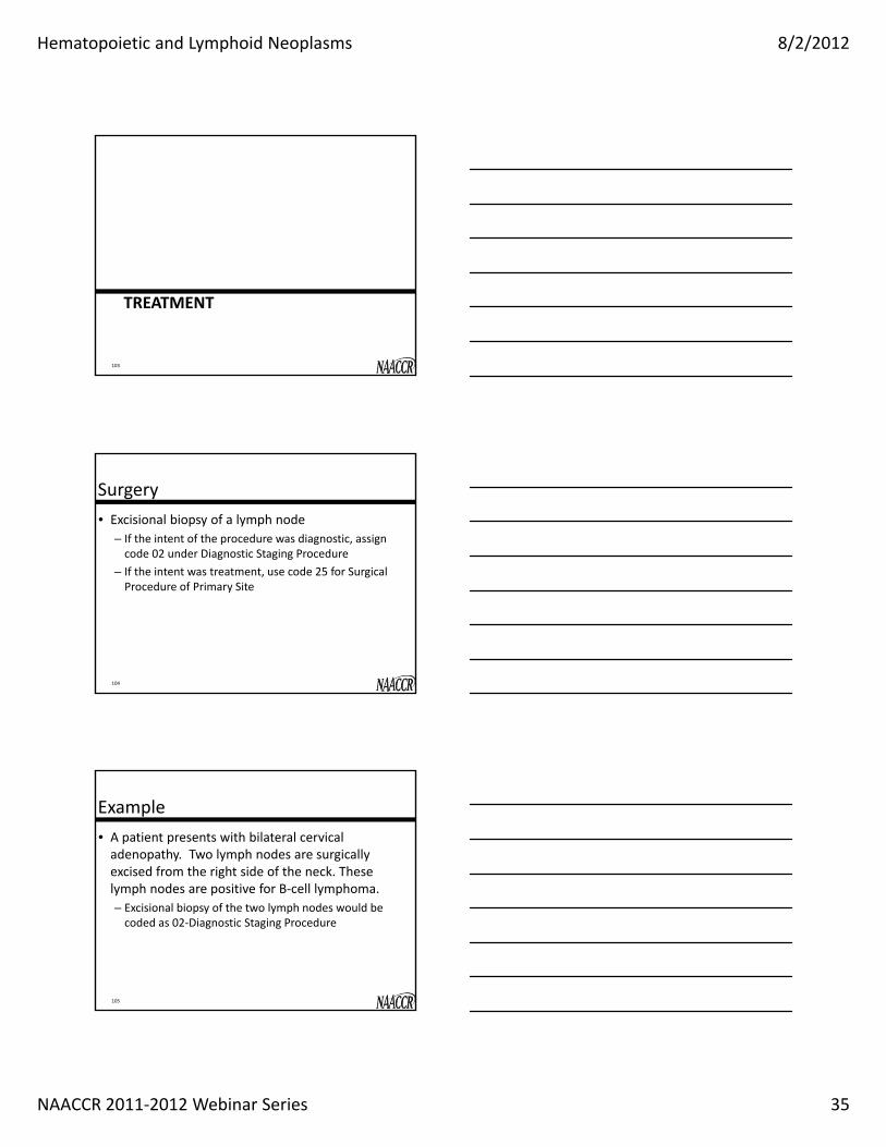
Surgery• Excisional biopsy of a lymph node
– If the intent of the procedure was diagnostic, assign code 02 under Diagnostic Staging Procedure
– If the intent was treatment, use code 25 for Surgical Procedure of Primary Site
104
Example• A patient presents with bilateral cervical adenopathy. Two lymph nodes are surgically excised from the right side of the neck. These lymph nodes are positive for B‐cell lymphoma.– Excisional biopsy of the two lymph nodes would be coded as 02‐Diagnostic Staging Procedure
105
Hematopoietic and Lymphoid Neoplasms 8/2/2012
NAACCR 2011‐2012 Webinar Series 36
Example 2: • Patient with palpable cervical lymph node presents for excisional biopsy; staging workup failed to reveal any additional disease– Assign code 25 for surgical procedure of primary site
106
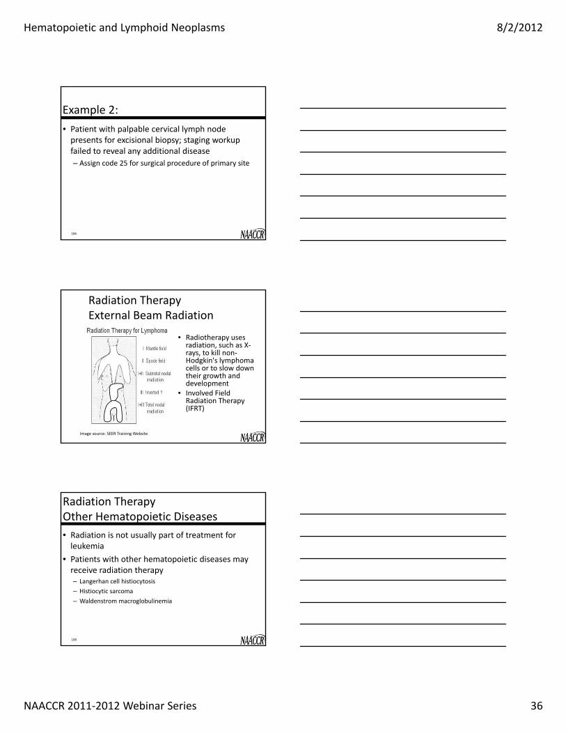
Radiation TherapyExternal Beam Radiation
• Radiotherapy uses radiation, such as X‐rays, to kill non‐Hodgkin's lymphoma cells or to slow down their growth and development
• Involved Field Radiation Therapy (IFRT)
Image source: SEER Training Website
Radiation TherapyOther Hematopoietic Diseases• Radiation is not usually part of treatment for leukemia
• Patients with other hematopoietic diseases may receive radiation therapy– Langerhan cell histiocytosis– Histiocytic sarcoma– Waldenstrom macroglobulinemia
108
Hematopoietic and Lymphoid Neoplasms 8/2/2012
NAACCR 2011‐2012 Webinar Series 37
Radiation Therapy for Lymphoma• Assign code 33, whole body in data item, regional treatment modality– Drugs combine the cell targeting ability of a monoclonal antibody with the additional cell killing ability of a radioactive particle, or radioisotope
• Bexxar (iodine‐131 tositumomab)• Zevalin (yttrium 90 ibritumomab tiuxitan)
109
Systemic Therapy• Primary treatment for most hematopoietic and lymphoid neoplasms– Varies widely based on diagnosis
• Use SEER Rx to determine if systemic treatment is chemotherapy, hormone therapy, immunotherapy, or other therapy.– http://seer.cancer.gov/tools/seerrx/
110
Systemic Therapies• Rituximab• CHOP• ABVD• Anthracycline
111
Hematopoietic and Lymphoid Neoplasms 8/2/2012
NAACCR 2011‐2012 Webinar Series 38
Hormone Therapy• Code hormonal agents given with chemotherapy regimens in the hormone therapy data item– Prednisone (ACVBP, CHOP, CNOP, EPOCH, MOCOP‐B, MOPP)
– Halotestin (EPOCH)
112
Immunotherapy• Monoclonal antibodies (MABs or MOABs)• Vaccines• T‐cell immunotherapy• Interferon alpha
113
Hematologic Transplant• Bone marrow transplant
– Code 11: autologous– Code 12: allogenic
• Peripheral blood stemcell transplant– Code 20
Image source: http://www.lymphoma‐net.org/transplantation.cfm114
Hematopoietic and Lymphoid Neoplasms 8/2/2012
NAACCR 2011‐2012 Webinar Series 39
Other Treatment• Aspirin
– Code as 'Other' therapy for essential thrombocythemia ONLY otherwise it is a differentiating agent (do not code)
• Anagrelide HCl– Code as 'Other' therapy for essential thrombocythemia ONLY otherwise it is a differentiating agent (do not code)
115
QUESTIONS?
116
Coming up!• 9/6/12
– Coding Pitfalls
• Register now for the 2012‐2013 Cancer Registry & Surveillance Webinar Series– http://www.naaccr.org/EducationandTraining/Webinars.aspx
117
And the winners of the fabulous prizes are….