Embed Size (px)
Citation preview

Harvard-MIT Division of Health Sciences and Technology HST.535: Principles and Practice of Tissue Engineering Instructor: I. V. Yannas
Collagen-GAG scaffolds for organ regeneration processes
Prof. I. V. Yannas
MIT
Reference: I.V.Yannas, Tissue and Organ Regeneration in Adults, Springer, 2001
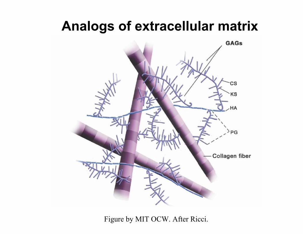
Analogs of extracellular matrix
Figure by MIT OCW. After Ricci.

Question to answer:Why use collagen-GAG scaffolds
to induce organ regeneration?
Study requirements for organ synthesis in vivo. Use chemical symbolism to simplify analysis.

Information stored in a chemical equation
Ammonia synthesis (F. Haber)T, P
3H2 + N2 → 2NH3 reactor
reactants → products
NOTE: The stoichiometry (masses on both sides) of a chemical equation expressesconservation of mass (Lavoisier)
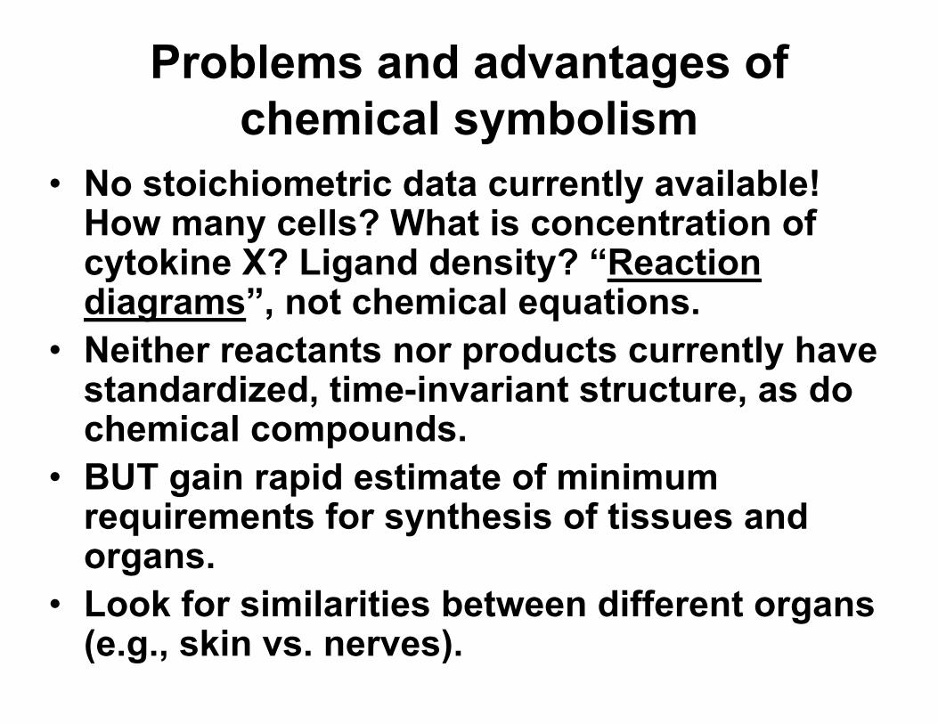
Problems and advantages of chemical symbolism
• No stoichiometric data currently available! How many cells? What is concentration ofcytokine X? Ligand density? “Reaction diagrams”, not chemical equations.
• Neither reactants nor products currently havestandardized, time-invariant structure, as do chemical compounds.
• BUT gain rapid estimate of minimumrequirements for synthesis of tissues and organs.
• Look for similarities between different organs(e.g., skin vs. nerves).

Transition to biologyI. Reactants
• Cells migrate, proliferate, synthesizematrices and cytokines, degrade matrices, etc.
• Cytokines and growth factors are solublemolecules that diffuse. They serve as“language” between cells.
• Matrices are insoluble macromolecular networks and do not diffuse. They controlcell behavior (phenotype) via integrinligand binding. Usually porous (“scaffolds”).

Transition to biologyII. Reactors
• In vitro reactors are dishes or flasks for cell culture.
• In vivo reactors are anatomical sites of organ loss in the living organism.
• Experimental in vivo reactors aregenerated by surgical excision (scalpel,laser, etc.).
• When organ synthesis takes place in vivoat the correct anatomical site of livingorganism it is referred to as “inducedregeneration”.

Skin: In vitro or in vivo synthesis?
Figure by MIT OCW.

Nerves: In vitro or in vivo?
Figure by MIT OCW.

• T• T• T
Standardized reactors
SKIN
PERIPHERAL NERVE
ransected nerveransected nerveransected nervedermis
epidermis
nerve stumps
dermis
epidermis
nerve stumps
dermis
epidermis
nerve stumps
Figures by MIT OCW.

Transition to biology. III. Products
• Organs are made up of tissues.• Products of the synthesis can be
tissues or organs. • Almost all organs are essentially
made up of three types of tissues: epithelial, basement membrane and stroma (connective tissue).

Members of the tissue triad
• EPITHELIA 100% cells. No matrix. No blood vessels.
• BASEMENT MEMBRANE No cells. 100% matrix. No blood vessels.
• STROMA (CONNECTIVE TISSUE) Cells. Matrix. Blood vessels.
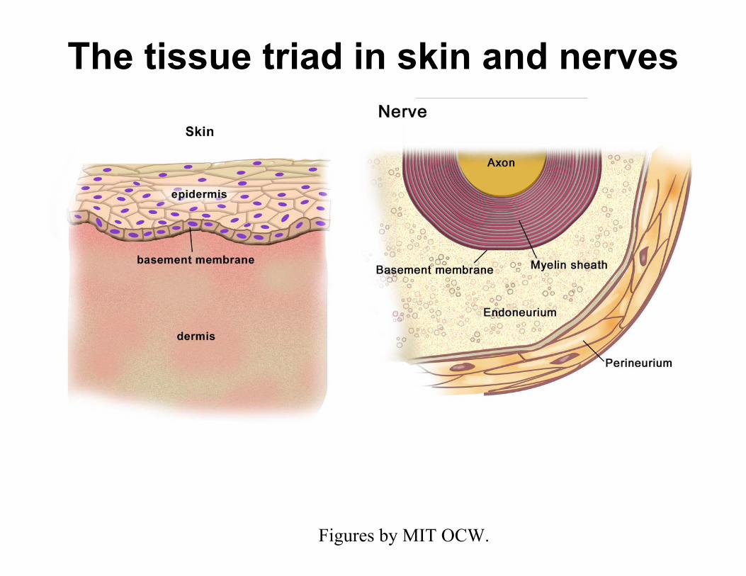
The tissue triad in skin and nerves
Figures by MIT OCW.

The central question in organ synthesis

Which tissues in the triad do notregenerate spontaneously?
• When excised from an organ, the epitheliaare regenerated spontaneously.Examples: the epidermis in skin, themyelin sheath in nerves.
• Likewise, the basement membrane regenerates spontaneously on the stroma.
• However, the stroma does not regeneratespontaneously. Examples: dermis in skin,endoneurium in nerves.

SKIN: The epidermis regenerates spontaneously
Epidermis lost. Dermis intact. Spontaneous regeneration
Figure by MIT OCW.

SKIN: Scar formation. The dermis does not regenerate.
Scar
Epidermis and dermis both Closure by contraction lost to severe injury and scar formation
Figure by MIT OCW.

NERVE: The injured myelin sheath regenerates spontaneously
Regeneratedmyelin
Injured myelin. Axoplasm
Myelin sheath
Endoneurium intact.
Figure by MIT OCW.
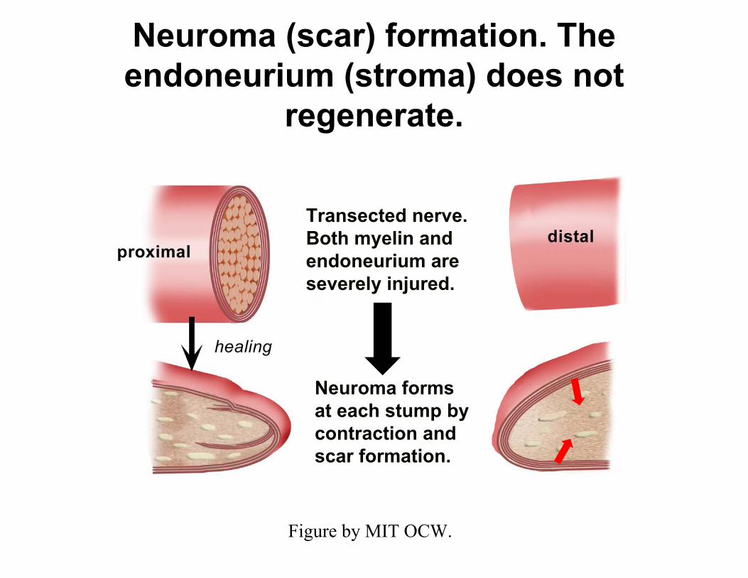
Neuroma (scar) formation. The endoneurium (stroma) does not
regenerate.
Transected nerve. Both myelin and endoneurium are severely injured.
Neuroma forms at each stump by contraction and scar formation.
Figure by MIT OCW.

Intact nerve fiber
Photo removed for copyright reasons.See Figure 2.5 in Yannas, I. V.Tissue and Organ Regeneration in Adults. New York: Springer, 2001. ISBN: 0387952144.
Spontaneously healed nerve fiber (scar)
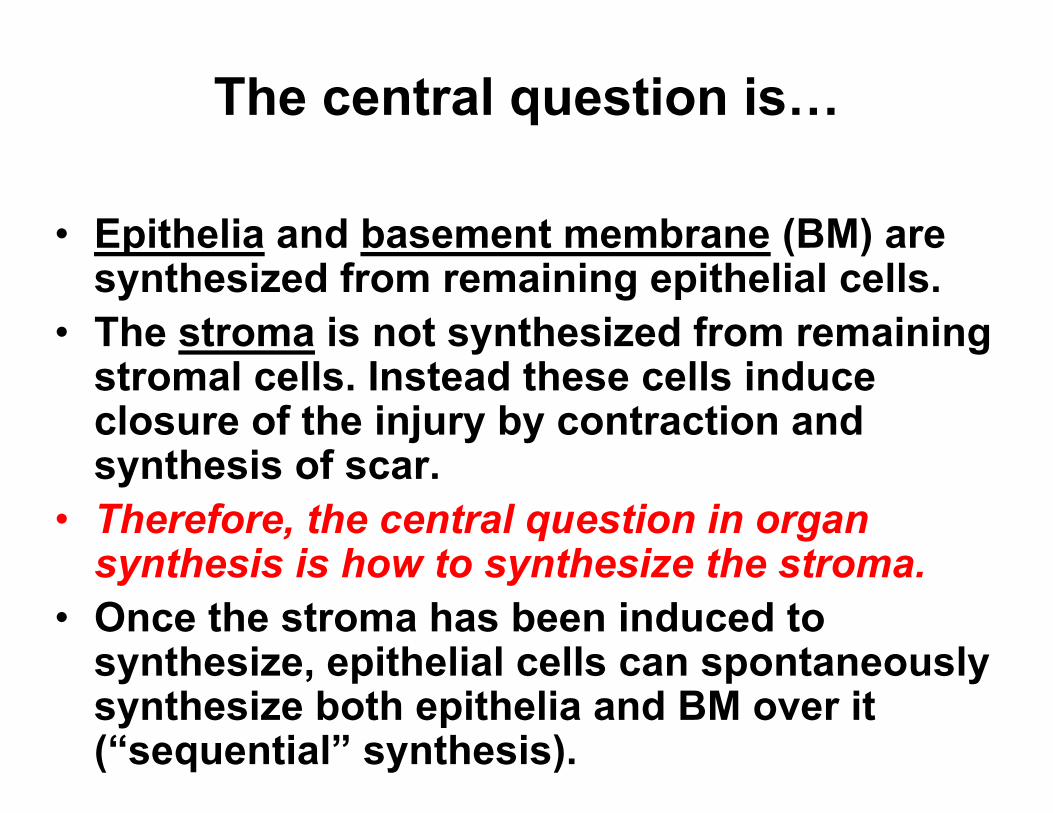
The central question is…
• Epithelia and basement membrane (BM) aresynthesized from remaining epithelial cells.
• The stroma is not synthesized from remainingstromal cells. Instead these cells induce closure of the injury by contraction andsynthesis of scar.
• Therefore, the central question in organsynthesis is how to synthesize the stroma.
• Once the stroma has been induced to synthesize, epithelial cells can spontaneouslysynthesize both epithelia and BM over it(“sequential” synthesis).
Which reactants are required to be supplied by the investigator to
synthesize an organ?
Use empirical trans-organ rulesto find requirements for addition of cells, scaffolds and growth factors

are
Required vs. redundant reactants
• Investigators typically supply (add) reactantsbased on favored hypotheses. Often,reactants supplied are not required tosynthesize tissue or organ.
• In vitro all reactants, including culturemedium, are supplied by investigator.
• In vivo the reactor spontaneously supplies exudate that contains certain reactants (endogenous reactants). The investigatorsupplies other reactants (exogenous).
• What are the minimal reactants that suffice to synthesize a tissue or organ? These are the“required” reactants.

Finally answer question:Why use collagen-GAG scaffolds to induce
organ regeneration?
Based on data from synthesis of skin and peripheral nerves :
• Synthesis of epithelia can be accomplished in vitro(does not require in vivo environment). It simplyrequires supply of epithelial cells and culturemedium. Scaffold not required.
• Synthesis of stroma has only been accomplished invivo. It requires supply only of an appropriatescaffold. Addition of stromal cells (e.g., fibroblasts) or growth factors (e.g., TGF-β, PDGF) is notrequired.
• An appropriate scaffold is required for organsynthesis. Epithelial cells speed up synthesis.Stromal cells (e.g., fibroblasts) or growth factors need not be added.

Various synthetic routes
Route 1: Sequential synthesis Stroma synthesized first using appropriate matrix (regeneration template). Epithelia andbasement membrane both synthesizedspontaneously later in contact with the newstroma by endogenous epithelial cells. Route 2: Simultaneous synthesis All three tissues can be simultaneouslysynthesized using template seeded withepithelial cells.Route 3: Modular organ synthesis? Synthesizeeach tissue in separate reactor, then combine.
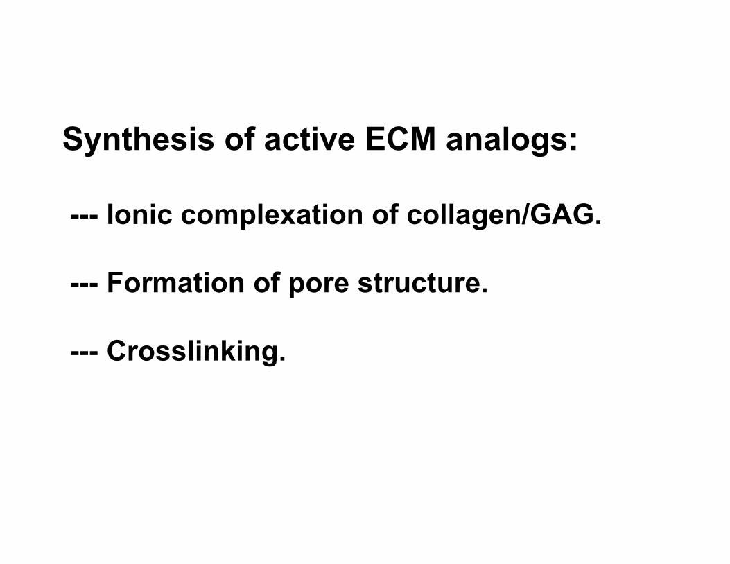
Synthesis of active ECM analogs:
--- Ionic complexation of collagen/GAG.
--- Formation of pore structure.
--- Crosslinking.

Crosslinking
Collagen Dispersion (or solution) GAG Solution
FLOW SHEET OF COLLAGEN-GAG MEMBRANE FORMATION PROCESS
Coprecipitation and Homogenization
Freeze-drying
Figure by MIT OCW.

Several slides removed due to copyright considerations.

Which collagen-GAG scaffolds are biologically active as regeneration templates?
Critical Structural Feature Role in regeneration
A. SKIN
Chem. Composition >2% GAG Ligand identity Deleted collagen quaternary structure Downregulation of contractile cells
Pore diameter 20—120 µm Ligand density
Degradation half-life 10-15 d Duration of ligands
B. NERVE
Chem. Composition [not studied] Deleted collagen quaternary structure [not studied] Pore diameter ∼ 5 µm Ligand density
Degradation half-life ∼ 1-10 wk Duration of ligands

Mechanism of regenerative activity of collagen-GAG scaffolds
1. Certain ECM analogs are biologically active scaffolds (regeneration templates) that induce regeneration of tissues and organs: skin, peripheral nerve and the conjunctiva (eye) in humans and experimental animals. 2. Regeneration templates lose their activity if the following structural features fall outside a narrow range: chemical composition, collagen quaternary structure, pore diameter, degradation rate. 3. The data suggest that templates induce regeneration in a defect by blocking selectively the contraction process that leads to closure of the defect in adults. 4. Templates block contraction by two basic mechanisms. First, by downregulating differentiation of fibroblasts to myofibroblasts. Second, by binding most of the contractile cells in the defect over a period corresponding to the duration of contraction in that defect. Binding requires the presence of appropriate ligands (chem. composition) at a minimal density (pore diameter) over a critical duration (degradation rate).

Summary1. Of three types of tissue in an organ only stroma fails
to regenerate spontaneously and needs to be induced to synthesize. Epithelia and basement membrane regenerate spontaneously.
2. There are three classes of “reactants”, i.e., cells, scaffolds and growth factors. Of these only an appropriate scaffold must be added to synthesize the stroma.
3. The appropriate scaffold (regeneration template) is synthesized to have the composition of an analog of the extracellular matrix, pore size within a critically defined range and degradation rate that matches the rate of tissue synthesis at the organ site.