Embed Size (px)
Citation preview
APPLIED AND ENVIRONMENTAL MICROBIOLOGY, Mar. 1981, p. 657-6630099-2240/81/030657-07$02.00/0
Vol. 41, No. 3
Coliform Species Recovered from Untreated Surface Waterand Drinking Water by the Membrane Filter, Standard, and
Modified Most-Probable-Number TechniquestT. M. EVANS, M. W. LECHEVALLIER, C. E. WAARVICK, AND RAMON J. SEIDLER*
Department ofMicrobiology, Oregon State University, Corvallis, Oregon 97331
The species of total coliforn bacteria isolated from drinking water and un-treated surface water by the membrane filter (MF), the standard most-probable-number (S-MPN), and modified most-probable-number (M-MPN) techniqueswere compared. Each coliform detection technique selected for a different profileof coliform species from both types ofwater samples. The MF technique indicatedthat Citrobacter freundii was the most common coliform species in water samples.However, the fernentation tube techniques displayed selectivity towards theisolation of Escherichia coli and Klebsiella. The M-MPN technique selected formore C. freundii and Enterobacter spp. from untreated surface water samplesand for more Enterobacter and Klebsiella spp. from drinking water samples thandid the S-MPN technique. The lack of agreement between the number ofcoliforms detected in a water sample by the S-MPN, M-MPN, andMF techniqueswas a result of the selection for different coliform species by the various tech-niques.
The four genera Escherichia, Klebsiella, En-terobacter, and Citrobacter are generally ac-cepted as comprising the total coliform popula-tion (4). Enumeration of this component of themicrobial aquatic ecosystem has been univer-sally applied to document the sanitary qualityof water. The usefulness of the total coliformcount as an indicator of bacterial water pollutionhas been questioned, partly because coliformdetection methods are potentially subject to in-terferences (14, 16). Interference with coliformdetection or coliform suppression in presumptivemedia (11) has been thought to result fromcompetition by noncoliform bacteria for nutri-ents (21). Other proposed causes of coliformsuppression are the production of inhibitoryproducts by noncoliform bacteria (16) and thefailure of brilliant green lactose bile broth torecover coliforms from gas-positive presumptivetubes (3, 22).
Recently, a modified most-probable-number(M-MPN) procedure was developed to docu-ment the magnitude of interference with totalcoliform detection in the standard MPN (S-MPN) technique (11). Coliform suppression inthe presumptive and confirmed tests was foundto contribute significantly to the underestima-tion of coliform numbers in the S-MPN tech-nique.
In addition to the quantitative impact of sup-
t Technical paper no. 5668, Oregon Agricultural Experi-ment Station, Corvallis, OR 97331.
pression on coliform enumeration, other factorsinfluence the qualitative recovery of the com-ponent coliform genera. It has been illustratedwith polluted specimens that the kind of waterexamined (sewage, unchlorinated sewage ef-fluent, surface water), as well as media andtechniques, will affect the isolation frequency ofthe four coliform genera (4, 9). Treatment of rawwater may also influence the percentage distri-bution of component coliform genera found.Clark and Pagel reported that the percentage ofEscherichia found in the component genera ofcontaminated drinking water samples was re-duced compared to the untreated surface watersource (4). Chlorination has been reported byothers to increase the percentage of Klebsiellain the component coliform genera isolated fromdrinking water samples (20).The purpose of this study was to determine
the influence ofStandardMethods (1) presump-tive media (lactose broth, lauryl tryptose broth)and the technique used (membrane filter [MF],MPN) on the recovery of coliforn genera fromchlorinated drinking water and the raw sourcewaters. In addition, recovery of the coliformspecies by the new M-MPN technique was com-pared to those species recovered by the standardMF and MPN procedures.
MATERIALS AND METHODS
Sampling area. Samples were collected from thefinished drinking water supply of an Oregon coastalcommunity serving 14,000 residents and from the two
657
on July 2, 2019 by guesthttp://aem
.asm.org/
Dow
nloaded from
APPL. ENVIRON. MICROBIOL.
coast range streams supplying raw water to the city.Chlorination was the only treatment applied to theraw water before entry into the distribution system(11). Chlorine was applied at a dose which resulted inan initial free chlorine concentration of approximately1.5 ppm.
Collection End microbiological techniques.Water samples were collected in sterile, 4-liter poly-propylene containers with (drinking water) or without(untreated surface water) added sodium thiosulfate.Samples were placed on ice and transported back tothe laboratory within 3 h after collection and analyzedwithin 7 h after collection.
Nine- or 15-tube S-MPN analyses were performedon each sample by accepted procedures (1) using lac-tose broth (LB; Difco lots 652242 and 662331) andlauryl tryptose broth (LTB; Difco lot 663637) pre-sumptive media in parallel analyses. Both presumptivemedia were supplemented with 18 mg of phenol red(Sigma) indicator per liter. Positive presumptive tubeswere submitted to the confirmed step using brilliantgreen bile lactose broth (Difco lot 666632). The com-pleted test was performed using m-Endo agar LES(Difco lot 663068) and Levine eosin methylene blue(Difco lot 610498). Typical and atypical colonies werepicked from the completion media and streaked ontoslants containing tryptic soy broth (Difco lot 663068)supplemented with 1.5% agar (Difco) and 0.3% yeastextract (Difco lot 656810). After 24 h of incubation at35°C, growth from the slant was removed for gramstaining and transferred to secondary broth tubes con-taining either LB or LTB.The M-MPN technique was performed with the
same media used in the S-MPN procedure with theexception that EC broth (Difco lot 641057) was addedas an additional confirmatory broth and incubated at35°C. The M-MPN scheme consisted of the S-MPNtechnique plus additional manipulations designed torecover coliforms from any tests which were negativein the S-MPN technique. A detailed description of theM-MPN technique can be found elsewhere (11).The MF total coliform detection technique was
performed by standard procedures (1). Gelman GN-6membranes (pore size, 0.45,um) and m-Endo agar LES(Difco lot 663068) were used. Duplicate volumes of250 and 100 ml of drinking water and 25-, 10-, and 1-ml volumes of untreated surface water were routinelyanalyzed. A minimum of 30% of the typical coliformcolonies on MF plates were submitted to LTB forverification (2). Colonies were randomly selected forverification by beginning at the upper left quadrant ofthe filter. As necessary, all typical colonies from ad-jacent grids were picked. In some drinking water sam-ples where the number of typical colonies was lessthan 10 per plate, colonies were picked from replicateplates. In this case, when possible, a total of 10 colonieswere verified.
Identification of total coliform bacteria. Threecompleted coliforms from the highest sample dilutionin each presumptive medium (LTB or LB) used in S-MPN parallel analysis were selected for identification(six in total). A maximum of three coliforms recoveredfrom false-negative S-MPN tests by the M-MPN pro-cedure were selected for identification from the high-est sample dilution in each presumptive medium (six
in total). In addition, 10 of the randomly selectedverified total coliforms recovered by the membranefilter technique were identified. All cultures were pu-rified by streaking onto m-Endo agar LES beforeidentification.
Coliforms were identified using triple sugar ironagar slants (Difco), the IMViC (indole, methyl red,Voges-Proskauer, citrate) tests, lysine and ornithinedecarboxylase broths, arginine dihydrolase broth, andmalonate, rhamnose, and sorbitol fermentation (10).The biochemical reactions of the coliform isolateswere compared to the reactions of coliforms obtainedfrom the American Type Culture Collection. Thiscomparison aided in the identification of the isolatesand ensured that correct reactions occurred in theidentification media. In addition, the API 20E system(Analytab Products, Plainview, N.Y.) was used to con-firm the identities of 5% of the isolates.A quality assurance program as outlined in Micro-
biological Methods for Monitoring the Environment(2) was used throughout the course of this study.
RESULTS
Identification of coliform isolates. Elevenspecies of gram-negative, facultatively anaero-bic, lactose-fermenting, and gas-producing rodswere isolated by one or more of the three coli-form enumeration techniques. Most of the spe-cies could readily be identified on the basis ofthe IMViC, lysine, ornithine, and arginine reac-tions (10). However, using these media, over 50%of the isolates comprising a single biotype couldnot be differentiated as belonging to the speciesSerratia liquefaciens or Citrobacter freundii.The use of additional diagnostic media (rham-nose and malonate fermentation, and deoxyri-bonuclease activity) indicated that these isolateswere hydrogen sulfide-negative biotypes of C.freundii (Table 1). The isolates fermented mal-onate and rhamnose and were negative for de-oxyribonuclease activity. These reactions are nottypical of the lactose-fermenting biotype of S.liquefaciens (13). Data published by Davis andEwing (6, 12) also indicate that the IMViC,lysine and ornithine decarboxylase, sorbitol, andrhamnose reactions of our isolates were repre-sentative of the hydrogen sulfide-negative bio-type of C. freundii. The deoxyribonucleic acidmole percent guanine and cytosine content ofthe isolates designated as C. freundii was 52 to53 mol%, which is also consistent with that of C.freundii (52.6 to 52.8 mol% [19, 24]) and notrepresentative of S. liquefaciens (53.4 to 53.8mol% [15]). Only 4% of the isolates identified inthis study as C. freundii were hydrogen sulfidepositive.Coliform organisms recovered by the MF,
S-MPN, and M-MPN techniques. Over 1,300coliform organisms (668 from untreated surfacewater, 668 from drinking water) were isolated
658 EVANS ET AL.
on July 2, 2019 by guesthttp://aem
.asm.org/
Dow
nloaded from
COLIFORM SPECIES RECOVERED FROM WATER 659
and identified. The isolates were obtained from36 untreated surface water and 193 drinking
water samples collected over a 1-year period.The incidence of coliform species detected bythe MF, S-MPN, and M-MPN techniques in thetwo types of samples examined is presented inTables 2 and 3. The coliform species isolated bythe M-MPN technique represent those obtainedfrom completed S-MPN tests plus additionalisolates recovered from false-negative S-MPNtests.The coliforn bacteria of primary interest be-
long to four genera: Citrobacter, Enterobacter,Escherichia, and Klebsiella (4). Cultures iden-tified as members of these four genera comprised
86% (M-MPN) to 95% (MF) of the total gram-
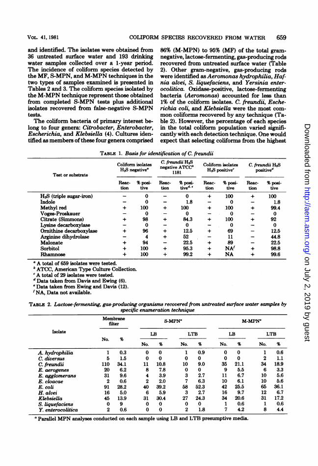
negative, lactose-fennenting, gas-producing rodsrecovered from untreated surface water (Table2). Other gram-negative, gas-producing rodswere identified as Aeromonas hydrophilia, Haf-nia alvei, S. liquefaciens, and Yersinia enter-ocolitica. Oxidase-positive, lactose-fermentingbacteria (Aeromonas) accounted for less than1% of the coliform isolates. C. freundii, Esche-richia coli, and Klebsiella were the most com-
mon coliforms recovered by any technique (Ta-ble 2). However, the percentage of each speciesin the total coliform population varied signifi-cantly with each detection technique. One wouldexpect that selecting coliforms from the highest
TABLE 1. Basis for identification of C. freundii
Coliform isolates C. freundiiH2uColHfor iolates C.fiHeundiiH2SH2S negativea negtive ATCCCbH2Spooitives posCtfvedTest or substrate 1181
Reac- % posi- Reac- % posi- Reac- % posi- Reac- % posi-tion tive tion tive e tion tive tion tive
H2S (triple sugar-iron) - 0 - 0 + 100 + 100Indole - 0 - 1.8 - 0 - 1.8Methyl red + 100 + 100 + 100 + 99.4Voges-Proskauer - 0 - 0 - 0 - 0Citrate (Simmons) + 98 + 84.3 + 100 + 92Lysine decarboxylase - 0 - 0 - 0 - 0Ornithine decarboxylase + 96 + 12.5 + 69 - 12.5Arginine dbiydrolase - 4 + 52 - 11 - 44.8Malonate + 94 - 22.5 + 89 - 22.5Sorbitol + 100 + 95.3 + NAf + 98.8Rhamnose + 100 + 99.2 + NA + 99.6
a A total of 659 isolates were tested.b ATCC, American Type Culture Collection.CA total of 29 isolates were tested.'Data taken from Davis and Ewing (6).e Data taken from Ewing and Davis (12).fNA, Data not available.
TABLE: 2. Lactose-fermenting, gas-producing organisms recovered from untreated surface water samples byspecific enumeration technique
Membrane S-MPNa M-MPNafilter
Isolate LB LTB LB LTBNo. %
No. % No. % No. % No. %
A. hydrophilia 1 0.3 0 0 1 0.9 0 0 1 0.6C. diversus 5 1.5 0 0 0 0 0 0 2 1.1C. freundii 110 34.1 11 10.8 10 9.0 35 21.1 34 18.9E. aerogenes 20 6.2 8 7.8 0 0 9 5.5 6 3.3E. agglomerans 31 9.6 4 3.9 3 2.7 11 6.7 10 5.6E. cloacae 2 0.6 2 2.0 7 6.3 10 6.1 10 5.6E. coli 91 28.2 40 39.2 58 52.3 42 25.5 65 36.1H. alvei 16 5.0 6 5.9 3 2.7 16 9.7 12 6.7Klebsiella 45 13.9 31 30.4 27 24.3 34 20.6 31 17.2S. liquefaciens 0 9 0 0 0 0 1 0.6 1 0.6Y. enterocolitica 2 0.6 0 0 2 1.8 7 4.2 8 4.4
a Parallel MPN analyses conducted on each sample using LB and LTB presumptive media.
VOL. 41, 1981
on July 2, 2019 by guesthttp://aem
.asm.org/
Dow
nloaded from
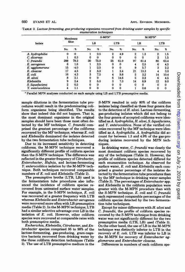
TABLE 3. Lactose-fermenting, gas-producing organisms recovered from drinking water samples by specificenumeration techniques
S-MPNa M-MPNaMembrane
Isolate filter LB LTB LB LTB
No. % No. % No. % No. % No. %
A. hydrophilia 0 0 1 2.5 2 4.9 2 1.3 2 1.5C. diversus 1 0.3 0 0 0 0 0 0 2 1.5C. freundii 298 79.3 30 75.0 25 61.0 97 61.4 85 63.4E. aerogenes 6 1.6 1 2.5 0 0 4 2.5 6 4.5E. agglomerans 15 4.0 1 2.5 0 0 9 5.7 3 2.2E. cloacae 17 4.5 2 5.0 1 2.4 21 13.3 4 3.0E. coli 16 4.3 3 7.5 4 9.8 5 3.2 14 10.4H. alvei 8 2.1 0 0 6 14.6 5 3.2 6 4.5Klebsiella 9 2.4 2 5.0 3 7.3 14 8.9 12 9.0S. liquefaciens 2 0.5 0 0 0 0 0 0 0 0Y. enterocolitica 4 1.1 0 0 0 0 1 0.6 0 0
a Parallel MPN analyses conducted on each sample using LB and LTB presumptive media.
sample dilutions in the fermentation tube pro-cedures would result in the predominating coli-form organisms being identified. The resultsshow that indeed this did not happen. That is,the most dominant organisms in the originalsamples should have been those most often de-tected by the MF technique. C. freundii com-prised the greatest percentage of the coliformsrecovered by the MF technique, whereas E. coliand Klebsiella dominated the isolates detectedby the two fermentation tube techniques.Due to its increased sensitivity in detecting
coliforms, the M-MPN technique recovered asignificantly different profile of coliform speciesthan the S-MPN technique. The difference wasreflected in the greater frequency of Citrobacter,Enterobacter, Hafnia, and lactose-fermentingY. enterocolitica isolation by the M-MPN tech-nique. Both techniques recovered comparablenumbers of E. coli and Klebsiella (Table 2).The presumptive broths (LTB, LB) used in
the fermentation tube procedures also influ-enced the incidence of coliform species re-covered from untreated surface water samples.For example, in the S-MPN technique, E. coliwas recovered at a greater frequency with LTBwhereas Klebsiella and Enterobacter aerogeneswere recovered more often with LB presumptivemedia (Table 2). In the M-MPN technique, LTBhad a similar selective advantage over LB in theisolation of E. coli. However, other coliformspecies were recovered at comparable rates withboth presumptive media.
Escherichia, Klebsiella, Citrobacter, and En-terobacter species comprised 95 to 98% of thelactose-fermenting, gas-producing, gram-nega-tive bacteria recovered from drinking water bythe three coliformn detection techniques (Table3). The use of LTB presumptive medium in the
S-MPN resulted in only 80% of the coliformisolates being classified as these four genera dueto the detection of H. alvei. Lactose-fermenting,gas-producing isolates which did not belong tothe four genera of accepted coliforms were iden-tified asA. hydrophilia, H. alvei, S. liquefaciens,and Y. enterocolitica. None of the verified col-onies recovered by the MF technique were iden-tified as A. hydrophilia. A. hydrophilia did ac-count for between 1.3 and 4.9% of the gas-pro-ducing isolates recovered by the MPN tech-niques.
In drinking water, C. freundii was clearly themost dominant coliform species recovered byany of the techniques (Table 3). However, theprofile of coliform species detected differed foreach enumeration technique. As observed forsurface water, E. coli and Klebsiella each com-prised a greater percentage of the isolates de-tected by the fermentation tube procedures thanby the MF technique in drinking water samples(Table 3). The percentages of Enterobacter spp.and Klebsiella in the coliform population weregreater with the M-MPN procedure than withthe S-MPN technique. E. coli and C. freundiieach represented comparable percentages of thecoliform species detected by the two fermenta-tion tube techniques.Except for minor differences with H. alvei and
C. freundii, the profile of coliform species re-covered by the S-MPN technique from drinkingwater was not significantly different for the twopresumptive media (LTB, LB) used (Table 3).On the other hand, the use ofLB in the M-MPNtechnique was distinctly inferior to LTB in therecovery of E. coli. LTB was inferior to LB inthe M-MPN for recovery of Enterobacter ag-glomerans and Enterobacter cloacae.
Differences in numbers of each coliform spe-
660 EVANS ET AL. APPL. ENVIRON. MICROBIOL.
on July 2, 2019 by guesthttp://aem
.asm.org/
Dow
nloaded from
COLIFORM SPECIES RECOVERED FROM WATER 661
cies detected by the M-MPN and S-MPN tech-niques are reflected by the incidence of thespecies isolated from false-negative S-MPNtests. Citrobacter, Enterobacter, Hafnia, Yer-sinia, Escherichia, Klebsiella, Serratia, andAeromonas, in decreasing order of occurrence,have been isolated from false-negative S-MPNtests of untreated surface water (Table 2). Citro-bacter, Enterobacter, Klebsiella, Escherichia,Hafnia, and Yersinia, in decreasing order ofoccurrence, were isolated from false-negative S-MPN tests of drinking water (Table 3).
Different presumptive media also influencedthe coliform species recovered from false-nega-tive S-MPN tests. For example, with surfacewater, E. coli and E. aerogenes were isolatedmore frequently from gas-negative LTB pre-sumptive tubes than from gas-negative LBtubes. The number of C. freundii, E. agglomer-ans, H. alvei, Klebsiella, S. liquefaciens, and Y.enterocolitica isolations from false-negative S-MPN tests was comparable with either pre-sumptive medium. In drinking water samples,E. aerogenes and E. coli were isolated morefrequently from false-negative S-MPN testswhen LTB presumptive medium was used andE. agglomerans, E. cloacae, and H. alvei whenLB presumptive medium was used. C. freundiiand Klebsiella were isolated at comparable rateswith either presumptive medium.
DISCUSSIONThe identification of coliforms isolated from
high-quality surface water and drinking waterby the MF, S-MPN, and M-MPN techniquesdemonstrated that each technique selected fora specific group of coliforms. A comparison ofthe coliform species identified from the samesamples indicated that the most numerous spe-cies recovered by the MF technique was C.freundii, whereas the two fermentation tubetechniques displayed a selectivity for E. coli andKlebsiella. These results differed from thosereported by Dutka and Tobin (9) who foundthat the MF technique relative to the S-MPNprocedure selected for E. coli and Klebsiellaspp. in creek water. Several factors may explainwhy the MF and the two fermentation tech-niques in this study recovered a different profileof coliform species from that reported by Dutkaand Tobin. The degree of pollution in the stream(geometric mean of 28,000 total coliforms per100 ml) studied by Dutka and Tobin was greaterthan in the stream (geometric mean of 160 totalcoliforms per 100 ml) examined in this investi-gation. In addition, Dutka and Tobin reportedthat Citrobacter was not among the coliformgenera isolated from creek water by the MF
technique. Nevertheless, the results of this andother investigations (9, 23) support the conclu-sion that the type of water examined and thetype of pollution in that water influences theselectivity patterns of the various coliform de-tection techniques.A significant difference was found in the pro-
files of coliform species recovered from drinkingwater and from the untreated surface watersource. For example, C. freundii, E. coli, andKlebsiella were the dominant coliform-speciesrecovered from untreated surface water, whereasC. freundii alone was the dominant species re-covered from contaminated drinking water sam-ples. All coliform detection techniques reflectedthis change in the profile of coliforin speciesobtained from the two types of water supplies.The presence of high numbers of C. freundii incontaminated drinking water samples may re-flect a resistance to chlorine by this organismrelative to other coliforms. High numbers of thehydrogen sulfide-negative biotype of C. freundiihave been reported in other aquatic environ-ments exposed to chlorine (7).The observed differences in the profiles of
coliforn species obtained by the MF and S-MPN techniques were more pronounced for sur-face water than for drinking water samples.These observations are consistent with colifornenumeration studies where the geometric meancoliform numbers recovered by the MF and S-MPN were the same for drinking water, but forsurface water the MF geometric mean wasnearly threefold higher than the S-MPN (11).The primary reason for the difference in geo-metric mean numbers for surface waters was theincreased detection of Citrobacter by the MFtechnique. The more pronounced differences inthe profiles of coliforms obtained by the M-MPN and S-MPN were caused by failures in theS-MPN to recover as many coliforn species insome 85 to 90% of the drinking water and surfacewater samples (11). Coliforms most frequentlyrecovered from false-negative S-MPN tests andthose which were responsible for the differencesin species profiles were Citrobacter, Enterobac-ter, and Klebsiella. The increased percent recov-ery of these genera should not obscure the factthat the M-MPN also outperforned the S-MPNin the actual number of E. coli isolates detected.In addition, the number of detectable coliform-positive drinking water samples based on the S-MPN or MF techniques was doubled by the M-MPN (11). This doubling in incidence and thefivefold increase in the M-MPN geometric meanover standard techniques was a result of theincreased detection of the three coliform genera.A 9- or 15-tube fermentation tube technique
VOL. 41, 1981
on July 2, 2019 by guesthttp://aem
.asm.org/
Dow
nloaded from
662 EVANS ET AL.
was used in the present study whereas drinkingwater-monitoring laboratories have the optionof using a 5-tube procedure. The majority of thefalse-negative S-MPN tests observed with con-taminated drfinking water samples occurred inthe fermentation tubes inoculated with 10-mlportions. Therefore, the selectivity patterns ob-served in the S-MPN in this investigation wouldoccur in monitoring laboratories as well.The nature of the enrichment processes oc-
curring in the two fermentation tube techniquesmay explain why the S-MPN and M-MPN pro-cedures recovered different groups of coliformspecies. Selection against a certain coliform spe-cies in the fermentation tube procedure mayresult from failure to produce gas in presumptivemedia due to unfavorable nutritional conditions(3, 5), competition for lactose (21), or the pro-duction of inhibitory products by noncoliformbacteria (16), and from the inhibitory nature ofthe confirmatory medium (22), or the failure toproduce typical colonies on the agar mediumused in the completed step (11).The use of different presumptive media in the
fermentation tube techniques and the use of m-Endo agar LES in the MF technique also influ-enced the selectivity patterns. LB, LTB, and m-Endo agar LES all yielded different profiles ofcoliform species. Other investigators have alsoreported that the media used in coliform detec-tion techniques greatly influenced the profile ofcoliforms obtained (4, 8, 9).Data presented here and elsewhere (11) indi-
cated that the selection for or against certaincoliformn species, i.e., Citrobacter, Enterobacter,and Klebsiella species, influenced the numberand species of coliforms detected in contami-nated drinking water samples and influencedcompliance with the Safe Drinking Water Act.The use of different coliform detection method-ologies will result in different numbers of coli-forms being recovered from aquatic environ-ments (raw sewage, sewage effluent, creeks, andchlorinated drinking water), where different col-iforn species predominate (17). Additional stud-ies must be conducted to determine if the selec-tion for or against certain coliform species in theS-MPN technique and the resulting false-nega-tive results are commonly occurring phenomena.Such investigations would underscore the neces-sity for revising current MF and MPN coliformenumeration techniques.The failure of the S-MPN technique to detect
Enterobacter and Citrobacter species in chlori-nated drinking water is disturbing and couldresult in such supplies being classified as potable.The occurrence of any coliforn species in drink-ing water should always cause concern. Citro-bacter and Enterobacter are present at 105 to
APPL. ENVIRON. MICROBIOL.
106 cells per gram of human feces (18) and aretherefore also present in sewage. Their presenceis at least a signal of inadequate raw watertreatment or perhaps is a result ofcontaminationfrom within the distribution system. Therefore,any selection against these or any other coli-forms by a coliform detection technique couldhave public health significance. The possibilityofselection for or against certain coliform groupsshould not be overlooked when new and moresensitive coliform detection techniques are beingdeveloped.
ACKNOWLEDGMENTSThis research was supported by cooperative agreement CR
806287 from the U.S. Environmental Protection Agency,Drinking Water Research Division. The suggestions and con-tributions of Harry D. Nash, U.S. Environmental ProtectionAgency, Office of Drinking Water Research, were greatlyappreciated.
LITERATURE CITED1. American Public Health Association. 1975. Standard
methods for the examination of water and waste water,14th ed. American Public Health Association, Inc.,Washington, D.C.
2. Bordner, R., and J. Winter (ed.). 1978. Microbiologicalmethods for monitoring the environment. U.S. Environ-mental Protection Agency, Cincinnati.
3. Chambers, C. W. 1950. Relationship of coliform bacteriato gas production in media containing lactose. PublicHealth Rep. 65:619-627.
4. Clark, J. A., and J. E. Pagel. 1977. Pollution indicatorbacteria associated with municipal raw and drinkingwater supplies. Can. J. Microbiol. 23:465-470.
5. Darby, C. W., and W. L Mallmann. 1939. Studies onmedia for coliform organisms. J. Am. Water WorksAssoc. 31:689-706.
6. Davis, B. R., and W. H. Ewing. 1971. Biochemicalcharacterization of Citrobacter freundii and Citrobac-ter diversus. DHEW Publication No. (CDC)76-8283.U.S. Department of Health, Education, and Welfare,Atlanta.
7. Davis, E. M., and S. R. Keen. 1974. Municipal waste-water bacteria capable of surviving chlorination. HealthLab. Sci. 111:268-274.
8. Dutka, B. J. 1973. Coliforms are an inadequate index ofwater quality. J. Environ. Health 36:39-48.
9. Dutka, B. J., and S. E. Tobin. 1976. Study on theefficiency of four procedures for enumerating coliformsin water. Can. J. Microbiol. 22:630-635.
10. Edwards, P. R., and W. H. Ewing. 1972. Identificationof Enterobacteriaceae. Burgess Publishing Co., Min-neapolis.
11. Evans, T. M., C. E. Waarvick, R. J. Seidler, and M.W. LeChevallier. 1981. Failure of the most-probable-number technique to detect coliforms in drinking waterand raw water supplies. Appl. Environ. Microbiol. 41:130-138.
12. Ewing, W. H., and B. R. Davis. 1972. Biochemicalcharacterization of Citrobacter diversus (Barkey/Werkman and Gillen) and designation of the neotypestrain. Int. J. Syst. Bacteriol. 22:12-18.
13. Ewing, W. H., B. RI Davis, M. A. Fife, and E. F.Lessel. 1973. Biochemical characterization of Serratialiquefaciens (Grimes and Hennerty) Bascomb et al.(formerly Enterobacter liquefaciens) and Serratia rub-idaea (Stapp) comb. nov. and designation of type andneotype strains. Int. J. Syst. Bacteriol. 23:217-225.
on July 2, 2019 by guesthttp://aem
.asm.org/
Dow
nloaded from
COLIFORM SPECIES RECOVERED FROM WATER 663
14. Geldreich, E. E., M. J. Allen, and R. H. Taylor. 1978.Interferences to coliform detection in potable watersupplies, p. 13-20. In C. W. Hendricks (ed.), Evaluationof the microbiology standards for drinking water. U. S.Environmental Protection Agency, Washington, D.C.
15. Grimont, P. A. D., F. Grimont, and M. P. Starr. 1978.Serratia proteamaculans (Paine and Stansfield) comb.nov., a senior subjective synonym of Serratia liquefa-ciens (Grimes and Hennerty) Bascomb et al. Int. J. Sys.Bacteriol. 28:503-510.
16. Hutchison, D., R. H. Weaver, and M. Scherago. 1943.The incidence and significance of microorganisms an-
tagonistic to Escherichia coli. J. Bacteriol. 45:29.17. Kabler, P. 1954. Water examinations by the membrane
filter and most probable number procedures. Am. J.Public Health 44:379-386.
18. Leclerc, H., D. A. Mossel, P. A. Trinel, and F. Gavini.1976. Microbiological monitoring-a new test for fecalcontamination, p. 23. In A. W. Hoadley and B. J. Dutka(ed.), Bacterial indicators/health hazards associated
with water. American Society for Testing and Materials,Philadelphia.
19. Popoff, M., and G. H. Stoleru. 1980. Taxonomic studyof Citrobacter freundii biochemical variants. Ann. Mi-crobiol. 131:189-218.
20. Ptak, D. J., W. Ginsburg, and B. F. Willey. 1973.Identification and incidence of Klebsiella in chlorinatedwater supplies. J. Am. Water Works Assoc. 65:604-608.
21. Reitler, R., and R. Seligmann. 1957. Pseudononas indrinking water. J. Appl. Bacteriol. 20:145-150.
22. Ruchhoft, C. C. 1935. Comparative studies of the mediafor determination of the coli-aerogenes group in wateranalysis. J. Am. Water Works Assoc. 27:1732-1745.
23. Shipe, E. L., and G. M. Cameron. 1954. A comparisonof the membrane filter with the most probable numbermethod for coliform determinations from several wa-
ters. Appl. Microbiol. 2:85-88.24. Starr, M. P., and M. Mandel. 1969. DNA base compo-
sition and taxonomy of phytopathogenic and other en-
terobacteria. J. Gen. Microbiol. 56:113-123.
VOL. 41, 1981
on July 2, 2019 by guesthttp://aem
.asm.org/
Dow
nloaded from


![Dec Ov Cy U I U Fine‑T VGG‑16 D L Network81 Page 4 of 8 SN Computer Science (2020) 1:81 SN Computer Science (FRR)of5.7%wereobtained[4 ].Thisworkwasfurther modiedandupdatedbyapplyingedge-basedsegmenta-](https://img.pdfslide.us/doc/110x75/5f0218ef7e708231d4028e86/dec-ov-cy-u-i-u-fineat-vgga16-d-l-network-81-page-4-of-8-sn-computer-science.jpg)