Embed Size (px)
Citation preview
Review paper �9 Springer-Verlag, 1991
Child's Nerv Syst (1991) 7:2-12
Cognitive deficits in long-term survivors of childhood brain tumors Tracy A. Glauser 1 and Roger J. Packer 1,2
1 Division of Child Neurology, The Children's Hospital Philadelphia, and Department of Neurology, University of Pennsylvania, Philadelphia, Pennsylvania, USA 2 Department of Neurology, Children's National Medical Center, George Washington University, Washington, D.C., USA
Received May 25, 1990/Revised August 24, 1990
Abstract. Improvements in survival for patients who had childhood brain tumors has led to an increasing emphasis on the quality of life for these long-term survivors. Initial survival studies relied on global descriptions of function- al abilities to assess cognitive deficits and reported that from 20% to 40% of long-term survivors had obvious partial disability and < 10% were severely disabled. For- mal neuropsychological testing has revealed that from 40% to 100% of long-term survivors have some form of cognitive deficit in various intelligence quotients, visual/ perceptual skills, learning abilities, and adaptive behav- ior. Prospective, controlled studies have found a younger age at diagnosis, radiotherapy, methotrexate chemother- apy, tumor location and time interval to testing to be important (alone or in combination) and related to a high risk of subsequent cognitive deficits. Some variables play an as yet unresolved role. However, despite the progress of the last decade, future prospective studies are needed to define the role of certain variables in the development of cognitive deficits that maximize survival while mini- mizing cognitive deficits.
Key words: Childhood brain tumors - Long-term sur- vivors - Cognitive deficits
Slow, but definite progress has occurred in the treatment of pediatric brain tumors during the past 20 years. As 5- and 10-year survival rates have improved, so has concern over the quality of life and cognitive deficits exhibited by the long-term survivors. This review examines the various studies that have molded our current understanding of this dilemma.
There are three sections in this review. The first re- views the neuropsychological tools used by studies de- signed to detect cognitive sequelae in long-term sur-
Offprint requests to: R.J. Packer, Department of Neurology, Chil- dren's National Medical Center, 111 Michigan Avenue, N.W., Washington, DC 20010-2970, USA
vivors. The second recounts studies published between 1968 and 1989 that depict the incidence and severity of these cognitive deficits. In the third section, the various factors purported to play a role in the pathogenesis of these cognitive deficits are described and discussed.
Assessment of cognitive deficits
Initial methods of evaluation
The first attempts at depicting cognitive deficits in the late 1960s and mid-1970s involving either an analysis of school performance or placement, the presence or ab- sence of "mental retardation" (determined without the use of norm-referenced tests) or a global assessment of daily functioning. Two major classifications based on daily functioning ability were proposed by Bouchard [8] and Bloom et al. [6] (Table 1).
Introduction of norm-referenced tests
Since the late 1970s studies of long-term survivors of childhood brain tumors have employed various norm- referenced tests to document cognitive deficits (Table 2).
General intelligence tests
The most frequently employed category of neuropsycho- metric tests are the general intelligence tests [34]. The Wechsler Intelligence Scale for Children - Revised (WISC-R), useful for children between 6 and 17 years old, calculates three separate intelligence quotients (IQ): a Full Scale IQ (FSIQ), a Verbal Scale IQ (VIQ), and a Performance Scale IQ (PIQ). Patients older than 16 years are assessed using the Wechsler Adult Intelligence Scale - Revised (WAIS-R), which also provides a FSIQ, a VIQ, and a PIQ. A third general intelligence test is the Stan- ford-Binet Intelligence Scale, used in children between the ages of 2 and 24 years.
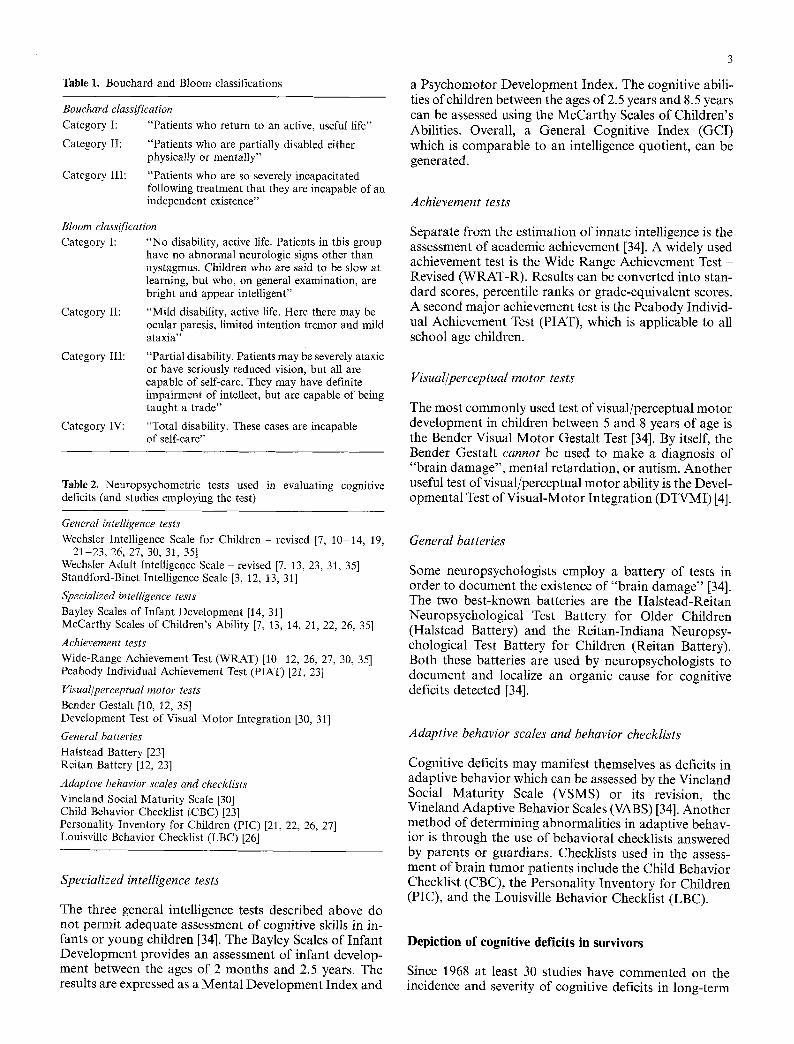
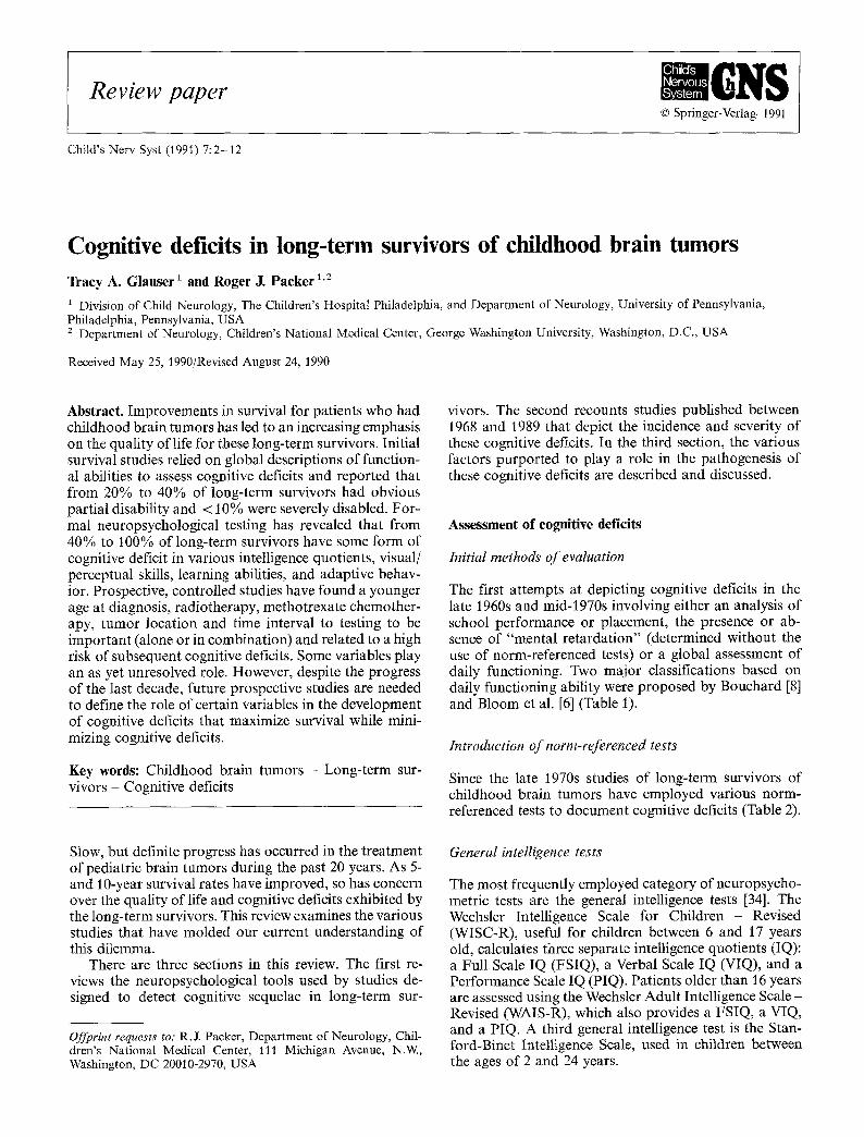
Table 1. Bouchard and Bloom classifications
Bouchard classification Category I: "Patients who return to an active, useful life"
Category II: "Patients who are partially disabled either physically or mentally"
Category III: "Patients who are so severely incapacitated following treatment that they are incapable of an independent existence"
a Psychomotor Development Index. The cognitive abili- ties of children between the ages of 2.5 years and 8.5 years can be assessed using the McCar thy Scales of Children's Abilities. Overall, a General Cognitive Index (GCI) which is comparable to an intelligence quotient, can be generated.
Achievement tests
Bloom classy'cation Category I: "No disability, active life. Patients in this group
have no abnormal neurologic signs other than nystagmus. Children who are said to be slow at learning, but who, on general examination, are bright and appear intelligent"
Category II: "Mild disability, active life. Here there may be ocular paresis, limited intention tremor and mild ataxia"
Category III: "Partial disability. Patients may be severely ataxic or have seriously reduced vision, but all are capable of self-care. They may have definite impairment of intellect, but are capable of being taught a trade"
Category IV: "Total disability. These cases are incapable of self-care"
Table2. Neuropsychometric tests used in evaluating cognitive deficits (and studies employing the test)
Separate f rom the estimation of innate intelligence is the assessment of academic achievement [34]. A widely used achievement test is the Wide Range Achievement Test - Revised (WRAT-R). Results can be converted into stan- dard scores, percentile ranks or grade-equivalent scores. A second major achievement test is the Peabody Individ- ual Achievement Test (PIAT), which is applicable to all school age children.
Visual/perceptual motor tests
The most commonly used test of visual/perceptual motor development in children between 5 and 8 years of age is the Bender Visual Motor Gestalt Test [34]. By itself, the Bender Gestalt cannot be used to make a diagnosis of "brain damage" , mental retardation, or autism. Another useful test of visual/perceptual motor ability is the Devel- opmental Test of Visual-Motor Integrat ion (DTVMI) [4].
General intelligence tests Wechsler Intelligence Scale for Children - revised [7, 10-14, 19,
21-23, 26, 27, 30, 31, 35] Wechsler Adult Intelligence Scale - revised [7, 13, 23, 31, 35] Standford-Binet Intelligence Scale [3, 12, 13, 31]
Specialized intelligence tests Bayley Scales of Infant Development [14, 31] McCarthy Scales of Children's Ability [7, 13, 14, 21, 22, 26, 35]
Achievement tests Wide-Range Achievement Test (WRAT) [10-12, 26, 27, 30, 35] Peabody Individual Achievement Test (PIAT) [21, 23]
Visual/perceptual motor tests Bender Gestalt [10, 12, 35] Development Test of Visual Motor Integration [30, 31]
General batteries Halstead Battery [23] Reitan Battery [12, 23]
Adaptive behavior scales and checklists Vineland Social Maturity Scale [30] Child Behavior Checklist (CBC) [23] Personality Inventory for Children (PIC) [21, 22, 26, 27] Louisville Behavior Checklist (LBC) [26]
Specialized intelligence tests
The three general intelligence tests described above do not permit adequate assessment of cognitive skills in in- fants or young children [34]. The Bayley Scales of Infant Development provides an assessment of infant develop- ment between the ages of 2 months and 2.5 years. The results are expressed as a Mental Development Index and
General batteries
Some neuropsychologists employ a battery of tests in order to document the existence of "brain damage" [34]. The two best-known batteries are the Halstead-Reitan Neuropsychological Test Battery for Older Children (Halstead Battery) and the Rei tan-Indiana Neuropsy- chological Test Battery for Children (Reitan Battery). Both these batteries are used by neuropsychologists to document and localize an organic cause for cognitive deficits detected [34].
Adaptive behavior scales and behavior checklists
Cognitive deficits may manifest themselves as deficits in adaptive behavior which can be assessed by the Vineland Social Maturi ty Scale (VSMS) or its revision, the Vineland Adaptive Behavior Scales (VABS) [34]. Another method of determining abnormalities in adaptive behav- ior is through the use of behavioral checklists answered by parents or guardians. Checklists used in the assess- ment of brain tumor patients include the Child Behavior Checklist (CBC), the Personality Inventory for Children (PIC), and the Louisville Behavior Checklist (LBC).
Depiction of cognitive deficits in survivors
Since 1968 at least 30 studies have commented on the incidence and severity of cognitive deficits in long-term
Table 3. Cognitive deficits in tumor studies (1968-1989)
Study (year, study size) Study format
Age at diagnosis (years)
Follow-up interval (years)
Results
Fessard (1968, n=11) Bloom et al. (1969, n=22)
Jenkin (1969, n=30)
Aron (1969, n = 5)
Marsa et al. (1973, n--18)
Abramson et al. (1974, n=65)
Onoyama et al. (1975, n =42)
Bamford et al. (1976, n=30) Gjerris (1976, n=74)
Harisiadis and Chang (1977, n = 11)
Mealey and Hall (1977, n= 9)
Farwell et al. (1978, n=18)
Raimondi and Tomita (1979, n= 13)
Hirsch et al. (1979, n=33)
Broadbent et al. (1981, n = 8) Berry (1981, n=68)
Spunberg et aL (1981, n= 14)
Danoff et al. (1982, n--38)
Duffner et al. (I983, n = 10)
Raimondi and Tomita (1983, n= 15) Chin and Maruyama (1984, n=23)
Kun et al. (1983, n=30)
Kun et al. (1983, n=18)
Mulhern and Kun (1985, n=26)
Ellenberg et al. (1987, n=43)
Packer et aL (1987, n=24)
Lebaron et al. (1988, n = 15)
Mulhern et al. (1988, n=7)
Duffner et al. (1988, n = 16)
Bordeaux et al. (1988, n = 14)
Packer et al. (1989, n=23)
R , U R , U
R, U
R, U
R , U
R , U
R , U
R , U R , U
P ,U
R , U
R , U
R , U
R ,C
R , U R , U
R, U
R , U
R , U
R , U R , U
R , U
R , U
P , U
P,C
R , U
R , U
P, U
P ,U
P ,C
P,C
<2 <15
<16
<15
9 (median)
<18
<15
<15 <14
6.25 (median)
8 (median)
<1.5
4.5 (median)
5 (median) <14
7 (median)
<2
7.9
3 (median)
<1 1.5-12
6 (median)
6 (median)
7.75
7.5 (mean)
7.75
8.3
8.4 (median)
8 (median)
10
7.7 (median)
2-14 5-17
4 19
1.8-22
1-11
2
3-15
8-18 15-40
5-13
3-13
1 - 2 3
2-10
> I
5-10 5-10
5-19
9.3 (mean)
1
1 - 1 0 8.5
(median) 1.75
(meadian) 1.8, 3.5
and 4.1 0.5
<4
5
1.6 (median)
0.7, 3.0 (median)
3-7
0.1-0.9
2
27% debilitated or backward 18% Bloom III or IV 30% slow learners 3% mental retardation 10% learning problems 40% borderline mental retardation 20% slow learner 39% Bouchard category II 6% retarded, 6% learning difficulty 22% Bouchard category II 1% Bouchard category III 21% Bouchard category II 10% Bouchard category III 43 % "educationally subnormal or severely subnormal" 5% "mentally retarded" 4% "dementia" 36% special education
22% mild mental retardation
22% institutionalized 11% school for retarded children 23% retarded
31% IQ<70, 27% Bloom III or IV 82% specific learning disabilities 38% "frank mental retardation" 12% mental retardation, paraplegia, blindness
44% FSIQ<70, 42% VIQ<70, 60% PIQ<70, 93% learning problems 17% IQ<70, 11% Bloom III or IV 37% emotional/behavioral problems 50% IQ < 80, 100% dementia, mental retardation, or learning problems 33% mental retardation 20% mental deficient
9% FSIQ<70, 11% VIQ<70, 8% PIQ<70, 63% learning disabled 28% FSIQ, VIQ, PIQ < 80 (1st evaluation); no significant change over 3 evaluation FSIQ 99, VIQ 103, PIQ 96, GCI 99; 19% had one or more subtests < 80 9% Bloom IV, 40% Bloom III
FSIQ 97, >50% had learning, fine motor, memory problems Mean FSIQ 77, 20% had FSIQ<70 20% Bloom III or IV 28% FSIQ <70, 71% learning disability 9 point drop in mean IQ over 3 or more years, 94% learning problems FSIQ 104-107, language and fine motor problems pre- and post-therapy RT group: FSIQ dropped 14 points; non-RT group: FSIQ no change
survivors of childhood brain tumors. Many of the earlier reports focused on factors involved in survival rather than long-term cognitive deficits and tended to be retro- spective and uncontrolled and relied on global descrip- tions of cognitive deficits. This section will review studies (beginning in 1968) that depict the cognitive deficits seen in long-term survivors of childhood brain tumors (Table 3).
In 1968, Fessard reported that 45% (5/11) of his long- term survivors were normal (all with posterior fossa tu- mors), but 27% (3/11) were deemed to have "debility or backwardness" [16].
In a landmark study published in 1969, Bloom de- scribed the outcome of 22 long-term survivors with prim- itive neuroectodermal tumor/medulloblastoma (PNET) [6]. All underwent surgery, most finished a complete ra- diotherapy course, and none received chemotherapy. Eighty-two percent (18/22) of the long-term survivors were either Bloom category I or II, 9% (2/22) category III, and 9% (2/22) category IV.
Two other studies published the same year shed light on the cognitive deficits seen in survivors of childhood PNET. Jenkin reported only 1 of 47 patients (3%) showed "progressively more evidence of mental retarda- tion" and 3 (10%) "dropped at least one grade during their subsequent school!ng" [20]. Aron described the out- come of 5 long-term survivors of PNET (treated with craniospinal radiation) and found 2 of 5 patients (40%) were either "borderline mental defective" or had "mental status slightly below normal" and one (20%) was a "slow learner" [2].
Bouchard's functional categories
Between 1973 and 1975 three articles appeared which utilized the Bouchard categories to describe long-term outcome. In 1973 Marsa reviewed the results of 18 chil- dren with astrocytic gliomas in different locations, treat- ed with different degrees of surgical resection and differ- ing amounts of radiotherapy [24]. Thirty-nine percent (7/18) had a fair quality of life (Bouchard category II), while the remainder were leading normal lives. Six per- cent (1/18) were mentally retarded and 6% (1/18) had learning difficulties.
The outcome of 65 long-term survivors of a variety of brain tumors treated in a heterogeneous fashion was de- scribed by Abramson et al. [1]. Twenty-two percent (14/ 65) were felt to be in Bouchard category II and 1% (1/65) was placed in category III. In 1975, Onomaya document- ed the Kyoto University Medical Center experience with 42 long-term survivors of childhood brain tumors; 21% (9/42) were Bouchard category II, 10% (4/42) were cate- gory III, and the remaining 69% (29/42) were category I [28].
Bamford's change in approach: focusing on deficits
In 1976, among an "unselected sample of 30 children," out of 64 long-term survivors Bamford et al. found 10%
(3/30) to be superior, 33% (10/30) average, 13% (4/30) below average, 7% (2/30) "severely subnormal" and 37% (I 1/30) "educationally subnormal" [3]. Emotional prob- lems, including depression, lability, suicide attempts, and aggressive behavior, plagued 43% (13/30) of the sur- vivors. Overall, 80% (24/30) had a residual problem with 20% (6/30) profoundly disabled.
End of the summary studies
Multiple articles appeared in the late 1970s that shed additional light on the incidence and magnitude of cogni- tive deficits in long-term survivors despite not employing formal neuropsychological testing. Gjerris found cogni- tive deficits, including "dementia" in 4% (3/74) and "mental retardation" in 5% (4/74) of patients evaluated [17]. Overall, 4% (3/74) were in need of nursing home care and 16% (12/74) "were receiving disablement pen- sion." Harisiadis found 36% (4/11) of long-term sur- vivors needed special education and exhibited language problems [18].
In 1977, Mealey concluded that 22% (2/9) of long- term survivors "required special education for mild men- tal deficiency," 67% were normal, and 11% (1/9)had a "nonincapacitating cerebellar ataxia" [25]. Among 18 children who survived longer than 1 year, Farwell found 22% to be "institutionalized" and 11% attending "schools for retarded children" [15].
Raimondi reported, in 1979, that 38% (5/13) of chil- dren surviving more than 2 years following diagnosis of PNET were "living completely normal lives," and 23 % (3/13) were noted to have "psychomotor retardation" [32]. A second article by Raimondi [33], focused on 15 long-term survivors of primary intracranial neoplasms. Overall, 33 % of the long-term survivors were "retarded"; all had received radiotherapy. The 6 patients who did not undergo radiotherapy had either teratomas or papillo- mas and were normal.
Introduction of detailed neuropsychologic testing
Formal neuropsychological testing was used by Hirsch in 1979 to detect cognitive deficits in 33 long-term survivors of PNET [19]. All underwent surgery and a course of radiotherapy. Craniospinal radiation consisted of 5000 rads to the posterior fossa and 3500 rads to the cerebral hemispheres and spinal cord (children under 3 years re- ceived 500 rads less to each region). Chemotherapy was used extensively, including intrathecal methotrexate, vin- cristine and 1-(-2-chloroethyl)-3-cyclohexyl-l-nitrosourea (CCNU).
Hirsch employed the WISC and two neuropsycholog- ical tests not used in the United States. He found 31% (8/26) had an IQ<70, 58% (15/26) had an IQ<90, and the remaining 11% (3/26) had an IQ > 90. Emotional and behavioral disorders were very common in the survivors, occurring in 93% (26/28). Fifteen percent of long-term survivors (5/33) were in Bloom category III and 12% (4/33) were in category IV. A major feature of Hirsch's

article was the use of a "control group" of 31 children with cerebellar astrocytoma. Among the astrocytoma group 19% (6/31) had an IQ< 70, 19% (6/31) had an IQ between 70 and 90, and the remaining 62% (19/31) had an IQ > 90. Emotional and behavioral disorders occurred in 59% (19/31).
PNET review articles
Broadbent et al. reported that out of 8 long-term sur- vivors with confirmed PNET, treated by surgery and then craniospinal irradiation, 62% (5/8) were at "normal schools" while the other 38% (3/8) showed evidence of "frank mental retardation" [9].
Berry and Jenkin described 68 long-term survivors and noted that 12% (8/68) exhibited "major deficits: mental retardation, paraplegia, and blindness" while 28% (19/68) were reported to have had unspecified "moderate neurological deficits" [5, 20].
NeuropsychoIogic testing and the 1980s
The two most important changes which occurred in the study of cognitive deficits during the first half of the 1980s were the increasing use of neuropsychological test- ing and the development of prospective studies.
The first study of this group was published by Spun- berg et al. [35]. Among 14 survivors of primary brain tumors (43% supratentorial, 57% infratentorial), diag- nosed under the age of two years, 79% (11/14) had under- gone surgery and all had received localized radiotherapy. Intelligence testing revealed 44% (4/9) had FSIQ < 70, 93% (12/13) had learning problems, and 46% (6/13) needed special education.
Danoff et al. (1982) reported the cognitive results of 38 children with a variety of brain tumors (71% supraten- torial, 29% infratentorial) [11]. All but 1 patient under- went surgery, all patients received radiotherapy, and none received chemotherapy. The radiotherapy ranged from 4000 to 6500 rad with 82% (31/38) receiving 5000 to 5600 rad. Seventeen percent (6/36) had an IQ < 70; 37% (14/36) had "emotional problems and behavioral distur- bances"; 11% (4/38) were classified as Bloom category III or IV.
Duff her et al. described the cognitive deficits in a group of 10 long-term survivors of posterior fossa tumors; all had undergone surgery, radiotherapy, and chemother- apy [12]. Radiotherapy included 2000 to 5040 rad to the posterior fossa with 2635 to 4000 rad to the whole brain. All patients received both intrathecal and intravenous methotrexate, intravenous vincristine, BCNU, and dex- amethasone. Fifty percent (5/10) had an IQ below 80, including 2 with IQ < 20. Duffner et al. found that all patients surveyed suffered from either dementia, learning disabilities, or mental retardation. A second study by Duffner et al. showed a drop in mean IQ among 16 chil- dren with brain tumors (63% supratentorial, 37% in- fratentorial) from a pretreatment level of 93.5 to 84.5 three or more years later [13]. All had undergone surgery
and radiotherapy, but only 31% (5/16) received chemotherapy (methotrexate in only 2 patients). Ninety- four percent (15/16) had learning disabilities and 69% needed special education.
In 1984, Chin and Maruyama reported the neuropsy- chological performance results of 10 children with PNET [10]. All had undergone postoperative radiation therapy. Passing note was made of the use of chemotherapy in these children without a full description of type, route of administration, or dose. Both patients (20%) less than 4 years old were deemed "mental deficient". Although this article used multiple neuropsychological tests, neither summary data nor analysis based on FSIQ, VIQ, or PIQ was presented.
Long-term cognitive deficits in a series of 24 children with only PNET treated between 1975 and 1984 was re- ported by Packer et al. in 1988 [30]. Following surgery, patients > 2 years old received 4000 cGy (median) cra- niospinal radiation with a local tumor boost to 5200 cGy (median) while children < 2 years old received between 1800 and 2000 cGy craniospinal radiation with a local tumor boost to between 4500 to 4600 cGy. Chemothera- py included CCNU, vincristine, and prednisone, but none received intrathecal medication. Neuropsychologi- cal tests revealed a median FSIQ (n = 17) of 97 (range 20-126), median VIQ of 101 and median PIQ of 94. Fifty-four percent (13/24) were in special education placement. Vineland social maturity scale assessments were abnormal in 25% (2/8) with a mean score of 88.
Lebaron et al. retrospectively examined the quality of life in 15 long-term survivors of mixed posterior fossa tumors [23]. All underwent surgical resection but only 11 had irradiation. One patient received an unknown type and amount of chemotherapy in addition to the radio- therapy. The mean FSIQ, VIQ, and PIQ were 77, 77, and 81, respectively; 20% (3/15) had FSIQ below 70. Both major general neuropsychological batteries were em- ployed. Two subtests involving cognitive flexibility (The Category Test and Trail Making Test B) revealed serious impairment in 20% (3/15) and 72% (5/7) of the children, respectively. Most (11/15) were either in special educa- tion, had repeated a grade, or had not returned to school.
Mulhern et al. reported the neuropsychologic func- tioning of 7 children with temporal lobe astrocyomas treated between 1975 and 1984 [27]. Following surgery, all underwent local radiotherapy (5000 to 5800 cGy) and 1 had 3600 cGy to the whole cranium. Twenty-eight per- cent (2/7) had FSIQ<70 while 43% (3/7) had 70 < FSIQ < 90; 43% (3/7) exhibited intellectual deterio- ration in FSIQ over the time of the study. Special educa- tional classes were needed in 71% (5/7) and 14% (1/7) were deemed "educable mentally retarded."
Prospective studies
Kun and Mulhern detailed the neuropsychological alter- ations over time in long-term survivors in a series of articles published between 1983 and 1985. The first arti- cle, in 1983, scrutinized 30 children with primary brain tumors (50% supratentorial, 50% infratentorial) treated
between 1979 and 1981 [22]: 87% (26/30) underwent surgery and 70% (21/30) received radiation therapy rang- ing from 4000 to 5800 rad. Adjuvant chemotherapy (BCNU, vincristine, and cyclophosphamide) was used in 3 patients. Overall, 9% (2/23) had FSIQ <70 and 7% (2/30) had memory scores below 70. Kun determined abnormal social-emotional functioning using the PIC and found that 62% (13/21) of the children tested scored in the "at risk" or "deviant" range. Sixty-three percent (10/16) of the patients needed special education. Their second article examined 18 children with serial neuropsy- chologic evaluations [21]. Initially, 28% (5/18) had FSIQ < 80 and 28% (5/18) had memory scores below 80; these percentages did not significantly differ at the second eval- uation.
The last article in this series prospectively examined 26 children with primary intracranial neoplasms (58% supratentorial, 42% infratentorial) [26]. Surgery was per- formed in 88% (23/26) of the cases and radiotherapy in 96% (25/26). Initial evaluations, performed post-surgery and preradiotherapy, revealed normal FSIQ, VIQ, PIQ, and CGI. Six months after radiotherapy was completed, the FSIQ, VIQ, PIQ and GCI were essentially un- changed. PIC or LBC revealed between 42% and 50% of the children were manifesting symptoms of emotional disturbance.
In the 1987 prospective study of Ellenberg et al., the intellectual outcome of 43 patients with primary intracra- nial neoplasms (49% supratentorial, 51% infratentorial) was serially assessed [14]. Ninety-three percent (40/43) underwent craniotomy and 86% (37/43) received radio- therapy, 43 % (14/43) of the patients received chemother- apy, including cytoxan [9], vincristine [9], prednisone [10], procarbazine [10], CCNU [3], 5-fluorouricil [2], but not methotrexate; 9% (4/43) of the patients were Bloom cat- egory IV, 40% (17/43) category III, 33% (14/43) category II, and 19% (8/43) category I.
Bordeaux et al. serially evaluated 14 children with pri- mary brain tumors treated between 1983 and 1985 [7]. Fifty percent (7/14) only had surgery while the other 50% (7/14) were treated by surgery followed by radiotherapy. Bordeaux analyzed the two groups separately. The 7 chil- dren treated by surgery alone (median age 124 months) had normal pre- and postsurgical FSIQ, VIQ, PIQ, and WRAT scores. Fine-motor speed, visual motor construc- tion, and psychomotor speed were statistically signifi- cantly lower than age norms, but no statistically signifi- cant changes over time were detected for any variable. In fact, mean scores improved after surgery in 22 out of 26 test variables.
All 7 children treated with surgery and then radiation received local irradiation; 4 also underwent-whole-brain irradiation, and 3 were given spinal radiotherapy. The mean pre- and postradiation FSIQ, VIQ, and PIQ were normal. Expressive language skills and fine-motor speed were statistically significantly lower than age norms, but there was no statistically significant change over time. In fact, group scores improved in 12 out of 26 test variables after radiation.
Packer conducted a prospective controlled study of the effects of radiotherapy in 18 children (median age 7.7
years) treated between 1983 and 1986. All patients under- went surgery, followed by radiation therapy. Children between 18 and 36 months of age at diagnosis received 2400 cGy of whole-brain radiotherapy plus a boost of 2400 to 2600 cGy to the primary tumor site. Children above 36 months of age at diagnosis received 3600 cGy of whole-brain radiotherapy plus a boost of 1800 to 2000 cGy to the tumor site. Seventy-two percent (13/18) of the patients received chemotherapy, including cis-plat- inure, CCNU, and vincristine. Neuropsychological test- ing revealed baseline FSIQ, VIQ, and PIQ for this group were 105, 109, and 102, respectively. By year 1 the FSIQ, VIQ, and PIQ had all dipped to 97, 106, and 97, respec- tively. However, by the 2-year mark the FSIQ, VIQ, and PIQ had further dropped to 91,102, and 97, respectively. Two years following therapy, 14% (2/14) had FSIQ < 70. Visual motor impairments were seen initially in 18% (2/ 11) and after 2 years 43% (6/14) exhibited impairment. Sixty-seven percent (12/18) of this group required special education. Packer's control group consisted of 14 pa- tients with cerebellar astrocytomas. None of the children received any radiation or chemotherapy. Baseline IQ scores were FSIQ 105, VIQ 109, and PIQ 98. After 1 year the scores were FSIQ 109, VIQ 115, and PIQ 101. After 2 years FSIQ was 106, VIQ 108, and PIQ 109. There was no increase during the 2 years of the study in the 14% to 21% of children exhibiting either fine motor, visual motor, memory, or language impairments at baseline. Seven percent of the control group required special edu- cation.
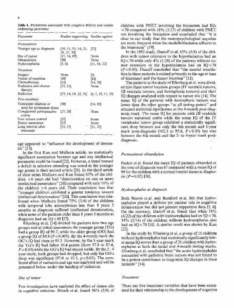
Parameters associated with cognitive deficits
The variables purported to be related to cognitive deficits can be divided into three groups: pretreatment, treat- ment, and post-treatment (Table 4).
Pretreatment variables
There are four pretreatment variables that have been ex- amined for their relationship to the development of cog- nitive deficits: age at diagnosis, site of tumor, obtunda- tion, or hydrocephalus.
Age at diagnosis
Danoff reported an IQ < 70 in 60% (3/5) of the children between 1 and 3 years old, 11% (3/26) of the children between 3 and 11 years old, and 0% (0/5) of the group older than 11 years old [t 1]. Chin and Maruyama found that patients less than 4 years old at the time of therapy had "major learning disabilities," while patients older than 8 years old had "no major intellectual deficits" [10]. Packer et al. reported a trend toward association between lower FSIQ and age less than 7 years old at diagnosis. The mean IQ for children less than 7 years old was 92, between 7 and 10 years old it was 93, and for patients over 10 it was 104 [30]. Duffner et al. reported that a younger
8
Table 4. Parameters associated with congitive deficits and studies addressing parameter
Parameter Studies supporting Studies against
Pretreatment Younger age at diagnosis
Site of tumor Obtundation Hydrocephalus
Treatment Surgery Extent of resection Chemotherapy Radiation and chemo-
therapy Radiation
Post-treatment Ventricular dilation or
need for permanent shunt Complicated postoperative
course Poor seizure control Tumor recurrence Long interval after
treatment
[10, 11, 13, 14, 21, [22] 26, 27, 30] [11, 14, 19] None [30] None [3, 6] [11, 14, 221
None [7] [30] [i4] [13] [14, 30] [12, 13] None
[113,.14, 19, 22, 31] [6;7, 10, 11, 35]
[301 [14, 19]
[27, 30 ] None
[27] None [27] None [13, 31] [21, 22]
age appeared to "influence the development of demen- tia" [13].
In the first Kun and Mulhern article, no statistically significant association between age and any intellectual parameter could be found [22]. However, a trend toward a deficit in selective attending was noted in the younger age group in their second article [21]. In the third article of their series Mulhern and Kun found 63% of the chil- dren < 6 years old had "deterioration on one or more intellectual parameters" [26] compared with only 11% of the children > 6 years old. Their conclusion was that "younger children exhibited a greater tendency toward intellectual deterioration" [26]. This conclusion was rein- forced when Mulhern found 75% (3/4) of the children with temporal lobe astrocytomas less than 8 years 5 months at diagnosis suffered intellectual deterioration, while none of the patients older than 8 years 5 months at diagnosis had an IQ < 80 [27].
Ellenberg et al. [14] divided his patients into two age groups and at initial assessment the younger group (YG) had a group IQ of 99.7, while the older group (OG) had a group IQ of 84.9 (P < 0.005). By the 4-month mark the OG's IQ had risen to 95.5. However, by the l-year mark the YG's IQ had fallen 10.4 points (from 97.8 to 87.4, P < 0.05) while the OG's IQ had stayed stable. By the 2 - 4 year mark, both groups had dropped, but only the OG's drop was significant (97.0 to 92.5, p<0.05). The com- bined effect of radiation and age was analyzed and will be presented below under the heading of radiation.
Site o f tumor
Few investigators have analyzed the effect of tumor site in cognitive outcome. Hirsch et al. found 56% (5/9) of
children with PNET involving the brainstem had IQs < 70 compared with 18 % (3/17) of children with PNET not involving the brainstem and concluded that "it is clear in our study that the neuropsychological sequelae are more frequent when the medulloblastoma adheres to the brainstem" [19].
In the 1982 study, Danoff et al. 62% (5/8) of the chil- dren with tumor extension to the hypothalamus had an IQ < 70 while only 4% (1/28) of the patients without tu- mor extension to the hypothalamus had an I Q < 7 0 (P<0.05). Danoff concluded that "the mental retarda- tion in these patients is related primarily to the age at time of treatment and the tumor location" [11].
The patients in the study of Ellenberg et al. were divid- ed into three tumor location groups (IV ventricle tumors, III ventricle tumors, and hemispheric tumors) and their IQ changes analyzed with respect to tumor site [141. The mean IQ of the patients with hemispheric tumors was lower than the other groups "a t all testing points" and attained statistical significance at the 4-month post-diag- nosis mark. The mean IQ for patients with III ventricle tumors remained stable while the mean IQ of the IV ventricular tumor group exhibited a statistically signifi- cant drop between not only the 4th month and l-year mark post-diagnosis (102.3 to 95.8, P<0.05) but also between the 4th month and the 2- to 4-year mark post- diagnosis.
Pretreatment obtundation
Packer et al. found the mean IQ of patients obtunded at the time of diagnosis was 67 compared with a mean IQ of 99 for the children with a normal mental status at diagno- sis ( P = 0.02) [30].
Hydrocephalus at diagnosis
Both Bloom et al. and Bamford et al. felt that hydro- cephalus played a definite yet unclear role in cognitive deterioration but did not present supportive data [3, 6]. On the contrary, Dafi0ff et al. found that while 18% (4/22) of the children with hydrocephalus had an IQ < 70, 14% (2/14) of the children without hydrocephalus also had an IQ < 70 [11]. A similar result was shown by Kun [221.
In the study by Ellenberg et al. a group of 11 children without hydrocephalus had statistically significantly low- er mean IQ scores than a group of 21 children with hydro- cephalus at both the initial and 4-month testing marks. Ellenberg et al. concluded that "the acute hydrocephalus associated with pediatric brain tumors was not found to be a potent contributor to long-term IQ changes in these patients" [14].
Trea tmen t
There are five treatment variables that have been exam- ined for their relationship to the development of cognitive
deficits: surgery, extent of resection, nonmethotrexate chemotherapy, radiation and methotrexate chemotherapy, and radiation.
Surgery
Bordeaux's 1988 study concluded that surgery itself was not associated "with acute effects on neuropsychological functions" since there was no statistically significant dif- ference between presurgical test scores and postoperative scores [71.
Extent of resection
Packer et al. reported patients with a total resection had a mean IQ of 98 compared to a mean IQ of 60 in patients with less than a total resection (P<0.025) [30]. In con- trast, Ellenberg et al. found "no significant difference in mean IQ between" patients with gross total resection and children having either a partial resection or biopsy "at I or 4 months postoperation or at long term follow-up" [14]. In the Ellenberg et al. study mean IQ scores for both the gross total resection group and the partial resection/ biopsy group ranged from approximately 88 to 98 at all testing points.
Nonmethotrexate chemotherapy
Methotrexate is a well-known neurotoxic chemothera- peutic agent and will be discussed in more detail in the next section. Ellenberg et al. found no significant differ- ence in mean IQ among the 9 patients who received non- methotrexate chemotherapy (cytoxan, vincristine, pred- nisone, procarbazine, CCNU, and 5-fiuoruracil) and the 25 children who received no chemotherapy at either "4 months postdiagnosis".. ."or at the 1 to 4 year follow-up examination" [14]. Packer et al. evaluated nonmethotrex- ate chemotherapy as a factor in cognitive outcome and did not find any statistically significant associations [30]. However, in Duffner's second study three patients treat- ed with nonmethotrexate-containing regimens suffered IQ drops of 14, 28, and 9 over 3 years. Duffner et al. concluded that even nonmethotrexate chemotherapy was a "significant factor in treatment associated dementia" [13].
Radiation and methotrexate chemotherapy
An important subsection of the radiation therapy contro- versy (elaborated on at length in the next section) is the effect on cognitive function when radiotherapy is com- bined with methotrexate (either intrathecal or intra- venous). Duffner found in her first study that all 10 of the patients treated with both methotrexate (both intrathecal and intravenous) and radiotherapy had either "dementia, learning disabilities, or evidence of intellectual retarda- tion" [12]. In her second study the combination of radio-
therapy and methotrexate in 2 patients led to IQ drops of 14 and 29 points. Duffner et al. concluded that "chemo- therapy, particularly a regimen involving methotrexate, appears to be a significant factor in treatment associated dementia" [13].
The combination of radiotherapy and methotrexate can give rise to the syndrome of necrotizing leukoen- cephalopathy (NP) [29]. NP consists clinically of a variety of symptoms (including developmental regression, de- mentia, spasticity, ataxia, seizures, hemiplegia, and pseu- dobulbar paresis), beginning usually 4 to 12 months after completing radiotherapy. Histologically, NP is charac- terized by multifocal areas of coagulation necrosis in the deep white matter with diffuse reactive astrocytosis. Al- though NP can occur in patients undergoing radiothera- py alone or receiving methotrexate alone, the combina- tion of the two treatment modalities significantly elevates the incidence of NP.
Radiation
Since radiation therapy (RT) was first shown to increase survival, its long-term effects on cognitive function have been very controversial. Bloom et al. felt that "serious late changes in the central nervous system" due to radia- tion "appear to be rare" [6].
Hirsch evaluated the effect of RT on cognitive devel- opment by comparing the group of 26 children with PNET with a "control" group of 31 children with astro- cytoma (treated only surgically) [19]. Hirsch et al. found a statistically significantly higher proportion of the PNET group had an IQ between 70 and 90, emotional and behavioral disorders, difficulties in spatial orientation, speech, writing, or reading, and academic failures. Con- versely, a statistically significant higher proportion of as- trocytomas had an IQ > 90. Although 31% of the PNET group had an IQ < 70 compared with 19% for the astro- cytoma group, this difference did not reach statistical significance. Hirsch et al. felt that the deficits in the PNET group were "probably due to the action of ionizing radi- ations on the vessels and cerebral parenchyma" [19].
Two articles published in the early 1980s refuted the damaging effects of RT. Spunberg et al. concluded that "the developing brain may not be as sensitive to irradia- tion as previously believed and that children, even under the age of 2, as in our series, tolerate radiotherapy to the brain surprisingly well" [35]. Danoffet al. also found "no apparent correlation between the volume of brain irradi- ated and mental retardation, as all patients who were mentally defective received local field treatment" [11]. However since 82% of the patients received between 5000 and 5600 rads, Danoff et al. were unable to assess the effect of RT tumor dose on IQ scores.
Conclusions about radiotherapy's cognitive effects in Duffner et al.'s first study were obscured by the use of concurrent methotrexate [12]; clearer conclusions arose from her second study. Duffner et al. found that "the age at the time of cranial irradiation" was a significant factor in "influencing intellectual decline" [13].
Kun and Mulhern found 53% (8/15) of the patients who had received either subtotal supratentorial or cranial
10
RT had one or more "intellectual delays" [22]. In con- trast, only 17% (1/6) of the children with posterior fossa tumors treated with local RT had evidence of intellectual delay. Kun concludes there is "a greater than normal risk for late neuropsychological alterations among children with supratentorial tumors and/or cranial irradiation" [22].
Chin and Maruyama reported only 20% (2/10) of the long-term PNET survivors were deemed mentally defi- cient. His conclusion was "for patients with medulloblas- toma, the incidence of mental or intellectual deficiency occurring after high-dose irradiation has been low" [10]. However, despite using neuropsychological tests, neither summary IQ data nor a comparison between IQ scores and RT tumor dose was presented.
In contrast, Ellenberg et al. [14] divided their 43 study patients into three groups (whole-brain radiation, local tumor irradiation, and no radiotherapy) and analyzed their neuropsychological test results. Four 4 months postoperatively, no difference in mean IQ between the three groups was found. However, over the next 6 months to 4 years the whole-brain RT group showed a statistically significant drop in mean IQ scores (100 to 87.7, P<0.01) while the other two groups' mean IQ scores remained stable. Multivariate analysis showed that "the IQ drops were attributable to radiation therapy rather than to tumor site" [14].
Ellenberg et al. then analyzed the combined effect of age and radiation [14]. The two age groups described in the age subsection above (younger and older) were fur- ther divided by amount of radiotherapy received (whole brain, local, none). At the 4-month postdiagnosis mark, there was no significant difference between the two age groups. However, at the I- to 4-year follow-up evalua- tions, the members of the younger group who had re- ceived whole-brain RT had the lowest mean IQ scores and had a statistically significant decline in IQ scores from 90.6 to 70.9 (P<0.005). Members of the older age group who received whole-brain RT had a trend toward an IQ decline over time (0.1 > P >0.05). Children under 5 years old who underwent whole-brain RT exhibited IQ score drops of 20 points or more. The average IQ drop for children between 6 and 8 years old who received whole- brain RT was approximately 10 points. "No consistent IQ declines" were seen in radiated patients older than 9 years old who had RT. There was essentially no change over time for either age group receiving local or no RT. In conclusion, Ellenberg states that "the most potentially devastating treatment in terms of cognitive sequelae for brain tumor patients is radiation therapy" and "the severity of the effects varies inversely with the age of the child."
Bordeaux concluded that radiotherapy was "not asso- ciated with ,acute effects on neuropsychological func- tions" [7]. However, a brief follow-up time (median 11 months) and small sample size could have been responsi- ble for missing radiation sequelae.
Packer et al.'s 1989 study was a prospective controlled look at the effect of RT on cognition [31]. Compared with baseline testing, the group receiving RT demonstrated a statistically significant decline in multiple IQ scores:
FSIQ at year 1 (P = 0.04), FSIQ at year 2 (P = 0.02), VIQ at year 2 (P = 0.04). In addition, WRAT scores exhibited a statistically significant decline between baseline and year 2 in spelling (P= 0.03) and reading (P=-0.03). The only parameter associated with this decline was age; younger children exhibited the greatest loss in intelli- gence. This inverse correlation with age was statistically significant in FSIQ (P<0.002) and VIQ (P<0.001), but not PIQ.
In the control group of children with cerebellar astro- cytomas no intelligence measure showed a statistically significant decline over time. Although the RT group and the control group had similar baseline FSIQ, VIQ, and PIQ, by the end of 2 years a statistically significant differ- ence in FSIQ was found between the RT group (14 point decline) and the control group (1 point gain) (P = 0.05). The changes in VIQ and PIQ over 2 years in each group did not reach statistical significance. The need for special help in school seen in the RT group was statistically sig- nificant. This was different from the control group (P=0.002). Despite the small study size, Packer etal. conclude that "whole-brain radiotherapy detrimentally effects cognitive function in children with brain tumors" and this "is especially true in younger patients" [31]. The mechanism for RT-induced damage is currently unclear.
Posttreatment
Postoperative hydrocephalus~need for permanent shunt. Hirsch et al. used postoperative computed axial tomog- raphy (CAT) scans of the brain and found "no correla- tion whatsoever between the IQ and the size of the ventri- cles" [19]. Ellenberg etal. divided their patients with hydrocephalus into those requiring a shunt and those not requiring a shunt because their hydrocephalus resolved after initial surgery [14]. Despite initial IQ scores in both groups being equivalent, by 4 months the IQ scores of the group requiring shunt placement were statistically signif- icantly different from the IQ scores of the nonshunt group. Ellenberg et al. concluded that the need for a per- manent shunt is not a potent predictor of long-term cog- nitive outcome.
In contrast to the above two studies, Packer et al. found the mean IQ of the shunted patients was 73 com- pared with the mean IQ of the unshunted patients of 99 (P < 0.05). In addition, Packer found a statistically signif- icant association between lower performance scores and the need for permanent shunting (P< 0.025). Packer et al. state: "The reasons for the association between lower intelligence a n d . . , the need for early cerebrospinal fluid shunting, is less clear" [30].
Complicated postoperative course. Packer et al. found a trend toward association between lower FSIQ scores and either a complicated postoperative course or postopera- tive infections [30]. Children with a complicated postop- erative course had a mean FSIQ of 83 at follow-up com- pared to a mean FSIQ of 97 for those with an uncompli- cated postoperative course. In addition the mean FSIQ for children who suffered postoperative bacterial ventri-
culitis/meningitis was 74 compared to 95 for those who did without. Mulhern reported that the deficits seen in his temporal lobe astrocytoma patients were associated with poor postoperative performance status [27]. However, no clear definition of postoperative performance status was provided, the sample size was small (n = 7), and no statis- tical analysis was presented on the data.
ll
mean IQ 2 years post-RT but a 9 point drop in mean IQ by 3 or more years post-RT [13]. In addition, over the study period a statistically significant proportion of the patients suffered a decrease of > 5 IQ points (P < 0.05). Packer et al. found progressive decline in FSIQ, VIQ, and WRAT in the patients receiving radiation over the 2 years of his study.
Tumor recurrence
Mulhern et al. also reported that the cognitive deficits seen in his temporal lobe astrocytoma patients were asso- ciated with tumor recurrence [27]. However, only 1 pa- tient had a tumor recurrence, the overall sample size was small (n -- 7), and no statistical analysis was presented on the data.
Poor seizure control
Another factor Mulhern et al. reported associated with the cognitive deficits was inadequate seizure control. Among the 5 children with postoperative seizure disor- ders were both children with an IQ < 70, all 3 patients with "intellectual deterioration," all 4 with organic brain syndromes, and 2 out of the 3 patients with an IQ be- tween 70 and 90 [27]. However, the overall sample size was small (n = 7), and no statistical analysis was pre- sented on the data.
Testing interval after treatment
In their first article Kun and Mulhern did not detect a "consistent trend toward deteriorating function in the 10 patients analyzed serially" nor did they find "a higher proportion of subnormal IQ results in patients studied 2 or more years after treatment" [22]. Their second study assessed 18 children serially tested at least twice. At the second evaluation (median 41 months post-RT), 67% (12/18) had FSIQ within 9 points of initial scores (median 22 months post-RT), while 3 improved more than 10 points and 3 dropped more than 10 points. At the third serial evaluation (median 49 months post-RT), 89% (8/9) had stability in their IQ and the other child had improve- ment in FSIQ scores from 83 to 105. Kun and Mulhern conclude "alterations in intellect are definable and, in large part, stable following treatment" [21]. Limitations in this study included a small sample size and retrospec- tive design.
The third study by Mulhern and Kun found no statis- tically significant difference in VIQ and CGI between the pre-RT evaluation and the 6-month post-RT evaluation. However, there was a statistically significant difference between the first and second FSIQ (P<0.01) and PIQ (P<0.02) with both FSIQ and PIQ scores higher at the second assessment [26].
In contrast, both Duffner and Packer have document- ed statistically significant progressive deterioration in cognitive function as testing interval following treatment increases. Duffner found less than a 1 point decrease in
Conclusion
Improvements in treatments for childhood brain tumors during the past three decades have ted to increasing em- phasis by patients, family members, and physicians on the quality of life for these long-term survivors. Until the late 1970s, global descriptions of functional abilities were used to assess cognitive deficits. Although these retro- spective uncontrolled studies covered multiple tumor types and various therapeutic modalities, it appeared that from 20% to 40% of long-term survivors had obvious partial disability and < 10% were severely disabled.
The use of formal neuropsychological testing has re- vealed that the above figures were underestimates of the magnitude of deficits endured by long-term survivors. Formal testing has revealed deficits in not only various intelligence quotients but also visual/perceptual skills, learning abilities, and adaptive behavior. It appears that from 40% to 100% of long-term survivors have some form of cognitive deficit that can be detected by neu- ropsychological testing.
During the last decade, prospective controlled studies have been conducted to assess the relative contributions of various pretreatment, treatment, and post-treatment variables in the development of cognitive deficits. Certain variables appear to be important (alone or in combina- tion) and related to a high risk of subsequent cognitive deficits, including a younger age at diagnosis, radiothera- py, methotrexate chemotherapy, and tumor location. In addition, more cognitive deficits are detected the larger the time lapse from treatment the testing is done. Surgery and preoperative hydrocephalus do not appear to play a role in the development of cognitive deficits. Some vari- ables, including preoperative obtundation, extent of re- section, tumor recurrence, postoperative course, postop- erative hydrocephalus and seizures, play as yet unre- solved roles.
Overall, the future is encouraging for children stricken with intracranial neoplasms. However, despite the pro- gress in the last decade, future prospective studies are needed to define the role of some variables in the develop- ment of cognitive deficits that maximize survival while minimizing cognitive deficits.
References
1. Abramson N, Raben M, Cavanaugh PJ (1974) Brain tumors in children: analysis of 136 cases. Radiology 112:669-672
2. Aron BS (1971) Medulloblastoma in children: twenty-two years' experience with radiation therapy. Am J Dis Child 121:314-317
12
3. Bamford FN, Jones PM, Pearson D, et al (1976) Residual dis- abilities in children treated for intracranial space-occupying lesions. Cancer 37:1149-1151
4. Beery K (1982) Manual for the development of test of visual motor integration. Modern Curriculum Press, Cleveland
5. Berry MP, Jenkin DT, Keen CW, et al (1981) Radiation treat- ment for medulloblastoma. J Neurosurg 55:43-51
6. Bloom HJG, Wallace ENK, Henk JM (1969) The treatment and prognosis of medulloblastoma in children: a study of 82 verified cases. Am J Roentgenol 105:43-62
7. Bordeaux JD, Dowell RE, Copeland DR, et al (1988) A pro- spective study of neuropsychological sequelae in children with brain tumors. J Child Neurol 3:63-68
8. Bouchard J (1966) Radiation therapy of tumors and disease of the nervous system. Lea & Febiger, Philadelphia
9. Broadbent VA, Barnes ND, Wheeler TK (1981) Medulloblas- toma in childhood: long-term results of treatment. Cancer 48:26-30
10. Chin HW, MaruyamaY (1984) Age at treatment and long-term performance results in medulloblastoma. Cancer 53:1952- 1958
11. Danoff BF, Cowchoek FS, Marquette C, etal (1982) Assess- ment of the long-term effects of primary radiation therapy for brain tumors in children. Cancer 49:1580-1586
12. Duffner PK, Cohen ME, Thomas P (1983) Late effects of treat- ment on the intelligence of children with posterior fossa tumors. Cancer 51:233 -237
13. Duffner PK, Cohen ME, Parker MS (1988) Prospective intel- lectual testing in children with brain tumors. Ann Neuro1 23:575-579
14. Ellenberg L, McComb JG, Siegel SE, et al (1987) Factors af- fecting intellectual outcome in pediatric brain tumor patients. Neurosurgery 21: 638 -644
15. Farwell JR, Dohrmann GJ, Flannery JT (1978) Intracranial neoplasms in infants. Arch Neurol 35:533-537
16. Fessard C (1968) Cerebral tumors in infancy: 66 clinicoanatom- ical case studies. Am J Dis Child 115:302-308
17. Gjerris F (1976) Clinical aspects and long-term prognosis of intracranial tumours in infancy and childhood. Dev Med Child Neurol 18:145-159
18. Harisiadis L, Chang CH (1977) Medulloblastoma in children: a correlation between staging and results of treatment. Int J Radiat Oncol Biol Phys 2:833-841
19. Hirsch JF, Renier D, Czernichow P, et al (1979) Medulloblas- toma in childhood. Survival and functional results. Acta Neu- rochir (Wien) 48:1 - 15
20. Jenkin RDT (1969) Medulloblastoma in childhood: radiation therapy. Can Med Assoc J 100:51-53
21. Kun LE, Mulhern RK (1983) Neuropsychologic function in children with brain tumors. II. Serial studies of intellect and time after treatment. Am J Clin Oncol 6:651-656
22. Kun LE, Mulhern RK, Crisco JJ (1983) Quality of life in chil- dren treated for brain tumors. J Neurosurg 58:1-6
23. LeBaron S, Zeltzer PM, Zeltzer LK, et al (1988) Assessment of quality of survival in children with medulloblastoma and cere- beUar astrocytoma. Cancer 62:1215-1222
24. Marsa GW, Probert JC, Rubenstein LJ, et al (1973) Radiation therapy in the treatment of childhood astrocytic gliomas. Can- cer 32:646-655
25. Mealey J, Hall PV (1977) Medulloblastoma in children. J Neu- rosurg 46:56-64
26. Mulhern RK, Kun LE (1985) Neuropsychologic function in children with brain tumors. III. Interval changes in the six months following treatment. Med Pediatr Oncol 13:318-324
27. Mulhern RK, Kovnar EH, Kun LE, et al (1988) Psychologic and neurologic function following treatment for childhood tem- poral lobe astrocytoma. J Child Neurol 3:47-52
28. Onoyama Y, Abe M, Takahashi M, et al (1975) Radiation ther- apy of brain tumors in children. Radiology 115:687-693
29. Packer RJ, Meadows AT, Rorke LB, et al (1987) Long-tei:m sequelae of cancer treatment on the central nervous system in childhood. Med Pediatr Oncol 15:241-253
30. Packer PJ, Sposto R, Atkins TE (1988) Quality of life in Chil- dren with primative neuroectodermal tumor (medulloblas- toma) of the posterior fossa. Pediatr Neurosci 13:169-175
31. Packer PJ, Sutton LN, Atkins TE, et al (1989) A prospective study of cognitive function in children receiving whole-brain radiotherapy and chemotherapy: 2-year results. J Neurosurg 70:707-713
32. Raimondi A J, Tomita T (1979) Medulloblastoma in childhood: comparative results of partial and total resection. Child's Brain 5:310-328
33. Raimondi AJ, Tomita T (1983) Brain tumors during the first year of life. Child's Brain 10:193-207
34. Sattler JM (1988) Assessment of children. Sattler, San Diego 35. Spunberg JJ, Chang CH, Goldman M, et al (1981) Quality of
long-term survival following irradiation for intracranial tumors in children under the age of two. Int J Radiat Oncol Biol Phys 7:727-736