Embed Size (px)
Citation preview

Seminar
1480 www.thelancet.com Vol 373 April 25, 2009
Coeliac diseaseAntonio Di Sabatino, Gino Roberto Corazza
Coeliac disease is a chronic infl ammatory disorder of the small bowel induced in genetically susceptible people by the irritant gluten and possibly other environmental cofactors. The disorder is characterised by a diverse clinical heterogeneity that ranges from asymptomatic to severely symptomatic, and it manifests with frank malabsorption, an increased morbidity attributable to the frequent association with autoimmune disorders and increased mortality resulting from the emergence of T-cell clonal proliferations that predispose the patient to enteropathy-type T-cell lymphoma. Our understanding of the molecular basis for this disorder has improved and enabled the identifi cation of targets for new therapies, although a strict gluten-free diet remains the mainstay of safe and eff ective treatment. In this Seminar we critically reassess the clinical and diagnostic aspects of this disease and new perspectives in its pathogenesis and treatment.
IntroductionCoeliac disease is a chronic infl ammatory disease characterised by fl attened villi on the small bowel mucosa, and is induced in genetically susceptible people by the ingestion of proline-rich and glutamine-rich proteins in wheat, rye, and barley. Researchers postulate that the condition fi rst developed after the last ice age in the fertile crescent of the Middle East with the cultivation of grains, and the fi rst description dates from the 1st and 2nd centuries CE.1 Coeliac disease has a diverse clinical heterogeneity, and increases both morbidity and mortality. However, knowledge of many aspects of this disorder is inadequate—even in the academic specialty setting.2
EpidemiologyThe accuracy of estimates of the true prevalence of coeliac disease has been substantially improved by the increased reliability of serological tests—namely for IgA antigliadin antibodies, initially, then for antiendomysial antibodies and IgA antihuman tissue transglutaminase (hTTG) antibodies. In large population samples, these tests enable screening for people who need a biopsy to confi rm intestinal coeliac lesions. By means of this approach, the prevalence of biopsy-proven coeliac disease in Finnish and Italian schoolchildren was reported to be 1:99 and 1:106, respectively.3,4 Similar rates of seroprevalence have been reported in adult populations in the UK (1:87)5 and USA (1:105),6 thus drawing attention to the very high frequency of the disease in white people. The disorder is less common in Hispanic Americans7 and is thought to be rare in central Africa and east Asia. The highest
rate of antiendomysial antibody positivity (5·6%) has been reported in Saharawi children.8
CausationCoeliac disease develops as a consequence of the encounter between an environmental trigger and a genetically predisposed host, with the possible participation of other environmental cofactors (fi gure 1).
TriggersGluten is a rubbery mass that consists of storage proteins that remain after starch is washed from wheat-fl our dough. These proteins have diff erent solubilities in alcohol–water solutions and, thus, can be roughly separated into two fractions—gliadins and glutenins. Gluten proteins have a complex chemistry and are responsible for the baking properties of wheat—water absorption capacity, cohesivity, viscosity, and dough elasticity.9 Analysis of gliadin has identifi ed more than a hundred components that can be grouped into four main types (ω5-, ω1,2-, α/β-, γ-gliadins). The immunogenicity and toxicity of several gliadin epitopes
Lancet 2009; 373: 1480–93
First Department of Medicine, Centro per lo
Studio e la Cura della Malattia Celiaca, Fondazione
IRCCS Policlinico San Matteo, University of Pavia, Pavia,
Italy (A Di Sabatino MD, Prof G R Corazza MD)
Correspondence to:Prof Gino Roberto Corazza,
Clinica Medica I, Fondazione IRCCS Policlinico San Matteo,
Università di Pavia, Piazzale Golgi 5, 27100 Pavia, Italy
Search strategy and selection criteria
We searched Medline using the medical subject heading (MeSH) terms “coeliac disease” and “celiac disease” for articles published between January, 1998, and January, 2009, but did not exclude commonly referenced and highly regarded older publications. We also searched the reference lists of review articles on coeliac disease.
Drugsinterferon alfa
Intestinal infectionsbacteria, viruses
Infant-feeding practices
Wheatgliadins and glutenins
Barleyhordeins
Ryesecalins
Trigger Host
Cofactors
HLA genesCOELIAC1 (6p21)
Non-HLA genesCOELIAC2 (5q31-33)
COELIAC3 (2q33)COELIAC4 (19p13·1)
Other risk variants(4q27, 1q31, 2q11-2q12, 3p21,
3q25-3q26, 6q25, 12q24)
Figure 1: Causative factors in coeliac disease

Seminar
www.thelancet.com Vol 373 April 25, 2009 1481
has been established.10 A distinction exists between a peptide being immunogenic or toxic. Lymphocyte-based systems are used to assess immuno stimulatory properties and, so far, all peptides that are immunostimulatory in vitro are toxic when tested in vivo. However, ex-vivo or in-vivo experiments are needed to confi rm toxicity. Investigators have, however, identifi ed a peptide that is toxic both in people11 and to coeliac small-intestinal explants12 that does not stimulate coeliac gluten-sensitive small-intestinal T cells in vitro. Thus, an absence of in-vitro stimulatory capacity of a peptide does not exclude it from causing toxic eff ects in patients.
Glutenins can be divided into groups of high molecular weight and low molecular weight. Immunogenicity13 and toxicity14 in the high-weight group have been shown. Storage proteins (prolamines), with a similar aminoacid composition to the gliadin fractions of wheat, have been identifi ed in barley (hordeins) and rye (secalines), and show a close relation to the taxonomy and toxic properties of wheat cereal that aff ect people with coeliac disease.15
Although several gluten epitopes are immuno stimulatory, some are more active than others. An immuno dominant peptide of 33 aminoacids (resi dues 57–89) identifi ed from an α-gliadin fraction has func tional properties attributable to many proline and glutamine residues.16 Proline gives the peptide increased resistance to gastrointestinal proteolysis (in people with and without coeliac disease), and causes a left-handed helical conformation, which strengthens binding with HLA-DQ2 and HLA-DQ8 molecules on antigen-presenting cells. Additionally, glutamine residues are a preferred substrate for tissue-transglutaminase-mediated deamida tion, which confers an enhanced immunogenicity.
GenesGenetic factors are a likely cause of coeliac disease on the basis of familial aggregation17 and a concordance rate of about 85% between monozygotic twins.18 Results from genetic linkage studies show that the disease is strongly associated with HLA-DQ genes. Most patients carry a variant of DQ2 (alleles DQA1*05/DQB1*02) and others carry a variant of DQ8 (alleles DQA1*03/DQB1*0302).19 Rare DQ2-/DQ8- patients carry alleles that code for one chain of the DQ2-encoded heterodimer (DQA1*05 or DQB1*02).20 The association between HLA genes (COELIAC1 locus on chromosome 6p21) and coeliac disease is very strong compared with other HLA-linked diseases; however, researchers estimate that the genetic eff ect attributable to HLA is 53%.21 Moreover, DQ2 is carried by roughly a third of the general population, thus suggesting that HLA is only partly the cause of the condition. The concordance rate between HLA-identical siblings is much lower than between monozygotic twins;18 thus, other non-HLA regions must be involved.22 Furthermore, researchers report that additional susceptibility might be conferred by: COELIAC2 (5q31–33),23 which contains cytokine gene clusters;
COELIAC3 (2q33)24 that encodes the negative co-stimulatory molecule CTLA4; and COELIAC4 (19p13.1),25 which contains the myosin IXB gene variant encoding an unconventional myosin that alters epithelial actin remodelling. Results of two genome-wide association studies have shown risk variants in the region harbouring interleukin 2 and interleukin 21 (4q27)26—which are both implicated in intestinal infl ammation—and six other genetic risk variants controlling immune responses (fi gure 1).27 Fine mapping and deep resequencing of these regions is needed to establish their causal relation to coeliac disease.
Environmental cofactorsSome drugs can have a role in enhancing a person’s susceptibility to gluten—Cammarota and colleagues28 concluded that a course of interferon alfa could activate coeliac disease in predisposed people. Intestinal infections might cause a transient rise in small-bowel permeability and could lead to up-regulation and release of tissue transglutaminase that, in turn, enhances gluten immunogenicity. Rod-shaped bacteria have been identifi ed in the intestinal epithelium in children with coeliac disease, although this colonisation could just be coincidental.29 Results of a longitudinal study show that a high frequency of rotavirus infections could raise the risk of coeliac disease in genetically predisposed children.30 The homology between the rotavirus-neutralising protein VP-7 and tissue transglutaminase might explain how rotavirus infection is implicated in the development of coeliac disease.31
Changes in infant-feeding practices might aff ect the rise and fall of the disease in Sweden. Results of a case–control study32 showed that the introduction of dietary gluten while infants were still being breastfed, and the introduction of small or medium amounts rather than large amounts, are independent protective factors against the disease in early and perhaps later childhood, whereas the timing of gluten introduction alone was irrelevant. However, these results were not confi rmed by a subsequent prospective study.33 Large follow-up studies are, therefore, needed to clarify how dietary cofactors aff ect the development of this condition before a child’s immunity is established and to identify primary prevention strategies.
PathophysiologyStudy of the pathogenesis of coeliac disease has focused on the mechanisms by which gluten peptides, after crossing the epithelium into the lamina propria, are deamidated by tissue transglutaminase and then presented by DQ2+ or DQ8+ antigen-presenting cells to pathogenic CD4+ T cells. Once activated, the CD4+ T cells drive a T-helper-cell type 1 response that leads to the development of coeliac lesions—namely intraepithelial and lamina propria infi ltration of infl ammatory cells, crypt hyperplasia, and villous atrophy (fi gure 2).

Seminar
1482 www.thelancet.com Vol 373 April 25, 2009
Epithelial translocation of gluten peptidesIn the intestinal lumen, gastrointestinal proteases are a fi rst defence against potentially toxic dietary proteins, including the incompletely digested gluten proteins.16 Gut permeability is enhanced in coeliac disease and gluten can reach the lamina propria through diff erent routes (fi gure 2). Investigators have postulated that there is a paracellular route on the basis of amplifi ed expression of zonulin—a protein implicated in the opening of tight junctions34—and T-helper-1-induced changes in the expression, localisation, or phosphorylation of epithelial junctional proteins in active disease.35 Additionally,
Wapenaar and co-workers36 identifi ed variants in genes coding for tight junctional proteins, suggesting that heritable factors might contribute to this eff ect. However, the paracellular passage of gluten is not proven, whereas study results have shown that the immunodominant α2-gliadin-33mer16 translocates into the lamina propria via an interferon-γ-dependent transcytosis,37 which suggests involvement of the transcellular route. Furthermore, the protected transport pathway, which is driven by retrotranscytosis of secretory IgA through transferrin receptor CD71,38 promotes the infl ux of intact, and thus harmful, gluten peptides.
Figure 2: Mechanisms of mucosal damage in coeliac diseaseGluten peptides can be transported across the intestinal epithelium either paracellularly (blue route) as a consequence of impaired mucosal integrity attributable to increased release of zonulin, or via transcytosis (green route), or retrotranscytosis of secretory IgA (sIgA) (red route) through transferrin receptor CD71. Deamidation or crosslinking of gluten by tissue transglutaminase (tTG) (beige panel) reinforces presentation of gluten peptides by dendritic cells to CD4+ T cells in the context of HLA-DQ2 or HLA-DQ8 molecules. Activated gluten-reactive CD4+ T-cells produce high levels of pro-infl ammatory cytokines, thus inducing a T-helper-cell–type-(Th)1 pattern dominated by interferon gamma (IFN-γ). Th-1 cytokines promote infl ammatory eff ects including fi broblast or lamina propria mononuclear cell (LPMC) secretion of matrix metalloproteinases (MMPs), which are responsible for degradation of extracellular matrix and basement membrane, and increased cytotoxicity of intraepithelial lymphocytes (IELs) or natural killer (NK) T cells. These latter facilitate the apoptotic death of enterocytes by the Fas/Fas ligand (FasL) system, or interleukin 15 (IL-15)-induced perforin–granzyme and NFG2D–MIC signalling pathways, thus leading to enterocyte apoptosis. Interferon alfa (IFN-α) released by activated dendritic cells perpetuates the infl ammatory reaction by inducing CD4+ T cells to produce IFN-γ. Additionally, through the production of Th-2 cytokines, activated CD4+ T-cells drive the activation and clonal expansion of B cells, which diff erentiate into plasma cells and produce antigliadin and anti-tTG antibodies. By interacting with the extracellular membrane-bound tTG (mtTG), tTG-autoantibody deposits in the basement-membrane region might induce enterocyte cytoskeleton changes with actin redistribution and consequent epithelial damage.
MMPs
CD71sIgA
Paracellular route
Gluten peptidesRe
trot
rans
cyto
sis
IFN-γ
CD8+IEL
FasL Fas
↑IL-15
↑Zo
nulin
Tran
scyt
osis
MIC
NKGD2
Perforinpores
T-NKcell
Granzyme
Deamidated gluten
Gluten-tTG complex
Cross-linking
Deamidation
Basement membrane
Th-1cytokines
mtTG
tTG
Fibroblasts, LPMCs
Th-2cytokines
B cell
CD4+T cell
IFN-αDendritic cell
HLA-DQ2/8
TCR
Plasmacell
Anti-tTG/antigliadinantibodies
Apoptosis
Apoptosis Apoptosis
Lysosome
Enterocyte

Seminar
www.thelancet.com Vol 373 April 25, 2009 1483
Modifi cation and presentation of gluten peptidesTissue transglutaminase, a calcium-dependent, ubiqui-tous enzyme that catalyses post-translational modifi cation of proteins and is released during infl ammation, could have at least two crucial roles in coeliac disease: as the main target autoantigen for antiendomysial antibodies and hTTG antibodies,39 and as a deamidating enzyme that raises the immunostimulatory eff ect of gluten.40,41 Expression and activity of tissue transglutaminase are raised in the mucosa of patients with coeliac disease,42 where, by deamidating glutamine to glutamic acid, this enzyme makes gliadin peptides negatively charged and therefore more capable of fi tting into pockets of the DQ2/DQ8 antigen-binding groove.41,43 Additional func-tions of the enzyme in coeliac disease consist of cross-linking gluten peptides, thus forming supra-molecular complexes, and catalysing either the binding of gluten peptides to interstitial collagen44 or the incorporation of histamine into gluten proteins (trans-amidation).45 All of these actions contribute to the formation of a wide range of T-cell-stimulatory epitopes that might be implicated in diff erent stages of the disease.46 The α2-gliadin-33mer fragment16 is the most immunogenic because it harbours six partly overlapping DQ2-restricted epitopes.47
Eff ector mechanismsIn the mucosa of patients with active coeliac disease, gluten-reactive CD4+ T cells produce several pro-infl ammatory cytokines, interferon γ being dominant,48,49 that trigger various eff ector mechanisms including raised secretion of tissue-damaging matrix metallo pro-teinases50 and heightened cytotoxicity of intraepithelial lymphocytes against enterocytes with increased entero-cyte apoptosis and villous fl attening (fi gure 2).51
Several aspects of the molecular mechanisms that drive the immune response in coeliac disease are unknown. Some proinfl ammatory cytokines are upregulated in active coeliac mucosa—namely interferon γ,48,49 interferon α,52 interleukin 6,53 interleukin 18,54 and interleukin 2155—however, paradoxically, tumour necrosis factor α,56 the most powerful promoter of infl ammation, and interleukin 12,48 the main cytokine that primes T cells for interferon γ production, are not raised. Di Sabatino and colleagues57 showed the pathogenic function of interferon-α-producing plasmacytoid dendritic cells, which draws attention to the crucial role of interferon α in promoting the diff erentiation of type 1 T-helper-cells and production of interferon γ.
T-cell-mediated immunity alone does not account for the expansion of cytotoxic CD8+ intraepithelial lymphocytes. Interleukin 15 is implicated in activating perforin–granzyme-dependent cytotoxicity by coeliac intraepithelial lymphocytes,58,59 and in promoting their expression of the natural-killer receptors CD94 and NKG2D,60,61 thus contributing to enhanced enterocyte apoptosis.62,63 Furthermore, interleukin 15 might have a
crucial role in the emergence of T-cell clonal prolifera-tions because of its antiapoptotic action on the intraepithelial lymphocytes,59 therefore predisposing patients to the malignant complications of coeliac disease.58
Some gluten peptides can directly induce mucosal damage via a non-T-cell-dependent pathway (innate response). The best characterised peptide is the non-immunodominant p31-43/49 fragment of α-gliadin that is thought to be unable to stimulate gluten-reactive CD4+ T cells.61,62 In coeliac mucosa, p31-43/49 induces interleukin 15 production, which in turn inhibits the immune-regulatory signalling of transforming growth factor β,63 promotes dendritic cell maturation, and causes epithelial stress.
Autoantibodies against tissue transglutaminase might contribute to mucosal damage by preventing the generation of the active form of transforming growth factor β,64 or through inducing enterocyte cytoskeleton changes with actin redistribution via their interaction with the extracellular-membrane-bound tissue trans-glutaminase,65 or by stabilising tissue transglutaminase in a catalytically advantageous conformation.66 Con-versely, because the enzyme autoantibodies might inhibit the activity of tissue transglutaminase,65 they could block their pathogenic role. However, inhibition is far from complete, and the residual enzyme activity could be suffi cient to exert its pathogenic role.
Some aspects of coeliac disease pathogenesis are unknown, including the relation between the events in
Panel 1: Range of clinical presentations in coeliac disease
Silent coeliac disease• Patients who do not complain of any symptoms and do not seek medical advice• Most of these patients are relatives of patients with known coeliac disease or
members of the general population found to be positive at the search for antiendomysial antibodies or hTTG antibodies
Minor coeliac disease• Patients complaining of trivial, transient, or apparently unrelated symptoms
(dyspepsia, abdominal discomfort and bloating, mild or occasional altered bowels habit without malabsorption mimicking irritable bowel syndrome, unexplained anaemia, isolated fatigue, cryptic hypertransaminasaemia, infertility, peripheral and central neurologic disorders, osteoporosis, short stature, dental enamel defects, dermatitis herpetiformis), or of isolated symptoms of autoimmune diseases often reported in association with coeliac disease
• Most of these patients are biopsied after positive search of antiendomysial antibodies or hTTG antibodies
Major coeliac disease• Patients complaining of frank malabsorption symptoms (diarrhoea which is often
nocturnal and with incontinence, steatorrhoea suggested by loose discoloured, greasy, and frothy stools that are diffi cult to fl ush away, weight loss and other features of malnutrition, cramps, tetany, and peripheral oedema due to electrolyte and albumin depletion); symptoms of other autoimmune diseases may be associated
• Most of these patients are biopsied only on the basis of symptoms
Seminar
1484 www.thelancet.com Vol 373 April 25, 2009
the epithelium, the contribution of innate or adaptive immunity, the role of regulatory T cells, and the possible function of gliadin peptides as ligands for mammalian-pattern-recognition receptors.
Clinical aspectsMode of presentationUntil 30 years ago, the use of intestinal biopsy was reserved for patients with symptoms of overt malabsorption and, consequently, the prevalence of malabsorption among patients with coeliac disease was very high. At the beginning of the 1980s, awareness of the disease improved and a lowered threshold for its investigation led us to acknowledge the more subtle and variable clinical expression of the condition. At the end of the 1980s—after the advent of serology—the number of patients with minor symptoms was twice the number of people with overt malabsorption, and this observation was accompanied by a signifi cant rise in the rate of diagnosis, a progressive lowering of patients’ age at diagnosis, and a reduction of the female to male ratio.67 The initial trend was seen in Italy and similar trends were confi rmed in the USA.68 The dis-ease is two or three times more common in women than in men, but this predominance falls after the age of 65 years.69
The clinical range of coeliac disease is very wide—patients can be asymptomatic to severely symptomatic. To categorise the possible forms of clinical presentations, terms such as atypical, typical, and classic should be discouraged, whereas the terms silent, minor, and major can characterise clinical presentation simply and clearly (panel 1). Use of the term silent has, however, been criticised because a thorough history, examination, or laboratory investigation might reveal very subtle abnormal changes,70 although many patients with silent disease are symptom-free.
Autoimmune and immune-mediated diseases often reported in association with coeliac disease are type 1 diabetes, autoimmune thyroiditis, autoimmune myo-carditis, idiopathic dilated cardiomyopathy, Sjögren’s syndrome, systemic lupus erythematosus, autoimmune hepatitis, autoimmune cholangitis, primary biliary cirrhosis, IgA defi ciency, Addison’s disease, IgA mesangial nephropathy, alopecia areata, neurological abnormalities, atopy, infl ammatory bowel disease, systemic and cutaneous vasculitis, psoriasis, juvenile idiopathic arthritis, and polymyositis.
The mechanisms that cause the severity of clinical presentation remain unknown. Researchers have showed that neither the degree of duodenal villous atrophy71 nor the extent of visible enteropathy assessed by capsule endoscopy72 correlates with presentation. These results should be corroborated by accurate tests such as morphometry to quantify mucosal damage and multiple biopsy collection and analysis to assess the length of small intestine aff ected.
ComorbiditySome patients with coeliac disease are aff ected by a loss of bone mass grossly proportional to the degree of malabsorption.73,74 In children, osteoporosis can be reversed by a gluten-free diet with restoration of normal peak densitometric values,75 however, in adults, bone-mineral density improves but rarely normalises.74 Accordingly, a higher risk of fractures has been reported in adults with coeliac disease.76,77
Screening bone-mineral density at diagnosis seems not to be justifi ed in view of a study78 showing a very low prevalence of osteoporosis in an unselected cohort of untreated patients with coeliac disease attending a district general hospital. Densitometry might be restricted to patients with major coeliac disease and particularly to those with previous pathological fractures, untreated hypogonadism, concomitant lactose intolerance, poor compliance with or unresponsiveness to a gluten-free diet, or those on steroids. Most bone-mineral density improve-ment occurs after one or two years of gluten withdrawal,74 thus, the optimal time of measurement might be at this time when densitometry is indicated to evaluate the need to supplement the gluten-free diet with mineral active drugs.
Autoimmune disorders occur much more frequently in patients with coeliac disease than in the general population. The prevalence of associated autoimmunity was shown to increase substantially with increasing age at diagnosis—which is taken as an index of the duration of exposure to gluten—in a cohort of coeliac children and adolescents.79 However, these results were not confi rmed in adult patients when mean duration of actual gluten exposure was considered rather than age at diagnosis.80 Protection against autoimmune associations has been held as one of the strongest arguments in favour of a prompt diagnosis and treatment, this issue should be carefully reconsidered.
The clinical relevance of autoimmunity in coeliac disease is threefold: fi rst, it further deteriorates the clinical course of coeliac disease; second, patients might present only with symptoms of secondary autoimmunity and this could favour the diagnosis of minor coeliac disease; and third, gluten withdrawal could improve the control of some associated autoimmune disorders.
Mild liver abnormalities are common in coeliac disease. Isolated hypertransaminasaemia, without specifi c histological changes and promptly reversible with a gluten-free diet, is the most common form of hepatic involvement. Other possible forms include autoimmune hepatitis, primary biliary cirrhosis, primary sclerosing cholangitis, and non-alcoholic fatty liver disease.81
A wide range of neurological and psychiatric disorders such as ataxia, peripheral neuropathy, epilepsy (mainly with occipital calcifi cations), headache, dementia, depres-sion, autism, and schizophrenia has been reported in association with coeliac disease.82 However, pathology studies and diet trials have not confi rmed the existence of

Seminar
www.thelancet.com Vol 373 April 25, 2009 1485
a link between autism and schizophrenia and coeliac disease. Furthermore, women with coeliac disease more frequently experience recurrent spontaneous miscarriage, delayed menarche, early menopause, and amenorrhoea. Fertility problems and loss of libido can also be present in male patients.83
Natural courseOur understanding of the natural course of coeliac disease is very limited. At diagnosis, we have diffi culty dating the development of intestinal lesions and symptoms (if any) and predicting what the spontaneous course of the disease would be without a gluten-free diet. We do know gluten might not be toxic from the fi rst contact. Children and adults with normal results on intestinal biopsy while taking a gluten-containing diet can subsequently develop villous atrophy that recovers after commencing a gluten-free diet.84 The recognition of a precoeliac state is usually retrospective and this condition has been termed
latent coeliac disease.85 Patients with positive coeliac-related anti bodies but with normal mucosa are regarded as having potential coeliac disease,86 although there is no evidence to support managing these patients with a gluten-free diet or careful follow-up until unequivocal mucosal fl attening is recorded.
Mortimer and colleagues reported that adult patients with coeliac disease, diagnosed in childhood and then lost to follow-up, had mild to moderate clinical complaints and persistence of fl at mucosa after many years of gluten exposure.87 Matysiak-Budnik and co-workers,88 who undertook a similar study in patients who resumed a gluten-containing diet, reported most patients had some villous atrophy. However, in 20% of patients duodenal biopsy remained normal, indicating an evolution towards latency that might be only transient, as suggested by a subsequent histological relapse in two patients and by the high frequency of coeliac antibodies in this subgroup. These results, therefore, do not disprove that coeliac disease is a permanent condition.
DiagnosisCase fi ndingThe burden of undetected disease is very high: a cautious prevalence estimate of 0·5% equates to around 2·5 million patients still undiagnosed in Europe, and researchers have calculated that the ratio between diagnosed and undiagnosed patients is as high as 1:7.89 Increased physician awareness of the clinical range of this disorder and a continued high threshold of suspicion are, therefore, needed. Researchers have shown that case-fi nding by measurement of anti endomysial antibodies or hTTG antibodies followed by histological confi rmation on duodenal biopsy is an accurate, cost-eff ective, and acceptable approach for diagnosis—even in primary care.90,91 Additionally, patients with conditions that are frequently associated with coeliac disease should be considered for screening (panel 2).92 This case-fi nding strategy is preferred to mass screening of the general
A B C
Panel 2: Risk groups for coeliac disease case-fi nding
• First-degree relatives • Dermatitis herpetiformis• Unexplained iron defi ciency anaemia• Autoimmune thyroiditis• Type 1 diabetes• Unexplained infertility• Recurrent abortion• Dental enamel hypoplasia• Cryptic hypertransaminasaemia• Autoimmune liver disease• Short stature• Delayed puberty• Down’s syndrome and Turner syndrome• Irritable bowel syndrome• Unexplained osteoporosis• Sjögren’s syndrome• Epilepsy with occipital calcifi cations
Figure 3: Grades A, B1, and B2 coeliac diseaseRepresentative pictures of coeliac disease grades. (A) Grade A (infi ltrative non-atrophic lesions, haematoxylin and eosin stain [H&E], and CD3 staining). (B) Grade B1 (atrophic with shortened but still detectable villi). (C) Grade B2 (atrophic with no longer detectable villi, H&E staining).95

Seminar
1486 www.thelancet.com Vol 373 April 25, 2009
population, although coeliac disease meets the criteria for mass screening for at least four reasons: the disease is common, there are simple and suffi ciently accurate screening tests, it has an agreed treatment, and it is burdened by the risk of complications. However, eff ectiveness, acceptability, and the costs of mass screening are unknown and there is no evidence that this alternative strategy would lead to reduced morbidity and mortality.
HistologyThe diagnosis of coeliac disease is based on the presence of characteristic lesions in small-intestinal biopsy samples. Four endoscopic biopsies are mandatory for absolute diagnostic confi dence,93 and the specimens should be correctly oriented and sectioned to avoid artifacts that frequently cause misdiagnosis. Biopsy samples are usually taken from the distal duodenum; however, isolated patchy villi atrophy of the duodenal bulb has been reported,94 so an additional biopsy from this region should also be taken.
The pathology of the disease can range from infi ltrative lesions characterised by increased intraepithelial lymphocyte with normal architecture to completely fl at mucosa. The Marsh classifi cation, as modifi ed by Oberhuber and colleagues,95 is used to grade the severity of these lesions. However, this classifi cation entails six diagnostic categories and can be unreliable. A simplifi ed grading system based on three morphologies has been proposed by Corazza and colleagues96 (fi gure 3): A, infi ltrative non-atrophic lesions; B1, atrophic lesions with shortened but still detectable villi; and B2, atrophic lesions with undetectable villi. This system has an inter-observer reproducibility that is signifi cantly higher than is the Marsh-Oberhuber classifi cation. Minor lesions, such as increased intraepithelial lymphocytes, are recognised to be important for diagnosis; however, isolated infi ltrative lesions have a low specifi city for coeliac disease and other causes of intraepithelial lymphocytosis have been recently reviewed.97 Moreover, villi atrophy is reported in other disorders (panel 3), and diagnosis of coeliac disease is,
therefore, confi rmed only by clinical symptoms, a positive serology, or histological improvement after commence-ment of a gluten-free diet. Assessment of clinical improvement in patients with silent or minor coeliac disease is very diffi cult and these patients could benefi t from a second post-treatment biopsy. A repeat biopsy after dietary intervention is not always necessary, but showing of histological improvement supports the diagnosis, checks dietary compliance, and reassures the patient. Furthermore, a second biopsy can be useful for patients with ambiguous histological changes, initial negative or discrepant serology, or continued symptoms.98
SerologyHill and colleagues99 reviewed studies assessing the accuracy of antibody tests. In 32 studies of antiendomysial antibody IgA, sensitivity ranged from 86% to 100% (mean 95%) and specifi city from 90% to 100% (mean 99%); in 27 studies for tissue transglutaminsase IgA, sensitivity ranged from 61% to 100% (mean 87%) and specifi city from 86% to 100% (mean 95%). These studies should, however, be interpreted with caution: all were done in research settings where accuracy is probably higher than in clinical practice, most assessed preselected serum samples and not prospectively enrolled individuals, and the use of guinea-pig enzyme instead of human-recombinant enzyme in some studies might have lowered the specifi city of the tissue transglutaminase results. Researchers of a prospective study that compared antiendomysial IgA with hTTG IgA reported a similar optimum sensitivity but a signifi cantly lower specifi city for the hTTG IgA.100 Antiactin antibodies, which correlate with intestinal damage, do not have a role in coeliac disease screening,101 whereas antibodies against deamidated gliadin peptides reinforce the accuracy of and perform better than hTTG antibodies and antiendomysial antibodies in the assessment of dietary compliance and post-treatment mucosal recovery.102 A potential pitfall of serology is the 10–16 fold increase of IgA defi ciency in the disease. Serum IgA should, therefore, always be checked and, if absent, measurement of antiendomysial IgG and hTTG IgG is recommended.103 Measurement of total IgA and antiendomysial IgA or hTTG IgA—depending on local facilities and laboratory expertise—is a suitable approach to screen for coeliac disease. However, although serology is a good method to select patients for biopsy, negative serology should not preclude biopsy examination in individuals for whom disease is suspected on clinical grounds.
EndoscopyTwo major endoscopic abnormal changes—the dis-appearance or reduction of Kerckring folds104 and the scalloped confi guration of reduced folds (fi gure 4)105—were identifi ed together. Conceivably, in these early studies, researchers overestimated the performance of endoscopic markers because of the high pretest probability of coeliac
Panel 3: Other causes of fl at mucosa
• Autoimmune enteropathy• Tropical sprue• Giardiasis• HIV enteropathy• Bacterial overgrowth• Crohn’s disease• Eosinophilic gastroenteritis• Cow’s milk enteropathy• Soy protein enteropathy• Primary immunodefi ciency• Graft-versus-host disease• Chemotherapy and radiation damage• Protein energy malnutrition
Seminar
www.thelancet.com Vol 373 April 25, 2009 1487
disease, and subsequent reports have downsized their sensitivity and specifi city.106,107 Controversies could be solved by defi ning the appropriate setting for endoscopy in coeliac disease. Endoscopic markers have no role in diagnosis when disease is suspected on clinical or serological grounds. In these patients, the decision to do biopsies should not depend on endoscopy. However, the recognition of abnormal changes on endoscopy done for other reasons might be crucial for patients in whom coeliac disease is not suspected. Immersion endoscopy has been proposed by Cammarota and colleagues108 as a cost-sparing and very accurate biopsy-avoiding technique, although it is unable to detect infi ltrative lesions and its use is not feasible in a routine endoscopy setting.
HLA typingThe negative predictive value of HLA-DQ2/DQ8 is almost absolute and is useful for ruling out coeliac disease in high-risk individuals such as fi rst-degree relatives and patients with type 1 diabetes. Negative results avoid future concerns about the condition and reduce the cost of further serological tests; the limiting factor is the high frequency of these predisposing genes in many of the risk groups susceptible to case-fi nding.109
Dietary treatmentThe only proven treatment for coeliac disease is strict and life-long adherence to a gluten-free diet. All food and drugs that contain gluten from wheat, rye, barley, and their derivatives must be eliminated because even small amounts can be harmful. Gluten contamination in gluten-free products cannot be completely avoided; results of a double-blind placebo–control trial established that 10 mg gluten per day is tolerated whereas 50 mg is harmful.110 Individual variability between patients, however, makes setting a universal safe threshold diffi cult.
Apart from the ubiquity of gluten in foodstuff s, another crucial limit of a gluten-free diet is patients’ compliance, which is imperfect—particularly in teenagers,111 adults,112 and patients diagnosed in screening programmes.113 The main factors associated with poor compliance are those that substantially decrease quality of life.114 Anxiety, constipation and intestinal bloating, changes in body composition and dietary intake, and poor vitamin status are additional minor but well defi ned side-eff ects associated with this lifelong treatment.
Although coeliac antibodies usually disappear after commencement of a gluten-free diet, they are not accurate for detecting dietary lapses115 or mucosal recovery.116 Antiendomysial antibodies are a more precise measure than are hTTG antibodies,115 and antibodies against deamidated gliadin might be eff ective.102 Dietary compliance assessed by a trained interviewer is considered the best measure of coeliac disease control
because it is low cost, non-invasive, and correlates to intestinal damage.117
Oats seem to be safe in coeliac disease, even after 5 years of exposure,118 but their inclusion in a gluten-free diet is restricted by reports of oat-sensitive patients.119,120 Avenin-reactive mucosal T cells have been identifi ed in these patients, and avenin peptides have sequences rich in proline and glutamine residues that closely resemble wheat-gluten epitopes.120 Patients’ education, close supervision with regular nutritional counselling, and maintenance of dietary adherence when travelling or dining out are crucial for achieving compliance.69 Patients should be encouraged to join a coeliac-disease support group because members are usually more knowledgeable and adherent to their diet than are non-members.
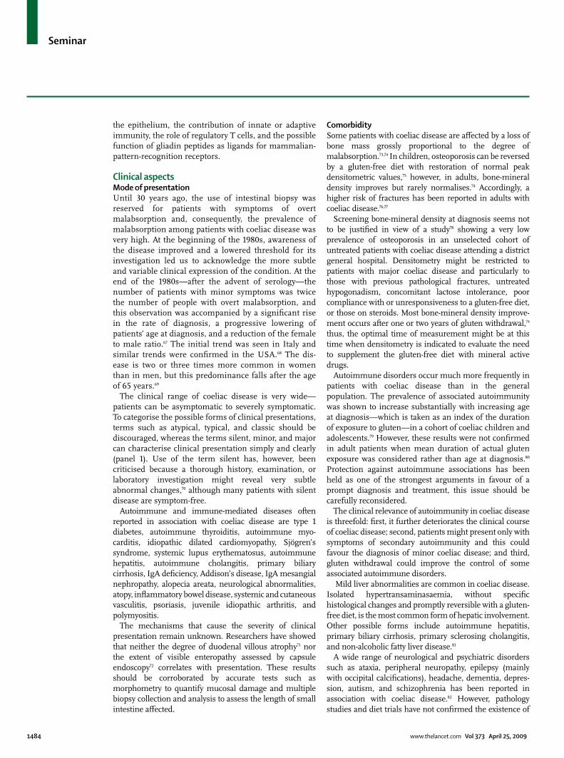
ComplicationsResults of studies from Italy121 and Sweden122 showed a twofold increased mortality in patients with coeliac disease, whereas this risk was only marginally raised in the UK.123 The risk of death rose with increasing delay in diagnosis and in patients with poor dietary compliance; non-Hodgkin lymphoma was the main cause of death.121
Patients diagnosed with the condition in childhood have a threefold increased risk of long-term mortality.124 However, this excess was mostly attributable to external causes as a possible result of behavioural changes
A B
Figure 4: Kerckring folds and scalloped confi gurationEndoscopic view of markedly reduced (A) and scalloped confi guration of folds (arrow) in active coeliac disease (B).
Type 1 Type 2
Unresponsive villous atrophy Yes Yes
Associated autoimmune diseases Yes No
Aberrant T-cell phenotype ≤10% of IELs >50% of IELs
Clonal TCR-γ gene rearrangement + ++
Chromosomal abnormalities No Yes
HLA-DQ2 homozygosity Uncommon Common
Associated ulcerative jejunoileitis Rare Common
Response to immunosuppressants (steroids, budesonide, azathioprine, tacrolimus, infl iximab)
Yes No
Risk of developing overt EATL Low 37–60% within 5 yrs
Mortality rate Slightly increased 5-yr survival <50%
EATL=enteropathy-associated T-cell lymphoma. IEL=intraepithelial lymphocyte. TCR=T-cell receptor.
Table 1: Comparison between refractory coeliac disease type 1 and type 2

Seminar
1488 www.thelancet.com Vol 373 April 25, 2009
associated with this chronic disease and its treatment. No increased risk of cancer or death has been reported in patients with dermatitis herpetiformis.125
Adult patients can develop complications such as refractory coeliac disease, ulcerative jejunoileitis, and enteropathy-associated T-cell lymphoma through a progressive accumulation in the intestinal epithelium of aberrant (CD3ε+, CD103+, CD8-, CD4-, T-cell-antigen receptor αβ-) and clonal (restricted rearrangements of T-cell-antigen receptor γ chain) intraepithelial lympho-
cytes,126 abnormally expanded by interleukin 15.58,59 These diseases are a continuum of each other, in which phenotypic disturbances of intraepithelial lymphocytes and chromosomal rearrangements127,128 cause the self-perpetuating, gluten-independent tissue damage of refractory coeliac disease and the subsequent uncontrolled expansion of a neoplastic T-cell clone of enteropathy-associated T-cell lymphoma.129
Refractory disease is defi ned by the absence of histological, and thus clinical, response to a gluten-free diet. Refractoryness is very often only apparent and mostly attributable to, dietary non-compliance or unintentional gluten intake. Other reasons for apparent refractoryness include misinterpretation of the original biopsy, slow histological recovery after diet, and misdiagnosis with other causes of villi atrophy (panel 3). Among them, autoimmune enteropathy may be easily diff erentiated by the presence of anti-enterocyte autoantibodies.130 Refractory coeliac disease has no relation to the persistence, after villi regrowth, of diarrhoea, which could be caused by concomitant irritable bowel syndrome, pancreatic insuffi ciency, microscopic colitis, and lactose mal absorption.131 Refractory coeliac disease could aff ect about 5% of patients with the condition,132,133 although this fi gure, which is derived from tertiary referral centres, might not refl ect true prevalence. There are two categories of true refractory disease—type 1 and type 2 (table 1).132–134
Ulcerative jejunoileitis, which shares many immuno-pathological features with type 2 refractory coeliac disease,135 is characterised by multiple ulcerations that evolve in strictures of the intestinal wall. Colicky central abdominal pain, distension, low-grade fever, diarrhoea, and weight loss are the main clinical manifestations. The rate of death is very high—resulting from obstruction, bleeding, and perforation.
Enteropathy-associated T-cell lymphoma is mainly localised in the proximal small intestine, is more prevalent in men over 60 years, and patients have a very poor outcome with a 2-year survival rate of 15–20%.133 Gross examination of the bowel reveals multifocal ulcerating nodules that can be accompanied by strictures and perforation (see fi gure 5 for histological features). Clinicians should be alerted to this complication by unexplained weight loss, abdominal pain, diarrhoea, loss of albumin and blood, increased lactate dehydrogenase, fever, and night sweating.136 Clinical suspicion should lead to an extensive diagnostic work-up (table 2); 18F-FDG-PET and histological identifi cation of lesions are regarded as the best options for diagnosis.
Refractory type 2 coeliac disease is resistant to available treatments, and although new drugs are being used, no randomised studies have been undertaken. These drugs include the antiCD52 antibody alemtuzumab,137 cladri-bine,138 and high-dose chemotherapy given before autologous haemopoietic stem-cell transplantation.139 The latter produced encouraging results in early-stage
Pros Cons
Small bowel enema Visualisation of the entire small bowel Only moderate accuracy
Abdominal computed tomography
Visualisation of extraintestinal fi ndings (lymphadenopathy, spleen atrophy)
No specifi c pattern identifi ed
PEG-enhanced magnetic resonance
Visualisation of extraintestinal fi ndings Accuracy not tested in complicated coeliac disease
¹⁸F-FDG-positron emission tomography
High sensitivity and adequate specifi city; possibly helpful in follow-up studies
Only preliminary prospective data; active peristalsis and infl ammation may aff ect specifi city
Capsule endoscopy Direct visualisation and magnifi cation of the entire small bowel; well tolerated; best sensitivity for ulcerations
Capsule retention in the event of strictures; unfeasibility for collecting biopsies
Double balloon enteroscopy
Feasibility for collecting biopsies; allows a defi nitive diagnosis
Only partial small bowel visualisation
Explorative laparotomy
Allows full thickness biopsies and operative procedures
Invasive
PEG=polyethylene glycol. FDG=fl uoro-deoxy-glucose.
Table 2: Diagnostic options in enteropathy-type T-cell lymphoma
Figure 5: Features of enteropathy-associated T-cell lymphoma Medium-sized to large tumour cells with round or angulated vescicular nuclei, prominent nucleoli, and moderate to abundant, pale-staining cytoplasm. A heavy eosinophil infi ltrate is evident between the tumour cells. Haematoxylin and eosin stain.
Seminar
www.thelancet.com Vol 373 April 25, 2009 1489
enteropathy-associated T-cell lymphoma140 but not in patients with advanced or relapsed disease.141 Further-more, the effi cacy of an anti-interleukin 15 monoclonal antibody aimed at reinducing the apoptotic machinery in aberrant intraepithelial lymphocytes is being investigated.58,59
A third of patients with untreated coeliac disease have haematological features of splenic hypofunction,142
which can be accompanied by spleen atrophy and depletion of IgM memory-B cells that protect against encapsulated bacteria.143 Although splenic hypofunction or atrophy is associated with a poor prognosis, its clinical relevance in coeliac disease has been overlooked—it is associated with increased auto-immunity144 and pneumococcal septicaemia in adult patients.145,146 Thus, patients with blood-fi lm features of splenic hypofunction—Howell Jolly bodies or red cells with membrane excavations—should receive pneumo-coccal-conjugate vaccine.
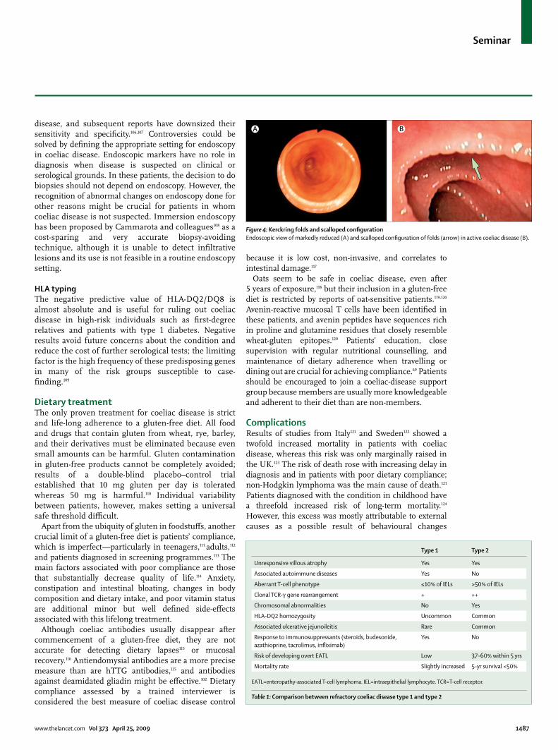
Future directionsImproved understanding of the molecular basis of coeliac disease has enabled researchers to suggest alternatives to a gluten-free diet. These novel treatments are aimed at blunting the immune stimulatory eff ects
of gluten (fi gure 6). However, we emphasise that some of these drugs (tissue-transglutaminase-inhibitors and monoclonal antibodies) have a poor safety profi le, and their hypothetical use could, thus, be reserved for complicated forms of the disease.
Alternative approaches to manage the condition aim to eliminate detrimental gluten peptides through genetic detoxifi cation or enzyme modifi cation. The development of grains that have low or no immunotoxicity can be achieved with transgenic technology that deletes or silences harmful gluten sequences147 or selectively breeding non-toxic wheat varieties.148,149 However, although the availability of non-toxic bread might resolve problems such as poor palatability and the high cost of gluten-free foods, patients’ social problems would nonetheless remain.
Gluten can be detoxifi ed within the intestine by oral administration of prolyl endopeptidases—enzymes that cleave the proline-rich and glutamine-rich immuno-stimulatory gluten peptides into small sequences with reduced toxic eff ects. In a pilot clinical study,150 patients with remitting coeliac disease were given a prolyl endopeptidase from Flavobacterium meningosepticum and challenged with oral gluten. The results were inconclusive: fat and xylose malabsorption, instead of
Gluten peptides
Zonulin
Interleukin 15
Tissue transglutaminase
HLA-DQ2/DQ8molecules
Dendritic cells
Interferon γ
T cells
Type 1 regulatoryT cells (Tr)
Adhesion molecules
Target Therapeutic agent Mechanism of action
Prolyl endopeptidases (PEP)
Zonulin receptor antagonist(AT-1001)
Anti-interleukin 15 antibody(AMG714)
Tissue transglutaminaseinhibitors
DQ2/DQ8 inhibitors
Peptide vaccines
Anti-interferon γ antibody(fontolizumab)
Anti CD3 antibody (visilizumab),anti CD4 antibody (cM-T412),anti CD25 antibody (daclizumab)
Human recombinantinterleukin 10 (Tenovil)
Anti-integrin α4 antibody(natalizumab); anti-integrinα4/β7 antibody (MLN-02);integrin α4 antagonist (T-0047)
Cleavage of proline-rich and glutamine-richgliadin peptides in safer sequences
Prevention of epithelial translocation ofgluten peptides into the lamina propria
Reduced cytolytic activity of intraepitheliallymphocytes against epithelial cells withconsequent decrease of enterocyteapoptosis
Blockade of deamidation and subsequentimmunological potentiation of glutenpeptides
Blockade of presentation of glutenpeptides with consequent silencing ofgluten-reactive T cells
Manipulation of dendritic cells in order tomake them a vehicle for peptide vaccines
Down-regulation of the Th1-mediatedinflammatory response
Silencing of gluten-reactive T cells
The interleukin-10-mediated expansion oftype 1 regulatory T cells may suppress theimmune response to gliadin
Blocking the cognate interaction betweenintegrin α4/β7 expressed on lymphocytesand MADCAM-1 expressed on mucosalendothelial cells may decrease lymphocyterecruitment in the gut
L&) 2/E)
N )"R, &@-"R
Endothelial cells
Integrin α4β7
MAdCAM-1
Apoptosis
Tr
T
T
T
T
Th1
CD4
CD25CD3
Interferon γ
Th0
CD8+IEL Enterocyte
Tight junctionopeningZonulin
Gluten peptides
IL-15
Dendritic cell
HLA-DQ2/8
TCR
tTG-deamidation
Figure 6: Novel treatments for coeliac disease

Seminar
1490 www.thelancet.com Vol 373 April 25, 2009
histology, was used to measure the effi cacy of the enzyme supplement, which did not resolve the malabsorption in about half of the patients. Additional management strategies could include the combination of two gastrically active enzymes (eg, a glutamine-specifi c endoprotease from barley and a prolyl endopeptidase from Sphingomonas capsulata are eff ective in detoxifying immunogenic peptides both in vitro and in animals);151 the use of a proline-endopeptidase from Aspergillus niger, which accelerates gluten degradation in a dynamic system that mimics in-vivo digestion;152 the addition to sourdough of lactobacilli proteolytic against proline-rich and glutamine-rich gluten peptides;153 or the pretreatment of fl our with a Streptomyces moboraensis-derived tissue transglutaminase that stops the immunostimulatory eff ects of fl our.154 These approaches are limited by the need for complete digestion of the toxic epitopes of gliadin, which has never been shown. These strategies should be investigated in large long-term clinical studies to assess safety and clinical eff ectiveness. The development of gluten-sensitive rhesus macaques could be a promising model of coeliac disease for testing these new treatments.155
Confl icts of interestWe declare that we have no confl icts of interest.
AcknowledgmentsWe thank Giovanni Gasbarrini, Monty Losowsky, and
Thomas MacDonald for inspiring our ongoing work on coeliac disease.
We thank Vincenzo Villanacci for providing fi gure 5.
References1 Losowsky MS. A history of coeliac disease. Dig Dis 2008; 26: 112–20.
2 Mann NS, Leung JW. Knowledge of some aspects of celiac sprue among professors of gastroenterology. Int Med J 2006; 13: 3–4.
3 Mäki M, Mustalahti K, Kokkonen J, et al. Prevalence of celiac disease among children in Finland. N Engl J Med 2003; 348: 2517–24.
4 Tommasini A, Not T, Kiren V, et al. Mass screening for coeliac disease using antihuman transglutaminase antibody assay. Arch Dis Child 2004; 89: 512–15.
5 West J, Logan RF, Hill PG, et al. Seroprevalence, correlates, and characteristics of undetected coeliac disease in England. Gut 2003; 52: 960–65.
6 Fasano A, Berti I, Gerarduzzi T, et al. Prevalence of celiac disease in at-risk and not-at-risk groups in the United States: a large multicenter study. Arch Intern Med 2003; 163: 286–92.
7 Hoff enberg EJ, MacKenzie T, Barriga KJ, et al. A prospective study of the incidence of childhood celiac disease. J Pediatr 2003; 143: 308–14.
8 Catassi C, Rätsch IM, Gandolfi L, et al. Why is coeliac disease endemic in the people of the Sahara? Lancet 1999; 354: 647–48.
9 Wieser H. Chemistry of gluten proteins. Food Microbiol 2007; 24: 115–19.
10 Kagnoff MF. Celiac disease: pathogenesis of a model immunogenetic disease. J Clin Invest 2007; 117: 41–49.
11 Sturgess R, Day P, Ellis HJ, et al. Wheat peptide challenge in coeliac disease. Lancet 1994; 343: 758–61.
12 Maiuri L, Ciacci C, Ricciardelli I, et al. Association between innate response to gliadin and activation of pathogenic T cells in coeliac disease. Lancet 2003; 362: 30–37.
13 Molberg Ø, Solheim Flaete N, Jensen T, et al. Intestinal T-cell responses to high-molecular-weight glutenins in celiac disease. Gastroenterology 2003; 125: 337–44.
14 Dewar DH, Amato M, Ellis HJ, et al. The toxicity of high molecular weight glutenin subunits of wheat to patients with coeliac disease. Eur J Gastroenterol Hepatol 2006; 18: 483–91.
15 Wieser H. The precipitating factor in coeliac disease. Baillieres Clin Gastroenterol 1995; 9: 191–207.
16 Shan L, Molberg Ø, Parrot I, et al. Structural basis for gluten intolerance in celiac sprue. Science 2002; 297: 2275–79.
17 Corazza GR, Valentini RA, Frisoni M, et al. Gliadin immune reactivity is associated with overt and latent enteropathy in relatives of celiac patients. Gastroenterology 1992; 103: 1517–22.
18 Nisticò L, Fagnani C, Coto I, et al. Concordance, disease progression, and heritability of coeliac disease in Italian twins. Gut 2006; 55: 803–08.
19 Sollid LM, Markussen G, Ek J, Gjerde H, Vartdal F, Thorsby E. Evidence for a primary association of celiac disease to a particular HLA-DQ alpha/beta heterodimer. J Exp Med 1989; 169: 345–50.
20 Karell K, Louka AS, Moodie SJ, et al. HLA types in celiac disease patients not carrying the DQA1*05-DQB1*02 (DQ2) heterodimer: results from the European Genetics Cluster on Celiac Disease. Hum Immunol 2003; 64: 469–77.
21 Sollid LM, Lie BA. Celiac disease genetics: current concepts and practical applications. Clin Gastroenterol Hepatol 2005; 3: 843–51.
22 Wolters VM, Wijmenga C. Genetic background of celiac disease and its clinical implications. Am J Gastroenterol 2008; 103: 190–95.
23 Greco L, Corazza GR, Babron MC, et al. Genome search in celiac disease. Am J Hum Genet 1998; 62: 669–75.
24 Holopainen P, Naluai AT, Moodie S, et al. Candidate gene region 2q33 in European families with coeliac disease. Tissue Antigens 2004; 63: 212–22.
25 Monsuur AJ, de Bakker PI, Alizadeh BZ, et al. Myosin IXB variant increases the risk of celiac disease and points toward a primary intestinal barrier defect. Nat Genet 2005; 37: 1341–44.
26 van Heel DA, Franke L, Hunt KA, et al. A genome-wide association study for celiac disease identifi es risk variants in the region harboring IL2 and IL21. Nat Genet 2007; 39: 827–29.
27 Hunt KA, Zhernakova A, Turner G, et al. Newly identifi ed genetic risk variants for celiac disease related to the immune response. Nat Genet 2008; 40: 395–402.
28 Cammarota G, Cuoco L, Cianci R, Pandolfi F, Gasbarrini G. Onset of coeliac disease during treatment with interferon for chronic hepatitis C. Lancet 2000; 356: 1494–45.
29 Forsberg G, Fahlgren A, Hörstedt P, Hammarström S, Hernell O, Hammarström ML. Presence of bacteria and innate immunity of intestinal epithelium in childhood celiac disease. Am J Gastroenterol 2004; 99: 894–904.
30 Stene LC, Honeyman MC, Hoff enberg EJ, et al. Rotavirus infection frequency and risk of celiac disease autoimmunity in early childhood: a longitudinal study. Am J Gastroenterol 2006; 101: 2333–40.
31 Zanoni G, Navone R, Lunardi C, et al. In celiac disease, a subset of autoantibodies against transglutaminase binds toll-like receptor 4 and induces activation of monocytes. PLoS Med 2006; 3: 1637–53.
32 Ivarsson A, Hernell O, Stenlund H, Persson LA. Breast-feeding protects against celiac disease. Am J Clin Nutr 2002; 75: 914–21.
33 Norris JM, Barriga K, Hoff enberg EJ, et al. Risk of celiac disease autoimmunity and timing of gluten introduction in the diet of infants at increased risk of disease. JAMA 2005; 293: 2343–51.
34 Lammers KM, Lu R, Brownley J, et al. Gliadin induces an increase in intestinal permeability and zonulin release by binding to the chemokine receptor CXCR3. Gastroenterology 2008; 135: 194–204.
35 Ciccocioppo R, Finamore A, Ara C, Di Sabatino A, Mengheri E, Corazza GR. Altered expression, localization, and phosphorylation of epithelial junctional proteins in celiac disease. Am J Clin Pathol 2006; 125: 502–11.
36 Wapenaar MC, Monsuur AJ, van Bodegraven AA, et al. Associations with tight junction genes PARD3 and MAGI2 in Dutch patients point to a common barrier defect for coeliac disease and ulcerative colitis. Gut 2008; 57: 463–67.
37 Schumann M, Richter JF, Wedell I, et al. Mechanisms of epithelial translocation of the alpha(2)-gliadin-33mer in coeliac sprue. Gut 2008; 57: 747–54.
38 Matysiak-Budnik T, Moura IC, Arcos-Fajardo M, et al. Secretory IgA mediates retrotranscytosis of intact gliadin peptides via the transferrin receptor in celiac disease. J Exp Med 2008; 205: 143–54.
39 Dieterich W, Ehnis T, Bauer M, et al. Identifi cation of tissue transglutaminase as the autoantigen of celiac disease. Nat Med 1997; 3: 797–801.

Seminar
www.thelancet.com Vol 373 April 25, 2009 1491
40 van de Wal Y, Kooy Y, van Veelen P, et al. Selective deamidation by tissue transglutaminase strongly enhances gliadin-specifi c T cell reactivity. J Immunol 1998; 161: 1585–88.
41 Molberg Ø, McAdam SN, Körner R et al. Tissue transglutaminase selectively modifi es gliadin peptides that are recognized by gut-derived T cells in celiac disease. Nat Med 1998; 4: 713–17.
42 Esposito C, Paparo F, Caputo I, et al. Expression and enzymatic activity of small intestinal tissue transglutaminase in celiac disease. Am J Gastroenterol 2003; 98: 1813–20.
43 Kim CY, Quarsten H, Bergseng E, Khosla C, Sollid LM. Structural basis for HLA-DQ2-mediated presentation of gluten epitopes in celiac disease. Proc Natl Acad Sci USA 2004; 101: 4175–79.
44 Dieterich W, Esslinger B, Trapp D, et al. Cross linking to tissue transglutaminase and collagen favours gliadin toxicity in coeliac disease. Gut 2006; 55: 478–84.
45 Qiao SW, Piper J, Haraldsen G, et al. Tissue transglutaminase-mediated formation and cleavage of histamine-gliadin complexes: biological eff ects and implications for celiac disease. J Immunol 2005; 174: 1657–63.
46 Vader LW, Kooy Y, Van Veelen P, et al. The gluten response in children with celiac disease is directed toward multiple gliadin and glutenin peptides. Gastroenterology 2002; 122: 1729–37.
47 Qiao SW, Bergseng E, Molberg Ø, et al. Antigen presentation to celiac lesion-derived T cells of a 33-mer gliadin peptide naturally formed by gastrointestinal digestion. J Immunol 2004; 173: 1757–62.
48 Nilsen EM, Jahnsen FL, Lundin KE, et al. Gluten induces an intestinal cytokine response strongly dominated by interferon gamma in patients with celiac disease. Gastroenterology 1998; 115: 551–63.
49 Monteleone I, Monteleone G, Del Vecchio Blanco G, et al. Regulation of the T helper cell type 1 transcription factor T-bet in coeliac disease mucosa. Gut 2004; 53: 1090–95.
50 Ciccocioppo R, Di Sabatino A, Bauer M, et al. Matrix metalloproteinase pattern in celiac duodenal mucosa. Lab Invest 2005; 85: 397–407.
51 Di Sabatino A, Ciccocioppo R, D’Alò S, et al. Intraepithelial and lamina propria lymphocytes show distinct patterns of apoptosis, whereas both the populations are active in Fas-based cytotoxicity in coeliac disease. Gut 2001; 49: 380–86.
52 Monteleone G, Pender SL, Wathen NC, MacDonald TT. Interferon-alpha drives T cell-mediated immunopathology in the intestine. Eur J Immunol 2001; 31: 2247–55.
53 Przemioslo RT, Kontakou M, Nobili V, Ciclitira PJ. Raised pro-infl ammatory cytokines interleukin 6 and tumour necrosis factor alpha in coeliac disease mucosa detected by immunohistochemistry. Gut 1994; 35: 1398–403.
54 Salvati VM, MacDonald TT, Bajaj-Elliott M, et al. Interleukin 18 and associated markers of T helper cell type 1 activity in coeliac disease. Gut 2002; 50: 186–90.
55 Fina D, Sarra M, Caruso R, et al. Interleukin 21 contributes to the mucosal T helper cell type 1 response in coeliac disease. Gut 2008; 57: 887–92.
56 Forsberg G, Hernell O, Melgar S, Israelsson A, Hammarström S, Hammarström ML. Paradoxical coexpression of proinfl ammatory and down-regulatory cytokines in intestinal T cells in childhood celiac disease. Gastroenterology 2002; 123: 667–78.
57 Di Sabatino A, Pickard KM, Gordon JN, et al. Evidence for the role of interferon-alfa production by dendritic cells in the Th1 response in celiac disease. Gastroenterology 2007; 133: 1175–87.
58 Mention JJ, Ben Ahmed M, Bègue B, et al. Interleukin 15: a key to disrupted intraepithelial lymphocyte homeostasis and lymphomagenesis in celiac disease. Gastroenterology 2003; 125: 730–45.
59 Di Sabatino A, Ciccocioppo R, Cupelli F, et al. Epithelium derived interleukin 15 regulates intraepithelial lymphocyte Th1 cytokine production, cytotoxicity, and survival in coeliac disease. Gut 2006; 55: 469–77.
60 Jabri B, Patey-Mariaud De Serre N, Cellier C, et al. Selective expansion of intraepithelial lymphocytes expressing the HLA-E-specifi c natural killer receptor CD94 in celiac disease. Gastroenterology 2000; 118: 867–79.
61 Hüe S, Mention JJ, Monteiro RC, et al. A direct role for NKG2D/MICA interaction in villous atrophy during celiac disease. Immunity 2004; 21: 367–77.
62 Benahmed M, Meresse B, Arnulf B, et al. Inhibition of TGF-beta signaling by IL-15: a new role for IL-15 in the loss of immune homeostasis in celiac disease. Gastroenterology 2007; 132: 994–1008.
63 Halttunen T, Maki M. Serum immunoglobulin A from patients with celiac disease inhibits human T84 intestinal crypt epithelial cell diff erentiation. Gastroenterology 1999; 116: 566–72.
64 Barone MV, Caputo I, Ribecco MT, et al. Humoral immune response to tissue transglutaminase is related to epithelial cell proliferation in celiac disease. Gastroenterology 2007; 132: 1245–53.
65 Esposito C, Paparo F, Caputo I, et al. Anti-tissue transglutaminase antibodies from coeliac patients inhibit transglutaminase activity both in vitro and in situ. Gut 2002; 51: 177–81.
66 Király R, Vecsei Z, Deményi T, Korponay-Szabó IR, Fésüs L. Coeliac autoantibodies can enhance transamidating and inhibit GTPase activity of tissue transglutaminase: dependence on reaction environment and enzyme fi tness. J Autoimmun 2006; 26: 278–87.
67 Corazza GR, Frisoni M, Treggiari EA, et al. Subclinical celiac sprue. Increasing occurrence and clues to its diagnosis. J Clin Gastroenterol 1993; 16: 16–21.
68 Murray JA, Van Dyke C, Plevak MF, Dierkhising RA, Zinsmeister AR, Melton LJ 3rd. Trends in the identifi cation and clinical features of celiac disease in a North American community, 1950-2001. Clin Gastroenterol Hepatol 2003; 1: 19–27.
69 Green PHR, Stavropoulos SN, Panagi SG, et al. Characteristics of adult celiac disease in the USA: results of a national survey. Am J Gastroenterol 2001; 96: 126–31.
70 Johnston SD, Watson RG, McMillan SA, Sloan J, Love AH. Coeliac disease detected by screening is not silent–simply unrecognized. QJM 1998; 91: 853–60.
71 Brar P, Kwon GY, Egbuna II, et al. Lack of correlation of degree of villous atrophy with severity of clinical presentation of coeliac disease. Dig Liver Dis 2007; 39: 26–29.
72 Murray JA, Rubio-Tapia A, Van Dyke CT, et al. Mucosal atrophy in celiac disease: extent of involvement, correlation with clinical presentation, and response to treatment. Clin Gastroenterol Hepatol 2008; 6: 186–93.
73 Corazza GR, Di Sario A, Cecchetti L, et al. Bone mass and metabolism in patients with celiac disease. Gastroenterology 1995; 109: 122–28.
74 Kemppainen T, Kröger H, Janatuinen E, et al. Osteoporosis in adult patients with celiac disease. Bone 1999; 24: 249–55.
75 Mora S, Barera G, Beccio S, et al. A prospective, longitudinal study of the long-term eff ect of treatment on bone density in children with celiac disease. J Pediatr 2001; 139: 516–21.
76 Ludvigsson JF, Michaelsson K, Ekbom A, Montgomery SM. Coeliac disease and the risk of fractures—a general population-based cohort study. Aliment Pharmacol Ther 2007; 25: 273–85.
77 Jafri MR, Nordstrom CW, Murray JA, et al. Long-term fracture risk in patients with celiac disease: a population-based study in Olmsted County, Minnesota. Dig Dis Sci 2008; 53: 964–71.
78 Lewis NR, Scott BB. Should patients with coeliac disease have their bone mineral density measured? Eur J Gastroenterol Hepatol 2005; 17: 1065–70.
79 Ventura A, Magazzù G, Greco L. Duration of exposure to gluten and risk for autoimmune disorders in patients with celiac disease. SIGEP Study Group for Autoimmune Disorders in Celiac Disease. Gastroenterology 1999; 117: 297–303.
80 Sategna Guidetti C, Solerio E, Scaglione N, Aimo G, Mengozzi G. Duration of gluten exposure in adult coeliac disease does not correlate with the risk for autoimmune disorders. Gut 2001; 49: 502–05.
81 Rubio-Tapia A, Murray JA. The liver in celiac disease. Hepatology 2007; 46: 1650–8.
82 Bushara KO. Neurologic presentation of celiac disease. Gastroenterology 2005; 128 (suppl): 92–97.
83 Rostami K, Steegers EA, Wong WY, Braat DD, Steegers-Theunissen RP. Coeliac disease and reproductive disorders: a neglected association. Eur J Obstet Gynecol Reprod Biol 2001; 96: 146–49.
84 Mäki M, Holm K, Koskimies S, Hällström O, Visakorpi JK. Normal small bowel biopsy followed by coeliac disease. Arch Dis Child 1990; 65: 1137–41.
85 Ferguson A, Arranz E, O’Mahony S. Clinical and pathological spectrum of coeliac disease—active, silent, latent, potential. Gut 1993; 34: 150–51.

Seminar
1492 www.thelancet.com Vol 373 April 25, 2009
86 Collin P, Helin H, Mäki M, Hällström O, Karvonen AL. Follow-up of patients positive in reticulin and gliadin antibody tests with normal small-bowel biopsy fi ndings. Scand J Gastroenterol 1993; 28: 595–98.
87 Mortimer PE, Stewart JS, Norman AP, Booth CC. Follow-up study of coeliac disease. Br Med J 1968; 3: 7–9.
88 Matysiak-Budnik T, Malamut G, de Serre NP, et al. Long-term follow-up of 61 coeliac patients diagnosed in childhood: evolution toward latency is possible on a normal diet. Gut 2007; 56: 1379–86.
89 Fasano A, Catassi C. Current approaches to diagnosis and treatment of celiac disease: an evolving spectrum. Gastroenterology 2001; 120: 636–51.
90 Hin H, Bird G, Fisher P, Mahy N, Jewell D. Coeliac disease in primary care: case fi nding study. BMJ 1999; 318: 164–67.
91 Korponay-Szabó IR, Szabados K, Pusztai J, et al. Population screening for coeliac disease in primary care by district nurses using a rapid antibody test: diagnostic accuracy and feasibility study. BMJ 2007; 335: 1244–47.
92 Mulder CJ, Bartelsman JF. Case-fi nding in coeliac disease should be intensifi ed. Best Pract Res Clin Gastroenterol 2005; 19: 479–86.
93 Pais WP, Duerksen DR, Pettigrew NM, Bernstein CN. How many duodenal biopsy specimens are required to make a diagnosis of celiac disease? Gastrointest Endosc 2008; 67: 1082–87.
94 Bonamico M, Thanasi E, Mariani P, et al. Duodenal bulb biopsies in celiac disease: a multicenter study. J Pediatr Gastroenterol Nutr 2008; 47: 618–22.
95 Oberhuber G, Granditsch G, Vogelsang H. The histopathology of coeliac disease: time for a standardized report scheme for pathologists. Eur J Gastroenterol Hepatol 1999; 11: 1185–94.
96 Corazza GR, Villanacci V, Zambelli C, et al. Comparison of the interobserver reproducibility with diff erent histologic criteria used in celiac disease. Clin Gastroenterol Hepatol 2007; 5: 838–43.
97 Brown I, Mino-Kenudson M, Deshpande V, Lauwers GY. Intraepithelial lymphocytosis in architecturally preserved proximal small intestinal mucosa: an increasing diagnostic problem with a wide diff erential diagnosis. Arch Pathol Lab Med 2006; 130: 1020–25.
98 Dewar DH, Ciclitira PJ. Clinical features and diagnosis of celiac disease. Gastroenterology 2005; 128 (suppl): 19–24.
99 Hill ID. What are the sensitivity and specifi city of serologic tests for celiac disease? Do sensitivity and specifi city vary in diff erent populations? Gastroenterology 2005; 128 (suppl): 25–32.
100 Carroccio A, Vitale G, Di Prima L, et al. Comparison of anti-transglutaminase ELISAs and an anti-endomysial antibody assay in the diagnosis of celiac disease: a prospective study. Clin Chem 2002; 48: 1546–50.
101 Fabbro E, Rubert L, Quaglia S, et al. Uselessness of anti-actin antibody in celiac disease screening. Clin Chim Acta 2008; 390: 134–37.
102 Kaukinen K, Collin P, Laurila K, Kaartinen T, Partanen J, Mäki M. Resurrection of gliadin antibodies in coeliac disease. Deamidated gliadin peptide antibody test provides additional diagnostic benefi t. Scand J Gastroenterol 2007; 42: 1428–33
103 Cataldo F, Lio D, Marino V, Picarelli A, Ventura A, Corazza GR. IgG(1) antiendomysium and IgG antitissue transglutaminase (anti-tTG) antibodies in coeliac patients with selective IgA defi ciency. Gut 2000; 47: 366–69.
104 Brocchi E, Corazza GR, Caletti G, Treggiari EA, Barbara L, Gasbarrini G. Endoscopic demonstration of loss of duodenal folds in the diagnosis of celiac disease. N Engl J Med 1988; 319: 741–44.
105 Jabbari M, Wild G, Goresky CA, Daly DS, et al. Scalloped valvulae conniventes: an endoscopic marker of celiac sprue. Gastroenterology 1988; 95: 1518–22.
106 Bardella MT, Minoli G, Radaelli F, Quatrini M, Bianchi PA, Conte D. Reevaluation of duodenal endoscopic markers in the diagnosis of celiac disease. Gastrointest Endosc 2000; 51: 714–16.
107 Shah VH, Rotterdam H, Kotler DP, Fasano A, Green PH. All that scallops is not celiac disease. Gastrointest Endosc 2000; 51: 717–20.
108 Cammarota G, Cesaro P, Martino A, et al. High accuracy and cost-eff ectiveness of a biopsy-avoiding endoscopic approach in diagnosing coeliac disease. Aliment Pharmacol Ther 2006; 23: 61–69.
109 Rashtak S, Murray JA. Tailored testing for celiac disease. Ann Intern Med 2007; 147: 339–41.
110 Catassi C, Fabiani E, Iacono G, et al A prospective, double-blind, placebo-controlled trial to establish a safe gluten threshold for patients with celiac disease. Am J Clin Nutr 2007; 85: 160–66.
111 Kumar PJ, Walker-Smith J, Milla P, Harris G, Colyer J, Halliday R. The teenage coeliac: follow up study of 102 patients. Arch Dis Child 1988; 63: 916–20.
112 O’Leary C, Wieneke P, Healy M, Cronin C, O’Regan P, Shanahan F. Celiac disease and the transition from childhood to adulthood: a 28-year follow-up. Am J Gastroenterol 2004; 99: 2437–41.
113 Fabiani E, Taccari LM, Rätsch IM, Di Giuseppe S, Coppa GV, Catassi C. Compliance with gluten-free diet in adolescents with screening-detected celiac disease: a 5-year follow-up study. J Pediatr 2000; 136: 841–43.
114 Lee A, Newman JM. Celiac diet: its impact on quality of life. J Am Diet Assoc 2003; 103: 1533–35.
115 Vahedi K, Mascart F, Mary JY, et al. Reliability of antitransglutaminase antibodies as predictors of gluten-free diet compliance in adult celiac disease. Am J Gastroenterol 2003; 98: 1079–87.
116 Sategna-Guidetti C, Grosso S, Bruno M, Grosso SB. Reliability of immunologic markers of celiac sprue in the assessment of mucosal recovery after gluten withdrawal. J Clin Gastroenterol 1996; 23: 101–04.
117 Pietzak MM. Follow-up of patients with celiac disease: achieving compliance with treatment. Gastroenterology 2005; 128 (suppl): 135–41.
118 Kemppainen T, Janatuinen E, Holm K, et al. No observed local immunological response aT cell level after fi ve years of oats in adult coeliac disease. Scand J Gastroenterol 2007; 42: 54–59.
119 Lundin KE, Nilsen EM, Scott HG, et al. Oats induced villous atrophy in coeliac disease. Gut 2003; 52: 1649–52.
120 Arentz-Hansen H, Fleckenstein B, Molberg Ø, et al. The molecular basis for oat intolerance in patients with celiac disease. PLoS Med 2004; 1: e1.
121 Corrao G, Corazza GR, Bagnardi V, et al. Mortality in patients with coeliac disease and their relatives: a cohort study. Lancet 2001; 358: 356–61.
122 Peters U, Askling J, Gridley G, Ekbom A, Linet M. Causes of death in patients with celiac disease in a population-based Swedish cohort. Arch Intern Med 2003; 163: 1566–72.
123 West J, Logan RF, Smith CJ, Hubbard RB, Card TR. Malignancy and mortality in people with coeliac disease: population based cohort study. BMJ 2004; 329: 716–19.
124 Solaymani-Dodaran M, West J, Logan RF. Long-term mortality in people with celiac disease diagnosed in childhood compared with adulthood: a population-based cohort study. Am J Gastroenterol 2007; 102: 864–70.
125 Lewis NR, Logan RF, Hubbard RB, West J. No increase in risk of fracture, malignancy or mortality in dermatitis herpetiformis: a cohort study. Aliment Pharmacol Ther 2008; 27: 1140–47.
126 Cellier C, Patey N, Mauvieux L, et al. Abnormal intestinal intraepithelial lymphocytes in refractory sprue. Gastroenterology 1998; 114: 471–81.
127 Verkarre V, Romana SP, Cellier C, et al. Recurrent partial trisomy 1q22-q44 in clonal intraepithelial lymphocytes in refractory celiac sprue. Gastroenterology 2003; 125: 40–46.
128 Obermann EC, Diss TC, Hamoudi RA, et al. Loss of heterozygosity at chromosome 9p21 is a frequent fi nding in enteropathy-type T-cell lymphoma. J Pathol 2004; 202: 252–62.
129 Cellier C, Delabesse E, Helmer C, et al. Refractory sprue, coeliac disease, and enteropathy-associated T-cell lymphoma. Lancet 2000; 356: 203–08.
130 Corazza GR, Biagi F, Volta U, Andreani ML, De Franceschi L, Gasbarrini G. Autoimmune enteropathy and villous atrophy in adults. Lancet 1997; 350: 106–09.
131 Fine KD, Meyer RL, Lee EL. The prevalence and causes of chronic diarrhea in patients with celiac sprue treated with a gluten-free diet. Gastroenterology 1997; 112: 1830–38.
132 Rubio-Tapia A, Kelly DG, Lahr BD, Dogan A, Wu TT, Murray JA. Clinical staging and survival in refractory celiac disease: a single center experience. Gastroenterology 2009; 136: 99–107.
133 Al-Toma A, Verbeek WH, Hadithi M, von Blomberg BM, Mulder CJ. Survival in refractory coeliac disease and enteropathy-associated T-cell lymphoma: retrospective evaluation of single-centre experience. Gut 2007; 56: 1373–78.
134 Malamut G, Afchain P, Verkarre V, et al. Presentation and long term follow-up of refractory celiac disease: comparison of type I with type II. Gastroenterology 2009; 136: 81–90.

Seminar
www.thelancet.com Vol 373 April 25, 2009 1493
135 Ashton-Key M, Diss TC, Pan L, Du MQ, Isaacson PG. Molecular analysis of T-cell clonality in ulcerative jejunitis and enteropathy-associated T-cell lymphoma. Am J Pathol 1997; 151: 493–98.
136 Gale J, Simmonds PD, Mead GM, Sweetenham JW, Wright DH. Enteropathy-type intestinal T-cell lymphoma: clinical features and treatment of 31 patients in a single center. J Clin Oncol 2000; 18: 795–803.
137 Vivas S, Ruiz de Morales JM, Ramos F, Suárez-Vilela D. Alemtuzumab for refractory celiac disease in a patient at risk for enteropathy-associated T-cell lymphoma. N Engl J Med 2006; 354: 2514–15.
138 Al-Toma A, Goerres MS, Meijer JW, et al. Cladribine therapy in refractory celiac disease with aberrant T cells. Clin Gastroenterol Hepatol 2006; 4: 1322–27.
139 Al-Toma A, Visser OJ, van Roessel HM, et al. Autologous hematopoietic stem cell transplantation in refractory celiac disease with aberrant T cells. Blood 2007; 109: 2243–49.
140 Bishton MJ, Haynes AP. Combination chemotherapy followed by autologous stem cell transplant for enteropathy-associated T cell lymphoma. Br J Haematol 2007; 136: 111–13.
141 Jantunen E, Juvonen E, Wiklund T, Putkonen M, Nousiainen T. High-dose therapy supported by autologous stem cell transplantation in patients with enteropathy-associated T-cell lymphoma. Leuk Lymphoma 2003; 44: 2163–64.
142 Corazza GR, Zoli G, Di Sabatino A, Ciccocioppo R, Gasbarrini G. A reassessment of splenic hypofunction in celiac disease. Am J Gastroenterol 1999; 94: 391–97.
143 Di Sabatino A, Rosado MM, Miele L, et al. Impairment of splenic IgM-memory but not switched-memory B cells in a patient with celiac disease and splenic atrophy. J Allergy Clin Immunol 2007; 120: 1461–63.
144 Di Sabatino A, Rosado MM, Cazzola P, et al. Splenic hypofunction and the spectrum of autoimmune and malignant complications in celiac disease. Clin Gastroenterol Hepatol 2006; 4: 179–86.
145 Thomas HJ, Wotton CJ, Yeates D, Ahmad T, Jewell DP, Goldacre MJ. Pneumococcal infection in patients with coeliac disease. Eur J Gastroenterol Hepatol 2008; 20: 624–28.
146 Ludvigsson JF, Olén O, Bell M, Ekbom A, Montgomery SM. Coeliac disease and risk of sepsis. Gut 2008; 57: 1074–80.
147 Vader LW, Stepniak DT, Bunnik EM, et al. Characterization of cereal toxicity for celiac disease patients based on protein homology in grains. Gastroenterology 2003; 125: 1105–13.
148 Molberg Ø, Uhlen AK, Jensen T, et al. Mapping of gluten T-cell epitopes in the bread wheat ancestors: implications for celiac disease. Gastroenterology 2005; 128: 393–401.
149 Spaenij-Dekking L, Kooy-Winkelaar Y, van Veelen P, et al. Natural variation in toxicity of wheat: potential for selection of nontoxic varieties for celiac disease patients. Gastroenterology 2005; 129: 797–806.
150 Pyle GG, Paaso B, Anderson BE, et al. Eff ect of pretreatment of food gluten with prolyl endopeptidase on gluten-induced malabsorption in celiac sprue. Clin Gastroenterol Hepatol 2005; 3: 687–94.
151 Gass J, Bethune MT, Siegel M, Spencer A, Khosla C. Combination enzyme therapy for gastric digestion of dietary gluten in patients with celiac sprue. Gastroenterology 2007; 133: 472–80.
152 Mitea C, Havenaar R, Drijfhout JW, Edens L, Dekking L, Koning F. Effi cient degradation of gluten by a prolyl endoprotease in a gastrointestinal model: implications for coeliac disease. Gut 2008; 57: 25–32.
153 Di Cagno R, De Angelis M, Auricchio S, et al. Sourdough bread made from wheat and nontoxic fl ours and started with selected lactobacilli is tolerated in celiac sprue patients. Appl Environ Microbiol 2004; 70: 1088–96.
154 Gianfrani C, Siciliano RA, Facchiano AM, et al. Transamidation of wheat fl our inhibits the response to gliadin of intestinal T cells in celiac disease. Gastroenterology 2007; 133: 780–89.
155 Bethune MT, Borda JT, Ribka E, et al. A non-human primate model for gluten sensitivity. PLoS ONE 2008; 3: e1614.