Embed Size (px)
Citation preview

CODE OF PRACTICE FOR RADIATION PROTECTION IN VETERINARY MEDICINE
RPII-02/3
November 2002
ii
Preface
This Code of Practice updates the Code of Practice on Radiation Protection in Veterinary Radiologyprepared by the Nuclear Energy Board in June 1989. The Code is designed to give guidance toveterinary surgeons to ensure that they, their employees and members of the public are adequatelyprotected from the hazards of ionising radiation arising from the use of X-ray equipment andradioactive substances in the practice of veterinary medicine. It reflects the regulations as specifiedin the Radiological Protection Act, 1991, (Ionising Radiation) Order, 2000 (S.I. No. 125 of 2000).
The Code has been prepared by staff of the Radiological Protection Institute’s Regulatory Service, inparticular Mr Jarlath Duffy, Mr David Fenton and Dr Ann McGarry, in consultation with Ms HesterMcAllister and Ms Cliona Skelly, Faculty of Veterinary Medicine, University College Dublin.
The words ‘shall’, ‘must’, and ‘should’ in this Code have been chosen with purpose. The words‘shall’ or ‘must’ indicate a mandatory requirement, while ‘should’ indicates an advisoryrecommendation that is highly desirable and that is to be implemented where feasible. Furthermore,since it is a condition of licence that the provisions of this Code be observed, the RadiologicalProtection Institute of Ireland (RPII) will not issue a licence where it believes that a mandatoryrequirement will not or cannot be met.
Use of the term ‘veterinary surgeon’ in this Code of Practice means a person whose name is on theRegister of Veterinary Surgeons maintained by the Veterinary Council under the provisions of theVeterinary Surgeons Act, 1931 (No. 36 of 1931), the Veterinary Surgeons Act, 1952 (No. 18 of 1952)and the Veterinary Surgeons Act, 1960 (No. 34 of 1960).
iii
Contents
1. Requirement for Radiation Protection in Veterinary Medicine 1
2. Statutory Requirements 2
3. Implications for the Practice of Veterinary Medicine 3
3.1 Justification 33.2 Risk Assessment 33.3 Optimisation – Compliance with this Code 33.4 Maximum Annual Dose 33.5 Classification of Areas 43.6 Requirements for Controlled and Supervised areas 53.7 Radiation Safety Procedures 53.8 Classification of Workers 53.9 Responsibility for Radiation Protection 6
3.9.1 Undertaking 63.9.2 Radiation Protection Adviser 63.9.3 Radiation Protection Officer 6
3.10 Provision of Information 63.11 Training/Competence 73.12 Monitoring of Work Environments 73.13 Monitoring of Persons 73.14 Accidental Exposure/Estimated Dose 83.15 Control of X-ray Equipment and Radioactive Substances 83.16 Duties of Workers 8
4. Radiography – Procedures and Facilities 9
4.1 General 94.2 Facilities 94.3 Radiography in Defined X-ray Rooms or Areas 114.4 Radiography Outside Defined X-ray Rooms or Areas 114.5 Reduction of Radiation Hazards 124.6 Fluoroscopic Procedures 134.7 Restraint of Animals 134.8 Film and Film Processing Equipment 15
5. Nuclear Medicine – Procedures and Facilities 16
5.1 General 165.2 Facilities 16
5.2.1 Stable/Horse box 165.2.2 Scanning Room 17
5.3 Procedures 17
Appendix 1 19
Appendix 2 23

1
1. Requirement for Radiation Protection in Veterinary Medicine
The ionising radiation used by veterinary surgeons in Ireland is mainly limited to the use of X-raysfor diagnostic purposes. However, the use of radioisotopes for diagnostic scintigraphy is increasing.In both of these practices there is considerable potential for persons involved to be exposed to theradiation hazards that could arise from them.
There are two kinds of biological consequences, which result from the radiation exposure of humans:
Somatic effects – affecting the individual exposed.Genetic effects – affecting the progeny of irradiated persons.
Somatic effects are further divided into two types.
Deterministic Effects
These are effects, which include, among others, the induction of cataract of the lens of the eye andskin erythema. They would not be expected to occur for exposures below a certain threshold value.The severity of the effect is assumed to be proportional to the dose received.
Stochastic Effects
These are effects, which include the induction of leukaemia and some other cancers. The probabilityof occurrence is assumed to be proportional to the dose received with no threshold value below whichno effects will be observed. Utilisation of badly adjusted equipment, and/or a poor technique mayunnecessarily increase the incidence of stochastic effects and could, in extreme cases, give rise to theinduction of a deterministic effect.
It is important to remember that the number and variety of radiation sources contributing to the totalexposure of the public is increasing. No user or group of users of ionising radiation has the right todefend its position on the basis that, in comparison with others, it is producing only a smallpopulation exposure. It is assumed that, for the purpose of establishing acceptable standards ofprotection from ionising radiation, any exposure, no matter how small, carries some risk. Themagnitude of this risk is also assumed to be proportional to the magnitude of the dose received. Allusers of radiation must therefore take every reasonable step to minimise the exposures for which theyare responsible.
2
2. Statutory Requirements
The principal relevant legal documents, which apply to the control of sources of ionising radiation inIreland, are:
The Radiological Protection Act, 1991 (No. 9 of 1991), which established the RadiologicalProtection Institute of Ireland (RPII).
The Radiological Protection Act, 1991, (Ionising Radiation) Order, 2000 (S.I. No. 125 of 2000),hereafter called the Order. This Order gives effect to Council Directive 96/29 Euratom of the 13 May1996 laying down the basic safety standards for the protection of the health of workers and thegeneral public against the dangers arising from ionising radiation. This Order prohibits any practiceinvolving the use of an irradiating apparatus or radioactive substance (except those which arespecifically exempted in the Order) save under licence from the Radiological Protection Institute.
3
3. Implications for the Practice of Veterinary Medicine
The following are the major implications of the above legislation for veterinary medicine in Ireland.
An undertaking (self employed person or employer) who possesses or intends to acquire X-rayequipment or radioactive substances in amounts greater than those specified in the Order is requiredto apply for a licence from the Institute. The application must be made to the Institute not later thanone month before the expected date of arrival of the equipment or source. In the case of a nuclearmedicine facility, the Institute should be advised as early as possible in the design stage. Theconditions attached to the licence will oblige the licensee to comply with the terms of this Code ofPractice and will include other conditions the Institute deems appropriate. The references to therelevant articles in the Order are quoted as appropriate.
3.1 Justification
All radiographic exposures shall be justified. This means that radiographic exposures shall only beundertaken if there is a definite indication for the procedure (Article 8).
3.2 Risk Assessment
Before commencing a practice, the undertaking, in consultation with the Radiological ProtectionOfficer (RPO) and Radiation Protection Adviser (RPA), shall make an assessment of the risks ofexposure to ionising radiation from the practice or from reasonably foreseeable accidents resultingfrom the practice for workers and members of the public who may be affected (Article 9). A riskassessment identifies the hazard, the risks from the hazard, the people at potential risk, the controlmeasures to be undertaken and the responsible persons. Guidance notes on undertaking a riskassessment are available from the Institute.
3.3 Optimisation – Compliance with this Code
The undertaking shall ensure that all exposures from the practice under its control are kept as low asreasonably achievable taking into account economic and social factors. In veterinary medicine,compliance with this Code of Practice will ensure that doses to staff and other persons are low.
3.4 Maximum Annual Dose
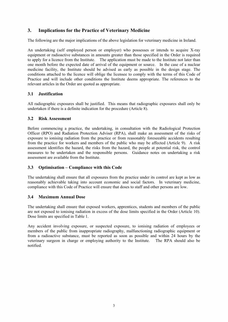
The undertaking shall ensure that exposed workers, apprentices, students and members of the publicare not exposed to ionising radiation in excess of the dose limits specified in the Order (Article 10).Dose limits are specified in Table 1.
Any accident involving exposure, or suspected exposure, to ionising radiation of employees ormembers of the public from inappropriate radiography, malfunctioning radiographic equipment orfrom a radioactive substance, must be reported as soon as possible and within 24 hours by theveterinary surgeon in charge or employing authority to the Institute. The RPA should also benotified.
4
Table 1
Dose Limitation for the Practice of Veterinary MedicineAs specified in Schedule 1 of S.I. No. 125 of 2000
Dose Limitsfor Exposed Workers
Dose Limits forApprentices andStudents
Dose limits forMembers of thePublic
Effective Dose 20 mSv in any year 20 mSv for personsaged 18 years or over
6 mSv per year forpersons aged between16 and 18 years
1 mSv in a year
Equivalent dose forlens of the eye
150 mSv in a year 50 mSv in a year 15 mSv in a year
Equivalent dose forthe skin (doseaveraged over area of1 cm2 regardless ofthe area exposed)
500 mSv in a year 150 mSv in a year 50 mSv in a year
Equivalent dose forthe hands, forearms,feet and ankles
500 mSv in a year 150 mSv in a year
3.5 Classification of Areas
The undertaking shall, having regard to the risk assessment, classify as appropriate a controlled orsupervised area taking into account the nature and extent of the radiological risks (Articles 15 & 16).
For X-ray units operating in fixed rooms or areas, the controlled area shall, subject to the followingconditions, exist between the ceiling and floor in a vertical direction extending two metres from eachedge of the table:
(i) Only one X-ray unit is operated at any one time and the beam is directed verticallydownwards onto the table.
(ii) The X-ray unit is fitted with a light beam diaphragm or other beam limiting device to ensurethat, at the maximum tube to table-top distance, the useful beam does not come within 10centimetres of the edge of the table.
(iii) The table is covered by 1 mm of lead over an area extending not less than 10 cm further ineach direction than the largest area of useful beam. It should be noted that this excludesPotter Bucky Tables, which have built-in shielding.
(iv) The equipment operates at less than 100 kV and the workload does not exceed 4 mA minutesin any week.
5
In practice, it is often simpler to designate the whole room as a controlled area. If the aboveconditions are met it shall not be necessary to designate a supervised area.
In situations where the conditions cannot be met, and in particular where X-ray equipment is operated‘in the field’ it may be difficult to rigorously classify areas. Notwithstanding this difficulty, theoperator shall take appropriate steps to control access to any area where elevated radiation levels mayexist. Classification of areas shall, in these situations, be made in consultation with the RPA.
For nuclear medicine facilities, the stable and the gamma camera room must be designated as acontrolled area once the technetium has been administered to the animal. The stable will remain acontrolled area after the animal has been removed, until any contamination has decayed tobackground levels.
3.6 Requirements for Controlled and Supervised areas
In the case of a controlled area the undertaking shall, as specified in Article 16, ensure that the area isdelineated, access is restricted to trained personnel and that a warning sign is displayed indicating thepresence of a controlled area and the nature of the risks.
The undertaking shall ensure in the case of a supervised area that radiological surveillance(measurement of external doserates) is undertaken to determine the nature and extent of theradiological risk, and that if appropriate, signs are displayed and the RPA is consulted in relation tothe work.
3.7 Radiation Safety Procedures
For the purposes of identifying the manner in which the safety, health and welfare of workers andother persons shall be served, the undertaking shall prepare a statement in writing of such procedures(radiation safety procedures) as he/she considers ought to be followed (Article 17).
The procedures should take into account the radiological risks involved and the assessmentsundertaken in the identification of hazards. The undertaking shall review the radiation safetyprocedures periodically, generally at least once during the licence period, and shall ensure that theyare observed in practice. The procedures shall be made available to the workers concerned and otherpersons who may be affected by them.
For veterinary surgeons in private practice with general radiographic equipment only, compliancewith this Code will suffice and no other radiation safety procedures are required. However, separateradiation safety procedures are required for all other cases.
3.8 Classification of Workers
An exposed worker is a person, either self-employed or working for an employer, liable to receive adose exceeding one or other of the dose levels equal to the dose limits for members of the public.
Category A
The undertaking shall classify as a Category A worker an exposed worker, who is liable to receive aneffective dose greater than 6 mSv in a period of 12 months or an equivalent dose greater than threetenths of the dose limits for the lens of the eye, or as the case may be, the skin, hands, forearms, feetand ankles specified in the Order.
At present, no veterinary surgeon has been classified as a Category A worker.
6
Category B
The undertaking shall classify as a Category B worker, an exposed worker, who is not classified as aCategory A worker. All veterinary surgeons and veterinary radiographers are currently classified asCategory B workers.
3.9 Responsibility for Radiation Protection
3.9.1 Undertaking
The undertaking means any natural or legal person who as a self employed person or employer as thecase may be, carries on or intends to carry on any practice to which the Order applies. In this contextit is the employer, principal partner or veterinary surgeon in charge.
3.9.2 Radiation Protection Adviser
The undertaking shall appoint in writing a suitable Radiation Protection Adviser (RPA) and shallconsult him/her as necessary regarding compliance with regulations (Article 19). The functions of theRPA shall include advising on all matters relating to radiation protection, quality control ofequipment and radioactive substances and in particular on those requirements for compliance withthis Code. The Institute should be contacted concerning the appointment of an RPA.
3.9.3 Radiation Protection Officer
In each practice the undertaking shall nominate a veterinary surgeon who is appropriately trained totake day-to-day responsibility for radiological protection. This person will be known as the RadiationProtection Officer (RPO). It shall be the responsibility of the RPO to ensure that the conditions of thelicence issued by the Institute and the requirements of this Code are adhered to; and that any staffwho are involved in work with radiographic equipment or radioactive substances are properly trained;and any female staff employed in the practice are made aware of the possible hazards of ionisingradiation during pregnancy.
While it is noted that on a stud farm, or in a university or research and development facility otherpersonnel such as the Health and Safety Officer or College RPO will have responsibilities in this area,the person assigned specific responsibility for radiological protection in a veterinary diagnostic X-raydepartment or a veterinary nuclear medicine department should, at a minimum, be a veterinaryradiographer and preferably a veterinary surgeon with relevant training and experience.
3.10 Provision of Information
Sufficient and appropriate information shall be provided to exposed workers, apprentices and studentsin relation to the health risks involved in their work. General radiation protection procedures, andprecautions to be taken in respect of the work shall be provided. Adequate information shall also begiven to other persons who are directly concerned with the work to ensure their health and safety asfar as reasonably practicable (Article 20).
Special attention should be applied to the protection of women of childbearing age and of pregnantwomen. It is the duty of the management of the practice to inform any such employee in his/herpractice of the possible hazards to the foetus from ionising radiation and to reschedule her duties tokeep her dose as low as reasonably achievable. A pregnant woman shall not hold animals duringradiography.
7
3.11 Training/Competence
Only persons who have completed or are undergoing a formal course of training in veterinaryradiology techniques and radiological protection measures may operate X-ray equipment. Theseinclude veterinary radiographers and veterinary surgeons. Furthermore, persons undergoing trainingmay only operate such equipment for purposes related to their training and under the supervision ofan appropriately qualified individual.
Veterinary surgeons involved in diagnostic imaging in veterinary nuclear medicine should have post-graduate training in a relevant discipline. This training shall be undertaken in consultation with theRPA.
Any person, such as a veterinary nurse or veterinary assistant, veterinary student, animal handler orany other person involved in the management of animals, who assists in carrying out X-rayexaminations or in a veterinary nuclear medicine unit, must have received instruction in radiologicalprotection measures appropriate to their work. It is the responsibility of the undertaking to ensure thattraining and information is provided (Article 20).
3.12 Monitoring of Work Environments
In accordance with Article 21 of the Order the undertaking is required to carry out dose rate andcontamination monitoring, where applicable, in the workplace. This monitoring shall be undertakenwith instruments that are calibrated, properly maintained and fit for their intended purpose. Recordsof any measurements taken and of the calibration certificates must be kept for inspection.
3.13 Monitoring of Persons
(i) Individual dose monitoring shall be provided for all staff working with ionising radiation(Article 22). Badges shall be worn on the trunk between the level of the shoulders and thehips and changed every eight weeks. An extremity badge shall also be worn on an indexfinger under a glove while handling unsealed sources.
For pregnant staff, the badge should be worn over the abdomen and should be changedfortnightly during the 8 – 15 week period of pregnancy and monthly for the remainder of thepregnancy. Alternatively, it may prove more convenient to change the badge fortnightly fromthe date of declaration of pregnancy until the individual goes on maternity leave.
All monitoring badges shall be obtained from an approved dosimetry service under the termsof Article 24 of the Order. Records of doses received by the staff, shall be kept for inspectionby officers of the Institute for at least 5 years. Dose limits are specified in Table 1.
Persons under 18 years of age may not be assigned to any work which could result in thembeing exposed workers. It shall also be ensured that staff, visitors and members of the publicare not exposed to ionising radiation in excess of the dose limits. The dose limits forapprentices and students who are not obliged to use sources in the course of their studies isthe same as the dose limits for members of the public.
(ii) If persons are needed to assist or hold animals during radiography they shall avoid theprimary beam and must be provided with a lead apron. The same person must not regularlyperform these duties.
(iii) During radiography an X-ray room shall not be used simultaneously for other veterinarypurposes.
8
3.14 Accidental Exposure/Estimated Dose
In the case of an accidental exposure where an individual dose measurement is impossible orinadequate, the dose should be estimated from other individuals wearing badges, who were in theimmediate vicinity or from area monitoring in the workplace or a combination of both. The workershall be notified of the dose estimate and the value reported to the approved dosimetry service(Article 23).
3.15 Control of X-ray Equipment and Radioactive Substances
Control of radioactive substances and irradiating apparatus shall be maintained as specified in Article28. This among other things requires the undertaking to maintain an up-to-date inventory of itemsheld, to keep records of use and disposal, to ensure secure and safe storage, and segregation andlabelling of radioactive substances.
The undertaking must ensure that all radiographic equipment and radioactive substances under his/herresponsibility conform to the standards outlined in this Code. If this is not so, then advice should besought from the RPA or from the Institute. All the radiological protection measures relating to staffand the public and the quality control procedures in this Code should be adhered to. The RPA shallinspect the veterinary practice to ascertain if adequate radiological protection measures are beingtaken. Institute inspectors may also carry out inspections of the practice.
3.16 Duties of Workers
An exposed worker shall not knowingly expose himself or herself or any other person to ionisingradiation to an extent greater than is reasonably necessary for the purpose of his or her work and shallexercise reasonable care while carrying out such work.
Full and proper use of any personal protective equipment shall be made and any defects in anyequipment or unusual occurrences likely to cause exposure shall be reported to the undertaking.
9
4. Radiography – Procedures and Facilities
4.1 General
Other than where manual constraint of the animal is unavoidable (see Section 4.7) the operator andother staff shall stand at least two metres away from the tube head and animal during exposure.
(i) Radiography shall be undertaken only if there is a definite indication for the procedure and ifit can be performed without undue radiation hazard.
(ii) Radiography shall be carried out only by appropriately trained and qualified personnel.
(iii) In radiography, no part of any person, even if shielded by protective lead lined clothing, shallbe exposed to the primary X-ray beam.
(iv) Only persons who are essential to the procedure shall be present during radiographic andfluoroscopic examinations. These persons shall be properly instructed and should understandtheir part in the proposed procedure. All such persons shall position themselves behindprotective screens, except where this is not practicable, in which case they shall wearprotective aprons and gloves and remain as far as practicable from the primary beam, theanimal and the X-ray tube assembly.
The veterinary surgeon in charge of X-ray equipment shall:
(i) Ensure that all X-ray equipment is kept in good working order. While the frequency ofperiodic servicing will depend on the degree of use of the equipment, servicing should becarried out as specified by the manufacturer/supplier/RPA/Institute as appropriate.
(ii) Notify the RPA and the Institute of damage or other incident (which could give rise to aradiation hazard), involving any X-ray unit or radioactive substance as soon as possible, butin any event, not later than 24 hours from the time of occurrence.
(iii) Notify the Institute immediately of the loss or theft of any licensed items.
(iv) Ensure that no X-ray unit is acquired or disposed of without the prior authorisation of theInstitute, in writing.
(v) Maintain records to include the following data relating to all licensed items:
(a) Date of purchase.(b) Date of withdrawal from service.(c) Dates of overhaul and repair.(d) Number of examinations performed per week.
(vi) Ensure that all records pertaining to this Code of Practice are readily available for inspectionby the inspectors of the Institute at all reasonable times.
4.2 Facilities
(i) In general, radiography may be considered in two categories.
(a) Radiography within a defined X-ray room or area.(b) Radiography outside a defined X-ray room or area when a mobile or portable X-ray
machine is taken to the animal.
10
(ii) X-ray machines shall have sufficient capacity to provide radiographs of good diagnosticquality. In addition, adequate facilities to provide control over the animal and protection ofthe operator are necessary. These are best provided in a defined X-ray room or area.Radiography outside such areas shall be carried out only where it is not practicable to bringthe animal to that area.
(iii) All X-ray equipment shall:
(a) be fitted with adequate shielding to ensure that the leakage radiation from the tube headdoes not exceed 1 mSv in 1 hour at 1 metre from the tube housing when operated at themaximum tube rating;
(b) have an exposure switch, which is able to terminate an exposure at a preset time. Theswitch shall require continuous pressure to maintain the production of X-rays and shall beoperable with the user standing at least 2 metres from the tube housing;
(c) be equipped with a warning device (normally a light) to indicate that the equipment isturned on and a second warning device to indicate when X-rays are being produced.(Ideally an audible alarm should also be fitted but it is noted that an audible alarm is notfitted on high frequency portable equipment and that the noise of the alarm may frightenan anxious animal);
(d) have all the control switches, meters and other displays clearly labelled to indicate theirfunctions;
(e) be fitted with a beam limiting device consisting of a light beam diaphragm;
(f) comply with any other standards for veterinary X-ray equipment which are currently inforce in Ireland or which the Institute may see fit to apply;
(iv) All X-ray equipment should be serviced at regular intervals in consultation with the RPA;
(v) An examination table shall be provided with either protective shielding equivalent to 0.5 mmlead on the sides or with protective shielding equivalent to 1 mm lead underneath the tabletop and any Potter-Bucky diaphragm incorporated in the table. The area of this lead shieldshall be greater than the maximum field size at the maximum tube focus to the tabletopdistance;
(vi) Sandbags, V-troughs, slings, adhesive tape or other positioning and immobilising devicesshall be available for supporting the animal during radiography;
(vii) Suitable cassette holders shall be available for use when using horizontal or angled X-raybeams. If they are not self-supporting, they shall be fitted with handles at least 1 metre longto ensure that a person holding them can remain well outside the primary beam;
(viii) Personal protective devices made of lead impregnated rubber or plastic such as aprons, glovesand shields suitable for hand and forearm, shall be provided for all persons who are requiredto be present during radiography and who are not protected by fixed or mobile protectivescreens. Gloves shall have a lead-equivalent thickness throughout of not less than 0.50 mm,and double sided aprons not less than 0.25 mm. These lead protective devices shall beexamined both visually and radiographically on a regular basis (e.g. three-monthly for apractice with a heavy X-ray work load), but at least annually to ensure that their shieldingefficiency has not become impaired by cracks due to sharp folds, penetrations which could be
11
caused by claws, or other damage. When not in use aprons shall be hung without folds onappropriate hangers.
4.3 Radiography in Defined X-ray Rooms or Areas
(i) For new installations, plans of the X-ray room shall be discussed with the RPA and whenfinalised, shall be submitted to the Institute.
(ii) The User in consultation with the RPA shall ensure that appropriate radiation safety riskassessments are made. These will be required in the following circumstances:
(a) Before the installation is put into routine use.
(b) If the installation or working procedures are modified. ‘Modified’ means a change in theamount of radiation, the manner of its use or a change in the X-ray equipment or itslocation. Such modifications may mean the original protection is no longer adequate.
(c) If the doses received by any person exceed, or are likely to exceed the appropriate dose-equivalent limits or are higher than normal for no obvious reason, or are significantlyhigher than average doses received in similar departments and practices.
(d) If changes are made in the immediate environs, for example a store or waiting area maybecome an office, resulting in change of occupancy.
(e) If there is a significant increase in the radiographic workload in the veterinary practice.
(iii) A defined X-ray room or area for veterinary radiography shall consist of a space of adequatedimensions, which offers radiation protection for persons both within and outside the area. Itshall possess:
(a) means of restricting access to the room area;
(b) X-ray warning signs at all entrances. It should be noted that the room shall be fitted witheither a warning system which indicates when X-rays are about to be produced and whichremains activated throughout the period of the exposure, or a device which prevents entryduring this period;
(c) facilities for positioning and immobilising the animal, and
(d) an X-ray machine of adequate capacity and appropriate type to undertake the requiredradiographic examination.
(iv) All small animal radiography should be carried out in a defined X-ray room or area.
(v) The need for structural shielding is reduced by ensuring that the X-ray beam can only bedirected vertically downwards with the animal placed on an examination table or on aconcrete or masonry floor. For horizontal X-ray beams additional shielding may be required.
4.4 Radiography Outside Defined X-ray Rooms or Areas
Radiography of animals outside defined X-ray rooms or areas (in other parts of the premises, or onvisits to farms, stables or kennels) is likely to add to the radiation risks for the following reasons:
(i) The usual ancillary and protective equipment may not be available.
12
(ii) It is likely to be more difficult to immobilise the animal.
(iii) The assistants may be untrained.
(iv) It is likely to be more difficult to prevent the presence of unauthorised persons duringradiography.
(v) There is a greater risk of irradiating persons in nearby areas.
When it is necessary to radiograph animals outside defined X-ray rooms or areas, it shall be ensuredthat:
(i) A suitable location is chosen with solid walls.
(ii) The necessary equipment, such as cassette holders, is available.
(iii) Sufficient lead protective clothing is available for all persons taking part.
(iv) The number of assistants is kept to the minimum necessary for the procedure.
(v) The nature of the procedure and the precautions to be observed are carefully explained to theassistants before the radiographic exposures are made.
(vi) Adequate precautions are taken to prohibit the access of unauthorised persons to the areaduring radiography (e.g. by display of warning signs, bollards or cones).
(vii) It should be ensured that no members of the public should be in the area during radiography.
(viii) Adequate supports for the X-ray tube assembly and cassettes are provided. Under nocircumstances is any person to hold these directly by hand.
(ix) Means are provided to achieve the correct alignment of the X-ray beam to the cassette and toensure that the X-ray beam is collimated and that the primary beam limits are clearly seen onthe final radiographs. Since the illumination of the light beam collimator may be ineffective,due to the light levels out-of-doors, there is a tendency to increase the area of the X-ray beamto an excessive size. From this point of view, it is preferable for outdoor radiography to bedone in the shade. Ideally, the animal should be radiographed in a stable or barn.
4.5 Reduction of Radiation Hazards
In addition to the measures outlined in Section 4 above the following items should be noted:
(i) The radiation dose to staff shall be minimised by:
(a) Taking all practical precautions to avoid unnecessary repetition of radiographs.
(b) Ensuring that the primary beam is restricted to the area to be examined and that the imageof the edges of the beam limiting device is visible on the radiograph.
(c) Using the fastest film and film-intensifying screen combination compatible with goodimage quality. This reduces dose and minimises the number of unsatisfactoryradiographs due to animal movement.
13
(d) Ensuring cleanliness and maintenance of cassettes and intensifying screens, therebyminimising repeat radiographs.
(e) Ensuring that all the assistants remain behind protective screens, or if there is no screen,wear protective clothing and position themselves as far as practicable from the X-ray tubeassembly, the animal and especially from the direction of the primary X-ray beam.
(f) Ensuring that the exposure is not made until the animal is properly restrained andpositioned.
(g) Ensuring that appropriate film processing facilities are available and are used correctly.
(ii) Cassette holders shall be used whenever a cassette cannot be supported on a table or on theground. No person shall directly hold the cassette manually. A person supporting a cassetteholder shall remain well outside the primary beam.
(iii) During radiography the X-ray tube assembly shall be rigidly supported by a holder or standwhich provides adequate stability and does not allow movement blurring of the radiograph.
(iv) Routine working radiation safety procedures for radiography shall be devised. Theprocedures shall be appropriate to the type of work carried out in the establishment and shallinclude necessary precautions to reduce radiation exposure. They shall be followed bypersons carrying out and assisting with radiography and shall be posted in the X-ray areas.
4.6 Fluoroscopic Procedures
Since the detail that can be visualised in fluoroscopy is inferior to that which can be seenradiographically, the additional risks of using fluoroscopy as a substitute for radiography are seldomjustified. Moreover because the clinical indications for such examinations are rare in veterinary workthey should not be undertaken in general veterinary practices but should be referred to establishmentsthat maintain specialist facilities and expertise, e.g., veterinary colleges. Furthermore, licences forfluoroscopy equipment will be granted by the Institute only to establishments with specialist facilitiesand suitably trained personnel. The following items should be considered:
(i) Fluoroscopy is potentially more hazardous than radiography, because the product of exposuretime and X-ray tube current is usually greater in the former and because the operators standnearer to the primary beam and the animal.
(ii) An X-ray image intensification system shall be used. It shall be properly installed andsubject to service and maintenance as specified by the manufacturer/supplier/RPA/Institute.A remote television display should be used for group viewing and teaching purposes.
4.7 Restraint of Animals
(i) The animal shall not be held for radiography except in exceptional circumstances when othermeans of immobilisation are not practicable e.g. due to the severity of the clinical conditionof the animal. Immobilisation should be achieved by mechanical means, or bytranquillisation or anaesthesia. These methods will eliminate or reduce the radiation hazardfrom manual restraint and assist in the reduction of image blurring due to movement. Adviceon mechanical restraints is given in Appendix 1.
14
(ii) When, in the rare and exceptional circumstances, manual restraint is necessary, the followingprocedures shall be adopted:
(a) The animal shall be restrained by the minimum number of persons necessary.
(b) All persons shall position themselves as far as practicable from the direction of theprimary X-ray beam, the animal and the X-ray tube housing. No part of any personshall be in the primary X-ray beam.
(c) Persons holding the animal shall wear protective lead aprons and lead gloves.
(d) If necessary, persons not normally exposed occupationally to ionising radiation (forinstance the owners of the animal) may be asked to hold the animal, provided thatsuch control will not significantly increase the radiation hazard of the procedure. Apregnant woman shall not hold animals during radiography.
(e) When it is necessary for staff to hold an animal during radiography, such individualsshall not be asked to hold animals repeatedly.
(iii) The radiography of large animals, e.g. horses and cattle, creates additional problems inrelation to radiation hazards for the following reasons:
(a) It is seldom practicable to anaesthetise the animal and some form of manual restraintis likely to be needed.
(b) It is often necessary for the film cassette holder to be supported manually in acassette holder.
(c) It is usually necessary for the useful beam to be directed horizontally. Thus, there isa greater risk of irradiating assistants.
(d) Those who restrain the animal or support the cassette holder are more likely to havetheir attention concentrated on their task rather than on avoiding the useful beam.
(e) Radiography of regions other than the lower limbs requires the use of considerablygreater exposure factors that will increase the hazard both from the primary beam andfrom scattered radiation.
(iv) In view of the additional radiation hazards in radiography of large animals, there is aparticular responsibility to ensure that, despite all difficulties, all precautions are observed.The following precautions shall be taken:
(a) The animal should be suitably tranquillised or anaesthetised whenever possible priorto radiography.
(b) All assistants shall wear protective clothing to give sufficient protection from thesource of radiation.
(c) All assistants not immediately required for the procedure shall remain at a safedistance. This is generally taken to be at least 2 metres from the X-ray tube andanimal.
15
4.8 Film and Film Processing Equipment
(i) Proper equipment for the receiving and development of the radiographic image, allied withcorrect equipment use in an appropriate dark room facility, can do much to reduce overall X-ray exposures. This can be achieved by keeping individual exposure times low and byminimising the number of repeat exposures.
(ii) For film processing, the solutions should be maintained at the temperature specified andreplenished or changed, with the frequency recommended by the manufacturer. Cold orexhausted developer can result in increased X-ray exposure times to obtain radiographs ofacceptable quality. For this reason, film processing tanks should normally be fitted withthermostatically controlled heaters. A floating lid on the developer tank will reduce theoxidation rate of the developer and prevent premature exhaustion. Where the X-ray filmthroughput is low, dish processing should be considered. A thermometer and stop watchshould be used for best results.
(iii) The films should be used and stored in accordance with the manufacturer’s instructions.
16
5. Nuclear Medicine – Procedures and Facilities
5.1 General
Equine scintigraphy is carried out with either a hand-held point counter or a gamma camera.Approximately 90% of veterinary scintigraphy procedures use 99mTc (technetium).
Facilities, such as the stable and the scanning area, should be centralised as far as possible, in order tominimise the need for transport of radioactive material on the site. Isotope reception, storage,preparation, administration and imaging should be in an area, which is easy to decontaminate.
The number of areas in which radioactive substances are to be used should be kept to a minimum.The work area in rooms where radionuclides are to be handled should be large enough to provideample space for staff, equipment and the animal. Ventilation, plumbing, electrical and floor loadingrequirements must also be considered.
The particular design features and protective shielding requirements should be related to the natureand the activity of the radionuclides to be used, their physical and chemical form and the procedures,which are to be carried out there.
5.2 Facilities
5.2.1 Stable/Horse box
To ensure that doses to exposed workers and non-occupationally exposed persons are as low asreasonable achievable the following design requirements should be considered:
(i) The stable should be large enough to satisfactorily house the animal and be constructed ofsuitable material, e.g. brick, concrete, etc.
(ii) The floor should be sealed concrete, with an absorbent covering (either a rubber waterproofcovering extending up the wall to a height of 10 cm or sufficient quantities of straw/sawdustor both). The floor level at the entrance or doorstep should be raised a few centimetres toprovide containment.
(iii) The walls of the stable should be sealed to about 2 metres in height, providing a clean surfaceequivalent to the height of the horse.
(iv) A mesh should be provided to prevent people petting the animal.
(v) To reduce the stable visit time, the plumbing should be arranged so that the water for thetrough may be turned on from outside the stable.
(vi) Hayracks should be installed to facilitate feeding from outside the box and adequate lightingshould be provided with an external switch.
(vii) The entrance door should be fitted with a slot for an appropriate, removable, radiationwarning sign.
(viii) Expert advice should be taken with respect to drainage from the stable.
17
5.2.2 Scanning Room
(i) The scanning room should be large enough to accommodate the animal, the staff and all theassociated equipment.
(ii) The floor should be of a concrete base with a durable ribbed rubber waterproof coveringextending up the walls to a height of about 10 cm.
(iii) The room should be equipped with a sink with elbow-operated taps. The sink outlet shouldbe directly connected to the main sewer to permit rapid dilution and to minimise thepossibility of contamination of other areas should the drain become blocked. All drain traps,where fitted, should be easily accessible for monitoring and should be labelled to indicate thatthey may contain radioactive contamination.
(iv) The entrance door should be wide enough to accommodate the animal to be studied and bemade of hardwood and painted with an oil based paint to make it waterproof, or some othersuitable material, to facilitate decontamination, if necessary. A slot should be provided onthe outside for a removable radiation warning sign.
(v) If a technetium generator is used then an efficient fume cupboard at least 2 metres wide mustbe provided. Generators must be installed behind 50 mm of lead.
The scanning room preparation area should also be fitted with or include:
(i) Lead lined bins, Cin bins or receptacles for contaminated instruments.
(ii) A lead shielded syringe and syringe carrier.
(iii) A calibrated contamination monitor and dose rate survey meter.
(iv) Warning cones or tripods with the appropriate radiation warning signs.
(v) Spare Lead pots/holders.
(vi) Face preparation shields and a locked press for storage of radioactive substances.
(vii) Disposable gloves, towels, overshoes, and gowns.
(viii) Suitable material such as a bag of sawdust, decon 90/Radiac wash or other agent to facilitatea clean up.
(ix) A dedicated shower facility for decontamination purposes should be available off the room oradjacent to the scanning room.
5.3 Procedures
The veterinary surgeon in charge shall ensure that the arrangements in the workplace with regard toradiation protection are appropriate to the nature of the installation, sources and to the magnitude andnature of the risks. The scope of the precautions and monitoring, as well as their type and quality,must be appropriate to the risks associated with the work involving exposure to ionising radiation.
The veterinary surgeon in charge of unsealed radioactive substances shall in addition to other relevantrequirements in this Code and the licence:

18
(i) Maintain records of the inventory of locations and quantities of all unsealed radioactivesubstances used and of the dates and method of disposal, where relevant.
(ii) Ensure that all radioactive substances are clearly labelled as such at all times.
(iii) Ensure that, when not in use, radioactive substances are segregated from non-radioactivesubstances and kept in secure and safe storage.
(iv) Measure external dose rates, indicating the nature and quality of the radiation.
(v) Provide suitable and sufficient measuring instruments for radiological surveillance ensuringthat the instruments are properly maintained, calibrated and fit for the intended purpose.
(vi) Ensure that individual dose monitoring is carried out by an approved dosimetry service andthat records are kept of the individual doses measured. An extremity badge in addition to awhole body badge shall be worn when handling unsealed sources.
(vii) Ensure that the Local Authority Fire Officer is informed annually of the location, nature andamount of radioactive substances held.
(viii) Ensure that the level of radioactive contamination on any surface does not exceed the valuesspecified in the licence.
(ix) Ensure that the unsealed radioactive substances are transported in accordance with licenceconditions.
(x) Transport arrangements should be undertaken in consultation with the RPA and the Institute.
19
Appendix 1Ancillary Equipment for Radiography
To position the animal correctly for radiography, special devices should be used to reduce to anabsolute minimum the number of occasions on which it is necessary for the animal to be heldmanually.
The following devices will be found useful:
(i) Small Animal Radiography
(a) Limb ties, ropes, gauze bandages
Various types of limb ties, ropes and bandaging may be tied around, or placed over,an anatomical region to fix it in position for radiography. They may also be used toremove an overlying anatomical region from the area of interest.
(b) Sand Bags
The sand bags should be contained in a sealed bag with an outer cover that can beremoved for cleaning. The bags should be floppy and pliant and made in a variety oflengths and widths so that they can be placed over a limb, or used as a ‘prop’, toposition an area for radiography.
(c) Positioning Troughs
These can be made of radiolucent timber, Perspex, or other sheet or foam plasticmaterial, usually, they are approximately V-shaped and may be constructed withadjustable sides. They are particularly useful for maintaining the animal in positionfor ventro-dorsal projections.
(d) Radiolucent Pads
Pads, made from radiolucent foam, plastic or rubber, can be purchased in a variety ofshapes and sizes and may be used to position the animal correctly. Plastic bags filledwith cotton wool will serve the same function.
(e) Cassette Holders
These may be simple devices, such as a welding clamp with a handle that can beattached to the cassette. Alternatively, they may be of a ‘picture-frame’ design,permitting the cassette to be slipped into a frame, to which a handle is attached.Adjustable cassette holders that may be clamped to the edge of the examination tableare very useful. A wall mounted cassette holder, adjustable in the vertical direction,can be used for standing lateral radiographs.
(f) Marking and labelling materials
These should be available to ensure that cassettes can be readily labelled andidentified in order to avoid having to repeat examinations.
20
(g) Other Devices
The animal can also be positioned using compression bands (fitted to some X-raytables) mouth gags, and suction cups which can be firmly fastened to the table (thecups may hold metal rods or padded metal plates that can be used to support theanimal). Birds or small mammals may be restrained by placing them inside Perspexcages or a short length of plastic tubing or piping with suitable ventilation.
(ii) Large Animal Radiography
(a) Cassette Holders
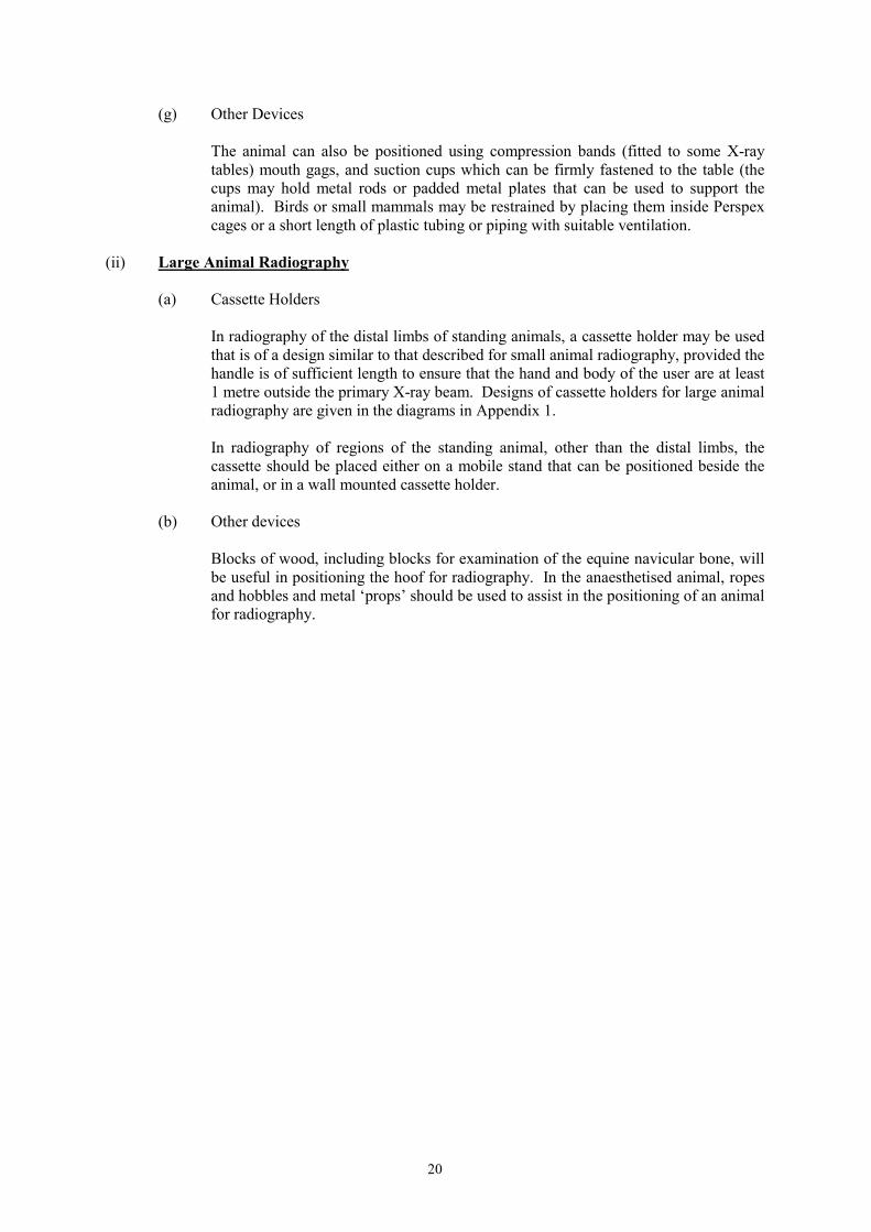
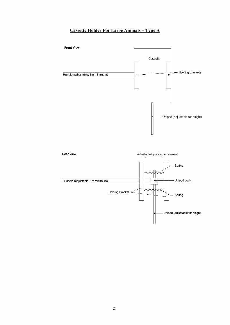
In radiography of the distal limbs of standing animals, a cassette holder may be usedthat is of a design similar to that described for small animal radiography, provided thehandle is of sufficient length to ensure that the hand and body of the user are at least1 metre outside the primary X-ray beam. Designs of cassette holders for large animalradiography are given in the diagrams in Appendix 1.
In radiography of regions of the standing animal, other than the distal limbs, thecassette should be placed either on a mobile stand that can be positioned beside theanimal, or in a wall mounted cassette holder.
(b) Other devices
Blocks of wood, including blocks for examination of the equine navicular bone, willbe useful in positioning the hoof for radiography. In the anaesthetised animal, ropesand hobbles and metal ‘props’ should be used to assist in the positioning of an animalfor radiography.
21
Cassette Holder For Large Animals – Type A
22
Cassette Holder For Large Animals – Type B
23
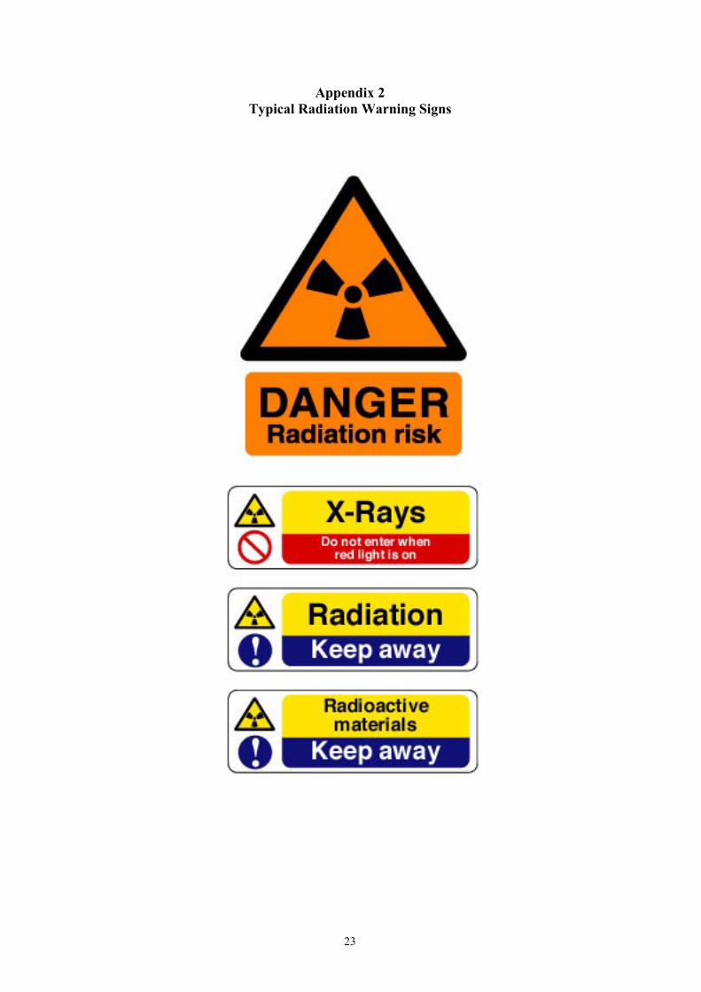
Appendix 2Typical Radiation Warning Signs