Embed Size (px)
Citation preview

Communication Vol. 262, No. 29. Issue of October 15, pp. 13881-13884,1987 THE JOURNAL OF BIOLOGICAL CHEMISTRY
Printed in U.S.A.
Cocrystals of Yeast Cytochrome c Peroxidase and Horse Heart Cytochrome c*
(Received for publication, April 27, 1987)
Thomas L. Poulos$g, Steven Sheriffll, and Andrew J. Howard$ From the $Protein Engineering Department, Genex Corporation, Gaithersburg, Maryland 20877 and the TNatwnul Institute of Diabetes and Digestive and Kidney
Bethesda, Maryland 20892 Diseases, National Institutes of Health,
Yeast cytochrome c peroxidase and horse heart cy- tochrome c have been cocrystallized in a form suitable €or x-ray diffTaction studies and the structure deter- mined at 3.3 A. The asymmetric unit contains a dimer of the peroxidase which was oriented and positioned in the unit cell using molecular replacement tech- niques. Similar attempts to locate the cytochrome c molecules were unsuccessful. The peroxidase dimer model was subjected to eight rounds of restrained pa- rameters least squares refinement after which the crystallographic R factor was 0.27 at 3.3 A. Exami- nation of a 2Fo-Fc electron density map showed large “empty” regions between peroxidase dimers with no indication of cytochrome c molecules. Electrophoretic analysis of the crystals demonstrated the presence of the peroxidase and cytochrome c in an approximate equal molar ratio. Therefore, while cytochrome c mol- ecules are present in the unit cell they are orientation- ally disordered and occupy the space between peroxi- dase dimers.
Biological electron transfer reactions require the formation of intermolecular complexes between electron donor and ac- ceptor protein molecules. Some of the more thoroughly stud- ied electron transfer reactions are those involving cytochrome c (Salemme, 1977; Ferguson-Miller et al., 1979). Using known crystal structures, computer graphics modeling has been em- ployed to develop hypothetical models for cytochrome c elec- tron transfer complexes (Salemme, 1977; Simondsen et at., 1982; Poulos and Finzel, 1984). The cytochrome c peroxidase- cytochrome c complex represents the only detailed model of a physiologically important complex involving cytochrome c (Poulos and Finzel, 1984). The peroxidase-cytochrome c com- plex has been investigated in several laboratories and cur- rently is one of the most actively studied electron transfer systems (Bechtold and Bosshard, 1985; Bisson and Capaldi, 1981; Cheung et al., 1986; Liang et al., 1987; Waldemeyer et al., 1982; Waldemeyer and Bosshard, 1985). The crystal struc- ture of the complex would provide important new information
* This work was supported in part by National Institutes of Health Grant GM 33688. The costs of publication of this article were defrayed in part by the payment of page charges. This article must therefore be hereby marked “advertisement” in accordance with 18 U.S.C. Section 1734 solely to indicate this fact.
5 Present address: Dept. of Chemistry/Biochemistry, University of Maryland and Center for Advanced Research in Biotechnology, 9600 Gudelsky Dr., Rockville, MD 20850.
in understanding protein-protein interactions and mecha- nisms of interprotein electron transfer reactions. We were very encouraged that the goal of determining the x-ray struc- ture of the complex might be realized once we succeeded in cocrystallizing the peroxidase and horse heart cytochrome c under conditions which should favor stabilization of the com- plex. However, as we describe in this report, the crystals consist of peroxidase dimers with orientationally disordered cytochrome c molecules trapped in the crystalline lattice.
MATERIALS AND METHODS
Crystallization-Cytochrome c peroxidase was purified from com- mercial bakers’ yeast according to Nelson et al. (1977) and twice crystallized by dialysis against distilled water. Sigma type VI horse heart cytochrome c was dissolved in distilled water, oxidized with ferricyanide, and chromatographed over a Sephadex G-75 column equilibrated with distilled water. The middle two-thirds of the cyto- chrome c band were collected. 1:l molar ratios of the peroxidase and cytochrome c were combined to a concentration of 0.1 mM followed by exhaustive dialysis against distilled water. After dialysis, the complex was concentrated to about 0.4 mM. Buffers were excluded from the crystallization medium to encourage electrostatic complex formation.
Crystallization was achieved using the hanging drop vapor diffusion method. 5 microliters of complex were combined with 5 r l of 20% methylpentanediol and vapor diffused against 0.5-1.0 ml of 20% methylpentanediol a t room temperature. Crystals (Fig. 1) appeared within 24 h and grew up to 0.5 mm and occasionally up to 1 mm. We also have succeeded in cocrystallizing the peroxidase with yeast iso- 1-cytochrome c, but these crystals were too thin for crystallographic studies.
Characterization of the Crystals-X-ray intensity data were col- lected with a Nicolet-Xentronics imaging proportional counter and a rotating anode x-ray source operating at 2.8 kilowatts. The unit cell dimensions and space group were determined directly from data obtained with the area detector as described by Gilliland et al. (1987). The autoindexing routine of the REFINE program in the XENGEN system’ was used to determine the unit cell parameters and were found to be a = b = 105.2 A and c = 186.6 A and a = B = y = 90”. These results indicated a tetragonal space group. Examination of individual frames of data with the aid of a TV monitor showed a 4- fold screw axis perpendicular to a 2-fold screw axis demonstrating that the space group must be P41212 or P43212. Assuming a molecular mass of 46,420 (34,030 for the peroxidase plus 12,354 for the cyto- chrome c) and two molecules of the complex/asymmetric unit, the Matthews coefficient, V, (Matthews, 1968), is 2.78 A3/dalton.
The crystal used for obtaining the initial data set for space group determination was removed from the capillary, dissolved in 1% so- dium dodecyl sulfate, and analyzed electrophoretically. As shown in Fig. 2, the crystal gave two bands corresponding to the peroxidase and cytochrome c. A sample of a 1:l mix of the two proteins was run on the same gel for comparison which indicates that the crystal also contained a 1:l ratio of the two proteins. In subsequent experiments, gels of the crystals were scanned and gave a peroxidase:cytochrome c ratio of 1.24.
Data Collection-A data set partially complete to 3.3 A was ob- tained from single crystal. A total of 84,187 observations of which 13,481 were unique scaled to give asn R,,, on intensities *= 0.098. The data set was 99% complete to 3.5 A and in the 3.5-3.3-A range, 35% complete. In the outermost shell of data between 3.5 and 3.3 A, the mean value of the ratio of the intensity to standard deviation was 2.05.
Molecular Replacement and Refinement-Molecular replacement was carried out using the MERLOT package of programs (Fitzgerald, 1987). The refined peroxidase (Finzel et al., 1984) and tuna cyto- chrome c (Takano and Dickerson, 1984) models, both of which are available from the Protein Data Bank (Bernstein et al., 1977), were
A. J. Howard, unpublished data.
13881

13882 Peroxidase-Cytochrome c Crystals
FIG. 1. Photograph of the peroxidase-horse heart cyto- chrome c cocrystals as they appear in the vapor diffusion well. The maximum dimension of a crystal shown is ahout 0.4 mm.
package of programs (Hendrickson and Konnert, 1980; Hendrickson, 1985), and Table I lists the refinement statistics. The final R factor after eight cycles of refinement- with no manual interventions to adjust the model was 0.27 at 3.3 A.
RESULTS AND DISCUSSION
Fig. 3 shows several sections of the 3.3-A 2Fo-Fc electron density map obtained after refinement of the peroxidase di- mer. What is most striking about this map is the large "empty" regions between peroxidase molecules. Some vestige of a cytochrome c molecule should have appeared in 2Fo-Fc, Fo- Fc, or Bijvoet difference electron density maps if one or more cytochrome c molecules occupied definite positions in the asymmetric unit. Bijvoet difference Fouriers should be partic- ularly sensitive to locating iron atoms. As a control to test the quality of the iron anomalous signal, the peroxidase iron atoms were removed from the model and a set of Fc (no iron) phases computed. The resulting Bijvoet difference map showed the peroxidase iron atoms as strong peaks with the
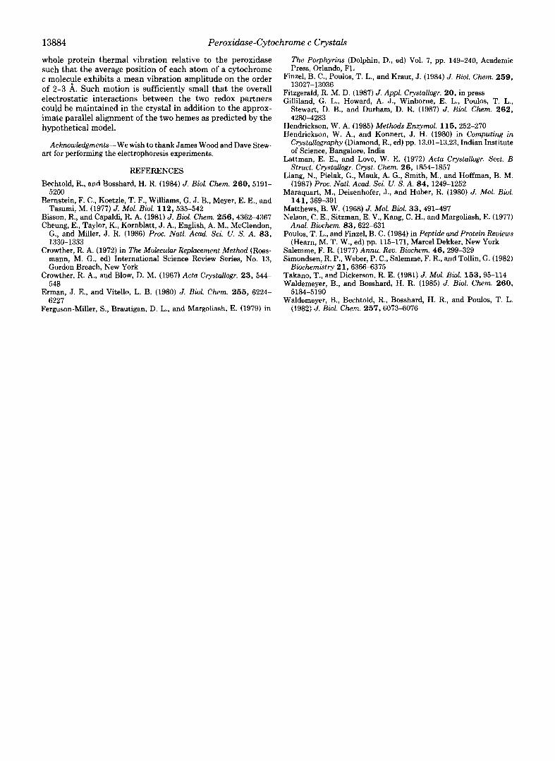
TABLE I Summary of refinement least squares statistics
rms deviation indicates the root mean square deviation of the final model from ideal bond distances and angles expected from small molecule crystal structures. The target rms represents an adjustable target root mean square deviation used as a weighting parameter during the restrained least-squares refinement.
Resolution range 10-3.3 A Reflections measured 13,481 Reflections used ( I > u n 8,999 R factor I Fo-Fc I /CFo 0.27
rms deviation from ideality
of final refined model deviation
Distances type target rms
A A
FIG. 2. Sodium dodecyl sulfate gel pattern of the cocrystal used for obtaining the initial x-ray intensity data set. Panels A and E are molecular weight standards. Panels B. C, and F are increasing amounts of a 1:l mix of the two proteins while panel D is the pattern of the single crystal.
used as probes. We expected the asymmetric unit to contain two peroxidase molecules and thus, two peaks should appear in the rotation function using the peroxidase model as a probe. Indeed, two peaks at 7.2 and 5.2 u were observed using the Crowther rotation function (Crowther, 1972) with data between 10.0 and 4.0 A and a 24-A radius of integration. The solution was relatively insensitive to the radius of integration used in the bange 20-30 A but was sensitive to resolution. Peaks in the 10.0-6.0-A range were closer to 3-4 u and were considerably more difficult to distinguish from the noise level than the clearly defined peaks obtained using the higher resolution data.
The Eulerian angles thus obtained were refined using the rotation function described by Lattman and Love (1972). In this case, the rotation search consisted of 9 steps in 2" increments around each of the three Eulerian angles obtained from the Crowther rotation func- tion. Next, each molecule was positioned in the unit cell using the translation function (Crow$her and Blow, 1967). The search was made in increments of 1.05 A along the a and b axes and 0.93 A along c. For both peroxidase molecules in the dimer, only one peak between 6 and 7 u appeared on each of the three Harker sections giving a clean and unambiguous solution. A translation search between the two molecules in the asymmetric unit also gave a unique solution and allowed for the relative positioning of each of the molecules. Finally, the program RMINIM was used to refine the orientation and trans- lation parameters and select the correct space group. The R value fell nearly 10 points to 0.45 when P43212 was used as opposed to P4,2,2 so the correct space group was the former. All attempts to locate the cytochrome c molecules using similar procedures were unsuccessful.
The oeroxidase dimer was refined usine the Hendrickson-Konnert
Through bonds 0.012 0.030 Through bond angles 0.020 0.040 Through dihedral angles 0.018 0.050 -
FIG. 3. Several sections of the f inal 3.3 A ZFo-Fc electron density map. The view is along the 4-fold (2) axis with one monomer

Peroxidase-Cytochrome c Crystals 13883
FIG. 4. Stereoscopic diagram of the hypothetical peroxidase-cytochrome c complex according to Poulos and Finzel (1984). The peroxidase is on the bottom and cytochrome c on the top. Critical carboxylate residues in the peroxidase and lysine residues in cytochrome c postulated to be involved in forming the electrostatic complex are labeled.
iron atom of molecule A of the peroxidase dimer appearing monomer, molecule B, because of the location of symmetry 5.8 times above background. Thus, the anomalous signal was related peroxidase molecules. Unfortunately, these results do sufficiently strong to have enabled location of the cytochrome not allow us to draw any firm conclusions on whether or not c iron atoms. Since the electrophoretic analysis of the crystals the hypothetical model is a valid representation of the “true” demonstrated the presence of cytochrome c in an approximate equal molar ratio to the peroxidase, we must conclude that the cytochrome c molecules are present but orientationally disordered and occupy the channels between peroxidase mol- ecules.
Why did the peroxidase crystallize rather than a specific and unique 1:l complex? At the very least, one might have expected that a stable 1:l complex should have prevented the peroxidase alone from crystallizing. By excluding buffers and using an organic solvent as a precipitating agent, the ex- tremely low ionic strength should have promoted complex formation. The equilibrium association constant is strongly dependent on ionic strength decreasing from 6 x lo6 M” at 0.01 M salt to 2.2 X lo3 M” a t 0.20 M salt (Erman and Vitello, 1980). Extrapolation to the ionic strength used for crystalli- zation suggests an association constant near 107-108 M” (Erman and Vitello, 1980). Nevertheless, crystallization of the peroxidase was favored (thermodynamically? kinetically?) over complex formation.
Next, we address the following question: is the surface of the peroxidase that the hypothetical model shows interacting with cytochrome c available for interaction with orientation- ally disordered cytochrome c molecules in the crystal? The answer is yes. One molecule of the peroxidase dimer, desig- nated molecule A, is oriented such that the surface we have postulated participates in complex formation with cytochrome c (Fig. 4) and is available for interaction with the cytochrome. Modeling of the hypothetical complex shows that a cyto- chrome c molecule could fit into the crystalline lattice between symmetry related peroxidase molecules and interact with the peroxidase according to the hypothetical model although some adjustments would be required in the positioning of the cy- tochrome in order to avoid unfavorably close contacts. Such interactions would not be possible with the second peroxidase
electron transfer complex. Finally, we consider two possible sources for the observed
disorder of cytochrome c: one static and one dynamic. The discussion that follows is very similar to that of Maraquart et al. (1980) who found that the entire Fc region in crystals of an intact antibody was disordered. If there were two distinct orientations of the cytochrome c molecules, we should have been able to observe some residual electron density for each of the orientations. It might also be possible to distinguish three orientations, but we doubt that more than three could be distinguished with the available 3.3-A data. Therefore, if there are four or more orientations of the cytochrome c mvlecules relative to the peroxidase molecules that differ by 2 A or more, we would expect the electron density correspond- ing to the location of the cytochrome c to appear as “flat” solvent as it does in our maps. The second possible source of disorder is dynamic. Dynamic disorder can sometimes be semi-quantitated with crystallographic B or temperature fac- tors. A temperature factor .Of 80 A’ corresponds to a mean amplitude of vibJation of 1 A and an 87% decrease in scatter- ing power at 5 A resolution with essentially no contribution to the diffraction pattern at 3 A resolution. Temperature factors this large for protein and especially solvent atoms are often very difficult to correctly model and usually are excluded from high resolution (1.5-2.0 A) refined structures. Theretore, we would expect that a thermal vibration of 2 to 3 A is sufficient to prevent the cytochrome c molecules from con- tributing significantly to the diffraction pattern even at very low resolution. Since the cytochrome c molecule is a highly ordered structure, the dynamic disorder is most likely due to thermal motion of the entire protein and not just individual cytochrome c atoms. Thus, a plausible explanation for why the cytochrome c molecules do not appear in the electron density maps is that the cytochrome c molecules undergo a
13884 Peroxidase-Cytochrome c Crystals
whole protein thermal vibration relative to the peroxidase such that the average position of each atom of a cytochrome c molecvle exhibits a mean vibration amplitude on the order of 2-3 A. Such motion is sufficiently small that the overall electrostatic interactions between the two redox partners could be maintained in the crystal in addition to the approx- imate parallel alignment of the two hemes as predicted by the hypothetical model.
Acknowledgments-We wish to thank James Wood and Dave Stew- art for performing the electrophoresis experiments.
REFERENCES Bechtold, R., and Bosshard, H. R. (1984) J. Biol. Chem. 260, 5191-
Bernstein, F. C., Koetzle, T. F., Williams, G. J . B., Meyer, E. E., and
Bisson, R., and Capaldi, R. A. (1981) J. Bwl. Chem. 256,4362-4367 Cheung, E., Taylor, K., Kornblatt, J. A., English, A. M., McClendon,
G., and Miller, J. R. (1986) Proc. Natl. Acad. Sci. U. S. A. 8 3 ,
Crowther, R. A. (1972) in The Molecular Replacement Method (Ross- mann, M. G., ed) International Science Review Series, No. 13, Gordon Breach, New York
Crowther, R. A., and Blow, D. M. (1967) Acta Crystallogr. 23, 544- 548
Erman, J. E., and Vitello, L. B. (1980) J. Biol. Chem. 2 5 5 , 6224- 6227
Ferguson-Miller, S., Brautigan, D. L., and Margoliash, E. (1979) in
5200
Tasumi, M. (1977) J. Mol. Biol. 112,535-542
1330-1333
The Porphyrins (Dolphin, D., ed) Vol. 7, pp. 149-240, Academic Press, Orlando, FL
Finzel, B. C., Poulos, T. L., and Kraut, J. (1984) J. Biol. Chem. 259 , 13027-13036
Fitzgerald, R. M. D. (1987) J. Appl. Crystallogr. 20, in press Gilliland, G. L., Howard, A. J., Winborne, E. L., Poulos, T. L.,
Stewart, D. B., and Durham, D. R. (1987) J. Biol. Chem. 262 , 4280-4283
Hendrickson, W. A. (1985) Methods Enzymol. 115, 252-270 Hendrickson, W. A., and Konnert, J. H. (1980) in Computing in
Crystallography (Diamond, R., ed) pp. 13.01-13.23, Indian Institute of Science, Bangalore, India
Lattman, E. E., and Love, W. E. (1972) Acta Crystallogr. Sect. B Struct. Crystallogr. Cryst. Chem. 26, 1854-1857
Liang, N., Pielak, G., Mauk, A. G., Smith, M., and Hoffman, B. M. (1987) Proc. Natl. Acad. Sci. U. S. A. 84, 1249-1252
Maraquart, M., Deisenhofer, J., and Huber, R. (1980) J. Mol. Biol.
Matthews, B. W. (1968) J. Mol. Biol. 33,491-497 Nelson, C. E., Sitzman, E. V., Kang, C. H., and Margoliash, E. (1977)
Poulos, T. L., and Finzel, B. C. (1984) in Peptide and Protein Reviews
Salemme, F. R. (1977) Annu. Rev. Biochem. 46, 299-329 Simondsen, R. P., Weber, P. C., Salemme, F. R., and Tollin, G. (1982)
Takano, T., and Dickerson, R. E. (1981) J. Mol. Biot. 153,95-114 Waldemeyer, B., and Bosshard, H. R. (1985) J. Biol. Chem. 2 6 0 ,
Waldemeyer, B., Bechtold, R., Bosshard, H. R., and Poulos, T. L.
141,369-391
Anal. Biochem. 83,622-631
(Hearn, M. T. W., ed) pp. 115-171, Marcel Dekker, New York
Biochemistry 21,6366-6375
5184-5190
(1982) J. Biol. Chem. 257,6073-6076