Embed Size (px)
Citation preview
Journal of Chromatography, 544 (1991) 233-247 Elsevier Science Publishers B.V., Amsterdam
CHROM. 23 086
Coated hydrophilic polystyrene-based packing materials”
YAN-BO YANG* and FRED E. REGNIER*
Department qf Biochemistry. Purdue University, West Lafayette. IN 47907 (lJ.S.A.j
ABSTRACT
A very hydrophilic high-performance liquid chromatographic base support was created from micro- particulate, macroporous poly(styrenedivinylbenzene) beads. An organic monomer containing cross-link- ing functionalities was coated on the poly(styrenedivinylbenzene), followed by a catalyzed cross-linking reaction. The coatings formed contain only stable chemical bonds (e.g., C-C, C-O-C), and easily-deriv- atized hydroxyl moieties. This coated base support was evaluated for hydrophilicity, chemical stability, solvent compatibility, rigidity, and irreversible adsorption. Derivatives of the coated base support were made and applied in various modes of chromatography.
INTRODUCTION
Highly-cross-linked poly(styreneedivinylbenzene) (PS-DVB) beads are of in- creasing interest to chromatographers due to their advantages over silica and conventional polymers (e.g., dextran and agarose) as packing materials of high-per- formance liquid chromatography (HPLC). Since its introduction by Moore [I] in 1964, the PS-DVB packing material has been mostly used in organic size-exclusion chromatography (SEC), and as a matrix in ion-exchange chromatography (IEC) for small molecules. The use of PS-DVB as a support in reversed-phase HPLC is rapidly increasing for both small molecules [2-lo] and biological macromolecules [l l-131.
PSDVB packings are organic, porous (and/or non-porous), spherical beads of polystyrene cross-linked with divinylbenzene. To achieve high mechanical stability, modern synthetic technology produces these polymeric beads with a very high degree of cross-linking, up to 90%. Thus, the highly cross-linked PS-DVB beads are not only chemically stable, inert, ion-free, insoluble in all non-oxidizing solvents, but also physically rigid and capable of higher mass transfer than conventional polystyrene packings. In spite of the above-mentioned properties, very few PS-DVB-based HPLC packings are currently available. Reasons for this are the inherent hydrophobicity of PS-DVB and lack of a variety of derivatives.
y Presented in part at the 12th International Symposium on Column Liquid Chromatography, Waxhington. DC, U.S.A., June19-24, 1988.
b Present address: The Separations Group, 17434 Mojave St., Hesperia, CA 92345, U.S.A.
0021-9673/91/$03.50 0 1991 Elsevier Science Publishers B.V.
234 Y.-B. YANG, F. E. REGNIER
Efforts have been made in recent years to overcome these problems through various chemical modifications [ 141 of the PS-DVB matrices. Carboxylated PS-DVB was produced [ 151 and used in reversed-phase and cation-exchange chromatography. A more hydrophilic PS-DVB support was also produced [ 16,171 by covalent bonding hydrophilic groups on the surface. This material was successfully used for SEC of proteins and in other modes of chromatography. In another case [ 18,191, an adsorbed polyethyleneimine coating was applied to sulphonated PS-DVB, yielding a strong anion-exchange packing material for protein separation. A graft co-polymer of polyoxyethylene and cross-linked polystyrene has been used as a support to bind peptides and proteins in some applications [20,21], but very few chromatographic properties have been reported. Polyoxirane-coated PS-DVB was also developed [22] for supports in normal-phase chromatography.
The work cited above has indeed broadened the application of PSDVB in chromatography. However, all existing packing materials are of limited application, particularly for biological macromolecules. Ideally, a “base support” should be: (1) chemically and mechanically stable, (2) hydrophilic, (3) charge free and (4) easily derivatizable. This paper introduces a very hydrophilic polystyrene-based packing material which was created by cross-linking a thin hydrophilic molecular layer applied to the hydrophobic PS-DVB resin. These packing materials are chemically and physically stable, and are easily modified. Several different applications, particularly in biopolymer separations, are demonstrated.
EXPERIMENTAL
Materials The microparticulate, macroporous PS-DVB resins PLRP-S 300 A and
PLRP-S 1000 A (15-25 pm, Polymer Labs., Church Stretton, U.K.) were mainly used as matrices for coated supports. The surface area of PLRP-S 300 A is 384 m’/g, and that of PLRP-S 1000 A, 267 m2/g, as measured by nitrogen adsorption [13]. These resins were designed as adsorbents for reversed-phase HPLC [9,1 I].
Tresyl chloride was purchased from Fluka (Ronkonkoma, NY, U.S.A.). Sodium m-periodate and potassium permagnate were from Sigma (St. Louis, MO, U.S.A.) and J. T. Baker (Phillipsburg, NJ, U.S.A.), respectively. Monochloroacetic acid was from Mallinckrodt (St. Louis, MO, U.S.A.). Other reagents were purchased from commercial sources and were used as received.
Apparatus Chromatography was performed using an LDC Constametric (I and IIG)
system with a gradient master and a mixer (Laboratory Data Control, Riviera Beach, FL, U.S.A.). Samples were injected onto columns using a Rheodyne Model 7125 manual loop injector (Rheodyne, Cotati, CA, U.S.A.) and detected with a Waters Lambda-Max Model 48 1 variable-wavelength liquid chromatography spectrophotom- eter (Milford, MA, U.S.A.). All chromatography was performed at ambient temper- ature using HPLC-grade solvents. Methanol, tetrahydrofuran (THF) and hexane were all purchased from Burdick and Jackson (Muskegon, MI, U.S.A.). All solvents were degassed under vacuum, with stirring, prior to use.
COATED HYDROPHILIC PS-BASED PACKING MATERIALS 235
Samples Polystyrene standards (molecular weights 500, 2000, 3550, 10 200, 17 500,
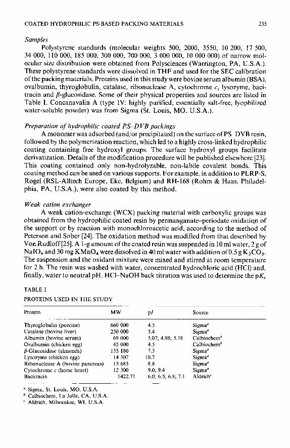
34 000, 110 000, 185 000, 300 000, 700 000, 3 000 000, 10 000 000) of narrow mol- ecular size distribution were obtained from Polysciences (Warrington, PA, U.S.A.). These polystyrene standards were dissolved in THF and used for the SEC calibration of the packing materials. Proteins used in this study were bovine serum albumin (BSA), ovalbumin, thyroglobulin, catalase, ribonuclease A, cytochrome c, lysozyme, baci- tracin and j?-glucosidase. Some of their physical properties and sources are listed in Table I. Concanavalin A (type IV: highly purified, essentially salt-free, lyophilized water-soluble powder) was from Sigma (St. Louis, MO, U.S.A.).
Preparation of hydrophilic coated PS-D VB packings A monomer was adsorbed (and/or precipitated) on the surface of PS-DVB resin,
followed by the polymerization reaction, which led to a highly cross-linked hydrophilic coating containing free hydroxyl groups. The surface hydroxyl groups facilitate derivatization. Details of the modification procedure will be published elsewhere [23]. This coating contained only non-hydrolyzable, non-labile covalent bonds. This coating method can be used on various supports. For example, in addition to PLRP-S, Rogel (RSL-Alltech Europe, Eke, Belgium) and RH-168 (Rohm & Haas, Philadel- phia, PA, U.S.A.), were also coated by this method.
Weak cation exchanger A weak cation-exchange (WCX) packing material with carboxylic groups was
obtained from the hydrophilic coated resin by permanganate-periodate oxidation of the support or by reaction with monochloroacetic acid, according to the method of Peterson and Sober [24]. The oxidation method was modified from that described by Von Rudloff [25]. A l-g amount of the coated resin was suspended in 10 ml water, 2 g of NaI04 and 30 mg KMn04 were dissolved in 40 ml water with addition of 0.5 g KzC03. The suspension and the oxidant mixture were mixed and stirred at room temperature for 2 h. The resin was washed with water, concentrated hydrochloric acid (HCI) and, finally, water to neutral pH. HCl-NaOH back titration was used to determine the pK,
TABLE I
PROTEINS USED IN THE STUDY
Protein MW PI Source
Thyroglobuhn (porcine) 660 000 4.5 Sigma” Catalase (bovine liver) 250 000 5.4 Sigma” Albumin (bovine serum) 69 000 5.07; 4.98; 5.18 Calbiochemb Ovalbumin (chicken egg) 45 000 4.5 Calbiochemb /I-Glucosidase (almonds) 135 180 7.3 Sigma” Lysozyme (chicken egg) 14 307 10.7 Sigma” Ribonuclease A (bovine pancreas) 13 683 8.8 Sigma” Cytochrome c (horse heart) 12 300 9.0; 9.4 Sigma” Bacitracin 1422.71 6.0; 6.5; 6.8; 7.1 Aldrich’
’ Sigma, St. Louis, MO, U.S.A. b Calbiochem, La Jolla, CA, U.S.A. ’ Aldrich, Milwaukee, WI, U.S.A.
236 Y.-B. YANG, F. E. REGNIER
and the capacity of the ionic group. About 0.5 mequiv./g of carboxylic groups were obtained by controlling the oxidation conditions.
Concanavalin A affinity packing Concanavalin A (Con A) was covalently bound to the coated PS-DVB resin
through tresylation of the hydroxyl groups on the surface as described by Nilsson and Mosbach [26]. S represents the support and L represents the protein ligand in the equations below.
S-CHI-OH + ClS02CH2CF3 -+ S-CH2-0S02CH2CF3
S-CH2-0S02CH2CF3 + L-NH2 + S-CH2-NH-L
One gram of the coated resin was washed with water (3 x 10 ml). The resin was transferred gradually to dried acetone through washing, then dispersed in 3 ml dried acetone and 200 ~1 pyridine. An loo-h1 volume of tresyl chloride was added, dropwise, to the vigorously stirring resin suspension over 4 min. This activated resin was washed with acetone and dilute aqueous HCl (pH 2.4) and stored at 4°C.
Con A was then coupled to the activated resin (via a stable amine bond) as follows: one gram of activated resin (in wet-cake form) was washed twice with precooled (OC) 20 mMNa3P04 + 0.8 MNaCl buffer (pH 7.38) and dispersed in 5 ml of the above buffer. To the suspension was added 100 mg Con A in 2 ml saturated NaCl solution. This reaction mixture was gently shaken at 4°C for 14 h. The resin was sequentially washed with (1) the coupling buffer, (2) dilute acetic acid (pH 2.8) (3) water, (4) sodium carbonate (pH 9.0), (5) water, and (6) 0.02 MTris-HCl (pH 6.71) + 0.5 M NaCl + 1 mA4 MnC12 + 1 mM CaC12.
The method described above can be used for any other protein which contains free amino groups; e.g., antibodies and antigens. MnC12 and CaC12 were used only for stabilizing Con A.
Pore size measurement In SEC, the elution volume (VE) of a molecule is defined as:
where Vi is the column interstitial volume, vr, the column internal pore volume, and I&,-, the size-exclusion distribution coefficient or the solute-accessible volume proportion in the inside of the pores. K SEC can be determined according to eqn. 1:
&EC = (VE - vi)/ VP (2) and
V* = Vo - Vi (3)
where V,, is the total void volume of the column, and can be measured by the elution volume of the smallest molecule. Vi can be obtained by the elution volume of a totally excluded molecule.
COATED HYDROPHILIC PS-BASED PACKING MATERIALS 237
Mean pore sizes of coated and uncoated resins were determined by the inverse-SEC method of Halasz and co-workers [27-291. This method has been shown to be suitable for polymeric HPLC packing materials [30]. A series of polystyrene standards of known molecular weight were used as the test probes and THF as the eluent. Benzene was assumed to be the smallest molecule to penetrate all pores. The exclusion value of the polystyrene standards, @, was then calculated from the molecular weight, MW:
@ = 0.63 MW”.5g A
The mean pore size, Qiso, was taken as the exclusion value of the polystyrene standard, which has a KsEC of 0.5. In other words, the mean pore size was assumed to be the exclusion value of the polystyrene molecule which can reach 50% of the total pore volume.
Column packing procedure All columns, except for the affinity column, were slurry packed using an HPLC
packing pump (Shandon Southern Instruments, Sewickley, PA, U.S.A.). Uncoated PSDVB was suspended and packed with isopropanol at 4500 p.s.i. The coated resins were suspended and packed with a mixture of water and methanol (1: 1) at 3500 p.s.i. As shown previously [14], these organic packing materials are better packed under lower pressure, using less-viscous solvent, at high flow-rate, and for a longer packing time.
The affinity column was packed using an Altex 110 pump (Beckman Instru- ments, Fullerton, CA, U.S.A.) at about 5 ml/min, using the chromatography buffer as the packing solvent.
RESULTS AND DISCUSSION
Evaluation of coated resins Irreversible adsorption of proteins is often a problem in chromatography,
especially on silica-based packings [3 1,321. On these packings, the silanol residues act as weak cation exchangers, which adsorb positively charged proteins, and repel negatively-charged proteins. In addition, metal ions remaining on the silica surface can cause adsorption of proteins through complex formation. With PS-DVB-based packings, the hydrophobic surface adsorbs proteins through hydrophobic interac- tions. This phenomenon has been used to physically immobilize proteins (e.g., antibodies or antigens) on the surface of polystyrene beads for immunoassay [33-351, although slow bleeding of proteins was observed. However, this is not acceptable in chromatographic packings, because it can result in poor recovery, as well as erroneous estimation of molecular weight in SEC.
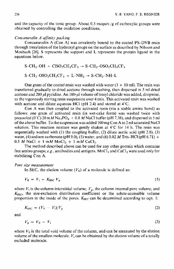
As observed in protein immobilization, irreversible adsorption results in a gradual increase in peak heights with each successive injection until a steady maximum is reached. Therefore, constant peak heights should be obtained with repeated injections if there is no irreversible adsorption of proteins by the support material. Successive injections of BSA were used to evaluate columns packed with coated PSDVB differing in coating coverage. Fig. 1 shows that irreversible adsorption of
238 Y.-B. YANG, F. E. REGNIER
0 3.34 pmol monomer tooting/m’
A 5.04 pmol monomer coating/m’
0 6.69 pmol monomer coating/m’
V 0.36 pmol monomer coating/m’
I OO
I I I 5 IO 15
INJECTION NUMBER
Fig. 1. Peak height versus injection number of BSA for resins of coated PLRP-S (15-25 pm, 300 A) with different quantities of coating (number of moles of monomers coated/m’). Column: 5 x 0.46 cm I.D.; mobile phase: 50 mM phosphate buffer (pH 7.0); flow-rate: 1.0 ml/min; detection: UV 280 nm. BSA was used as the sample.
protein on the support was negligible when the concentration of monomer in the coating was higher than 5 pmol/m’, which corresponds to a monolayer coating.
As mentioned above, charged groups on the support can contribute to the adsorption and exclusion of proteins, causing the elution volume of proteins to deviate from the true size-exclusion volume. This is most pronounced at lower mobile phase ionic strength [31]. If the support carries negative charges, basic proteins elute with a larger volume due to ionic interaction and acidic proteins would elute at a lower volume due to ion exclusion; the opposite occurs on a positively-charged support. Pfannkoch et al. [31] showed that most commercially available SEC supports (both silica-based and organic-based) carry negative charges. This feature of the coated PS-DVB packing material was tested using thyroglobulin, catalase, bovine serum albumin, ribonuclease A, cytochrome c and bacitracin. These proteins all differ in molecular weight and isoelectric points (pl) (see Table I). No changes in elution volume were observed for all the proteins tested, except cytochrome c, when the concentration of sodium phosphate buffer was varied from lo-500 mM at pH 7.0. Although cytochrome c (the basic protein) exhibited slight adsorption at very low ionic strength, ribonuclease A (another basic protein) did not. The reason for this is unknown. Perhaps the slight adsorption of cytochrome c could be due to structural changes when the salt concentration was very low [36].
All of the preceding experiments were conducted with neutral pH mobile phase. Changes in mobile phase pH can influence elution volume (due to protein-support interactions) if the support contains ionizable groups. Thus, chromatography of a number of acidic and basic proteins (thyroglobulin, ovalbumin, lysozyme, ribonu- clease A and bacitracin) across the pH range 2-12 was used to check for the presence of ionizable moieties on this packing material. Constant elution volumes for both acidic and basic proteins were obtained over this pH range. This indicates that the coated resin possesses neither positively nor negatively ionizable groups.
COATED HYDROPHILIC PS-BASED PACKING MATERIALS 239
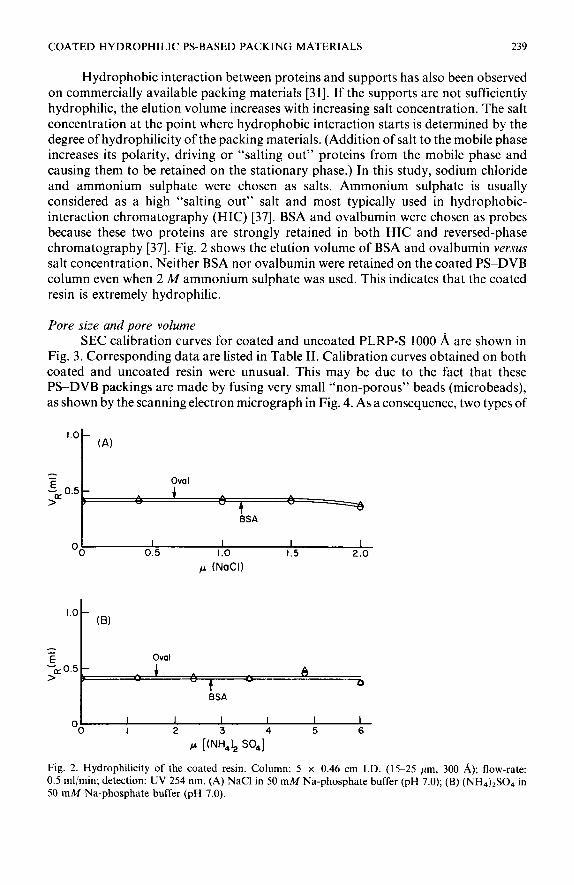
Hydrophobic interaction between proteins and supports has also been observed on commercially available packing materials [31]. If the supports are not sufficiently hydrophilic, the elution volume increases with increasing salt concentration. The salt concentration at the point where hydrophobic interaction starts is determined by the degree of hydrophilicity of the packing materials. (Addition of salt to the mobile phase increases its polarity, driving or “salting out” proteins from the mobile phase and causing them to be retained on the stationary phase.) In this study, sodium chloride and ammonium sulphate were chosen as salts. Ammonium sulphate is usually considered as a high “salting out” salt and most typically used in hydrophobic- interaction chromatography (HIC) [37]. BSA and ovalbumin were chosen as probes because these two proteins are strongly retained in both HIC and reversed-phase chromatography [37]. Fig. 2 shows the elution volume of BSA and ovalbumin versus salt concentration. Neither BSA nor ovalbumin were retained on the coated PS-DVB column even when 2 M ammonium sulphate was used. This indicates that the coated resin is extremely hydrophilic.
Pore size and pore volume SEC calibration curves for coated and uncoated PLRP-S 1000 A are shown in
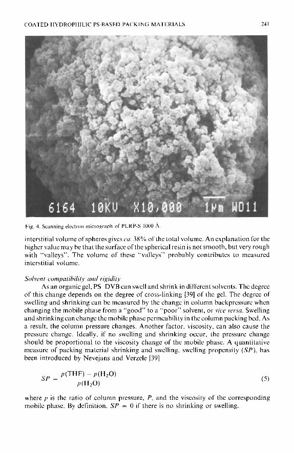
Fig. 3. Corresponding data are listed in Table II. Calibration curves obtained on both coated and uncoated resin were unusual. This may be due to the fact that these PS-DVB packings are made by fusing very small “non-porous” beads (microbeads), as shown by the scanning electron micrograph in Fig. 4. As a consequence, two types of
1.0 - (A)
= 5 OWI
>” 0.5
-
3 4 ” -3 BSA
OO I I I I
0.5 I.0 1.5 2.0
p (NaCI)
OO I I I I I I I 2
P [&& SO41
4 5 6
Fig. 2. Hydrophilicity of the coated resin. Column: 5 x 0.46 cm I.D. (15-25 pm, 300 A); flow-rate: 0.5 ml/min; detection: UV 254 nm. (A) NaCl in 50 mM Na-phosphate buffer (pH 7.0); (B) (NH&SO4 in 50 mM Na-phosphate buffer (pH 7.0).

240 Y.-B. YANG, F. E. REGNIER
6-
6
0 Uncoated PLRP-S 1000~
A Coated PLRP-S ICGO~
z4
-: i:--;
F
2
0 I I I I I 1.5 2.0 2.5 3.0 3.5 4.0
ELUTION VOLUME (ml)
Fig. 3. SEC calibration curves of coated and uncoated 1000 A PSDVB resins. Column: 25 x 0.46 cm I.D. (15-25 pm); polystyrene standards as the test probes, THF as mobile phase.
pores are present. One is formed by interstitial spaces between the microbeads. This is reflected by the upper part of the curve, which shows a mean pore size of 772 A on the uncoated resin. The other is the micropores of the “non-porous” microbeads. These contribute to the lower part of the curve. The mean pore diameter obtained from the lower part of the curve of uncoated resin is 12 A. This is consistent with the value (10 A) obtained on “non-porous” polystyrene packing by Nevejans and Verzele [38]. The mean pore size obtained from the whole calibration curve of uncoated resin is 600 A. It should be pointed out that the molecular weight standards used for the calibration are neither large enough to reach the exclusion limit nor small enough to reach the total permeation limit. This could result in some error in estimating mean pore size. After coating, the total pore volume of this resin was greatly decreased but, interestingly, the mean pore size increased. A mean pore size of 625 A was obtained from the whole calibration curve of the coated resin, 870 A on the upper part, and 21 8, on the lower part of the curve. Apparently, after coating, the pore volume fraction distributed among smaller pores was decreased. More importantly, although the mean pore size is changed, the molecular weight range between the exclusion and the total permeation limit does not change. The interstitial volume (Vi) of coated and uncoated PLRP-S 1000 8, seems to be very high, 56% of the total column volume for uncoated resin and 45% of the total column volume for coated resin. A theoretical calculation for
TABLE II
PHYSICAL FEATURES OF THE RESINS
Vi/Vc = Column interstitial porosity; VP/V, = column internal porosity; V,/V, = column porosity.
VilVc VP1 vc VOIVC @so (4
PLRP-S 1000 A 4.138 3.733 2.325 1.408 0.56 0.34 0.90 600 Coated PLRP-S 1000 A 4.138 2.12 1.893 0.827 0.45 0.20 0.66 625
COATED HYDROPHILIC PS-BASED PACKING MATERIALS 241
Fig. 4 Scanning electron micrograph 01‘ PLRP-S 1000 A.
interstitial volume of spheres gives CU. 38% of the total volume. An explanation for the higher value may be that the surface of the spherical resin is not smooth, but very rough with “valleys”. The volume of these “valleys” probably contributes to measured interstitial volume.
Solvent compatibility and rigidity
As an organic gel, PSDVB can swell and shrink in different solvents. The degree of this change depends on the degree of cross-linking [39] of the gel. The degree of swelling and shrinking can be measured by the change in column backpressure when changing the mobile phase from a “good” to a “poor” solvent, or vice versa. Swelling and shrinking can change the mobile phase permeability in the column packing bed. As a result, the column pressure changes. Another factor, viscosity, can also cause the pressure change. Ideally, if no swelling and shrinking occur, the pressure change should be proportional to the viscosity change of the mobile phase. A quantitative measure of packing material shrinking and swelling, swelling propensity (SP), has been introduced by Nevejans and Verzele [39]
sp = PUHF) - PWZO)
PWZO)
where p is the ratio of column pressure, P, and the viscosity of the corresponding mobile phase. By definition, SP = 0 if there is no shrinking or swelling.
242 Y.-B. YANG, F. E. REGNIER
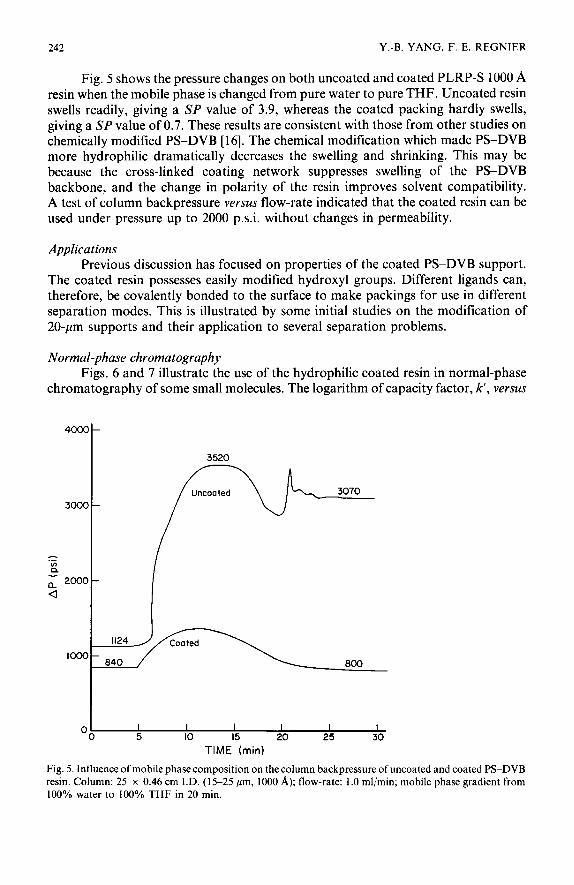
Fig. 5 shows the pressure changes on both uncoated and coated PLRP-S 1000 A resin when the mobile phase is changed from pure water to pure THF. Uncoated resin swells readily, giving a SP value of 3.9, whereas the coated packing hardly swells, giving a SP value of 0.7. These results are consistent with those from other studies on chemically modified PS-DVB [ 161. The chemical modification which made PS-DVB more hydrophilic dramatically decreases the swelling and shrinking. This may be because the cross-linked coating network suppresses swelling of the PS-DVB backbone, and the change in polarity of the resin improves solvent compatibility. A test of column backpressure versus flow-rate indicated that the coated resin can be used under pressure up to 2000 p.s.i. without changes in permeability.
Applications Previous discussion has focused on properties of the coated PS-DVB support.
The coated resin possesses easily modified hydroxyl groups. Different ligands can, therefore, be covalently bonded to the surface to make packings for use in different separation modes. This is illustrated by some initial studies on the modification of 20-pm supports and their application to several separation problems.
Normal-phase chromatography Figs. 6 and 7 illustrate the use of the hydrophilic coated resin in normal-phase
chromatography of some small molecules. The logarithm of capacity factor, k’, versus
4mo-
3520
01 I I I I I I
0 5 IO I5 20 25 30
TIME (min)
Fig. 5. Influence of mobile phase composition on the column backpressure of uncoated and coated PS-DVB resin. Column: 25 x 0.46 cm I.D. (15-25 pm, 1000 A); flow-rate: 1 .O ml/min; mobile phase gradient from 100% water to 100% THF in 20 min.
COATED HYDROPHILIC PS-BASED PACKING MATERIALS 243
2.C
1.:
I.C
0.5
-k
W -0
C
-0.5
-1.c
-1.4
I-
i-
log (% B)
P O-NH,
OH
* rlME (min)
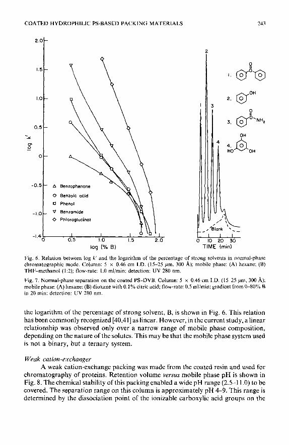
Fig. 6. Relation between log k’ and the logarithm of the percentage of strong solvents in normal-phase chromatographic mode. Column: 5 x 0.46 cm I.D. (15-25 pm, 300 A); mobile phase: (A) hexane; (B) THF-methanol (1:2); flow-rate: I.0 ml/min; detection: UV 280 nm.
Fig. 7. Normal-phase separation on the coated PS-DVB. Column: 5 x 0.46 cm I.D. (15-25 pm, 300 A); mobile phase: (A) hexane; (B) dioxane with 0.1% citric acid; flow-rate: 0.5 ml/min; gradient from O-80% B in 20 min: detection: UV 280 nm.
the logarithm of the percentage of strong solvent, B, is shown in Fig. 6. This relation has been commonly recognized [40,41] as linear. However, in the current study, a linear relationship was observed only over a narrow range of mobile phase composition, depending on the nature of the solutes. This may be that the mobile phase system used is not a binary, but a ternary system.
Weak cation-exchanger A weak cation-exchange packing was made from the coated resin and used for
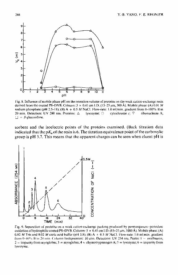
chromatography of proteins. Retention volume versus mobile phase pH is shown in Fig. 8. The chemical stability of this packing enabled a wide pH range (2.5-l 1.0) to be covered. The separation range on this column is approximately pH 4-9. This range is determined by the dissociation point of the ionizable carboxylic acid groups on the
244 Y.-B. YANG. F. E. KEONIEK
‘0 I 2 3 4 5 6 7 8 9 IO II
PH
Fig. 8. Influence of mobile phase pH on the retention volume of proteins on the weak cation-exchange resin derived from the coated PS-DVB. Column: 5 x 0.41 cm I.D. (15-25 pm, 300 A). Mobile phase: (A) 0.01 M sodium phosphate (pH 2.5-l 1); (B) A + 0.5 M NaCl. Flow-rate: I .O ml/min, gradient from O-100% B in 20 min. Detection: UV 280 nm. Proteins: LI = lysozyme; 0 = cytochrome c; c7 = ribonuclease A; 0 = /I-glucosidase.
sorbent and the isoelectric points of the proteins examined. (Back titration data indicated that the pK, of the resin is 6. The titration equivalence point of the carboxylic group is pH 3.7. This means that the apparent charges can be seen when eluent pH is
, , , , , , , , ,
0 8 16 24 32 1 TIME (min)
Fig. 9. Separation of proteins on a weak cation-exchange packing produced by permanganatc-periodale oxidation of hydrophilic coated PS-DVB. Column: 5 x 0.41 cm I.D. (15-25 pm, 1000 A). Mobile phase: (A) 0.02 M Tris and 0.02 M citric acid buffer (pH 5.8): (B) A + 0.5 M NaCl. Flow-rate: 1 .O ml/min, gradient from O-40”% B 111 20 min. Column backpressure: 10 atm. Detection: UV 254 nm. Peaks: I = ovalbumin;
2 = impurity from myoglobin; 3 = myoglobin; 4 = chymotrypsinogen A; 5 = lysozyme; 6 = impurity from
lysozyme.
COATED HYDROPHILIC PS-BASED PACKING MATERIALS 245
higher than 3.7.) Cytochrome c behaves differently from the other proteins tested. At and above pH 6, it was eluted between lysozyme and ribonuclease A in the order of pZ points (see Table I). But it was irreversibly adsorbed (not eluted) from the column at pH 4.5 and eluted as two peaks at pH 2.5. This may either be due to a pH-induced conformational change or dimerization [36,42]. A protein separation at pH 5.8 is shown in Fig. 9.
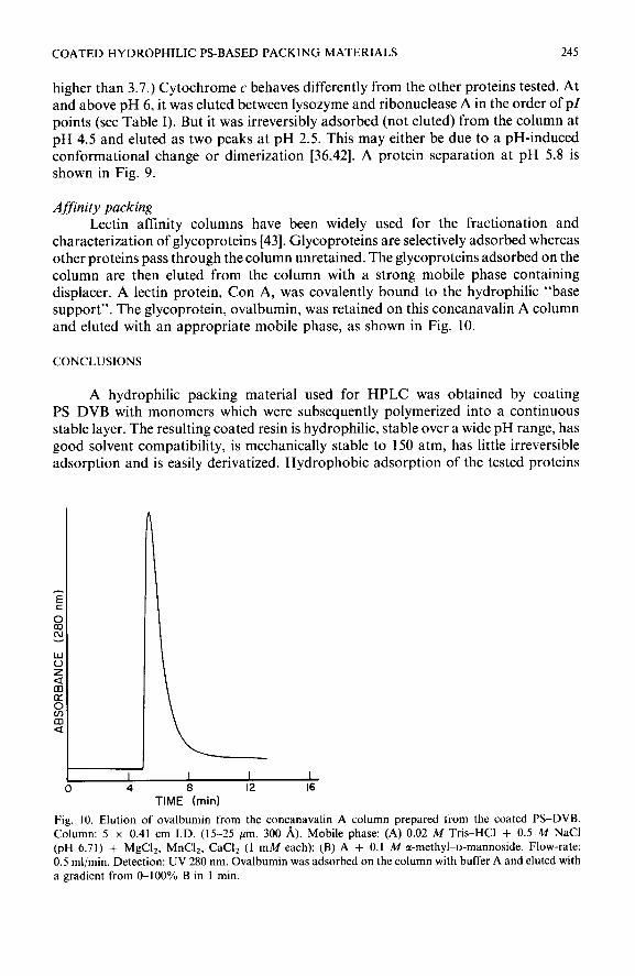
Affinity packing Lectin affinity columns have been widely used for the fractionation and
characterization of glycoproteins [43]. Glycoproteins are selectively adsorbed whereas other proteins pass through the column unretained. The glycoproteins adsorbed on the column are then eluted from the column with a strong mobile phase containing displacer. A lectin protein, Con A, was covalently bound to the hydrophilic “base support”. The glycoprotein, ovalbumin, was retained on this concanavalin A column and eluted with an appropriate mobile phase, as shown in Fig. 10.
CONCLUSIONS
A hydrophilic packing material used for HPLC was obtained by coating PSDVB with monomers which were subsequently polymerized into a continuous stable layer. The resulting coated resin is hydrophilic, stable over a wide pH range, has good solvent compatibility, is mechanically stable to 150 atm, has little irreversible adsorption and is easily derivatized. Hydrophobic adsorption of the tested proteins
I I I I
4 8 12 16 TIME (mid
Fig. IO. Elution of ovalbumin from the concanavalin A column prepared from the coated PS-DVB.
Column: 5 x 0.41 cm I.D. (15-25 pm, 300 A). Mobile phase: (A) 0.02 M Tris-HC1 + 0.5 M NaCl (pH 6.71) + MgCl*, MnC12, CaCI, (1 mM each); (B) A + 0.1 M a-methyl-D-mannoside. Flow-rate: 0.5 ml/min. Detection: UV 280 nm. Ovalbumin was adsorbed on the column with buffer A and eluted with a gradient from O-100% B in 1 min.
246 Y.-B. YANG, F. E. REGNIER
onto the coated support matrix could not be induced even with 2 M (NH4)2S04. Electrostatic adsorption of protein was not observed with mobile phase above 0.01 M salt concentration. A wide range of derivatization procedures can be used with this support, making the material useful for a variety of applications.
ACKNOWLEDGEMENTS
We thank Dr. C. Bracker and A. Bracker for their help in using the electron microscopy equipment (located in the Electron Microscopy Center in Agriculture, Agricultural Experiment Station, Purdue University). Dr. H. Weiner, M. Rounds and C. Desilets are acknowledged for their helpful comments regarding this manuscript. This research was supported by grants from the National Institute of Health (GM 25431) and the National Science Foundation (8613167). This is Journal Paper No. 12304 of the Purdue University Agricultural Experiment Station.
REFERENCES
1 J. Moore, J. Polym. Sci., Part A-2, (1964) 835.
2 S. Coppi, A. Betti, C. Bighi, G. P. Cartoni and F. Coccioli, J. Chromatogr., 442 (1988) 97.
3 D. P. Lee and J. Kindsvater, An&. Chem., 52 (1980) 2425.
4 Z. Iskandarani and D. Pietrzyk, Anal. Chem., 53 (1981) 489.
5 D. P. Lee, J. Chromutogr. Sci., 20 (1982) 203.
6 R. Greyson and A. Patch, J. Chromufogr., 242 (1982) 349.
7 H. A. McLeod and G. Laver, J. Chromufogr., 244 (1982) 385.
8 J. Bontemps, L. Bettendorff, J. Lombet, G. Dandrifosse, E. Schoffeniels, F. Nevejans, Y.-B. Yang and M. Verzele, Chromutogruphiu, 18 (I 984) 424.
9 J. V. Dawkins, L. L. Lloyd and F. P. Warner, J. Chromutogr., 352 (1986) 157.
10 L. D. Bowers and S. Pedigo, J. Chromutogr., 371 (1986) 243.
I I K. A. Tweeten and T. N. Tweeten, J. Chromutogr., 359 (1986) I1 I. 12 D. P. Lee, J. Chromurogr., 443 (1988) 143.
13 L. L. Lloyd, Z. Dryzek, D. B. Harrison and F. P. Warner, presented at the 10th Internutionu/S,vmposium
on Column Liquid Chromutogruphy, Sun Fruncisco, CA, U.S.A., Muy 18-23, 1986, abstract No. 515. 14 Y.-B. Yang, Ph.D. Dissertation, State University of Ghent, Ghent. 1986. 15 Y.-B. Yang, F. Nevejans and M. Verzele, Chromutogruphiu, 20 (1985) 735.
16 Y.-B. Yang and M. Verzele, J. Chromutogr., 387 (1987) 197.
17 Y.-B. Yang and M. Verzele, J. Chromutogr., 391 (1987) 383.
18 M. A. Rounds, W. D. Rounds and F. E. Regnier, J. Chromutogr., 397 (1987) 25.
19 M. A. Rounds and F. E. Regnier, J. Chromutogr., 443 (1988) 73.
20 E. Bayer, presented at the 10th International Symposium on Column Liquid Chromutogruphy, Sun
Francisco, CA, U.S.A.. May 18-23. 1986. abstract No. 504. 21 E. Bayer, German Pu/., DE 3 500 IX0 A I ( 19X4). 22 M. Verzele, F. Van Damme, C. Dewaele and M. Ghijs, Chromutogruphiu, 24 (1987) 302.
23 L. Varaday, F. Regnier, Y.-B. Yang and S. Cook, U.S. Put. Appl., 469 956, tiled on Jan. 25, 1990. 24 E. A. Peterson and H. A. Sober, J. Am. Chem. Sot., 78 (1956) 753.
25 E. Von Rudloff, Can. J. Chem., 43 (1965) 1784.
26 K. Nilsson and K. Mosbach, Biochem. Biophys. Res. Commun., 120 (1981) 449.
27 I. Hal&z and K. Martin, Angew. Chem., Inr. Ed. Engl., 17 (1978) 901. 28 R. Nikolov, W. Werner and 1. Hal&z, J. Chromurogr. Sci., 18 (1980) 207.
29 W. Werner and I. Hal&z, J. Chromutogr. Sci., 18 (1980) 277.
30 F. Nevejans and M. Verzele, Chromutogruphiu, 20 (1985) 173.
31 E. Pfannkoch, K. C. Lu, F. E. Regnier and H. G. Barth, J. Chromutogr. Sci., 18 (1980) 430.
32 D. E. Schmidt, Jr., R. W. Giese, D. Conron and B. L. Karger, Anal. Chem., 52 (1980) 177.
33 L. J. Janis and F. E. Regnier, J. Chromatogr., 444 (1988) I.
COATED HYDROPHILIC PS-BASED PACKING MATERIALS 241
34 W. Gastra, in J. M. Walker (Editor), Methods in Molecular Biology: Proteins, Vol. I, Humana Press, Clifton, NJ, 1984, p. 349.
35 L. J. Kricka, L&and Binder Assays, Marcel Dekker, New York, 1985, p. 78. 36 E. Margohash and J. Lustgarten, J. Biol. Chun., 237 (1962) 3397. 37 J. L. Fausnaugh, L. A. Kennedy and F. E. Regnier, J. Chromafogr., 317 (1984) 141. 38 F. Nevejans and M. Verzele, J. Chromatogr., 406 (1987) 325. 39 F. Nevejans and M. Verzele, J. Chromafogr., 350 (1985) 145. 40 L. R. Snyder, Anal. Chem., 46 (1974) 1384. 41 S. Hara and S. Ohnishi, J. Liq. Chromatogr., 7 (1984) 59. 42 R. Lemberg and J. Barrett, Cytochromes, Academic Press, London, 1973, p. 182. 43 T. Osawa and T. Tsuji. Annu. Rev. Biochem., 56 (1987) 21.