Embed Size (px)
Citation preview
Co-Option of Pre-Existing Pathways During Rhizobium-Legume
Symbiosis Evolution
ALESSANDRA LILLO
Thesis committee Thesis supervisor Prof. dr. Ton Bisseling Professor of Molecular Biology (Development Biology of Plants) Wageningen University Thesis co-supervisor Dr. ir. René Geurts Assistant professor, Laboratory of Molecular Biology Wageningen University Other members Dr. Duur K. Aanen, Wageningen University Dr. ir. Tijs Ketelaar, Wageningen University Dr. Jan A.L. van Kan, Wageningen University Prof. dr. Harro J. Bouwmeester, Wageningen University This research was conducted under the auspices of the Graduate School of Experimental Plant Sciences

Co-Option of Pre-Existing Pathways During Rhizobium-Legume
Symbiosis Evolution
Alessandra Lillo
Thesis submitted in fulfilment of the requirement for the degree of doctor
at Wageningen University by the authority of the Rector Magnificus
Prof. dr. M.J. Kropff, in the presence of the
Thesis Committee appointed by Academic Board to be defended in public
on Wednesday 5 September 2012 at 11 a.m. in the Aula.
Alessandra Lillo Co-Option of Pre-Existing Pathways During Rhizobium-Legume Symbiosis Evolution 152 pages Thesis, Wageningen University, Wageningen, NL (2012) With references and summaries in Dutch and English ISBN 978-94-6173-344-3
5
Contents
CHAPTER 1 7 Exploiting an Ancient Signalling Machinery to Enjoy a Nitrogen Fixing Symbiosis CHAPTER 2 23 Lateral Root Primordium Formation in Medicago truncatula and Lotus japonicus Involves Similar Cortical Cell Divisions as Root Nodule Formation CHAPTER 3 43 A Phylogenetic Strategy Based on a Legume-Specific Whole Genome Duplication Yields Symbiotic Cytokinin Type-A Response Regulators CHAPTER 4 71 NADPH oxidase MtRBOHA and MtRBOHG Have a Dual Function in Medicago-Rhizobium Nodule Symbiosis CHAPTER 5 101 Medicago NADPH Oxidase MtRBOHC is Nod Factor Induced CHAPTER 6 115 General Conclusions English Summary 137
Samenvatting 143 Acknowledgments 147 Curriculum Vitae 149 Education Statement 151


CHAPTER 1
Exploiting an Ancient Signalling Machinery to Enjoy a Nitrogen Fixing Symbiosis1
Alessandra Lillo, Ton Bisseling, and René Geurts
Adapted version from: Geurts, R., Lillo, A., Bisseling, T., (2012)
Exploiting an ancient signalling machinery to enjoy a nitrogen fixing symbiosis. Current Opinion in Plant Biology, 1-6.
Department of Plant Sciences, Laboratory of Molecular Biology, Wageningen University, Droevendaalsesteeg 1, 6708 PB, Wageningen, The Netherlands (A.L., T.B., R.G.); and College of Science, King Saud University, Post Office Box 2455, Riyadh 11451, Saudi Arabia (T.B). 1 This work was supported by the Dutch Science Foundation (NWO-vidi 864.06.007 to R.G.) and the E.U. Research Training Network “NODPERCEPTION” (MRTNCT-2006-035546) (to A.L., T.B., and R.G.)

Chapter 1
8
Abstract Almost for a century now it is speculated that a transfer of the largely legume-specific symbiosis with nitrogen fixing rhizobium would be profitable in agriculture (Burrill and Hansen, 1917; Charpentier and Oldroyd, 2010). Till now such step was not achieved, despite intensive research in this era. Novel insights in the underlying signalling networks leading to intracellular accommodation of rhizobium as well as mycorrhizal fungi of the Glomeromycota order show extensive commonalities between both interactions. As mycorrhizae symbiosis can be established basically with most higher plant species it raises questions why only in a few taxonomic lineages the underlying signalling network could be hijacked by rhizobium. Unravelling this, will lead to insights that are essential to achieve an old dream.

Exploiting an Ancient Signalling Machinery to Enjoy Symbiosis
9
Introduction Rhizobium bacteria and arbuscular mycorrhizal fungi of the Glomeromycota phylum can both establish an endosymbiosis with plants that facilitates growth in a nitrogen or phosphate deficient environment, respectively. Mycorrhizal fungi are obligatory biotrophs. Their hyphae penetrate the root intercellularly or intracellularly, depending on the plant host, and subsequently form arbuscules in inner cortical cells. These arbuscules are highly branched intracellular hyphae that are surrounded by a membrane formed by the host. This peri-arbuscular membrane functions as a symbiotic interface as transporters are present that facilitate exchange of nutrients. These are mainly phosphates, but also nitrates that are taken up by the mycelium outside the plant root (the so called extraradical mycelium) (Smith et al., 2011; Javot et al., 2011). In return the fungus obtains photosynthates of the plant for which it has specific monosaccharide transporters in its arbuscular membrane (Helber et al., 2011). In between the peri-arbuscular membrane and the branched hyphae a structured plant cell wall is practically absent to maximize reciprocal exchange between both organisms. Unlike mycorrhizal fungi, some soil bacteria of the Rhizobaceae family -collectively called rhizobia- have a dual lifestyle. They can be free living in soil, but in case the appropriate legume host is present they can establish a biotrophic endosymbiosis. For this they carry a set of symbiotic genes that are located on a large symbiotic (sym) plasmid(s) or are present as symbiotic islands in the genome. These symbiotic genes do include; (I) genes required for nitrogen fixation (the nif and fix genes) and (II) genes essential to establish symbiosis (the nod, nol and noe genes). This second set of genes encodes the machinery that is essential for biosynthesis and secretion of lipo-chitooligosaccharides (LCOs) that function as signal molecules and are named Nod factors. Nod factors are perceived by the host plant and set in motion symbiotic engagement.
Chapter 1
10
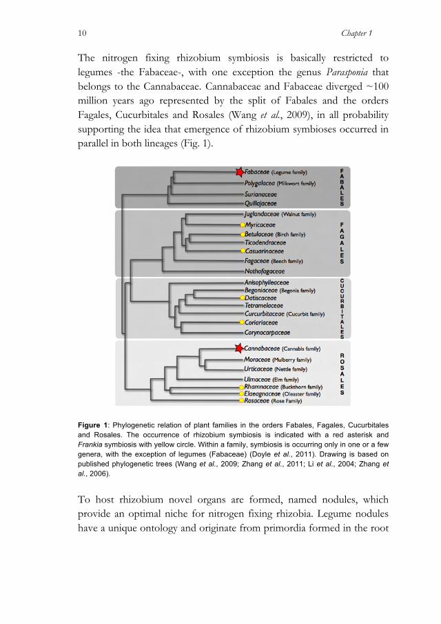
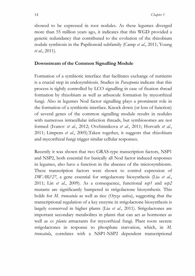
The nitrogen fixing rhizobium symbiosis is basically restricted to legumes -the Fabaceae-, with one exception the genus Parasponia that belongs to the Cannabaceae. Cannabaceae and Fabaceae diverged ~100 million years ago represented by the split of Fabales and the orders Fagales, Cucurbitales and Rosales (Wang et al., 2009), in all probability supporting the idea that emergence of rhizobium symbioses occurred in parallel in both lineages (Fig. 1).
Figure 1: Phylogenetic relation of plant families in the orders Fabales, Fagales, Cucurbitales and Rosales. The occurrence of rhizobium symbiosis is indicated with a red asterisk and Frankia symbiosis with yellow circle. Within a family, symbiosis is occurring only in one or a few genera, with the exception of legumes (Fabaceae) (Doyle et al., 2011). Drawing is based on published phylogenetic trees (Wang et al., 2009; Zhang et al., 2011; Li et al., 2004; Zhang et al., 2006).
To host rhizobium novel organs are formed, named nodules, which provide an optimal niche for nitrogen fixing rhizobia. Legume nodules have a unique ontology and originate from primordia formed in the root
Exploiting an Ancient Signalling Machinery to Enjoy Symbiosis
11
cortex. Legume nodules contain a large central tissue of which its infected cells harbour hundreds of rhizobia. In most legumes these bacteria are hosted individually, or in small clusters, surrounded by a plant-derived membrane; a unit that is called symbiosome. This membrane compartment facilitates exchange of fixed ammonium for other nutrients, including photosynthates. To reach the nodule primordia, in general sophisticated intracellular infection threads are formed. Starting at a root hair that curls around a (single) bacterium a membrane bound tubular infection thread is formed that guides the clonally propagating microsymbiont to the nodule primordium. Subsequently, bacteria are released from the infection thread and develop in their symbiotic form. Infection threads that enter nodule cells are bound by a thick cell wall and to release rhizobia from such infection thread cell wall-free patches are created, so-called unwalled droplets. At such sites, bacteria are in close contact with the surrounding host membrane enabling pinching off of symbiosomes. In contrast to legumes, Parasponia nodules seem much more primitive. Nodule ontology resembles that of a lateral root. Parasponia is infected intercellularly by rhizobium and only in the nodule cortex rhizobium triggers formation of intracellular infection threads. These are invaginations of the plasmamembrane and are bound by a thick cell wall. From these infection threads, fixation threads are formed. Fixation threads are also bound by a plant membrane and a plant cell wall, however this cell wall is markedly thinner than the infection thread cell wall. This type of fixation threads also occurs in nodules of some basal legumes. Rhizobium is not released as symbiosomes from the fixation threads most likely due to the presence of this cell wall. Commonalities in Signalling Since several years it is known that in legumes rhizobium and mycorrhizal fungi signals activate different receptors, but in turn these
Chapter 1
12
activate a common signalling module that subsequently diverges in the two symbiotic interactions (Radutoiu et al., 2003). This commonality in symbiotic signalling has been characterized in two model legume species, Lotus japonicus and Medicago truncatula, and is shown to occur in other legumes and non-legumes that interact with mycorrhizal fungi (Gutjahr et al., 2008; Kouchi et al., 2010). The common signalling module stretches from a plamamembrane receptor kinase (named LjSYMRK in L. japonicus and MtDMI2 in M. truncatula), a cation channel located in the nuclear envelope (LjCASTOR, LjPOLLUX/MtDMI1), and a nuclear localized protein complex of a calcium Calmodulin dependent kinase (CCaMK) and a coiled-coil protein (LjCYCLOPS/MtIPD3). Furthermore, several subunits of the nuclear pore have been found to be essential for rhizobium and mycorrhizae induced signalling (For recent reviews see: Kouchi et al., 2010; Oldroyd et al., 2011). An essential step in rhizobium symbiosis is the recognition of Nod factors, which holds for (almost all) legumes as well as for Parasponia species. In legumes, Nod factors are perceived by two distinct transmembrane LysM-type receptor kinases (LjNFR1, LjNFR5 / MtLYK3, MtNFP). Studies in heterologous systems indicate that these receptors can form a heterodimeric complex (Madsen et al., 2011), whereas in legumes itself the subcellular regulation is highly dynamic and affected upon Nod factor signalling (Haney et al., 2011). Upon Nod factor perception these LysM-type receptor kinases, together with the common signalling module, set in motion bacterial entry as well as root nodule organogenesis. Legume LysM-type Nod factor receptors are not essential for mycorrhization. However, two complementary approaches strongly support the idea that mycorrhizal fungi activate LysM-type receptor kinases. Recently it was shown that the model mycorrhizal fungus Glomus intraradices also produces LCOs, molecules that are named Myc factors (Maillet et al., 2011). Application of these Myc factors to plant roots increases mycorrhization at least twofold, an effect that seems generic in higher plants as it can be triggered in legumes as well as non-legumes (Maillet et al., 2011).

Exploiting an Ancient Signalling Machinery to Enjoy Symbiosis
13
Myc factors and Nod factors are structurally very similar (Maillet et al., 2011). As the rhizobium symbiosis is relatively young in comparison to mycorrhizal symbiosis it suggests that Nod factor perception evolved from the mycorrhizal fungal symbiosis. Strong support for this came from studies on Parasponia andersonii. Parasponia-rhizobium symbiosis is relatively young based on the close phylogenetic relation of Parasponia species with non-nodulating sister species in the Trema genus (Sytsma et al., 2002; Yesson et al., 2004). Therefore co-evolution of rhizobium and Parasponia has been relatively limited when compared to legumes. In line with this it is anticipated that in Parasponia the LysM-type receptor kinase family is less diverged, similar as seen in other non-legumes species (Zhang et al., 2007; Zhang et al., 2009). In comparison L. japonicus and M. truncatula have at least 2 members of the LjNFR5/MtNFP-type receptor kinases, of which one is a Nod factor receptor, whereas the second Nod factor receptor, LjNFR1/MtLYK3, underwent even series of duplications (Zhang et al., 2007; Limpens et al., 2003; Arrighi et al., 2006; Lohmann et al., 2010). In contrast P. andersonii only has a single NFR5/NFP-type receptor, PaNFP. Functional analysis revealed that PaNFP has a dual symbiotic function. It controls the formation of the symbiotic interface of rhizobium as well as mycorrhizal fungi (Op den Camp et al., 2011). Together with the observation that mycorrhizal fungi secrete LCOs this leads to the hypothesis that not only the common signalling module, but also the rhizobium Nod factor perception mechanism is recruited from endomycorrhizae. The duplications of Nod factor receptors in M. truncatula and L. japonicus do not stand on their own. Like most plant lineages also the legume lineage experienced whole genome duplications (WGDs) (Cannon et al., 2006). One such duplication event occurred early in evolution of the Papilionoideae about 58 million years ago. This legume subfamily represents most nodulating legumes, including all prominent crop species. Hundreds of paralogous gene pairs that originate from this duplication showed to be maintained in M. truncatula, L. japonicus and soybean (Glycine max), and for many of these one or even both genes
Chapter 1
14
showed to be expressed in root nodules. As these legumes diverged more than 55 million years ago, it indicates that this WGD provided a genetic redundancy that contributed to the evolution of the rhizobium nodule symbiosis in the Papilionoid subfamily (Camp et al., 2011; Young et al., 2011). Downstream of the Common Signalling Module Formation of a symbiotic interface that facilitates exchange of nutrients is a crucial step in endosymbiosis. Studies in Parasponia indicate that this process is tightly controlled by LCO signalling in case of fixation thread formation by rhizobium as well as arbuscule formation by mycorrhizal fungi. Also in legumes Nod factor signalling plays a prominent role in the formation of a symbiotic interface. Knock down (or loss of function) of several genes of the common signalling module results in nodules with numerous intracellular infection threads, but symbiosomes are not formed (Ivanov et al., 2012; Ovchinnikova et al., 2011; Horvath et al., 2011; Limpens et al., 2005).Taken together, it suggests that rhizobium and mycorrhizal fungi trigger similar cellular responses. Recently it was shown that two GRAS-type transcription factors, NSP1 and NSP2, both essential for basically all Nod factor induced responses in legumes, also have a function in the absence of the microsymbionts. These transcription factors were shown to control expression of DWARF27, a gene essential for strigolactone biosynthesis (Liu et al., 2011; Lin et al., 2009). As a consequence, functional nsp1 and nsp2 mutants are significantly hampered in strigolactone biosynthesis. This holds for M. truncatula as well as rice (Oryza sativa), suggesting that the transcriptional regulation of a key enzyme in strigolactone biosynthesis is largely conserved in higher plants (Liu et al., 2011). Strigolactones are important secondary metabolites in plants that can act as hormones as well as ex planta attractants for mycorrhizal fungi. Plant roots secrete strigolactones in response to phosphate starvation, which, in M. truncatula, correlates with a NSP1-NSP2 dependent transcriptional
Exploiting an Ancient Signalling Machinery to Enjoy Symbiosis
15
activation of MtDWARF27 (Liu et al., 2011). This supports that NSP1 and NSP2 have a function in mycorrhizal symbiosis and suggests that this function might have been recruited during evolution of rhizobium symbiosis. However, to date only very little evidence supports a role of strigolactone signalling in rhizobium symbiosis (Soto et al., 2010; Foo and Davies, 2011). Further, all NSP mediated responses in the rhizobium symbiosis depend on Nod factor perception and the common signalling module. In contrast, strigolactone biosynthesis does not depend on the common signalling module. Therefore it is very well possible that not primarily the strigolactone function of NSP1-NSP2 has been recruited in rhizobium symbiosis, but rather that both transcription factors gained novel primary targets in case of legume nodulation. The question remains how mycorrhizae and rhizobium LCO induced signalling on one hand can trigger similar cellular responses and on the other hand can control symbiosis specific responses of which nodule formation in case of rhizobium is most prominent. Both, mycorrhizae and rhizobium LCOs trigger Ca2+ oscillations in the perinuclear region within minutes after application. It was hypothesized that the amplitude and oscillation frequencies were different and that CCaMK, possibly in conjunction with interacting proteins like LjCYCLOPS/MtIPD3, can translate these different calcium signatures in specific responses (Kosuta et al., 2008). However, studies using a nuclear-targeted version of the Ca2+ sensor cameleon reveals that the Ca2+ oscillation responses triggered by rhizobium and a mycorrhizal fungus are indistinguishable (Sieberer et al., 2012). This makes it unlikely that a signature in Ca2+ oscillation is a discriminating factor between both symbionts. Instead a difference is observed between (cortical) cells that perceive LCOs and cells that become actually intracellularly infected by either rhizobium or mycorrhizal fungus. In latter case, the cells display and enhanced amplitude in Ca2+ oscillations (Sieberer et al., 2012). Still, CCaMK might be a component that can discriminate differences in input signal; e.g. in strength of the signal. A recent study shows that rhizobium and mycorrhzal symbioses have different requirements for binding of
Chapter 1
16
Calmodulin (CaM) to CCaMK (Shimoda et al., 2012). CaM is a Ca2+ binding protein that functions as messenger to transduce signals and in such could be a determinant of CCaMK specificity. However, the CaM binding domain in CCaMK is highly conserved and trans-complementation experiments demonstrated that non-legume CCaMK could functionally complement a corresponding mutation in legumes. This makes it unlikely that this domain obtained different properties in legumes to serve the rhizobium symbiosis. However, it does illustrate that there are more stringent demands to CCaMK functioning in rhizobium symbiosis when compared to mycorrhizae. In trying to understand the difference in rhizobium and mycorrhizal induced responses it is important to realise that our knowledge on ‘when, where, which and how much’ LCOs are needed in the different steps to achieve a symbiosis with rhizobium or a mycorrhizal fungus is still scanty. The differences in demands for Nod factor receptor is clearly illustrated by a M. truncatula Mtlyk3 splicing mutant (hcl-4) that produces markedly reduced levels of functional receptor protein (90% reduction of correctly spliced mRNA) (Smit et al., 2007). In this mutant root hair curling is not affected, whereas infection thread formation is almost completely blocked. Nevertheless both processes depend on the activation of the same common signalling pathway, including CCaMK. As mycorrhizal fungi seem to produce extremely low quantities of these LCOs when compared to rhizobium (Maillet et al., 2011), it is possible that the difference in response is also a matter of amounts of ligand. Evolutionary Constraints and Multiple Events As discussed above, it seems very probable that the evolution of rhizobium nodule symbiosis in legumes as well as in Parasponia involved the recruitment of the mycorrhizal LCO perception mechanism as well as the common signalling module. This suggests that genetic constraints rather than invention of novelties determined the evolution of the rhizobium endosymbiosis. As the essential genes are likely present in all
Exploiting an Ancient Signalling Machinery to Enjoy Symbiosis
17
plant species that are able to establish an endomycorrhizal symbiosis, it in theory provides a red-carpet welcome for microbes to evolve a (symbiotic) biotrophic relation. This raises the question whether the common signalling pathway has been recruited in evolution more than twice. Some observations indicate that this is indeed the case. Current knowledge about legume phylogeny in relation to occurrence of rhizobium symbiosis suggests that this character could have evolved up to 6 times within the Fabaceae (Doyle, 2011). However, no evidence yet has been provided that in all these cases the same set of genes has been co-opted. First such evidence has been obtained in a different plant-nitrogen fixing endosymbiosis; namely between the gram-positive bacteria of the genus Frankia and species collectively known as actinorhizal plants. Actinorhizal plants make lateral root-like nodules similar as found on Parasponia roots. Filamentous Frankia hyphae infect actinorhizal roots intercellularly, but once inside the nodule cortical cells are infected intracellularly to form fixiation thread-like structures. Actinorhyzal plants do not form a single phylogenetic lineage and most probably the symbiosis with Frankia species evolved several times in parallel (Fig. 1) (Doyle, 2011). Studies in two unrelated actinorhizal species, Datisca glomerata and Casuarina glauca revealed that gene homologs of the legume Nod factor signalling pathway are expressed in young root nodules (Hocher et al., 2011). RNA interference knockdown experiments revealed that the LRR-type receptor kinase SYMRK/DMI2 is essential for nodule formation as well as intracellular infection (Gherbi et al., 2008; Markmann et al., 2008). This makes it probable that also in actinorhizal species the common signalling module has been recruited. These findings suggest that parallel evolutionary events of nitrogen fixing nodular endosymbioses with either rhizobium or Frankia at least in part leaned on the ancient and widespread mycorrhizal symbiosis. Taken into account these recent insights in the genetic constraints underlying nitrogen fixing endosymbioses, it raises questions why not more plant species have gained such -at first sight- profitable interaction. Current advances in genomics and metabolomics provide unprecedented

Chapter 1
18
new tools to tackle this question. Thereby it should be the ambition to provide a proof of concept, and demonstrate that a transfer of nitrogen fixing symbiosis is achievable.
Exploiting an Ancient Signalling Machinery to Enjoy Symbiosis
19
References Arrighi, J-F., Barre, A., Ben Amor, B., et al., (2006) The Medicago truncatula lysin
motif-receptor-like kinase gene family includes NFP and new nodule-expressed genes. Plant Physiology, 142(1), 265–279.
Burrill, T.J. and Hansen, R., (1917) Is symbiosis possible between legume bacteria and non-legume plants? In Agricultural Experiment Station. Illinois University Press, pp. 115–181.
Camp, den, R.H.O., De Mita, S., Lillo, A., Cao, Q., Limpens, E., Bisseling, T. and Geurts, R., (2011) A phylogenetic strategy based on a legume-specific whole genome duplication yields symbiotic cytokinin type-A Response Regulators. Plant Physiology, 2013–2022.
Cannon, S.B., Sterck, L., Rombauts, S., et al., (2006) Legume genome evolution viewed through the Medicago truncatula and Lotus japonicus genomes. Proceedings of the National Academy of Sciences of the United States of America, 103(40), 14959–14964.
Charpentier, M. and Oldroyd, G., (2010) How close are we to nitrogen-fixing cereals? Current Opinion in Plant Biology, 13(5), 556–564.
Doyle, J.J., (2011) Phylogenetic perspectives on the origins of nodulation. Molecular Plant-Microbe Interactions, 24(11), 1289–1295.
Foo, E. and Davies, N.W., (2011) Strigolactones promote nodulation in pea. Planta, 234(5), 1073–1081.
Gherbi, H., Markmann, K., Svistoonoff, S., et al., (2008) SymRK defines a common genetic basis for plant root endosymbioses with arbuscular mycorrhiza fungi, rhizobia, and Frankiabacteria. Proceedings of the National Academy of Sciences of the United States of America, 105(12), 4928–4932.
Gutjahr, C., Banba, M., Croset, V., An, K., Miyao, A., An, G., Hirochika, H., Imaizumi-Anraku, H. and Paszkowski, U., (2008) Arbuscular mycorrhiza-specific signaling in rice transcends the common symbiosis signaling pathway. The Plant Cell, 20(11), 2989–3005.
Haney, C.H., Riely, B.K., Tricoli, D.M., Cook, Doug R, Ehrhardt, D.W. and Long, S.R., (2011) Symbiotic rhizobia bacteria trigger a change in localization and dynamics of the Medicago truncatula Receptor Kinase LYK3. The Plant Cell, 23(7), 2774–2787.
Helber, N., Wippel, K., Sauer, N., Schaarschmidt, S., Hause, B. and Requena, N., (2011) A versatile monosaccharide transporter that operates in the arbuscular mycorrhizal fungus Glomus sp is crucial for the symbiotic relationship with plants. The Plant Cell, 23(10), 3812–3823.
Hocher, V., Alloisio, N., Auguy, F., et al., (2011) Transcriptomics of actinorhizal symbioses reveals homologs of the whole common symbiotic signaling cascade. Plant Physiology, 156(2), 700–711.
Horvath, B., Yeun, L.H., Domonkos, A., et al., (2011) Medicago truncatula IPD3 is a member of the common symbiotic signaling pathway required for rhizobial and mycorrhizal symbioses. Molecular Plant-Microbe Interactions, 24(11), 1345–1358.
Chapter 1
20
Ivanov, S., Fedorova, E.E., Limpens, E., De Mita, S., Genre, A., Bonfante, P. and Bisseling, T., (2012) Rhizobium-legume symbiosis shares an exocytotic pathway required for arbuscule formation. Proceedings of the National Academy of Sciences of the United States of America.
Javot, H., Penmetsa, R.V., Breuillin, F., Bhattarai, K.K., Noar, R.D., Gomez, S.K., Zhang, Q., Cook, Douglas R and Harrison, M.J., (2011) Medicago truncatula mtpt4 mutants reveal a role for nitrogen in the regulation of arbuscule degeneration in arbuscular mycorrhizal symbiosis. The Plant Journal, 68(6), 954–965.
Kosuta, S., Hazledine, S., Sun, J., Miwa, H., Morris, R.J., Downie, J.A. and Oldroyd, G.E.D., (2008) Differential and chaotic calcium signatures in the symbiosis signaling pathway of legumes. Proceedings of the National Academy of Sciences of the United States of America, 105(28), 9823–9828.
Kouchi, H., Imaizumi-Anraku, H., Hayashi, M., Hakoyama, T., Nakagawa, T., Umehara, Y., Suganuma, N. and Kawaguchi, M., (2010) How many peas in a pod? Legume genes responsible for mutualistic symbioses underground. Plant and Cell Physiology, 51(9), 1381–1381.
Li, R-Q., Chen, Z-D., Lu, A., Soltis, D.E., Soltis, P.S. and Manos, P.S., (2004) Phylogenetic relationships in Fagales based on DNA sequences from three genomes. International Journal of Plant Sciences, 165(2), 311–324.
Limpens, E., Franken, C., Smit, P., Willemse, J., Bisseling, T. and Geurts, R., (2003) LysM domain receptor kinases regulating rhizobial Nod factor-induced infection. Science, 302(5645), 630–633.
Limpens, E., Mirabella, R., Fedorova, E., Franken, C., Franssen, H., Bisseling, T. and Geurts, R., (2005) Formation of organelle-like N2-fixing symbiosomes in legume root nodules is controlled by DMI2. Proceedings of the National Academy of Sciences of the United States of America, 102(29), 10375–10380.
Lin, H., Wang, R., Qian, Q., et al., (2009) DWARF27, an iron-containing protein required for the biosynthesis of strigolactones, regulates rice tiller bud outgrowth. The Plant Cell, 21(5), 1512–1525.
Liu, W., Kohlen, W., Lillo, A., et al., (2011) Strigolactone biosynthesis in Medicago truncatula and rice requires the symbiotic GRAS-type transcription factors NSP1 and NSP2. The Plant Cell, 23(10), 3853–3865.
Lohmann, G.V., Shimoda, Y., Nielsen, M.W., et al., (2010) Evolution and regulation of the Lotus japonicus LysM receptor gene family. Molecular Plant-Microbe Interactions, 23(4), 510–521.
Madsen, E.B., Antolín-Llovera, M., Grossmann, C., et al., (2011) Autophosphorylation is essential for the in vivo function of the Lotus japonicus Nod factor receptor 1 and receptor-mediated signalling in cooperation with Nod factor receptor 5. The Plant Journal, 65(3), 404–417.
Maillet, F., Poinsot, V., André, O., et al., (2011) Fungal lipochitooligosaccharide symbiotic signals in arbuscular mycorrhiza. Nature, 469(7328), 58–63.
Exploiting an Ancient Signalling Machinery to Enjoy Symbiosis
21
Markmann, K., Giczey, G. and Parniske, M., (2008) Functional adaptation of a plant receptor-kinase paved the way for the evolution of intracellular root symbioses with bacteria. PLoS Biology, 6(3), e68.
Oldroyd, G.E., Murray, J.D., Poole, P.S. and Downie, J.A., (2011) The rules of engagement in the legume-rhizobial symbiosis. Genetics, 45(1), 119–144.
Op den Camp, R., Streng, A., De Mita, S., et al., (2011) LysM-type mycorrhizal receptor recruited for rhizobium symbiosis in nonlegume Parasponia. Science, 331(6019), 909–912.
Ovchinnikova, E., Journet, E.-P., Chabaud, M., et al., (2011) IPD3 controls the formation of nitrogen-fixing symbiosomes in pea and Medicago Spp. Molecular Plant-Microbe Interactions, 24(11), 1333–1344.
Radutoiu, S., Madsen, L.H., Madsen, E.B., et al., (2003) Plant recognition of symbiotic bacteria requires two LysM receptor-like kinases. Nature, 425(6958), 585–592.
Shimoda, Y., Han, L., Yamazaki, T., Suzuki, R., Hayashi, M. and Imaizumi-Anraku, H., (2012) Rhizobial and fungal symbioses show different requirements for Calmodulin binding to Calcium Calmodulin-dependent Protein Kinase in Lotus japonicus. The Plant Cell, 24(1), 304–321.
Sieberer, B.J., Chabaud, M., Fournier, J., Timmers, A.C.J. and Barker, D.G., (2012) A switch in Ca2+ spiking signature is concomitant with endosymbiotic microbe entry into cortical root cells of Medicago truncatula. The Plant Journal, 69(5), 822–830.
Smit, P., Limpens, E., Geurts, R., Fedorova, E., Dolgikh, E., Gough, C. and Bisseling, T., (2007) Medicago LYK3, an entry receptor in rhizobial nodulation factor signaling. Plant Physiology, 145(1), 183–191.
Smith, S.E., Jakobsen, I., Grønlund, M. and Smith, F.A., (2011) Roles of arbuscular mycorrhizas in plant phosphorus nutrition: interactions between pathways of phosphorus uptake in arbuscular mycorrhizal roots have important implications for understanding and manipulating plant phosphorus acquisition. Plant Physiology, 156(3), 1050–1057.
Soto, M.J., Fernández-Aparicio, M., Castellanos-Morales, V., García-Garrido, J.M., Ocampo, J.A., Delgado, M.J. and Vierheilig, H., (2010) First indications for the involvement of strigolactones on nodule formation in alfalfa (Medicago sativa). Soil Biology and Biochemistry, 42(2), 383–385.
Sytsma, K.J., Morawetz, J., Pires, J.C., Nepokroeff, M., Conti, E., Zjhra, M., Hall, J.C. and Chase, M.W., (2002) Urticalean rosids: circumscription, rosid ancestry, and phylogenetics based on rbcL, trnL-F, and ndhF sequences. American Journal of Botany, 89(9), 1531–1546.
Wang, H., Moore, M.J., Soltis, P.S., et al., (2009) Rosid radiation and the rapid rise of angiosperm-dominated forests. Proceedings of the National Academy of Sciences of the United States of America, 106(10), 3853–3858.
Yesson, C., Russell, S.J., Parrish, T., Dalling, J.W. and Garwood, N.C., (2004) Phylogenetic framework for Trema (Celtidaceae). Plant Systematics and Evolution, 248(1), 85–109.
Chapter 1
22
Young, N.D., Debellé, F., Oldroyd, G.E.D., et al., (2011) The Medicago genome provides insight into the evolution of rhizobial symbioses. Nature.
Zhang, L-B., Simmons, M.P., Kocyan, A. and Renner, S.S., (2006) Phylogeny of the Cucurbitales based on DNA sequences of nine loci from three genomes: implications for morphological and sexual system evolution. Molecular Phylogenetics and Evolution, 39(2), 305–322.
Zhang, S-D., Soltis, D.E., Yang, Y., Li, D-Z. and Yi, T-S., (2011) Multi-gene analysis provides a well-supported phylogeny of Rosales. Molecular Phylogenetics and Evolution, 60(1), 21–28.
Zhang, X-C., Cannon, S.B. and Stacey, G., (2009) Evolutionary genomics of LysM genes in land plants. BMC Evolutionary Biology, 9, 183.
Zhang, X-C., Wu, X., Findley, S., Wan, J., Libault, M., Nguyen, H.T., Cannon, S.B. and Stacey, G., (2007) Molecular evolution of lysin motif-type receptor-like kinases in plants. Plant Physiology, 144(2), 623–636.
CHAPTER 2
Lateral Root Primordium Formation in Medicago truncatula and Lotus japonicus
Involves Cortical Cell Divisions Similar as Root Nodule Formation1
Alessandra Lillo, Ton Bisseling and René Geurts
Department of Plant Sciences, Laboratory of Molecular Biology, Wageningen University, Droevendaalsesteeg 1, 6708 PB, Wageningen, The Netherlands (A.L., T.B., and R.G.); College of Science, King Saud University, Post Office Box 2455, Riyadh 11451, Saudi Arabia (T.B.)
1 This work was supported by the Dutch Science Foundation (NWO-vidi 864.06.007 to R.G.) and the E.U. Research Training Network “NODPERCEPTION” (MRTNCT-2006-035546) (to A.L., T.B., and R.G.)
Chapter 2
24
Abstract
Nodule and lateral root formation have been proposed to share part of their developmental programs according to the idea that nodules evolved from pre-existing lateral roots (Nutman, 1948; Hirsch et al., 1997; Mathesius et al., 2000; Wopereis et al., 2000; Gonzalez-Rizzo et al., 2006). Although homology between these organs is evident in non-legume species able to establish a nodular nitrogen fixing symbiosis (Trinick, 1979; Callaham and Torrey, 1977), in case of legumes such resemblance, between nodule and lateral root developmental programs, is less obvious. Especially the mature organs have a distinct structure. In legumes, however, lateral root development has not been extensively studied. No clear data are available concerning the first stages of lateral root primordium formation. In contrast, nodule development has been studied in more detail. Here a better characterization of the first stages of lateral root development in the model legumes Medicago truncatula and Lotus japonicus is presented. It is shown that the cell layers recruited during lateral root primordium formation are very similar to the one’s involved in nodule primordium formation. This led to the speculation that part of the signalling network controlling lateral root primordium formation has been recruited during the evolution of nodules. So, lateral root formation have been analysed in symbiotic M. truncatula mutants affected in root nodule formation. Furthermore, the effect of environmental factors, such as external fixed nitrogen that is known to influence nodule primordium formation, has been studied. These experiments revealed that the lateral root developmental program is very robust, as it is not affected by any of the factors tested.
Lateral root primordium formation in involves cortical cell divisions
25
Introduction Plants of a single angiosperm lineage, the so-called N2-fixing clade, are able to enter a nodular endosymbiosis with nitrogen-fixing bacteria (Soltis et al., 1999; Soltis et al., 1995). Within this N2-fixing clade, only Fabaceae and the genus Parasponia of Cannabaceae have evolved such symbiosis with gram-negative bacteria, collectively known as rhizobia, while some other plants within this clade enter in symbiosis with an actinomycete of the genus Frankia. Within the N2-fixing clade, it is likely that nodulation evolved several times independently (Doyle et al., 2011). Since long it has been speculated that the pre-existing lateral root developmental program has been co-opted during evolution of nodulation (Nutman, 1948; Hirsch et al., 1997; Mathesius et al., 2000; Wopereis et al., 2000; Gualtieri and Bisseling, 2000; Gonzalez-Rizzo et al., 2006). Homology between these organs is evident in non-legume nitrogen-fixing species. Such nodules clearly resemble lateral roots in terms of cytology and ontology. Non-legume nitrogen-fixing nodules originate largely from the pericycle cell layer and have a central vascular bundle surrounded by a cortex in which bacterial infection takes place (Hirsch, 1992; Vasse et al., 1990). In case of legume nodules, however, such morphological similarity is less obvious. Legume nodules have a distinct ontology compared to lateral roots. They have a large central zone that contains cells that accommodate the endosymbiont bacteria and is surrounded by peripheral tissues, including vascular bundles, embedded in the nodule parenchyma. Furthermore, legume nodules largely originate from cortical cell layers (Truchet et al., 1989; van Brussel et al., 1992; Yang et al., 1994; Timmers et al., 1999), whereas lateral roots are believed to derive from the pericycle cell layer (Libbenga and Harkes, 1973; Hirsch et al., 1997), though only limited number of studies have been conducted. Here we aim to provide a better characterization of the first stages of lateral root development in the model legumes Medicago truncatula (Medicago) and Lotus japonicus (Lotus).

Chapter 2
26
In the generic plant model Arabidopsis thaliana (Arabidopsis), lateral roots are initiated exclusively in pericycle cell files positioned opposite a xylem pole (Dubrovsky et al., 2000; Beeckman et al., 2001; Kurup et al., 2005). Within these pericycle cell files, a small group of adjacent cells, called the lateral root founder cell population, are primed (specified) to form a lateral root primordium (De Smet et al., 2007; Baluska et al., 2010). The underlying mechanism of cell priming remains to be elucidated. Two mechanisms have been proposed, both finding their origin in the basal root meristem: (1) periodic fluctuations in auxin signalling (De Smet et al., 2007; Dubrovsky et al., 2008; Fukaki and Tasaka, 2009), and (2) oscillations in gene expression regulated by an endogenous mechanism other than auxin (Moreno-Risueno and Benfey, 2011). Starting exclusively from dividing pericycle founder cells, the formation of a lateral root primordium has been subdivided in subsequent phases preceding lateral root emergence (Malamy and Benfey, 1997). The founder cells undergo polarized asymmetric anticlinal divisions, creating two small daughter cells flanked by two larger daughter cells. The two small daughter cells continue to divide creating a group with a maximum of ten small cells that are flanked by two large daughter cells. Subsequently, the central small cells divide periclinally, giving rise to a primordium composed of an inner and an outer cell layer. Several rounds of subsequent cell divisions take place. One round of periclinal cell divisions in the outer layer creates a three-layered lateral root primordium, then a second round of periclinal cell divisions in the inner layer adds a fourth layer giving rise to a primordium with two inner and two outer cell layers. Next, anticlinal cell divisions take place so that the lateral root primordium is midway through the parent cortex. From this point on, series of cell expansions and division events occur, giving rise to an emerging lateral root. Studies on lateral root development in other plant species then Arabidopsis are scarce. In cereals it was shown that lateral roots originate from the endodermis and the pericycle, similar as seen in Arabidopsis

Lateral root primordium formation in involves cortical cell divisions
27
(Hochholdinger and Zimmermann, 2008). However, older studies suggest that the precise ontology of the lateral root developmental program can differ between species. For instance, in case of Allium cepa (Casero et al., 1996) and the legume Glycine max (soybean) (Bryne et al., 1977), cortical cell divisions occur during the formation of the lateral root primordium. Moreover, in Cucurbita maxima lateral root primordia are initiated in the pericycle and also endodermis and cortical cells contribute to the formation of the primordia (Mallory et al., 1970). Apart from soybean (Bryne et al., 1977), lateral root development has not been studied in legumes. Although no data are available concerning the precise cell layers involved in lateral root primordium formation in legumes, it is commonly believed that exclusively pericycle cells give rise to the lateral root primordium according to Arabidopsis (Oldroyd et al., 2011; Hirsch et al., 1997). In contrast, nodule development has been studied in far more detail, especially in the model legumes Medicago and Lotus. These species make distinct nodule types. Medicago develops indeterminate nodules with an active apical meristem, whereas Lotus forms determinate nodules that loose the meristematic activity soon after initiation. In Medicago the first cell divisions that occur during the symbiotic interaction are anticlinal divisions of pericycle cells positioned opposite the protoxylem poles (Timmers et al., 1999). Following the mitotic activation of pericycle cells, inner cortical cells divide anticlinally first, and periclinally after, forming the nodule primordium. The endodermis undergoes only a very limited number of divisions. Activated cells of the inner cortical layers give rise to the nodule primordium (Timmers et al., 1999). Similarly, also in Lotus cortical cells give rise to the nodule primordium, however, in this case, mainly cells form the middle and outer cortical layers (Szczyglowski et al., 1998). Although lateral root formation has not been studied in either model legumes, and despite the fact that old literature indicates specie-specific morphological variation in this process, the findings in Arabidopsis are generalized. To determine which cell layers contribute to lateral root
Chapter 2
28
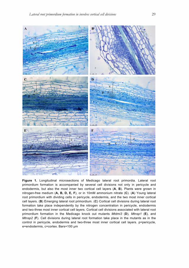
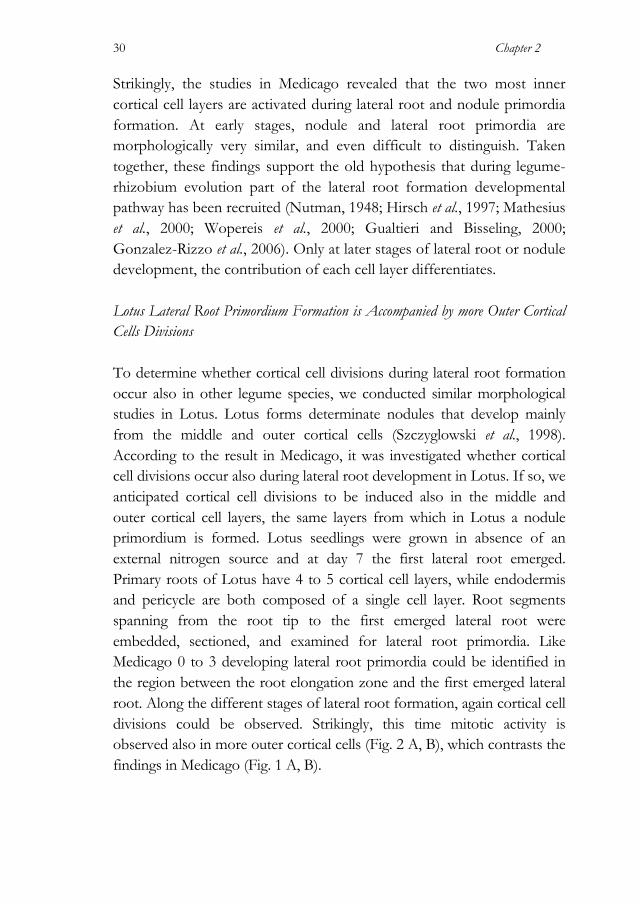
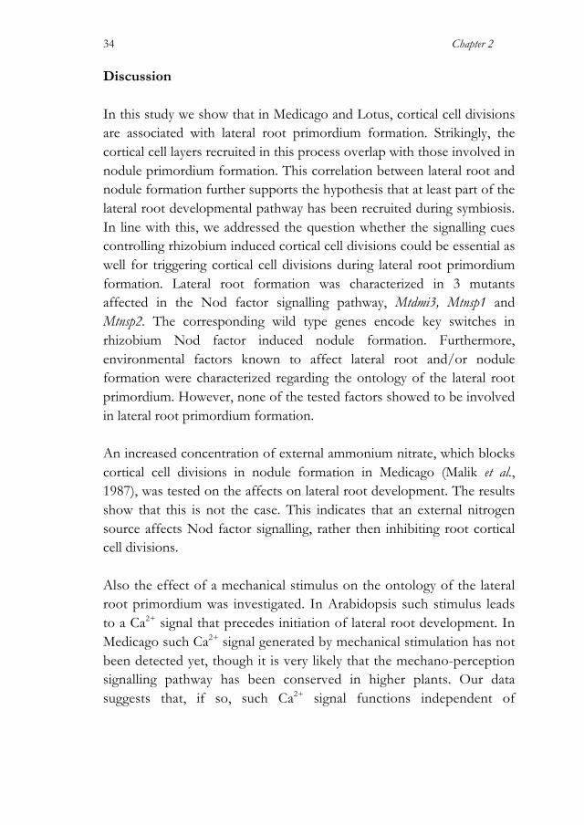
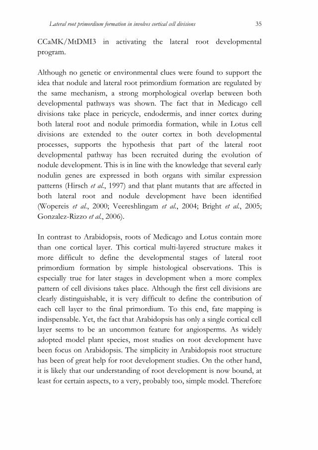
development, the first stages of this process in both species Medicago and Lotus were characterized. We show that in these legumes not only pericycle, but also endodermis and cortical cell layers contribute to lateral root primordium formation. Moreover, in these two species a striking correlation is found in the number and position of cortical cells recruited during lateral root and nodule primordia formation. To analyse this morphological correlation, it was investigated whether the cortical cell divisions in the lateral root developmental program are regulated by the same signalling mechanisms that control nodule primordium formation. Results Medicago Lateral Root Primordium Formation is Accompanied by Inner Cortical Cell Divisions To determine the ontology of lateral roots in Medicago, seedlings (n=10) were grown vertically on a modified (nitrogen-free) Fahraeus medium (Fahraeus, 1957), at 21°C. Under such conditions, the timing of the formation of first lateral root is highly synchronized. The first lateral root emerged 10 days after germination at approximately 5 cm above of the root tip. Root segments spanning from the root tip to the youngest emerged lateral root were embedded, and longitudinal sections were made. These sections were examined for lateral root primordia. Medicago roots have a single epidermal layer, ~5 cortical cell layers, a single endodermal cell layer, and, adjacent to the vascular bundle, a single pericycle cell layer. In these sections 0 to 3 lateral root primordia were present. Strikingly, cell divisions associated with lateral root formation occurred not only in the pericycle, but also in the 2 most inner cortical cell layers (layer 4 and 5) (Fig. 1A). The finding that in Medicago root cortical cell divisions occur during lateral root primordium development, shows that these cells have the ability to divide during non-symbiotic development, rather than cortical cell divisions are exclusively triggered upon rhizobium signalling.
Lateral root primordium formation in involves cortical cell divisions
29
Figure 1. Longitudinal microsections of Medicago lateral root primordia. Lateral root primordium formation is accompanied by several cell divisions not only in pericycle and endodermis, but also the most inner two cortical cell layers (A, B). Plants were grown in nitrogen-free medium (A, B, D, E, F), or in 10mM ammonium nitrate (C). (A) Young lateral root primordium with dividing cells in pericycle, endodermis, and the two most inner cortical cell layers. (B) Emerging lateral root primordium. (C) Cortical cell divisions during lateral root formation take place independently by the nitrogen concentration in pericycle, endodermis and two-three most inner cortical cell layers. Cortical cell divisions associated with lateral root primordium formation in the Medicago knock out mutants Mtdmi3 (D), Mtnsp1 (E), and Mtnsp2 (F). Cell divisions during lateral root formation take place in the mutants as in the control in pericycle, endodermis and two-three most inner cortical cell layers. p=pericycle, e=endodermis, c=cortex. Bars=100 µm
c
p e
A B
C D
pec
c
pe
cep
E F
p ec
Chapter 2
30
Strikingly, the studies in Medicago revealed that the two most inner cortical cell layers are activated during lateral root and nodule primordia formation. At early stages, nodule and lateral root primordia are morphologically very similar, and even difficult to distinguish. Taken together, these findings support the old hypothesis that during legume-rhizobium evolution part of the lateral root formation developmental pathway has been recruited (Nutman, 1948; Hirsch et al., 1997; Mathesius et al., 2000; Wopereis et al., 2000; Gualtieri and Bisseling, 2000; Gonzalez-Rizzo et al., 2006). Only at later stages of lateral root or nodule development, the contribution of each cell layer differentiates. Lotus Lateral Root Primordium Formation is Accompanied by more Outer Cortical Cells Divisions To determine whether cortical cell divisions during lateral root formation occur also in other legume species, we conducted similar morphological studies in Lotus. Lotus forms determinate nodules that develop mainly from the middle and outer cortical cells (Szczyglowski et al., 1998). According to the result in Medicago, it was investigated whether cortical cell divisions occur also during lateral root development in Lotus. If so, we anticipated cortical cell divisions to be induced also in the middle and outer cortical cell layers, the same layers from which in Lotus a nodule primordium is formed. Lotus seedlings were grown in absence of an external nitrogen source and at day 7 the first lateral root emerged. Primary roots of Lotus have 4 to 5 cortical cell layers, while endodermis and pericycle are both composed of a single cell layer. Root segments spanning from the root tip to the first emerged lateral root were embedded, sectioned, and examined for lateral root primordia. Like Medicago 0 to 3 developing lateral root primordia could be identified in the region between the root elongation zone and the first emerged lateral root. Along the different stages of lateral root formation, again cortical cell divisions could be observed. Strikingly, this time mitotic activity is observed also in more outer cortical cells (Fig. 2 A, B), which contrasts the findings in Medicago (Fig. 1 A, B).

Lateral root primordium formation in involves cortical cell divisions
31
Figure 2. Longitudinal microsections of Lotus lateral root primordium. Plants were grown in nitrogen-free medium. (A) Young lateral root primordium with dividing pericycle, endodermis, and all inner cortical cell layers except the most outer one. (B) Emerging lateral root primordium. p= pericycle, e=endodermis, c=cortex. Bars=100 µm Cortical Participation in Lateral Root Development is Independent of the Nitrogen Status of the Plant A well-known character of root nodule development is that fixed nitrogen sources inhibits it (Carroll et al., 1985; Schnabel et al., 2010). 10 mM ammonium nitrate is sufficient to block rhizobium induced symbiotic cell divisions in the root cortex (Malik et al., 1987). To address the question whether an external nitrogen source restrains cortical cell divisions during lateral root formation, Medicago seedlings (n=10) where grown for 10 days under nutrient non-permissive (10 mM ammonium nitrate), or permissive (nitrogen-free) symbiotic conditions. In both growth conditions the frequency of primordium formation was similar, 0 to 3 developing lateral root primordia could be identified in the region between the root elongation zone and the first emerged lateral root. This is in line with a previous study that showed that the average number of lateral roots per root in Medicago is not affected by ammonium nitrate (Yendrek et al., 2010). To investigate whether the presence of fixed nitrogen in the medium affects the ontology of lateral root primordia, we sectioned the root segment between meristem and first emerging lateral root. Generally, 0 to 3 primorida were present in this region of the root. Irrespective on the presence of ammonium nitrate, lateral root formation is accompanied
A B
c
c

Chapter 2
32
by cell divisions of pericycle and inner cortex cell layers 4 and 5 (Fig. 1C). Therefore, we conclude that the ontology of lateral root primordia is not affected by the presence of a fixed nitrogen source in the medium. Lateral Root Primordium Associated Cortical Cell divisions are Independent of the Nod Factor Signalling Cascade and Physically Induced Calcium Responses Nodule formation is controlled by rhizobium Nodulation (Nod) factors. Nod factors are lipochitooligosaccharide signals that trigger key symbiotic responses in the host legume root by a specific signalling pathway; the so-called Nod factor signalling pathway. This pathway includes several genes that are essential not only for rhizobium Nod factor induced signalling, but also for mycorrhizal symbiosis. Among these is a nuclear localized calcium/calmodulin-dependent protein kinase CCAMK, named MtDMI3 in Medicago. It is postulated that CCAMK is regulated by Ca2+ and subsequently activates two GRAS-type transcription factors NSP1 and NSP2. Since CCAMK is essential and sufficient to activate root nodule developmental program (Gleason et al., 2006; Tirichine et al., 2007), the Medicago CCAMK knockout mutant, Mtdmi3, was investigated to determine whether lateral root primordium formation requires part of the Nod factor signalling pathway. Root segments between meristem and first emerging lateral root were sectioned. These studies revealed that also in the Mtdmi3 mutant lateral root formation involves cell divisions of the pericycle and inner cortex cell layer 4 and 5 (Fig. 1C). Therefore, a functional CCaMK is not essential for cortical cell divisions during lateral root formation. Arabidopsis lateral root formation can be triggered by mechanical stimulation through an increase of cytosolic Ca2+ (Monshausen et al., 2009). We questioned whether in Medicago CCaMK is involved in the transduction of such Ca2+ signal, especially in mitotic activation of cortical cells. First, an assay to induce lateral root development upon

Lateral root primordium formation in involves cortical cell divisions
33
transient bending of Medicago roots was developed. Four days old Medicago seedling roots were transiently bended, similar as described for Arabidopsis (Monshausen et al., 2009). Two days post treatment the plant roots were analysed for lateral root primordia. For both wild type and Mtdmi3 roots (n=10), in ~70% of the cases lateral root primordium formation is triggered at the convex side of the bending. Next, cortical cell divisions are associated with the lateral root primordia were investigated. No differences were observed in the number cell layers taking part in lateral root formation between Mtdmi3 and wild type roots (data not shown). Likewise, Mtdmi3 knockout mutant roots grown just straight showed no differences in lateral root primordium morphology (Fig. 1A, 1D). Concluding, CCaMK/MtDMI3 is not required in lateral root primordium development. Nod factor signalling also requires transcription factors, among which are the GRAS-type proteins NSP1 and NSP2 (Kalò et al., 2005; Smit et al., 2005; Heckmann et al., 2006; Murakami et al., 2006). Both transcription factors are essential for nearly all rhizobium-induced symbiotic responses, including Nod factor-induced cortical cell divisions (Catoira et al., 2000; Oldroyd and Long, 2003; Smit et al., 2005), while under non-symbiotic conditions they control strigolactones biosynthesis (Liu et al., 2011). Strigolactones are known to play a role in root development (Ruyter-Spira et al., 2011; Kapulnik et al., 2011; Liu et al., 2010). In line with these findings, we raised the question whether strigolactones play a role in lateral root primordium ontology in Medicago. To this end lateral root development in Mtnsp1 and Mtnsp2 knockout mutants was characterized. Ten days post germination, root segments spanning from the root tip to the first emerged lateral root were embedded, sectioned, and examined as described before. As in wild type, in both nsp mutants lateral root formation is accompanied by cell divisions in pericycle, endodermis and the 2 most inner cortical cell layers (layer 4 and 5) (Fig.1 E-F). This indicates that the ontology of the lateral root primordium in Medicago is independent of NSP1 and NSP2.
Chapter 2
34
Discussion In this study we show that in Medicago and Lotus, cortical cell divisions are associated with lateral root primordium formation. Strikingly, the cortical cell layers recruited in this process overlap with those involved in nodule primordium formation. This correlation between lateral root and nodule formation further supports the hypothesis that at least part of the lateral root developmental pathway has been recruited during symbiosis. In line with this, we addressed the question whether the signalling cues controlling rhizobium induced cortical cell divisions could be essential as well for triggering cortical cell divisions during lateral root primordium formation. Lateral root formation was characterized in 3 mutants affected in the Nod factor signalling pathway, Mtdmi3, Mtnsp1 and Mtnsp2. The corresponding wild type genes encode key switches in rhizobium Nod factor induced nodule formation. Furthermore, environmental factors known to affect lateral root and/or nodule formation were characterized regarding the ontology of the lateral root primordium. However, none of the tested factors showed to be involved in lateral root primordium formation. An increased concentration of external ammonium nitrate, which blocks cortical cell divisions in nodule formation in Medicago (Malik et al., 1987), was tested on the affects on lateral root development. The results show that this is not the case. This indicates that an external nitrogen source affects Nod factor signalling, rather then inhibiting root cortical cell divisions. Also the effect of a mechanical stimulus on the ontology of the lateral root primordium was investigated. In Arabidopsis such stimulus leads to a Ca2+ signal that precedes initiation of lateral root development. In Medicago such Ca2+ signal generated by mechanical stimulation has not been detected yet, though it is very likely that the mechano-perception signalling pathway has been conserved in higher plants. Our data suggests that, if so, such Ca2+ signal functions independent of
Lateral root primordium formation in involves cortical cell divisions
35
CCaMK/MtDMI3 in activating the lateral root developmental program. Although no genetic or environmental clues were found to support the idea that nodule and lateral root primordium formation are regulated by the same mechanism, a strong morphological overlap between both developmental pathways was shown. The fact that in Medicago cell divisions take place in pericycle, endodermis, and inner cortex during both lateral root and nodule primordia formation, while in Lotus cell divisions are extended to the outer cortex in both developmental processes, supports the hypothesis that part of the lateral root developmental pathway has been recruited during the evolution of nodule development. This is in line with the knowledge that several early nodulin genes are expressed in both organs with similar expression patterns (Hirsch et al., 1997) and that plant mutants that are affected in both lateral root and nodule development have been identified (Wopereis et al., 2000; Veereshlingam et al., 2004; Bright et al., 2005; Gonzalez-Rizzo et al., 2006). In contrast to Arabidopsis, roots of Medicago and Lotus contain more than one cortical layer. This cortical multi-layered structure makes it more difficult to define the developmental stages of lateral root primordium formation by simple histological observations. This is especially true for later stages in development when a more complex pattern of cell divisions takes place. Although the first cell divisions are clearly distinguishable, it is very difficult to define the contribution of each cell layer to the final primordium. To this end, fate mapping is indispensable. Yet, the fact that Arabidopsis has only a single cortical cell layer seems to be an uncommon feature for angiosperms. As widely adopted model plant species, most studies on root development have been focus on Arabidopsis. The simplicity in Arabidopsis root structure has been of great help for root development studies. On the other hand, it is likely that our understanding of root development is now bound, at least for certain aspects, to a very, probably too, simple model. Therefore
Chapter 2
36
we argue that lateral root studies should now be extended to other species. Our work represents a first step toward this aim. Materials and Methods Plant Materials and Growth Conditions Medicago truncatula lines Jemalong A17, Mtnsp1-1 (B85) (Catoira et al., 2000; Smit et al., 2005), Mtnsp2-2 (0-4) (Oldroyd and Long, 2003; Kaló et al., 2005), and Mtdmi3-1 (TRV25) (Mitra et al., 2004), were used. The seeds were scarified in concentrated sulphuric acid for 8 min, rinsed with water, surface-sterilized in 4% sodium hypochlorite, rinsed for 6 times in sterile water, imbibed for 2 hours in sterile water, and plated on 1% deionized water agar plates. The seeds were subsequently vernalized for 24 hours at 4°C and were germinated by incubating at room temperature overnight. Medicago germinated seedlings were grown vertically on modified Fahraeus medium plates with (10mM ammonium nitrate) or without nitrogen (Fahraeus, 1957) in a growth chamber at 20°C and 16/8 hours day/night regime. Lotus japonicus B-129 Gifu was used as a wild type. The seeds were scarified in concentrated sulphuric acid for 15 min, rinsed with sterile water, surface-sterilized in 4% sodium hypochlorite, rinsed 6 times with sterile water, imbibed in sterile water for 2 hours water, and plated on 1% deionized water agar plates. The seeds were subsequently vernalized for 48 hours at 4°C and subsequently germinated by incubating at room temperature for 48 hours. Lotus germinated seedlings were grown vertically on modified (nitrogen-free) Fahraeus medium (Fahraeus, 1957) in a growth chamber at 20°C and 16/8 hours day/night regime. Root Bending Assay The bent was imposed for at least 1 minute with the aid of 2 glasses capillary on 4 days old seedling. In the proximal elongation/mature zone
Lateral root primordium formation in involves cortical cell divisions
37
roots were manually reoriented with an angle of 90 degrees and a pencil mark was left on the plate at the place of the bending. The petri dishes were left such that the root apex, at the moment of the reorientation, was aligned perpendicular to the gravity vector. 2 days after, bended roots segments of circa 2 cm were collected at the place of the mark and embedded for histochemical analysis. Histochemical Analysis and Microscopy Roots were fixed in 5% glutaraldehyde (v/v) in 0.1 M phosphate buffer (pH7.2) for at least 1 hour under vacuum. The fixed roots were subsequently washed tree times for 15 min in 0.1 M phosphate buffer (pH7.2) and one time for 15 min in water. Ethanol dehydration series was carried out as followed: 10 min in 10%, 30%, 50%, 70%, 90%, and 100% ethanol. The dehydrated roots were embedded in Technovit 7100 (Heraeus-Kulzer, Wehrheim, Germany). Microtome sections of 4 µm using a microtome (Reichert-Jung, Leica, Holland), stained by 1% toluidine blue (Sigma, Germany), mounted in Canada balsam (MERCK, Holland), and analysed using a Leica DM5500B microscope equipped with a Leica DFC425C camera (Leica microsystems, Wetzlar, Germany). Images were digitally processed using Photoshop CS3 (Adobe Systems, San Jose, California).
Chapter 2
38
References Baluska, F., Mancuso, S., Volkmann, D. and Barlow, P.W., (2010) Root apex
transition zone: a signalling-response nexus in the root. Trends in Plant Science, 15(7), 402–408.
Beeckman, T., Burssens, S. and Inzé, D., (2001) The peri-cell-cycle in Arabidopsis. Journal of Experimental Botany, 52, 403–411.
Bright, L.J., Liang, Y., Mitchell, D.M. and Harris, J.M., (2005) The LATD gene of Medicago truncatula is required for both nodule and root development. Molecular Plant-Microbe Interactions, 18(6), 521–532.
Bryne, J.M., Pesacreta, T.C. and Fox, J.A., (1977) Development and structure of the vascular connection between the primary and secondary root of Glycine max (L.) Merr. American Journal of Botany, 64(8), 946–959.
Callaham, D. and Torrey, J.G., (1977) Prenodule formation and primary nodule development in roots of Comptonia (Myricaceae). Canadian Journal of Botany, 55(17), 2306–2318.
Carroll, B.J., McNeil, D.L. and Gresshoff, P.M., (1985) Isolation and properties of soybean [Glycine max (L.) Merr.] mutants that nodulate in the presence of high nitrate concentrations. Proceedings of the National Academy of Sciences of the United States of America, 82(12), 4162–4166.
Casero, P.J., Casimiro, I. and Lloret, P.G., (1996) Pericycle proliferation pattern during the lateral root initiation in adventitious roots of Allium cepa. Protoplasma, 191(3-4), 136–147.
Catoira, R., Galera, C., de Billy, F., et al., (2000) Four genes of Medicago truncatula controlling components of a nod factor transduction pathway. The Plant Cell, 12(9), 1647–1666.
De Smet, I., Tetsumura, T., De Rybel, B., et al., (2007) Auxin-dependent regulation of lateral root positioning in the basal meristem of Arabidopsis. Development, 134(4), 681–690.
Doyle, J.J., (2011) Phylogenetic perspectives on the origins of nodulation. Molecular Plant-Microbe Interactions, 24(11), 1289–1295.
Dubrovsky, J.G., Doerner, P.W., Colón-Carmona, A. and Rost, T.L., (2000) Pericycle cell proliferation and lateral root initiation in Arabidopsis. Plant Physiology, 124(4), 1648–1657.
Dubrovsky, J.G., Sauer, M., Napsucialy-Mendivil, S., Ivanchenko, M.G., Friml, J., Shishkova, S., Celenza, J. and Benková, E., (2008) Auxin acts as a local morphogenetic trigger to specify lateral root founder cells. Proceedings of the National Academy of Sciences of the United States of America, 105(25), 8790–8794.
Fahraeus, G., (1957) The infection of clover root hairs by nodule bacteria studied by a simple glass slide technique. Journal of general microbiology, 16(2), 374–381.
Fukaki, H. and Tasaka, M., (2009) Hormone interactions during lateral root formation. Plant Molecular Biology, 69(4), 437–449.
Lateral root primordium formation in involves cortical cell divisions
39
Gleason, C., Chaudhuri, S., Yang, T., Muñoz, A., Poovaiah, B.W. and Oldroyd, G.E.D., (2006) Nodulation independent of rhizobia induced by a calcium-activated kinase lacking autoinhibition. Nature, 441(7097), 1149–1152.
Gonzalez-Rizzo, S., Crespi, M. and Frugier, F., (2006) The Medicago truncatula CRE1 cytokinin receptor regulates lateral root development and early symbiotic interaction with Sinorhizobium meliloti. The Plant Cell, 18(10), 2680–2693.
Gualtieri, G. and Bisseling, T., (2000) The evolution of nodulation. Plant Molecular Biology, 42(1), 181–194.
Hirsch, A.M., (1992) Developmental biology of legume nodulation. New Phytologist, (122), 211–237.
Hirsch, A.M., Larue, T.A. and Doyle, J., (1997) Is the legume nodule a modified root or stem or an organ sui generis? Critical Reviews in Plant Sciences, 16(4), 361–392.
Hochholdinger, F. and Zimmermann, R., (2008) Conserved and diverse mechanisms in root development. Current Opinion in Plant Biology, 11(1), 70–74.
Kaló, P., Gleason, C., Edwards, A., et al., (2005) Nodulation signaling in legumes requires NSP2, a member of the GRAS family of transcriptional regulators. Science, 308(5729), 1786–1789.
Kapulnik, Y., Delaux, P-M., Resnick, N., et al., (2011) Strigolactones affect lateral root formation and root-hair elongation in Arabidopsis. Planta, 233(1), 209–216.
Kurup, S., Runions, J., Köhler, U., Laplaze, L., Hodge, S. and Haseloff, J., (2005) Marking cell lineages in living tissues. The Plant Journal, 42(3), 444–453.
Libbenga, K.R. and Harkes, P.A.A., (1973) Initial proliferation of cortical cells in the formation of root nodules in Pisum sativum L. Planta, 114, 17–28.
Liu, W., Chen, A-M., Luo, L., Sun, J., Cao, L-P., Yu, G-Q., Zhu, J-B. and Wang, Y-Z., (2010) Characterization and expression analysis of Medicago truncatula ROP GTPase family during the early stage of symbiosis. Journal of Integrative Plant Biology, 52(7), 639–652.
Liu, W., Kohlen, W., Lillo, A., et al., (2011) Strigolactone biosynthesis in Medicago truncatula and rice requires the symbiotic GRAS-type transcription factors NSP1 and NSP2. The Plant Cell, 23(10), 3853–3865.
Malamy, J.E. and Benfey, P.N., (1997) Organization and cell differentiation in lateral roots of Arabidopsis thaliana. Development, 124(1), 33–44.
Malik, N.S., Calvert, H.E. and Bauer, W.D., (1987) Nitrate induced regulation of nodule formation in soybean. Plant Physiology, 84(2), 266–271.
Mallory, T.E., Chiang, S-H., Cutter, E.G. and E M Gifford, J., (1970) Sequence and pattern of lateral root formation in five selected species. American Journal of Botany, 57(7), 800–809.
Mathesius, U., Weinman, J.J., Rolfe, B.G. and Djordjevic, M.A., (2000) Rhizobia can induce nodules in white clover by “hijacking” mature cortical cells activated during lateral root development. Molecular Plant-Microbe Interactions, 13(2), 170–182.

Chapter 2
40
Mitra, R.M., Gleason, C.A., Edwards, A., Hadfield, J., Downie, J.A., Oldroyd, G.E.D. and Long, S.R., (2004) A Ca2+/calmodulin-dependent protein kinase required for symbiotic nodule development: Gene identification by transcript-based cloning. Proceedings of the National Academy of Sciences of the United States of America, 101(13), 4701–4705.
Monshausen, G.B., Bibikova, T.N., Weisenseel, M.H. and Gilroy, S., (2009) Ca2+ regulates reactive oxygen species production and pH during mechanosensing in Arabidopsis roots. The Plant Cell, 21(8), 2341–2356.
Moreno-Risueno, M.A. and Benfey, P.N., (2011) Time-based patterning in development: The role of oscillating gene expression. Transcription, 2(3), 124–129.
Nutman, P.S., (1948) Physiological studies on nodule formation. Annals of Botany, 12(2), 81–96.
Oldroyd, G.E., Murray, J.D., Poole, P.S. and Downie, J.A., (2011) The rules of engagement in the legume-rhizobial symbiosis. Genetics, 45(1), 119–144.
Oldroyd, G.E.D. and Long, S.R., (2003) Identification and characterization of nodulation-signaling pathway 2, a gene of Medicago truncatula involved in Nod actor signaling. Plant Physiology, 131(3), 1027–1032.
Ruyter-Spira, C., Kohlen, W., Charnikhova, T., et al., (2011) Physiological effects of the synthetic strigolactone analog GR24 on root system architecture in Arabidopsis: another belowground role for strigolactones? Plant Physiology, 155(2), 721–734.
Schnabel, E., Mukherjee, A., Smith, L., Kassaw, T., Long, S. and Frugoli, J., (2010) The lss supernodulation mutant of Medicago truncatula reduces expression of the SUNN gene. Plant Physiology, 154(3), 1390–1402.
Smit, P., Raedts, J., Geurts, R.7, (2005) NSP1 of the GRAS protein family is essential for rhizobial Nod factor-induced transcription. Science, 308(5729), 1789–1791.
Soltis, D.E., Soltis, P.S., Morgan, D.R., Swensen, S.M., Mullin, B.C., Dowd, J.M. and Martin, P.G., (1995) Chloroplast gene sequence data suggest a single origin of the predisposition for symbiotic nitrogen fixation in angiosperms. Proceedings of the National Academy of Sciences of the United States of America, 92(7), 2647–2651.
Soltis, P.S.P., Soltis, D.E.D. and Chase, M.W.M., (1999) Angiosperm phylogeny inferred from multiple genes as a tool for comparative biology. Nature, 402(6760), 402–404.
Szczyglowski, K., Robert S Shaw, Wopereis, J., Copeland, S., Hamburger, D., Kasiborski, B., Dazzo, F.B. and Frans J de Bruijn, (1998) Nodule organogenesis and symbiotic mutants of the model legume Lotus japonicus. Molecular Plant-Microbe Interactions, 11(7), 684–697.
Timmers, A.C., Auriac, M.C. and Truchet, G., (1999) Refined analysis of early symbiotic steps of the Rhizobium-Medicago interaction in relationship with microtubular cytoskeleton rearrangements. Development, 126(16), 3617–3628.
Lateral root primordium formation in involves cortical cell divisions
41
Tirichine, L., Sandal, N., Madsen, L.H., Radutoiu, S., Albrektsen, A.S., Sato, S., Asamizu, E., Tabata, S. and Stougaard, J., (2007) A gain-of-function mutation in a cytokinin receptor triggers spontaneous root nodule organogenesis. Science, 315(5808), 104–107.
Trinick, M.J., (1979) Structure of nitrogen-fixing nodules formed by Rhizobium on roots of Parasponia andersonii Planch. Canadian journal of microbiology, 25(5), 565–578.
Truchet, G., Camut, S., Billy, F., Odorico, R. and Vasse, J., (1989) The Rhizobium-legume symbiosis Two methods to discriminate between nodules and other root-derived structures. Protoplasma, 149(2-3), 82–88.
van Brussel, A.A., Bakhuizen, R., van Spronsen, P.C., Spaink, H.P., Tak, T., Lugtenberg, B.J. and Kijne, J.W., (1992) Induction of pre-infection thread structures in the leguminous host plant by mitogenic lipo-oligosaccharides of Rhizobium. Science, 257(5066), 70–72.
Vasse, J., de Billy, F., Camut, S. and Truchet, G., (1990) Correlation between ultrastructural differentiation of bacteroids and nitrogen fixation in alfalfa nodules. Journal of bacteriology, 172(8), 4295–4306.
Veereshlingam, H., Haynes, J.G., Dickstein, R.6, (2004) nip, a symbiotic Medicago truncatula mutant that forms root nodules with aberrant infection threads and plant defense-like response. Plant Physiology, 136(3), 3692–3702.
Wopereis, J., Pajuelo, E., Dazzo, F.B., Jiang, Q., Gresshoff, P.M., De Bruijn, F.J., Stougaard, J. and Szczyglowski, K., (2000) Short root mutant of Lotus japonicus with a dramatically altered symbiotic phenotype. The Plant Journal, 23(1), 97–114.
Yang, W-C., de Blank, C., Meskiene, I., Hirt, H., Bakker, J., van Kammen, A., Franssen, H. and Bisseling, T., (1994) Rhizobium Nod factors reactivate the cell cycle during infection and nodule primordium formation, but the cycle is only completed in primordium formation. The Plant Cell, 6(10), 1415–1426.
Yendrek, C.R., Lee, Y-C., Morris, V., et al., (2010) A putative transporter is essential for integrating nutrient and hormone signaling with lateral root growth and nodule development in Medicago truncatula. The Plant Journal, 62(1), 100–112.

CHAPTER 3
A Phylogenetic Strategy
Based on a Legume-Specific Whole Genome Duplication Yields
Symbiotic Cytokinin Type-A Response Regulators1
Rik H.M. Op den Camp, Alessandra Lillo2, Stèphane De Mita2, Qingqin Cao, Erik Limpens, Ton Bisseling, and René Geurts
Adapted version from: Camp den, R.H.O., De Mita, S., Lillo, A.,
Cao, Q., Limpens, E., Bisseling, T. and Geurts, R., (2011) A phylogenetic strategy based on a legume-specific whole genome duplication yields symbiotic cytokinin type-A Response Regulators.
Plant Physiology, 2013–2022.
Department of Plant Sciences, Laboratory of Molecular Biology, Wageningen University, Droevendaalsesteeg 1, 6708 PB, Wageningen, The Netherlands (R.H.M.O.d.C., S.D.M., A.L., Q.C., E.L., T.B., R.G.); Institut de Recherche pour le Développement Montpellier, 34394 Montpellier cedex 5, France (S.D.M.); Department of Biotechnology, Beijing University of Agriculture, Huilongguan Changping District, Beijing, China 102206 (Q.C.); and College of Science, King Saud University, Post Office Box 2455, Riyadh 11451, Saudi Arabia (T.B.) 1 This work was supported by the Dutch Science Foundation (NWO-vidi 864.06.007 to R.G.) and the E.U. Research Training Network “NODPERCEPTION” (MRTNCT-2006-035546) (to A.L., T.B., and R.G.)
2 These authors contributed equally to this work
Chapter 3
44
Abstract Legumes host their rhizobium symbiont in novel root organs called nodules. Nodules originate from differentiated root cortical cells that dedifferentiate and subsequently form nodule primordia, a process controlled by cytokinin. A whole genome duplication has occurred at the root of the legume Papilionoideae subfamily. We hypothesize that gene pairs originating from this duplication event and are conserved in distinct Papilionoideae lineages have evolved symbiotic functions. A phylogenetic strategy was applied to search for such gene pairs to identify novel regulators of nodulation, using the cytokinin phosphorelay pathway as a test case. In this way, two paralogous type-A cytokinin Response Regulators were identified that are involved in root nodule symbiosis. MtRR9 and MtRR11 in Medicago truncatula, and an ortholog in Lotus japonicus, are rapidly induced upon rhizobium Nod factor signalling. Constitutive expression of MtRR9 results in arrested primordia that have emerged from cortical, endodermal, and pericycle cells. In legumes, lateral root primordia are not exclusively formed from pericycle cells but also require the involvement of the root cortical cell layer. Therefore, the MtRR9 induced foci of cell divisions show a strong resemblance to lateral root primordia, suggesting an ancestral function of MtRR9 in this process. Together, these findings provide a proof of principle for the applied phylogenetic strategy to identify genes with a symbiotic function in legumes.

A Phylogenetic Strategy Yields Symbiotic Cytokinin Type-A RR
45
Introduction Most legumes (Fabaceae) can establish a unique endosymbiosis with nitrogen-fixing soil bacteria, collectively named rhizobium. Rhizobium bacteria grant their hosts access to combined nitrogen. To achieve this, root nodules are formed, which are unique plant organs that provide optimal conditions for rhizobium to fix nitrogen. The rhizobium-legume symbiosis is set in motion by bacterial signal molecules, named Nod factors. Nod factors are perceived by plant-specific LysM domain transmembrane receptors, which in turn activate downstream signalling networks essential for nodule organogenesis (Kouchi et al., 2010). Among the downstream signalling networks is the cytokinin phosphorelay pathway (Frugier et al., 2008). How legumes have recruited such genes to function in symbiosis remains largely unknown. Recently, it was shown that legumes of the large Papilionoideae subfamily (Papilionoids) underwent a whole genome duplication (WGD) (Cannon et al., 2006). This duplication event occurred early in Papilionoid evolution; it is estimated to have occurred 56 to 65 million years ago (Fawcett et al., 2009; Cannon et al., 2010). Papilionoids represent all major legume crops, and rhizobium symbiosis is common to most of the ~13000 species (Gepts et al., 2005). We hypothesize that the Papilionoid-specific WGD has contributed substantially to the makeup of root nodules in this subfamily, even though rhizobium symbiosis itself possibly evolved at an earlier time point (Fawcett et al., 2009; Cannon et al., 2010). To test this hypothesis, we focused on the cytokinin phosphorelay signalling pathway. The role of cytokinin signalling in root nodule symbiosis is demonstrated by physiological and molecular genetic studies. Early studies showed that, in some legume species, initiation of nodule organogenesis could be mimicked by external cytokinin application. For example, in Medicago sativa (alfalfa), Lotus japonicus (lotus), and Trifolium repens (white clover), the formation of nodule-like structures can be triggered with an architecture similar to Nod factor induced nodules (Cooper and Long,
Chapter 3
46
1994; Mathesius, Charon, Rolfe, Kondorosi and Crespi, 2000a; Heckmann et al., 2011). In addition, in many legume species, it is shown that externally applied cytokinin leads to induction of symbiotic genes, which can also be activated by Nod factors (Frugier et al., 2008). Genetic integration of the cytokinin phosphorelay pathway in Nod factor signalling is best demonstrated by gain-of-function and loss-of-function mutants of the His kinase cytokinin receptor (HK) LjLHK1/ MtCRE1 in lotus and Medicago truncatula (medicago). A functional LjLHK1/MtCRE1 gene is indispensable for nodule formation, and a dominant positive mutation in the receiver domain even leads to spontaneous nodule formation (Gonzalez-Rizzo et al., 2006; Murray et al., 2007; Tirichine et al., 2007; Ovchinnikova et al., 2011; Plet et al., 2011). Spontaneous nodulation driven by the gain-of-function HK mutant requires other components of the Nod factor-induced signalling pathway, e.g. NSP2 and NIN, which highlights the inter- twining of both networks. LjLHK1/MtCRE1 also functions in lateral root formation, indicating that the symbiotic activity of these HKs is derived from this non-symbiotic process (Gonzalez-Rizzo et al., 2006; Murray et al., 2007; Tirichine et al., 2007; Plet et al., 2011). Examples of cytokinin signalling related to root development are the control meristem size, cell differentiation, vasculature development, and lateral root primordium initiation (Bishopp et al., 2009). The latter process generally is considered to occur in the root pericycle, whereas in legumes root nodules, primordia are largely formed from root cortical cells (Laplaze et al., 2007; Crespi and Frugier, 2008; Péret et al., 2009). The cytokinin phosphorelay pathway consists of four signalling components: histidine kinase cytokinin receptors (HKs), phosphotransfer proteins (HPs), and two types of response regulators (RRs). Upon activation, HK phosphorylates an HP. Subsequently HP migrates to the nucleus and transfers the phosphate to a type-B RR, which in turn acts as a transcriptional activator. Among the primary targets of type-B RRs are so-called type-A RRs. Both RR types are homologous in sequence, although type-A RRs lack a putative DNA-

A Phylogenetic Strategy Yields Symbiotic Cytokinin Type-A RR
47
binding domain. It is generally assumed that type-A RRs act as negative regulators of cytokinin signalling (Müller and Sheen, 2007). In line with the symbiotic role of LjHK1/MtCRE1, it can be anticipated that other components of the cytokinin phosphorelay pathway have also evolved to function in symbiotic signalling. One such gene is the A-type RR MtRR4, which functions down- stream of MtCRE1 (Plet et al., 2011). In this study, the cytokinin phosphorelay components from three Papilionoid legume species for which substantial genome information is available; namely medicago, lotus, and Glycine max (soybean) were analysed to find gene pairs that were maintained from the Papilionoid specific WGD. We used as criterion that both gene copies should be maintained in all three legume species and that the timing of the duplication should match the WGD event. One such gene pair, encoding type-A RRs, was found. Functional studies revealed that these genes are transcriptionally induced upon Nod factor signalling in both medicago and lotus. For the medicago genes MtRR9 and MtRR11, we show that their induction depends on the nuclear localized Calcium Calmodulin Kinase (CCaMK); a key element in Nod factor signalling (Levy, 2004; Mitra, Gleason, Edwards, Hadfield, Downie, Oldroyd and Long, 2004a; Smit et al., 2005). Ectopic expression of MtRR9 results in arrested lateral primordia that are associated with multiple cortical and pericycle cell divisions. These data provide a proof of principle for the phylogenetic strategy based on a legume-specific WGD to identify genes involved in rhizobium symbiosis. Results One Gene Pair of Type-A RRs is Maintained Upon Papilionoid Specific WGD The genes encoding components of the cytokinin phosphorelay pathway are well characterized in Arabidopsis thaliana (arabidopsis), which facilitated the identification of legume genes of this pathway. To test whether some of these genes are specifically duplicated in Papilionoid
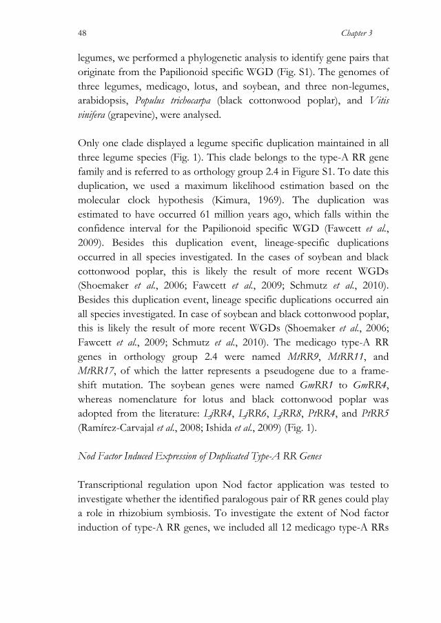
Chapter 3
48
legumes, we performed a phylogenetic analysis to identify gene pairs that originate from the Papilionoid specific WGD (Fig. S1). The genomes of three legumes, medicago, lotus, and soybean, and three non-legumes, arabidopsis, Populus trichocarpa (black cottonwood poplar), and Vitis vinifera (grapevine), were analysed. Only one clade displayed a legume specific duplication maintained in all three legume species (Fig. 1). This clade belongs to the type-A RR gene family and is referred to as orthology group 2.4 in Figure S1. To date this duplication, we used a maximum likelihood estimation based on the molecular clock hypothesis (Kimura, 1969). The duplication was estimated to have occurred 61 million years ago, which falls within the confidence interval for the Papilionoid specific WGD (Fawcett et al., 2009). Besides this duplication event, lineage-specific duplications occurred in all species investigated. In the cases of soybean and black cottonwood poplar, this is likely the result of more recent WGDs (Shoemaker et al., 2006; Fawcett et al., 2009; Schmutz et al., 2010). Besides this duplication event, lineage specific duplications occurred ain all species investigated. In case of soybean and black cottonwood poplar, this is likely the result of more recent WGDs (Shoemaker et al., 2006; Fawcett et al., 2009; Schmutz et al., 2010). The medicago type-A RR genes in orthology group 2.4 were named MtRR9, MtRR11, and MtRR17, of which the latter represents a pseudogene due to a frame-shift mutation. The soybean genes were named GmRR1 to GmRR4, whereas nomenclature for lotus and black cottonwood poplar was adopted from the literature: LjRR4, LjRR6, LjRR8, PtRR4, and PtRR5 (Ramírez-Carvajal et al., 2008; Ishida et al., 2009) (Fig. 1). Nod Factor Induced Expression of Duplicated Type-A RR Genes Transcriptional regulation upon Nod factor application was tested to investigate whether the identified paralogous pair of RR genes could play a role in rhizobium symbiosis. To investigate the extent of Nod factor induction of type-A RR genes, we included all 12 medicago type-A RRs

A Phylogenetic Strategy Yields Symbiotic Cytokinin Type-A RR
49
in this analysis. For 6 genes expression in roots could be detected, of which three were transcriptionally activated 3 hours after application of Sinorhizobium meliloti Nod factors, including MtRR9 and MtRR11 (Fig. 2A; Fig. S2). This suggests that both genes could have a function early in symbiotic signalling. Besides MtRR9 and MtRR11, also MtRR8 was strongly induced, whereas MtRR5 was downregulated (Fig. S2). MtRR8 is the putative ortholog of AtARR5 (Fig. S1); a gene widely used as a cytokinin responsive marker in a diverse range of species, including legumes (D'Agostino et al., 2000; Lohar et al., 2004). Noteworthy, we did not find Nod factor induced transcriptional activation of MtRR4, a type-A RR that is transcriptionally activated upon rhizobium inoculation within 24 h (Gonzalez-Rizzo et al., 2006; Plet et al., 2011). To determine whether the Nod factor induced expression of the duplicated gene pair in orthology group 2.4 is conserved in legumes, we studied the expression of the orthologous lotus genes LjRR4, LjRR6, and LjRR8 (Fig. 1).
Figure 1. Maintained duplication of type-A RR genes in legumes. Red star marks legume specific duplication in this selection of orthology group 2.4 (complete tree of type-A RRs is shown in Fig. S1). Tree is constructed using maximum likelihood phylogeny (PhyML version 3.0; Anisimova and Gascuel, 2006) and branch support test from 1,000 bootstrap repetitions. Glycine max (Gm), Lotus japonicus (Lj), Medicago truncatula (Mt), Populus trichocarpa (Pt).
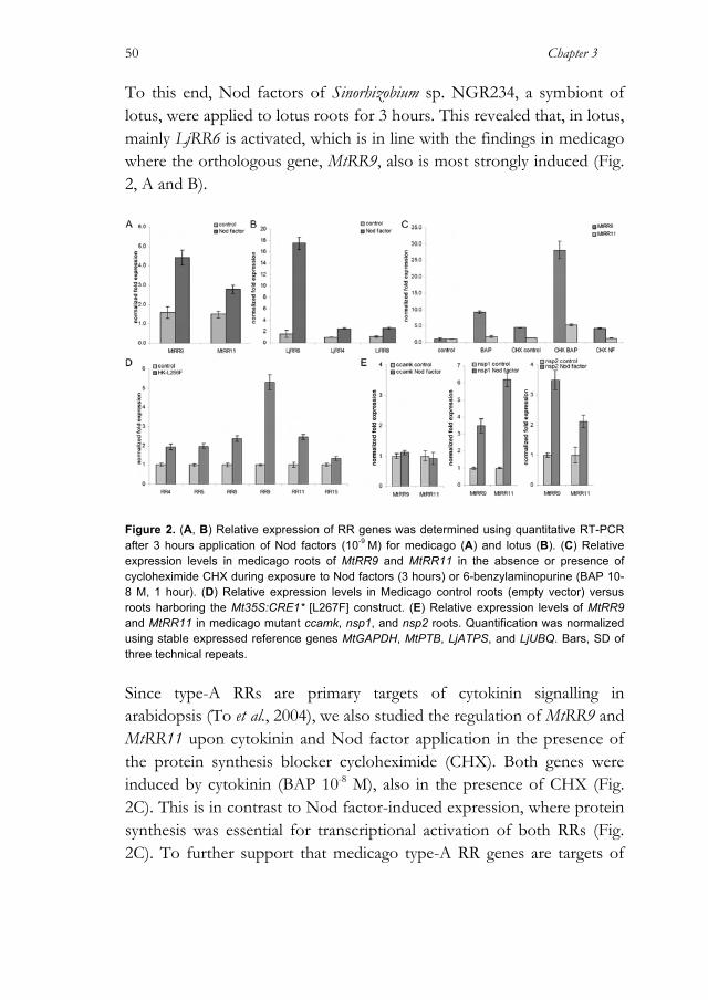
Chapter 3
50
To this end, Nod factors of Sinorhizobium sp. NGR234, a symbiont of lotus, were applied to lotus roots for 3 hours. This revealed that, in lotus, mainly LjRR6 is activated, which is in line with the findings in medicago where the orthologous gene, MtRR9, also is most strongly induced (Fig. 2, A and B).
Figure 2. (A, B) Relative expression of RR genes was determined using quantitative RT-PCR after 3 hours application of Nod factors (10-9 M) for medicago (A) and lotus (B). (C) Relative expression levels in medicago roots of MtRR9 and MtRR11 in the absence or presence of cycloheximide CHX during exposure to Nod factors (3 hours) or 6-benzylaminopurine (BAP 10-8 M, 1 hour). (D) Relative expression levels in Medicago control roots (empty vector) versus roots harboring the Mt35S:CRE1* [L267F] construct. (E) Relative expression levels of MtRR9 and MtRR11 in medicago mutant ccamk, nsp1, and nsp2 roots. Quantification was normalized using stable expressed reference genes MtGAPDH, MtPTB, LjATPS, and LjUBQ. Bars, SD of three technical repeats.
Since type-A RRs are primary targets of cytokinin signalling in arabidopsis (To et al., 2004), we also studied the regulation of MtRR9 and MtRR11 upon cytokinin and Nod factor application in the presence of the protein synthesis blocker cycloheximide (CHX). Both genes were induced by cytokinin (BAP 10-8 M), also in the presence of CHX (Fig. 2C). This is in contrast to Nod factor-induced expression, where protein synthesis was essential for transcriptional activation of both RRs (Fig. 2C). To further support that medicago type-A RR genes are targets of
A Phylogenetic Strategy Yields Symbiotic Cytokinin Type-A RR
51
the cytokinin phosphorelay pathway, we isolated RNA from medicago roots transformed with the gain-of-function MtCRE1 construct (35S:MtCRE1* [L267F]), which causes spontaneous nodule formation (Ovchinnikova et al., 2011). Quantitative RT-PCR on root RNA showed that from all type-A RR genes, MtRR9 was most strongly induced, and also MtRR4, MtRR5, MtRR8, and MtRR11 were activated (Fig. 2D). These results show that these five genes are indeed primary targets of cytokinin signalling downstream of MtCRE1 and suggest that their expression is under direct control of a type-B RR. In legumes, Nod factor signalling is achieved by a conserved signalling pathway that contains several key proteins, including a nuclear localized CCaMK and two GRAS-type transcription factors, NSP1 and NSP2 (Mitra, Gleason, Edwards, Hadfield, Downie, Oldroyd and Long, 2004a; Kaló et al., 2005; Smit et al., 2005). CCaMK, NSP1, and NSP2 are reported to be essential for the induction of nearly all symbiotic genes by Nod factors (Mitra, Shaw and Long, 2004b). We studied the transcriptional regulation of MtRR9 and MtRR11 upon Nod factor application in the medicago Nod factor signalling knockout mutants Mtdmi3/ccamk, Mtnsp1, and Mtnsp2 to determine whether induction depends on these key symbiotic genes. This revealed that the induction of MtRR9 and MtRR11 was dependent on CCaMK but could be triggered in both nsp mutants (Fig. 2E). This suggests that Nod factor activation of the cytokinin phosphorelay pathway can occur independently from both GRAS-type regulators resulting in bifurcation of Nod factor induced signalling downstream of CCaMK. A similar bifurcation of Nod factor signalling downstream of CCaMK has also been shown in lotus (Madsen et al., 2010). All together, these studies suggest that legume type-A RR genes have gained a function in Nod factor-induced root nodule formation. MtRR9 is Induced in the Nod Factor Susceptible Zone To study the symbiotic regulation of MtRR9 and MtRR11 in more detail, the spatial expression pattern of both genes was determined using GUS
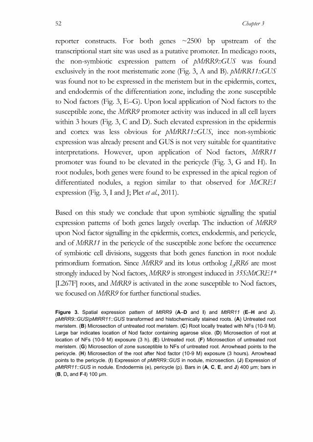
Chapter 3
52
reporter constructs. For both genes ~2500 bp upstream of the transcriptional start site was used as a putative promoter. In medicago roots, the non-symbiotic expression pattern of pMtRR9::GUS was found exclusively in the root meristematic zone (Fig. 3, A and B). pMtRR11::GUS was found not to be expressed in the meristem but in the epidermis, cortex, and endodermis of the differentiation zone, including the zone susceptible to Nod factors (Fig. 3, E–G). Upon local application of Nod factors to the susceptible zone, the MtRR9 promoter activity was induced in all cell layers within 3 hours (Fig. 3, C and D). Such elevated expression in the epidermis and cortex was less obvious for pMtRR11::GUS, ince non-symbiotic expression was already present and GUS is not very suitable for quantitative interpretations. However, upon application of Nod factors, MtRR11 promoter was found to be elevated in the pericycle (Fig. 3, G and H). In root nodules, both genes were found to be expressed in the apical region of differentiated nodules, a region similar to that observed for MtCRE1 expression (Fig. 3, I and J; Plet et al., 2011). Based on this study we conclude that upon symbiotic signalling the spatial expression patterns of both genes largely overlap. The induction of MtRR9 upon Nod factor signalling in the epidermis, cortex, endodermis, and pericycle, and of MtRR11 in the pericycle of the susceptible zone before the occurrence of symbiotic cell divisions, suggests that both genes function in root nodule primordium formation. Since MtRR9 and its lotus ortholog LjRR6 are most strongly induced by Nod factors, MtRR9 is strongest induced in 35S:MtCRE1* [L267F] roots, and MtRR9 is activated in the zone susceptible to Nod factors, we focused on MtRR9 for further functional studies. Figure 3. Spatial expression pattern of MtRR9 (A–D and I) and MtRR11 (E–H and J). pMtRR9::GUS/pMtRR11::GUS transformed and histochemically stained roots. (A) Untreated root meristem. (B) Microsection of untreated root meristem. (C) Root locally treated with NFs (10-9 M). Large bar indicates location of Nod factor containing agarose slice. (D) Microsection of root at location of NFs (10-9 M) exposure (3 h). (E) Untreated root. (F) Microsection of untreated root meristem. (G) Microsection of zone susceptible to NFs of untreated root. Arrowhead points to the pericycle. (H) Microsection of the root after Nod factor (10-9 M) exposure (3 hours). Arrowhead points to the pericycle. (I) Expression of pMtRR9::GUS in nodule, microsection. (J) Expression of pMtRR11::GUS in nodule. Endodermis (e), pericycle (p). Bars in (A, C, E, and J) 400 µm; bars in (B, D, and F-I) 100 µm.
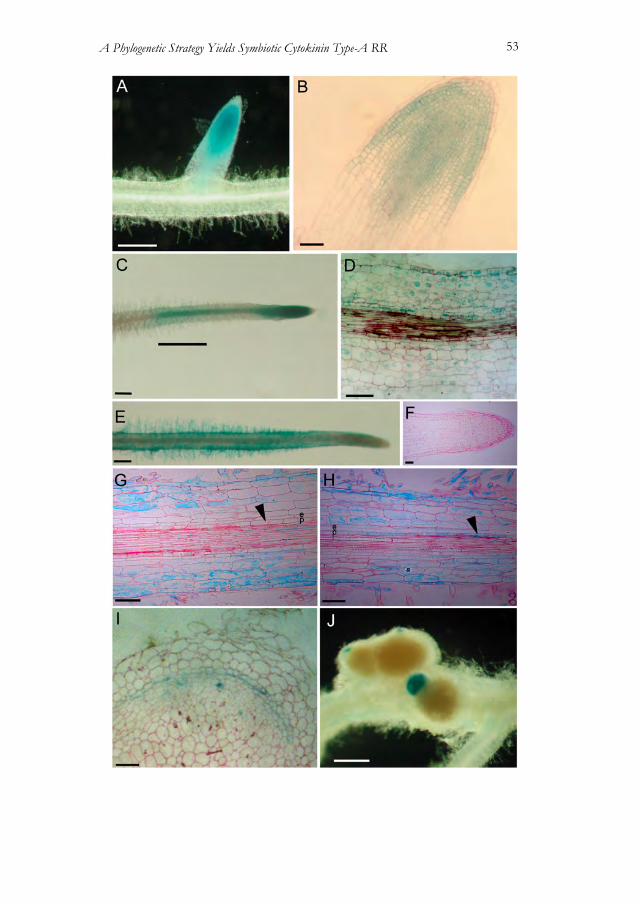
A Phylogenetic Strategy Yields Symbiotic Cytokinin Type-A RR
53
Chapter 3
54
Ectopic Expression of MtRR9 Results in Arrested Primordia To investigate the role of MtRR9 in root nodule primordium formation, we conducted ectopic expression as well as RNA interference (RNAi) experiments. First, we made an RNAi construct to target MtRR9 and introduced it into medicago roots by Agrobacterium rhizogenes mediated transformation. Because MtRR9 is highly homologous to MtRR11 as well as several other type-A RRs, we determined the specificity of this targeting construct. Therefore, the expression of all six root expressed type-A RRs was quantified by RT-PCR. Analysis showed that this RNAi construct affects MtRR9 and MtRR11, but also MtRR5. The latter is the closest homolog of MtRR9 and MtRR11, though it showed opposite regulation by Nod factors when compared to MtRR9/MtRR11 (Fig. S1; Fig. S2). mRNA levels of all three genes show a knockdown level of approximately 50% in medicago RNAi roots (Fig. 4A). We searched for a primordium formation phenotype in the RNAi plants and noted that the RNAi roots had ~33% fewer emerged lateral roots when compared with the wild type plants (n=46, Mann and Whitney U test, P Subsequently 0.05) (Fig. 4, B and C). Inoculation of these RNAi roots also resulted in a decreased nodulation efficiency of ~33% of the average number of nodules per transgenic root (n=37, Mann and Whitney U test, P<0.05) (Fig. 4D). These findings indicate that the type-A RR genes MtRR9 and MtRR11, and possibly MtRR5, are required both for nodule organogenesis and for lateral root formation. Figure 4. (A) Quantification of root-expressed type-A RRs. Relative expression levels in medicago pooled control roots (empty vector) versus roots harboring the MtRR9-RNAi construct. Quantification was normalized using stably expressed reference genes MtGAPDH and MtPTB. Bars represtent SD of three technical repeats. (B, C) Number of emerged lateral roots (B) and lateral root primordia (C) per transgenic root of medicago plants harboring either a control (empty vector) or the MtRR9-RNAi construct. Asterisk indicates that the difference in number of emerged lateral roots between control and MtRR9-RNAi is statistically significant (Mann-Whitney U test, P<0.05). Error bars represent SE (n = 46). (D) Number of nodules per transgenic root of medicago plants harboring either a control (empty vector) or the MtRR9-RNAi construct. Asterisk indicates that the difference in nodule number between control and MtRR9-RNAi is statistically significant (Mann-Whitney U test, P<0.05). Error bars inticate SE (n = 37). Two independent biological replicates were performed for all experiments (A–D).
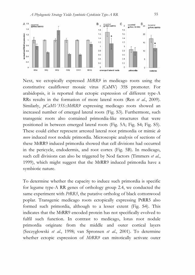
A Phylogenetic Strategy Yields Symbiotic Cytokinin Type-A RR
55
Next, we ectopically expressed MtRR9 in medicago roots using the constitutive cauliflower mosaic virus (CaMV) 35S promoter. For arabidopsis, it is reported that ectopic expression of different type-A RRs results in the formation of more lateral roots (Ren et al., 2009). Similarly, pCaMV35S::MtRR9 expressing medicago roots showed an increased number of emerged lateral roots (Fig. S3). Furthermore, such transgenic roots also contained primordia-like structures that were positioned in between emerged lateral roots (Fig. 5A; Fig. S4; Fig. S5). These could either represent arrested lateral root primordia or mimic de novo induced root nodule primordia. Microscopic analysis of sections of these MtRR9 induced primordia showed that cell divisions had occurred in the pericycle, endodermis, and root cortex (Fig. 5B). In medicago, such cell divisions can also be triggered by Nod factors (Timmers et al., 1999), which might suggest that the MtRR9 induced primordia have a symbiotic nature. To determine whether the capacity to induce such primordia is specific for legume type-A RR genes of orthology group 2.4, we conducted the same experiment with PtRR5, the putative ortholog of black cottonwood poplar. Transgenic medicago roots ectopically expressing PtRR5 also formed such primordia, although to a lesser extent (Fig. S4). This indicates that the MtRR9 encoded protein has not specifically evolved to fulfil such function. In contrast to medicago, lotus root nodule primordia originate from the middle and outer cortical layers (Szczyglowski et al., 1998; van Spronsen et al., 2001). To determine whether ectopic expression of MtRR9 can mitotically activate outer

Chapter 3
56
cortical cells, we introduced CaMV35Sp::MtRR9 into lotus roots. Also, lotus primordia are induced, as in medicago (Fig. 5D). Sectioning revealed that in lotus, not only pericycle and inner cortical cells divided but also cells in the middle and outer cortical layers (Fig. 5E). This shows that the location of MtRR9 induced cell divisions coincides with the spatial position of symbiotic divisions in the cortex. Furthermore, it indicates that there is not a specific function of MtRR9 dedicated to indeterminate type nodulation.
Figure 5. MtRR9 constitutive expression results in primordia. (A, D) Primordia on roots of medicago (A) and lotus (D) as a result of constitutive expression of MtRR9 (pCaMV35S::MtRR9). Transgenic roots were selected based on DsRed fluorescence. (B) Microsection of a medicago primordium shows cell divisions in the inner cortical cell layers and pericycle. (E) Microsection of a lotus primordium with cell divisions in inner and outer cortical cell layers and pericycle. Microsection of young lateral root primordia of medicago (E) and lotus (F). Divisions in the outer cortex are marked with arrowheads. Cortex (c), endodermis (e), pericycle (p). Bars in (A, C) 400 µm, bars in (B, D-F) 100 µm.
The general view is that lateral root primordia develop from the pericycle and endodermal cell layers (Péret et al., 2009). However, for many species, including some legumes, it has been reported that cortical cell divisions can also accompany lateral root development (Tschermak-Woess and Dolezal, 1953; Mallory et al., 1970; McCully, 1975; Bryne et al., 1977; Casero et al., 1996; Mathesius, Weinman, Rolfe and Djordjevic,
A Phylogenetic Strategy Yields Symbiotic Cytokinin Type-A RR
57
2000b). To better compare the MtRR9 induced primordia, we studied the involvement of cortical cells during lateral root primordium formation in lotus and medicago. In both species, we observed that the formation of a lateral root primordium in the pericycle cell layer is associated with cell divisions in the endodermal and cortical cell layers (Fig. 5, C). Interestingly, in lotus, the cell divisions in the cortex were extended more to the outer cortical cell layers when compared with medicago, similar to what was observed upon Nod factor induced nodule primordium formation (Szczyglowski et al., 1998; van Spronsen et al., 2001). Since both species display root cortical cell divisions during lateral root formation, it suggests that ectopic expression of MtRR9 results in arrested primordia that are the result of activation of shared developmental programs essential for nodule as well as lateral root formation. Discussion In this study, we present a phylogenetic strategy to identify genes originating from the Papilionoid specific WGD that have gained a function in rhizobium symbiosis. To test this strategy, we focused on the cytokinin phosphorelay pathway because it is presumed to be an integrative part of rhizobium induced signalling (Frugier et al., 2008). A total of 22 orthology groups were investigated and resulted in the identification of a single conserved gene pair originating from this WGD. We demonstrate that the encoded type-A RRs are part of the Nod factor induced symbiotic signalling cascade. This shows that despite massive gene loss upon the WGD event that occurred early in the evolution of the Papilionoid subfamily, duplicated gene pairs that have contributed to the evolution of the rhizobium symbiosis in this subfamily, can be identified. Therefore, the presented phylogenetic approach can be a useful tool to identify novel genes that function in rhizobium symbiosis. Type-A RRs are generally considered to be negative regulators of cytokinin phosphorelay signalling (Hwang and Sheen, 2001; Osakabe et

Chapter 3
58
al., 2002; Kiba et al., 2003; To et al., 2004; Hirose et al., 2007). In accordance with this view, we hypothesize that MtRR9 and MtRR11 are also part of a negative feedback mechanism on the cytokinin phosphorelay signalling. Cytokinin plays a negative role in lateral root initiation, and it has been shown that arabidopsis type-A RR mutants may fulfil a key function to control this inhibition (To et al., 2004; Ren et al., 2009). The function of MtRR9 as a negative regulator is based on the finding that ectopic MtRR9 expression results in more lateral roots, which is in line with similar findings for ectopic expression of type-A RRs in arabidopsis and with the fact that lowered endogenous levels of cytokinin lead to a higher lateral root density (Laplaze et al., 2007; Nibau et al., 2008; Ren et al., 2009). Furthermore, we observed an increased number of arrested lateral primordia. Besides its negative role in the initiation of lateral roots, cytokinin is known to regulate, antagonistically to auxin, the proper patterning of the embryonic root meristem (Müller and Sheen, 2008). Whether similar genetic mechanisms regulate lateral root meristem development is unknown. We anticipate that ectopic expression of a negative regulator of cytokinin signalling may disturb proper lateral root meristem patterning, resulting in arrested lateral primordia. In contrast to ectopic expression of type-A RR genes, type-A RR arabidopsis mutants have fewer lateral roots, which reflect the negative role of cytokinin in lateral root initiation. Type-A RRs function redundantly because inhibitory effects were only observed when multiple members were knocked out (To et al., 2004). Our RNAi construct was designed with the intention only to target MtRR9, but because of high homology also other type-A RRs were also knocked down. Therefore, the observed lowered amount of emerged lateral roots on the RNAi roots are probably due to the combined knockdown of multiple type-A RRs, suggesting redundant functioning of these genes. Notably also a decrease in nodule number is observed in knockdown roots. This seems a paradox. As cytokinin is promoting root nodule formation, one would anticipate that the downregulation of cytokinin inhibitor genes would
A Phylogenetic Strategy Yields Symbiotic Cytokinin Type-A RR
59
promote root nodule formation. So far, we do not have a mechanistic explanation for this finding, though we anticipate that the positive effect of cytokinin on root nodule formation acts only transiently, subsequently resulting in a new auxin maximum in the developing root nodule primordium (Plet et al., 2011). The three genes targeted by RNAi show opposite regulation by Nod factors; MtRR9 and MtRR11 were transcriptionally activated, whereas MtRR5 was down-regulated. This may provide an explanation for the observed phenotype because precise cytokinin signalling may be crucial for nodule development. Constitutive knockdown of MtRR9, MtRR11, and MtRR5 therefore may act negatively on root nodule formation as well. Strikingly, the arrested primordia that are obtained in MtRR9 over-expression roots are composed of cells that originate from the cortex, endodermis, and pericycle. This observation made us to investigate the ontogeny of lateral root primordia in lotus and medicago. It was found that in both legumes, the cortical ground tissue also contributes substantially to this developmental process. Furthermore, we noticed that -to some extent- the spatial position of mitotically active cortical cells in lateral root primordia coincides with the spatial position of nodule primordia (Chapter 2, this Thesis). This suggests that the potential to mitotically reactivate cortical cells is not an exclusive characteristic of Rhizobium spp. Nod factor-induced signalling but an intrinsic feature of these cells. However, the fact that root cortical cells are mitotically activated during lateral root development is not a legume-specific characteristic because it is reported for several non-legume species as well (Tschermak-Woess and Dolezal, 1953; Mallory et al., 1970; Casero et al., 1996). The precise function of the dividing cortical cells during lateral root primordium formation remains unknown. Two possible functions can be hypothesized. Either the re-differentiated cortical cells become an integrative part of the primordium; alternatively, these divisions facilitate lateral root emergence through the cortex. The latter hypothesis is proposed for plants that have multiple cortex layers (Péret et al., 2009), including medicago and lotus, which have at least five cortical cell layers.

Chapter 3
60
Besides MtRR9, other type-A RRs were shown to function in rhizobium symbiosis (Gonzalez-Rizzo et al., 2006; Vernié et al., 2008). Although their exact molecular functioning remains elusive, we can now position MtRR9 and MtRR11 in the Nod factor signalling network. We demonstrate that these genes are transcriptionally activated upon rhizobium Nod factor signalling. This Nod factor-induced expression is not dependent on the GRAS-type transcription factor complex MtNSP1-MtNSP2, whereas it requires MtCCaMK/MtDMI3, a nuclear localized and calcium- regulated kinase that functions upstream of the MtNSP1-MtNSP2 transcription factor complex (Kouchi et al., 2010). Furthermore, we found that Nod factor- induced MtRR9 and MtRR11 expression requires de novo protein synthesis, indicating that Nod factor regulated gene products have a positive effect on the cytokinin signalling pathway. These could be newly synthesized enzymes involved in reallocation or metabolism of bioactive cytokinin. Such a presumed cytokinin signal is then likely perceived by the HK receptor MtCRE1, which has several type-A RRs among its downstream targets including MtRR4, MtRR45, MtRR48, MtRR49, and MtRR411 (Plet et al., 2011). The identification of a novel gene pair involved in the rhizobium-legume symbiosis by using a phylogenetic approach based on the Papilionoid specific WGD provides a proof of principle for the feasibility of this approach. Therefore, we propose that this phylogenetic strategy can be used on a genome-wide scale to identify new (candidate) genes involved in rhizobium symbiosis, even when such gene pairs share redundant functions, which hampers their identification by forward genetic screens. Materials and Methods Vectors and Constructs MtRR9 and PtRR5 full length genomic sequence and MtRR9 RNAi target sequence were derived by PCR amplification using the primers listed in Table S1. The genes were cloned into a pENTR-D-Topo vector
A Phylogenetic Strategy Yields Symbiotic Cytokinin Type-A RR
61
(Invitrogen) creating pENTR1-2_MtRR9 and pENTR1-2_PtRR5. The CaMV35S promoter and terminator were cloned into a pENTR4-1 and pENTR2-3 (Invitrogen), thereby creating two modified pENTR clones: pENTR4-1_p35S and pENTR2_T35S. All three pENTR vectors were combined into the binary destination vector pKGW-RR-MGW by a multisite gateway reaction (Invitrogen). pKGW-RR-MGW contains pAtUBQ10::DsRED1 of pRedRoot as selection marker (Limpens et al., 2004). The MtRR9 RNAi target sequence was cloned into the DsRed modified gateway vector pK7GWIWG2(II) driven by the CaMV35S promoter as described by Limpens et al. (2005). 35S:MtCRE1* [L267F] was used as described by Ovchinnikova et al. (2011). The putative promoter region of MtRR9 and MtRR11, ~2500 bp upstream of the translational start site, was PCR amplified using primers listed in Table S1. The putative promoters were cloned into a pENTR-D-Topo, thereby creating pENTR1-2_pMtRR9 and pENTR1- 2_pMtRR11. Subsequently, each promoter was recombined into pKGWFS7- RR containing a GUS-GFP fusion reporter as well as pAtUBQ10::DsRed1 as a selectable marker (Karimi et al., 2002). All cloning vectors and constructs are available upon request from our laboratory or via the Functional Genomics unit of the Department of Plant Systems Biology (Vlaams Instituut voor Biotechnologie-Ghent University). Plant Materials and Treatments For the quantitative RT-PCR on type-A RR genes, Medicago truncatula (medicago) and Lotus japonicus (lotus) germinated seedlings were grown vertically on modified Fahraeus medium agar plates with 0.2 mM Ca(NO3)2 (low nitrate) on top of filter paper for 48 hours (Fahraeus, 1957). Then water dissolved Nod factors (~10-9 M) )(Sinorhizobium sp. NGR234 Nod factors for lotus and Sinorhizobium meliloti Nod factors for medicago) or water as a control were pipetted on top of every root (Hussain et al., 1999). Roots were exposed for 3 hours; subsequently, 1
Chapter 3
62
cm root pieces were cut just above the root tip and were snap-frozen (n=15). For CHX experiments, plants were grown in modified Fahraeus slides using modified liquid Fahraeus medium (Heidstra et al., 1994) with low nitrate. A single germinated seedling was placed in each slide, and medium was exchanged every 24 hours. Experiments were done with plants grown for 48 h in Fahraeus slides. Plants in the slides were treated either with 6-benzylaminopurine (BAP, 10-8 M) purified Nod factors (~10-9 M), 50 mM CHX, 50 mM CHX + Nod factors (~10-9 M), 50 mM CHX + 10-8 M BAP for 3 hours, or Fahraeus medium as a control. Subsequently root pieces were snap-frozen as described above. For all experiments, plants were grown in an environmentally controlled growth chamber at 20ºC with a 16-hours-light/8-hours-dark cycle and 70% relative humidity. Quantitative RT-PCR RNA was isolated from snap-frozen root samples using the plant RNA kit (E.Z.N.A, Omega Bio-Tek) as described in the manufacturer’s protocol. Complementary DNA was synthesized from 1 mg of total RNA using the iScript cDNA synthesis kit (BioRad, Hercules, USA) as described in the manufacturer’s protocol. Quantitative RT-PCR was performed using SYBR Green-based detection (Eurogentec, Maastricht, the Netherlands). Experimental setup and execution were conducted using a MyIQ optical cycler, according to protocol provided by the manufacturer (BioRad, Hercules, USA). All primers, including the genes used for normalization (MtGAPDH, MtPTB, LjATPS, and LjUBQ) are given in Table S1. As a control for the experimental set up of each Nod factor induced sample, the induction of NIN in both medicago and lotus were checked and confirmed (data not shown). Data analysis was performed using BioRad iQ5 software. Baselines were set at 100 relative fluorescence units to calculate the threshold cycle values. Threshold cycle values of 31 and higher were excluded from the analysis, though still checked for transcriptional induction (Table S1). A representative sample of three independent biological replicates is shown in all figures.
A Phylogenetic Strategy Yields Symbiotic Cytokinin Type-A RR
63
Plant Transformation and Nodulation Assay
Agrobacterium rhizogenes mediated hairy roots transformation was used to transform medicago (Jemalong A17) as described in (Limpens, 2004), with the adaptation that 0.2 mM Ca(NO3)2 (low nitrate) was used in Fahraeus medium instead. Transgenic roots were selected based on DsRED1 expression. Three weeks after transformation, transgenic roots from promoter studies and ectopic expression studies were transferred to low nitrate Fahraeus plates. MtRR9 RNAi and empty vector control plants were investigated for lateral roots and primordia 10 days after transfer to Fahraeus plates. After transformation, MtRR9 RNAi and empty vector control plants were grown and inoculated for 3 weeks in perlite as described by Limpens et al. (2004). pMtRR9::GUS and pMtRR11::GUS transformed plants were inoculated and grown in perlite in the same way. The differentiation zone (at ~0.7 cm above the tip) of pMtRR9::GUS and pMtRR11::GUS transgenic roots were exposed on Fahraeus plates for 3 hours to 2-3 mm thin slices of Nod factor (10-9 M) or deionized water dissolved in low-melting point water-agarose, respectively. Afterward, these roots were fixed and sectioned as described by Limpens et al. (2005). Histochemical GUS staining was performed as described in Supplemental Protocols. pCaMV35S::MtRR9 and pCaMV35S::PtRR5 roots were investigated for lateral roots and primordia 10 days after transfer to Fahraeus plates. Primordia were fixed and sectioned as described in Supplemental Protocols. All statistical tests were executed using SigmaStat software version 3.5 (Systat Software, San Jose, California).
Phylogeny
The phylogenetic trees were reconstructed using the maximum likelihood method implemented in the software PhyML version 3.0 (Anisimova and Gascuel, 2006; Guindon and Gascuel, 2003). More details are described in Supplemental Protocols. Sequence data from this

Chapter 3
64
article can be found in the GenBank/EMBL data libraries under accession number JQ013379. Supplemental data Supplemental data available online at plantphysiol.org
A Phylogenetic Strategy Yields Symbiotic Cytokinin Type-A RR
65
References Anisimova, M. and Gascuel, O., (2006) Approximate likelihood-ratio test for
branches: a fast, accurate, and powerful alternative. Systematic biology, 55(4), 539–552.
Bishopp, A., Help, H. and Helariutta, Y., (2009) Cytokinin signaling during root development. International Review of Cell and Molecular Biology, 276, 1–48.
Bryne, J.M., Pesacreta, T.C. and Fox, J.A., (1977) Development and structure of the vascular connection between the primary and secondary root of Glycine max (L.) Merr. American Journal of Botany, 64(8), 946–959.
Cannon, S.B., Ilut, D., Farmer, A.D., Maki, S.L., May, G.D., Singer, S.R. and Doyle, J.J., (2010) Polyploidy did not predate the evolution of nodulation in all legumes. PloS ONE, 5(7), e11630.
Cannon, S.B., Sterck, L., Rombauts, S., et al., (2006) Legume genome evolution viewed through the Medicago truncatula and Lotus japonicus genomes. Proceedings of the National Academy of Sciences of the United States of America, 103(40), 14959–14964.
Casero, P.J., Casimiro, I. and Lloret, P.G., (1996) Pericycle proliferation pattern during the lateral root initiation in adventitious roots of Allium cepa. Protoplasma, 191(3-4), 136–147.
Cooper, J.B. and Long, S.R., (1994) Morphogenetic rescue of Rhizobium meliloti nodulation mutants by trans-zeatin secretion. The Plant Cell, 6(2), 215–225.
Crespi, M. and Frugier, F., (2008) De novo organ formation from differentiated cells: root nodule organogenesis. Science signaling, 1(49), re11.
D'Agostino, I.B., Deruere, J. and Kieber, J.J., (2000) Characterization of the response of the Arabidopsis Response Regulator gene family to cytokinin. Plant Physiology, 124(4), 1706–1717.
Fahraeus, G., (1957) The infection of clover root hairs by nodule bacteria studied by a simple glass slide technique. Journal of general microbiology, 16(2), 374–381.
Fawcett, J.A., Maere, S. and van de Peer, Y., (2009) Plants with double genomes might have had a better chance to survive the Cretaceous-Tertiary extinction event. Proceedings of the National Academy of Sciences of the United States of America, 106(14), 5737–5742.
Frugier, F., Kosuta, S., Murray, J.D., Crespi, M. and Szczyglowski, K., (2008) Cytokinin: secret agent of symbiosis. Trends in Plant Science, 13(3), 115–120.
Gepts, P., Beavis, W.D., Brummer, E.C., Shoemaker, R.C., Stalker, H.T., Weeden, N.F. and Young, N.D., (2005) Legumes as a model plant family. Genomics for food and feed report of the Cross-Legume advances through genomics conference. In pp. 1228–1235.
Gonzalez-Rizzo, S., Crespi, M. and Frugier, F., (2006) The Medicago truncatula CRE1 cytokinin receptor regulates lateral root development and early symbiotic interaction with Sinorhizobium meliloti. The Plant Cell, 18(10), 2680–2693.

Chapter 3
66
Guindon, S.S. and Gascuel, O.O., (2003) A simple, fast, and accurate algorithm to estimate large phylogenies by maximum likelihood. Systematic biology, 52(5), 696–704.
Heckmann, A.B., Sandal, N., Bek, A.S., Madsen, L.H., Jurkiewicz, A., Nielsen, M.W., Tirichine, L. and Stougaard, J., (2011) Cytokinin induction of root nodule primordia in Lotus japonicus is regulated by a mechanism operating in the root cortex. Molecular Plant-Microbe Interactions, 24(11), 1385–1395.
Heidstra, R., Geurts, R., Franssen, H., Spaink, H.P., Van Kammen, A. and Bisseling, T., (1994) Root hair deformation activity of nodulation factors and their fate on Vicia sativa. Plant Physiology, 105(3), 787–797.
Hirose, N.N., Makita, N.N., Kojima, M.M., Kamada-Nobusada, T.T. and Sakakibara, H.H., (2007) Overexpression of a type-A response regulator alters rice morphology and cytokinin metabolism. Plant and Cell Physiology, 48(3), 523–539.
Hussain, A.A., Jiang, Q., Broughton, W.J. and Gresshoff, P.M., (1999) Lotus japonicus nodulates and fixes nitrogen with the broad host range Rhizobium sp. NGR234. Plant Cell Physiol, 894–899.
Hwang, I. and Sheen, J., (2001) Two-component circuitry in Arabidopsis cytokinin signal transduction. Nature, 413(6854), 383–389.
Ishida, K., Niwa, Y., Yamashino, T. and Mizuno, T., (2009) A genome-wide compilation of the two-component systems in Lotus japonicus. DNA Research, 16(4), 237–247.
Kaló, P., Gleason, C., Edwards, A., et al., (2005) Nodulation signaling in legumes requires NSP2, a member of the GRAS family of transcriptional regulators. Science, 308(5729), 1786–1789.
Karimi, M., Inzé, D. and Depicker, A., (2002) GATEWAY vectors for Agrobacterium-mediated plant transformation. Trends in Plant Science, 7(5), 193–195.
Kiba, T., Yamada, H., Sato, S., Kato, T., Tabata, S., Yamashino, T. and Mizuno, T., (2003) The type-A Response Regulator, ARR15, acts as a negative regulator in the cytokinin-mediated signal transduction in Arabidopsis thaliana. Plant and Cell Physiology, 44(8), 868–874.
Kimura, M., (1969) The rate of molecular evolution considered from the standpoint of population genetics. Proceedings of the National Academy of Sciences of the United States of America, 63(4), 1181–1188.
Kouchi, H., Imaizumi-Anraku, H., Hayashi, M., Hakoyama, T., Nakagawa, T., Umehara, Y., Suganuma, N. and Kawaguchi, M., (2010) How many peas in a pod? Legume genes responsible for mutualistic symbioses underground. Plant and Cell Physiology, 51(9), 1381–1381.
Laplaze, L., Benkova, E., Casimiro, I., et al., (2007) Cytokinins act directly on lateral root founder cells to inhibit root initiation. The Plant Cell, 19(12), 3889–3900.
Levy, J., (2004) A putative Ca2+ and Calmodulin-dependent protein Kinase required for bacterial and fungal symbioses. Science, 303(5662), 1361–1364.
A Phylogenetic Strategy Yields Symbiotic Cytokinin Type-A RR
67
Limpens, E., (2004) RNA interference in Agrobacterium rhizogenes-transformed roots of Arabidopsis and Medicago truncatula. Journal of Experimental Botany, 55(399), 983–992.
Lohar, D.P., Schaff, J.E., Laskey, J.G., Kieber, J.J., Bilyeu, K.D. and Bird, D.M., (2004) Cytokinins play opposite roles in lateral root formation, and nematode and Rhizobial symbioses. The Plant Journal, 38(2), 203–214.
Madsen, L.H., Tirichine, L., Jurkiewicz, A., Sullivan, J.T., Heckmann, A.B., Bek, A.S., Ronson, C.W., James, E.K. and Stougaard, J., (2010) The molecular network governing nodule organogenesis and infection in the model legume Lotus japonicus. Nature Communications, 1, 1–12.
Mallory, T.E., Chiang, S-H., Cutter, E.G. and E M Gifford, J., (1970) Sequence and pattern of lateral root formation in five selected species. American Journal of Botany, 57(7), 800–809.
Mathesius, U., Charon, C., Rolfe, B.G., Kondorosi, A. and Crespi, M., (2000a) Temporal and spatial order of events during the induction of cortical cell divisions in white clover by Rhizobium leguminosarum bv. trifolii inoculation or localized cytokinin addition. Molecular Plant-Microbe Interactions, 13(6), 617–628.
Mathesius, U., Weinman, J.J., Rolfe, B.G. and Djordjevic, M.A., (2000b) Rhizobia can induce nodules in white clover by “hijacking” mature cortical cells activated during lateral root development. Molecular Plant-Microbe Interactions, 13(2), 170–182.
McCully, M., (1975) The development of lateral roots. In J. Torrey and D. Clarkson, eds. The development and function of roots: third Cabot symposium. London: Academic Press Inc, pp. 105–124.
Mitra, R.M., Gleason, C.A., Edwards, A., Hadfield, J., Downie, J.A., Oldroyd, G.E.D. and Long, S.R., (2004a) A Ca2+/calmodulin-dependent protein kinase required for symbiotic nodule development: Gene identification by transcript-based cloning. Proceedings of the National Academy of Sciences of the United States of America, 101(13), 4701–4705.
Mitra, R.M., Shaw, S.L. and Long, S.R., (2004b) Six nonnodulating plant mutants defective for Nod factor-induced transcriptional changes associated with the legume-rhizobia symbiosis. Proceedings of the National Academy of Sciences of the United States of America, 101(27), 10217–10222.
Murray, J.D., Karas, B.J., Sato, S., Tabata, S., Amyot, L. and Szczyglowski, K., (2007) A cytokinin perception mutant colonized by Rhizobium in the absence of nodule organogenesis. Science, 315(5808), 101–104.
Müller, B. and Sheen, J., (2007) Advances in cytokinin signaling. Science, 318(5847), 68–69.
Müller, B. and Sheen, J., (2008) Cytokinin and auxin interaction in root stem-cell specification during early embryogenesis. Nature, 453(7198), 1094–1097.
Nibau, C., Gibbs, D.J. and Coates, J.C., (2008) Branching out in new directions: the control of root architecture by lateral root formation. The New Phytologist, 179(3), 595–614.
Chapter 3
68
Osakabe, Y., Miyata, S., Urao, T., Seki, M., Shinozaki, K. and Yamaguchi-Shinozaki, K., (2002) Overexpression of Arabidopsis response regulators, ARR4/ATRR1/IBC7 and ARR8/ATRR3, alters cytokinin responses differentially in the shoot and in callus formation. Biochemical and biophysical research communications, 293(2), 806–815.
Ovchinnikova, E., Journet, E-P., Chabaud, M., et al., (2011) IPD3 controls the formation of nitrogen-fixing symbiosomes in pea and Medicago Spp. Molecular Plant-Microbe Interactions, 24(11), 1333–1344.
Péret, B., Larrieu, A. and Bennett, M.J., (2009) Lateral root emergence: a difficult birth. Journal of Experimental Botany, 60(13), 3637–3643.
Plet, J., Wasson, A., Ariel, F., Le Signor, C., Baker, D., Mathesius, U., Crespi, M. and Frugier, F., (2011) MtCRE1-dependent cytokinin signaling integrates bacterial and plant cues to coordinate symbiotic nodule organogenesis in Medicago truncatula. The Plant Journal, 65(4), 622–633.
Ramírez-Carvajal, G.A., Morse, A.M. and Davis, J.M., (2008) Transcript profiles of the cytokinin response regulator gene family in Populus imply diverse roles in plant development. The New Phytologist, 177(1), 77–89.
Ren, B., Liang, Y., Deng, Y., Chen, Q., Zhang, J., Yang, X. and Zuo, J., (2009) Genome-wide comparative analysis of type-A Arabidopsis response regulator genes by overexpression studies reveals their diverse roles and regulatory mechanisms in cytokinin signaling. Cell Research, 19(10), 1178–1190.
Schmutz, J., Cannon, S.B., Schlueter, J., et al., (2010) Genome sequence of the palaeopolyploid soybean. Nature, 463(7278), 178–183.
Shoemaker, R.C., Schlueter, J. and Doyle, J.J., (2006) Paleopolyploidy and gene duplication in soybean and other legumes. Current Opinion in Plant Biology, 9(2), 104–109.
Smit, P., Raedts, J., Geurts, R.7, (2005) NSP1 of the GRAS protein family is essential for rhizobial Nod factor-induced transcription. Science, 308(5729), 1789–1791.
Szczyglowski, K., Robert S Shaw, Wopereis, J., Copeland, S., Hamburger, D., Kasiborski, B., Dazzo, F.B. and Frans J de Bruijn, (1998) Nodule organogenesis and symbiotic mutants of the model legume Lotus japonicus. Molecular Plant-Microbe Interactions, 11(7), 684–697.
Timmers, A.C., Auriac, M.C. and Truchet, G., (1999) Refined analysis of early symbiotic steps of the Rhizobium-Medicago interaction in relationship with microtubular cytoskeleton rearrangements. Development, 126(16), 3617–3628.
Tirichine, L., Sandal, N., Madsen, L.H., Radutoiu, S., Albrektsen, A.S., Sato, S., Asamizu, E., Tabata, S. and Stougaard, J., (2007) A gain-of-function mutation in a cytokinin receptor triggers spontaneous root nodule organogenesis. Science, 315(5808), 104–107.
To, J.P.C., Haberer, G., Ferreira, F.J., Deruere, J., Mason, M.G., Schaller, G.E., Alonso, J.M., Ecker, J.R. and Kieber, J.J., (2004) Type-A Arabidopsis response regulators are partially redundant negative regulators of cytokinin signaling. The Plant Cell, 16(3), 658–671.

A Phylogenetic Strategy Yields Symbiotic Cytokinin Type-A RR
69
Tschermak-Woess, E. and Dolezal, R., (1953) Durch seitenwurzelbildung indzierte und spontane mitosen in den dauergeweben der wurzel. Oesterreichische Botanische Zeitschrift, 100, 358–402.
van Spronsen, P.C., Grønlund, M., Pacios Bras, C., Spaink, H.P. and Kijne, J.W., (2001) Cell biological changes of outer cortical root cells in early determinate nodulation. Molecular Plant-Microbe Interactions, 14(7), 839–847.
Vernié, T., Moreau, S., de Billy, F., et al., (2008) EFD Is an ERF transcription factor involved in the control of nodule number and differentiation in Medicago truncatula. The Plant Cell, 20(10), 2696–2713.

CHAPTER 4
NADPH Oxidase MtRBOHA and MtRBOHG
Have a Dual Function in Medicago-Rhizobium Nodule Symbiosis1
Alessandra Lillo, Stéphane De Mita, Sergey Ivanov, Elena Fedorova,
Ton Bisseling, and René Geurts
Department of Plant Sciences, Laboratory of Molecular Biology, Wageningen University, Droevendaalsesteeg 1, 6708 PB, Wageningen, The Netherlands (A.L., S.D.M., S.I., N.L., E.F., T.B., R.G.); IRD Montpellier, 911 avenue Agropolis BP 64501, 34394 Montpellier cedex 5, France (S.D.M.); K.A. Timiryazev Institute of Plant Physiology, Russian Academy of Sciences, Botanicheskaya 35, Moscow 127392, Russia (S.I, E.F); College of Science, King Saud University, Post Office Box 2455, Riyadh 11451, Saudi Arabia (T.B.)
1 This work was supported by the Dutch Science Foundation (NWO-vidi 864.06.007 to R.G.) and the E.U. Research Training Network “NODPERCEPTION” European Union grant (MRTNCT-2006-035546 to R.G., T.B. and A.L.)
Chapter 4 72
Abstract
Reactive oxygen species (ROS) accumulation is associated with several processes of the legume-rhizobium symbiosis (Peleg-Grossman and Volpin, 2007; Cárdenas et al., 2008; Jamet et al., 2007; Rubio et al., 2004; Santos et al., 2001). NADPH oxidases have been proposed as one of the most important sources of ROS (Peleg-Grossman and Volpin, 2007; Mando et al., 2009), though in legumes the corresponding genes are only poorly functionally characterized. Here we present two paralogous genes encoding NADPH oxidases, MtRBOHA and MtRBOHG, that have a symbiotic function. We show that both genes have very similar expression patterns being highly expressed in the apical part of the nodule, suggesting redundant functions. Knockdown of MtRBOHA and MtRBOHG expression by RNAi revealed a complex symbiotic phenotype. Mutant nodules are either devoided of infected cells, or, when cells are infected, premature senescence takes place in patchy sectors of the nitrogen fixation zone. Sub-cellular localization of both proteins visualized an association with infection threads in the infection zone of the nodule, concomitant to rhizobium release. By studying MtRBOHA and MtRBOHG localization in two mutants, Mtipd3, which is defected in bacterial release, and Mtdfn1, which is defected in symbiosome development, we argue that both NADPH oxidases function during and/or just after bacterial release from infection threads to establish sustainable symbiosomes.
MtRBOHA and MtRBOHG Have a Dual Function in Nodule Symbiosis 73
Introduction
The interaction between legume plants and nitrogen-fixing rhizobia has evolved in a very effected symbiotic relation over a period of 60 million years. To achieve this, several evolutionary hurdles had to be taken, including avoidance of pathogenic responses as well as adaptation of cellular machineries to support release and maintenance of symbiotic bacteria. However, to date little is known about the adaptation of such machineries. Reactive oxygen species (ROS) are known to play an important role in cell growth and differentiation. In plants ROS are produced via an NADPH oxidase dependent pathway. Several NADPH oxidase encoding genes are highly expressed in legume root nodules (Marino et al., 2010) suggesting that they are playing a role in root nodule symbiosis as well. Furthermore, NADPH oxidases are known to be activated in response to pathogens (Torres et al., 2002; Proels et al., 2010), a process that has to be repressed during symbiosis in legumes (Peleg-Grossman et al., 2009). Here we present two paralogous NADPH oxidases, MtRBOHA and MtRBOHG, that function in the infection zone of the root nodule, concomitant to rhizobium release.
Legumes host their rhizobial partners in specific root organs, so-called nodules. Nodules are fully adapted to support the symbiotic relation. They have a large central zone where most cells are infected by rhizobia. Inside a single cell, hundreds of bacteria can be present as transient organelle-like structures, the so-called symbiosomes. Symbiosomes contain a plant-derived membrane that facilitates exchange of compounds in two directions. The central zone of the nodule is surrounded by peripheral tissue that includes cortex, endodermis and vascular bundles. Furthermore, most legume species, e.g. Medicago truncatula (Medicago), have an apical meristem, which is responsible for an indeterminate growth fate. Nodules are formed in response to bacterial signalling molecules named Nodulation (Nod) factors. These signals are essential not only to activate the developmental program leading to nodule organogenesis, but also to guide intracellular bacterial
Chapter 4 74
infection. Generally, infection is initiated by Nod factor producing rhizobia. Rhizobia manipulate root hair growth in such that root hairs form curls, capturing the bacteria in closed infection pockets. There, the bacteria find the appropriate environment to divide and initiate the formation of infection threads. Infection threads are formed upon local degradation of the cell wall and subsequent invagination of the plasma membrane (Brewin, 2004). Focal deposition of new cell wall material occurs at the tip of the threads by which infection threads grow. New infection threads are formed in the underlying cortical cells that subsequently guide the invading bacteria toward the nodule primordium (Timmers et al., 1999; Gage, 2004; Miyahara et al., 2010; Brewin, 2004).
Individual rhizobia are taken up in the newly formed nodule primordial cells. This occurs via an endocytosis-like process (Verma, 1992; Jones et al., 2007; Limpens et al., 2009). When just released from infection threads, symbiosomes are immature and do not fix nitrogen yet. In the maturation process they first divide, and subsequently differentiate. In some legume species, like Medicago, symbiosomes elongate and become terminally differentiated into a nitrogen-fixing form. This process requires nodule-specific cysteine-rich peptides (NCRs) that are transported to the symbiosome via a nodule specific signal peptidase. Mutations in this process inhibit symbiosome differentiation, as in case of the Medicago Mtdnf1 mutant that is affected in the peptidase complex (Wang et al., 2010; van de Velde et al., 2010; Haag et al., 2011). In indeterminate nodules, as in the model Medicago, the apical meristem remains active for several weeks. Meristematic cells, typically small and rich in cytoplasm, continuously divide providing new cells that will differentiate along the nodule longitudinal axis. New infection threads will be formed in the cells of the proximal zone of the meristem from which rhizobia will be released. These cytoplasmic rhizobia are surrounded by plant-derived membrane and called symbiosomes. This region of the nodule is called infection zone. Interestingly, in this zone all key genes of the Nod factor signalling network appear to be active (Bersoult et al., 2005; Limpens et al., 2005).

MtRBOHA and MtRBOHG Have a Dual Function in Nodule Symbiosis 75
Mutations in some of these genes specifically block the infection process. Of these, the Medicago Mtipd3 mutant has the most prominent infection phenotype. MtIPD3 encodes a nuclear localized coiled-coiled protein that interacts with MtCCaMK; a master regulator in root nodule organogenesis and rhizobium infection (Benaben et al., 1995; Messinese et al., 2007; Ovchinnikova et al., 2011). An Mtipd3 knockout mutant forms mutant nodules with two different phenotypes; none of which contains symbiosomes. This because either infection threads do not penetrate the root cortex, so the nodule is devoid of rhizobia, or, when infection threads reach the primordium, bacteria release does not take place (Benaben et al., 1995; Ovchinnikova et al., 2011). These features make the symbiotic mutants Mtdnf1 and Mtipd3 interesting tools to study cellular machineries involved in symbiosome formation.
NADPH oxidases have been extensively studied in Arabidopsis thaliana (Arabidopsis). Generally, NADPH oxidases carry 6 transmembrane domains indicating that the proteins function in plasma membrane or other membraneous structures. NADPH oxidases contain a highly conserved cytoplasmic N-terminal region that includes four EF-hand motifs (Oda et al., 2010). Protein activity is modulated by Ca2+ binding to these motifs, but also by direct interaction with a small GTPase (Sagi and Fluhr, 2001; Takeda et al., 2008; Ogasawara et al., 2008). NADPH oxidases are involved in a wide range of processes. They are essential for (polar) cell growth and cell wall loosening (Foreman et al., 2003; Liszkay et al., 2004; Monshausen et al., 2007; Takeda et al., 2008). Furthermore, NADPH oxidases have found to be critical in stomata closure (Kwak et al., 2003) as well as in root development were they trigger the transition from cell proliferation to cell differentiation (Tsukagoshi et al., 2010). Besides processes that affect cell shape, NADPH oxidases are also activated upon wounding (Miller et al., 2009) and in response to microbial pathogens (Torres et al., 2002). Thereby, the produced ROS can fulfil a variety of functions, including mechanical, affecting plant cell wall lignification (Hamann et al., 2009; Denness et al., 2011), and signalling roles, acting as signal that is transmitted systemically (Miller et al., 2009).

Chapter 4 76
In legumes NADPH oxidases are only poorly functionally characterized. ROS accumulation is associated with several symbiotic processes including early symbiotic responses, and senescence of symbiosomes containing cells (Peleg-Grossman and Volpin, 2007; Cárdenas et al., 2008; Jamet et al., 2007; Rubio et al., 2004; Santos et al., 2001). Several studies have proposed that during the rhizobium-legume symbiosis plant defense reactions are suppressed, including ROS accumulation (Chang et al., 2009). In Medicago it was found that upon rhizobium Nod factor signalling the expression of NADPH oxidases genes transiently decreases. This reduction in NADPH oxidases expression has been suggested to be associated with a reduction in ROS production needed for root hair swelling (Lohar et al., 2007). Moreover, different hypotheses on ROS functioning during infection thread growth have been formulated (Wisniewski and Rathbun, 2000; Jamet et al., 2007). For instance, it is known that superoxyde (O2
.-) produced by NADPH oxidases may be further converted, by superoxide dismutases, into hydrogen peroxide (H2O2). H2O2 is known to be required for peroxidase-dependent cell wall rigidification, a process that occurs via lignification and cross-linking proline-rich proteins. As rhizobial infection threads contain several symbiosis specific proline-rich proteins (e.g. ENOD11 and ENOD12) ROS production could be essential for proper infection thread growth (Bradley et al., 1992; Scott-Craig et al., 1995; Cook et al., 1995; Passardi et al., 2004). Infection thread rigidification could limit bacterial invasion, an hypothesis which is in line with the fact that many infection threads are prematurely aborted and only a few will successfully infect nodules. Further support for this hypothesis is the finding that rhizobia actively regulate the ROS balance in the lumen of the infection threads, e.g. by expressing the H2O2 catalases KATB and KATC genes. Sinorhizobium meliloti overexpressing KATB reduce more efficiently exogenous ROS produced by the plant in the lumen of the infection threads. This leads to a delay of nodulation combined with an enlargement of infection threads (Jamet et al., 2007). Moreover, a S. meliloti katBkatC double mutant nodulates poorly and displays abnormal infection. After release into plant cells, bacteria fail to differentiate into bacteroids and undergo rapid senescence (Jamet et al., 2003).
MtRBOHA and MtRBOHG Have a Dual Function in Nodule Symbiosis 77
Recently it was shown that in Medicago a specific NADPH oxidase, MtRBOHA, functions in the fixation zone of the root nodule, underlining a long-term production of ROS in this symbiotic organ (Marino et al., 2010). In legumes like Medicago, NADPH oxidase genes form a small gene family that underwent several gene duplication events (Marino et al., 2010). Here we focused on two NADPH oxidases, MtRBOHA and its paralog MtRBOHG. Both genes are highly expressed in the infection zone of the nodule. Studying the function of MtRBOHA and MtRBOHG revealed that both proteins are essential to establish sustainable symbiosomes. Based on the sub-cellular localization studies in wild type nodules as well as in Medicago Mtipd3 and Mtdnf1 mutant backgrounds, we conclude that both proteins localize in discrete areas of the infection zone that are associated with bacterial release. Results MtRBOHA and MtRBOHG are Paralogous Genes with a Similar Symbiotic Expression Profile Phylogenetic studies on NADPH oxidase genes point to duplication events in the legume lineage (Marino et al., 2010). However, the closest non-legume specie included in this study is Arabidopsis, which belongs to the Malvidae clade, whereas legumes are part of the Fabidae clade. We first addressed the question to what extend the duplications in the NADPH oxidases genes are indeed legumes specific. Our phylogenetic analysis were extended to 8 non-legume species phylogenetically positioned between legumes and Arabidopsis. In this analysis we included also the basal legume Chamaecrista fasciculata (Chamaecrista) of the Caesalpiniod subfamily, whereas the model legumes Medicago and Lotus japonicus (Lotus) belong to the Papilionoid subfamily (Lavin et al., 2005). For Chamaecrista a large number of expressed sequence tags (ESTs) have been generated (Cannon et al., 2010). The phylogenetic reconstruction revealed that two lineages of NADPH oxidase genes were duplicated specifically in the Papilionoid subfamily (Fig. 1).
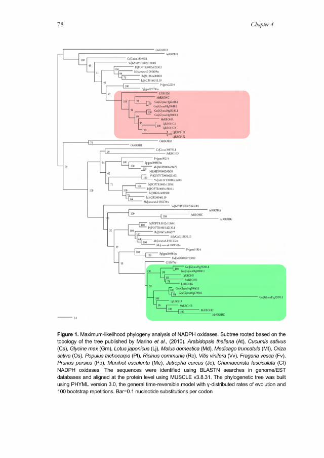
Chapter 4 78
Figure 1. Maximum-likelihood phylogeny analysis of NADPH oxidases. Subtree rooted based on the topology of the tree published by Marino et al., (2010). Arabidopsis thaliana (At), Cucumis sativus (Cs), Glycine max (Gm), Lotus japonicus (Lj), Malus domestica (Md), Medicago truncatula (Mt), Oriza sativa (Os), Populus trichocarpa (Pt), Ricinus communis (Rc), Vitis vinifera (Vv), Fragaria vesca (Fv), Prunus persica (Pp), Manihot esculenta (Me), Jatropha curcas (Jc), Chamaecrista fasciculata (Cf) NADPH oxidases. The sequences were identified using BLASTN searches in genome/EST databases and aligned at the protein level using MUSCLE v3.8.31. The phylogenetic tree was built using PHYML version 3.0, the general time-reversible model with γ-distributed rates of evolution and 100 bootstrap repetitions. Bar=0.1 nucleotide substitutions per codon
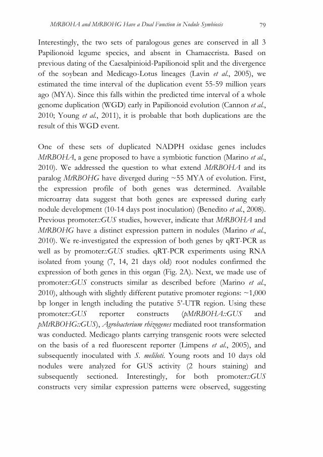
MtRBOHA and MtRBOHG Have a Dual Function in Nodule Symbiosis 79
Interestingly, the two sets of paralogous genes are conserved in all 3 Papilionoid legume species, and absent in Chamaecrista. Based on previous dating of the Caesalpinioid-Papilionoid split and the divergence of the soybean and Medicago-Lotus lineages (Lavin et al., 2005), we estimated the time interval of the duplication event 55-59 million years ago (MYA). Since this falls within the predicted time interval of a whole genome duplication (WGD) early in Papilionoid evolution (Cannon et al., 2010; Young et al., 2011), it is probable that both duplications are the result of this WGD event. One of these sets of duplicated NADPH oxidase genes includes MtRBOHA, a gene proposed to have a symbiotic function (Marino et al., 2010). We addressed the question to what extend MtRBOHA and its paralog MtRBOHG have diverged during ~55 MYA of evolution. First, the expression profile of both genes was determined. Available microarray data suggest that both genes are expressed during early nodule development (10-14 days post inoculation) (Benedito et al., 2008). Previous promoter::GUS studies, however, indicate that MtRBOHA and MtRBOHG have a distinct expression pattern in nodules (Marino et al., 2010). We re-investigated the expression of both genes by qRT-PCR as well as by promoter::GUS studies. qRT-PCR experiments using RNA isolated from young (7, 14, 21 days old) root nodules confirmed the expression of both genes in this organ (Fig. 2A). Next, we made use of promoter::GUS constructs similar as described before (Marino et al., 2010), although with slightly different putative promoter regions: ~1,000 bp longer in length including the putative 5’-UTR region. Using these promoter::GUS reporter constructs (pMtRBOHA::GUS and pMtRBOHG::GUS), Agrobacterium rhizogenes mediated root transformation was conducted. Medicago plants carrying transgenic roots were selected on the basis of a red fluorescent reporter (Limpens et al., 2005), and subsequently inoculated with S. meliloti. Young roots and 10 days old nodules were analyzed for GUS activity (2 hours staining) and subsequently sectioned. Interestingly, for both promoter::GUS constructs very similar expression patterns were observed, suggesting
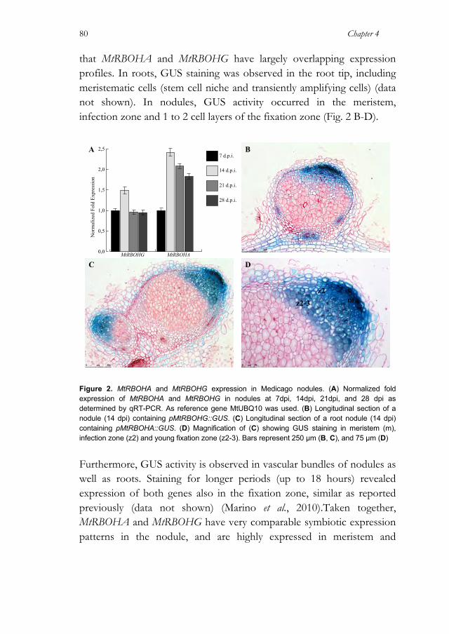
Chapter 4 80
that MtRBOHA and MtRBOHG have largely overlapping expression profiles. In roots, GUS staining was observed in the root tip, including meristematic cells (stem cell niche and transiently amplifying cells) (data not shown). In nodules, GUS activity occurred in the meristem, infection zone and 1 to 2 cell layers of the fixation zone (Fig. 2 B-D).
Figure 2. MtRBOHA and MtRBOHG expression in Medicago nodules. (A) Normalized fold expression of MtRBOHA and MtRBOHG in nodules at 7dpi, 14dpi, 21dpi, and 28 dpi as determined by qRT-PCR. As reference gene MtUBQ10 was used. (B) Longitudinal section of a nodule (14 dpi) containing pMtRBOHG::GUS. (C) Longitudinal section of a root nodule (14 dpi) containing pMtRBOHA::GUS. (D) Magnification of (C) showing GUS staining in meristem (m), infection zone (z2) and young fixation zone (z2-3). Bars represent 250 µm (B, C), and 75 µm (D)
Furthermore, GUS activity is observed in vascular bundles of nodules as well as roots. Staining for longer periods (up to 18 hours) revealed expression of both genes also in the fixation zone, similar as reported previously (data not shown) (Marino et al., 2010).Taken together, MtRBOHA and MtRBOHG have very comparable symbiotic expression patterns in the nodule, and are highly expressed in meristem and
A
C
B
D
0,0
0,5
1,0
1,5
2,0
2,5
28 d.p.i.
21 d.p.i.
14 d.p.i.
7 d.p.i.
MtRBOHAMtRBOHG
Nor
mal
ized
Fol
d Ex
pres
sion
mz2
z2-3
MtRBOHA and MtRBOHG Have a Dual Function in Nodule Symbiosis 81
infection zone. In line with this we hypothesize that both genes fulfill a function in the rhizobial infection process. MtRBOHA and MtRBOHG are Essential to Establish Sustainable Symbiosomes To get insights in the symbiotic functioning of MtRBOHA and MtRBOHG, we conducted RNAi experiments. A previous RNAi study that focussed specifically on MtRBOHA revealed that knocking down levels of this gene do not impaire nodules functionality, although nitrogen fixation activity is slightly reduced (Marino et al., 2010). Since we expected MtRBOHA and MtRBOHG to be functionally redundant, we aimed to knockdown both genes simultaneously. An RNAi construct driven by the CaMV 35S promoter was made to encompass the complete coding region of MtRBOHA (35S:RNAiMtRBOHA). As MtRBOHA and MtRBOHG coding sequences have 83% identity on DNA sequence level, including several regions longer than 24 bp, we anticipated that this RNAi construct will target degradation of mRNAs of both genes. This construct was subsequently introduced in Medicago using A. rhizogenes and plants carrying transgenic roots were selected based on red fluorescence (Limpens et al., 2005; Limpens, 2004). No phenotypic effect on root development was observed in these roots. To determine the knockdown levels of MtRBOHA and MtRBOHG mRNA was isolated from transgenic roots and used as template for qRT-PCR experiments. This revealed varialbe knockdown levels between different transgenic roots. Generally MtRBOHA was effected more severealy than MtRBODG (Fig. 3D). Intrestingly, we noted also a knockdown of MtRBOHD (Fig. 3D). MtRBOHD is considered to be a pseudogene due to an insertion of a retrotransposable element in the putative coding region (Marino et al., 2010). Next, MtRBOHA/MtRBOHG RNAi roots were inoculated with S. melioti. Fourteen days post inoculation MtRBOHA/MtRBOHG RNAi knockdown roots showed nearly two fold reduction in the nodule number per root when compared to plant roots transformed with the
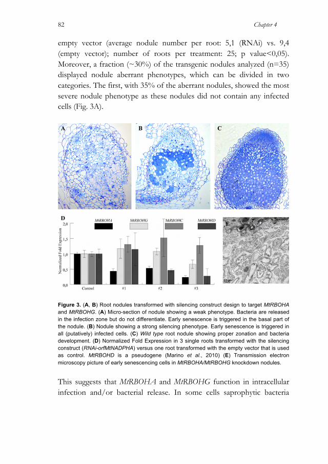
Chapter 4 82
empty vector (average nodule number per root: 5,1 (RNAi) vs. 9,4 (empty vector); number of roots per treatment: 25; p value<0,05). Moreover, a fraction (~30%) of the transgenic nodules analyzed (n=35) displayed nodule aberrant phenotypes, which can be divided in two categories. The first, with 35% of the aberrant nodules, showed the most severe nodule phenotype as these nodules did not contain any infected cells (Fig. 3A).
Figure 3. (A, B) Root nodules transformed with silencing construct design to target MtRBOHA and MtRBOHG. (A) Micro-section of nodule showing a weak phenotype. Bacteria are released in the infection zone but do not differentiate. Early senescence is triggered in the basal part of the nodule. (B) Nodule showing a strong silencing phenotype. Early senescence is triggered in all (putatively) infected cells. (C) Wild type root nodule showing proper zonation and bacteria development. (D) Normalized Fold Expression in 3 single roots transformed with the silencing construct (RNAi-orfMtNADPHA) versus one root transformed with the empty vector that is used as control. MtRBOHD is a pseudogene (Marino et al., 2010) (E) Transmission electron microscopy picture of early senescencing cells in MtRBOHA/MtRBOHG knockdown nodules.
This suggests that MtRBOHA and MtRBOHG function in intracellular infection and/or bacterial release. In some cells saprophytic bacteria

MtRBOHA and MtRBOHG Have a Dual Function in Nodule Symbiosis 83
were found, suggesting that these nodule cells are dead. In the second category, 65% of the abberrant nodules, nodules displayed a more mosaic phenotype. While in most cells succesful symbiosis occured as in wild type, several patches of senescing cells were found in the nitrogen fixation zone (Fig. 3B). Subsequent transmission electron microscope (EM) studies revealed that cells became normally infected, though degenerated due to early senescence (Fig. 3E). Taken together, we conclude that MtRBOHA and MtRBOHG display pleiotropy in symbiotic root nodules, as they are essential for bacterial infection as well as to sustain the lifespan of infected cells. MtRBOHA and MtRBOHG Localize at the Sites of Bacterial Release MtRBOHA and MtRBOHG are highly expressed in the distal region of the infection zone and, in a substantial fraction of the RNAi nodules, no infected cells are found. This suggests that both proteins function in the bacterial infection. Therefore we aimed to unravel their function in this process. To this end, the sub-cellular localization of MtRBOHA and MtRBOHG in young nodules was determined. We made use of translational GFP-fusions of MtRBOHA and MtRBOHG driven either by their native promoter regions or by the strong Arabidopsis UBIQUITIN3 (AtUBQ3) promoter (Limpens et al., 2009). To our knowledge GFP-tagged RBOHs localization studies in plants have been done only in case of AtRHD2 in Arabidopsis. In this case the fusion protein is functional when GFP is fused to the N-teriminus of the protein (Takeda et al., 2008). In line with this study, we made N-terminal GFP fusions for MtRBOHA and MtRBOHG constructs (GFP:MtRBOHA and GFP:MtRBOHA). These constructs, driven either by their native or the AtUBQ3 promoter, were introduced in Medicago using A. rhizogenes. Subsequently, plants carrying transgenic roots were inoculated with S. meliloti. First, we investigated whether we could detect GFP signal in roots. In case of the native promoters the GFP fluorescence was relatively weak

Chapter 4 84
(data not shown). In contrast, when GFP:MtRBOHA and GFP:MtRBOHG were driven by the AtUBQ3 promoter, a clear signal was observed at the periphery of cells and most likely marked the plasma-membrane. To visualize plasma-membrane localization, transgenic roots were treated with FM4-64, a lipophylic styryl dye that fluoresces upon insertion into membrane. FM4-64 and GFP signals from GFP:MtRBOHA as well as GFP:MtRBOHG co-localized (Fig. 4 A-B). This proofed that both NADPH oxidases are plasma membrane localized. Strikingly, the fluorescent signal was much weaker in root nodules when compared to roots. This was independent of the promoter used. The AtUBQ3 promoter is also highly active in root nodules (Limpens et al., 2009), suggesting that the presence of both proteins is post-translationally regulated in nodules. We applied immunodetection using GFP specific antibodies to visualize GFP-MtRBOHA or GFP-MtRBOHG. In both cases, GFP antibodies marked specifically cells in the apical region of the nodule, including meristematic cells and cells of the infection zone. In the fixation zone no clear signal was present. In the meristematic zone of the nodule, specific signal was detected in the periphery of the cells, in a punctuated pattern. A similar pattern was found also in the uninfected cells of the infection zone. In infected cells such localization was not detected. Instead, infected cells most proximal to the meristem displayed a strong signal in discrete areas of infection threads (Fig. 4D). In this zone of the nodule, infection threads just have entered cells and bacterial release has not yet taken place (Vasse et al., 1990). Based on the known biological processes in which NADPH oxidases function in plants, three different scenarios can be postulated about the symbiotic role of the ROS produced by MtRBOHA and MtRBOHG in the infection zone of the nodule. (I) In analogy to AtRHD2 in root hair growth, both proteins could function in the cellular machinery supporting (polar) infection thread growth, (II) the ROS produced by

MtRBOHA and MtRBOHG Have a Dual Function in Nodule Symbiosis 85
MtRBOHA and MtRBOHG are essential for bacterial release from the infection threads, and/or (III) MtRBOHA and MtRBOHG function during early symbiosome development. We used Medicago symbiotic mutants to determine which of these three scenarios is most likely.
Figure 4. Localization of GFP-MtRBOHA in Medicago roots and the infection zone of nodules. (A-B) Confocal image of a pUBQ10::GFP:MtRBOHA transformed root. (A) The GFP signal is present in every cell and co-localizes with FM4-64 (red) to the plasma membrane (B). (C-D) Immunolocalization of GFP:MtRBOHA driven by its native promoter (anti-GFP and secondary antibody Alexa488-tagged resulting in a green signal) and FM4-64 counter-staining (red signal). (C) Nodule infection zone. No robust signal is present in uninfected cells, whereas in cells being invaded by bacteria Alexa488 fluorescence appears in discrete areas (red arrows). (D) Discrete areas labelled by Alexa488 are associated with bacteria (yellow arrows) getting released from the infection thread and not with bacteria that are still packed in the infection thread (asterisk). Bars = 10 µm.
A B
C D
*
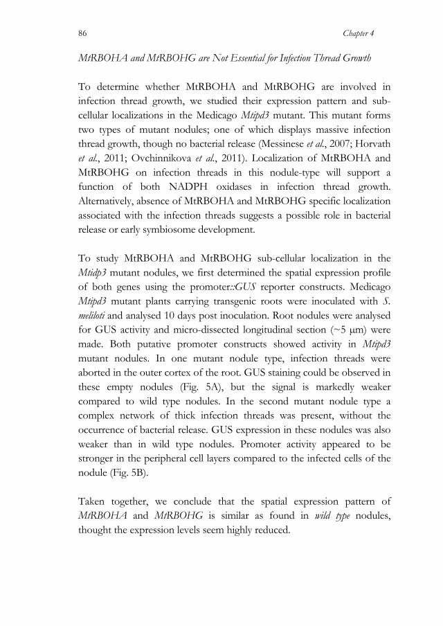
Chapter 4 86
MtRBOHA and MtRBOHG are Not Essential for Infection Thread Growth To determine whether MtRBOHA and MtRBOHG are involved in infection thread growth, we studied their expression pattern and sub-cellular localizations in the Medicago Mtipd3 mutant. This mutant forms two types of mutant nodules; one of which displays massive infection thread growth, though no bacterial release (Messinese et al., 2007; Horvath et al., 2011; Ovchinnikova et al., 2011). Localization of MtRBOHA and MtRBOHG on infection threads in this nodule-type will support a function of both NADPH oxidases in infection thread growth. Alternatively, absence of MtRBOHA and MtRBOHG specific localization associated with the infection threads suggests a possible role in bacterial release or early symbiosome development. To study MtRBOHA and MtRBOHG sub-cellular localization in the Mtidp3 mutant nodules, we first determined the spatial expression profile of both genes using the promoter::GUS reporter constructs. Medicago Mtipd3 mutant plants carrying transgenic roots were inoculated with S. meliloti and analysed 10 days post inoculation. Root nodules were analysed for GUS activity and micro-dissected longitudinal section (~5 µm) were made. Both putative promoter constructs showed activity in Mtipd3 mutant nodules. In one mutant nodule type, infection threads were aborted in the outer cortex of the root. GUS staining could be observed in these empty nodules (Fig. 5A), but the signal is markedly weaker compared to wild type nodules. In the second mutant nodule type a complex network of thick infection threads was present, without the occurrence of bacterial release. GUS expression in these nodules was also weaker than in wild type nodules. Promoter activity appeared to be stronger in the peripheral cell layers compared to the infected cells of the nodule (Fig. 5B). Taken together, we conclude that the spatial expression pattern of MtRBOHA and MtRBOHG is similar as found in wild type nodules, thought the expression levels seem highly reduced.
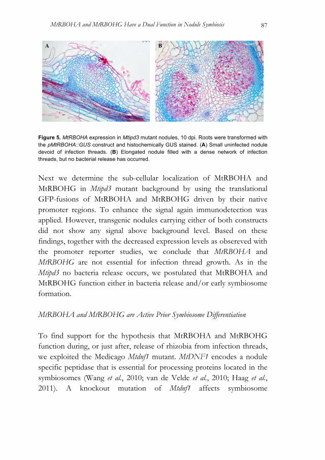
MtRBOHA and MtRBOHG Have a Dual Function in Nodule Symbiosis 87
Figure 5. MtRBOHA expression in Mtipd3 mutant nodules, 10 dpi. Roots were transformed with the pMtRBOHA::GUS construct and histochemically GUS stained. (A) Small uninfected nodule devoid of infection threads. (B) Elongated nodule filled with a dense network of infection threads, but no bacterial release has occurred. Next we determine the sub-cellular localization of MtRBOHA and MtRBOHG in Mtipd3 mutant background by using the translational GFP-fusions of MtRBOHA and MtRBOHG driven by their native promoter regions. To enhance the signal again immunodetection was applied. However, transgenic nodules carrying either of both constructs did not show any signal above background level. Based on these findings, together with the decreased expression levels as obsereved with the promoter reporter studies, we conclude that MtRBOHA and MtRBOHG are not essential for infection thread growth. As in the Mtipd3 no bacteria release occurs, we postulated that MtRBOHA and MtRBOHG function either in bacteria release and/or early symbiosome formation. MtRBOHA and MtRBOHG are Active Prior Symbiosome Differentiation To find support for the hypothesis that MtRBOHA and MtRBOHG function during, or just after, release of rhizobia from infection threads, we exploited the Medicago Mtdnf1 mutant. MtDNF1 encodes a nodule specific peptidase that is essential for processing proteins located in the symbiosomes (Wang et al., 2010; van de Velde et al., 2010; Haag et al., 2011). A knockout mutation of Mtdnf1 affects symbiosome
A B

Chapter 4 88
development; symbiosomes divide but remain small. So, Mtdnf1 nodules contain several cell layers within the central zone filled with immature bacteroids. These cells subsequently undergo early senescence. Introducing the translational GFP-fusions constructs GFP:MtRBOHA and GFP:MtRBOHG driven by their native promoters, revealed that both genes are expressed in the distal part of the infection zone. As in nodules of wild type plants, the signal was present in discrete areas associated with infection threads (Fig. 6). No signal could be detected associated with symbiosomes.
Figure 6. Localization of GFP-MtRBOHA in Medicago Mtdnf1 nodules. Confocal image of a pUBQ10::GFP:MtRBOHA Mtdnf1 transformed root. Discrete areas labelled by Alexa488 are associated with the infection thread, at the site of putative bacteria release. Bars = 20 µm.
As in Mtdnf1 nodules symbiososmes stay immature and undergo early senescence, this suggest that MtRBOHA and MtRBOHG do not function in this
process. Taken together our data strongly suggest that both NADPH oxidases are involved in bacterial release from infection threads or immediately after during early symbiosome development. Discussion In this study we show that in Medicago 2 paralogous genes encoding NADPH oxidases, MtRBOHA and MtRBOHG, function during, or just after, bacterial release from infection threads. We found that MtRBOHA and MtRBOHG have also pleitoropic functions in root nodules, preventing premature senescence of nitrogen fixing symbiosomes.
20 µm
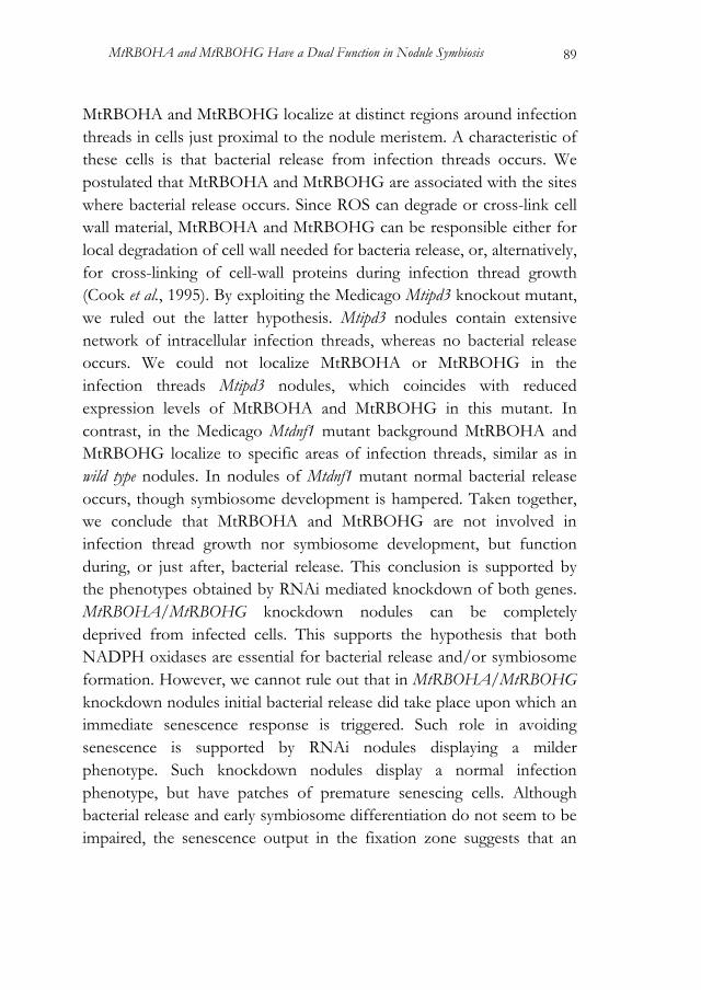
MtRBOHA and MtRBOHG Have a Dual Function in Nodule Symbiosis 89
MtRBOHA and MtRBOHG localize at distinct regions around infection threads in cells just proximal to the nodule meristem. A characteristic of these cells is that bacterial release from infection threads occurs. We postulated that MtRBOHA and MtRBOHG are associated with the sites where bacterial release occurs. Since ROS can degrade or cross-link cell wall material, MtRBOHA and MtRBOHG can be responsible either for local degradation of cell wall needed for bacteria release, or, alternatively, for cross-linking of cell-wall proteins during infection thread growth (Cook et al., 1995). By exploiting the Medicago Mtipd3 knockout mutant, we ruled out the latter hypothesis. Mtipd3 nodules contain extensive network of intracellular infection threads, whereas no bacterial release occurs. We could not localize MtRBOHA or MtRBOHG in the infection threads Mtipd3 nodules, which coincides with reduced expression levels of MtRBOHA and MtRBOHG in this mutant. In contrast, in the Medicago Mtdnf1 mutant background MtRBOHA and MtRBOHG localize to specific areas of infection threads, similar as in wild type nodules. In nodules of Mtdnf1 mutant normal bacterial release occurs, though symbiosome development is hampered. Taken together, we conclude that MtRBOHA and MtRBOHG are not involved in infection thread growth nor symbiosome development, but function during, or just after, bacterial release. This conclusion is supported by the phenotypes obtained by RNAi mediated knockdown of both genes. MtRBOHA/MtRBOHG knockdown nodules can be completely deprived from infected cells. This supports the hypothesis that both NADPH oxidases are essential for bacterial release and/or symbiosome formation. However, we cannot rule out that in MtRBOHA/MtRBOHG knockdown nodules initial bacterial release did take place upon which an immediate senescence response is triggered. Such role in avoiding senescence is supported by RNAi nodules displaying a milder phenotype. Such knockdown nodules display a normal infection phenotype, but have patches of premature senescing cells. Although bacterial release and early symbiosome differentiation do not seem to be impaired, the senescence output in the fixation zone suggests that an
Chapter 4 90
altered ROS balance in the infection zone at the place of bacterial release has a pleiotropic effect several cell layers after. Therefore, we conclude that MtRBOHA and MtRBOHG have a pleiotropic function in symbiosis. Both proteins are essential during or just after rhizobium release from infection threads, as well to sustain the life span of infected cells. MtRBOHA and MtRBOHG have overlapping expression profiles and knockdown experiments suggest that both genes are redundant in their symbiotic function. This indicates that the ancestral gene already fulfilled a similar symbiotic function in root nodules. The timing of the duplication of these genes coincides with an event of whole genome duplication early in the Papillionoid lineage (Cannon et al., 2006; 2010; Young et al., 2011). Therefore, we assume that the gene pair MtRBOHA and MtRBOHG is a remaining of this whole genome duplication event. Similarly, a second paralogous gene pair encoding NADPH oxidases is present of which one copy has diverged further; namely MtRBOHE, MtRBOHB, MtRBOHC and MtRBOHD. These gene duplications are conserved in Lotus, Medicago and soybean, three species that represent lineages that diverged 55 MYA. This suggests that there is positive selection to maintain both copies, though it remains unclear whether this is due to sub- and/or neo-functionalization or a gene dosage dependency. As it is assumed that the ancestral Papilionoid legumes gained rhizobium symbiosis prior this whole genome duplication event (Cannon et al., 2006; Cannon et al., 2009; Cannon et al., 2010; Young et al., 2011), it is in line with our finding that MtRBOHA and MtRBOHG have a overlapping symbiotic function in root nodules. MtRBOHA and MtRBOHG, most likely, do not function exclusively in rhizobium legume symbiosis. Based on their expression pattern we conclude that they also have a function in the root meristematic zone as well as several other organs. Interestingly, the closest Arabidopsis homolog and putative ortholog, AtRBOHB, is involved in seed after-ripening and germination. ROS produced by AtRBOHB are proposed to

MtRBOHA and MtRBOHG Have a Dual Function in Nodule Symbiosis 91
intervene in ABA perception and/or signalling, or alternatively activate post-translational modifications by carbonylation of proteins (Müller et al., 2009). It is unclear whether MtRBOHA and MtRBOHG are expressed in Medicago pods during seed ripening, but if we assume that this is rather the basal function in angiosperms, we can presume that MtRBOHA and/or MtRBOHG could have a similar non-symbiotic function. MtRBOHA and MtRBOHG promoters are highly active in the apical part of Medicago nodules, where the expression seems stronger when compared to the root meristematic zone. In contrast however, GFP-fusion proteins as generated by the use of translational GFP fusion constructs were poorly visible in nodules. To detect reliably GFP-MtRBOHA and GFP-MtRBOHG immunodetection needed to be applied. Also, in nodules the signal could not be enhanced by using a stronger promoter (AtUBQ3 promoter). This in contrast to roots where clear plasma membrane localization of GFP-MtRBOHA and GFP-MtRBOHG was detectable. For the Arabidopsis homolog/ortholog, AtRBOHB, alternative splicing is reported (Müller et al., 2009). Therefore, one could hypothesize that such mechanism is conserved in Medicago MtRBOHA and MtRBOHG. However, since we used a translational fusion of the cDNA that was easily detected in roots, but not in nodules, we can role out that such mechanism is affecting the introduced trans genes. To explain the difference in level of MtRBOHA and MtRBOHG proteins between root and nodules, we hypothesize that a different mechanism controls the half-life of both proteins and that this mechanism is more active in nodules than in roots. The number of genes known to control cellular responses in nodules is limited. This is probably due to gene redundancy in combination with pleiotropic functioning. We found that both MtRBOHA and MtRBOHG have been recruited to control a cellular response in nodules. Based on subcellular localization studies in combination with mutant analysis we argue that both proteins have a function in ealry
Chapter 4 92
steps of symbiosome formation. As MtRBOHA and MtRBOHG are maintained as a paralogous gene pair in distinct legume species, it underlines the importance of the duplication event. In line with this we argue that a phylogeny-based strategy that aims to identify conserved gene duplicates in Papilionoid legumes can be a powerful approach to identify novel symbiotic genes. Materials and methods Plant Material and Growth Conditions In this study M. truncatula accession Jemalong A17, Mtipd3 (Benaben et al., 1995) and Mtdnf1 were used. Transgenic roots were obtained by Agrobacterium rhizogenes MSU440 (A. rhizogenes) mediated root trans-formation (Limpens, 2004). After transformation and selection, plants with transgenic roots were transferred to perlite saturated with liquid Fahraeus medium (Fahraeus, 1957) and inoculated with S. meliloti strain 2011 or S. meliloti 2011 containing pHC60 (Limpens et al., 2003) OD600 0.1, 2 ml per plant) and grown in at 21°C under 16h light/8h darkness conditions. Root nodules were collected for analysis 7, 10, 14 and 28 days post inoculation (dpi). Cloning Medicago genomic DNA, or cDNA obtained from root nodules, was used as template in PCR reactions using Phusion High-Fidelity DNA Polymerase (Finnzymes) and gene or promoter specific primers. Primers used to amplify target sequences were designated with additional attB sites for Gateway cloning. Gene specific primers: gMtRBOHA-f 5’-gaatggaaattgatcaagagaacaaa-3’, gMtRBOHA-r 5’-cattggaaggatcaaaagc-3’, gMtRBOHG-f 5’-agatggagattagtcaagagagagagaa-3’, gMtRBOHG-r 5’-cgctccaaatctaactaacaatcaga-3’. Promoter specific primers were designed to amplify native promoter regions (3 kb 5’-upstream sequence): pMtRBOHA-f 5’-ttcctaattattgttttacttttcttcaca-3’, pMtRBOHA-r 5’-

MtRBOHA and MtRBOHG Have a Dual Function in Nodule Symbiosis 93
gttgctgttttgtttttctgaa-3’, pMtRBOHG-f 5’-attgatatgctcgaagtaaaaatgc-3’, pMtRBOHG-r 5’-ctccgatcgatcaactcgtag-3’. Gateway technology (Invitrogen) was used to create constructs for GFP translational fusions, promoter::GUS reporter studies, and RNA interference gene knock down studies (Karimi et al., 2002). For this an additional round of PCR amplification has been done using the same primers but now extended with the appropriate att sequences needed for cloning. To generate GFP-MtRBOHA and GFP-MtRBOHG translational fusions, GFP was fused to N-terminal end of MtRBOHA or MtRBOHG as described for Arabidopsis AtRHD2 (Takeda et al., 2008), either driven by their native promoter regions or the strong Arabidopsis UBIQUITIN3 (AtUBQ3) promoter (Limpens et al., 2009). Primers used to generate DNA fragments for RNA interference: RNAi-f 5’-gaatggaaattgatcaagagaacaaa-3’, RNAi-r 5’-tctaaaaattctctttatgaaaatcaaact-3’. Gene Expression Studies Total RNA was extracted from roots and nodules using E.Z.N.A. Plant RNA Mini Kit (Omega biotek). cDNA synthesized from 1 µg total RNA was used for quantitative RT-PCR using the iQ SYBR Green Supermix (Bio-Rad) and gene specific primers: qMtRBOHA-f 5’-cgctgaatacaccgagttca-3’, qMtRBOHA-r 5’-gtccagcagactcaacagca-3’, qMtRBOHG-f 5’-ctgtgtgccatggcttcagttgtta-3’, qMtRBOHG-r 5’-gcatcctaccacaccttattcacttcc-3’. qRT-PCR was performed on a MyiQ Real-Time Detection System (Bio-Rad). Gene-expression profiles were normalized against the transcription level of reference gene MtUBQ10. Confocal Laser–Scanning Microscopy GFP-fused proteins were visualized in whole roots and hand sectioned nodules. Imaging was done using a Zeiss LSM 5 Pascal confocal laser–scanning microscope (Carl Zeiss, GmbH). Immuno-detection was performed as described previously (Limpens et al., 2009). Goat serum or 3% BSA was used as blocking agent. Polyclonal rabbit anti-GFP
Chapter 4 94
antibodies (Molecular Probes) in dilution 1:200 and secondary anti-rabbit Alexa 488 antibodies (Molecular Probes) were used. Sections were counterstained by FM4-64 (30 µg/mL). GUS-Staining and Sectioning Transgenic roots and nodules were collected and washed twice in 0.1 M sodium phosphate buffer, pH 7.2, incubated in β-glucoronidase buffer under vacuum conditions at room temperature for 30 minutes to allow the buffer to replace oxygen in the tissue, incubated at 37°C for 0-2 hours or overnight to enable the enzymatic reaction and embedded for sectioning following the Technovit 7100 protocol (Technovit). Sectioning was performed on a microtome machine. Sections were mounted on microscope slides, counterstained stained with ruthenium red and analysed using a Nikon Optiphot-2 microscope. Micro-Sectioning and Staining RNAi nodules were fixed in 4% paraformaldehyde with 3% glutaraldehyde in 50 mM phosphate buffer (pH7.4) and sectioning following the Technovit 7100 protocol (Technovit). Sectioning was performed on a microtome machine. Sections were counterstained using 0.05% toluidine blue and analysed under a Nikon Optiphot-2 microscope. Statistics Analysis The mean nodule number was obtained from a sample size of 20-30 transgenic roots and a Least Significant Difference (LSD) test was performed on the ln values of the original data (which gave a normal distribution). SPSS software (version 16.0) was employed to analyse the data. Differences were considered significant at p < 0.05.
MtRBOHA and MtRBOHG Have a Dual Function in Nodule Symbiosis 95
References
Benaben, V., Duc, G., Lefebvre, V. and Huguet, T., (1995) TE7, an inefficient symbiotic mutant of Medicago truncatula Gaertn. cv Jemalong. Plant Physiology, 107(1), 53–62.
Benedito, V.A., Torres-Jerez, I., Murray, J.D., et al., (2008) A gene expression atlas of the model legume Medicago truncatula. The Plant Journal, 55(3), 504–513.
Bersoult, A.A., Camut, S.S., Cullimore, J.V.J.6, (2005) Expression of the Medicago truncatula DMI2 gene suggests roles of the symbiotic nodulation receptor kinase in nodules and during early nodule development. Molecular Plant-Microbe Interactions, 18(8), 869–876.
Bradley, D.J., Kjellbom, P. and Lamb, C.J., (1992) Elicitor- and wound-induced oxidative cross-linking of a proline-rich plant cell wall protein: a novel, rapid defense response. Cell, 70(1), 21–30.
Brewin, N., (2004) Plant cell wall remodelling in the Rhizobium-legume symbiosis. Critical Reviews in Plant Sciences, 23(4), 293–316.
Cannon, S.B., Ilut, D., Farmer, A.D., Maki, S.L., May, G.D., Singer, S.R. and Doyle, J.J., (2010) Polyploidy did not predate the evolution of nodulation in all legumes. PloS ONE, 5(7), e11630.
Cannon, S.B., May, G.D. and Jackson, S.A., (2009) Three sequenced legume genomes and many crop species: rich opportunities for translational genomics. Plant Physiology, 151(3), 970–977.
Cannon, S.B., Sterck, L., Rombauts, S., et al., (2006) Legume genome evolution viewed through the Medicago truncatula and Lotus japonicus genomes. Proceedings of the National Academy of Sciences of the United States of America, 103(40), 14959–14964.
Cárdenas, L., Martínez, A., Sánchez, F. and Quinto, C., (2008) Fast, transient and specific intracellular ROS changes in living root hair cells responding to Nod factors (NFs). The Plant Journal, 56(5), 802–813.
Chang, C., Damiani, I., Puppo, A. and Frendo, P., (2009) Redox changes during the legume-rhizobium symbiosis. Molecular Plant-Microbe Interactions, 2(3), 370–377.
Cook, D., Dreyer, D., Bonnet, D., Howell, M., Nony, E. and VandenBosch, K., (1995) Transient induction of a peroxidase gene in Medicago truncatula precedes infection by Rhizobium meliloti. The Plant Cell, 7(1), 43–55.
Denness, L., McKenna, J.F., Segonzac, C., Wormit, A., Madhou, P., Bennett, M., Mansfield, J., Zipfel, C. and Hamann, T., (2011) Cell Wall Damage-Induced Lignin Biosynthesis Is Regulated by a Reactive Oxygen Species- and Jasmonic Acid-Dependent Process in Arabidopsis. Plant Physiology, 156(3), 1364–1374.
Fahraeus, G., (1957) The infection of clover root hairs by nodule bacteria studied by a simple glass slide technique. Journal of general microbiology, 16(2), 374–381.
Foreman, J., Demidchik, V., Bothwell, J.H.F., et al., (2003) Reactive oxygen species produced by NADPH oxidase regulate plant cell growth. Nature, 422(6930), 442–446.
Chapter 4 96
Gage, D.J., (2004) Infection and invasion of roots by symbiotic, nitrogen-fixing rhizobia during nodulation of temperate legumes. Microbiology and molecular biology reviews : MMBR, 68(2), 280–300.
Haag, A.F., Baloban, M., Sani, M., et al., (2011) Protection of Sinorhizobium against host cysteine-rich antimicrobial peptides is critical for symbiosis. PLoS Biology, 9(10), e1001169.
Hamann, T., Bennett, M., Mansfield, J. and Somerville, C., (2009) Identification of cell-wall stress as a hexose-dependent and osmosensitive regulator of plant responses. The Plant Journal, 57(6), 1015–1026.
Horvath, B., Yeun, L.H., Domonkos, A., et al., (2011) Medicago truncatula IPD3 is a member of the common symbiotic signaling pathway required for rhizobial and mycorrhizal symbioses. Molecular Plant-Microbe Interactions, 24(11), 1345–1358.
Jamet, A., Mandon, K., Puppo, A. and Hérouart, D., (2007) H2O2 is required for optimal establishment of the Medicago sativa/Sinorhizobium meliloti symbiosis. Journal of bacteriology, 189(23), 8741–8745.
Jamet, A., Sigaud, S., van de Sype, G., Puppo, A. and Hérouart, D., (2003) Expression of the bacterial catalase genes during Sinorhizobium meliloti-Medicago sativa symbiosis and their crucial role during the infection process. Molecular Plant-Microbe Interactions, 16(3), 217–225.
Jones, K.M., Kobayashi, H., Davies, B.W., Taga, M.E. and Walker, G.C., (2007) How rhizobial symbionts invade plants: the Sinorhizobium–Medicago model. Nature Reviews Microbiology, 5(8), 619–633.
Karimi, M., Inzé, D. and Depicker, A., (2002) GATEWAY vectors for Agrobacterium-mediated plant transformation. Trends in Plant Science, 7(5), 193–195.
Kwak, J.M., Mori, I.C., Pei, Z-M., et al., (2003) NADPH oxidase AtrbohD and AtrbohF genes function in ROS-dependent ABA signaling in Arabidopsis. The EMBO journal, 22(11), 2623–2633.
Lavin, M., Herendeen, P.S. and Wojciechowski, M.F., (2005) Evolutionary rates analysis of Leguminosae implicates a rapid diversification of lineages during the tertiary. Systematic biology, 54(4), 575–594.
Limpens, E., (2004) RNA interference in Agrobacterium rhizogenes-transformed roots of Arabidopsis and Medicago truncatula. Journal of Experimental Botany, 55(399), 983–992.
Limpens, E., Franken, C., Smit, P., Willemse, J., Bisseling, T. and Geurts, R., (2003) LysM domain receptor kinases regulating rhizobial Nod factor-induced infection. Science, 302(5645), 630–633.
Limpens, E., Ivanov, S., van Esse, W., Voets, G., Fedorova, E. and Bisseling, T., (2009) Medicago N2-fixing symbiosomes acquire the endocytic identity marker Rab7 but delay the acquisition of vacuolar identity. The Plant Cell, 21(9), 2811–2828.
Limpens, E., Mirabella, R., Fedorova, E., Franken, C., Franssen, H., Bisseling, T. and Geurts, R., (2005) Formation of organelle-like N2-fixing symbiosomes in legume root nodules is controlled by DMI2. Proceedings of the National Academy of Sciences of the United States of America, 102(29), 10375–10380.
MtRBOHA and MtRBOHG Have a Dual Function in Nodule Symbiosis 97
Liszkay, A., van der Zalm, E. and Schopfer, P., (2004) Production of reactive oxygen intermediates (O2.-, H2O2, and .OH) by maize roots and their role in wall loosening and elongation growth. Plant Physiology, 136(2), 3114–3123.
Lohar, D.P., Haridas, S., Gantt, J.S. and VandenBosch, K.A., (2007) A transient decrease in reactive oxygen species in roots leads to root hair deformation in the legume-rhizobia symbiosis. New Phytologist, 173(1), 39–49.
Mando, K., Pauly, N., Boscari, A., Brouquisse, R., Frendo, P., Demple, B. and Puppo, A., (2009) ROS in the legume-Rhizobium symbiosis. In L. A. Rio and A. Puppo, eds. Reactive Oxygen Spieces in Plant Signaling. Signaling and Communication in Plants, pp. 135–147.
Marino, D., Andrio, E., Danchin, E.G.J., Oger, E., Gucciardo, S., Lambert, A., Puppo, A. and Pauly, N., (2010) A Medicago truncatula NADPH oxidase is involved in symbiotic nodule functioning. New Phytologist, 189(2), 580–592.
Messinese, E., Mun, J-H., Yeun, L.H., et al., (2007) A novel nuclear protein interacts with the symbiotic DMI3 calcium- and calmodulin-dependent protein kinase of Medicago truncatula. Molecular Plant-Microbe Interactions, 20(8), 912–921.
Miller, G., Schlauch, K., Tam, R., Cortes, D., Torres, M.A., Shulaev, V., Dangl, J.L. and Mittler, R., (2009) The plant NADPH oxidase RBOHD mediates rapid systemic signaling in response to diverse stimuli. Science signaling, 2(84), ra45.
Miyahara, A., Richens, J., Starker, C., Morieri, G., Smith, L., Long, S., Downie, J.A. and Oldroyd, G.E.D., (2010) Conservation in function of a SCAR/WAVE component during infection thread and root hair growth in Medicago truncatula. Molecular Plant-Microbe Interactions, 23(12), 1553–1562.
Monshausen, G.B., Bibikova, T.N., Messerli, M.A., Shi, C. and Gilroy, S., (2007) Oscillations in extracellular pH and reactive oxygen species modulate tip growth of Arabidopsis root hairs. Proceedings of the National Academy of Sciences of the United States of America, 104(52), 20996–21001.
Müller, K., Carstens, A.C., Linkies, A., Torres, M.A. and Leubner-Metzger, G., (2009) The NADPH-oxidase AtrbohB plays a role in Arabidopsis seed after-ripening. The New Phytologist, 184(4), 885–897.
Oda, T., Hashimoto, H., Kuwabara, N., et al., (2010) Structure of the N-terminal regulatory domain of a plant NADPH oxidase and its functional implications. The Journal of Biological Chemistry, 285(2), 1435–1445.
Ogasawara, Y., Kaya, H., Hiraoka, G., et al., (2008) Synergistic activation of the Arabidopsis NADPH oxidase AtrbohD by Ca2+ and phosphorylation. The Journal of Biological Chemistry, 283(14), 8885–8892.
Ovchinnikova, E., Journet, E-P., Chabaud, M., et al., (2011) IPD3 controls the formation of nitrogen-fixing symbiosomes in pea and Medicago Spp. Molecular Plant-Microbe Interactions, 24(11), 1333–1344.
Passardi, F., Penel, C. and Dunand, C., (2004) Performing the paradoxical: how plant peroxidases modify the cell wall. Trends in Plant Science, 9(11), 534–540.
Chapter 4 98
Peleg-Grossman, S. and Volpin, H., (2007) Root hair curling and Rhizobium infection in Medicago truncatula are mediated by phosphatidylinositide-regulated endocytosis and reactive oxygen species. Journal of Experimental Botany.
Peleg-Grossman, S., Golani, Y., Kaye, Y., Melamed-Book, N. and Levine, A., (2009) NPR1 protein regulates pathogenic and symbiotic interactions between Rhizobium and legumes and non-legumes. PloS ONE, 4(12), e8399.
Proels, R.K., Oberhollenzer, K., Pathuri, I.P., Hensel, G., Kumlehn, J. and Hückelhoven, R., (2010) RBOHF2 of barley is required for normal development of penetration resistance to the parasitic fungus Blumeria graminis f. sp. hordei. Molecular Plant-Microbe Interactions, 23(9), 1143–1150.
Rubio, M.C., James, E.K., Clemente, M.R., Bucciarelli, B., Fedorova, M., Vance, C.P. and Becana, M., (2004) Localization of superoxide dismutases and hydrogen peroxide in legume root nodules. Molecular Plant-Microbe Interactions, 17(12), 1294–1305.
Sagi, M. and Fluhr, R., (2001) Superoxide production by plant homologues of the gp91phox NADPH oxidase. Modulation of activity by calcium and by tobacco mosaic virus infection. Plant Physiology, 126(3), 1281–1290.
Santos, R., Herouart, D., Sigaud, S., Touati, D. and Puppo, A., (2001) Oxidative burst in alfalfa-Sinorhizobium meliloti symbiotic interaction. Molecular Plant-Microbe Interactions, 14(1), 86–89.
Scott-Craig, J., Kerby, K., Stein, B. and Somerville, S., (1995) Expression of an extracellular peroxidase that is induced in barley (Hordeum vulgare) by the powdery mildew pathogen (Erysiphe graminis f. sp. hordei). Physiological and Molecular Plant Pathology, 47(6), 407–418.
Takeda, S., Gapper, C., Kaya, H., Bell, E., Kuchitsu, K. and Dolan, L., (2008) Local positive feedback regulation determines cell shape in root hair cells. Science, 319(5867), 1241–1244.
Timmers, A.C., Auriac, M.C. and Truchet, G., (1999) Refined analysis of early symbiotic steps of the Rhizobium-Medicago interaction in relationship with microtubular cytoskeleton rearrangements. Development, 126(16), 3617–3628.
Torres, M.A., Dangl, J.L. and Jones, J.D.G., (2002) Arabidopsis gp91phox homologues AtrbohD and AtrbohF are required for accumulation of reactive oxygen intermediates in the plant defense response. Proceedings of the National Academy of Sciences of the United States of America, 99(1), 517–522.
Tsukagoshi, H., Busch, W. and Benfey, P.N., (2010) Transcriptional regulation of ROS controls transition from proliferation to differentiation in the root. Cell, 143(4), 606–616.
van de Velde, W., Zehirov, G., Szatmari, A., et al., (2010) Plant peptides govern terminal differentiation of bacteria in symbiosis. Science, 327(5969), 1122–1126.
Vasse, J., de Billy, F., Camut, S. and Truchet, G., (1990) Correlation between ultrastructural differentiation of bacteroids and nitrogen fixation in alfalfa nodules. Journal of bacteriology, 172(8), 4295–4306.
Verma, D., (1992) Signals in root nodule organogenesis and endocytosis of Rhizobium. The Plant Cell, 4(4), 373–382.

MtRBOHA and MtRBOHG Have a Dual Function in Nodule Symbiosis 99
Wang, D., Griffitts, J., Starker, C., Fedorova, E., Limpens, E., Ivanov, S., Bisseling, T. and Long, S., (2010) A nodule-specific protein secretory pathway required for nitrogen-fixing symbiosis. Science, 327(5969), 1126–1129.
Wisniewski, J. and Rathbun, E., (2000) Involvement of diamine oxidase and peroxidase in insolubilization of the extracellular matrix: implications for pea nodule initiation by Rhizobium leguminosarum. Molecular Plant-Microbe Interactions.
Young, N.D., Debellé, F., Oldroyd, G.E.D., et al., (2011) The Medicago genome provides insight into the evolution of rhizobial symbioses. Nature.

CHAPTER 5
Medicago NADPH Oxidase MtRBOHC is Nod Factor Induced1
Alessandra Lillo, Na Li, Ton Bisseling and René Geurts
Department of Plant Sciences, Laboratory of Molecular Biology, Wageningen University, Droevendaalsesteeg 1, 6708 PB, Wageningen, The Netherlands (A.L., T.B., and R.G.); College of Science, King Saud University, Post Office Box 2455, Riyadh 11451, Saudi Arabia (T.B.)
1 This work was supported by the Dutch Science Foundation (NWO-vidi 864.06.007 to R.G.) and the E.U. Research Training Network “NODPERCEPTION” (MRTNCT-2006-035546) (to A.L., T.B., and R.G.)

Chapter 5
102
Abstract Root hair growth is essential to develop a successful rhizobium symbiosis in most legume species. Root hairs are the first plant cells that morphologically respond to rhizobia. Upon bacterial attachment, root hairs continuously redirect their growth thereby forming curls where rhizobia are entrapped. The curl guarantees an optimal microenvironment to start infection thread formation (Esseling et al., 2003). It has been hypothesized that in legumes part of the root hair tip-growth machinery has been recruited during the evolution of symbiosis (Arrighi et al., 2008), so that at least part of this machinery is shared in symbiotic and non-symbiotic conditions. In Arabidopsis thaliana (Arabidopsis) root hair growth is controlled by the NADPH oxidase AtRHD2. Recently it was found that in legumes RHD2 homologous genes underwent a series of duplications (Marino et al., 2010) (Chapter 4, this Thesis). While in Arabidopsis NADPH oxidases genes have been intensively studied, in legumes most NADPH oxidases have not been functionally characterized. We studied whether these RHD2 homologous genes in Medicago truncatula gained a symbiotic function. Here we show that the expression of the NADPH oxidase gene MtRBOHC, a close homolog of AtRHD2, is induced upon rhizobium signalling.
Medicago NADPH Oxidase MtRBOHC is Nod Factor Induced
103
Introduction The mutualistic interaction between nitrogen fixing rhizobium bacteria and legume plants evolved over a period of 60 million years, in which it co-opted several cellular machineries to support this very effective endosymbiosis. Among the cellular machineries for which recruitment in symbiosis has been hypothesized, root hair tip growth raised our interest for three reasons. First, in most legumes root hair tip-growth is essential for the development of a successful symbiotic relationship at early stage of the interaction (Fahraeus, 1957; Esseling et al., 2004). Second, mutant(s) affected in early symbiotic signalling are also affected in root hair growth, thereby strongly supporting the hypothesis that at least part of the root hair tip-growth machinery has been co-opted (Esseling et al., 2003; Cárdenas et al., 2008). Third, recently it was found that in legumes a gene known to be essential in root hair tip growth in Arabidopsis, RHD2, encoding a NADPH oxidase, experienced a series of duplication events. We investigated whether these RHD2 homologous genes gained a symbiotic function. Here we present a study on a Medicago truncatula (Medicago) NADPH oxidase, MtRBOHC, one of the closest homologs of AtRHD2 that is induced early during symbiosis. Root hair tip growth is a highly oriented cell expansion mechanism that is first established and then maintained in such that root hairs elongate exclusively at their apical end. Root hair tip-growth is well studied in Arabidopsis where the mechanism is maintained by a complex network of gradients of an oscillatory tip-focused Ca2+, pH and reactive oxygen species (ROS). Together these gradients maintain a polarized secretion system at the apical end of the growing root hair (Foreman et al., 2003; Monshausen et al., 2007). This secretion system includes longitudinally oriented actin cables for myosin-mediated organelle transport and fine F-actin structures for secretory vesicles transport to sites of their fusion with the plasma membrane. Studies in Arabidopsis revealed that among the components needed for root hair tip-growth maintenance, a specific

Chapter 5
104
NADPH oxidase appears to be essential; namely AtRHD2 (Foreman et al., 2003). AtRHD2 is polar localized at the tip of the root hair, and becomes activated by Ca2+ resulting in localized production of ROS at the tip of the growing root hair. In turn, through a positive feedback regulation, AtRHD2 activation establishes and maintains the Ca2+ gradient needed for tip-growth in expanding root hair cells (Takeda et al., 2008). In addition to its signalling role in mediating Ca2+ gradients, it is proposed that ROS produced by AtRHD2 are essential for regulating cell wall extensibility (Monshausen et al., 2007). Root hair growth is not a continuous process, but rather occurs in phases, where ROS -and pH- oscillations in the apoplast modulate discrete episodes of polar growth. Growth phases coincide with low apoplastic ROS just behind the apex (cell wall loosening) followed by stasis phases correlated with a high ROS accumulation, proposed to trigger cell wall rigidification and consequent restriction of the growth site (Monshausen et al., 2007). Consequently, Arabidopsis Atrhd2 knockout mutants that are defective in a key component of tip-growth, display short root hairs with uncontrolled cell expansion. Most probably, this uncontrolled cell expansion leads the growing root hairs to bulge or to burst according with alkali or acidic pH. In case of legumes such as Medicago, root hairs are among the first cells to respond morphologically to so-called rhizobium Nodulation (Nod) factor signalling molecules (Heidstra et al., 1994). Inline with this is the hypothesis that some components of the tip-growth mechanism are specifically induced shortly after Nod factor application. Nod factors locally secreted by rhizobium manipulate root hairs growth continuously redirecting the growth direction and leading to curling root hair curling (Esseling et al., 2003). The newly formed curl captures the bacterium in a closed infection pocket. There, in the appropriate environment of the tight curl, bacteria dividing induce infection thread formation. Since root hair curling is essential for a successful symbiotic infection, this process has been extensively studied at a morphological level, although mechanistically is still undeciphered. To study Nod factor induced root
Medicago NADPH Oxidase MtRBOHC is Nod Factor Induced
105
hair responses, two assays have been used that differ in the Nod factors application technique; local application to a single root hair at a spot very close to the growing root hair tip (spot inoculation), or application to the whole root (Fahraeus, 1957; Heidstra et al., 1994; de Ruijter et al., 1998; Miller et al., 1999; Sieberer and Emons, 2000; Catoira et al., 2000; Esseling et al., 2003). While with the application to the whole root only root hair swelling and deformation/branching are induced, in case of spot inoculation even root hair curling can be mimicked (Esseling et al., 2003). It is interesting to note that in either assay, root hairs can respond to Nod factors also by establishing a new site of tip-growth resulting in root hair branching. This indicates that Nod Factors can influence not only existing tip-growth machineries, but also can dictate formation of new growth foci. The root hair tip-growth machinery is a highly plastic system, regulated by a wide range of environmental signals including mechanical stimulation (López-Bucio et al., 2003; Bloch et al., 2011; Monshausen et al., 2009). Among the Nod factor signalling genes, the LRR-type receptor kinase MtDMI2 is required for maintenance of the tip-growth machinery under mechanical stress conditions (Esseling et al., 2004). Growing root hairs have a specific cytoarchitecture with the sub-apex of the root hair filled with cytoplasm, and the nucleus at the base of this area, while the shank of the root hair is filled with the central vacuole and cytoplasm. When subjected to a mechanical stimulus, growing root hairs immediately display cytoarchitecture reorganization. The vacuole overtakes the nucleus and, subsequently the cytoplasmic rich sub-apical region decreases in size. This response is typical for root hairs that terminating growth. However, Medicago wild type root hairs show a recovery in cytoarchitecture and restart growing within 15 minutes after stress application. In contrast, root hairs of Mtdmi2 mutant plant are impaired in cytoarchitecture and growing recovery (Esseling et al., 2004). Since both Nod factor signalling and root hair mechanical stimulation trigger Ca2+ influx (Monshausen et al., 2009), it can be speculated that this is a shared function impaired in the Mtdmi2 mutant. However, this is

Chapter 5
106
not the case since Ca2+ influx upon Nod factor signalling is not impaired in this mutant (Shaw and Long, 2003). Medicago has 5 close homologs of Arabidopsis AtRHD2 (Marino et al., 2010). Two of these genes, MtRBOHA and MtRBOHG, have a redundant function in the root nodule, where they are associated with bacterial release from the infection threads (Chapter 4, this Thesis). Among the other 3 Medicago NADPH oxidases, MtRBOHD is most probably a pseudogene (Marino et al., 2010), whereas there are no data in literature about MtRBOHB and MtRBOHC. Since in Arabidopsis, AtRHD2 has been shown to be essential for root hair tip-grow (Foreman et al., 2003), it can be speculated that in legumes such function is conserved among one (or more) of its homologs. We hypothesize that in legumes part of the root hair tip-growing machinery has been recruited during evolution of symbiosis. In line with this we questioned whether at least one of the Medicago RHD2-like genes could fulfil such symbiotic function. Based on gene expression studies, MtRBOHC is an interesting candidate since it is transcriptionally induced upon Nod factor signalling. However, Arabidopsis Atrhd2 mutant trans-complementation studies and Medicago Tnt1-tagged MtrbohC mutant phenotype analysis indicate that AtRHD2-like function is not conserved in MtRBOHC. Results One Medicago AtRHD2-like Gene is Specifically Up-Regulated during Nodulation Previous phylogenetic studies as presented in Chapter 4 of this Thesis revealed the presence of two sets of paralogous gene pairs in the NADPH oxidase gene family, which are conserved in Medicago, Lotus japonicus (Lotus), and Glycine max (soybean). Focussing on Medicago the two duplicated gene sets include: MtRBOHA and MtRBOHG (set I); MtRBOHE, MtRBOHB, MtRBOHC, and MtRBOHD (set II). Since MtRBOHA and MtRBOHG show no function in root hair growth
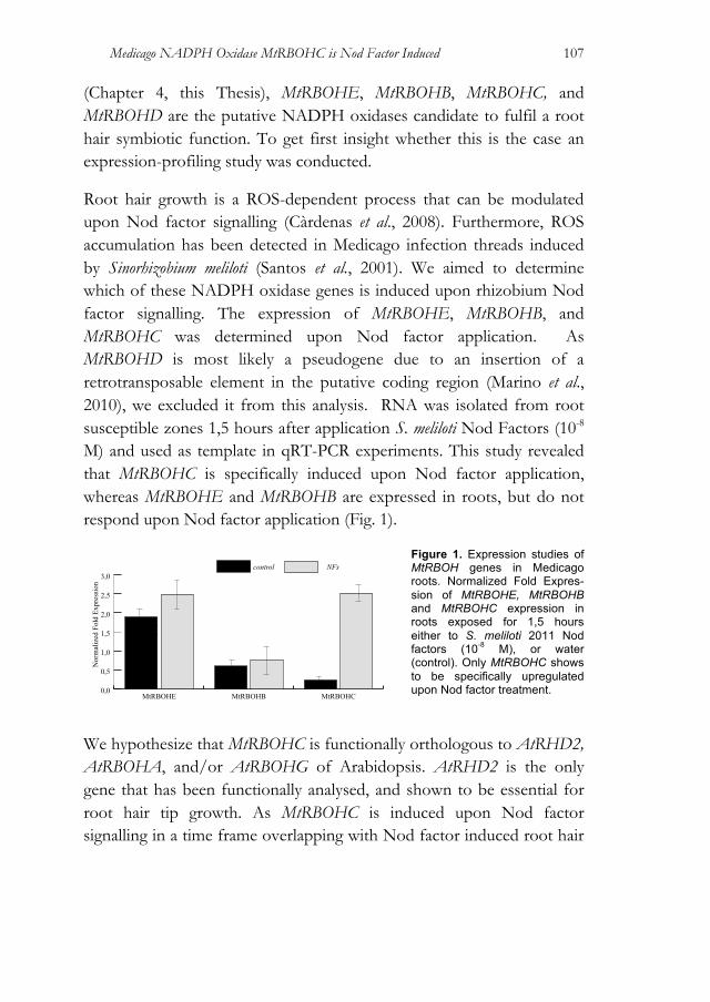
Medicago NADPH Oxidase MtRBOHC is Nod Factor Induced
107
(Chapter 4, this Thesis), MtRBOHE, MtRBOHB, MtRBOHC, and MtRBOHD are the putative NADPH oxidases candidate to fulfil a root hair symbiotic function. To get first insight whether this is the case an expression-profiling study was conducted.
Root hair growth is a ROS-dependent process that can be modulated upon Nod factor signalling (Càrdenas et al., 2008). Furthermore, ROS accumulation has been detected in Medicago infection threads induced by Sinorhizobium meliloti (Santos et al., 2001). We aimed to determine which of these NADPH oxidase genes is induced upon rhizobium Nod factor signalling. The expression of MtRBOHE, MtRBOHB, and MtRBOHC was determined upon Nod factor application. As MtRBOHD is most likely a pseudogene due to an insertion of a retrotransposable element in the putative coding region (Marino et al., 2010), we excluded it from this analysis. RNA was isolated from root susceptible zones 1,5 hours after application S. meliloti Nod Factors (10-8 M) and used as template in qRT-PCR experiments. This study revealed that MtRBOHC is specifically induced upon Nod factor application, whereas MtRBOHE and MtRBOHB are expressed in roots, but do not respond upon Nod factor application (Fig. 1).
Figure 1. Expression studies of MtRBOH genes in Medicago roots. Normalized Fold Expres-sion of MtRBOHE, MtRBOHB and MtRBOHC expression in roots exposed for 1,5 hours either to S. meliloti 2011 Nod factors (10-8 M), or water (control). Only MtRBOHC shows to be specifically upregulated upon Nod factor treatment.
We hypothesize that MtRBOHC is functionally orthologous to AtRHD2, AtRBOHA, and/or AtRBOHG of Arabidopsis. AtRHD2 is the only gene that has been functionally analysed, and shown to be essential for root hair tip growth. As MtRBOHC is induced upon Nod factor signalling in a time frame overlapping with Nod factor induced root hair
0,0
0,5
1,0
1,5
2,0
2,5
3,0NFscontrol
MtRBOHCMtRBOHBMtRBOHE
Nor
mal
ized
Fol
d Ex
pres
sion
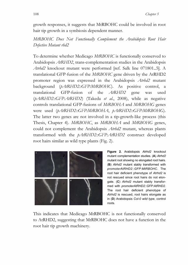
Chapter 5
108
growth responses, it suggests that MtRBOHC could be involved in root hair tip growth in a symbiosis dependent manner.
MtRBOHC Does Not Functionally Complement the Arabidopsis Root Hair Defective Mutant rhd2
To determine whether Medicago MtRBOHC is functionally conserved to Arabidopsis AtRHD2, trans-complementation studies in the Arabidopsis Atrhd2 knockout mutant were performed [ref. Salk line 071801_5]. A translational GFP-fusion of the MtRBOHC gene driven by the AtRHD2 promoter region was expressed in the Arabidopsis Atrhd2 mutant background (pAtRHD2::GFP:MtRBOHC). As positive control, a translational GFP-fusion of the AtRHD2 gene was used (pAtRHD2::GFP:AtRHD2) (Takeda et al., 2008), while as negative controls translational GFP-fusions of MtRBOHA and MtRBOHG genes were used (pAtRHD2::GFP:MtRBOHA, pAtRHD2::GFP:MtRBOHG). The latter two genes are not involved in a tip-growth-like process (this Thesis, Chapter 4). MtRBOHC, as MtRBOHA and MtRBOHG genes, could not complement the Arabidopsis Atrhd2 mutant, whereas plants transformed with the pAtRHD2::GFP:AtRHD2 construct developed root hairs similar as wild type plants (Fig. 2).
Figure 2. Arabidopsis Atrhd2 knockout mutant complementation studies. (A) Atrhd2 mutant root showing no elongated root hairs. (B) Atrhd2 mutant stably transformed with promoterAtRHD2::GFP:MtRBOHC. The root hair deficient phenotype of Atrhd2 is not rescued since root hairs do not elon-gate. (C) Atrhd2 mutant stably transfor-med with promoterAtRHD2::GFP:AtRHD2. The root hair deficient phenotype of Atrhd2 is rescued, root hairs elongate as in (D) Arabidopsis Col-0 wild type, control roots.
This indicates that Medicago MtRBOHC is not functionally conserved to AtRHD2, suggesting that MtRBOHC does not have a function in the root hair tip growth machinery.
A B
C D

Medicago NADPH Oxidase MtRBOHC is Nod Factor Induced
109
Tnt1-tagged MtRBOHC Mutant Does Not Show a Root Hair Phenotype To get insight in the function of MtRBOHC, a Medicago line harbouring a Tnt1 retrotransposon insertion in the coding region of this gene was analysed (Tadege et al., 2008). PCR with Tnt1 and MtRBOHC specific primers confirmed the insertion in NF4406 line (Insertion 9). In total NF4406 line contained 19 Tnt1 insertions and was homozygote for the insertion in MtRBOHC exon 10 (the MtRBOHC gene contains 12 exons). NF4406 was selfed and seeds of plants homozygote for the Mtrbohc mutant alleles were collected.
First 10 homozygotes Tnt1-tagged Mtrbohc seedlings were analysed for root hair defective phenotype when compared with Medicago R108 (control). In Tnt1-tagged Mtrbohc seedlings, root hairs show no altered phenotype when compared to wild type plants (Fig. 3).
Figure 3. Elongating root hairs of the Medicago Mtrbohc knockout line NF4406 (A) and wild type Medicago R108 (B). No difference in root hair length is observed. Next, Tnt1-tagged Mtrbohc seedlings were tested for root hair deformation phenotype upon Nod factor (10-8 M) treatment. No difference between Medicago R108 wild type and the Tnt1-tagged Mtrbohc line was detected (data not shown). Therefore, MtRBOHC does not have an essential function in the root hair tip growth machinery.
A B
Chapter 5
110
Conclusions The role of MtRBOHC in root hair tip-growth was investigated. Based on transcomplementation studies in the Arabidopsis Atrhd2 mutant and mutant analysis in Medicago, most probably the AtRHD2-like function is not conserved in MtRBOHC. Furthermore, no obvious phenotype was detectable in Tnt1-tagged Mtrbohc seedlings grown in symbiotic and non-symbiotic conditions, indicating that MtRBOHC is presumably required for other processes and/or is redundant in function. Yet the question how Nod factor signalling triggers root hair morphological responses remains unsolved. We postulated that the root hair tip-growth machinery is conserved in plants and that based on phylogeny MtRBOHC is orthologous to Arabidopsis AtRHD2. As no experimental evidence could be provided that this is the case, it suggests that MtRBOHC is neo-functionalized and other NADPH oxidase genes fulfil the AtRHD2-like function in Medicago. Based on expression and phylogeny studies can be argued that there are two other candidate genes that could fulfil such function; MtRBOHB and MtRBOHF, respectively. The first is phylogenetically closer to AtRHD2 (Chapter 4, Fig. 1), but the second is expressed in root hairs (Marino et al., 2010). Considering the expression profile, MtRBOHF therefore seems to be a good candidate. If this were the case, it would imply that the common ancestor of AtRHD2 and MtRBOHF had already a tip-growth function. Yet, it is unlikely that a single gene is essential for root hair tip-growth in Medicago, as no such (symbiotic) mutant has been identified in genetic screens. A good indication on the functionality and on the putative redundancy can be provided by sub-cellular localization studies of the different NADPH oxidases proteins. GFP-tagged versions of the proteins were developed for trans-complementation studies in the Arabidopsis Atrhd2 mutant background. This will enable us to perform studies in this direction. Protein localization on the dome of the bulging root hair

Medicago NADPH Oxidase MtRBOHC is Nod Factor Induced
111
would indicate that the protein is recognized but the tip-growth localization system and properly located. The transgenic lines that have been created in this study are suited for such experiments. This can provide novel insight in neo-functionalization of the NADPH oxidases proteins in Medicago. Materials and Methods Plant Material and Growth Conditions Arabidopsis seeds were sterilized in 5% sodium hypochlorite, washed in water and sown on Murashige and Skoog (Duchefa, Haarlem, The Netherlands) medium (pH 5.8) containing 1% sucrose and 0.8% Phytagel. Atrhd2 mutant used in this study was the Salk line 071801_5 showing severe short root hair phenotype (Fig. 2). Cloning and Plant Transformation Medicago genomic DNA or cDNA obtained from root nodules as a template to amplify by PCR DNA fragments using Phusion™ High-Fidelity DNA Polymerase (Finnzymes) and gene specific primers. Long primers used to generate coding sequences of genes of interest were designated with additional attB sites. AttB sequences: forward (f) 5’-ggggacagctttcttgtacaaagtcg-3’, and reverse primers (r) 5’-ggggacaactttgtataataaagttg-3’. Gene specific primers: gMtRBOHA-f 5’-gaatggaaattgatcaagagaacaaa-3’, gMtRBOHA-r 5’-cattggaaggatcaaaagc-3’; gMtRBOHC-f 5’-cggtgagaaacttagaggtccgaaa-3’, gMtRBOHC-r 5’-ttatctgtctctcttacacacaatgtcc-3’; gMtRBOHG-f 5’-agatggagattagtcaagagaga gagaa-3’, gMtRBOHG-r 5’-cgctccaaatctaactaacaatcaga-3’. The Gateway® technology (Invitrogen) was used to create genetic constructs for GFP-fusions. To generate GFP-MtRBOHA, GFP-MtRBOHC, GFP-MtRBOHG, and GFP-AtRHD2 translational fusions, GFP was fused to N-terminal end of MtRBOHA, MtRBOHC, MtRBOHG, and AtRHD2 driven by AtRHD2 promoter exactly as described previously

Chapter 5
112
(Takeda et al., 2008). Using Gateway technology (Invitrogen; http://www.invitrogen.com/), the whole cassette was introduced into the vector pBnRGW (Mlynárová et al., 2007). The final binary vector was introduced into Agrobacterium tumefaciens strain AGLO (Lazo et al., 1991) was used for plant transformation by the floral dipping method as described (Clough and Bent, 1998). Gene Expressions Studies For the quantitative RT-PCR on RBOH genes, Medicago truncatula accession Jemalong A17 germinated seedlings were grown vertically on modified Fahraeus medium agar plates without nitrate on top of filter paper for 48 hours (Fahraeus, 1957). Then, water-dissolved Sinorhizobium meliloti (S. meliloti) strain 2011 Nod factors (approximately 10-9 M) or water as a control were pipetted on top of every root. Roots were exposed for 1,5 hour; subsequently, 1 cm root pieces were cut just above the root tip and were snap-frozen (n=15). Total RNA was extracted from snap-frozen roots samples using E.Z.N.A.™ Plant RNA Mini Kit (Omega biotek). Equal amounts of total RNA were used to perform gene analysis expression of MtRBOH genes by quantitative RT-PCR using iQ SYBR Green Supermix (Bio-Rad) and gene specific primers: qMtRBOHC-f 5’-ggcatggatgtcacaaaaac-3’, qMtRBOHC-r 5’-gaaccattgcgatgtactctttc-3’, qMtRBOHB-f 5’-agaaaagcacattgtgtgtaacg-3’, qMtRBOHB-r 5’-gtggatttctccttcaacttcg-3’, qMtRBOHE-f 5’-aacatgggaggatcttctgc-3’, qMtRBOHE-r 5’-tgttctcaggagcaatgagttc-3’. Detection of fluorescent signal was performed on a My iQ Real-Time Detection System (Bio-Rad). Gene-expression profiles were normalized against the transcription level of reference gene MtUBQ10.
Medicago NADPH Oxidase MtRBOHC is Nod Factor Induced
113
References Arrighi, J., Godfroy, O., de Billy, F., Saurat, O., Jauneau, A. and Gough, C.,
(2008) The RPG gene of Medicago truncatula controls Rhizobium-directed polar growth during infection. Proceedings of the National Academy of Sciences of the United States of America, 105(28), 9817–9822.
Bloch, D., Monshausen, G., Gilroy, S. and Yalovsky, S., (2011) Co-regulation of root hair tip growth by ROP GTPases and nitrogen source modulated pH fluctuations. Plant Signaling & Behavior, 6(3), 426–429.
Cannon, S.B., Ilut, D., Farmer, A.D., Maki, S.L., May, G.D., Singer, S.R. and Doyle, J.J., (2010) Polyploidy did not predate the evolution of nodulation in all legumes. PloS ONE, 5(7), e11630.
Catoira, R., Galera, C., de Billy, F., et al., (2000) Four genes of Medicago truncatula controlling components of a nod factor transduction pathway. The Plant Cell, 12(9), 1647–1666.
Cárdenas, L., Martínez, A., Sánchez, F. and Quinto, C., (2008) Fast, transient and specific intracellular ROS changes in living root hair cells responding to Nod factors (NFs). The Plant Journal, 56(5), 802–813.
Clough, S.J. and Bent, A.F., (1998) Floral dip: a simplified method for Agrobacterium-mediated transformation of Arabidopsis thaliana. The Plant Journal, 16(6), 735–743.
de Ruijter, N., Rook, M. and Bisseling, T., (1998) Lipochito-oligosaccharides re-initiate root hair tip growth in Vicia sativa with high calcium and spectrin-like antigen at the tip. Plant Journal.
Esseling, J.J., Lhuissier, F.G.P. and Emons, A.M.C., (2004) A nonsymbiotic root hair tip growth phenotype in NORK-mutated legumes: implications for nodulation factor-induced signaling and formation of a multifaceted root hair pocket for bacteria. The Plant Cell, 16(4), 933–944.
Esseling, J.J., Lhuissier, F.G.P. and Emons, A.M.C., (2003) Nod factor-induced root hair curling: continuous polar growth towards the point of nod factor application. Plant Physiology, 132(4), 1982–1988.
Fahraeus, G., (1957) The infection of clover root hairs by nodule bacteria studied by a simple glass slide technique. Journal of general microbiology, 16(2), 374–381.
Foreman, J., Demidchik, V., Bothwell, J.H.F., et al., (2003) Reactive oxygen species produced by NADPH oxidase regulate plant cell growth. Nature, 422(6930), 442–446.
Heidstra, R., Geurts, R., Franssen, H., Spaink, H.P., Van Kammen, A. and Bisseling, T., (1994) Root hair deformation activity of nodulation factors and their fate on Vicia sativa. Plant Physiology, 105(3), 787–797.
Lazo, G.R., Stein, P.A. and Ludwig, R.A., (1991) A DNA transformation-competent Arabidopsis genomic library in Agrobacterium. Nature Biotechnology, 9(10), 963–967.
Chapter 5
114
López-Bucio, J., Cruz-Ramı ́rez, A. and Herrera-Estrella, L., (2003) The role of nutrient availability in regulating root architecture. Current Opinion in Plant Biology, 6(3), 280–287.
Marino, D., Andrio, E., Danchin, E.G.J., Oger, E., Gucciardo, S., Lambert, A., Puppo, A. and Pauly, N., (2010) A Medicago truncatula NADPH oxidase is involved in symbiotic nodule functioning. New Phytologist, 189(2), 580–592.
Miller, D.D., de Ruijter, N.C.A., Bisseling, T. and Emons, A.M.C., (1999) The role of actin in root hair morphogenesis: studies with lipochito-oligosaccharide as a growth stimulator and cytochalasin as an actin perturbing drug. The Plant Journal, 17(2), 141–154.
Mlynárová, L., Nap, J-P. and Bisseling, T., (2007) The SWI/SNF chromatin-remodeling gene AtCHR12 mediates temporary growth arrest in Arabidopsis thaliana upon perceiving environmental stress. The Plant Journal, 51(5), 874–885.
Monshausen, G.B., Bibikova, T.N., Messerli, M.A., Shi, C. and Gilroy, S., (2007) Oscillations in extracellular pH and reactive oxygen species modulate tip growth of Arabidopsis root hairs. Proceedings of the National Academy of Sciences of the United States of America, 104(52), 20996–21001.
Monshausen, G.B., Bibikova, T.N., Weisenseel, M.H. and Gilroy, S., (2009) Ca2+ regulates reactive oxygen species production and pH during mechanosensing in Arabidopsis roots. The Plant Cell, 21(8), 2341–2356.
Santos, R., Herouart, D., Sigaud, S., Touati, D. and Puppo, A., (2001) Oxidative burst in alfalfa-Sinorhizobium meliloti symbiotic interaction. Molecular Plant-Microbe Interactions, 14(1), 86–89.
Shaw, S.L. and Long, S.R., (2003) Nod factor elicits two separable calcium responses in Medicago truncatula root hair cells. Plant Physiology, 131(3), 976–984.
Sieberer, B. and Emons, A.M.C., (2000) Cytoarchitecture and pattern of cytoplasmic streaming in root hairs of Medicago truncatula during development and deformation by nodulation factors. Protoplasma, 214, 118–127.
Tadege, M., Wen, J., He, J., et al., (2008) Large-scale insertional mutagenesis using the Tnt1 retrotransposon in the model legume Medicago truncatula. The Plant Journal, 54(2), 335–347.
Takeda, S., Gapper, C., Kaya, H., Bell, E., Kuchitsu, K. and Dolan, L., (2008) Local positive feedback regulation determines cell shape in root hair cells. Science, 319(5867), 1241–1244.
CHAPTER 6
General Conclusions
Alessandra Lillo
Department of Plant Sciences, Laboratory of Molecular Biology, Wageningen University, Droevendaalsesteeg 1, 6708 PB, Wageningen, The Netherlands
1 This work was supported by the E.U. Research Training Network “NODPERCEPTION” (MRTNCT-2006-035546)

Chapter 6
116
Introduction By entering an endosymbiotic relation with rhizobia, legumes bypass one of the most severe problems plants usually encounter during their life cycle in natural environments: shortage of fixed nitrogen. In current agriculture, chemical fertilizers are used to overcome such problem for crop species, yet this solution is far from meeting environmental demands (Dawson and Hilton, 2011; Brentrup and Palliere, 2008). The endosymbiotic relation with rhizobia guarantees a reliable nitrogen source to legumes. Therefore, this symbiosis makes legumes an extremely important system to study. Although many steps have been made toward the understanding of the legume-rhizobium symbiosis, the path to enable extension of this symbiosis to non-legume crops seems still to be long. During the last two decades, legume root nodule symbiosis has been dissected and forward genetics screens, focussed on complete loss of nodulation ability, are now believed to have reached saturation. The genetic approach in two model legumes, Medicago truncatula (Medicago) and Lotus japonicus (Lotus), has been very powerful to identify key regulators in root nodule formation, although such approach also has obvious limitations, as in the case of functional redundancy and/or pleiotropic effects (caused by the knockout of genes that do not function exclusively in symbiosis). Therefore, in this Thesis an alternative approach was exploited. Recent studies in the reference species Medicago, Lotus and soybean (Glycine max) have revealed the presence of numerous paralogous gene pairs in their genomes. Many of these originate from a whole genome duplication (WGD) event that has occurred in a common ancestral legume species. Strikingly, a substantial fraction of gene pairs are conserved in the three reference species. As Medicago, Lotus and soybean represent major phylogenetic clades that have diverged early in evolution of the legume Papilionoid subfamily, the question was raised to what extend this WGD has contributed to the evolution of Rhizobium root

General Conclusions
117
nodules (Camp et al., 2011; Young et al., 2011). This work aimed on the functional characterization of 3 such gene pairs; two in the NADPH oxidase gene family, and one in the cytokinin phosphorelay pathway, namely type-A cytokinin Response Regulators (RRs). These studies provided experimental evidence that 2 of these duplicated paralogous gene pairs, maintained in 3 unrelated legume species, fulfil symbiotic functions and that the functioning of the paralogs is redundant. Strikingly, ectopic expression of either both RR genes resulted in arrested primordia that had a chimeric nature with nodule as well as lateral root characteristics. This triggered the study on the ontology of lateral root primordia formation in more detail. A Whole Genome Duplication Has Shaped Rhizobium Symbiosis in Papilionoid Legumes
Rhizobium root nodule symbiosis occurs in the vast majority of the 18,000 legume species, though it is absent from the two earliest diverging lineages of the family: Cercideae and Detarieae, respectively. Most probably, nodulation evolved after the origin of the family, approximately 60 million years ago (Lavin et al., 2005; Sprent, 2008). Although nodulation occurs in the two largest clades, the monophyletic subfamily Papilionoideae and the Mimosoideae (Mimosoids) superclade (also containing the Caesalpineae-Cassieae lineages), nodulation is not present in every species/genus in either of both clades. Because of the absence of nodulation in some species within the legume family, phylogenetic studies have tried to decipher whether nodulation evolved once, followed by trait losses in some species, or evolved multiple times independently (Doyle et al., 2011). Lack of phylogenetic resolution and of nodulation data are currently the biggest obstacles to answer this question. Based on current knowledge it has been suggested that there could have been five to six independent origins of nodulation already within the Mimosoids superclade and possibly one or two in the Papilionoids (Doyle et al., 1997; Doyle et al., 2011).
Chapter 6
118
Fifty-six to 65 million years ago an ancestral legume species within the Papilionoids experienced a WGD event (Fawcett et al., 2009; Cannon et al., 2010; Young et al., 2011). As the occurrence of this WGD can be traced in Medicago, Lotus and soybean, their ancestral legume has given rise at least to the Indigoferoid, Milettioid and Hologalegina lineages that enclose mainly nodulating species. Polyploidy is considered to be a major force in the advancement of new functions, since it generates a dramatic increase in raw genetic material on which evolution can act (van de Peer et al., 2009). WGDs have been implicated in the emergence of new traits, such as the origin of flowering plants (De Bodt et al., 2005). In line with this it was investigated whether the WGD as found in Papilionoid legumes predates the occurrence of nodulation in this subfamily. Expression studies in Medicago revealed that a substantial number of paralogous gene pairs have a similar symbiotic expression profile. This suggests that the ancestral genes already had symbiotic functions. Consequently, the WGD should have occurred after the emergence of nodulation (Young et al., 2011). So, although polyploidy was not an essential contribution to the emergence of rhizobium symbiosis in Papilionoid legumes, it gave rise to an immediate increased in gene redundancy that could have contributed to further specialization of the symbiotic interaction. It is commonly accepted that soon after a WGD, massive gene loss takes place (Scannell et al., 2007), while some gene pairs are maintained by chance others sub/neo-functionalize or contribute to advantageous gene dosage effects (Innan and Kondrashov, 2010) (Hughes, 1994). Among the first category two symbiotic genes, initially identified by forward genetic screens, have been categorized. These include the LysM-type rhizobium Nod factor receptor MtNFP/LjNFR5 and the ethylene response factor ERN1 (Op den Camp et al.; 2011; Young et al., 2011). Studies on paralogs of these genes revealed that these genes function in the endomycorrhizal symbiosis (Young et al., 2011; Op den Camp et al., 2011; Gomez et al.,

General Conclusions
119
2009). Since long ago, it has been known that mycorrhizae and rhizobium symbioses in legumes share some common key signalling genes (Duc et al., 1989). This so-called common symbiotic signalling pathway has been unravelled in Medicago and Lotus by means of forward genetics. This genetic signalling network comprises a conserved set of genes encoding a plasma membrane receptor kinase (MtDMI2 and LjSYMRK), several components in the nuclear envelope including a cation channel (MtDMI1, LjCASTOR and LjPOLLUX), a nuclear localized Calcium Calmodulin dependent Kinase (CCaMK; MtDMI3 and LjCCaMK) and a CCaMK interacting protein (MtIPD3 and LjCYCLOPS). Despite the dual function of this pathway in mycorrhizal and rhizobial endosymbiosis, none of these genes have a paralogous counterpart retained that originated from the Papilionoid specific WGD. This in contrast to the LysM-type receptors and the ERN-type transcription factors that act upstream and downstream of the common signaling pathway. The reason that MtNFP/LjNFR5 and ERN1 have been found in forward genetic screens indicate that these genes have neo-functionalized after the WGD, possibly to create specificity in both symbioses. Forward genetic screens will be unsuited to identify symbiotic genes among duplicated gene pairs that are (partially) redundant in function. So a strategy based on phylogenetic reconstruction of orthology groups to identify legume specific gene duplicates was adopted (Op den Camp et al., 2011; De Mita & Geurts, unpublished). Subsequent expression profiling pinpointed those gene paralogs that are either induced upon rhizobium inoculation and/or expressed in root nodules. From the genes identified two duplication events in the NADPH oxidase family as well as one duplication in the so-called cytokinin Response Regulators were the focus of this Thesis. Functional analyses of the corresponding genes proofed their redundant symbiotic functioning, underlining the strength of this phylogenetic approach to identify partial redundant gene pairs.
Chapter 6
120
Symbiotic RBOH Gene Paralogs Control Symbiosome Maintenance In a Redundant Fashion NADPH oxidase activity is an important source of reactive oxygen species (ROS) (Peleg-Grossman and Volpin, 2007; Mando et al., 2009). NADPH oxidases have been hypothesized to be involved in a wide range of functions, including symbiosis, but no direct evidence yet has been provided. ROS accumulation is associated with several symbiotic processes from early responses to the presence of the symbiotic partner, down to senescence of symbiosomes containing cells (Cárdenas et al., 2008; Peleg-Grossman and Volpin, 2007; Jamet et al., 2007; Rubio et al., 2004; Limpens et al., 2005; Santos et al., 2001). Despite their importance, there is only limited knowledge on the biological role of NADPH oxidases in symbiosis. We identified two gene pairs encoding NADPH oxidases that are maintained in Medicago, Lotus and soybean. In Medicago these include the pairs MtRBOHA-MtRBOHG as well as MtRBOHB, MtRBOHC, MtRBOHD and MtRBOHE. The second gene set, represented by MtRBOHE, MtRBOHB, MtRBOHC and MtRBOHD, not only has been maintained after the Papilionoid-specific WGD, but experienced even further duplications. These genes represent the closest homologs to Arabidopsis thaliana (Arabidopsis) AtRHD2, a key component in root hair tip-growth (Foreman et al., 2003). To date little is known about key genes involved in the adaptation of the root hair tip-growth machinery essential for rhizobium infection. Root hair growth is triggered as symbiotic response upon rhizobium Lipo-chitooligosaccharides (LCOs, also named Nod factors). This rhizobium induced root hair growth is often visible in the changed growth direction of the hair and therefore called root hair deformation (Heidstra et al., 1994). Eventually, symbiotic root hair tip growth results in a curl that encloses a rhizobium bacterium. We found in Medicago transcriptional activation of MtRBOHC upon Nod factor induced signalling. In line with this finding we hypothesized that MtRBOHC fulfils an AtRHD2-like function. However, mutant analysis and

General Conclusions
121
complementation studies did not provide support for this hypothesis, suggesting that MtRBOHC does not fulfil an AtRHD2-like function. The second gene pair encoding NADPH oxidases, MtRBOHA and MtRBOHG, has an overlapping expression pattern. Both genes are expressed under symbiotic and non-symbiotic conditions. They are expressed in root tips, including meristematic cells, and in the meristem, infection zone and one to two cell layers of the fixation zone of the nodule. In Medicago both MtRBOHA and MtRBOHG were shown to be essential to sustain symbiosomes. Nodules formed on MtRBOHA/MtRBOHG knockdown roots show two phenotypes. Either no release of bacteria from infection threads is occurring by which the nodule is not infected, or, in case infection is successful, patches of premature senescing cells are present in the nitrogen fixation zone (Chapter 4, this Thesis). The block in release suggests a positive role of ROS in release. It has been shown that release requires a specific exocytotic pathway by which a cell wall free region, unwalled droplet, is formed (Ivanov et al., 2012). So, it can be hypothesized that the NADPH activity is either required to maintain a focused calcium gradient essential for vesicle transport, or alternatively its cross-linking abilities of hydroxyl-proline riche matrix proteins to provide rigidity (Takeda et al., 2008; Monshausen et al., 2007; Foreman et al., 2003; Carol and Dolan, 2006). MtRBOHA and MtRBOHG genes do not seem to have sub-functionalized in relation to their functioning in root and nodule nor in expression in these tissues. In line with this it can be argued that both copies are either only partially redundant, or alternatively are maintained because of positive dosage effects. Partial redundancy can be explained, since their expression patterns and/or functions in other tissues and/or conditions remain to be elucidated. Partial redundancy might confer an evolutionary advantage, this in contrast to complete genetic redundancy, considered to be evolutionary unstable in the long term. Yet, there are at least two hypotheses that aim to explain complete gene redundancy; the (relative) dosage balance hypothesis (Papp et al., 2003; Freeling and Thomas, 2006; Birchler and Veitia, 2007), and the (absolute) dosage
Chapter 6
122
hypothesis (Bekaert et al., 2011). According to the relative dosage balance hypothesis, the redundancy in a module or network could be needed to preserve stoichiometric balances amongst the individual components (Veitia et al., 2008). If this was the case, most probably also enzymes needed for ROS scavenging mechanisms, should have been maintained as gene pair after the WGD event. Alternatively, the absolute increase in the concentration of the product of the maintained gene pairs is beneficial. Selection on absolute gene dosage has been shown for duplicated genes from different organisms (Kondrashov and Kondrashov, 2006), including plants (van Hoof et al., 2001; Widholm et al., 2001). A way to discriminate between these two hypotheses is to investigate the evolutionary context of enzymatic scavenging mechanisms present in the apoplast (e.g. ascorbate peroxidase (APX). If some of them have been maintained in gene pairs and they are co-expressed in the nodule in an overlapping pattern with MtRBOHA and MtRBOHG, these would be the best candidate genes to support the relative dosage balance hypothesis. As suggested by De Smet et al. (2012), for the future understanding of the evolution of regulatory networks after WGDs it is essential to take into account predictions of the entire network instead of only focusing on the divergence of homologous gene pairs. To do so, the use of computational methods is strongly needed. First attempts into this direction have already been made outside plant research (Navlakha and Kingsford, 2011; Pinney et al., 2007; Conant and Wolfe, 2006; Xie et al., 2011). Paralogous Cytokinin Response Regulators are Induced Upon Rhizobium Nod Factor Signalling In a focussed approach we addressed the question whether conserved paralogous gene pairs are present in the cytokinin phosphorelay pathway. Cytokinin signalling is plays a pivotal role in legume root nodule formation, yet the precise mechanism of action has still to be
General Conclusions
123
deciphered. The cytokinin phosphorelay pathway consists of four signalling components: histidine kinase receptors (HKs), phosphotransfer proteins (HPs) and two types of response regulators (RRs): type-A and -B, respectively. Upon phosphorylation, type-B RRs are essential for transcriptional activation of many targets, among them A-type RRs. A small gene family encodes all 4 components. For example Arabidopsis has 6 HP, 3 HK and 33 RR genes. We determined whether within these gene families’ paralogous gene pairs are maintained in the Papilionoid legume species Medicago, Lotus and soybean. One gene pair encoding type-A response regulators, named MtRR9 and MtRR11 in Medicago, was found to be maintained. Type-A response regulators are considered as negative regulators of cytokinin signalling (Müller and Sheen, 2007) and they are transcriptionally activated upon cytokinin signalling, although the precise function of MtRR9 and MtRR11 in Rhizobium symbiosis has not fully resolved. We show that MtRR9 and MtRR11 are involved in root nodule formation since the reduction of their transcript levels by RNAi results in a reduced number of nodules. Promoter reporter studies revealed that MtRR9 and MtRR11 have a different expression pattern in the root that does not overlap, whereas during nodulation the expression patterns of both genes largely overlap. This suggests that the ancestral gene already had such cis regulatory element responsible for the symbiotic expression profile. So, most probably the symbiotic function of MtRR9 and MtR11 did not diverged. Instead, sub-functionalization has taken place in their non-symbiotic functioning. Cortical Cell Divisions Associated with Lateral Foot Formation in Medicago and Lotus Ectopic expression of MtRR9 and MtRR11 in Medicago roots revealed arrested primordia that originate from pericycle and cortical cells. As it is generally known that legume nodules originate from the root cortex, whereas lateral root primordial find their origin in the pericycle, we argued that such structures found upon ectopic MtRR9 expression are
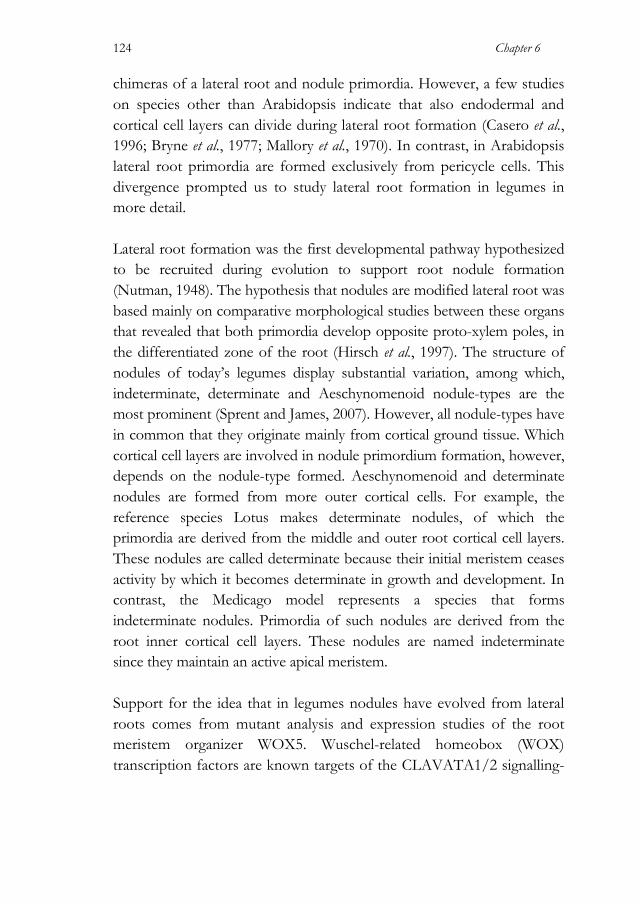
Chapter 6
124
chimeras of a lateral root and nodule primordia. However, a few studies on species other than Arabidopsis indicate that also endodermal and cortical cell layers can divide during lateral root formation (Casero et al., 1996; Bryne et al., 1977; Mallory et al., 1970). In contrast, in Arabidopsis lateral root primordia are formed exclusively from pericycle cells. This divergence prompted us to study lateral root formation in legumes in more detail. Lateral root formation was the first developmental pathway hypothesized to be recruited during evolution to support root nodule formation (Nutman, 1948). The hypothesis that nodules are modified lateral root was based mainly on comparative morphological studies between these organs that revealed that both primordia develop opposite proto-xylem poles, in the differentiated zone of the root (Hirsch et al., 1997). The structure of nodules of today’s legumes display substantial variation, among which, indeterminate, determinate and Aeschynomenoid nodule-types are the most prominent (Sprent and James, 2007). However, all nodule-types have in common that they originate mainly from cortical ground tissue. Which cortical cell layers are involved in nodule primordium formation, however, depends on the nodule-type formed. Aeschynomenoid and determinate nodules are formed from more outer cortical cells. For example, the reference species Lotus makes determinate nodules, of which the primordia are derived from the middle and outer root cortical cell layers. These nodules are called determinate because their initial meristem ceases activity by which it becomes determinate in growth and development. In contrast, the Medicago model represents a species that forms indeterminate nodules. Primordia of such nodules are derived from the root inner cortical cell layers. These nodules are named indeterminate since they maintain an active apical meristem. Support for the idea that in legumes nodules have evolved from lateral roots comes from mutant analysis and expression studies of the root meristem organizer WOX5. Wuschel-related homeobox (WOX) transcription factors are known targets of the CLAVATA1/2 signalling-
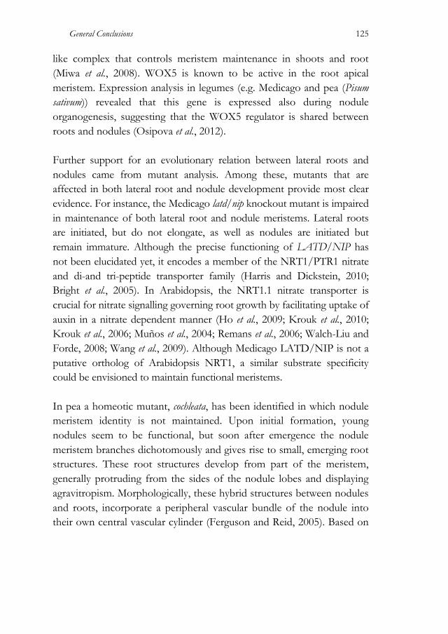
General Conclusions
125
like complex that controls meristem maintenance in shoots and root (Miwa et al., 2008). WOX5 is known to be active in the root apical meristem. Expression analysis in legumes (e.g. Medicago and pea (Pisum sativum)) revealed that this gene is expressed also during nodule organogenesis, suggesting that the WOX5 regulator is shared between roots and nodules (Osipova et al., 2012). Further support for an evolutionary relation between lateral roots and nodules came from mutant analysis. Among these, mutants that are affected in both lateral root and nodule development provide most clear evidence. For instance, the Medicago latd/nip knockout mutant is impaired in maintenance of both lateral root and nodule meristems. Lateral roots are initiated, but do not elongate, as well as nodules are initiated but remain immature. Although the precise functioning of LATD/NIP has not been elucidated yet, it encodes a member of the NRT1/PTR1 nitrate and di-and tri-peptide transporter family (Harris and Dickstein, 2010; Bright et al., 2005). In Arabidopsis, the NRT1.1 nitrate transporter is crucial for nitrate signalling governing root growth by facilitating uptake of auxin in a nitrate dependent manner (Ho et al., 2009; Krouk et al., 2010; Krouk et al., 2006; Muños et al., 2004; Remans et al., 2006; Walch-Liu and Forde, 2008; Wang et al., 2009). Although Medicago LATD/NIP is not a putative ortholog of Arabidopsis NRT1, a similar substrate specificity could be envisioned to maintain functional meristems. In pea a homeotic mutant, cochleata, has been identified in which nodule meristem identity is not maintained. Upon initial formation, young nodules seem to be functional, but soon after emergence the nodule meristem branches dichotomously and gives rise to small, emerging root structures. These root structures develop from part of the meristem, generally protruding from the sides of the nodule lobes and displaying agravitropism. Morphologically, these hybrid structures between nodules and roots, incorporate a peripheral vascular bundle of the nodule into their own central vascular cylinder (Ferguson and Reid, 2005). Based on
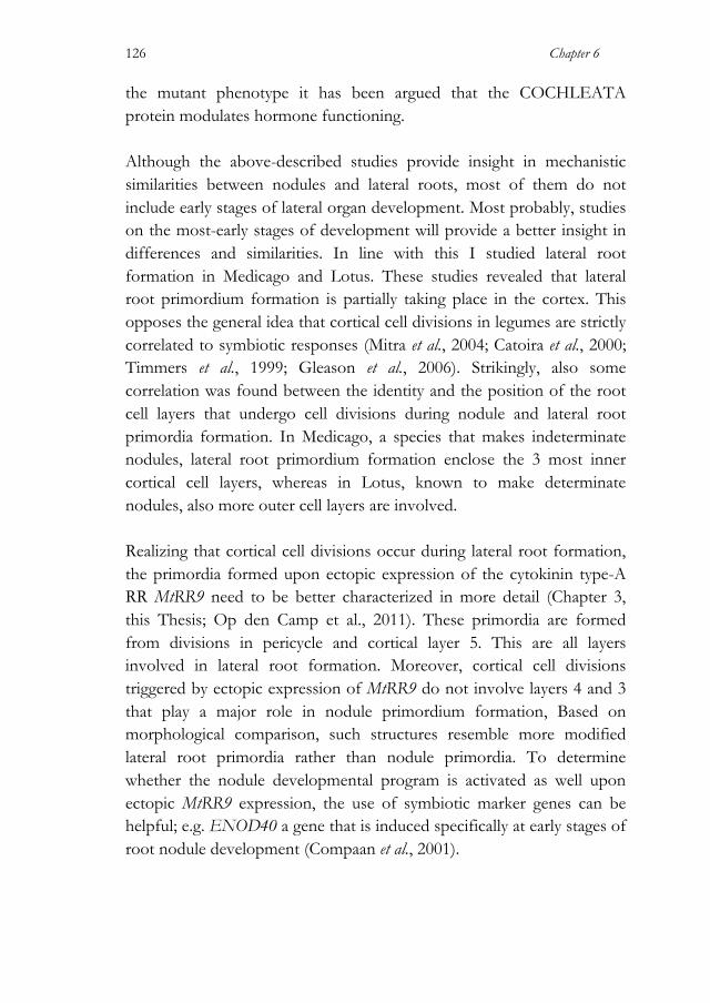
Chapter 6
126
the mutant phenotype it has been argued that the COCHLEATA protein modulates hormone functioning. Although the above-described studies provide insight in mechanistic similarities between nodules and lateral roots, most of them do not include early stages of lateral organ development. Most probably, studies on the most-early stages of development will provide a better insight in differences and similarities. In line with this I studied lateral root formation in Medicago and Lotus. These studies revealed that lateral root primordium formation is partially taking place in the cortex. This opposes the general idea that cortical cell divisions in legumes are strictly correlated to symbiotic responses (Mitra et al., 2004; Catoira et al., 2000; Timmers et al., 1999; Gleason et al., 2006). Strikingly, also some correlation was found between the identity and the position of the root cell layers that undergo cell divisions during nodule and lateral root primordia formation. In Medicago, a species that makes indeterminate nodules, lateral root primordium formation enclose the 3 most inner cortical cell layers, whereas in Lotus, known to make determinate nodules, also more outer cell layers are involved. Realizing that cortical cell divisions occur during lateral root formation, the primordia formed upon ectopic expression of the cytokinin type-A RR MtRR9 need to be better characterized in more detail (Chapter 3, this Thesis; Op den Camp et al., 2011). These primordia are formed from divisions in pericycle and cortical layer 5. This are all layers involved in lateral root formation. Moreover, cortical cell divisions triggered by ectopic expression of MtRR9 do not involve layers 4 and 3 that play a major role in nodule primordium formation, Based on morphological comparison, such structures resemble more modified lateral root primordia rather than nodule primordia. To determine whether the nodule developmental program is activated as well upon ectopic MtRR9 expression, the use of symbiotic marker genes can be helpful; e.g. ENOD40 a gene that is induced specifically at early stages of root nodule development (Compaan et al., 2001).

General Conclusions
127
Also, it was investigated whether the underlying mechanisms that control cortical cell divisions during lateral root and nodule primordia are shared. To do so, lateral root development was studied under several conditions as well as in 3 symbiotic mutants. These include environmental factors such as external nitrate and mechanical stress and the Medicago mutants ccamk, nsp1 and nsp2. The later 3 genes are essential for rhizobium Nod factor induced cortical cell divisions. Knockout mutations in either of these genes fully block nodule primordium formation. Studying lateral root formation in these mutants reveals that the lateral root developmental program in Medicago is extremely robust. None of the considered conditions affected the lateral root developmental program. Priming of Lateral Root Founder Cells is Taking Place Exclusively in Pericycle Cells The current model(s) on lateral root development are exclusively based on studies in Arabidopsis. In this species lateral root initiation starts with the selection of few cells located in three adjacent files of the pericycle opposite to a xylem pole. These are the so-called founder cells (Dubrovsky et al., 2000; Beeckman et al., 2001). Founder cells are the only cells that divide and give rise to the lateral root primordium (Kurup et al., 2005). Based on auxin reporter studies it is hypothesized that founder cells are primed for lateral root formation, which is thought to occur already in the basal meristem (De Smet et al., 2007). While in Arabidopsis exclusively pericycle cells (are primed and subsequently) divide during lateral root initiation, in Medicago and Lotus multiple cell layers undergo cell division. Therefore, it is interesting to question whether in analogy to Arabidopsis, also in Medicago and Lotus exclusively cells in the pericycle are primed as lateral root founder cells, or other cell layers (endodermis and cortex) are primed as well. Based on time-frame studies in Medicago it can be concluded that cells in the pericycle are the first to divide during lateral root primordium formation, subsequently followed by cells in the inner cortex, whereas the

Chapter 6
128
endodermis undergoes only a limited number of cell divisions (Kulikova & Bisseling; unpublished). This suggests that an auxin response maximum is first generated in the pericycle and subsequently extended to the more outer cell layers. If so, priming of cells in cell layers other than the pericycle would not be essential. Early Stages in Lateral Root and Nodule Primordium Formation are Very Similar Both, nodule and lateral root primordia are associated with auxin accumulation. Recently, a predictive model has been developed showing how theoretically an auxin maximum can be created in a multi cell layered root cortex (Denium et al., 2012). This showed that a decreased auxin efflux activity in all cell layers of a cortical segment of the root is sufficient to create an auxin maximum in the inner cortical cells, as occurs in the Medicago root. Auxin efflux is mainly under control of auxin efflux carriers of the PIN family. These membrane proteins have specific sub-cellular locations. Cytokinin is known to modulate endocytic PIN trafficking, thereby decreasing membrane bound PIN (Besnard et al., 2011). Cytokinin promotes nodule primordium formation in legumes. Such Nod factor induced cytokinin responses occur homogenously in all cell layers as exemplified with MtRR9 and MtRR11 promoter reporter studies (Chapter 2 this thesis; Op den Camp et al., 2011). So, we hypothesize that Nod factor induced cytokinin responses modulate the subcellular PIN localization, subsequently leading to a decrease in auxin efflux in these cells. According to the model of Denium et al. (2012) this reduction in auxin efflux in all cortical cell layers can be sufficient to induce a local auxin maximum in the cortex. Moreover, it is hypothesized that subtle differences in the distribution of lateral positioned PIN proteins could be key factor in determining which cortical cell layers give rise to determinate and indeterminate nodules (Denium et al., 2012). Currently, there are no studies done that describe the PIN distribution in roots of a legume species. To validate the

General Conclusions
129
proposed model such studies will be essential to understand mechanistically the patterning of nodule as well as lateral root formation in legumes.
Chapter 6
130
References Beeckman, T., Burssens, S. and Inzé, D., (2001) The peri-cell-cycle in Arabidopsis.
Journal of Experimental Botany, 52, 403–411.
Besnard, F., Vernoux, T. and Hamant, O., (2011) Organogenesis from stem cells in planta: multiple feedback loops integrating molecular and mechanical signals. Cellular and Molecular Life Sciences, 68(17), 2885–2906.
Bekaert, M., Edger, P.P., Pires, J.C. and Conant, G.C., (2011) Two-phase resolution of polyploidy in the Arabidopsis metabolic network gives rise to relative and absolute dosage constraints. The Plant Cell, 23(5), 1719–1728.
Birchler, J.A. and Veitia, R.A., (2007) The gene balance hypothesis: from classical genetics to modern genomics. The Plant Cell, 19(2), 395–402.
Brentrup, F. and Palliere, C., (2008) GHG emissions and energy efficiency in European nitrogen fertiliser production and use. In Proceedings of the International Fertiliser Society. International Fertiliser Society, pp. 1–25.
Bright, L.J., Liang, Y., Mitchell, D.M. and Harris, J.M., (2005) The LATD gene of Medicago truncatula is required for both nodule and root development. Molecular Plant-Microbe Interactions, 18(6), 521–532.
Bryne, J.M., Pesacreta, T.C. and Fox, J.A., (1977) Development and structure of the vascular connection between the primary and secondary root of Glycine max (L.) Merr. American Journal of Botany, 64(8), 946–959.
Camp, den, R.H.O., De Mita, S., Lillo, A., Cao, Q., Limpens, E., Bisseling, T. and Geurts, R., (2011) A phylogenetic strategy based on a legume-specific whole genome duplication yields symbiotic cytokinin type-A Response Regulators. Plant Physiology, 2013–2022.
Cannon, S.B., Ilut, D., Farmer, A.D., Maki, S.L., May, G.D., Singer, S.R. and Doyle, J.J., (2010) Polyploidy did not predate the evolution of nodulation in all legumes. PloS ONE, 5(7), e11630.
Carol, R.J. and Dolan, L., (2006) The role of reactive oxygen species in cell growth: lessons from root hairs. Journal of Experimental Botany, 57(8), 1829–1834.
Casero, P.J., Casimiro, I. and Lloret, P.G., (1996) Pericycle proliferation pattern during the lateral root initiation in adventitious roots of Allium cepa. Protoplasma, 191(3-4), 136–147.
Catoira, R., Galera, C., de Billy, F., et al., (2000) Four genes of Medicago truncatula controlling components of a nod factor transduction pathway. The Plant Cell, 12(9), 1647–1666.
Cárdenas, L., Martínez, A., Sánchez, F. and Quinto, C., (2008) Fast, transient and specific intracellular ROS changes in living root hair cells responding to Nod factors (NFs). The Plant Journal, 56(5), 802–813.
Compaan, B., Yang, W-C., Bisseling, T. and Franssen, H., (2001) ENOD40 expression in the pericycle precedes cortical cell division in Rhizobium-legume interaction and the highly conserved internal region of the gene does not encode a peptide. Plant and Soil, 230, 1–8.
General Conclusions
131
Conant, G.C. and Wolfe, K.H., (2006) Functional partitioning of yeast co-expression networks after genome duplication. PLoS Biology, 4(4), e109–e109.
Dawson, C.J. and Hilton, J., (2011) Fertiliser availability in a resource-limited world: production and recycling of nitrogen and phosphorus. Food Policy, 1–9.
De Bodt, S., Maere, S. and van de Peer, Y., (2005) Genome duplication and the origin of angiosperms. Trends in Ecology & Evolution, 20(11), 591–597.
Denium, E. E., Geurts, R., Bisseling, T., Mulder, B. M., (2012) Modeling a cortical auxin maximum for nodulation: different signatures of potential strategies. Frontiers in Plant Science. In press.
De Smet, I., Tetsumura, T., De Rybel, B., et al., (2007) Auxin-dependent regulation of lateral root positioning in the basal meristem of Arabidopsis. Development, 134(4), 681–690.
Doyle, J., Doyle, J., Ballenger, J., Dickson, E., Kajita, T. and Ohashi, H., (1997) A phylogeny of the chloroplast gene rbcL in the Leguminosae: taxonomic correlations and insights into the evolution of nodulation. American Journal of Botany, 84(4), 541.
Doyle, J.J., (2011) Phylogenetic perspectives on the origins of nodulation. Molecular Plant-Microbe Interactions, 24(11), 1289–1295.
Dubrovsky, J.G., Doerner, P.W., Colón-Carmona, A. and Rost, T.L., (2000) Pericycle cell proliferation and lateral root initiation in Arabidopsis. Plant Physiology, 124(4), 1648–1657.
Duc, G., Trouvelot, A., Gianinazzi-Pearson, V. and Gianinazzi, S., (1989) First report of non-mycorrhizal plant mutants (Myc−) obtained in pea (Pisum sativum L.) and fababean (Vicia faba L.). Plant Science, 60(2), 215–222.
Fawcett, J.A., Maere, S. and van de Peer, Y., (2009) Plants with double genomes might have had a better chance to survive the Cretaceous-Tertiary extinction event. Proceedings of the National Academy of Sciences of the United States of America, 106(14), 5737–5742.
Ferguson, B.J. and Reid, J.B., (2005) Cochleata: getting to the root of legume nodules. Plant and Cell Physiology, 46(9), 1583–1589.
Foreman, J., Demidchik, V., Bothwell, J.H.F., et al., (2003) Reactive oxygen species produced by NADPH oxidase regulate plant cell growth. Nature, 422(6930), 442–446.
Freeling, M. and Thomas, B.C., (2006) Gene-balanced duplications, like tetraploidy, provide predictable drive to increase morphological complexity. Genome Research, 16(7), 805–814.
Gleason, C., Chaudhuri, S., Yang, T., Muñoz, A., Poovaiah, B.W. and Oldroyd, G.E.D., (2006) Nodulation independent of rhizobia induced by a calcium-activated kinase lacking autoinhibition. Nature, 441(7097), 1149–1152.

Chapter 6
132
Gomez, S.K., Javot, H., Deewatthanawong, P., Torres-Jerez, I., Tang, Y., Blancaflor, E.B., Udvardi, M.K. and Harrison, M.J., (2009) Medicago truncatula and Glomus intraradices gene expression in cortical cells harboring arbuscules in the arbuscular mycorrhizal symbiosis. BMC Plant Biology, 9, 10.
Harris, J.M. and Dickstein, R., (2010) Control of root architecture and nodulation by the LATD/NIP transporter. Plant Signaling & Behavior, 5(11), 1365–1369.
Heidstra, R., Geurts, R., Franssen, H., Spaink, H.P., Van Kammen, A. and Bisseling, T., (1994) Root hair deformation activity of nodulation factors and their fate on Vicia sativa. Plant Physiology, 105(3), 787–797.
Hirsch, A.M., Larue, T.A. and Doyle, J., (1997) Is the legume nodule a modified root or stem or an organ sui generis? Critical Reviews in Plant Sciences, 16(4), 361–392.
Ho, C-H., Lin, S-H., Hu, H-C. and Tsay, Y-F., (2009) CHL1 functions as a nitrate sensor in plants. Cell, 138(6), 1184–1194.
Hughes, A.L., (1994) The evolution of functionally novel proteins after gene duplication. Proceedings. Biological sciences / The Royal Society, 256(1346), 119–124.
Innan, H. and Kondrashov, F., (2010) The evolution of gene duplications: classifying and distinguishing between models. Nature Reviews: Genetics, 11(2), 97–108.
Ivanov, S., Fedorova, E.E., Limpens, E., De Mita, S., Genre, A., Bonfante, P. and Bisseling, T., (2012) Rhizobium-legume symbiosis shares an exocytotic pathway required for arbuscule formation. Proceedings of the National Academy of Sciences of the United States of America.
Jamet, A., Mandon, K., Puppo, A. and Hérouart, D., (2007) H2O2 is required for optimal establishment of the Medicago sativa/Sinorhizobium meliloti symbiosis. Journal of bacteriology, 189(23), 8741–8745.
Kondrashov, F.A. and Kondrashov, A.S., (2006) Role of selection in fixation of gene duplications. Journal of theoretical biology, 239(2), 141–151.
Krouk, G., Lacombe, B., Bielach, A., et al., (2010) Nitrate-regulated auxin transport by NRT1.1 defines a mechanism for nutrient sensing in plants. Developmental Cell, 18(6), 927–937.
Krouk, G.G., Tillard, P.P. and Gojon, A.A., (2006) Regulation of the high-affinity NO3- uptake system by NRT1.1-mediated NO3- demand signaling in Arabidopsis. Plant Physiology, 142(3), 1075–1086.
Kurup, S., Runions, J., Köhler, U., Laplaze, L., Hodge, S. and Haseloff, J., (2005) Marking cell lineages in living tissues. The Plant Journal, 42(3), 444–453.
Lavin, M., Herendeen, P.S. and Wojciechowski, M.F., (2005) Evolutionary rates analysis of Leguminosae implicates a rapid diversification of lineages during the tertiary. Systematic biology, 54(4), 575–594.
Mallory, T.E., Chiang, S-H., Cutter, E.G. and E M Gifford, J., (1970) Sequence and pattern of lateral root formation in five selected species. American Journal of Botany, 57(7), 800–809.
General Conclusions
133
Mando, K., Pauly, N., Boscari, A., Brouquisse, R., Frendo, P., Demple, B. and Puppo, A., (2009) ROS in the legume-Rhizobium symbiosis. In L. A. Rio and A. Puppo, eds. Reactive Oxygen Spieces in Plant Signaling. Signaling and Communication in Plants, pp. 135–147.
Mitra, R.M., Shaw, S.L. and Long, S.R., (2004) Six nonnodulating plant mutants defective for Nod factor-induced transcriptional changes associated with the legume-rhizobia symbiosis. Proceedings of the National Academy of Sciences of the United States of America, 101(27), 10217–10222.
Miwa, H., Kinoshita, A., Fukuda, H. and Sawa, S., (2008) Plant meristems: CLAVATA3/ESR-related signaling in the shoot apical meristem and the root apical meristem. Journal of plant research, 122(1), 31–39.
Monshausen, G.B., Bibikova, T.N., Messerli, M.A., Shi, C. and Gilroy, S., (2007) Oscillations in extracellular pH and reactive oxygen species modulate tip growth of Arabidopsis root hairs. Proceedings of the National Academy of Sciences of the United States of America, 104(52), 20996–21001.
Muños, S.S., Cazettes, C.C., Fizames, C.C., Gaymard, F.F., Tillard, P.P., Lepetit, M.M., Lejay, L.L. and Gojon, A.A., (2004) Transcript profiling in the chl1-5 mutant of Arabidopsis reveals a role of the nitrate transporter NRT1.1 in the regulation of another nitrate transporter, NRT2.1. The Plant Cell, 16(9), 2433–2447.
Müller, B. and Sheen, J., (2007) Cytokinin signaling pathway. Signal Transduction Knowledge Environment (STKE), 2007(407), cm4–cm4.
Navlakha, S. and Kingsford, C., (2011) Network archaeology: uncovering ancient networks from present-day interactions. PLoS Computational Biology, 7(4), e1001119.
Nutman, P.S., (1948) Physiological studies on nodule formation. Annals of Botany, 12(2), 81–96.
Camp, den, R.H.O., De Mita, S., Lillo, A., Cao, Q., Limpens, E., Bisseling, T. and Geurts, R., (2011) A phylogenetic strategy based on a legume-specific whole genome duplication yields symbiotic cytokinin type-A Response Regulators. Plant Physiology, 2013–2022.
Osipova, M.A., Mortier, V., Demchenko, K.N., Tsyganov, V.E., Tikhonovich, I.A., Lutova, L.A., Dolgikh, E.A. and Goormachtig, S., (2012) WUSCHEL-RELATED HOMEOBOX5 gene expression and interaction of CLE peptides with components of the systemic control add two pieces to the puzzle of autoregulation of nodulation. Plant Physiology, 158(3), 1329–1341.
Papp, B., Pál, C. and Hurst, L.D., (2003) Dosage sensitivity and the evolution of gene families in yeast. Nature, 424(6945), 194–197.
Peleg-Grossman, S. and Volpin, H., (2007) Root hair curling and Rhizobium infection in Medicago truncatula are mediated by phosphatidylinositide-regulated endocytosis and reactive oxygen species. Journal of Experimental Botany.
Pinney, J.W., Amoutzias, G.D., Rattray, M. and Robertson, D.L., (2007) Reconstruction of ancestral protein interaction networks for the bZIP transcription factors. Proceedings of the National Academy of Sciences of the United States of America, 104(51), 20449–20453.
Chapter 6
134
Remans, T., Nacry, P., Pervent, M., Filleur, S., Diatloff, E., Mounier, E., Tillard, P., Forde, B.G. and Gojon, A., (2006) The Arabidopsis NRT1.1 transporter participates in the signaling pathway triggering root colonization of nitrate-rich patches. Proceedings of the National Academy of Sciences of the United States of America, 103(50), 19206–19211.
Rubio, M.C., James, E.K., Clemente, M.R., Bucciarelli, B., Fedorova, M., Vance, C.P. and Becana, M., (2004) Localization of superoxide dismutases and hydrogen peroxide in legume root nodules. Molecular Plant-Microbe Interactions, 17(12), 1294–1305.
Santos, R., Herouart, D., Sigaud, S., Touati, D. and Puppo, A., (2001) Oxidative burst in alfalfa-Sinorhizobium meliloti symbiotic interaction. Molecular Plant-Microbe Interactions, 14(1), 86–89.
Scannell, D.R., Frank, A.C., Conant, G.C., Byrne, K.P., Woolfit, M. and Wolfe, K.H., (2007) Independent sorting-out of thousands of duplicated gene pairs in two yeast species descended from a whole-genome duplication. Proceedings of the National Academy of Sciences of the United States of America, 104(20), 8397–8402.
Sprent, J.I., (2008) 60Ma of legume nodulation. What“s new? What”s changing? Journal of Experimental Botany, 59(5), 1081–1084.
Sprent, J.I. and James, E.K., (2007) Legume evolution: where do nodules and mycorrhizas fit in? Plant Physiology, 144(2), 575–581.
Takeda, S., Gapper, C., Kaya, H., Bell, E., Kuchitsu, K. and Dolan, L., (2008) Local positive feedback regulation determines cell shape in root hair cells. Science, 319(5867), 1241–1244.
Timmers, A.C., Auriac, M.C. and Truchet, G., (1999) Refined analysis of early symbiotic steps of the Rhizobium-Medicago interaction in relationship with microtubular cytoskeleton rearrangements. Development, 126(16), 3617–3628.
van de Peer, Y., Maere, S. and Meyer, A., (2009) The evolutionary significance of ancient genome duplications. Nature Reviews: Genetics, 10(10), 725–732.
van Hoof, N.A., Hassinen, V.H., Hakvoort, H.W., Ballintijn, K.F., Schat, H., Verkleij, J.A., Ernst, W.H., Karenlampi, S.O. and Tervahauta, A.I., (2001) Enhanced copper tolerance in Silene vulgaris (Moench) Garcke populations from copper mines is associated with increased transcript levels of a 2b-type metallothionein gene. Plant Physiology, 126(4), 1519–1526.
Veitia, R.A., Bottani, S. and Birchler, J.A., (2008) Cellular reactions to gene dosage imbalance: genomic, transcriptomic and proteomic effects. Trends in Genetics, 24(8), 390–397.
Walch-Liu, P. and Forde, B.G., (2008) Nitrate signalling mediated by the NRT1.1 nitrate transporter antagonises l-glutamate-induced changes in root architecture. The Plant Journal, 54(5), 820–828.
Wang, R., Xing, X., Wang, Y., Tran, A. and Crawford, N.M., (2009) A genetic screen for nitrate regulatory mutants captures the nitrate transporter gene NRT1.1. Plant Physiology, 151(1), 472–478.

General Conclusions
135
Widholm, J.M., Chinnala, A.R., Ryu, J-H., Song, H-S., Eggett, T. and Brotherton, J.E., (2001) Glyphosate selection of gene amplification in suspension cultures of 3 plant species. Physiologia Plantarum, 112(4), 540–545.
Xie, D., Chen, C-C., He, X., Cao, X. and Zhong, S., (2011) Towards an evolutionary model of transcription networks. PLoS Computational Biology, 7(6), e1002064–e1002064.
Young, N.D., Debellé, F., Oldroyd, G.E.D., et al., (2011) The Medicago genome provides insight into the evolution of rhizobial symbioses. Nature.


English Summary Fixed nitrogen is one of the most limiting factors for plant growth. The major biological source of fixed nitrogen in ecosystems is provided by nitrogen-fixing bacteria that are able to break the triple bond of N2 molecules and convert it into ammonium via an enzymatic (nitrogenase) activity (Kouchi et al., 2010; Zehr et al., 2003). One of the most important nitrogen-fixing systems is the rhizobium root nodule symbiosis. In this interaction phylogenetically diverse nitrogen fixing rhizobium bacteria colonize nodules on the root of their host plants and find the appropriate conditions to provide fixed nitrogen in exchange for carbohydrates (Kouchi et al., 2010). This efficient endosymbiosis is restricted to two taxonomic lineages: the legume family (Fabaceae) and the genus Parasponia in the Cannabis family (Cannabaceae) (Soltis et al., 1999; Soltis et al., 1995). The ability of these species to achieve a rhizobium endosymbiosis guarantees them a reliable source of nitrogen. In this Thesis I have studied the legume-rhizobium symbiosis, starting from the idea that part of pre-existing signalling pathways have been co-opted during evolution of this mutualistic interaction. It is well known that parts of the signalling pathway that is essential for the more ancestral mycorrhizal symbiosis are recruited to support the rhizobium symbiosis (Kouchi et al., 2010; Ivanov et al., 2012). In line with this, it is hypothesized that also other ancestral pathways are recruited during evolution of nodulation. For example, the debate on whether nodule and lateral root formation share parts of their developmental programs has been going on for long (Nutman, 1948; Hirsch et al., 1997; Mathesius et al., 2000; Wopereis et al., 2000; Gonzalez-Rizzo et al., 2006). In Chapter 2 we studied lateral root primordium formation in the model legumes Medicago truncatula (Medicago) and Lotus japonicus (Lotus). Although it is commonly believed that exclusively pericycle cells give rise to the lateral root primordium, similar as seen in Arabidopsis thaliana (Arabidopsis) (Oldroyd et al., 2011; Hirsch et al., 1997), we provide morphological
English Summary
138
evidence that in the studied legume species this is not the case. In both, Lotus and Medicago, also root cortical cell divisions occur during lateral root formation. Furthermore, we found a striking correlation in the cell layers that are recruited during lateral root and nodule primordium formation. This supports the hypothesis that at least parts of the lateral root developmental program have been recruited during evolution of symbiotic root nodules. Gene duplications -of which a whole genome duplication (WGD) is the most dramatic variant- are known as important driving forces in evolution of new traits. 56 to 65 million years ago an ancestral legume species within the Papilionoidae subfamily (Papilionoids) experienced a WGD event and subsequently gave rise to several major phylogenetic crowns (Lavin et al., 2005). Three of these major lineages are represented by Medicago, Lotus and soybean (Glycine max) (Fawcett et al., 2009; Cannon et al., 2010; Young et al., 2011). I hypothesize that among the orthologous gene pairs maintained in these 3 species are genes that are essential for nodulation. Such genes are yet unidentified by forward genetic screens, because of their (partial) redundancy. I adopted a phylogenetic strategy to identify new candidate genes involved in the legume-Rhizobium symbiosis (Chapter 3, 4 & 5). In a targeted approach, we focussed on the cytokinin phosphorelay pathway, since cytokinin is well known to be involved during nodule organogenesis. This resulted in the identification of one gene pair encoding type-A Response Regulators (RRs). Both these genes, named MtRR9 and MtRR11 in Medicago, are rapidly activated upon rhizobial signalling, whereas all other type-A RR genes are not. Constitutive expression of these type-A RRs is sufficient to trigger cortical cell divisions, suggesting a positive regulatory role for these proteins in root nodule formation. Yet the illustrated role for MtRR9 and MtRR11 in rhizobial symbiosis provides a proof of principle of this method to identify gene pairs involved in legume specific characters (Chapter 3). An unbiased search for paralogous gene pairs revealed two conserved gene
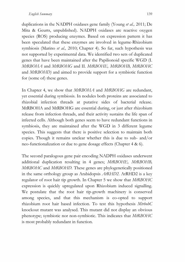
English Summary
139
duplications in the NADPH oxidases gene family (Young et al., 2011; De Mita & Geurts, unpublished). NADPH oxidases are reactive oxygen species (ROS) producing enzymes. Based on expression pattern it has been speculated that these enzymes are involved in legume-Rhizobium symbiosis (Marino et al., 2010; Chapter 4). So far, such hypothesis was not supported by experimental data. We identified two sets of duplicated genes that have been maintained after the Papilionoid specific WGD (I. MtRBOHA and MtRBOHG and II. MtRBOHE, MtRBOHB, MtRBOHC and MtRBOHD) and aimed to provide support for a symbiotic function for (some of) these genes. In Chapter 4, we show that MtRBOHA and MtRBOHG are redundant, yet essential during symbiosis. In nodules both proteins are associated to rhizobial infection threads at putative sides of bacterial release. MtRBOHA and MtRBOHG are essential during, or just after rhizobium release from infection threads, and their activity sustains the life span of infected cells. Although both genes seem to have redundant functions in symbiosis, they are maintained after the WGD in 3 different legume species. This suggests that there is positive selection to maintain both copies. Though it remains unclear whether this is due to sub- and/or neo-functionalization or due to gene dosage effects (Chapter 4 & 6). The second paralogous gene pair encoding NADPH oxidases underwent additional duplication resulting in 4 genes; MtRBOHE, MtRBOHB, MtRBOHC and MtRBOHD. These genes are phylogenetically positioned in the same orthology group as Arabidopsis AtRHD2. AtRHD2 is a key regulator of root hair tip growth. In Chapter 5 we show that MtRBOHC expression is quickly upregulated upon Rhizobium induced signalling. We postulate that the root hair tip-growth machinery is conserved among species, and that this mechanism is co-opted to support rhizobium root hair based infection. To test this hypothesis MtrbohC knockout mutant was analysed. This mutant did not display an obvious phenotype; symbiotic nor non-symbiotic. This indicates that MtRBOHC is most probably redundant in function.

English Summary
140
References Cannon, S.B., Ilut, D., Farmer, A.D., Maki, S.L., May, G.D., Singer, S.R. and
Doyle, J.J., (2010) Polyploidy did not predate the evolution of nodulation in all legumes. PloS ONE, 5(7), e11630.
Fawcett, J.A., Maere, S. and van de Peer, Y., (2009) Plants with double genomes might have had a better chance to survive the Cretaceous-Tertiary extinction event. Proceedings of the National Academy of Sciences of the United States of America, 106(14), 5737–5742.
Gonzalez-Rizzo, S., Crespi, M. and Frugier, F., (2006) The Medicago truncatula CRE1 cytokinin receptor regulates lateral root development and early symbiotic interaction with Sinorhizobium meliloti. The Plant Cell, 18(10), 2680–2693.
Hirsch, A.M., Larue, T.A. and Doyle, J., (1997) Is the legume nodule a modified root or stem or an organ sui generis? Critical Reviews in Plant Sciences, 16(4), 361–392.
Ivanov, S., Fedorova, E.E., Limpens, E., De Mita, S., Genre, A., Bonfante, P. and Bisseling, T., (2012) Rhizobium-legume symbiosis shares an exocytotic pathway required for arbuscule formation. Proceedings of the National Academy of Sciences of the United States of America.
Kouchi, H., Imaizumi-Anraku, H., Hayashi, M., Hakoyama, T., Nakagawa, T., Umehara, Y., Suganuma, N. and Kawaguchi, M., (2010) How many peas in a pod? Legume genes responsible for mutualistic symbioses underground. Plant and Cell Physiology, 51(9), 1381–1381.
Lavin, M., Herendeen, P.S. and Wojciechowski, M.F., (2005) Evolutionary rates analysis of Leguminosae implicates a rapid diversification of lineages during the tertiary. Systematic biology, 54(4), 575–594.
Marino, D., Andrio, E., Danchin, E.G.J., Oger, E., Gucciardo, S., Lambert, A., Puppo, A. and Pauly, N., (2010) A Medicago truncatula NADPH oxidase is involved in symbiotic nodule functioning. New Phytologist, 189(2), 580–592.
Mathesius, U., Weinman, J.J., Rolfe, B.G. and Djordjevic, M.A., (2000) Rhizobia can induce nodules in white clover by “hijacking” mature cortical cells activated during lateral root development. Molecular Plant-Microbe Interactions, 13(2), 170–182.
Nutman, P.S., (1948) Physiological studies on nodule formation. Annals of Botany, 12(2), 81–96.
Oldroyd, G.E., Murray, J.D., Poole, P.S. and Downie, J.A., (2011) The rules of engagement in the legume-rhizobial symbiosis. Genetics, 45(1), 119–144.
Soltis, D.E., Soltis, P.S., Morgan, D.R., Swensen, S.M., Mullin, B.C., Dowd, J.M. and Martin, P.G., (1995) Chloroplast gene sequence data suggest a single origin of the predisposition for symbiotic nitrogen fixation in angiosperms. Proceedings of the National Academy of Sciences of the United States of America, 92(7), 2647–2651.
Soltis, P.S.P., Soltis, D.E.D. and Chase, M.W.M., (1999) Angiosperm phylogeny inferred from multiple genes as a tool for comparative biology. Nature, 402(6760), 402–404.

English Summary
141
Wopereis, J., Pajuelo, E., Dazzo, F.B., Jiang, Q., Gresshoff, P.M., De Bruijn, F.J., Stougaard, J. and Szczyglowski, K., (2000) Short root mutant of Lotus japonicus with a dramatically altered symbiotic phenotype. The Plant Journal, 23(1), 97–114.
Young, N.D., Debellé, F., Oldroyd, G.E.D., et al., (2011) The Medicago genome provides insight into the evolution of rhizobial symbioses. Nature.
Zehr, J.P., Jenkins, B.D., Short, S.M. and Steward, G.F., (2003) Nitrogenase gene diversity and microbial community structure: a cross-system comparison. Environmental microbiology, 5(7), 539–554.

Samenvatting Beschikbaarheid van gebonden stikstof is een belangrijke beperking in de groei van planten. Biologische stikstoffixatie door bacteriën is de meest prominente manier om stikstof in ecosystemen te krijgen. Deze stikstofbindende bacteriën zijn instaat met behulp van het nitrogenase enzymcomplex de drievoudige binding in moleculaire stikstofmoleculen (N2) te breken en N2 om te zetten in ammonium. Een van de prominente stikstofbindende systemen is de rhizobium-wortelknolsymbiose. Deze symbiose leidt tot knolletjes op het wortelsyteem van gastheerplanten. In deze knolletjes worden de rhizobium bacteriën gehuisvest, en vinden zij de juiste condities om stikstof te binden en deze met de waardplant uit te ruilen voor koolhydraten. Het aantal plantensoorten dat een rhizobium-wortelknolsymbiose kan aangaan is beperkt tot twee taxa; de peulvruchtenfamilie (Fabaceae) en het geslacht Parasponia in de Cannabisfamilie (Cannabaceae). Dit vermogen van deze soorten geeft ze de beschikking over een permanente bron van gebonden stikstof. In dit proefschrift heb ik de rhizobium-wortelknolsymbiose in peulvruchten onderzocht, beginnend met de hypothese dat een deel van reeds bestaande signaaltransductie-cascades zijn gerekruteerd gedurende de evolutie van deze interactie. Het is al langer bekend dat delen van een symbiotische signaaltransductie-cascade ook gebruikt worden door symbiotische mycorrhizaschimmels om planten wortels te infecteren. De mycorrhiza-symbiose is wijd verspreid en komt voor bij veel plantensoorten. Ook is deze symbiose veel ouder dan rhizobium-wortelknolsymbiose. Daarom wordt aangenomen dat rhizobium delen van de mycorrhiza-symbiose signaaltransductiecascade heeft gebruikt om vlinderbloemige plantensoorten (en Parasponia) te infecteren. In lijn hiermee wordt verondersteld dat ook andere signaaltransductiecascades, of ontwikkelingsprogramma’s, zijn gerekruteerd tijdens evolutie van de wortelknolsymbiose. Bijvoorbeeld, een voor de hand liggende vraag is of wortelknolletjes zijn voortgekomen uit het ontwikkelingsprogramma van
Samenvatting 144
zijwortels. In hoofdstuk 2 is daarom het zijwortel-ontwikkelingsprogramma van de vlinderbloemige modelplanten Medicago truncatula (Medicago) en Lotus japonicus (Lotus) bestudeerd. In het algemeen wordt aangenomen dat zijwortels zich uitsluitend ontwikkelen uit pericykelcellen, terwijl wortelknolletjes voortkomen uit de cortexcellen. Ons onderzoek laat echter zien dat dit in Lotus en Medicago niet zo strikt is. In beide soorten dragen ook corticale celdelingen bij aan zijwortelontwikkeling. Hierbij is een opvallende correlatie gevonden tussen de corticale cellen die delen gedurende zijwortelontwikkeling en de corticale cellen die het wortelknolletje kunnen vormen. Dit ondersteunt de hypothese dat tenminste een deel van de zijwortelontwikkelings-programma is gebruikt voor de vorming van wortelknolletjes. Gen-duplicaties, met een genoomduplicatie als meest ultieme vorm, zijn een belangrijke drijvende kracht in de evolutie van nieuwe eigenschappen. Ergens 56-65 miljoen jaar geleden is er een genoomduplicatie opgetreden in een voorouderlijke peulvruchtensoort die aan de basis heeft gestaan van de vlinderbloemigen-subfamilie (Papilionoidae). De 3 vlinderbloemige modelsoorten waarvoor genoom data beschikbaar zijn, Medicago, Lotus en soja (Glycine max), vertegenwoordigen 3 belangrijke groepen binnen deze subfamilie welke 55 miljoen jaar geleden zijn gedivergeerd. Na een genoomduplicatie gaan veel genen weer verloren, omdat er een tweede kopie als backup aanwezig is. Alleen als één of beide genen een nieuwe, of een meer specifieke functie verwerven, dan zullen beide genen behouden blijven. Wij veronderstellen dat de genoomduplicatie heeft bijgedragen aan de evolutie van de Rhizobium-wortelknolsymbiose in de vlinderbloemigen-subfamilie. Daarbij stel ik de hypothese dat orthologe genenparen die gehandhaafd zijn in Medicago, Lotus en soja, een belangrijke rol hebben gespeeld in de evolutie van vlinderbloemige-specifieke eigenschappen, zoals de rhizobium-wortelknolsymbiose. Zulke genen zijn nog niet geïdentificeerd in genetische screens, waarschijnlijk door redundantie-effecten. Daarom is er een fylogenetische strategie gebruikt om nieuwe kandidaat-genen, betrokken in rhizobium-wortelknolsymbiose, te identificeren (hoofdstuk 3, 4 & 5).

Samenvatting 145
In een gerichte aanpak is de cytokinine phosphorelay signalleringscascade bestudeerd op de aanwezigheid van gen-duplicaties. Cytokinine is betrokken bij wortelknolvorming. Dit onderzoek resulteerde in de identificatie van een genenpaar dat codeert voor type-A Response Regulatoren (RRs). Beide genen, genaamd MtRR9 en MtRR11 in Medicago, worden geactiveerd door Rhizobium geinduceerde signalering, terwijl dat niet het geval is voor de overige type-A RR-genen. Constitutieve expressie van MtRR9/MtRR11 leidt tot corticale celdelingen, hetgeen duidt op een positieve regulerende rol van deze eiwitten in wortelknolvorming. Deze vinding onderstreept de bruikbaarheid van een fylogenetische strategie om nieuwe symbiotische genen te identificeren (hoofdstuk 3). In een genoombrede aanpak zijn twee geconserveerde gen duplicaties gevonden in de familie van NADPH oxidase genen. NADPH oxidasen produceren reactieve zuurstofradicalen (reactive oxygen species; ROS). Het expressie patroon van de gedupliceerde genen suggereert dat ze een rol vervullen in de Rhizobium wortelknolsymbiose. De toetsing van deze hypothese is beschreven in Hoofdstuk 4 & 5. In hoofdstuk 4 laten we zien dat MtRBOHA en MtRBOHG, hoewel redundant in functie, essentieel zijn voor de wortelknolsymbiose. In wortelknolletjes zijn beide eiwitten geassocieerd zijn met rhizobiuminfectie en nodig tijdens, of direct na het vrijkomen van Rhizobium uit de infectiedraden. Het uitschakelen van beide NADH oxidasen ondermijnt de fitness van geïnfecteerde wortelknolcellen. Hoewel beide genen redundant in functie zijn, zijn ze toch gedurende de evolutie als genenpaar behouden. Dit duidt op een selectiedruk op behoud van beide genen. Echter het onderliggende mechanisme hiervoor is nog niet opgehelderd (hoofdstuk 4 en 6). Het tweede paraloge genenpaar dat codeert voor NADPH oxidasen heeft additionele duplicaties ondergaan dat in Medicago uiteindelijk heeft geresulteerd in 4 genen; MtRBOHE, MtRBOHB, MtRBOHC en

Samenvatting 146
MtRBOHD. Deze genen behoren tot dezelfde orthologie-groep als het Arabidopsis AtRHD2 gen. AtRHD2 is een belangrijke regulator van tipgroei in wortelharen. In hoofdstuk 5 laten we zien dat MtRBOHC expressie wordt geïnduceerd door Rhizobium. We veronderstellen dat het mechanisme van wortelhaargroei is geconserveerd is hogere planten en dat Rhizobium gebruik maakt van dit mechanisme om wortelharen te infecteren. Om deze hypothese te testen is er een MtrbohC knock-out mutant geanalyseerd. Deze mutant heeft echter geen duidelijk afwijkend fenotype. Dit geeft aan dat de functie van MtRBOHC redundant is.

Acknowledgments I would like to acknowledge the Marie Curie PhD fellowship program “NODPERCEPTION network” for supporting my PhD research project and all the others research projects too: 7 more PhDs and 2 Post-Docs all around Europe. Especially thank to Dr. Julie Cullimore (INRA, Toulouse, France) for leading the network and caring for the scientific and personal development of each of the researchers involved. I am very thankful to our collaborators Prof. Dr. Allan Downie and Dr. Giles Oldroyd (John Innes Center, England). I am especially indebted with Prof. Dr. Allan Downie to have accepted me under his mentorship, and to have guided me with his truly inspiring scientific approach through new concepts and ideas. Thanks to Giulia Morieri (John Innes Center, England), Anna Pietraszewska (Amsterdam University, the Netherlands), and Malick Mbengue (INRA, Toulouse, France) to have sparkled the network meetings with both science discussions and comradeship. I express my gratitude to Eduardo Andres Martinez (Grenoble, France) for providing the Nod factors used in this Thesis (and not only in this Thesis).
Working at Wageningen University was a wonderful experience. I would like to thank my promoter, Prof. Dr. Ton Bisseling, and my co-supervisor, Dr. Ir. René Geurts to have offered me the PhD student position at the Molecular Biology Laboratory. Thanks for the scientific discussions, the revisions and comments on this Thesis. I am grateful to Dr. Stéphane De Mita for his phylogenetic studies that deeply contributed to this work, for his support, and for his patience. I am thankful to Dr. Sergey Ivanov for his prodigality in long scientific discussions and technical support, and, above all, for his friendship. I consider myself very lucky to have shared teaching duties with Jan Verver. Last but not least, a special thank to Dr. Olga Kulikova for her support. I would like to thank all the people at the Laboratory of Molecular Biology, every single one of them.
Acknowledgments 148
The help of Ir. Chris Bosch (Biosystematics Group, Wageningen University), Prof. Dr. Hans de Vries, and Dr. Giulia Pagliarani (Plant Research International, Wageningen University) was essential to find the botanical illustration of Medicago truncatula for the cover of this Thesis. I would like to thank Cristina Llavata Peris (Biochemistry, Wageningen) as well for her valuable contribution to the cover. The kind help of my father during the layout and pre-printing operations was greatly appreciated. Dr. Stefania Montersino (Biochemistry, Wageningen) and Dr. Sergey Ivanov honored me for being my paranymphs. Many people (of many nationalities) contributed to make my life in Wageningen wonderful. Above all: Stefania Montersino, Sergey Ivanov, Christoph Bücherl, Cristina Llavata Peris, Giulia Pagliarani, Norma Fàbregas, Chiara Guarnerio and Sara Martinelli.
149
Curriculum Vitae Alessandra Lillo was born on July 6th, 1980, in Arzignano (Vi), Italy, few minutes after her twin brother, Galileo. After finishing the high school (gymnasium) at “Liceo-ginnasio Tito Livio” in Milan in 1999, she started her study in Plant Biotechnology at Verona University. In 2004 Alessandra performed her first research project as exchange student (Erasmus fellowship) at the Laboratory of Molecular Biology, Wageningen University, under the supervision of Dr. Henk Franssen and Prof. Dr. Ton Bisseling. At the same time Alessandra, working in team with Chiara Guarnerio, contributed to unravel the role of ENOD40 in nodule development. For her MSc Thesis research project in 2005, Alessandra joined the Laboratory of Cell Biology, Verona University, and performed functional studies of ENOD40 in BY-2 tobacco cells. Soon after obtaining the MSc degree in Plant Biotechnology in 2006, Alessandra joined again the Laboratory of Molecular Biology, this time as PhD student under the supervision of Dr. Ir. René Geurts and Prof. Dr. Ton Bisseling. The first 3 years of her PhD project were supported by the Marie Curie PhD fellowship via the NODPERCEPTION network, where Alessandra was under the mentorship of Prof. Dr. Allan Downie, John Innes Center, Norwich. Her research on the co-option of pre-existing pathways during rhizobium-legume symbiosis evolution resulted in the publication of this Thesis in 2012.

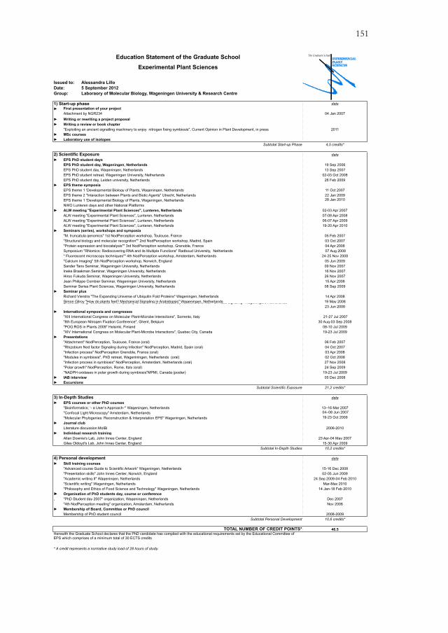
151
Alessandra Lillo5 September 2012Laboraory of Molecular Biology, Wageningen University & Research Centre
date!
04 Jan 2007! !
2011! !
4,5 credits*
date!
19 Sep 200613 Sep 2007
02-03 Oct 200826 Feb 2009
! 11 Oct 200722 Jan 200928 Jan 2010
! 02-03 Apr 200707-08 Apr 200806-07 Apr 200919-20 Apr 2010
! 05 Feb 200703 Oct 200704 Apr 2008 07 Aug 2008
24-25 Nov 200805 Jun 200909 Nov 2007 16 Nov 2007 26 Nov 200715 Apr 200808 Sep 2009
! 14 Apr 200819 May 200823 Jun 2008
! 21-27 Jul 2007
30 Aug-03 Sep 200808-10 Jul 200919-23 Jul 2009
! 06 Feb 200704 Oct 200703 Apr 200802 Oct 200827 Nov 200824 Sep 2009
19-23 Jul 2009! 05 Dec 2008!
21,2 credits*
date!
13–16 Mar 2007 04–08 Jun 2007 19-23 Oct 2009
! 2006-2010
! 23 Apr-04 May 2007
15-30 Apr 200910,2 credits*
date!
15-16 Dec 200802-05 Jun 2009
24 Sep 2009-04 Feb 2010Mar-May 2010
14 Jan-18 Feb 2010! , Dec 2007
Nov 2008!
2008-200910,6 credits*
46.5
"Advanced course Guide to Scientific Artwork" Wageningen, Netherlands
"Bioinformatics; ‘- a User’s Approach-‘" Wageningen, Netherlands
"Rhizobium Nod factor Signaling during Infection" NodPerception, Madrid, Spain (oral)"Attachment" NodPerception, Toulouse, France (oral)
"Polar growth" NodPerception, Rome, Italy (oral)
Excursions
Allan Downie's Lab, John Innes Center, England
"XIV International Congress on Molecular Plant-Microbe Interactions", Quebec City, Canada
Journal club
Giles Oldoyd's Lab, John Innes Center, England
Simon Gilroy "How do plants feel? Mechanical Signaling in Arabidopsis" Wageningen, Netherlands
IAB interview
"Modules in symbiosis", PhD retreat, Wageniningen, Netherlands (oral)"Infection process in symbiosis" NodPerception, Amsterdam, Netherlands (oral)
"NADPH oxidases in polar growth during symbiosis"MPMI, Canada (poster)
"Molecular Phylogenies: Reconstruction & Interpretation EPS" Wageningen, Netherlands
Education Statement of the Graduate School
Experimental Plant Sciences
First presentation of your project
Subtotal Start-up Phase
1) Start-up phase
Attachment by NGR234
Writing a review or book chapter"Exploiting an ancient signalling machinery to enjoy nitrogen fixing symbiosis", Current Opinion in Plant Development, in press
Writing or rewriting a project proposal
Issued to:
MSc courses
Date:Group:
Laboratory use of isotopes
EPS theme symposia
NWO Lunteren days and other National Platforms
"Protein expression and biocatalysis"" 3rd NodPerception workshop, Grenoble, France
ALW meeting "Experimental Plant Sciences", Lunteren, Netherlands
EPS theme 1 'Developmental Biology of Plants, Wageningen, Netherlands
EPS theme 1 'Developmental Biology of Plants, Wageningen, NetherlandsEPS theme 2 "Interaction between Plants and Biotic Agents" Utrecht, Netherlands
ALW meeting "Experimental Plant Sciences", Lunteren, Netherlands
"M. truncatula genomics" 1st NodPerception workshop, Toulouse, France "Structural biology and molecular recognition"" 2nd NodPerception workshop, Madrid, Spain
ALW meeting "Experimental Plant Sciences", Lunteren, NetherlandsALW meeting "Experimental Plant Sciences", Lunteren, NetherlandsSeminars (series), workshops and symposia
2) Scientific Exposure
EPS PhD student day, Wageningen, NetherlandsEPS PhD student retreat, Wageningen University, NetherlandsEPS PhD student day, Leiden university, Netherlands
EPS PhD student day, Wageningen, NetherlandsEPS PhD student days
Subtotal Personal Development
EPS courses or other PhD courses
"PhD Student day 2007" organization, Wageningen, Netherlands
4) Personal development
Membership of PhD student councilMembership of Board, Committee or PhD council
Organisation of PhD students day, course or conference
"4th NodPerception meeting" organization, Amsterdam, Netherlands
"Philosophy and Ethics of Food Science and Technology" Wageningen, Netherlands"Scientific writing" Wageningen, Netherlands
"Confocal Light Microscopy" Amsterdam, Netherlands
Skill training courses
"Academic writing II" Wageningen, Netherlands
Subtotal In-Depth Studies
Literature discussion MolBi
* A credit represents a normative study load of 28 hours of study.
TOTAL NUMBER OF CREDIT POINTS*Herewith the Graduate School declares that the PhD candidate has complied with the educational requirements set by the Educational Committee of EPS which comprises of a minimum total of 30 ECTS credits
Ineke Braakman Seminar, Wageningen University, Netherlands
Symposium "RNomics: Rediscovering RNA and its Multiple Functions" Radboud University, Netherlands
Hiroo Fukuda Seminar, Wageningen University, Netherlands
" Fluorescent microscopy techniques"" 4th NodPerception workshop, Amsterdam, Netherlands"Calcium imaging" 5th NodPerception workshop, Norwich, England Sander Tans Seminar, Wageningen University, Netherlands
Jean Philippe Combier Seminar, Wageningen University, NetherlandsSeminar Series Plant Sciences, Wageningen University, NetherlandsSeminar plus
"Presentation skills" John Innes Center, Norwich, England
Richard Vierstra "The Expanding Universe of Ubiquitin Fold Proteins" Wageningen, Netherlands
Presentations
3) In-Depth Studies
"POG:ROS in Plants 2009" Helsinki, Finland
"Infection process" NodPerception Grenoble, France (oral)
International symposia and congresses"XIII International Congress on Molecular Plant-Microbe Interactions", Sorrento, Italy "8th European Nitrogen Fixation Conference", Ghent, Belgium
Subtotal Scientific Exposure
Zhenbiao Yang "Coordination of cell polarity formation in plant tissues by hormone and ROP GTPase signaling" Wageningen, Netherlands
Individual research training
152
Financial support from Wageningen University for printing this Thesis is gratefully acknowledged.
On the Cover: Medicago truncatula Gaertn.,
drawn by Dulic Amsler, in Michael Zohary, Flora Palaestina, Part II: Plates, no. 216. Jerusalem: The Israel Academy of Sciences and Humanities, 1987.
© The Israel Academy of Sciences and Humanities. Reproduced by permission.
Pre-printing: Fotolito Galli Thierry, Milan, Italy
Printed and bound by: Global Print, Gorgonzola (Mi), Italy


![Comparison between the reference [i]Rhizobium … · Common bean plants were inoculated with rhizobia strains: Rhizobium tropici CIAT899 or Rhizobium etli 12a3, ... +216 94120093](https://img.pdfslide.us/doc/110x75/5b915a7d09d3f26a278b6ea5/comparison-between-the-reference-irhizobium-common-bean-plants-were-inoculated.jpg)