Embed Size (px)
Citation preview
CO2 Acts as a Signalling Molecule in Populations of theFungal Pathogen Candida albicansRebecca A. Hall1,2, Luisa De Sordi1, Donna M. MacCallum2, Husnu Topal3, Rebecca Eaton1, James W.
Bloor1, Gary K. Robinson1, Lonny R. Levin4, Jochen Buck4, Yue Wang5, Neil A. R. Gow2, Clemens
Steegborn3,6, Fritz A. Muhlschlegel1,7*
1 School of Biosciences, University of Kent, Canterbury, Kent, United Kingdom, 2 School of Medical Sciences, Institute of Medical Sciences, University of Aberdeen,
Aberdeen, United Kingdom, 3 Department of Physiological Chemistry, Ruhr-University Bochum, Bochum, Germany, 4 Department of Pharmacology, Weill Medical College
of Cornell University, New York, New York, United States of America, 5 Institute of Molecular and Cell Biology, Agency for Science, Technology and Research, Proteos,
Singapore, 6 Department of Biochemistry, University of Bayreuth, Bayreuth, Germany, 7 East Kent Hospitals University NHS Foundation Trust, Clinical Microbiology Service,
William Harvey Hospital, Ashford, Kent, United Kingdom
Abstract
When colonising host-niches or non-animated medical devices, individual cells of the fungal pathogen Candida albicansexpand into significant biomasses. Here we show that within such biomasses, fungal metabolically generated CO2 acts as acommunication molecule promoting the switch from yeast to filamentous growth essential for C. albicans pathology. Wefind that CO2-mediated intra-colony signalling involves the adenylyl cyclase protein (Cyr1p), a multi-sensor recently found tocoordinate fungal responses to serum and bacterial peptidoglycan. We further identify Lys 1373 as essential for CO2/bicarbonate regulation of Cyr1p. Disruption of the CO2/bicarbonate receptor-site interferes selectively with C. albicansfilamentation within fungal biomasses. Comparisons between the Drosophila melanogaster infection model and the mousemodel of disseminated candidiasis, suggest that metabolic CO2 sensing may be important for initial colonisation andepithelial invasion. Our results reveal the existence of a gaseous Candida signalling pathway and its molecular mechanismand provide insights into an evolutionary conserved CO2-signalling system.
Citation: Hall RA, De Sordi L, MacCallum DM, Topal H, Eaton R, et al. (2010) CO2 Acts as a Signalling Molecule in Populations of the Fungal Pathogen Candidaalbicans. PLoS Pathog 6(11): e1001193. doi:10.1371/journal.ppat.1001193
Editor: Aaron P. Mitchell, Carnegie Mellon University, United States of America
Received February 25, 2010; Accepted October 13, 2010; Published November 18, 2010
Copyright: � 2010 Hall et al. This is an open-access article distributed under the terms of the Creative Commons Attribution License, which permits unrestricteduse, distribution, and reproduction in any medium, provided the original author and source are credited.
Funding: This work was funded by the MRC, BBSRC and Nuffield Foundation (all to FAM). Work in the CS laboratory was funded by DFG priority program 1160(grant STE1701/2 to CS). The funders had no role in study design, data collection and analysis, decision to publish, or preparation of the manuscript.
Competing Interests: The authors have declared that no competing interests exist.
* E-mail: [email protected]
Introduction
Candida albicans is the predominant fungal pathogen of humans.
In healthy individuals C. albicans resides as a commensal of the
gastrointestinal, oral and vaginal tracts. C. albicans can cause
superficial infections which, although not life threatening, provide
discomfort to the individual and require treatment with antifungals
which is a constant drain on hospitals resources. However, C.
albicans infections are life threatening when the individual’s
immune system becomes compromised as a result of age, cancer,
chemotherapy hospitalisation and AIDS. Under these circum-
stances superficial infections may readily develop into systemic
disease where mortality rates are reported to be up to 40%, which
is higher than those for most bacterial infections [1,2,3]. For
example, oropharyngeal candidiasis is common in patients with
haematological malignancies (up to 60%) and those undergoing
radiotherapy [4,5,6]. Here, a few fungal cells develop into
biomasses measuring several millimetres in diameter that pene-
trate and invade the underlying tissue, eventually leading to
dissemination of Candida into the blood stream and subsequently
systemic infection [7].
Development from superficial infection to invasive disease is
mediated by many well characterised virulence factors including
morphological transition. C. albicans can exist in yeast, pseudohy-
phal and true hyphal growth forms, all of which are important for
the virulence of the organism [8]. Yeast cells are thought to be
essential for growth and dissemination [9], while the hyphal forms
are essential for invading mucosal membranes [9]. This morpho-
logical transition is mediated by host environmental cues including
temperature, pH, serum, O2, and CO2, which the pathogen
encounters during disease progression [5,10,11].
The virulence-associated morphological transitions of C. albicans
are largely controlled through the secondary messenger cAMP. In
C. albicans, cAMP is synthesised by the fungal adenylyl cyclase (AC),
Cyr1p [12], a member of the Class III nucleotidyl cyclase family
[13]. Activity of Cyr1p governs most processes essential to C. albicans
virulence including tissue adhesion followed by the invasion of the
underlying host-barriers, and biofilm formation [14]. C. albicans AC
activity is subject to both positive and negative regulation, with an
increasing number of molecules directly interacting with specific
domains of the protein [10,15,16]. For example, bacterial
peptidoglycan stimulates Cyr1p via the enzyme’s leucine-rich-
region [16], and CO2/HCO32 directly activates the Cyr1p C-
terminal catalytic domain [10]. These forms of regulation enable C.
albicans to recognise and respond (via filamentation) to specific host
environmental conditions during disease progression.
PLoS Pathogens | www.plospathogens.org 1 November 2010 | Volume 6 | Issue 11 | e1001193
In addition to host environmental cues, the morphological
transition of C. albicans is also regulated by soluble chemical
mediators, termed quorum sensing molecules (QSMs). QSMs are
secreted into the environment by a variety of microorganisms [for
recent reviews see 17,18], and upon reaching threshold
concentrations, impact on microbial behaviour by influencing
expression of virulence determinants [19]. QSMs including the
self-generated sesquiterpene farnesol [20] and 3-oxo-C12
homoserine lactone (HSL) secreted by Pseudomonas aeruginosa
[21] inhibit C. albicans filamentation though cAMP dependent
signalling cascades [22].
Further to soluble chemical mediators, volatile compounds can
also act as signalling molecules. For example, in Saccharomyces
cerevisiae, nutrient limited yeast cells release volatile ammonia,
which when sensed by another colony inhibits its growth in the
direction of the signal [23]. CO2 is a volatile gas that has recently
been described as a predominant regulator of C. albicans virulence
factors and has been shown to effect the virulence of other
microbial species [24,25]. In C. albicans CO2 functions in two
processes key to pathogenicity, one metabolic and the other cell
signalling to promote filamentation [10]. In biological systems
CO2 is maintained in equilibrium with its hydrated form, HCO32,
via the actions of carbonic anhydrase. HCO32 is required for
metabolism, but when at high concentrations HCO32 directly
activates adenylyl cyclase increasing cytosolic cAMP and promot-
ing filamentation [10]. To date, only the effects of high (5%)
exogenous CO2 concentrations have been investigated in
microbial species. However, microbes continuously secrete
metabolically generated CO2 into their immediate microenviron-
ment at levels perceived to be lower than 5%. Here, we investigate
the effects of self generated CO2 on pathogenicity associated traits
of C. albicans. Previously we identified the carbonic anhydrase,
Nce103p, as being essential for growth under CO2 limiting
conditions [10]. Now we explore a new application of the mutant
strain Dnce103 as a CO2 biosensor to report on CO2 concentra-
tions within fungal biomasses. Using our CO2-dependent bio-
sensing strain, we demonstrate that build-up of self-generated,
metabolic CO2 occurs in a fungal population. Furthermore, we
show that CO2 mediates its effects as a hierarchy, with low
concentrations of CO2 functioning to fill metabolic demand, then
once CO2 exceeds a critical threshold, it promotes filamentation
and subsequent surface invasion of the pathogen. We show that
microbial CO2, like environmental CO2, is sensed by the AC
catalytic domain and identify a bicarbonate receptor site in Cyr1p.
Results
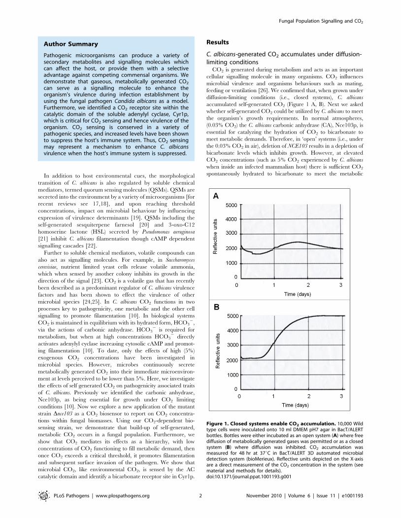
C. albicans-generated CO2 accumulates under diffusion-limiting conditions
CO2 is generated during metabolism and acts as an important
cellular signalling molecule in many organisms. CO2 influences
microbial virulence and organisms behaviours such as mating,
feeding or ventilation [26]. We confirmed that, when grown under
diffusion-limiting conditions (i.e., closed systems), C. albicans
accumulated self-generated CO2 (Figure 1 A, B). Next we asked
whether self-generated CO2 could be utilized by C. albicans to meet
the organism’s growth requirements. In normal atmospheres,
(0.03% CO2) the C. albicans carbonic anhydrase (CA), Nce103p, is
essential for catalyzing the hydration of CO2 to bicarbonate to
meet metabolic demands. Therefore, in ‘open’ systems (i.e., under
the 0.03% CO2 in air), deletion of NCE103 results in a depletion of
bicarbonate levels which inhibits growth. However, at elevated
CO2 concentrations (such as 5% CO2 experienced by C. albicans
when inside an infected mammalian host) there is sufficient CO2
spontaneously hydrated to bicarbonate to meet the metabolic
Author Summary
Pathogenic microorganisms can produce a variety ofsecondary metabolites and signalling molecules whichcan affect the host, or provide them with a selectiveadvantage against competing commensal organisms. Wedemonstrate that gaseous, metabolically generated CO2
can serve as a signalling molecule to enhance theorganism’s virulence during infection establishment byusing the fungal pathogen Candida albicans as a model.Furthermore, we identified a CO2 receptor site within thecatalytic domain of the soluble adenylyl cyclase, Cyr1p,which is critical for CO2 sensing and hence virulence of theorganism. CO2 sensing is conserved in a variety ofpathogenic species, and increased levels have been shownto suppress the host’s immune system. Thus, CO2 sensingmay represent a mechanism to enhance C. albicansvirulence when the host’s immune system is suppressed.
Figure 1. Closed systems enable CO2 accumulation. 10,000 Wildtype cells were inoculated onto 10 ml DMEM pH7 agar in BacT/ALERTbottles. Bottles were either incubated as an open system (A) where freediffusion of metabolically generated gases was permitted or as a closedsystem (B) where diffusion was inhibited. CO2 accumulation wasmeasured for 48 hr at 37uC in BacT/ALERT 3D automated microbialdetection system (bioMerieux). Reflective units depicted on the X-axisare a direct measurement of the CO2 concentration in the system (seematerial and methods for details).doi:10.1371/journal.ppat.1001193.g001
Fungal Population Signalling and CO2
PLoS Pathogens | www.plospathogens.org 2 November 2010 | Volume 6 | Issue 11 | e1001193
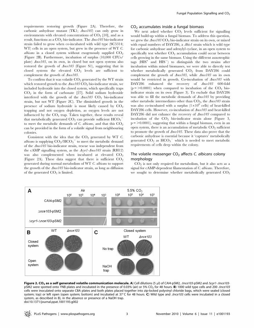
requirements restoring growth (Figure 2A). Therefore, the
carbonic anhydrase mutant (TK1; Dnce103) can only grow in
environments with elevated concentrations of CO2 [10], and as a
result, functions as a CO2 bio-indicator. The Dnce103 bio-indicator
strain failed to grow when co-incubated with wild type (SC5314;
WT) cells in an open system, but grew in the presence of WT C.
albicans in a closed system without exogenously supplied CO2
(Figure 2B). Furthermore, incubation of surplus (10,000 CFUs/
plate) Dnce103, on its own, in closed but not open systems also
restored the growth of Dnce103 (Figure S1), suggesting that in
closed systems the elevated CO2 levels are sufficient to
complement the growth of Dnce103.
To confirm that it was volatile CO2 generated by the WT strain
which restored growth to the Dnce103 CO2 bio-indicator strain, we
included hydroxide into the closed system, which specifically traps
CO2 in the form of carbonate [27]. Solid sodium hydroxide
interfered with the growth of the Dnce103 CO2 bio-indicator
strain, but not WT (Figure 2C). The diminished growth in the
presence of sodium hydroxide is most likely caused by CO2
trapping and not oxygen depletion, as oxygen levels are not
influenced by the CO2 trap. Taken together, these results reveal
that metabolically generated CO2 can provide sufficient HCO32
to meet the metabolic demands of C. albicans, and that this CO2
can be provided in the form of a volatile signal from neighbouring
colonies.
Consistent with the idea that the CO2 generated by WT C.
albicans is supplying CO2/HCO32 to meet the metabolic demand
of the Dnce103 bio-indicator strain, rescue was independent from
the cAMP signalling system, as the Dcyr1-Dnce103 strain (RH12)
was also complemented when incubated at elevated CO2
(Figure 2A). These data suggest that there is sufficient CO2
generated during normal metabolism of WT C. albicans to support
the growth of the Dnce103 bio-indicator strain, as long as diffusion
of the generated CO2 is limited.
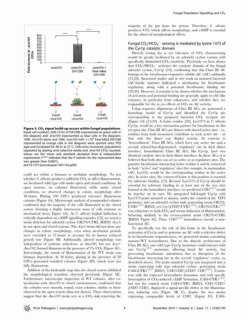
CO2 accumulates inside a fungal biomassWe next asked whether CO2 levels sufficient for signalling
would build-up within a fungal biomass. To address this question,
we grew the Dnce103 CO2 bio-indicator strain on its own, or mixed
with equal numbers of DAY286, a Dhis1 strain which is wild type
for carbonic anhydrase and adenylyl cyclase, in an open system to
specifically test whether CO2 accumulation could occur between
cells growing in the same biomass. Using the different auxotrophic
tags (HIS+ and HIS2) to distinguish the two strains after
incubation within mixed biomasses, we were able to directly test
whether metabolically generated CO2 from DAY286 could
complement the growth of Dnce103, while Dnce103 on its own
would be restricted in growth. Co-incubation of Dnce103 with
DAY286 enhanced the recovery of Dnce103 600-fold
(p = .0.0001) when compared to incubation of the CO2 bio-
indicator strain on its own (Figure 3). To exclude that DAY286
was able to fill the metabolic demands of Dnce103 by providing
other metabolic intermediates other than CO2, the Dnce103 strain
was also co-incubated with a surplus (16106 cells) of heat-killed
DAY286 cells. However, co-incubation of Dnce103 and heat-killed
DAY286 did not enhance the recovery of Dnce103 compared to
incubation of the CO2 bio-indicator strain alone (Figure 3,
p = .0.0001), suggesting that within a fungal biomass, even in an
open system, there is an accumulation of metabolic CO2 sufficient
to promote the growth of Dnce103. These data also prove that the
carbonic anhydrase is essential because it ‘captures’ metabolically
generated CO2 as HCO32 which is needed to meet metabolic
requirements of cells deep within the colony.
The volatile messenger CO2 affects C. albicans colonymorphology
CO2 is not only required for metabolism, but it also acts as a
signal for cAMP-dependent filamentation of C. albicans. Therefore,
we sought to determine whether metabolically generated CO2
Figure 2. CO2 as a self generated volatile communication molecule. A) Cell dilutions (5 ml) of CAI4-pSM2, Dnce103-pSM2 and Dcyr1-Dnce103-pSM2 were spotted onto YNB plates and incubated in the presence of 0.03% (air) or 5% CO2 for 48 hours. B) 1000 wild type cells and 200 Dnce103cells were inoculated onto separate CBA plates and both plates placed together into zip-locked polyvinyl chloride bags, which were sealed (closedsystem; top) or left open (open system; bottom) and incubated at 37uC for 48 hours. C) Wild type and Dnce103 cells were incubated in a closedsystem, as described in B), in the absence or presence of a NaOH trap.doi:10.1371/journal.ppat.1001193.g002
Fungal Population Signalling and CO2
PLoS Pathogens | www.plospathogens.org 3 November 2010 | Volume 6 | Issue 11 | e1001193
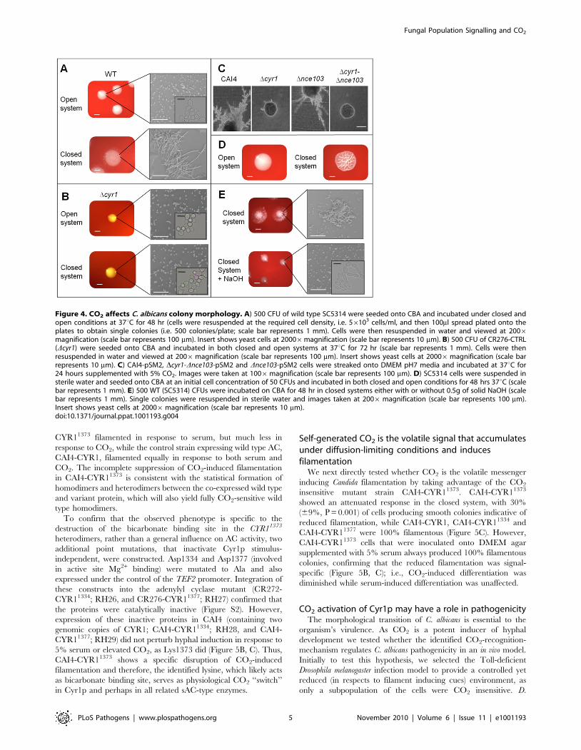
could act within a biomass to modulate morphology. To test
whether C. albicans produces sufficient CO2 to affect filamentation,
we incubated wild type cells under open and closed conditions. In
open systems, no colonies filamented, while under closed
conditions, we observed changes in colony morphology after
48 hours. Plating 500 CFUs produced extensive filamentous
colonies (Figure 4A). Microscopic analysis of resuspended colonies
confirmed that the majority of the cells filamented in the closed
system (forming a highly interwoven mass of cells resistant to
mechanical stress; Figure 4A). As C. albicans hyphal induction is
critically dependent on cAMP signalling cascades [12], we tested a
strain deficient for adenylyl cyclase CR276-CTRL (RH20; Dcyr1)
in our open and closed systems. The Dcyr1 strain did not show any
changes in colony morphology, even when incubation periods
were extended to 72 hours to account for its known reduced
growth rate (Figure 4B). Additionally, altered morphology was
independent of carbonic anhydrase, as Dnce103, but not Dcyr1-
Dnce103, formed filaments in the presence of 5% CO2 (Figure 4C).
Interestingly, the extent of filamentation of the WT strain was
biomass dependent. At 48 hours, plating in the presence of 50
CFUs generated wrinkled colonies (Figure 4D), which were not
fully filamentous.
Addition of the hydroxide trap into the closed system inhibited
the morphological transition observed previously (Figure 4E).
Furthermore, microscopic inspection of WT colonies, after co-
incubation with Dnce103 in closed environments, confirmed that
the colonies were smooth, round, yeast colonies, similar to those
observed in the open system (data not shown). These observations
suggest that the Dnce103 strain acts as a CO2 sink removing the
majority of the gas from the system. Therefore, C. albicans
produces CO2 which affects morphology, and cAMP is essential
for the observed morphological effects.
Fungal CO2/HCO32 sensing is mediated by lysine 1373 of
the Cyr1p catalytic domainDirectly testing the in vivo relevance of CO2 chemosensing
would be greatly facilitated by an adenylyl cyclase variant with
specifically diminished CO2 sensitivity. Previously we have shown
that CO2/HCO32 activates the catalytic domain of the fungal
adenylyl cyclase, Cyr1p [10], confirming that this Class III AC
belongs to the bicarbonate-responsive soluble AC (sAC) subfamily
[13,28]. Structural studies and in vitro work on mutated bacterial
sAC-family enzymes indicated a mechanism for bicarbonate
regulation, along with a potential bicarbonate binding site
[29,30]. However, it remains to be shown whether the mechanism
of activation and potential binding site generally apply to sAC-like
enzymes, in particular from eukaryotes, and whether they are
responsible for the in vivo effects of CO2 on AC activity.
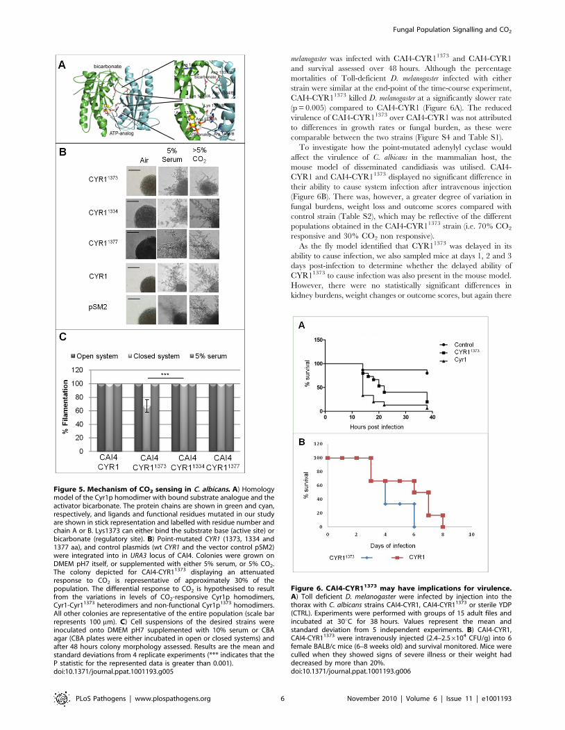
Using sequence alignments of Class III ACs, we generated a
homology model of Cyr1p and identified the Cyr1p site
corresponding to the proposed bacterial CO2 receptor site
(Figure 5A) [13,29]. A lysine residue [29], Lys1373 in C. albicans
Cyr1p, would be a key interaction partner for bicarbonate in this
receptor site. Class III ACs are dimers with shared active sites – i.e.
residues from both monomers contribute to each active site – so
that only the dimer can display activity. In contrast to
‘heterodimeric’ Class III ACs, which have one active site and a
second, related-but-degenerated, ‘regulatory’ site in their dimer
interface, homodimeric Class III ACs, like Cyr1p, have two
identical catalytic sites in their dimer interface. In these ACs, it is
believed that both sites can act as active or as regulatory sites. The
putative bicarbonate-interacting lysine residue is strictly conserved
in both ‘‘active’ and ‘regulatory’ sites (for example, in mammalian
sAC, Lys334, would be the corresponding residue in the active
site). In active sites, the conserved lysine at this position is essential
for substrate binding [13]. Because Lys1373 of Cyr1p should be
essential for substrate binding in at least one of the two sites
formed at the homodimer interface, we predicted CYR11373 would
be inactive on its own. We integrated full-length Cyr1p with
Lys1373 point mutated to alanine, under the control of the TEF2
promoter, into an adenylyl cyclase null, generating strain CR276-
CYR11373 (RH22; cyr1/cyr1:pTEF2 CYR11373). CR276-CYR11373
was refractory to both CO2 and serum induction of filamentation,
behaving similarly to the vector-control strain CR276-CTRL
(RH20; Figure S2). Thus, CYR11373 homodimers encode a non-
functional AC.
To specifically test the role of this lysine in the bicarbonate
activation of Cyr1p and to generate an AC with a selective defect
in its bicarbonate responsiveness, we generated strains containing
mutant/WT heterodimers. Due to the dimeric architecture of
Class III ACs, one wild type Cyr1p monomer could interact with
one Cyr1p1373 monomer, allowing basal AC activity, but
preventing bicarbonate stimulation due to disruption of the
bicarbonate interacting site in the second ‘regulatory’ centre (as
described above). The point mutated Cyr1p was integrated into a
strain expressing wild type adenylyl cyclase, generating strain
CAI4-CYR11373 (RH25; CYR/CYR1/pTEF2 CYR11373). Consis-
tent with the expected heterodimer formation and with specific
interruption of CO2-induced cAMP formation, CAI4-CYR11373,
but not the control strain CAI4-CYR1 (RH24; CYR1/CYR1/
pTEF2 CYR1), displayed a signal-specific defect to the filamenta-
tion inducing cues (Figure 5B, C), despite the two strains
expressing comparable levels of CYR1, (Figure S3). CAI4-
Figure 3. CO2 signal build-up occurs within fungal populations.Equal cell numbers (500 CFUs) of DAY286 (represented as green cells inthe diagram) and Dnce103 (represented as blue cells in the diagram),1000 Dnce103 alone and 1000 Dnce103 with 16106 heat-killed DAY286(represented as orange cells in the diagram) were spotted onto YPDagar and incubated for 48 hr at 37uC. Cells were recovered, populationsseparated by plating onto selective media and Dnce103 CFUs counted.Values are the mean and standard deviation from 8 independentexperiments (**** indicates that the P statistic for the represented datawas greater than 0.0001).doi:10.1371/journal.ppat.1001193.g003
Fungal Population Signalling and CO2
PLoS Pathogens | www.plospathogens.org 4 November 2010 | Volume 6 | Issue 11 | e1001193
CYR11373 filamented in response to serum, but much less in
response to CO2, while the control strain expressing wild type AC,
CAI4-CYR1, filamented equally in response to both serum and
CO2. The incomplete suppression of CO2-induced filamentation
in CAI4-CYR11373 is consistent with the statistical formation of
homodimers and heterodimers between the co-expressed wild type
and variant protein, which will also yield fully CO2-sensitive wild
type homodimers.
To confirm that the observed phenotype is specific to the
destruction of the bicarbonate binding site in the CYR11373
heterodimers, rather than a general influence on AC activity, two
additional point mutations, that inactivate Cyr1p stimulus-
independent, were constructed. Asp1334 and Asp1377 (involved
in active site Mg2+ binding) were mutated to Ala and also
expressed under the control of the TEF2 promoter. Integration of
these constructs into the adenylyl cyclase mutant (CR272-
CYR11334; RH26, and CR276-CYR11377; RH27) confirmed that
the proteins were catalytically inactive (Figure S2). However,
expression of these inactive proteins in CAI4 (containing two
genomic copies of CYR1; CAI4-CYR11334; RH28, and CAI4-
CYR11377; RH29) did not perturb hyphal induction in response to
5% serum or elevated CO2, as Lys1373 did (Figure 5B, C). Thus,
CAI4-CYR11373 shows a specific disruption of CO2-induced
filamentation and therefore, the identified lysine, which likely acts
as bicarbonate binding site, serves as physiological CO2 ‘‘switch’’
in Cyr1p and perhaps in all related sAC-type enzymes.
Self-generated CO2 is the volatile signal that accumulatesunder diffusion-limiting conditions and inducesfilamentation
We next directly tested whether CO2 is the volatile messenger
inducing Candida filamentation by taking advantage of the CO2
insensitive mutant strain CAI4-CYR11373. CAI4-CYR11373
showed an attenuated response in the closed system, with 30%
(69%, P = 0.001) of cells producing smooth colonies indicative of
reduced filamentation, while CAI4-CYR1, CAI4-CYR11334 and
CAI4-CYR11377 were 100% filamentous (Figure 5C). However,
CAI4-CYR11373 cells that were inoculated onto DMEM agar
supplemented with 5% serum always produced 100% filamentous
colonies, confirming that the reduced filamentation was signal-
specific (Figure 5B, C); i.e., CO2-induced differentiation was
diminished while serum-induced differentiation was unaffected.
CO2 activation of Cyr1p may have a role in pathogenicityThe morphological transition of C. albicans is essential to the
organism’s virulence. As CO2 is a potent inducer of hyphal
development we tested whether the identified CO2-recognition-
mechanism regulates C. albicans pathogenicity in an in vivo model.
Initially to test this hypothesis, we selected the Toll-deficient
Drosophila melanogaster infection model to provide a controlled yet
reduced (in respects to filament inducing cues) environment, as
only a subpopulation of the cells were CO2 insensitive. D.
Figure 4. CO2 affects C. albicans colony morphology. A) 500 CFU of wild type SC5314 were seeded onto CBA and incubated under closed andopen conditions at 37uC for 48 hr (cells were resuspended at the required cell density, i.e. 56103 cells/ml, and then 100ml spread plated onto theplates to obtain single colonies (i.e. 500 colonies/plate; scale bar represents 1 mm). Cells were then resuspended in water and viewed at 2006magnification (scale bar represents 100 mm). Insert shows yeast cells at 20006magnification (scale bar represents 10 mm). B) 500 CFU of CR276-CTRL(Dcyr1) were seeded onto CBA and incubated in both closed and open systems at 37uC for 72 hr (scale bar represents 1 mm). Cells were thenresuspended in water and viewed at 2006magnification (scale bar represents 100 mm). Insert shows yeast cells at 20006magnification (scale barrepresents 10 mm). C) CAI4-pSM2, Dcyr1-Dnce103-pSM2 and Dnce103-pSM2 cells were streaked onto DMEM pH7 media and incubated at 37uC for24 hours supplemented with 5% CO2. Images were taken at 1006magnification (scale bar represents 100 mm). D) SC5314 cells were suspended insterile water and seeded onto CBA at an initial cell concentration of 50 CFUs and incubated in both closed and open conditions for 48 hrs 37uC (scalebar represents 1 mm). E) 500 WT (SC5314) CFUs were incubated on CBA for 48 hr in closed systems either with or without 0.5g of solid NaOH (scalebar represents 1 mm). Single colonies were resuspended in sterile water and images taken at 2006 magnification (scale bar represents 100 mm).Insert shows yeast cells at 20006magnification (scale bar represents 10 mm).doi:10.1371/journal.ppat.1001193.g004
Fungal Population Signalling and CO2
PLoS Pathogens | www.plospathogens.org 5 November 2010 | Volume 6 | Issue 11 | e1001193
melanogaster was infected with CAI4-CYR11373 and CAI4-CYR1
and survival assessed over 48 hours. Although the percentage
mortalities of Toll-deficient D. melanogaster infected with either
strain were similar at the end-point of the time-course experiment,
CAI4-CYR11373 killed D. melanogaster at a significantly slower rate
(p = 0.005) compared to CAI4-CYR1 (Figure 6A). The reduced
virulence of CAI4-CYR11373 over CAI4-CYR1 was not attributed
to differences in growth rates or fungal burden, as these were
comparable between the two strains (Figure S4 and Table S1).
To investigate how the point-mutated adenylyl cyclase would
affect the virulence of C. albicans in the mammalian host, the
mouse model of disseminated candidiasis was utilised. CAI4-
CYR1 and CAI4-CYR11373 displayed no significant difference in
their ability to cause system infection after intravenous injection
(Figure 6B). There was, however, a greater degree of variation in
fungal burdens, weight loss and outcome scores compared with
control strain (Table S2), which may be reflective of the different
populations obtained in the CAI4-CYR11373 strain (i.e. 70% CO2
responsive and 30% CO2 non responsive).
As the fly model identified that CYR11373 was delayed in its
ability to cause infection, we also sampled mice at days 1, 2 and 3
days post-infection to determine whether the delayed ability of
CYR11373 to cause infection was also present in the mouse model.
However, there were no statistically significant differences in
kidney burdens, weight changes or outcome scores, but again there
Figure 5. Mechanism of CO2 sensing in C. albicans. A) Homologymodel of the Cyr1p homodimer with bound substrate analogue and theactivator bicarbonate. The protein chains are shown in green and cyan,respectively, and ligands and functional residues mutated in our studyare shown in stick representation and labelled with residue number andchain A or B. Lys1373 can either bind the substrate base (active site) orbicarbonate (regulatory site). B) Point-mutated CYR1 (1373, 1334 and1377 aa), and control plasmids (wt CYR1 and the vector control pSM2)were integrated into in URA3 locus of CAI4. Colonies were grown onDMEM pH7 itself, or supplemented with either 5% serum, or 5% CO2.The colony depicted for CAI4-CYR11373 displaying an attenuatedresponse to CO2 is representative of approximately 30% of thepopulation. The differential response to CO2 is hypothesised to resultfrom the variations in levels of CO2-responsive Cyr1p homodimers,Cyr1-Cyr11373 heterodimers and non-functional Cyr1p1373 homodimers.All other colonies are representative of the entire population (scale barrepresents 100 mm). C) Cell suspensions of the desired strains wereinoculated onto DMEM pH7 supplemented with 10% serum or CBAagar (CBA plates were either incubated in open or closed systems) andafter 48 hours colony morphology assessed. Results are the mean andstandard deviations from 4 replicate experiments (*** indicates that theP statistic for the represented data is greater than 0.001).doi:10.1371/journal.ppat.1001193.g005
Figure 6. CAI4-CYR11373 may have implications for virulence.A) Toll deficient D. melanogaster were infected by injection into thethorax with C. albicans strains CAI4-CYR1, CAI4-CYR11373 or sterile YDP(CTRL). Experiments were performed with groups of 15 adult files andincubated at 30uC for 38 hours. Values represent the mean andstandard deviation from 5 independent experiments. B) CAI4-CYR1,CAI4-CYR11373 were intravenously injected (2.4–2.56104 CFU/g) into 6female BALB/c mice (6–8 weeks old) and survival monitored. Mice wereculled when they showed signs of severe illness or their weight haddecreased by more than 20%.doi:10.1371/journal.ppat.1001193.g006
Fungal Population Signalling and CO2
PLoS Pathogens | www.plospathogens.org 6 November 2010 | Volume 6 | Issue 11 | e1001193
was greater variability in the CAI4-CYR11373 data, which was not
observed for the CAI4-CYR1 strain (Table S2). The differences in
outcome between the two infection models may be expected.
Although the CAI4-CYR11373 strain is reduced in its ability to
filament in response to elevated CO2 it is responsive to serum or
elevated temperature, cues absent in the fly model.
Discussion
CO2 is a biologically important molecule and has major
implications for disease progression. As well as host derived CO2,
microorganisms themselves generate and secrete metabolic CO2
into their microenvironment which has the potential to impact on
the organism’s virulence. We observed that fungal derived,
metabolic CO2 accumulated in C. albicans biomasses to sufficient
levels to first provide HCO32 as a metabolic intermediate to
promote growth and then subsequently to induce the morpholog-
ical transition crucial for C. albicans pathogenicity through
activation of Cyr1p via lysine residue 1373.
CO2 is produced by multiple metabolic processes and the data
presented here suggest that nutrient availability affects production
rates. For instance, we found that fungal biomasses grown on
nutrient rich media (YPD) were able to support the growth of over
ten times the amount of our bio-indicator strain (Dnce103)
compared to those grown on nutrient limiting media (YNB; data
not shown). This result may reflect the increased flux through
metabolic pathways as the organism utilises the available nutrients.
In accordance with this Ghosh et al. recently proposed that the
catalysis of arginine to urea and urea’s subsequent breakdown to
CO2 produces sufficient CO2 to induce C. albicans germ tube
formation when engulfed by macrophages [31]. Therefore,
arginine biosynthesis maybe a key contributor to CO2 production
in C. albicans.
Accumulation of metabolically generated CO2 in race tubes has
been shown to impact on asexual spore development in Neurospora
crassa [32,33]. Here, simple displacement of the accumulated CO2
(by inverting the tubes) restores conidial banding. These results
suggest that the heavier density of CO2 compared to O2 and N2
allow it to accumulate in a system more freely rather than diffusing
away. In accordance with this, we found that growth of the
Dnce103 strain was enhanced at the bottom of the colony (17-fold,
P = 0.001) where agar invasion was observed to stem from the
centre of the colony, suggesting that the concentration of CO2 is
highest at the lower extremities of the biomass (data not shown).
The ability to accumulate in a system is essential for
communication molecules, with many molecules only having an
impact once a threshold concentration is reached. However,
unlike conventional QSMs, CO2 may not be specifically generated
for the purpose of communication. This is mainly due to the lack
of evidence for a single pathway controlling CO2 output, although
the work of Ghosh et al suggest that arginine biosynthesis may play
a significant role in the production of CO2 in C. albicans [31].
Therefore, it is more likely that the organisms have evolved to
sense and respond to CO2 gradients as a form of diffusion sensing
rather than CO2 being a true quorum sensing molecule.
However, the interplay between CO2 production and other
microbial species maybe relevant. When colonising mucosal
membranes and epithelia C. albicans will be in contact with other
microbes residing in the same niche. For example we found that
under diffusion limiting conditions significantly fewer colony
forming units (10-fold less) of Escherichia coli or Pseudomonas aeruginosa
were required to restore growth of the CO2 bio-indicator strain,
Dnce103, compared to wild type C. albicans (data not shown). Given
that C. albicans is found in mixed microbial biofilms on medical
devices it is interesting to speculate about the role the
metabolically generated CO2 in biofilm establishment and
maintenance.
Signalling molecules normally interact with membrane associ-
ated receptors to initiate intracellular signalling cascades termi-
nating in a transcriptional response which subsequently induces
the desired effect. Unlike most signalling molecules, CO2 enters
the cell by simple diffusion and is maintained in the cell through
hydration to HCO32 via the actions of carbonic anhydrase.
Although HCO32 is a metabolic intermediate and will feed into
various metabolic processes, a conserved HCO32 binding site was
identified in the adenylyl cyclase, Cyr1p, involving lysine residue
1373, which enables CO2/HCO32 to bind and directly stimulate
Cyr1p and hence activate cAMP dependent signalling cascades.
Mutation of the HCO32 binding site resulted in a subpopulation
of cells that were CO2 non responsive.
Introduction of the CO2 sensing deficient strain (CAI4-
CYR11373) into the Toll-deficient D. melanogaster infection model
highlighted its reduced ability to kill the host. In the mouse model
for disseminated candidiasis this attenuated virulence was not
observed. However, this was hypothesised as the mutated strain
remained fully responsive to other host environmental cues,
including the elevated temperature and presence of serum in
mammals, which are absent in the fly infection model. Taking this
into consideration we hypothesise that the ability to sense and
respond to metabolically generated CO2 gradients is important
during colonisation and initial invasion of mucosal membranes
lining the oral and vaginal tracts during superficial infections
where environmental CO2 conditions are low and not as
important during systemic infection (Figure 7). Here, in an
expanding fungal biomass self produced metabolic CO2 gradually
accumulates and once reaching threshold concentrations directly
activates the soluble adenylyl cyclase, Cyr1p via the catalytic,
bicarbonate receptor site. The resulting increase in cytosolic
cAMP, in conjunction with other epithelial adhesion mechanisms,
functions to induce the morphological switch in C. albicans. Hyphal
formation results in the penetration and invasion of the underlying
epithelial cells, which subsequently enhances the dissemination of
the fungal pathogen. Our data supports this as we routinely found
enhanced levels of Dnce103 cells in the biomass sections that were
invading into the agar, similar to what is observed in oropharyn-
geal candidiasis, suggesting that cells towards the bottom of the
biomass are exposed to higher concentrations of CO2 than cells on
the surface, which would support hyphal development. Therefore,
we hypothesise that during superficial infections that occur in
niches where environmental CO2 concentrations are low (for
example, on the skin and mucosal membranes lining the oral
cavity) C. albicans can use self generated, metabolic CO2 to
enhance adhesion and promote filamentation of the underlining
cells increasing the opportunity for dissemination into the
bloodstream.
In line with CO2 playing an enhancing role in microbial
virulence, hypercapnia (elevated CO2) has recently been shown to
inhibit the production of anti-microbial peptides in Drosophila [34].
Furthermore, elevated CO2 levels suppress the mammalian
inflammatory response [35,36,37]. Therefore, pathogen associat-
ed, metabolically generated CO2 may play multiple roles in the
infection process. One would operate at a local level, suppressing
the host’s immune system in the underlining epithelia and
rendering the host susceptible to infection. Secondly, high CO2
would enhance the microbe’s pathogenicity, providing more
opportunity for host cell invasion.
In conclusion, Cyr1p is a multifunctional sensor that is essential
to fungal pathology. It contains multiple domains that mediate
Fungal Population Signalling and CO2
PLoS Pathogens | www.plospathogens.org 7 November 2010 | Volume 6 | Issue 11 | e1001193
signal-specific enzyme activation in C. albicans in response to
diverse filamentation-inducing molecules. We have now identified
the mechanism by which this AC is stimulated in vitro and in vivo by
CO2, supplied by the environment or the fungal biomass itself.
Our results give novel molecular insights into this pathogenicity
mechanism, as well as an evolutionary conserved CO2-chemore-
ception system. Interfering with fungal CO2-sensing may reveal
novel approaches for therapeutic intervention.
Methods
Ethics statementAll animal experimentation was done in accordance with
United Kingdom Home Office regulations and was approved by
both the Home Office and the University of Aberdeen ethical
review committee. All mice were checked and weighed at least
once daily, and if they showed any signs of severe disease and/or
had lost 20% of their original body weight mice were humanely
terminated immediately. Mice sampled at defined time points
were also humanely terminated prior to aseptic removal of kidneys
for burden determination.
Strains and mediaC. albicans strains and transforming plasmids used in this study
are listed in Table S3. Columbia blood agar plates (CBA), a
quality-controlled growth medium routinely used in diagnostic
microbiology laboratories, supplemented with 5% defibrinated
horse blood were either purchased premade, or were made from
Columbia blood agar base [38] from Oxoid (2.3% peptone, 0.1%
starch, 0.5% NaCl, 1% agar, pH 7.3). Dulbecco’s Modified Eagle
Medium (DMEM) without bicarbonate and pyruvate was
obtained from GIBCO and used at pH7, (1.34% DMEM,
3.57% HEPES supplemented to a final concentration of 2%
glucose). YNB and YPD were made as described previously [10].
Where supplementation with 5% CO2 was required, plates were
incubated in a CO2 incubator (Infors HT Minitron) enriched with
5% (vol/vol) CO2. Solidified or serum supplemented media
contained 2% agar and 5% horse serum.
Toll transheterozygotes flies were generated by crossing flies
carrying a loss of function allele of Toll (Tl1-RXA; obtained from the
Tubingen Drosophila Stock Collection) and flies carrying a
thermo-sensitive allele of Toll, with a strong phenotype at 29uC(Tl3; obtained from the Bloomington Stock Center). All stocks
were maintained on standard fly medium at 25uC, except during
infection experiments where flies were incubated at 30uC.
Open and closed systemsFor diffusion-permitting (open) systems, plates were incubated
in the standard way with no additional sealing mechanism. To
generate a diffusion-limiting (closed) environment, standard 10 cm
petri dishes containing CBA (20 ml) were sealed with two layers of
laboratory sealing film (Parafilm) followed by three layers of
standard cling-film (low density polyvinyl chloride). To minimise
diffusion the sealing process was repeated twice. When plates were
to be incubated in parallel, standard petri dishes were placed into,
zip-locked polyvinyl chloride bags (15.5623 cm). To ensure that
the bags were air tight they were sealed mechanically with an
additional polyethylene bar making the bags both air and water
tight.
Measurement of CO2 accumulation in diffusion-limitingconditions
Sodium hydroxide was used as a CO2-trap as described by the
equation below. Plates were incubated in air tight plastic bags
containing a separate vial of 4M NaOH, or 0.5g of solid NaOH
crystals for 48 hrs. To measure CO2 accumulation the BacT/
ALERT system [39] was used with some modifications. The
prefilled bottles were emptied in a sterile environment, media
replaced with 20 ml of solidified DMEM pH7 and the agar surface
seeded with 10,000 SC5314 cells. Bottles for incubation in closed
systems were sealed as described for agar plates. CO2 accumu-
lation was directly measured using a BacT/ALERT 3D
automated microbial detection system (bioMerieux) where micro-
bial CO2 production is assessed by a colorimetric sensor and
detection system (red L.E.D and red-light-absorbing photodiode).
Emitted light is recorded as a voltage signal that is directly
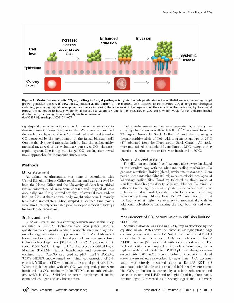
Figure 7. Model for metabolic CO2 signalling in fungal pathogenicity. As the cells proliferate on the epithelial surface, increasing fungalgrowth generates pockets of elevated CO2 located at the bottom of the biomass. Cells exposed to the elevated CO2 undergo morphologicalswitching, promoting hyphal development and hence increasing the adherence of the organism. At the same time, the protruding hyphae wouldexpose the pathogen to host environmental signals like serum, pH and further increases in CO2 levels, which would further enhance hyphaldevelopment, increasing the opportunity for tissue invasion.doi:10.1371/journal.ppat.1001193.g007
Fungal Population Signalling and CO2
PLoS Pathogens | www.plospathogens.org 8 November 2010 | Volume 6 | Issue 11 | e1001193
proportional to the reflective light and hence the concentration of
CO2 in the bottle.
CO2z2NaOH?Na2CO3zH2O
Heterogeneous populations of CAI4 and Dnce103Heterogeneous cell suspensions containing equal proportions
(500 cells/ml) of DAY286 and Dnce103 were spotted (1 ml total)
onto individual YPD or YNB plates and incubated at 37uC for
48 hrs. Initially 1 ml of sterile water was used to wash the single
colony from the plate with light agitation of the agar to remove
adhered cells. From the recovered 800 ml, 200 ml was plated onto
YNB, 5% CO2 to promote growth of the strictly CO2-requiring
strain Dnce103 strain only (DAY286 will not grow under these
conditions as it is Dhis1/Dhis1). Stability of the different phenotypic
markers was verified upon replica-plating of colonies. The number
of colonies was counted, and after taking into account the dilution
factor, related back to the initial number of colonies in the cell
suspension. Initial cell suspensions were always replica plated onto
YNB and YPD to obtain the average starting cell concentration for
each strain.
Molecular modelling of a nucleotide and bicarbonatecomplex of homodimeric Cyr1p catalytic domain
The amino acid sequence of Cyr1p was duplicated and aligned
with the sequences of two chains of a homodimeric substrate
analogue complex of CyaC from Spirulina platensis (PDB ID 1WC0;
[30]) by using Genedoc (http://www.psc.edu/biomed/genedoc).
A homology model for Cyr1p was generated with this alignment
using Modeller [40], and nucleotide and divalent ions positioned
by superposition with the experimentally determined CyaC
complex structure. Bicarbonate was then positioned manually at
the site proposed for binding in bacterial sAC-like enzymes [13].
The model was visualized using Pymol (DeLano Scientific; http://
www.pymol.org).
Site directed mutagenesis of CYR1Lys 1373, Asp 1334 and Asp 1377 were point mutated to Ala by
site directed mutagenesis using the following sets of primers
(mutations underlined) 1373F-tggatatgaagtggcgactgaaggtgatg and
Primer 7-ctatttaagttcattaactgttttcatgat, Primer 8-aacttgtttcactcc-
cagca and 1373R-atcaccttcagtcgccacttcatatccac, 1334F-ggttttc-
actgcgatcaaaaactcaac and Primer 7, 1334R-gttgagtttttgatcgcagt-
gaaaacc and Primer 8, 1377F-gactgaaggtgcggcgttcatgg and Primer
7, 1377R-ccatgaacgccgcaccttcagtc and Primer 8. The resulting
PCR fragments were ligated into the SpeI and BamHI restriction
sites of pSM2. The 59 domain of CYR1 together with the TEF2
promoter were subsequently ligated into the pSM2 plasmid using
XbaI and HindIII (site located with C-terminal domain of CYR1)
restriction sites forming pACL1, pACL2, and pACL3. Full-length,
native CYR1 cloned into pFM2 under the control of the TEF2
promoter was subsequently restricted using SacI and BamHI
restriction sites and ligated into pSM2 forming plasmid pSMTC.
Plasmids pACL1, pACL2, pACL3, pSMTC and pSM2 (vector
control) were integrated into the URA3 locus of CR276 and CAI4,
generating strains RH20-25 (Figure 7A) using standard heat-shock
procedures as previously described [41].
Southern blot analysisSingle copy integration of pACL1 was confirmed for five
resulting CAI4 transformants by southern analysis using DIG
High primer DNA labelling and detection (Roche) as per the
manufacturer’s recommendations. DNA probe (1 kb) was PCR
amplified using primer-44 59TTGGTGACATTGAGGCGTTA
and primer-47 59GTTCAATTGTCATTCCGGCAT.
Semi-quantitative RT-PCRTo assess transcript levels of CYR1 total RNA was extracted from
cultures (50 ml YPD) grown to OD600 0.5. Cells were harvested
through centrifugation and immediately frozen in liquid nitrogen.
Samples were disrupted using a Mikro-dismembrator S (Sartouis) at
2000 rpm, 2 minutes and RNA immediately extracted using the
Qiagen RNeasy Kit according to the manufacturer’s recommen-
dations. CYR1 expression levels (native CYR1 and CYR11373) were
analysed by semi quantitative RT-PCR using the BioRad one-step
RT-PCR Kit with Syber Green (primers CYR1-F 59GACGA-
CAACAAACGTGCCAGAACA and CYR1-R 59 AATCACG-
TGCTGAAACATGGTCCC). CYR1 levels were normalised to
ACT1.
Disruption of NCE103The strain, RH12 (Dnce103 Dcyr1), was constructed in the Dcyr1
background strain, CR276, using the HisG-URA3-HisG cassette to
disrupt the 847 bp NCE103 open reading frame (GenBank
association number EAL03010) from positions +153 to +807 as
described previously [10]. Correct integration of the HisG-URA3-
HisG cassette into the NCE103 locus was confirmed by PCR.
Mouse infection modelsSurvival experiments. CAI4-CYR1 and CAI4-CYR11373
were grown on YPD plates at 30uC for 16 hrs and cells were
washed off plates with saline, the resulting cell suspensions washed
twice with sterile saline and then resuspended in saline to provide
inocula for infection. For each C. albicans strain 6 female (6–8
weeks old) BALB/c mice (Harlan, UK) were intravenously
challenged with 2.4–2.56104 CFU/g of each strain. Mice were
monitored and weighed at least once daily, with mice culled when
they displayed signs of severe illness, or when their weight
decreased by 20%. When culled, the kidneys were aseptically
removed and burdens determined. Survival data were compared
by the Kaplan-Meier Log-rank statistic.
Outcome scores. For each C. albicans strain 9 mice were
challenged intravenously, as described above, with three mice
sampled on days 1, 2 and 3 post-infection. For each mouse kidney
burdens and percentage weight change were determined, with the
two parameters used to determine infection outcome scores [42].
Kidney burdens, weight change and outcome scores were
compared by the Mann-Whitney U statistic.
Supporting Information
Figure S1 Dnce103 can promote its own growth under diffusion
limiting conditions. 10,000 CFUs of Dnce103 were plated onto
CBA media and incubated in open or closed systems for 48 hr.
Found at: doi:10.1371/journal.ppat.1001193.s001 (0.58 MB TIF)
Figure S2 Mutations in CYR1 affect adenylyl cyclase activity as
homo, but not heterodimers (related to Figure 5). The desired
point mutated adenylyl cyclase genes or control plasmids were
integrated into the URA3 locus of the adenylyl cyclase mutant
(CR276). Resulting transformants were screened on DMEM pH7,
DMEM pH7 supplemented with 5% serum and DMEM pH7
incubated in atmospheres of 5.5% CO2. Plates were incubated at
37uC for 24 hrs. Scale bar represents 100 mm.
Fungal Population Signalling and CO2
PLoS Pathogens | www.plospathogens.org 9 November 2010 | Volume 6 | Issue 11 | e1001193
Found at: doi:10.1371/journal.ppat.1001193.s002 (0.58 MB
TIF)
Figure S3 Expression levels of CYR1 constructs (related to
Figure 5 and Figure 6). A) Schematic diagram of the CYR1 locus
and the CAI4 URA3 locus containing the integrated cassette. B)Strains were checked for single copy integration of plasmids
containing CYR1 and CYR11373. Genomic DNA from the
parental strain CAI4 (Lane 1), CAI4-CYR1, strains (Lanes 2
and 3) and CAI4-CYR11373 strains (Lanes 4–8) was digested with
Hind III and detected using 1Kb probe specific to the 39 CYR1
open reading frame. C) Expression levels of CYR1 in the parental
control strain, CAI4-CYR1 and CAI4-CYR11373 as analysed by
semi quantitative RT-PCR. Values are the mean and standard
deviation from two independent experiments.
Found at: doi:10.1371/journal.ppat.1001193.s003 (0.45 MB TIF)
Figure S4 CAI4-CYR11373, CAI4-CYR1 and CAI4-pSM2 have
the same growth rates (related to Figure 6). Overnight cultures
were diluted to an initial OD600 0.1 in fresh YPD and growth rate
followed at 37uC, 150 rpm for 9 hours. Values represent the mean
and standard deviation from two independent experiments.
Found at: doi:10.1371/journal.ppat.1001193.s004 (0.14 MB TIF)
Table S1 Fungal burden in the D. melanogaster infection model
(related to Figure 6A). Flies were homogenised in sterile water and
CFUs determined on YPD agar supplemented with chloram-
phenicol.
Found at: doi:10.1371/journal.ppat.1001193.s005 (0.05 MB RTF)
Table S2 Mouse infection parameters measured on day 1–3
post-infection (related to Figure 6B). For each C. albicans strain 9
mice were challenged intravenously, with three mice sampled on
days 1, 2 and 3 post-infection.
Found at: doi:10.1371/journal.ppat.1001193.s006 (0.08 MB RTF)
Table S3 Strains used in the study.
Found at: doi:10.1371/journal.ppat.1001193.s007 (0.12 MB RTF)
Acknowledgments
We would like to thank Dr C. Gourlay and Dr. F. Cottier for fruitful
discussions, Kara J. Turner for technical assistance and Mark Baker for
advice on CO2 detection systems.
Author Contributions
Conceived and designed the experiments: RAH DMM GKR YW CS
FAM. Performed the experiments: RAH LDS DMM HT RE YW.
Analyzed the data: RAH LDS JWB GKR LRL JB YW NARG CS FAM.
Contributed reagents/materials/analysis tools: JWB YW NARG. Wrote
the paper: RAH LDS DMM GKR LRL JB CS FAM.
References
1. Almirante B, Rodrıguez D, Park BJ, Cuenca-Estrella M, Planes AM, et al. (2005)
Epidemiology and Predictors of Mortality in Cases of Candida BloodstreamInfection: Results from Population-Based Surveillance, Barcelona, Spain, from
2002 to 2003. J Clin Microbiol 43: 1829–1835.
2. Klevay MJ, Ernst EJ, Hollanbaugh JL, Miller JG, Pfaller MA, et al. (2008)
Therapy and outcome of Candida glabrata versus Candida albicans bloodstreaminfection. Diag Microbiol Infect Dis 60: 273–277.
3. Leroy O, Gangneux J-P, Montravers P, Mira J-P, Gouin F, et al. (2009)
Epidemiology, management, and risk factors for death of invasive Candidainfections in critical care: A multicenter, prospective, observational study in
France (2005–2006). Crit Care Med 37: 1612–1618.
4. Bodey GP (1986) Candidiasis in cancer patients. Am J Med 77: 13–19.
5. Odds FC (1988) Candida and candidosis A review and bibliography.
6. Scully C, el-Kabir M, Samaranayake L (1994) Candida and oral candidosis: a
review. Crit Rev Oral Biol Med 5: 125–157.
7. Farah C, Ashman R, Challacombe S (2000) Oral Candidosis. Clin Dermatol 18:
553–562.
8. Lo H-J, Kohler JR, DiDomenico B, Loebenberg D, Cacciapuoti A, et al. (1997)
Nonfilamentous C. albicans Mutants Are Avirulent. Cell 90: 939–949.
9. Saville SP, Lazzell AL, Monteagudo C, Lopez-Ribot JL (2003) Engineered
Control of Cell Morphology In Vivo Reveals Distinct Roles for Yeast andFilamentous Forms of Candida albicans during Infection. Eukaryotic Cell 2:
1053–1060.
10. Klengel T, Liang W-J, Chaloupka J, Ruoff C, Schroppel K, et al. (2005) Fungal
Adenylyl Cyclase Integrates CO2 Sensing with cAMP Signaling and Virulence.Curr Biol 15: 2021–2026.
11. Buffo J, Herman M, Soll D (1984) A characterization of pH regulateddimorphism in Candida albicans. Mycopathologia 85: 21–30.
12. Rocha CRC, Schroppel K, Harcus D, Marcil A, Dignard D, et al. (2001)Signaling through Adenylyl Cyclase Is Essential for Hyphal Growth and
Virulence in the Pathogenic Fungus Candida albicans. Mol Biol Cell 12:
3631–3643.
13. Kamenetsky M, Middelhaufe S, Bank EM, Levin LR, Buck J, et al. (2006)
Molecular Details of cAMP Generation in Mammalian Cells: A Tale of TwoSystems. J Mol Biol 362: 623–639.
14. Verstrepen KJ, Klis FM (2006) Flocculation, adhesion and biofilm formation inyeasts. Mol Microbiol 60: 5–15.
15. Fang H-M, Wang Y (2006) RA domain-mediated interaction of Cdc35 withRas1 is essential for increasing cellular cAMP level for Candida albicans hyphal
development. Mol Microbiol 61: 484–496.
16. Xu X-L, Lee RTH, Fang H-M, Wang Y-M, Li R, et al. (2008) Bacterial
Peptidoglycan Triggers Candida albicans Hyphal Growth by Directly Activatingthe Adenylyl Cyclase Cyr1p. Cell Host & Microbe 4: 28–39.
17. Hogan DA (2006) Talking to Themselves: Autoregulation and Quorum Sensingin Fungi. Eukaryot Cell 5: 613–619.
18. Shank E, Kolter R (2009) New developments in microbial interspecies signaling.Curr Opin Microbiol 12: 1–10.
19. Hughes DT, Sperandio V (2008) Inter-kingdom signalling: communication
between bacteria and their hosts. Nat Rev Micro 6: 111–120.
20. Hornby JM, Jensen EC, Lisec AD, Tasto JJ, Jahnke B, et al. (2001) Quorum
Sensing in the Dimorphic Fungus Candida albicans Is Mediated by Farnesol. Appl
Environ Microbiol 67: 2982–2992.
21. Hogan DA, Vik A, Kolter R (2004) A Pseudomonas aeruginosa quorum-sensing
molecule influences Candida albicans morphology. Mol Microbiol 54: 1212–1223.
22. Davis-Hanna A, Piispanen AE, Stateva LI, Hogan DA (2008) Farnesol and
dodecanol effects on the Candida albicans Ras1-cAMP signalling pathway and the
regulation of morphogenesis. Mol Microbiol 67: 47–62.
23. Palkova Z, Janderova B, Gabriel J, Zikanova B, Pospisek M, et al. (1997)
Ammonia mediates communication between yeast colonies. Nature 390:
532–536.
24. Bahn Y-S, Cox GM, Perfect JR, Heitman J (2005) Carbonic Anhydrase and
CO2 Sensing during Cryptococcus neoformans Growth, Differentiation, and
Virulence. Curr Biol 15: 2013–2020.
25. Gewiss Mogensen E, GuilhemJanbon, JamesChaloupka, ClemensSteegborn,
Man ShunFu, et al. (2006) Cryptococcus neoformans Senses CO2 through the
Carbonic Anhydrase Can2 and the Adenylyl Cyclase Cac1. Eukaryotic Cell 5:
103–111.
26. Sharabi K, Lecuona E, Helenius IT, Beitel G, Sznajder JI, et al. (2009) Sensing,
physiological effects and molecular response to elevated CO2 levels in
eukaryotes. Journal of Cellular and Molecular Medicine 9999.
27. Christensen BE, Facer JF (1939) Simple wet combustion method for the
determination of carbon, oxygen equivalence and empirical formula by iodic
acid oxidation. J Am Chem Soc 61: 3001–3005.
28. Chen Y, Cann MJ, Litvin TN, Iourgenko V, Sinclair ML, et al. (2000) Soluble
Adenylyl Cyclase as an Evolutionarily Conserved Bicarbonate Sensor. Science
289: 625–628.
29. Cann MJ, Hammer A, Zhou J, Kanacher T (2003) A Defined Subset of Adenylyl
Cyclases Is Regulated by Bicarbonate Ion. J Biol Chem 278: 35033–35038.
30. Steegborn C, Litvin TN, Levin LR, Buck J, Wu H (2005) Bicarbonate activation
of adenylyl cyclase via promotion of catalytic active site closure and metal
recruitment. Nat Struct Mol Biol 12: 32–37.
31. Ghosh S, Navarathna DHMLP, Roberts DD, Cooper JT, Atkin AL, et al. (2009)
Arginine-Induced Germ Tube Formation in Candida albicans Is Essential for
Escape from Murine Macrophage Line RAW 264.7. Infect Immun 77:
1596–1605.
32. Belden WJ, Larrondo LF, Froehlich AC, Shi M, Chen C-H, et al. (2007) The
band mutation in Neurospora crassa is a dominant allele of ras-1 implicating RAS
signaling in circadian output. Genes & Dev 21: 1494–1505.
33. Park S, Lee K (2004) Inverted race tube assay for circadian clock studies of the
Neurospora accessions. Fungal Genet Newslett 51: 12–14.
34. Helenius IT, Krupinski T, Turnbull DW, Gruenbaum Y, Silverman N, et al.
(2009) Elevated CO2 suppresses specific Drosophila innate immune responses
and resistance to bacterial infection. Proceedings of the National Academy of
Sciences 106: 18710–18715.
35. De Smet, Hilde R, Bersten AD, Barr HA, Doyle IR (2007) Hypercapnic acidosis
modulates inflammation, lung mechanics, and edema in the isolated perfused
lung. Journal of Critical Care 22: 305–313.
Fungal Population Signalling and CO2
PLoS Pathogens | www.plospathogens.org 10 November 2010 | Volume 6 | Issue 11 | e1001193

36. Halbertsma FJJ, Vaneker M, Pickkers P, Snijdelaar DG, van Egmond J, et al.
(2008) Hypercapnic acidosis attenuates the pulmonary innate immune responsein ventilated healthy mice. Critical Care Medicine 36: 2403–2406.
37. O’Croinin DF, Nichol AD, Hopkins N, Boylan J, O’Brien S, et al. (2008)
Sustained hypercapnic acidosis during pulmonary infection increases bacterialload and worsens lung injury. Critical Care Medicine 36: 2128–2135.
38. Ellner PD, Stoessel CJ, Drakeford E, Vasi F (1966) A new culture medium forclinical bacteriology. American Journal of Clinical Pathology 45: 502–504.
39. Thorpe TC, Wilson ML, Turner JE, DiGuiseppi JL, Willert M, et al. (1990)
BacT/Alert: an automated colorimetric microbial detection system. J ClinMicrobiol 28: 1608–1612.
40. Sali A, Blundell TL (1993) Comparative protein modelling by satisfaction of
spatial restraints. J Mol Biol 234: 779–815.
41. Walther A, Wendland J (2003) An improved transformation protocol for the
human fungal pathogen Candida albicans. Curr Genetics 42: 339–343.
42. MacCallum DM, Coste A, Ischer F, Jacobsen MD, Odds FC, et al. (2010)
Genetic Dissection of Azole Resistance Mechanisms in Candida albicans and Their
Validation in a Mouse Model of Disseminated Infection. Antimicrob Agents
Chemother 54: 1476–1483.
Fungal Population Signalling and CO2
PLoS Pathogens | www.plospathogens.org 11 November 2010 | Volume 6 | Issue 11 | e1001193