Embed Size (px)
Citation preview
CNNM2 Mutations Cause Impaired Brain Developmentand Seizures in Patients with HypomagnesemiaFrancisco J. Arjona1., Jeroen H. F. de Baaij1., Karl P. Schlingmann2., Anke L. L. Lameris1,
Erwin van Wijk3, Gert Flik4, Sabrina Regele2, G. Christoph Korenke5, Birgit Neophytou6,
Stephan Rust7, Nadine Reintjes8, Martin Konrad2, Rene J. M. Bindels1, Joost G. J. Hoenderop1*
1 Department of Physiology, Radboud Institute for Molecular Life Sciences, Radboud university medical center, Nijmegen, The Netherlands, 2 Department of General
Pediatrics, University Children’s Hospital, Munster, Germany, 3 Department of Otorhinolaryngology, Radboud university medical center, Nijmegen, The Netherlands,
4 Department of Organismal Animal Physiology, Institute for Water and Wetland Research, Radboud University Nijmegen, Nijmegen, The Netherlands, 5 Department of
Neuropediatrics, Children’s Hospital, Oldenburg, Germany, 6 Department of Neuropediatrics, St. Anna Children’s Hospital, Medical University Vienna, Vienna, Austria,
7 Leibniz Institute of Arteriosclerosis Research, University of Munster, Munster, Germany, 8 Institute of Human Genetics, University of Cologne, Cologne, Germany
Abstract
Intellectual disability and seizures are frequently associated with hypomagnesemia and have an important geneticcomponent. However, to find the genetic origin of intellectual disability and seizures often remains challenging because ofconsiderable genetic heterogeneity and clinical variability. In this study, we have identified new mutations in CNNM2 in fivefamilies suffering from mental retardation, seizures, and hypomagnesemia. For the first time, a recessive mode ofinheritance of CNNM2 mutations was observed. Importantly, patients with recessive CNNM2 mutations suffer from brainmalformations and severe intellectual disability. Additionally, three patients with moderate mental disability were shown tocarry de novo heterozygous missense mutations in the CNNM2 gene. To elucidate the physiological role of CNNM2 andexplain the pathomechanisms of disease, we studied CNNM2 function combining in vitro activity assays and the zebrafishknockdown model system. Using stable Mg2+ isotopes, we demonstrated that CNNM2 increases cellular Mg2+ uptake inHEK293 cells and that this process occurs through regulation of the Mg2+-permeable cation channel TRPM7. In contrast,cells expressing mutated CNNM2 proteins did not show increased Mg2+ uptake. Knockdown of cnnm2 isoforms in zebrafishresulted in disturbed brain development including neurodevelopmental impairments such as increased embryonicspontaneous contractions and weak touch-evoked escape behaviour, and reduced body Mg content, indicative of impairedrenal Mg2+ absorption. These phenotypes were rescued by injection of mammalian wild-type Cnnm2 cRNA, whereasmammalian mutant Cnnm2 cRNA did not improve the zebrafish knockdown phenotypes. We therefore concluded thatCNNM2 is fundamental for brain development, neurological functioning and Mg2+ homeostasis. By establishing the loss-of-function zebrafish model for CNNM2 genetic disease, we provide a unique system for testing therapeutic drugs targetingCNNM2 and for monitoring their effects on the brain and kidney phenotype.
Citation: Arjona FJ, de Baaij JHF, Schlingmann KP, Lameris ALL, van Wijk E, et al. (2014) CNNM2 Mutations Cause Impaired Brain Development and Seizures inPatients with Hypomagnesemia. PLoS Genet 10(4): e1004267. doi:10.1371/journal.pgen.1004267
Editor: Ali G. Gharavi, Columbia University, United States of America
Received November 10, 2013; Accepted February 5, 2014; Published April 3, 2014
Copyright: ! 2014 Arjona et al. This is an open-access article distributed under the terms of the Creative Commons Attribution License, which permitsunrestricted use, distribution, and reproduction in any medium, provided the original author and source are credited.
Funding: This work was supported by grants from the Netherlands Organization for Scientific Research (ZonMw 9120.8026, NWO ALW 818.02.001), a DutchKidney Foundation Innovation Grant (IP11.46), the EURenOmics project from the European Union seventh Framework Programme (FP7/2007–2013, agreement nu305608) and NWO Vici Grant to JGJH (016.130.668). This work was further supported by the Hans-Joachim-Bodlee-Stifung and by the Peter-Stiftung. The fundershad no role in study design, data collection and analysis, decision to publish, or preparation of the manuscript.
Competing Interests: The authors have declared that no competing interests exist.
* E-mail: [email protected]
. These authors contributed equally to this work.
Introduction
Brain defects including seizures, migraine, depression andintellectual disability are frequently associated with hypomagne-semia [1]. Indeed, low Mg2+ concentrations may cause epilep-tiform activity during development [2]. Specifically, the Mg2+
channel transient receptor potential melastatin 7 (TRPM7) isessential for brain function and development [3]. Interestingly,patients with genetic defects in TRPM6, a close homologue ofTRPM7, may have neurological complications [4]. AlthoughTRPM6 and TRPM7 share similar Mg2+ transporting properties,they are differentially expressed and regulated [5]. TRPM7 is aubiquitously expressed protein regulating intracellular Mg2+
levels in a broad range of cells, whereas TRPM6 is localized inthe luminal membrane of renal and intestinal epithelia involvedin Mg2+ absorption [1,6–8]. Recently, we have identifiedmutations in the gene encoding cyclin M2 (CNNM2) in twounrelated families with dominant isolated hypomagnesemia(CNNM2 [MIM 607803]) [9]. Patients suffered from symptomsassociated with low serum Mg2+ levels (0.3–0.5 mM) such astremors, headaches and muscle weakness. The role of CNNM2 inthe kidney for the maintenance of serum Mg2+ levels can betraced to the distal convoluted tubule (DCT), where also TRPM6is expressed. Here, CNNM2 is present in the basolateralmembrane of DCT cells and its expression is regulated bydietary Mg2+ availability [9–10].
PLOS Genetics | www.plosgenetics.org 1 April 2014 | Volume 10 | Issue 4 | e1004267
Although CNNM2 has been proposed as a Mg2+ transporter inoverexpression studies in Xenopus oocytes [11], Mg2+ transportcould not be directly measured in mammalian cells using patchclamp analysis [9]. On the other hand, modelling of the CNNM2cystathionine b-synthase (CBS) domain resulted in the identifica-tion of a Mg2+-ATP binding site, suggesting a role in Mg2+ sensingwithin the cell [12]. Consequently, the molecular mechanismexplaining the role of CNNM2 in DCT-mediated Mg2+ transportremains to be elucidated.
The CNNM2 gene is ubiquitously expressed in mammaliantissues, most prominently in kidney, brain and lung [12–13].Although the role of CNNM2 beyond the kidney has never beenstudied, genome wide association studies have related the CNNM2locus to blood pressure, coronary artery disease and schizophrenia,suggesting an important role of CNNM2 in the cardiovascularsystem and brain [14–15]. CNNM2 is widely conserved amongspecies. In zebrafish (Danio rerio), a frequently used model for ionhomeostasis and human genetic diseases in general [16–17], thecnnm2 gene is duplicated and two paralogues, cnnm2a and cnnm2b,are described [18]. Both paralogues share a high conservation withhuman CNNM2 (79% amino acid identity). In detail, transcriptsare abundantly expressed in zebrafish brain and in ionoregulatoryorgans such as kidney and gills, which act as a pseudokidney in fish[18]. Consistent with the regulation of CNNM2 transcripts inmammals [11], the expression of cnnm2a and cnnm2b is regulatedby Mg2+ in vivo [18].
In the present study, we aim to elucidate the function ofCNNM2 in brain and kidney. Hence, we can demonstrate thegenetic origin of symptoms in five unrelated families suffering froma distinct phenotype of mental retardation, seizures and hypo-magnesemia, where we have identified novel mutations in theCNNM2 gene. By combining functional analyses and a loss-of-function approach in the zebrafish model, we provide functionalevidence for a key role of CNNM2 in brain development,neurological activity and renal Mg2+ handling.
Results
PatientsPatients F1.1 and F1.2 presented in the neonatal period with
cerebral convulsions. Serum Mg2+ levels at manifestation werefound to be 0.5 mM in both patients (Table 1). Convulsions wererefractory to conventional antiepileptic medications. IntravenousMg2+ supplementation with ,1 mmol/kg body weight/day wasinitiated after oral Mg2+ failed to correct serum Mg2+ levels.However, seizure activity continued even in face of normomagne-semia. An extensive analysis for infectious causes or inborn errorsof metabolism did not yield any positive results. Ultrasoundexamination of the kidneys did not reveal nephrocalcinosis,whereas basal ganglia calcifications were noted in early centralnervous system (CNS) sonographies. During follow-up, severedevelopmental delay was noted accompanied by microcephaly(head circumference below third percentile for age and sex in bothpatients). A magnetic resonance imaging (MRI) at 5.5 years of agein patient F1.1 showed wide supratentorial outer cerebrospinalliquor spaces with failure of opercularization together with asignificantly reduced myelinization of the white matter tract(Figure 1C–D). The severe degree of intellectual disability, whichbecame apparent with increasing age comprised major deficits incognitive function, the inability to verbally communicate, andseverely limited motor skills. Both children are not able to performmain activities of daily living and require full-time care by anattendant. Seizure activity is sufficiently controlled in the olderbrother by valproate and lamotrigine, electroencephalography(EEGs) merely shows generalized slowing, but no epileptic activity.In contrast, the younger sister suffers from ongoing generalized,myoclonic seizures despite antiepileptic treatment with valproateand levetiracetame. Laboratory investigations during follow-updemonstrated persistent hypomagnesemia of ,0.6 mM despiteoral Mg2+ supplementation.
Patients F2.1, F3.1, and F4.1 presented with seizures duringinfancy (between 4 and 12 months of age). Laboratory evaluationyielded isolated hypomagnesemia of ,0.5 mM (Table 1). Urineanalyses demonstrated inappropriate fractional excretion for Mg2+
in face of persistent hypomagnesemia. In addition, the renal Mg2+
leak was verified by Mg2+ loading tests in patients F2.1 and F4.1 asdescribed before [19]. Urinary calcium excretion rates werenormal, renal ultrasound excluded the presence of nephrocalci-nosis. After acute therapy with intravenous Mg2+, the patientsreceived a continuous oral Mg2+ supplementation of 0.5 to1 mmol/kg body weight/day of elemental Mg2+. This oral therapyhowever failed to correct the hypomagnesemia, serum Mg2+
remained in the subnormal range in all three children. Because ofrecurrent cerebral seizures, patients F2.1 to F4.1 received diverseantiepileptic medications. Currently only patient F4.1 is stilltreated with clobazam.
In all three patients (F2.1 to F4.1), a significant degree ofintellectual disability was already noted in early childhood withdelayed speech development, but also impaired motor as well ascognitive skills. In addition, patient F4.1 was noted to exhibitdisturbed social interaction, abnormal verbal and non-verbalcommunication, as well as stereotyped behaviour and finallyreceived the formal diagnosis of early onset autism. All threepatients F2.1 to F4.1 were not able to attend regular schools.Standardized intelligence testing in patients F2.1 and F3.1revealed a significant degree of mental retardation (see Table 1).While patients F2.1 and F3.1 are currently living with theirparents, patient F4.1 is placed in a home for children with mentalillness because of episodes of violence and destructive behaviour.
Author Summary
Mental retardation affects 1–3% of the population and hasa strong genetic etiology. Consequently, early identifica-tion of the genetic causes of mental retardation is ofsignificant importance in the diagnosis of the disease, aspredictor of the progress of the disease and for thedetermination of treatment. In this study, we identifymutations in the gene encoding for cyclin M2 (CNNM2)to be causative for mental retardation and seizures inpatients with hypomagnesemia. Particularly, in patientswith a recessive mode of inheritance, the intellectualdisability caused by dysfunctional CNNM2 is dramaticallysevere and is accompanied by severely limited motor skillsand brain malformations suggestive of impaired earlybrain development. Although hypomagnesemia has beenassociated to several neurological diseases, Mg2+ status isnot regularly assessed in patients with seizures and mentaldisability. Our findings establish CNNM2 as an importantprotein for renal magnesium handling, brain developmentand neurological functioning, thus explaining the physiol-ogy of human disease caused by (dysfunctional) mutationsin CNNM2. CNNM2 mutations should be taken intoaccount in patients with seizures and mental disability,specifically in combination with hypomagnesemia.
CNNM2 Mutations Impair Brain and Kidney Function
PLOS Genetics | www.plosgenetics.org 2 April 2014 | Volume 10 | Issue 4 | e1004267
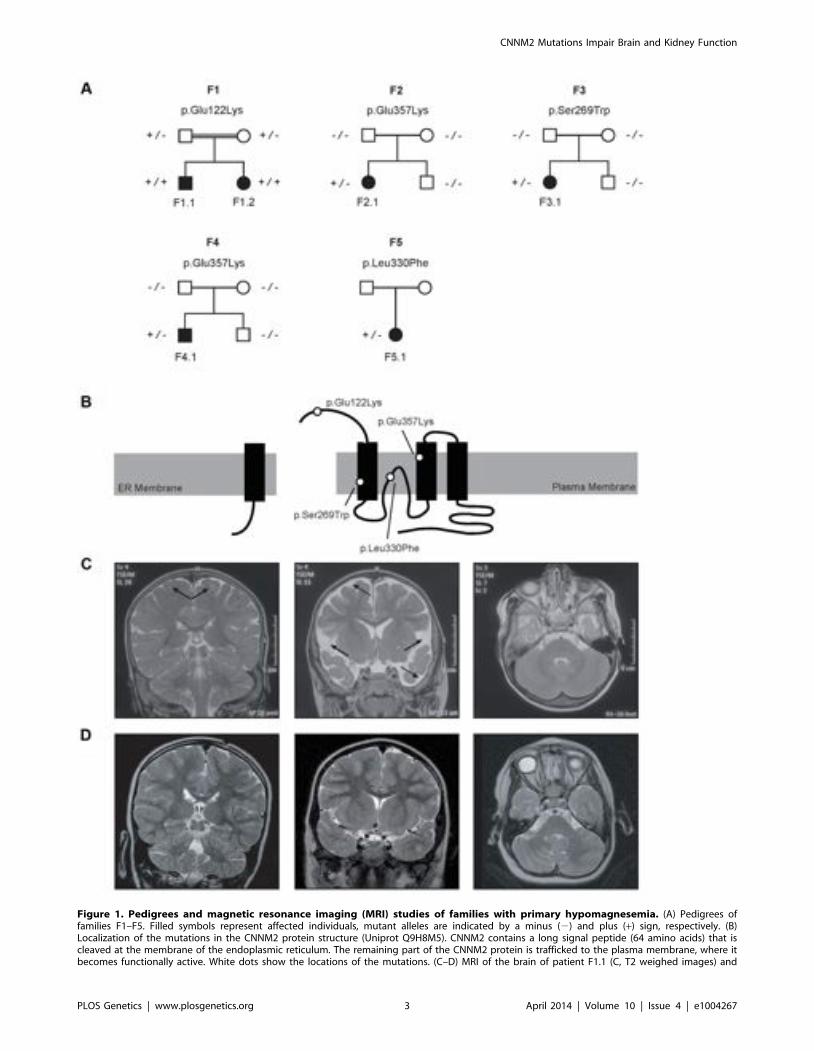
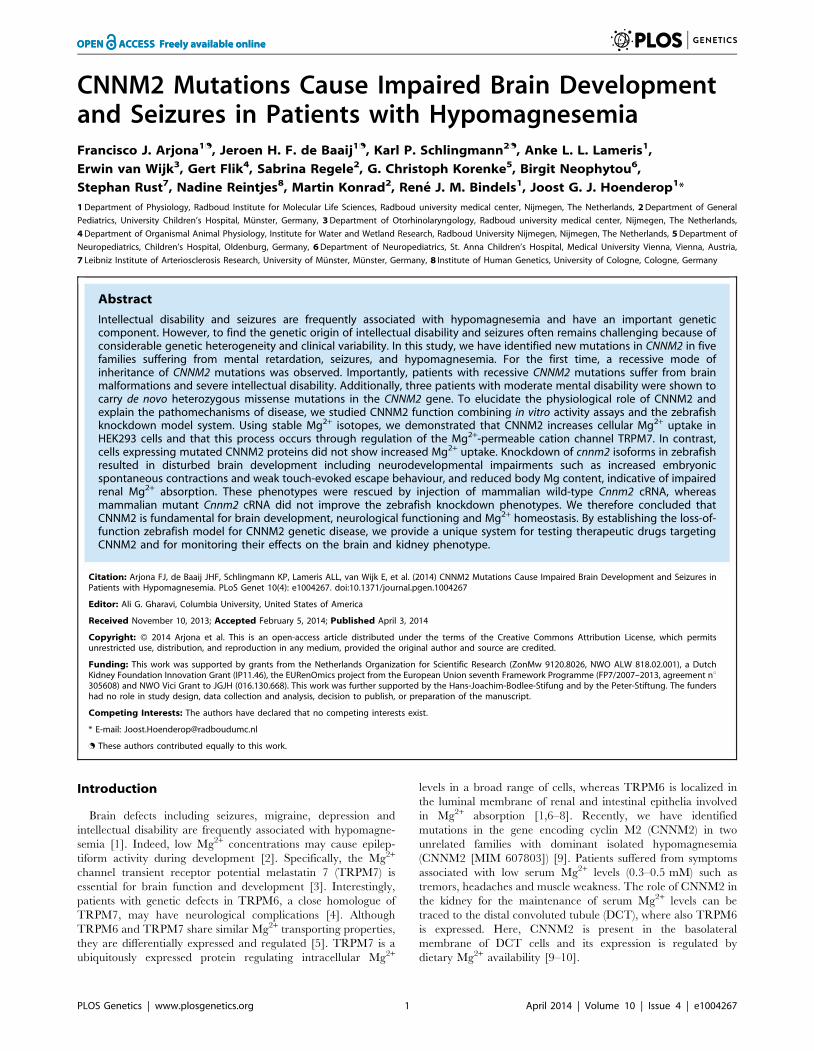
Figure 1. Pedigrees and magnetic resonance imaging (MRI) studies of families with primary hypomagnesemia. (A) Pedigrees offamilies F1–F5. Filled symbols represent affected individuals, mutant alleles are indicated by a minus (2) and plus (+) sign, respectively. (B)Localization of the mutations in the CNNM2 protein structure (Uniprot Q9H8M5). CNNM2 contains a long signal peptide (64 amino acids) that iscleaved at the membrane of the endoplasmic reticulum. The remaining part of the CNNM2 protein is trafficked to the plasma membrane, where itbecomes functionally active. White dots show the locations of the mutations. (C–D) MRI of the brain of patient F1.1 (C, T2 weighed images) and
CNNM2 Mutations Impair Brain and Kidney Function
PLOS Genetics | www.plosgenetics.org 3 April 2014 | Volume 10 | Issue 4 | e1004267
The parents of all three children (F2.1–F4.1) had normal serumMg2+ levels and no signs of intellectual disability.
Finally, patient F5.1 presented with muscle spasms anddysesthesia in adolescence. Serum Mg2+ levels were found to below (,0.6 mM). Because of concomitant borderline hypokalemia,she was suspected to have Gitelman syndrome ([MIM 263800])and received oral Mg2+ and K+ supplements. Also this patientexhibited a mild degree of intellectual disability. Unfortunately,she was not available for further examination.
CNNM2 mutations in patients with mental retardationCommon genetic causes of mental retardation were excluded in
patients F1.1 and F2.1 by array CGH (comparative genomichybridization). The presence of two affected siblings together withthe suspected parental consanguinity in family F1 suggested anautosomal-recessive pattern of inheritance. Therefore, we subject-ed patients F1.1 and F1.2 to homozygosity mapping which, at acut-off size of .1.7 megabases (Mb), yielded eleven criticalintervals on autosomes with a cumulative size of 62 Mb. The genelist generated from these loci included 322 RefSeq genes andputative transcripts. CNNM2 in a critical interval of 7.1 Mb onchromosome 10 emerged as the most promising candidate gene
because of its known role in Mg2+ metabolism [9,11–12,20].Conventional Sanger sequencing of the complete coding region ofthe CNNM2 gene revealed a homozygous mutation, c.364G.A,leading to a non-conservative amino acid substitution of glutamateto lysine at position 122 of the CNNM2 protein (p.Glu122Lys,Figure 1A–B). The mutation was present in heterozygous state inboth parents. After discovery of this homozygous mutation inpatients F1.1 and F1.2, a larger cohort (n = 34) of patients withMg2+ deficiency of unknown origin was screened for mutations inthe CNNM2 gene. Mutations in heterozygous state were discov-ered in patients F2.1 to F5.1 (Table 1). However, sequencing of thecomplete coding region and adjacent exon-intron boundaries didnot reveal a second pathogenic allele. Next, we examined theCNNM2 gene in parents and unaffected siblings of families F2 toF4. The mutations previously identified in our patients were notdetected in either of the parents pointing to de novo mutationalevents. Interestingly, patients F2.1 and F4.1 exhibited the samemutation, p.Glu357Lys (c.1069G.A), affecting a highly conservedamino acid residue in the 2nd membrane-spanning domain of theCNNM2 protein. Also the p.Ser269Trp (c.806C.G) mutationdetected in patient F3.1 affects a highly conserved residue locatedin the 1st transmembrane domain. All three mutations were
patient F2.1 (D, T2 weighed images). Left: Coronal images demonstrating a defect in myelinization of U-fibers (arrows) in patient F1.1 in contrast to anormal myelin pattern in patient F2.1. Center: Coronal T2 weighted images showing widened outer cerebrospinal liquor spaces (dashed arrows) andlack of opercularization (solid arrows) in patient F1.1, whereas a regular brain volume and insular lobe is observed in patient F2.1. Right: Absence ofcerebellar structural abnormalities in patients F1.1 and F2.1 on axial T2 weighted images at the level of the trigeminal nerve.doi:10.1371/journal.pgen.1004267.g001
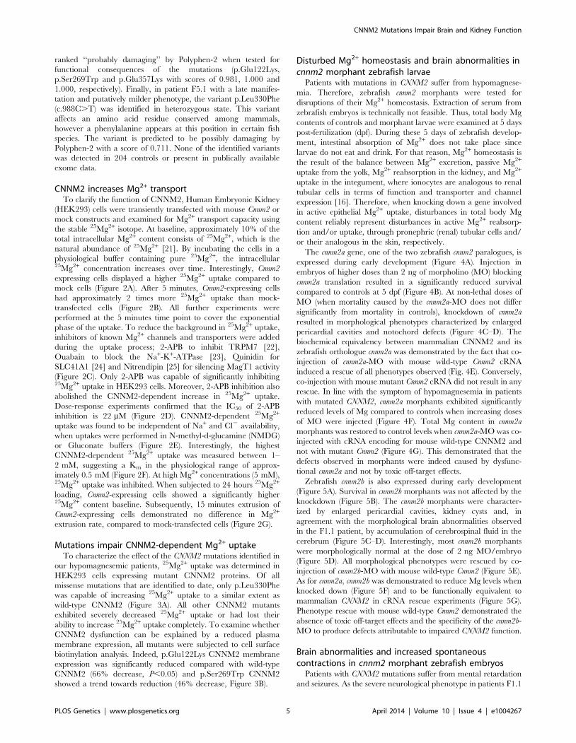
Table 1. Clinical and biochemical data of patients.
Patient F1.1 F1.2 F2.1 F3.1 F4.1 F5.1
Gender Male Female Female Female Male Female
Ethnicity Serbian Serbian German German German Polish
Age at manifestation 1 day 6 days 7 months 1 years 4 months 16 years
Follow-up 12 years 8 years 12 years 20 years 12 years None
Symptoms atmanifestation
Seizures Seizures Seizures Seizures,Paresthesia
Seizures Myoclonus,Paresthesia
Treatment Valproate,Lamotrigine
Valproate,Levetiracetame
Phenobarbital Valproate Clobazam Unknown
Neuroimaging Myelinizationdefects,Opercularizationdefect, Widenedouter cerebrospinalliquor spaces
Normal Normal Normal Unknown
Mental retardation Severe Severe Moderate Moderate Moderate Mild
Cognitive function IQ 55–57 IQ 55–59 Autism Unknown
Speech/Communication No verbalspeech, Limitedcommunication skills
No verbalspeech, Limitedcommunication skills
Expressivelanguagedisorder
Expressivelanguagedisorder
Limitedspeech andvocabulary
Unknown
Additional symptoms Very limitedmotor skills
Very limitedmotor skills
Impairedmotor skills,severe obesity
Impairedmotor skills,severe obesity
Impairedmotor skills
Unknown
Initial serum Mg2+ (mmol/L) 0.5 0.5 0.56 0.44 0.5 0.66
Follow-up serum Mg2+ (mmol/L) 0.66 0.54 0.56 0.53 0.68 -
Mutation (DNA level) c.364G.A c.364G.A c.1069G.A c.806C.G c.1069G.A c.988C.T
Mutation (Protein level) p.Glu122Lys p.Glu122Lys p.Glu357Lys p.Ser269Trp p.Glu357Lys p.Leu330Phe
Zygosity Homozygous Homozygous Heterozygous Heterozygous Heterozygous Heterozygous
doi:10.1371/journal.pgen.1004267.t001
CNNM2 Mutations Impair Brain and Kidney Function
PLOS Genetics | www.plosgenetics.org 4 April 2014 | Volume 10 | Issue 4 | e1004267
ranked ‘‘probably damaging’’ by Polyphen-2 when tested forfunctional consequences of the mutations (p.Glu122Lys,p.Ser269Trp and p.Glu357Lys with scores of 0.981, 1.000 and1.000, respectively). Finally, in patient F5.1 with a late manifes-tation and putatively milder phenotype, the variant p.Leu330Phe(c.988C.T) was identified in heterozygous state. This variantaffects an amino acid residue conserved among mammals,however a phenylalanine appears at this position in certain fishspecies. The variant is predicted to be possibly damaging byPolyphen-2 with a score of 0.711. None of the identified variantswas detected in 204 controls or present in publically availableexome data.
CNNM2 increases Mg2+ transportTo clarify the function of CNNM2, Human Embryonic Kidney
(HEK293) cells were transiently transfected with mouse Cnnm2 ormock constructs and examined for Mg2+ transport capacity usingthe stable 25Mg2+ isotope. At baseline, approximately 10% of thetotal intracellular Mg2+ content consists of 25Mg2+, which is thenatural abundance of 25Mg2+ [21]. By incubating the cells in aphysiological buffer containing pure 25Mg2+, the intracellular25Mg2+ concentration increases over time. Interestingly, Cnnm2expressing cells displayed a higher 25Mg2+ uptake compared tomock cells (Figure 2A). After 5 minutes, Cnnm2-expressing cellshad approximately 2 times more 25Mg2+ uptake than mock-transfected cells (Figure 2B). All further experiments wereperformed at the 5 minutes time point to cover the exponentialphase of the uptake. To reduce the background in 25Mg2+ uptake,inhibitors of known Mg2+ channels and transporters were addedduring the uptake process; 2-APB to inhibit TRPM7 [22],Ouabain to block the Na+-K+-ATPase [23], Quinidin forSLC41A1 [24] and Nitrendipin [25] for silencing MagT1 activity(Figure 2C). Only 2-APB was capable of significantly inhibiting25Mg2+ uptake in HEK293 cells. Moreover, 2-APB inhibition alsoabolished the CNNM2-dependent increase in 25Mg2+ uptake.Dose-response experiments confirmed that the IC50 of 2-APBinhibition is 22 mM (Figure 2D). CNNM2-dependent 25Mg2+
uptake was found to be independent of Na+ and Cl2 availability,when uptakes were performed in N-methyl-d-glucamine (NMDG)or Gluconate buffers (Figure 2E). Interestingly, the highestCNNM2-dependent 25Mg2+ uptake was measured between 1–2 mM, suggesting a Km in the physiological range of approx-imately 0.5 mM (Figure 2F). At high Mg2+ concentrations (5 mM),25Mg2+ uptake was inhibited. When subjected to 24 hours 25Mg2+
loading, Cnnm2-expressing cells showed a significantly higher25Mg2+ content baseline. Subsequently, 15 minutes extrusion ofCnnm2-expressing cells demonstrated no difference in Mg2+
extrusion rate, compared to mock-transfected cells (Figure 2G).
Mutations impair CNNM2-dependent Mg2+ uptakeTo characterize the effect of the CNNM2 mutations identified in
our hypomagnesemic patients, 25Mg2+ uptake was determined inHEK293 cells expressing mutant CNNM2 proteins. Of allmissense mutations that are identified to date, only p.Leu330Phewas capable of increasing 25Mg2+ uptake to a similar extent aswild-type CNNM2 (Figure 3A). All other CNNM2 mutantsexhibited severely decreased 25Mg2+ uptake or had lost theirability to increase 25Mg2+ uptake completely. To examine whetherCNNM2 dysfunction can be explained by a reduced plasmamembrane expression, all mutants were subjected to cell surfacebiotinylation analysis. Indeed, p.Glu122Lys CNNM2 membraneexpression was significantly reduced compared with wild-typeCNNM2 (66% decrease, P,0.05) and p.Ser269Trp CNNM2showed a trend towards reduction (46% decrease, Figure 3B).
Disturbed Mg2+ homeostasis and brain abnormalities incnnm2 morphant zebrafish larvae
Patients with mutations in CNNM2 suffer from hypomagnese-mia. Therefore, zebrafish cnnm2 morphants were tested fordisruptions of their Mg2+ homeostasis. Extraction of serum fromzebrafish embryos is technically not feasible. Thus, total body Mgcontents of controls and morphant larvae were examined at 5 dayspost-fertilization (dpf). During these 5 days of zebrafish develop-ment, intestinal absorption of Mg2+ does not take place sincelarvae do not eat and drink. For that reason, Mg2+ homeostasis isthe result of the balance between Mg2+ excretion, passive Mg2+
uptake from the yolk, Mg2+ reabsorption in the kidney, and Mg2+
uptake in the integument, where ionocytes are analogous to renaltubular cells in terms of function and transporter and channelexpression [16]. Therefore, when knocking down a gene involvedin active epithelial Mg2+ uptake, disturbances in total body Mgcontent reliably represent disturbances in active Mg2+ reabsorp-tion and/or uptake, through pronephric (renal) tubular cells and/or their analogous in the skin, respectively.
The cnnm2a gene, one of the two zebrafish cnnm2 paralogues, isexpressed during early development (Figure 4A). Injection inembryos of higher doses than 2 ng of morpholino (MO) blockingcnnm2a translation resulted in a significantly reduced survivalcompared to controls at 5 dpf (Figure 4B). At non-lethal doses ofMO (when mortality caused by the cnnm2a-MO does not differsignificantly from mortality in controls), knockdown of cnnm2aresulted in morphological phenotypes characterized by enlargedpericardial cavities and notochord defects (Figure 4C–D). Thebiochemical equivalency between mammalian CNNM2 and itszebrafish orthologue cnnm2a was demonstrated by the fact that co-injection of cnnm2a-MO with mouse wild-type Cnnm2 cRNAinduced a rescue of all phenotypes observed (Fig. 4E). Conversely,co-injection with mouse mutant Cnnm2 cRNA did not result in anyrescue. In line with the symptom of hypomagnesemia in patientswith mutated CNNM2, cnnm2a morphants exhibited significantlyreduced levels of Mg compared to controls when increasing dosesof MO were injected (Figure 4F). Total Mg content in cnnm2amorphants was restored to control levels when cnnm2a-MO was co-injected with cRNA encoding for mouse wild-type CNNM2 andnot with mutant Cnnm2 (Figure 4G). This demonstrated that thedefects observed in morphants were indeed caused by dysfunc-tional cnnm2a and not by toxic off-target effects.
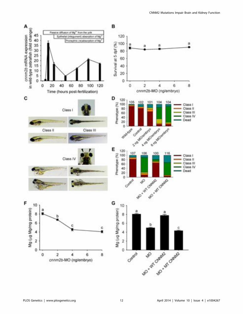
Zebrafish cnnm2b is also expressed during early development(Figure 5A). Survival in cnnm2b morphants was not affected by theknockdown (Figure 5B). The cnnm2b morphants were character-ized by enlarged pericardial cavities, kidney cysts and, inagreement with the morphological brain abnormalities observedin the F1.1 patient, by accumulation of cerebrospinal fluid in thecerebrum (Figure 5C–D). Interestingly, most cnnm2b morphantswere morphologically normal at the dose of 2 ng MO/embryo(Figure 5D). All morphological phenotypes were rescued by co-injection of cnnm2b-MO with mouse wild-type Cnnm2 (Figure 5E).As for cnnm2a, cnnm2b was demonstrated to reduce Mg levels whenknocked down (Figure 5F) and to be functionally equivalent tomammalian CNNM2 in cRNA rescue experiments (Figure 5G).Phenotype rescue with mouse wild-type Cnnm2 demonstrated theabsence of toxic off-target effects and the specificity of the cnnm2b-MO to produce defects attributable to impaired CNNM2 function.
Brain abnormalities and increased spontaneouscontractions in cnnm2 morphant zebrafish embryos
Patients with CNNM2 mutations suffer from mental retardationand seizures. As the severe neurological phenotype in patients F1.1
CNNM2 Mutations Impair Brain and Kidney Function
PLOS Genetics | www.plosgenetics.org 5 April 2014 | Volume 10 | Issue 4 | e1004267
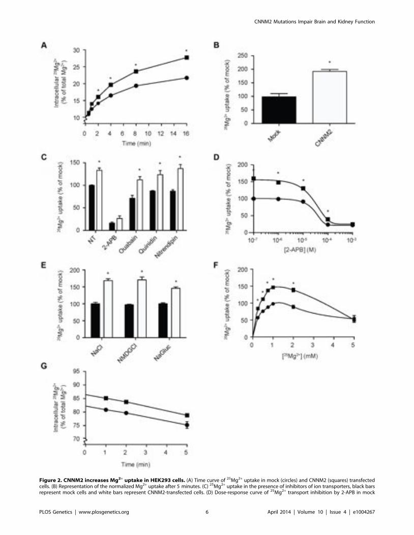
Figure 2. CNNM2 increases Mg2+ uptake in HEK293 cells. (A) Time curve of 25Mg2+ uptake in mock (circles) and CNNM2 (squares) transfectedcells. (B) Representation of the normalized Mg2+ uptake after 5 minutes. (C) 25Mg2+ uptake in the presence of inhibitors of ion transporters, black barsrepresent mock cells and white bars represent CNNM2-transfected cells. (D) Dose-response curve of 25Mg2+ transport inhibition by 2-APB in mock
CNNM2 Mutations Impair Brain and Kidney Function
PLOS Genetics | www.plosgenetics.org 6 April 2014 | Volume 10 | Issue 4 | e1004267
and F1.2 was diagnosed early after birth (Table 1), wehypothesized that the deleterious effects of mutant CNNM2 couldresult from early developmental defects in brain primordia. Inzebrafish, the segmental organization of the brain rudiment, andmorphologically visible boundaries and primordia are establishedat 25 hours post-fertilization (hpf). At this stage, maldevelopmentof the midbrain hindbrain boundary (MHB) is observed in cnnm2amorphant embryos (Figure 6A–B). Interestingly, these phenotypescould not be rescued by exposure to media with high Mg2+
concentrations (Figure 6B), even though these media significantlyincreased the Mg content of morphant embryos (Figure 6C). Moreimportantly, phenotypes were rescued by co-injection with themouse orthologue cRNA and not by co-injection with the mutanttranscript (Figure 6D). In addition to brain developmental defects,the frequency of spontaneous embryonic contractions was increasedin cnnm2a morphants compared to controls (Figure 6E), which couldindicate that (motor) neurons are hyperexcitable [26]. Thisphenotype was not rescued by exposure to high Mg2+ concentra-tions in the medium (Figure 6E). In contrast, co-injection of thecnnm2a-MO with mouse wild-type Cnnm2 cRNA did result in arescue of the neurological functioning (Figure 6F). Conspicuously,co-injection with the mutant Cnnm2 cRNA even worsened thismotor neuronal phenotype by increasing the number of spontane-ous contractions significantly compared to embryos injected onlywith cnnm2a-MO (Figure 6F; Movies S1, S2, S3).
In the case of cnnm2b morphants, enlarged tectums were alsopresent in a 30% of morphants in addition to the defects in theMHB, phenotypes that were not rescued by exposure to high Mg2+
concentrations (Figure 7A–C) but by co-injection of cnnm2b-MOwith mouse wild-type Cnnm2 cRNA (Figure 7D). Spontaneouscontraction frequency was increased in cnnm2b morphants(Figure 7E), restored to control levels with overexpression of mousewild-type Cnnm2 (Figure 7F), and 4-fold increased with overexpres-sion of mouse mutant Cnnm2 compared to cnnm2b morphantsinjected solely with cnnm2b-MO (Figure 7F; Movies S4, S5, S6).
The validated cRNA rescue controls proved the specificity ofthe brain defects observed, attributable to dysfunctional ortholo-gues of CNNM2 for both translation blocking MOs used.
Weaker touch-evoked escape behaviour in cnnm2morphant zebrafish larvae
As our in vitro data pointed to a putative interaction betweenCNNM2 and TRPM7 and zebrafish morphants presented braindevelopmental defects, the touch-evoked escape behaviour inzebrafish was evaluated, a parameter largely dependent on TRPM7activity in sensory neurons and/or brain development [27–29].Indeed, in cnnm2a and cnnm2b morphants (at 5 dpf), touch-evokedescape behaviour was significantly weaker than that in controls(Figures S1, S2). Additionally, this phenotype was rescued by co-injection of the MO with wild-type Cnnm2 cRNA and not by mutantCnnm2 cRNA. As for the other phenotypes, cRNA rescues provedthe causality between the weak touch-evoked escape behaviour inmorphants and dysfunctional cnnm2 paralogues.
Discussion
In the present study, a severe brain phenotype consisting ofcerebral seizures, mental retardation and brain malformations in
patients with hypomagnesemia was shown to be caused bymutations in CNNM2. Our experiments established CNNM2 as anew essential gene in brain development, neurological functioningand Mg2+ homeostasis. This notion is supported by the followingobservations; i) hypomagnesemic patients with CNNM2 mutationssuffer from seizures, mental disability, and if mutations are presentin recessive state, brain malformations are observed in addition; ii)Mg2+ supplementation does not improve the neurological pheno-type of the patients; iii) CNNM2 increases Mg2+ uptake inHEK293 cells, whereas mutant CNNM2 does not; iv) knockdownof CNNM2 orthologues in zebrafish results in impaired develop-ment of the brain, abnormal neurodevelopmental phenotypesmanifested as altered locomotor and touch-evoke escape behav-iours, and Mg wasting; v) the zebrafish phenotype can be rescuedby injection of mouse Cnnm2 cRNA.
In addition to the previously reported dominant mode ofinheritance [9], the genetic findings in our patients supportheterogenous patterns of inheritance. In family F1, a recessivemode of CNNM2 inheritance was observed. The homozygousCNNM2 p.Glu122Lys mutation in this family resulted in themanifestation of a neonatal onset and a considerably more severecerebral involvement than in the remaining patients. Yet, a centralnervous system (CNS) phenotype with seizures and intellectualdisability, which was not reported previously, represented thecardinal clinical symptom in all of our patients. Seizuresconstituted the major symptom at manifestation coinciding withhypomagnesemia, but were also seen during follow-up despiteMg2+ supplementation. Pronounced Mg2+ deficiency reflected byseverely low serum Mg2+ levels clearly represents a promotiveelement in the development of seizures. However, the persistenceof seizure activity despite Mg2+ supplementation might point to agenuine disturbance in brain function caused by defectiveCNNM2. Accordingly, the extent of hypomagnesemia found inthe two siblings with the recessive mutation, F1.1 and F1.2, wasidentical to other patients (F2.1–F5.1) with heterozygous CNNM2mutations and a milder neurological phenotype. Continuous oralMg2+ supplementation stabilized serum Mg2+ levels in thesubnormal range, however a complete normalization of Mg2+
metabolism could not be achieved in any of the patients.In three out of five families (F2, F3 and F4), the mutation of the
patient was not present in the parents. This finding supports therecent advancements evidencing that de novo mutations provide animportant mechanism in the development of mental disabilitydisorders [30]. Remarkably, the same de novo p.Glu357Lysmutation was identified in two unrelated individuals. Althoughthe mutation rate along the human genome varies significantly[31], the chance to observe an identical de novo base pair change intwo individuals is extremely small, supporting causality of thismutation. The clinical and genetic findings observed in patientF5.1 should be interpreted with caution. Though the p.Leu330Phevariant was not present in controls or exome variant databases, thepresence of an innocuous polymorphism cannot be completelyexcluded. The clinical phenotype with a milder degree ofintellectual disability and a later manifestation with hypomagne-semic symptoms during adolescence support a partial loss ofCNNM2 function caused by the p.Leu330Phe variant. Unfortu-nately, the patient was not available for clinical re-evaluation.
(circles) and CNNM2 (squares) transfected cells. (E) The effect of Na+ and Cl2 availability on 25Mg2+ uptake in mock (black bars) and CNNM2 (whitebars) transfected cells. (F) 25Mg2+ uptake as a function of extracellular 25Mg2+ availability in mock (circles) and CNNM2 (squares) transfected cells. (G)25Mg2+ extrusion in mock (circles) and CNNM2 (squares) transfected cells. Each data point represent the mean of 3 independent experiments 6 SEM.* indicates significant differences compared to mock (P,0.05).doi:10.1371/journal.pgen.1004267.g002
CNNM2 Mutations Impair Brain and Kidney Function
PLOS Genetics | www.plosgenetics.org 7 April 2014 | Volume 10 | Issue 4 | e1004267
Figure 3. CNNM2 mutations impair Mg2+ uptake in HEK293 cells. (A) Time curve of 25Mg2+ uptake in mock, wild-type CNNM2 and mutantCNNM2 transfected cells. Symbols indicate cells transfected with the vector empty (N mock) or containing Cnnm2 sequences encoding for wild-typeor mutant CNNM2 proteins (& CNNM2, m CNNM2-p.Glu122Lys, . CNNM2-p.Ser269Trp, X CNNM2-p.Leu330Phe, # CNNM2-p.Glu357Lys, %CNNM2-p.Thr568Ile). Each data point represent the mean of 3 independent experiments 6 SEM. * indicates significant differences compared to mock
CNNM2 Mutations Impair Brain and Kidney Function
PLOS Genetics | www.plosgenetics.org 8 April 2014 | Volume 10 | Issue 4 | e1004267
Over the recent years, the function of CNNM2 in the context ofMg2+ handling has been heavily debated [9,11–13,20]. Therefore,an important question is how CNNM2 mutations cause impairedMg2+ metabolism and lead to CNS dysfunction. Functional studiesin HEK293 cells demonstrated a putative role in cellular Mg2+
transport. Overexpression of CNNM2 increased cellular Mg2+
uptake, which was abrogated by introduction of the CNNM2mutants identified in our patients. The p.Ser269Trp andp.Glu357Lys mutants as well as the previously publishedp.Thr568Ile mutant [9], all identified in heterozygous state, failedto enhance the cellular Mg2+ uptake, indicating a loss-of-functionin mutated CNNM2. The recessively inherited p.Glu122Lysmutant identified in patients F1.1 and F1.2 displayed a small butsignificant residual function, while Mg2+ uptake was almostcompletely retained for mutant p.Leu330Phe. Biotinylationexperiments demonstrated a trafficking defect of p.Glu122Lysand p.Ser269Trp mutants supporting a loss-of-function nature ofCNNM2 mutations. Together, these findings argue for distinctdegrees of severity of the disease depending on the number ofaffected alleles. Furthermore, a small residual function ofp.Glu122Lys is in line with the lack of a clinical phenotype inthe parents of family F1. The parents, however, declined athorough evaluation of their Mg2+ status.
To further analyze the relevance of CNNM2 for brain andMg2+ metabolism deduced from the human disease model, thetranslation of orthologues of CNNM2 (cnnm2a and cnnm2b) wasknocked down in zebrafish. In line with the human disease, theconcentration of total body Mg was decreased in zebrafish cnnm2aand cnnm2b morphants when compared to controls. The decreasein total body Mg content is interpreted as a decrease in the renalabsorption and/or skin uptake (through ionocytes analogous torenal tubular cells) of the ionic fraction, Mg2+, since only Mg2+ istransported transcellularly and no intestinal Mg2+ uptake takesplace in zebrafish larvae. Additionally, Mg losses observed inmorphant larvae were rescued by expression of wild-type Cnnm2,but not by expression of mutant Cnnm2. This demonstrates thespecificity of our MO antisense oligos, as well as the functionalequivalence between mammalian CNNM2 and its zebrafishorthologues.
Consistent with the human pathology, knockdown of cnnm2a orcnnm2b induced brain malformations. Specifically, the brainphenotype observed in cnnm2b morphants resembles that foundin patient F1.1 showing enlarged outer cerebrospinal liquorspaces. This provides further consistency to link CNNM2dysfunction with the brain morphological defects found in thishomozygous patient. The absence of outer cerebrospinal liquorspaces in the cerebrum of cnnm2a morphants shows that cnnm2paralogues in zebrafish are a case of subfunctionalization at thelevel of the cerebrum. CNS malformations were rescued inmorphants by co-injection with mouse wild-type Cnnm2.
In homozygous patients, the neurological defects becameevident early after birth. In line with a developmental role forCNNM2 within the CNS, gene expression of zebrafish cnnm2a andcnnm2b peaked within the first 24 hpf. In addition, in situhybridization located cnnm2a expression specifically in the MHB[32], an organizing center in the neural tube that determinesneural fate and differentiation in the CNS during development[33–34]. Indeed, the most striking brain developmental defect in
our study is maldevelopment of the MHB. These defects wererescued with Cnnm2 cRNA. Interestingly, the brain phenotypesobserved in both zebrafish and patients were independent ofMg2+, as Mg2+ supplementation was unsuccessful to rescue thephenotypes. Thus, our findings suggest that a brain-specificCNNM2 function is crucial for the development of constitutiveregions of the CNS, which in the zebrafish model is illustrated bydefects in the MHB.
At 25 hpf, a time point in which the locomotor behaviour isunaffected by the brain and only depends on signals from thespinal cord [27], zebrafish morphant embryos displayed anincreased frequency of spontaneous contractions, especially whenthe MOs were co-injected with mutant Cnnm2. This hyperexcit-ability of motor neurons suggests a function of zebrafish Cnnm2proteins in the regulation of the activity of the neurologicalnetwork in the spinal cord or in the synaptic junctions with musclefibbers. Consistent with these findings, patients with mutations inCNNM2 presented impaired motor skills, which were severe in thecase of homozygous patients.
In the CNS, TRPM7 is essential during early development[35], as it modulates neurotransmitter release in sensory neurons[36–37]. Specifically, when using 2-APB, an inhibitor of TRPM7[22], CNNM2-dependent Mg2+ transport was abolished inHEK293 cells. Remarkably, in a similar fashion to trpm7 mutantsin zebrafish [28–29], cnnm2a or cnnm2b morphants showed weakertouch-evoked escape behaviour compared to controls. In 5 dpflarvae, and unlike in 25 hpf embryos, locomotor behaviourselicited by touch require the involvement of high brain structures[27]. Therefore, it is reasoned that CNNM2 conditions locomo-tor behaviour with an etiology that can be related to lack ofexcitation of sensory neurons via TRPM7 and/or to the defects inearly brain development observed in zebrafish morphantembryos. In kidney, where CNNM2 is expressed at thebasolateral membrane in DCT, specific regulation of TRPM7Mg2+ reabsorption is unlikely, since TRPM6 is the main Mg2+
transporter in this segment. TRPM7 is a ubiquitously expressedgene regulating cellular Mg2+ metabolism, which is for instanceinvolved in regulation of brain Mg2+ levels [6]. Therefore, onecould hypothesize that CNNM2 may regulate other proteins inaddition to TRPM7 in kidney for the control of Mg2+
reabsorption, which remain to be identified.In conclusion, our findings of CNNM2 mutations in patients
with hypomagnesemia and severe neurological impairment widenthe clinical spectrum of CNNM2-related disease. By establishing azebrafish CNNM2 loss-of-function model of the genetic disease,we provide a unique model for the testing of novel therapeuticdrugs targeting CNNM2.
Materials and Methods
Ethics statementAll genetic studies were approved by the ethics committee of the
Westfalische Wilhelms University, Munster. All patients or theirparents provided written informed consent in accordance to theDeclaration of Helsinki. All animal experiments were performed inagreement with European, National and Institutional regulations.Animal experimentation and analysis was restricted to the first fivedays post-fertilization (dpf).
(P,0.05). (B) Representative immunoblots showing that p.Glu122Lys and p.Ser269Trp mutations reduce CNNM2 membrane expression (upper blot)and a CNNM2 expression control (lower blot). Quantification of cell surface expression of wild-type (WT) and mutant CNNM2 proteins corrected fortotal protein expression. Results are the mean 6 SEM of 3 independent experiments. * indicate significant differences compared to WT CNNM2transfected cells (P,0.05).doi:10.1371/journal.pgen.1004267.g003
CNNM2 Mutations Impair Brain and Kidney Function
PLOS Genetics | www.plosgenetics.org 9 April 2014 | Volume 10 | Issue 4 | e1004267
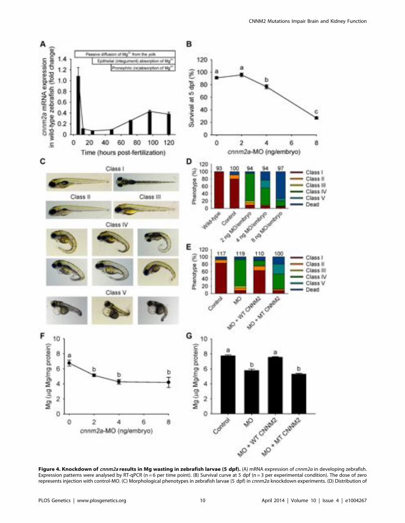
Figure 4. Knockdown of cnnm2a results in Mg wasting in zebrafish larvae (5 dpf). (A) mRNA expression of cnnm2a in developing zebrafish.Expression patterns were analysed by RT-qPCR (n = 6 per time point). (B) Survival curve at 5 dpf (n = 3 per experimental condition). The dose of zerorepresents injection with control-MO. (C) Morphological phenotypes in zebrafish larvae (5 dpf) in cnnm2a knockdown experiments. (D) Distribution of
CNNM2 Mutations Impair Brain and Kidney Function
PLOS Genetics | www.plosgenetics.org 10 April 2014 | Volume 10 | Issue 4 | e1004267
PatientsWe studied a cohort of six patients from five families with
hypomagnesemia and mental retardation. Patients F1.1 to F4.1 arefollowed in secondary or tertiary care neuropediatric centres.Neuroimaging was performed in F1.1, F2.1, F3.1, and F4.1 bycranial MRI (magnetic resonance imaging). Psychological diagnosticevaluation in patients F2.1 and F3.1 was performed using SnijdersOomen Non-Verbal (SON) Intelligence Test (revised) 5.5–17 years.Copy number variations (CNVs) associated with neurodevelopmentaldelay and intellectual disability were excluded in patients F1.1 andF2.1 by array CGH (comparative genomic hybridization) using theSureprint G3 Human CGH Microarray kit (Agilent Technologies,Boeblingen, Germany) in patient F1.1 and using the AffymetrixCytogenetics Whole-Genome 2.7 Array in patient F2.1.
Homozygosity mapping and CNNM2 mutational analysisGenomic DNA of affected individuals and available family
members was extracted from whole blood using standard methods.A genome scan for shared homozygous regions was performed inthe two affected children F1.1 and F1.2 with suspected parentalconsanguinity. Samples were genotyped on an Illumina human660W Quad beadchip SNP array (Illumina, Eindhoven, TheNetherlands). Merlin 1.1.2 (University of Michigan, Ann Arbor,MI, USA) was used to determine homozygous regions by linkageanalysis. As exact information on pedigree structure was missing,we used a 1.7 Mb threshold for regions identical by descent that isvery rarely crossed by non-consanguineous samples, but allows toidentify most of the true homozygosity regions if parentalconsanguinity is present [38]. A list of candidate genes withinthe identified homozygous intervals was generated includingknown Refseq genes as well as novel transcripts using EnsemblGenome assembly GRCh37 via biomart (www.ensembl.org). At acut-off size of .1.7 Mb, eleven critical intervals were yielded onautosomes with a cumulative size of 62 Mb. The gene listgenerated from these loci included 322 RefSeq genes and putativetranscripts, including CNNM2 in a critical interval of 7.1 Mb onchromosome 10. The entire coding region and splice-sites of themost promising candidate gene CNNM2 were sequenced fromboth strands (Genbank: NM_017649.4, Uniprot: Q9H8M5). Afterdiscovery of a homozygous mutation in the index family F1, themutational screening was extended to patients with hypomagne-semia without mutations in known genes involved in hereditarymagnesium wasting. The presence of newly identified CNNM2sequence variations was tested in at least 204 ethnically matchedcontrol alleles and compared to publically available exome data(www.1000genomes.org; evs.gs.washington.edu). Additionally, allidentified mutations were ranked for potential damage on proteinfunction using Polyphen-2 (genetics.bwh.harvard.edu/cgi-bin/ggi/ggi2.cgi).
DNA constructsMouse wild-type Cnnm2 construct was cloned into the pCINeo
HA IRES GFP vector as described previously [12]. Cnnm2
mutations were inserted in the construct using the QuikChangesite-directed mutagenesis kit (Stratagene, La Jolla, CA, USA)according to the manufacturer’s protocol. All constructs wereverified by sequence analysis. Primer sequences used for cloning ormutagenesis PCR are reported in Table S1.
Cell cultureHEK293 cells were grown in Dulbecco’s modified eagle’s
medium (DMEM, Bio Whittaker-Europe, Verviers, Belgium)containing 10% (v/v) fetal calf serum (PAA, Liz, Austria), 2 mML-glutamine and 10 mg/mL non-essential amino acids, at 37uC ina humidity-controlled incubator with 5% (v/v) CO2. The cellswere transiently transfected with the respective DNA constructsusing Lipofectamin 2000 (Invitrogen, Breda, The Netherlands) at1:2 DNA:Lipofectamin ratio for 48 hours unless otherwise stated.
Cell surface biotinylationHEK293 cells were transfected with wild-type and mutant
CNNM2 constructs for 48 hours. Subsequently, cell surfaceproteins were biotinylated as described previously [39]. Briefly,cell surface proteins were biotinylated for 30 min at 4uC in0.5 mg/mL sulfo-NHS-LC-LC-biotin (Pierce, Rockford, IL,USA). Cells were washed and lysed in lysis buffer (150 mM NaCl,5 mM EGTA, Triton 1% (v/v), 1 mg/ml pepstatin, 1 mM PMSF,5 mg/ml leupeptin, 5 mg/ml aproptin, 50 mM Tris/HCl, pH 7.5).10% (v/v) of the sample was taken as input control and the rest ofthe protein lysates were incubated overnight with NeutrAvidin-agarose beads (Pierce, Rockford, IL, USA) at 4uC. The next day,unbound protein was discarded by washing the beads 5 times withlysis buffer. The remaining protein lysates were denatured inLaemmli containing 100 mM DTT for 30 min at 37uC andsubsequently subjected to SDS-PAGE. Then, immunoblots wereincubated with mouse anti-HA 1:5,000 primary antibodies (CellSignaling Technology, Danvers, MA, USA) and peroxidaseconjugated sheep anti-mouse secondary antibodies 1:10,000(Jackson Immunoresearch, Suffolk, UK).
Magnesium transport assaysHEK293 cells were transfected with wild-type and mutant
CNNM2 constructs for 48 hours and seeded on poly-L-lysine(Sigma, St Louis, MO, USA) coated 12-well plates. Mg2+ uptakewas determined using a stable 25Mg2+ isotope (Cortecnet, VoisinsLe Bretonneux, France), which has a natural abundance of 610%.Cells were washed with basic uptake buffer (125 mM NaCl, 5 mMKCl, 0.5 mM CaCl2, 0.5 mM Na2HPO4, 0.5 mM Na2SO4,15 mM HEPES/NaOH, pH 7.5) and subsequently placed inbasic uptake buffer containing 1 mM 25Mg2+ (purity 698%) for5 minutes unless stated differently. After washing three times withice-cold PBS, the cells were lysed in HNO3 ($65%, Sigma) andsubjected to ICP-MS (inductively coupled plasma mass spectrom-etry) analysis. For extrusion experiments, cells were transfectedwith wild-type or mutant CNNM2 constructs for 24 hours. After24 hours, cells were placed in culture medium containing 1 mM
morphological phenotypes in zebrafish larvae (5 dpf) untreated (wild-type) or injected with different doses of cnnm2a-MO or control-MO. Numberson top of the bars indicate the number of animals in each experimental condition. (E) Distribution of morphological phenotypes in zebrafish larvae at5 dpf in rescue experiments. The wild-type phenotype (class I) was restored in morphants by co-injection of cnnm2a-MO (2 ng MO/embryo) withwild-type (WT) CNNM2 cRNA (50 pg cRNA/embryo), but not with mutant (MT, p.Glu357Lys) CNNM2 cRNA (50 pg cRNA/embryo). (F) Magnesiumcontent in zebrafish injected with different doses of cnnm2a-MO, the dose of zero represents injection with control-MO (n = 10 per experimentalcondition except in 8 ng MO-injected zebrafish where n = 5). (G) Rescue of Mg wasting in morphant zebrafish by co-injection of cnnm2a-MO (2 ngMO/embryo) with cRNA encoding for wild-type (WT) CNNM2 (50 pg cRNA/embryo). Co-injection with cRNA encoding for mutant (MT, p.Glu357Lys)CNNM2 (50 pg cRNA/embryo) did not restore Mg levels (n = 10 per experimental condition). Data are presented as mean 6 SEM. Different lettersindicate significant differences between mean values in experimental groups (P,0.05).doi:10.1371/journal.pgen.1004267.g004
CNNM2 Mutations Impair Brain and Kidney Function
PLOS Genetics | www.plosgenetics.org 11 April 2014 | Volume 10 | Issue 4 | e1004267
CNNM2 Mutations Impair Brain and Kidney Function
PLOS Genetics | www.plosgenetics.org 12 April 2014 | Volume 10 | Issue 4 | e1004267
25Mg2+ (purity 698%) for an additional 48 hours. Before the startof the experiment, the cells were briefly washed in basic uptakebuffer and subsequently placed in basic uptake buffer containing0.5 mM Mg2+ (containing 610% 25Mg2+) for 5 minutes. Afterwashing three times with ice-cold PBS, the cells were lysed in nitricacid and subjected to ICP-MS analysis.
Morpholino knockdown and rescue experimentsWild-type Tupfel long-fin zebrafish were bred and raised under
standard conditions (28.5uC and 14 h of light: 10 h of dark cycle)in accordance with international and institutional guidelines.Zebrafish eggs were obtained from natural spawning. Thefollowing antisense oligonucleotides (MOs) were raised againstthe translational start site of cnnm2a and cnnm2b, along with thestandard mismatch control MO (Gene Tools, Philomath, OR,USA): cnnm2a, 59-GCGGTCCATTGCTCTGCCATGTTGA-39;cnnm2b, 59-ACCGACGGTTCTGCCATGTTGATAA-39; andthe negative control (standard mismatch MO), directed against ahuman b-globin intron mutation, 59-CCTCTTACCTCAGTTA-CAATTTATA. The underlined areas indicate the complementa-ry sequences to the initial methionines of cnnm2a and cnnm2b. MOswere diluted in deionized, sterile water supplemented with 0.5%(w/v) phenol red and injected in a volume of 1 nl into the yolk ofone- to two-cell stage embryos using a Pneumatic PicoPumppv280 (World Precision Instruments, Sarasota, FL, USA). Wild-type (uninjected) embryos were also included in the experiments tocontrol for the effects of the injection procedure per se. Todetermine the most effective dose of the cnnm2a- and cnnm2b-MO,2, 4 and 8 ng were injected. In these experiments, control embryoswere injected with 8 ng of the standard mismatch control MO (thehighest dose). After injection, embryos from the same experimentalcondition were placed in 3 Petri dishes (at a maximum density of45 embryos/dish, allowing statistical comparisons between surviv-als in the different experimental conditions) and cultured at 28.5uCin E3 embryo medium (5 mM NaCl, 0.17 mM KCl, 0.33 mMCaCl2, 0.33 mM MgSO4), which was refreshed daily. As criteriafor subsequent experiments, the dose of MO that caused majoreffects and induced a percentage of mortality non-significantlydifferent from controls was injected (2 ng for cnnm2a-MO and 8 ngfor cnnm2b-MO). In experiments that implied exposure to highMg2+ concentrations, the high Mg2+ medium had a composition of5 mM NaCl, 0.17 mM KCl, 0.33 mM CaCl2 and 25 mMMgSO4. In order to control for the specificity of the MOsblocking the translation of cnnm2a and cnnm2b, as well as for toxicoff-target effects, in vivo cRNA rescue experiments were performed[40]. For these experiments, mouse wild-type CNNM2 andmutant (p.Glu357Lys) CNNM2 cRNAs were prepared using themMESSAGE mMACHINE Kit (Ambion, Austin, TX, USA)according to the manufacturer’s instructions. The cRNAs, in anamount of 50 pg, as based on other studies [26], were (co)injected
together with MOs as described above. Zebrafish embryos andlarvae were phenotyped at 25 hpf or 5 dpf, respectively.
Analysis of phenotypes in embryos and larvaeFor the analyses of brain phenotypes, the brain rudiment of
zebrafish embryos at 25 hpf was observed for morphologicalchanges under a Leica MZFLIII microscope (Leica MicrosystemsLtd, Heerburgg, Germany). Morphological phenotypes, whichalso included the brain, were also analysed in larvae at 5 dpf.Embryos or larvae were classified into different classes ofphenotypes on the basis of comparisons with stage-matchedcontrol embryos of the same clutch. In cnnm2a morphant embryos(25 hpf), two phenotype classes were distinguished: class I, normal;and class II, embryos with underdeveloped MHB. In cnnm2amorphant larvae (5 dpf), the following classes were distinguished:class I, normal; class II, normal with non-inflated swim bladder;class III, larvae with enlarged pericardial cavity (edema) and non-inflated swim bladder; class IV, larvae with notochord defects,enlarged pericardial cavity and non-inflated swim bladder; andclass V, larvae with severely enlarged pericardial cavity, notochorddefects and non-inflated swim bladder. In cnnm2b morphantembryos (25 hpf), three different phenotypes were distinguished:class I, normal; class II, embryos with underdeveloped MHB; andclass III, embryos with underdeveloped MHB and enlargedtectum. In cnnm2b morphant larvae (5 dpf), the number of differentphenotypes distinguished were four: class I, normal; class II,normal with non-inflated swim bladder; class III, larvae withenlarged pericardial cavity (edema) and non-inflated swimbladder; and class IV, larvae with widened outer cerebrospinalfluid spaces, kidney cysts, severely enlarged pericardial cavity andnon-inflated swim bladder. Representative images were obtainedwith a DFC450C camera (Leica Microsystems Ltd) afteranaesthetising embryos or larvae with tricaine/Tris pH 7.0solution. Prior to anaesthesia and image acquisition, zebrafishembryos were manually dechorionated.
Magnesium determinations in embryos and larvaeZebrafish embryos or larvae were anesthetized with tricaine/
Tris pH 7.0 solution and 5–7 individuals were pooled as onesample. Samples were then snap frozen in liquid nitrogen andstored at 280uC in order to ensure euthanasia of animals andremained at these storage conditions until the beginning of theanalytical procedures.
Analytical procedures started by quickly washing the sampleswith nanopure water in order to avoid contamination of remainingwaterborne Mg2+. The washing procedure was repeated twice.Fish were then dried at 65uC for 1.5 hours, at which time 2.5 ml ofHNO3 ($65%, Sigma) was added to each tube. Samples weredigested at 65uC during 1.5 hours. After, digested samples werediluted 1:10 with 22.5 ml nanopure water. The total Mg content in
Figure 5. Knockdown of cnnm2b results in Mg wasting and brain malformations in zebrafish larvae (5 dpf). (A) mRNA expression ofcnnm2b in developing zebrafish. Expression patterns were analysed by RT-qPCR (n = 6 per time point). (B) Survival curve at 5 dpf (n = 3 perexperimental condition). The dose of zero represents injection with control-MO. (C) Morphological phenotypes in zebrafish larvae (5 dpf) in cnnm2bknockdown experiments. (D) Distribution of morphological phenotypes in zebrafish larvae (5 dpf) untreated (wild-type) or injected with differentdoses of cnnm2b-MO or control-MO. Brain malformations (widened cerebrospinal fluid spaces, class IV phenotype) are prominent in morphantsinjected with 4–8 ng MO/embryo. Numbers on top of the bars indicate the number of animals in each experimental condition. (E) Distribution ofmorphological phenotypes in zebrafish larvae at 5 dpf in rescue experiments. The wild-type phenotype (class I) was restored in morphants by co-injection of cnnm2b-MO (8 ng MO/embryo) with wild-type (WT) CNNM2 cRNA (50 pg cRNA/embryo), but not with mutant (MT, p.Glu357Lys) CNNM2cRNA (50 pg cRNA/embryo). (F) Magnesium content in zebrafish injected with different doses of cnnm2b-MO. The dose of zero represents injectionwith control-MO (n = 10 per experimental condition). (G) Rescue of Mg wasting in morphant zebrafish by co-injection of cnnm2b-MO (8 ng MO/embryo) with cRNA encoding for wild-type (WT) CNNM2 (50 pg cRNA/embryo). Co-injection with cRNA encoding for mutant (MT, p.Glu357Lys)CNNM2 (50 pg cRNA/embryo) did not restore Mg levels (n = 10 per experimental condition). Data are presented as mean 6 SEM. Different lettersindicate significant differences between mean values in experimental groups (P,0.05).doi:10.1371/journal.pgen.1004267.g005
CNNM2 Mutations Impair Brain and Kidney Function
PLOS Genetics | www.plosgenetics.org 13 April 2014 | Volume 10 | Issue 4 | e1004267
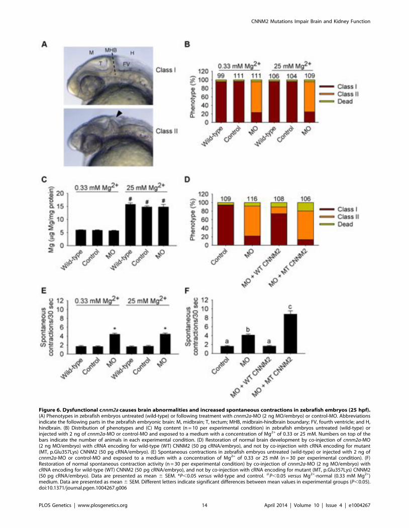
Figure 6. Dysfunctional cnnm2a causes brain abnormalities and increased spontaneous contractions in zebrafish embryos (25 hpf).(A) Phenotypes in zebrafish embryos untreated (wild-type) or following treatment with cnnm2a-MO (2 ng MO/embryo) or control-MO. Abbreviationsindicate the following parts in the zebrafish embryonic brain: M, midbrain; T, tectum; MHB, midbrain-hindbrain boundary; FV, fourth ventricle; and H,hindbrain. (B) Distribution of phenotypes and (C) Mg content (n = 10 per experimental condition) in zebrafish embryos untreated (wild-type) orinjected with 2 ng of cnnm2a-MO or control-MO and exposed to a medium with a concentration of Mg2+ of 0.33 or 25 mM. Numbers on top of thebars indicate the number of animals in each experimental condition. (D) Restoration of normal brain development by co-injection of cnnm2a-MO(2 ng MO/embryo) with cRNA encoding for wild-type (WT) CNNM2 (50 pg cRNA/embryo), and not by co-injection with cRNA encoding for mutant(MT, p.Glu357Lys) CNNM2 (50 pg cRNA/embryo). (E) Spontaneous contractions in zebrafish embryos untreated (wild-type) or injected with 2 ng ofcnnm2a-MO or control-MO and exposed to a medium with a concentration of Mg2+ of 0.33 or 25 mM (n = 30 per experimental condition). (F)Restoration of normal spontaneous contraction activity (n = 30 per experimental condition) by co-injection of cnnm2a-MO (2 ng MO/embryo) withcRNA encoding for wild-type (WT) CNNM2 (50 pg cRNA/embryo), and not by co-injection with cRNA encoding for mutant (MT, p.Glu357Lys) CNNM2(50 pg cRNA/embryo). Data are presented as mean 6 SEM. *P,0.05 versus wild-type and control. #P,0.05 versus Mg2+-normal (0.33 mM Mg2+)medium. Data are presented as mean 6 SEM. Different letters indicate significant differences between mean values in experimental groups (P,0.05).doi:10.1371/journal.pgen.1004267.g006
CNNM2 Mutations Impair Brain and Kidney Function
PLOS Genetics | www.plosgenetics.org 14 April 2014 | Volume 10 | Issue 4 | e1004267
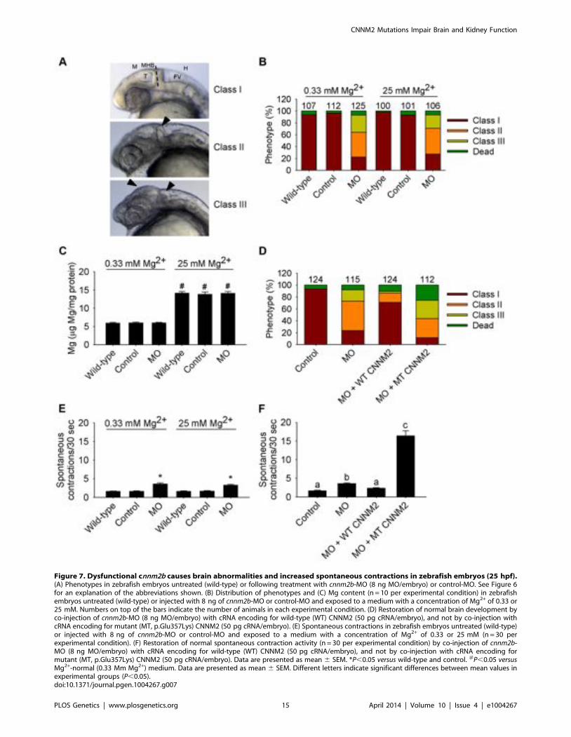
Figure 7. Dysfunctional cnnm2b causes brain abnormalities and increased spontaneous contractions in zebrafish embryos (25 hpf).(A) Phenotypes in zebrafish embryos untreated (wild-type) or following treatment with cnnm2b-MO (8 ng MO/embryo) or control-MO. See Figure 6for an explanation of the abbreviations shown. (B) Distribution of phenotypes and (C) Mg content (n = 10 per experimental condition) in zebrafishembryos untreated (wild-type) or injected with 8 ng of cnnm2b-MO or control-MO and exposed to a medium with a concentration of Mg2+ of 0.33 or25 mM. Numbers on top of the bars indicate the number of animals in each experimental condition. (D) Restoration of normal brain development byco-injection of cnnm2b-MO (8 ng MO/embryo) with cRNA encoding for wild-type (WT) CNNM2 (50 pg cRNA/embryo), and not by co-injection withcRNA encoding for mutant (MT, p.Glu357Lys) CNNM2 (50 pg cRNA/embryo). (E) Spontaneous contractions in zebrafish embryos untreated (wild-type)or injected with 8 ng of cnnm2b-MO or control-MO and exposed to a medium with a concentration of Mg2+ of 0.33 or 25 mM (n = 30 perexperimental condition). (F) Restoration of normal spontaneous contraction activity (n = 30 per experimental condition) by co-injection of cnnm2b-MO (8 ng MO/embryo) with cRNA encoding for wild-type (WT) CNNM2 (50 pg cRNA/embryo), and not by co-injection with cRNA encoding formutant (MT, p.Glu357Lys) CNNM2 (50 pg cRNA/embryo). Data are presented as mean 6 SEM. *P,0.05 versus wild-type and control. #P,0.05 versusMg2+-normal (0.33 Mm Mg2+) medium. Data are presented as mean 6 SEM. Different letters indicate significant differences between mean values inexperimental groups (P,0.05).doi:10.1371/journal.pgen.1004267.g007
CNNM2 Mutations Impair Brain and Kidney Function
PLOS Genetics | www.plosgenetics.org 15 April 2014 | Volume 10 | Issue 4 | e1004267
each sample was determined with a commercial colorimetric assay(Roche Diagnostics, Woerden, The Netherlands) following themanufacturer’s protocol. Blanks (HNO3 diluted 1:10 withnanopure water) were added during assays and values were equalto zero. Within-run precision and accuracy was controlled bymeans of an internal control Precinorm (Roche Diagnostics).Samples from embryos exposed to 25 mM Mg2+ were furtherdiluted (1:20). Furthermore, samples were normalized by proteincontent, which was determined in 1:20 (embryos at 25 hpf) or 1:50(larvae at 5 dpf) diluted samples using the Pierce BCA proteinassay kit (Pierce Biotechnology, Rockford, IL, USA).
Total RNA isolation, cDNA synthesis and quantitativereal-time PCR analysis
Zebrafish embryos or larvae at specific developmental times (6,12, 24, 48, 72, 96 and 120 hpf) were anaesthetised with tricaine/Tris pH 7 solution and 10 individuals were pooled as one sample.RNA isolation, cDNA synthesis and quantitative real-time PCR(RT-qPCR) measurements were carried out as previouslydescribed using validated cnnm2a and cnnm2b primers [18].Samples were normalized to the expression level of the house-keeping gene elongation factor-1a (elf1a) [18]. Relative mRNAexpression was analysed using the Livak method (22DDCt), whereresults are expressed relative to the gene expression at 6 hpf (timepoint chosen as calibrator).
Spontaneous contraction analysis and touch-evokedescape behaviour
At 25 hpf, 10 zebrafish embryos per Petri dish (n = 30 perexperimental condition) were randomly selected. The number ofcomplete body contractions each zebrafish made in 30 secondperiod was counted and was used as indicative of motor neuronactivity [27]. Representative videos of each experimental conditionwere taken using Leica Application Suite (Leica Microsystems Ltd)and a Leica MZFLIII microscope (Leica Microsystems Ltd)equipped with a DFC450C camera (Leica Microsystems Ltd).
For the analysis of the touch-evoked escape behaviour, 10zebrafish larvae per Petri dish (n = 30 per experimental condition)were randomly selected. Touch-evoked escape behaviours wereelicited by touching a larva in the tail up to 6 times with a pair offorceps at 5 dpf. Three categories were distinguished, responders,late responders and non-responders, to which the following scoreswere given: 3 points for responders: fish quickly react (swimmingor flicking the tail) to the stimuli after 1 or 2 twitches; 2 points forlate responders: fish react (swimming or flicking the tail) to thestimuli after 3, 4 or 5 twitches; and 1 point for non-responders: fishdo not react to the stimuli after more than 5 twitches. Represen-tative videos were recorded with the system described above.
Statistical analysisAll results are depicted as mean 6 standard error of the mean
(SEM). Statistical analyses were conducted by one- or two-way (forexperiments where zebrafish embryos were exposed to differentMg2+ concentrations, then two factors of variance appear: Mg2+
concentration and treatment) ANOVA. Where appropriate, datawere logarithmically transformed to fulfil the requirements forANOVA, but all data are shown in their decimal values for clarity.When data did not comply with the premises of the parametricANOVA, data were analyzed using a Kruskal-Wallis ANOVA onranks. Tukey’s post-test was used to identify significantly differentgroups. Statistical significance was accepted at P,0.05.
Supporting Information
Figure S1 Impairment of touch-evoked escape behaviour incnnm2a morphant zebrafish. Touch-evoked escape behaviour scorein zebrafish cnnm2a morphants at 5 dpf after injection of 2 ngcontrol-MO/embryo, 2 ng cnnm2a-MO/embryo, 2 ng cnnm2a-MO/embryo+50 pg wild-type (WT) CNNM2 cRNA/embryo, or2 ng cnnm2a-MO/embryo+50 pg mutant (MT, p.Glu357Lys)CNNM2 cRNA/embryo. Three categories were distinguished,responders, late responders and non-responders, to which thefollowing scores were given: 3 points for responders: fish quicklyreact (swimming or flicking the tail) to the stimuli after 1 or 2twitches; 2 points for late responders: fish react (swimming orflicking the tail) to the stimuli after 3, 4 or 5 twitches; and 1 pointfor non-responders: fish do not react to the stimuli after more than5 twitches. The upper part of the figure shows frames of videosshowing touch-evoked escape contractions at 5 dpf of control andmorphant zebrafish larvae. Time of each video frame is indicatedin centisenconds (cs). Data are shown as mean 6 SEM (n = 30).Different letters indicate significant differences between meanvalues in experimental groups (P,0.05).(TIF)
Figure S2 Impairment of touch-evoked escape behaviour incnnm2b morphant zebrafish. Touch-evoked escape behaviour scorein zebrafish cnnm2b morphants at 5 dpf after injection of 8 ngcontrol-MO/embryo, 8 ng cnnm2b-MO/embryo, 8 ng cnnm2b-MO/embryo+50 pg wild-type (WT) CNNM2 cRNA/embryo, or2 ng cnnm2b-MO/embryo+50 pg mutant (MT, p.Glu357Lys)CNNM2 cRNA/embryo. Three categories were distinguished,responders, late responders and non-responders, to which thefollowing scores were given: 3 points for responders: fish quicklyreact (swimming or flicking the tail) to the stimuli after 1 or 2twitches; 2 points for late responders: fish react (swimming orflicking the tail) to the stimuli after 3, 4 or 5 twitches; and 1 pointfor non-responders: fish do not react to the stimuli after more than5 twitches. The upper part of the figure shows frames of videosshowing touch-evoked escape contractions at 5 dpf of control andmorphant zebrafish larvae. Time of each video frame is indicatedin centisenconds (cs). Data are shown as mean 6 SEM (n = 30).Different letters indicate significant differences between meanvalues in experimental groups (P,0.05).(TIF)
Movie S1 Spontaneous contraction frequency in zebrafishembryos (25 hpf) injected with a dose of 2 ng control-MO/embryo.(AVI)
Movie S2 Spontaneous contraction frequency in zebrafishembryos (25 hpf) injected with a dose of 2 ng cnnm2a-MO/embryo.(AVI)
Movie S3 Spontaneous contraction frequency in zebrafishembryos (25 hpf) co-injected with a dose of 2 ng cnnm2a-MO/embryo and 50 pg of mutant (p.Glu357Lys) CNNM2 cRNA.(AVI)
Movie S4 Spontaneous contraction frequency in zebrafishembryos (25 hpf) injected with a dose of 8 ng control-MO/embryo.(AVI)
Movie S5 Spontaneous contraction frequency in zebrafishembryos (25 hpf) injected with a dose of 8 ng cnnm2b-MO/embryo.(AVI)
CNNM2 Mutations Impair Brain and Kidney Function
PLOS Genetics | www.plosgenetics.org 16 April 2014 | Volume 10 | Issue 4 | e1004267
Movie S6 Spontaneous contraction frequency in zebrafishembryos (25 hpf) co-injected with a dose of 8 ng cnnm2b-MO/embryo and 50 pg of mutant (p.Glu357Lys) CNNM2 cRNA.(AVI)
Table S1 Primer sequences for mutagenesis PCR.(DOCX)
Acknowledgments
The authors are grateful to the patients for their participation in this study.We thank Lonneke Duijkers, Jelle Eygensteyn, Margo Dona, Sami
Mohammed and Andreas Kompatscher for excellent technical support.We thank W. Schwindt, Department of Clinical Radiology, and HeymutOmran, Department of General Pediatrics, University Hospital Munsterfor help with the interpretation of neuroimaging studies.
Author Contributions
Conceived and designed the experiments: FJA JHFdB KPS MK RJMBJGJH. Performed the experiments: FJA JHFdB KPS SRe GCK BN SRuNR. Analyzed the data: FJA JHFdB KPS GCK BN SRu MK RJMBJGJH. Contributed reagents/materials/analysis tools: ALLL EvW GF.Wrote the paper: FJA JHFdB KPS MK RJMB JGJH.
References
1. Dimke H, Monnens L, Hoenderop JG, Bindels RJ (2012) Evaluation ofHypomagnesemia: Lessons From Disorders of Tubular Transport. Am J KidneyDis 62: 377–83.
2. Moser J, Kilb W, Werhahn KJ, Luhmann HJ (2006) Early developmentalalterations of low-Mg2+ -induced epileptiform activity in the intact corticohip-pocampal formation of the newborn mouse in vitro. Brain Res 1077: 170–177.
3. Jin J, Wu LJ, Jun J, Cheng X, Xu H, et al. (2012) The channel kinase, TRPM7,is required for early embryonic development. Proc Natl Acad Sci USA 109:E225–E233.
4. van de Graaf SF, Bindels RJ, Hoenderop JG (2007) Physiology of epithelial Ca2+
and Mg2+ transport. Rev Physiol Biochem Pharmacol 158: 77–160.5. Runnels LW (2011) TRPM6 and TRPM7: A Mul-TRP-PLIK-cation of channel
functions. Curr Pharm Biotechnol 12: 42–53.6. Hoenderop JG, Bindels RJ (2008) Calciotropic and magnesiotropic TRP
channels. Physiology 23: 32–40.7. Thebault S, Alexander RT, Tiel Groenestege WM, Hoenderop JG, Bindels RJ
(2009) EGF increases TRPM6 activity and surface expression. J Am SocNephrol 20: 78–85.
8. Voets T, Nilius B, Hoefs S, van der Kemp AW, Droogmans G, et al. (2004)TRPM6 forms the Mg2+ influx channel involved in intestinal and renal Mg2+
absorption. J Biol Chem 279: 19–25.9. Stuiver M, Lainez S, Will C, Terryn S, Gunzel D, et al. (2011) CNNM2,
encoding a basolateral protein required for renal Mg2+ handling, is mutated indominant hypomagnesemia. Am J Hum Genet 88: 333–343.
10. de Baaij JH, Groot Koerkamp MJ, Lavrijsen M, van Zeeland F, Meijer H, et al.(2013) Elucidation of the distal convoluted tubule transcriptome identifies newcandidate genes involved in renal magnesium handling. Am J Physiol RenalPhysiol 305: F1563–73. doi:10.1152/ajprenal.00322.2013.
11. Goytain A, Quamme GA (2005) Functional characterization of ACDP2 (ancientconserved domain protein), a divalent metal transporter. Physiol Genomics 22:382–389.
12. de Baaij JH, Stuiver M, Meij IC, Lainez S, Kopplin K, et al. (2012) Membranetopology and intracellular processing of cyclin M2 (CNNM2). J Biol Chem 287:13644–13655.
13. Wang CY, Shi JD, Yang P, Kumar PG, Li QZ, et al. (2003) Molecular cloningand characterization of a novel gene family of four ancient conserved domainproteins (ACDP). Gene 306: 37–44.
14. Ripke S, Sanders AR, Kendler KS, Levinson DF, Sklar P, et al. (2011) Genome-wide association study identifies five new schizophrenia loci. Nat Genet 43: 969–976.
15. Newton-Cheh C, Johnson T, Gateva V, Tobin MD, Bochud M, et al. (2009)Genome-wide association study identifies eight loci associated with bloodpressure. Nat Genet 41: 666–676.
16. Hwang P-P, Chou M-Y (2013) Zebrafish as an animal model to study ionhomeostasis. Pflugers Arch 465: 1233–1247.
17. Lieschke GJ, Currie PD (2007) Animal models of human disease: zebrafish swiminto view. Nat Rev Genet 8: 353–367.
18. Arjona FJ, Chen YX, Flik G, Bindels RJ, Hoenderop JG (2013) Tissue-specificexpression and in vivo regulation of zebrafish orthologues of mammalian genesrelated to symptomatic hypomagnesemia. Pflugers Arch 465: 1409–1421.
19. Walder RY, Landau D, Meyer P, Shalev H, Tsolia M, et al. (2002) Mutation ofTRPM6 causes familial hypomagnesemia with secondary hypocalcemia. NatGenet 31: 171–174.
20. Sponder G, Svidova S, Schweigel M, Vormann J, Kolisek M (2010) Splice-variant 1 of the ancient domain protein 2 (ACDP2) complements the
magnesium-deficient growth phenotype of Salmonella enterica sv. typhimurium strainMM281. Magnes Res 23: 105–114.
21. White JR, Cameron AE (1948) The Natural Abundance of Isotopes of StableElements. Phys Rev 74: 991–1000.
22. Chokshi R, Fruasaha P, Kozak JA (2012) 2-aminoethyl diphenyl borinate (2-APB) inhibits TRPM7 channels through an intracellular acidification mecha-nism. Channels 6: 362–369.
23. Whittam R, Wheeler KP (1961) The sensitivity of a kidney ATPase to ouabainand to sodium and potassium. Biochim Biophys Acta 51: 622–624.
24. Kolisek M, Nestler A, Vormann J, Schweigel-Rontgen M (2012) Human geneSLC41A1 encodes for the Na+/Mg(2)+ exchanger. Am J Physiol Cell Physiol 302:C318–C326.
25. Goytain A, Quamme GA (2005) Identification and characterization of a novelmammalian Mg2+ transporter with channel-like properties. BMC Genomics 6: 48.
26. Mahmood F, Mozere M, Zdebik AA, Stanescu HC, Tobin J, et al. (2013)Generation and validation of a zebrafish model of EAST (epilepsy, ataxia,sensorineural deafness and tubulopathy) syndrome. Dis Model Mech 6: 652–660.
27. Saint-Amant L, Drapeau P (1998) Time course of the development of motorbehaviors in the zebrafish embryo. J Neurobiol 37: 622–632.
28. Elizondo MR, Arduini BL, Paulsen J, MacDonald EL, Sabel JL, et al. (2005)Defective skeletogenesis with kidney stone formation in dwarf zebrafish mutantfor trpm7. Curr Biol 15: 667–671.
29. Low SE, Amburgey K, Horstick E, Linsley J, Sprague SM, et al. (2011) TRPM7is required within zebrafish sensory neurons for the activation of touch-evokedescape behaviors. J Neurosci 31: 11633–11644.
30. Veltman JA, Brunner HG (2012) De novo mutations in human genetic disease.Nat Rev Genet 13: 565–575.
31. Hodgkinson A, Eyre-Walker A (2011) Variation in the mutation rate acrossmammalian genomes. Nat Rev Genet 12: 756–766.
32. Thisse B, Thisse C (2004) Fast Release Clones: A High Throughput ExpressionAnalysis. ZFIN Direct Dat Submission (Http://zfin.org).
33. Scholpp S, Brand M (2009) Neural Patterning: Midbrain–Hindbrain Boundary.In: Larry RS, editor. Encyclopedia of Neuroscience. Oxford: Academic Press.pp. 205–211.
34. Dworkin S, Jane SM (2013) Novel mechanisms that pattern and shape themidbrain-hindbrain boundary. Cell Mol Life Sci 70: 3365–3374.
35. Jin J, Wu LJ, Jun J, Cheng X, Xu H, et al. (2012) The channel kinase, TRPM7,is required for early embryonic development. Proc Natl Acad Sci USA 109:E225–E233.
36. Krapivinsky G, Mochida S, Krapivinsky L, Cibulsky SM, Clapham DE (2006)The TRPM7 ion channel functions in cholinergic synaptic vesicles and affectstransmitter release. Neuron 52: 485–496.
37. Brauchi S, Krapivinsky G, Krapivinsky L, Clapham DE (2008) TRPM7facilitates cholinergic vesicle fusion with the plasma membrane. Proc Natl AcadSci USA 105: 8304–8308.
38. Hildebrandt F, Heeringa SF, Ruschendorf F, Attanasio M, Nurnberg G, et al.(2009) A systematic approach to mapping recessive disease genes in individualsfrom outbred populations. PLOS Genet 5: e10000353.
39. Gkika D, Mahieu F, Nilius B, Hoenderop JG, Bindels RJ (2004) 80K-H as a newCa2+ sensor regulating the activity of the epithelial Ca2+ channel transientreceptor potential cation channel V5 (TRPV5). J Biol Chem 279: 26351–26357.
40. Eisen JS, Smith JC (2008) Controlling morpholino experiments: don’t stopmaking antisense. Development 135: 1735–1743.
CNNM2 Mutations Impair Brain and Kidney Function
PLOS Genetics | www.plosgenetics.org 17 April 2014 | Volume 10 | Issue 4 | e1004267