Embed Size (px)
Citation preview
CMC Certification Review Course: Handout
Session #: 2
Congestive Heart Failure Cardiomyopathy
Presented by:
Leanna R. Miller RN, MN, CCRN-CSC, PCCN-CMC, CEN, CNRN, CMSRN, NP
An AACN Critical Care Publication 101 Columbia Aliso Viejo, CA 92656-1491 ©2013 American Association of Critical-Care Nurses All rights reserved. AACN grants permission for a single individual to print one copy of this electronic publication. No additional copies are permitted. No part of this electronic publication may be reproduced, uploaded, stored in a retrieval system, or transmitted, in any form or by any means (electronic, photocopying, recording or otherwise) without the prior written permission of AACN.

3/28/2013
1
EXIT Closes themodule
BACK Displays
previous slide
PAUSE/PLAY Pauses or
resumes audio
NEXT Displays next slide
PROGRESS BAR Purple shows your progress within the module
AUDIOMutes
audio for entire
module
TABLE of SLIDESNavigate to any slide by clicking on its tab.
CURRENT SLIDE
COMPLETED SLIDE
BOOKMARK
COMPLETED/TOTAL TIME
How to use this module:
CMC Review CourseSession 2:
Congestive Heart Failure Cardiomyopathy
Leanna R. Miller
RN, MN, CCRN-CSC, PCCN-CMC, CEN, CNRN, CMSRN, NP
3/28/2013
2
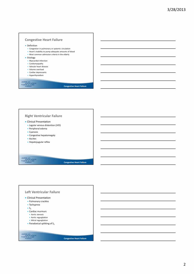
� Definition
� Congestion in pulmonary or systemic circulation
� Heart’s inability to pump adequate amounts of blood
� Most common admission criteria in the elderly
� Etiology
� Myocardial infarction
� Cardiomyopathy
� Valvular heart disease
� Volume overload
� Cardiac depressants
� Hyperthyroidism
Congestive Heart Failure
Congestive Heart Failure
� Clinical Presentation
� Jugular venous distention (JVD)
� Peripheral edema
� Cyanosis
� Congestive hepatomegaly
� Ascites
� Hepatojugular reflex
Right Ventricular Failure
Congestive Heart Failure
� Clinical Presentation
� Pulmonary crackles
� Tachypnea
� S3
� Cardiac murmurs
� Aortic stenosis
� Aortic regurgitation
� Mitral regurgitation
� Paradoxical splitting of S2
Left Ventricular Failure
Congestive Heart Failure

3/28/2013
3
� Lab
� CBC
� Blood urea nitrogen (BUN)
� Creatinine
� Liver enzymes
� Thyroid stimulating hormone (TSH)
� B-type natriuretic peptide (BNP)
� Imaging
� Chest x-ray
� 2-dimensional (2-D) echocardiography
� Cardiac catheterization
CHF: Diagnostics
Congestive Heart Failure
� Substance secreted from the ventricles of the heart in response to changes in pressure that occur when heart failure develops and worsens
� Increases when HF symptoms worsen and decreases when HF is stable
� Quick, inexpensive test that enhances current diagnostic assessment tools
� Allows the correct diagnosis of HF
� Future research� Determine prognosis
� Decisions regarding treatment
BNP
Congestive Heart Failure
� < 100 picograms/milliliter (pg/mL) indicates no HF
� 100 − 300 ρg/mL suggests HF is present
� > 300 − < 600 pg/mL indicates mild HF
� > 600 − < 900 pg/mL indicates moderate HF
� > 900 pg/mL indicates severe HF
BNP: Interpretation
Congestive Heart Failure

3/28/2013
4
� Systolic Dysfunction
� Diuretics
� Furosemide
� Aldosterone inhibitor
� ACE inhibitor
� Beta-blockers
� Digitalis
CHF: Goals of Treatment
Congestive Heart Failure
� Systolic Dysfunction (cont.)
� Direct vasodilating drugs
� Hydralazine (Apresoline)
� Isosorbide (Isordil)
� Indications
� Unable to tolerate ACE inhibitor
� ACE inhibitor not effective
CHF: Goals of Treatment
Congestive Heart Failure
� Systolic Dysfunction (cont.)
� Anticoagulants
� Indications
� Atrial fibrillation
� History of embolism
� Antidysrhythmic agents
� Amiodarone
� Modest effect in reducing mortality
� Pulmonary toxicity
CHF: Goals of Treatment
Congestive Heart Failure

3/28/2013
5
� Diastolic Dysfunction: hypertension (HTN)
� Calcium channel blockers
� ACE inhibitors
� Beta-blockers
� Diuretics
CHF: Goals of Treatment
Congestive Heart Failure
� Diastolic Dysfunction: aortic stenosis
� Diuretics
� No ACE inhibitors, nitrates, or digitalis
(except in rate control)
� Aortic valve replacement
CHF: Goals of Treatment
Congestive Heart Failure
� Diastolic Dysfunction: aortic and mitral
regurgitation
� ACE inhibitors with diuretics
� Hydralazine with nitrates if ACE inhibitor is not tolerated
CHF: Goals of Treatment
Congestive Heart Failure

3/28/2013
6
� Diastolic Dysfunction: mitral stenosis
� Diuretics
� Beta-blocker, digitalis, and/or verapamil to
control heart rate
� Repair or replace mitral valve
� Balloon valvuloplasty
CHF: Goals of Treatment
Congestive Heart Failure
Practice Exam Questions
Congestive Heart Failure
A. No indication of CHF
B. Mild CHF
C. Moderate CHF. BNP levels < 100 pg/mL indicates no
HF. BNP levels from 100 – 300 pg/mL indicates HF is
present. Levels > 300 pg/mL indicates mild HF; levels
> 600 pg/mL indicates moderate HF, while levels >
900 pg/mL indicates severe HF.
D. Severe CHF
The patient’s BNP on admission is 625 pg/mL.
This is consistent with:
Congestive Heart Failure
Question #1 - Answer

3/28/2013
7
A. Edema
B. Decreased appetite
C. Profound fatigue. Fatigue was present in patients
before dyspnea. Most patients ignored the fatigue;
however, it increased with worsening NYHA class.
D. Activity intolerance
The symptom of HF associated with worsening New York
Heart Association (NYHA) functional class and a reliable
indicator of hospitalization is:
Congestive Heart Failure
Question #2 - Answer
A. Spironilactone (Aldactone)
B. Metaprolol (Lopressor). Metaprolol and carvedilol
have been found to reduce M&M by 50%. An ACE
inhibitor reduces M&M by 10%. Spironilactone
reduces M&M by an additional 10%. No reduction
directly related to the use of a statin.
C. Lisinopril (Zestril, Prinivil)
D. Simvastatin (Zocor)
Your patient has been diagnosed with systolic HF. The
medication that reduces morbidity and mortality by 50% is:
Congestive Heart Failure
Question #3 - Answer
A. BNP > 300 pg/mL
B. C-reactive protein
C. Wall motion abnormalities on echocardiogram
D. EF < 40%. EF is the factor that differentiates between
diastolic and systolic failure. BNP is an indicator of the
severity of the HF. C-reactive protein elevates in all
inflammatory diseases, and wall motion changes are
related to the etiology of the HF.
The definitive diagnostic indicator for systolic HF is:
Congestive Heart Failure
Question #4 - Answer

3/28/2013
8
Cardiomyopathy
� Anatomic and pathologic diagnosis
� Muscle or electrical dysfunction
� Heterogeneous group of diseases of the
myocardium
� Ventricular hypertrophy and dilatation
Cardiomyopathy
Cardiomyopathy
� Primary
� Genetic
� Mixed
� Acquired
� Secondary
� Infiltrative
� Toxic
� Inflammatory
Classifications
Cardiomyopathy

3/28/2013
9
� Clinical Manifestations
� Highest incidence in middle age
� African-American 2x more frequent than Caucasian
� Men 3x more frequent than women
� Symptoms may have gradual onset
� Acute presentation
� Misdiagnosed as viral upper respiratory infection in young
adults
� Uncommon to find specific myocardial disease on
endomyocardial biopsy
Dilated (Congestive) Cardiomyopathy (DCM)
Cardiomyopathy
� Clinical Presentation
� Right and left HF
� Diagnosis
� Chest x-ray
� Cardiomegaly
� Pulmonary congestion
Dilated Cardiomyopathy
Cardiomyopathy
� Diagnosis
� ECG
� Biventricular hypertrophy
� AF
� Echo
� Diminished wall motion
� Reduced EF
Dilated Cardiomyopathy
Cardiomyopathy

3/28/2013
10
� Diagnosis
� Cardiac catheterization
� Elevated pulmonary artery occlusion pressure (PAOP) and
pulmonary artery pressure (PAP)
� CO and EF
� Mitral valve abnormality
Dilated Cardiomyopathy
Cardiomyopathy
Dilated Cardiomyopathy
Cardiomyopathy
� Manage CHF
� Oxygen to achieve O2 saturation 90%
� ACE inhibitors
� Vasodilators
� Diuretics
� Inotrope (digitalis)
DCM: Management
Cardiomyopathy

3/28/2013
11
� Beta-blocker (antagonist)
� Carvedilol (Coreg)
� Bisoprolol (Zebeta)
� Metoprolol (Lopressor)
� Calcium channel blockers
� Not first-line therapy
DCM: Medications
Cardiomyopathy
� Antidysrhythmic
� ICD
� Anticoagulant
� Coumadin
� AF
� Stroke
� Thrombus
DCM: Treatments
Cardiomyopathy
� Immunosuppressant
� Dual chamber pacing
� Surgery
� Mitral valve replacement (MVR)
� Heart transplant
� Diet and physical exercise
� Decrease O2 demand
� Activity restriction
� Sodium restrictions
� Anxiolytics as needed
DCM: Patient Management
Cardiomyopathy

3/28/2013
12
� Dysrhythmias
� AF
� Ventricular
� Systemic emboli
DCM: Managing Complications
Cardiomyopathy
� Cardiac transplantation
� DCM is the most common indication for cardiac
transplantation
� Survival after transplant is:
� 80% at 1 year
� 70% at 5 years
� LV reduction procedures
� LV reshaping
DCM: Manage Symptoms
Cardiomyopathy
� Idiopathic hypertrophic subaortic stenosis
(IHSS)
� Hypertrophy of the heart muscle
� Rigid, noncompliant ventricle
� Decreased preload and CO
� Outflow tract obstruction
� Decreased coronary and cerebral blood flow
Hypertrophic Cardiomyopathy (HCM): Introduction
Cardiomyopathy
3/28/2013
13
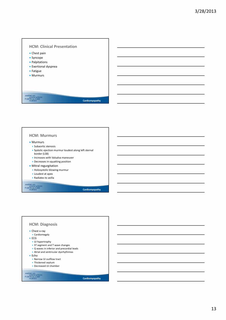
� Chest pain
� Syncope
� Palpitations
� Exertional dyspnea
� Fatigue
� Murmurs
HCM: Clinical Presentation
Cardiomyopathy
� Murmurs
� Subaortic stenosis
� Systolic ejection murmur loudest along left sternal
border (LSB)
� Increases with Valsalva maneuver
� Decreases in squatting position
� Mitral regurgitation
� Holosystolic blowing murmur
� Loudest at apex
� Radiates to axilla
HCM: Murmurs
Cardiomyopathy
� Chest x-ray
� Cardiomegaly
� ECG
� LV hypertrophy
� ST segment and T wave changes
� Q waves in inferior and precordial leads
� Atrial and ventricular dysrhythmias
� Echo
� Narrow LV outflow tract
� Thickened septum
� Decreased LV chamber
HCM: Diagnosis
Cardiomyopathy

3/28/2013
14
� Cardiac catheterization
� Decreased LV compliance
� Mitral regurgitation
� Hyperdynamic systolic function
� LV outflow obstruction
HCM: Diagnosis
Cardiomyopathy
� General
� Fluid and sodium restriction
� O2 therapy
� Rest and exercise restriction
� Cardioversion for AF
� Intra-aortic balloon pump (IABP)
HCM: Management
Cardiomyopathy
� Beta-blockers
� Decrease ventricular contractility
� Increase ventricular volume and outflow
� Antimicrobials
� Endocarditis prophylaxis
HCM: Medications
Cardiomyopathy

3/28/2013
15
� Digitalis
� Avoid
� Worsens symptoms
� Diuretics
� Cautious use
HCM: Medication Management
� Beta agonist
� Worsens outflow
gradient
� Beta – blocker
� Improves diastolic filling
� For angina, presyncope,
dyspnea, and sudden
death
Cardiomyopathy
� Calcium antagonist
� Verapamil
� Improves diastolic filling
� Nifedipine (Adalat, Procardia)
� Diminishes chest pain
� Antidysrhythmic
� Amiodarone (Cordarone)
� Sotalol (Betapace)
� Anticoagulant
� For AF
HCM: Medication Management
Cardiomyopathy
� DDD pacemaker
� Reduces 25% of gradient
� Improves exercise capacity
� Worsens hemodynamic variables
� ICD
� Helps prevent sudden death
� Septal ablation
� Surgical treatment
� Myectomy
� MVR
HCM: Management
Cardiomyopathy

3/28/2013
16
� Mural thrombus
� Pulmonary embolus
� Severe HF
� Sudden cardiac death
HCM: Complications
Cardiomyopathy
� Young age (< 35)
� “Malignant” family history of SCD
� Aborted SCD
� Sustained VT or SVT
� Non-sustained VT on holter monitor
� AF
SCD: Risk Factors
Cardiomyopathy
� Dilated left ventricle
� NYHA Class III or IV
� Syncope
� Severe hypertrophy (LV > 3 cm)
� Abnormal blood pressure response to exercise
� Coronary artery disease
� Strenuous exercise or work
SCD: Risk Factors
Cardiomyopathy

3/28/2013
17
Hypertrophic Cardiomyopathy
Genotype positive, phenotype
negative
Longitudinal follow-up
No or mild symptoms
No treatment
Drug Therapy
HCM: Treatment Algorithm
Cardiomyopathy
Hypertrophic Cardiomyopathy
Heart Failure
Non-obstructive
Drug Therapy
Transplantation
Obstructive
Drug Therapy
Surgery
Pacer
HCM: Treatment Algorithm
Cardiomyopathy
Hypertrophic Cardiomyopathy
High Clinical or Genetic Risk of Death
Amiodarone
Therapy
ICD
HCM: Treatment Algorithm
Cardiomyopathy

3/28/2013
18
Dilated Cardiomyopathy Hypertrophic
Cardiomyopathy
LV Volume
End-diastolic
End-systolic
Increased
Markedly Increased
Normal
Decreased
LV Mass Increased Markedly Increased
Mass/volume ratio
Systolic Function
Ejection Fraction
Decreased
Decreased
Increased
Normal or Increased
Myocardial Shortening
Wall Stress
Decreased
Increased
Increased
Decreased
Diastolic Function
Chamber Stiffness
Myocardial Stiffness
Decreased
Increased
Increased
Increased
DCM vs. HCM
Cardiomyopathy
� Introduction
� Fibrous infiltration of heart
� Heart becomes noncompliant
� Decreased preload and contractility leads to
decreased CO
Restrictive Cardiomyopathy
Cardiomyopathy
� Pathophysiology
� Wall of ventricle becomes stiff, but not thickened
� Resists normal filling
� Ventricular systolic pressure remains normal
� Diastolic pressure elevates
� CO decreases
Restrictive Cardiomyopathy
Cardiomyopathy
3/28/2013
19
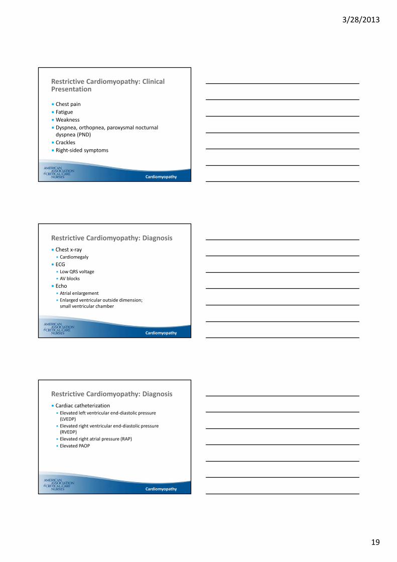
� Chest pain
� Fatigue
� Weakness
� Dyspnea, orthopnea, paroxysmal nocturnal
dyspnea (PND)
� Crackles
� Right-sided symptoms
Restrictive Cardiomyopathy: Clinical Presentation
Cardiomyopathy
� Chest x-ray
� Cardiomegaly
� ECG
� Low QRS voltage
� AV blocks
� Echo
� Atrial enlargement
� Enlarged ventricular outside dimension;
small ventricular chamber
Restrictive Cardiomyopathy: Diagnosis
Cardiomyopathy
� Cardiac catheterization
� Elevated left ventricular end-diastolic pressure
(LVEDP)
� Elevated right ventricular end-diastolic pressure
(RVEDP)
� Elevated right atrial pressure (RAP)
� Elevated PAOP
Restrictive Cardiomyopathy: Diagnosis
Cardiomyopathy
3/28/2013
20
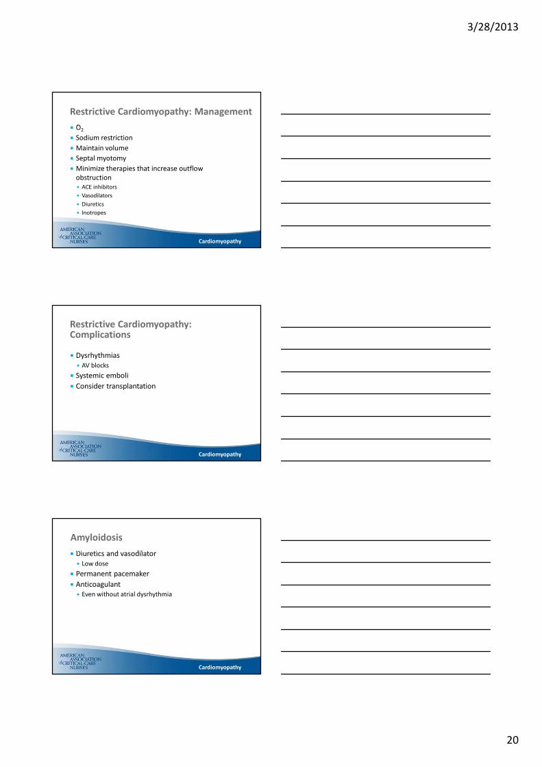
� O2
� Sodium restriction
� Maintain volume
� Septal myotomy
� Minimize therapies that increase outflow
obstruction
� ACE inhibitors
� Vasodilators
� Diuretics
� Inotropes
Restrictive Cardiomyopathy: Management
Cardiomyopathy
� Dysrhythmias
� AV blocks
� Systemic emboli
� Consider transplantation
Restrictive Cardiomyopathy: Complications
Cardiomyopathy
� Diuretics and vasodilator
� Low dose
� Permanent pacemaker
� Anticoagulant
� Even without atrial dysrhythmia
Amyloidosis
Cardiomyopathy
3/28/2013
21
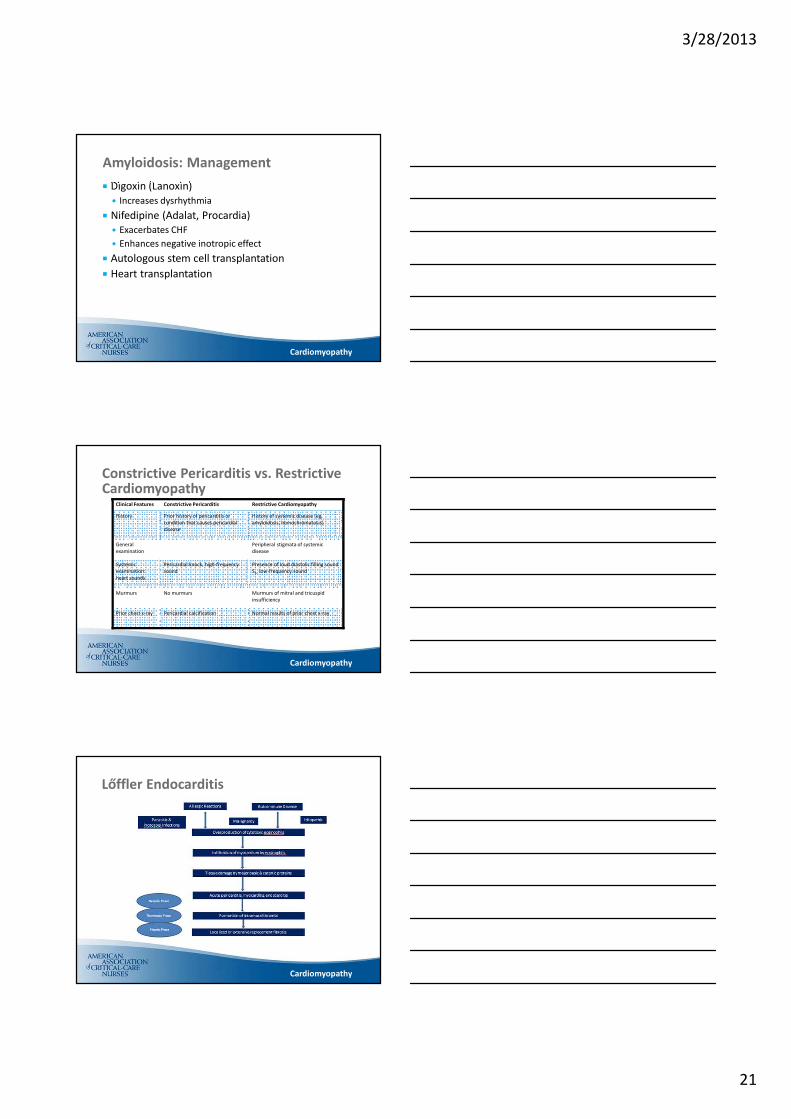
� Digoxin (Lanoxin)
� Increases dysrhythmia
� Nifedipine (Adalat, Procardia)
� Exacerbates CHF
� Enhances negative inotropic effect
� Autologous stem cell transplantation
� Heart transplantation
Amyloidosis: Management
Cardiomyopathy
Clinical Features Constrictive Pericarditis Restrictive Cardiomyopathy
History Prior history of pericarditis or
condition that causes pericardial
disease
History of systemic disease (eg,
amyloidosis, hemochromatosis)
General
examination
Peripheral stigmata of systemic
disease
Systemic
examination:
heart sounds
Pericardial knock, high-frequency
sound
Presence of loud diastolic filling sound
S3, low-frequency sound
Murmurs No murmurs Murmurs of mitral and tricuspid
insufficiency
Prior chest x-ray Pericardial calcification Normal results of prior chest x-ray
Constrictive Pericarditis vs. Restrictive Cardiomyopathy
Cardiomyopathy
Lőffler Endocarditis
Cardiomyopathy

3/28/2013
22
Allergic Reactions
Parasitic & Protozoal InfectionsMalignancy
Autoimmune Disease
Idiopathic
Overproduction of cytotoxic eosinophils
Infiltration of myocardium by eosinophils
Tissue damage by major basic & catonic proteins
Acute pericarditis, myocarditis, endocarditis
Formation of intramural thrombi
Localized or extensive replacement fibrosis
Thrombotic
Phase
Necrotic
Phase
Fibrotic
Phase
Lőffler Endocarditis
� Symptoms
� Weight loss
� Fever
� Cough
� Rash
� CHF
Lőffler Endocarditis
� Diagnosis
� ECG
� Atrial fibrillation + RBBB
� Echo & cardiac
catheterization
� Probable mitral valve and
tricuspid valve
regurgitation
� Complications
� Systemic emboli
Cardiomyopathy
� Treatment
� Steroid
� Cytotoxic agents
� Hydroxyurea
� Symptom control
Lőffler Endocarditis
Cardiomyopathy

3/28/2013
23
Practice Exam Questions
Cardiomyopathy
A. Volume. Preload must be maintained. Any
medication that increases outflow tract
obstruction will exacerbate symptoms. Avoid
inotropes, dilators, and diuretics.
B. Dobutamine
C. Lasix
D. Nitrates
Your patient has been diagnosed with restrictive
cardiomyopathy. An appropriate intervention would
include administration of:
Question #1 - Answer
Cardiomyopathy
A. Cardiac transplantation as soon as possible
B. Surgical manipulation of the septum
C. An implantable cardioverter-defibrillator. These
patients are at high risk of ventricular fibrillation.
Usually, drug therapy with amiodarone and the
insertion of an ICD is the treatment of choice.
D. A beta-blocker to reduce workload
Patients with hypertrophic cardiomyopathy with high
clinical or genetic risk of sudden death should receive:
Cardiomyopathy
Question #2 - Answer
3/28/2013
24
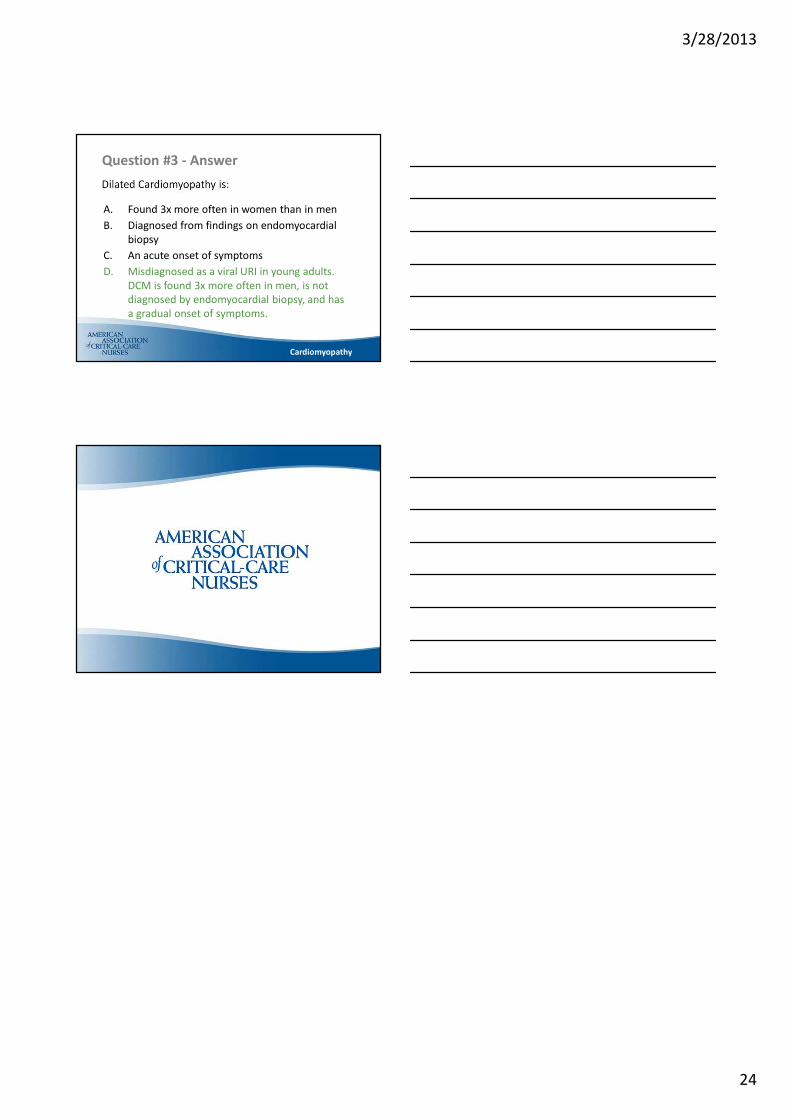
A. Found 3x more often in women than in men
B. Diagnosed from findings on endomyocardial
biopsy
C. An acute onset of symptoms
D. Misdiagnosed as a viral URI in young adults.
DCM is found 3x more often in men, is not
diagnosed by endomyocardial biopsy, and has
a gradual onset of symptoms.
Cardiomyopathy
Question #3 - Answer


![[HF] FREEWEIGHT PRODUCTS - HOIST Fitness · [hf] flat bench hf-5163 [hf] 7-position folding f.i.d. bench hf-5167 new! warranty new! warranty [hf] 7-position f.i.d. olympic bench hf-5170](https://img.pdfslide.us/doc/110x75/5b5909d87f8b9ad0048c899a/hf-freeweight-products-hoist-fitness-hf-flat-bench-hf-5163-hf-7-position.jpg)

![MPIA 15 459 1133 - · Dst: Type: icmp time exceeded in-transit [tos OxcO] /-----, -----\ > > > > > > > > > ® . MPIA 15 459 11302/17/99 , !](https://img.pdfslide.us/doc/110x75/5ab91efd7f8b9ac10d8dd314/mpia-15-459-1133-type-icmp-time-exceeded-in-transit-tos-oxco-.jpg)