Embed Size (px)
Citation preview
Clinical StudyUltrasonic Surgical Aspirator to Treat Deep Infrabony Defects:A New Flapless Minimally Invasive Approach
Carlo Ghezzi,1 Camilla Donghi,1 Luca Ferrantino ,2 Elena Varoni ,3
and Giovanni Lodi 3
1Private Practice, Via Verdi 4, Settimo Milanese, 20019 Milan, Italy2Department of Oral Rehabilitation, Istituto Stomatologico Italiano, University of Milan, Milan, Italy3Department of Biomedical, Surgical and Dental Sciences, Universita Degli Studi di Milano, Milan, Italy
Correspondence should be addressed to Luca Ferrantino; [email protected]
Received 8 February 2018; Revised 25 May 2018; Accepted 2 June 2018; Published 29 July 2018
Academic Editor: Atsutoshi Yoshimura
Copyright© 2018CarloGhezzi et al.*is is an open access article distributed under the Creative Commons Attribution License,which permits unrestricted use, distribution, and reproduction in any medium, provided the original work is properly cited.
*e primary outcome of the present study was to assess the percentage of pocket closure, and the secondary aim was to evaluatethe clinical performance in terms of clinical attachment level (CAL) gain, probing pocket depth (PPD) reduction, and gingivalrecession (REC) after the use of cavitron ultrasonic surgical aspirator (CUSA) in deep infrabony defects. Fourteen deep infrabonydefects in 11 patients who were previously treated with active periodontal therapy followed by one year of supportive periodontaltherapy (at least three sessions) were additionally treated by the aid of CUSA. Eighty-six percent of the initial defects (12 out of 14)resulted in a PD< 5mm, showing complete resolution six months after CUSA treatment, without any adverse event and withnegligible pain (VAS from 0 to 3). CUSA showed potential as a method to promote pocket healing, reduce PPD, and increaseclinical attachment (P< 0.001) in deep infrabony defects. *is trial is registered with ClinicalTrials.gov NCT03567161.
1. Introduction
Deep periodontal pockets, which are associated withinfrabony defects, are specific risk factors for periodontaldisease progression and tooth loss [1, 2]. In the past, theinterest of researchers on regeneration focused to developmaterials as a type of bone substitute and membrane orbiological mediator to improve result in tissue regeneration[3–6], but recently, interest has been moving to the tissuemanagement to achieve better result introducing the min-imally invasive surgical approaches (MIS) [7–13].
*e innovative aspects of the MIS technique are rep-resented by a flap design [12–18] to permit preservation ofinterdental space, minimizing vertical release in order toobtain adhesion andmaturation with slight trauma, togetherwith primary intention wound closure to achieve peri-odontal tissue regeneration [11, 17–24].
In this context, we have to consider other new studies inwhich authors define and compare the performance of theminimally invasive nonsurgical technique (MINST) to the
minimally invasive surgical approach [25]. MINST has beenintroduced as a concept that aims at obtaining extensivesubgingival debridement with a retention of the preoperativegingival architecture, creating a minimal wound, and gentlehandling of the soft and hard tissues to stimulate the for-mation of a stable blood clot by natural filling of theinfrabony defect [25–28].
Cavitron ultrasonic surgical aspirator (CUSA) is awell-known technology that is used in medicine for dif-ferent purposes; its most frequent applications are inneurosurgery and liver disease [29–31]. CUSA has provento be effective in biofilm disruption and cell stimulation[32]. *e hypothesis is that the employment of CUSA fornonsurgical treatment of infrabony defects, thanks to itsabilities to disrupt, fragment, and aspirate granulationtissue, will allow the formation of larger and more stableblood clot.
*e purpose of this study was to test CUSA in non-surgical treatment of infrabony defects to promote pocketclosure.
HindawiAdvances in MedicineVolume 2018, Article ID 3612359, 8 pageshttps://doi.org/10.1155/2018/3612359
2. Materials and Methods
*is was a Phase 2 noncontrolled clinical trial performed onpatients with infrabony defects to test whether the em-ployment of CUSA for treating periodontal patients.
(1) provides benefits in terms of a PD reduction andCAL gain;
(2) is comfortable for both the patient and the operator;(3) is free from adverse events.
All subjects included in the study were consecutiveperiodontal patients attending a private clinic in SettimoMilanese (Milan, Italy) who were treated by two operators(CG and CD) with a similar experience in nonsurgicalproduce who performed a specific training for CUSA ona periodontal model. *ey were selected on the basis of thefollowing criteria.
Inclusion Criteria
(1) Having received a diagnosis of chronic periodon-titis (Armitage 1999)
(2) Being treated by full-mouth debridement andsupportive periodontal treatment (SPT) in the lastyear (at least three sessions) by one of the authors
(3) Having at least one residual pocket ≥5mm with anintrabony component at least ≥2mm
Exclusion Criteria
(1) Smoking more than 10 cigarettes per day(2) Pregnancy(3) Irregular compliance during SPT in the last year(4) Systemic conditions or therapies known to affect
the healing potential of periodontal tissues(e.g., uncontrolled diabetes, oncological conditions,and immunosuppressant drugs)
All patients were informed on the objective of the studiesand provided informed consent.
*e clinical procedure was always performed in a singlesession. Before intervention, all cases received local anaes-thesia with 1 :100,000 mepivacaine. All residual pockets≥5mm underwent the following:
(1) Ultrasonic debridement: to minimize trauma tothe soft tissues, we used piezoelectric devices with
specific thin and delicate tips (EMS Electro MedicalSystems S.A. Chemin de la Vuarpilliere, 31 1260,Lyon, Switzerland).
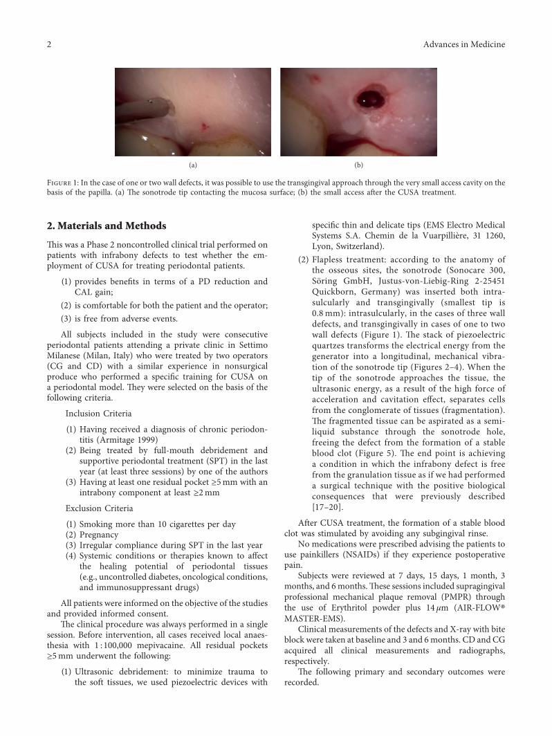
(2) Flapless treatment: according to the anatomy ofthe osseous sites, the sonotrode (Sonocare 300,Soring GmbH, Justus-von-Liebig-Ring 2-25451Quickborn, Germany) was inserted both intra-sulcularly and transgingivally (smallest tip is0.8 mm): intrasulcularly, in the cases of three walldefects, and transgingivally in cases of one to twowall defects (Figure 1). *e stack of piezoelectricquartzes transforms the electrical energy from thegenerator into a longitudinal, mechanical vibra-tion of the sonotrode tip (Figures 2–4). When thetip of the sonotrode approaches the tissue, theultrasonic energy, as a result of the high force ofacceleration and cavitation effect, separates cellsfrom the conglomerate of tissues (fragmentation).*e fragmented tissue can be aspirated as a semi-liquid substance through the sonotrode hole,freeing the defect from the formation of a stableblood clot (Figure 5). *e end point is achievinga condition in which the infrabony defect is freefrom the granulation tissue as if we had performeda surgical technique with the positive biologicalconsequences that were previously described[17–20].
After CUSA treatment, the formation of a stable bloodclot was stimulated by avoiding any subgingival rinse.
No medications were prescribed advising the patients touse painkillers (NSAIDs) if they experience postoperativepain.
Subjects were reviewed at 7 days, 15 days, 1 month, 3months, and 6months.*ese sessions included supragingivalprofessional mechanical plaque removal (PMPR) throughthe use of Erythritol powder plus 14 μm (AIR-FLOW®MASTER-EMS).
Clinical measurements of the defects and X-ray with biteblock were taken at baseline and 3 and 6months. CD and CGacquired all clinical measurements and radiographs,respectively.
*e following primary and secondary outcomes wererecorded.
(a) (b)
Figure 1: In the case of one or two wall defects, it was possible to use the transgingival approach through the very small access cavity on thebasis of the papilla. (a) *e sonotrode tip contacting the mucosa surface; (b) the small access after the CUSA treatment.
2 Advances in Medicine
Primary Outcomes
(i) Pocket closure proportion (PPD< 5mm)(ii) Probing depth (PPD) reduction(iii) CAL gain(iv) Gingival recession
Secondary Outcomes
(i) Comfort and acceptability of the patient duringand after the procedure, as measured by in-terviews, use of painkillers in the following threedays, and the visual analogue scale (VAS) after oneweek;
(ii) Comfort and convenience of the operator duringthe procedure, as measured by interviews at theend of the procedure;
(iii) Adverse events.
Clinical data from all patients were entered into anExcel �le and checked for entry errors. Continuous var-iables were expressed as the mean ± standard deviation(SD). Dichotomous data were expressed as a percentage.
�e comparison between baseline and 6 months after�apless treatment was performed by applying a Wilcoxonsigned-rank test. All calculations were performed usingStata version 11.1 (College Station, TX, USA). �e defectwas used as a statistical unit, and a P value <0.05 wasconsidered statistically signi�cant.
3. Results
A total of 14 defects in 11 patients were treated and includedin this case series. �e demographic and clinical baselinecharacteristics of the 11 subjects are depicted in Table 1. �eaverage age was 56± 8 years. Within the 14 treated defects,�ve were in the mandible and nine in the maxilla. Sevendefects a�ected the lateral and central incisors, and threewere adjacent to premolars and four to molars. �e meanPPD at baseline was 8.6± 1.5mm, with an average gingivalrecession of 2.6± 2.1mm, and therefore, the mean CAL was11.1± 3mm. Table 2 shows the characteristics of the 14infrabony defects included in this study.
Clinical measurements were taken during the lastfollow-up visit, six months after CUSA treatment (Table 2).�e CUSA procedure achieved pocket closure(PPD< 5mm) in 86% of the defects (12 out of 14).
At that time point, themean values of PPD, CAL, and RECwere 3.9± 1.4mm, 6.8± 1.9mm, and 2.8± 1.6mm, re-spectively; the di�erences with baseline data were statisticallysigni�cant for PPD and CAL (P< 0.01). �e mean PPD re-ductionwas 4.7± 2mm, andmeanCAL gain was 4.3± 2.2mm.
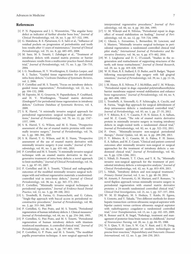
X-rays of the selected defects are presented inFigures 6–9. At the end of the procedure, all patients re-ported negligible discomfort; none took any painkillers inthe following week or more. �e mean VAS was 1.18± 1.11(the distribution among the study population is shown inFigure 10). �e VAS values ranged between 0 and 3; noadverse event was recorded.
4. Discussion
Systematic review studies [33, 34] revealed that both con-ventional nonsurgical and surgical therapies were e�ectivemethods for making improvements in terms of CAL gainand PD reduction.
However, in recent studies inwhichMINSTwas performedin initially deep pockets (PD> 6mm), there was a greater CALgain and PD reduction; the change in these clinical parameterswas similar, showing a mean CAL gain of 2.56mm and PDreduction of 3.13mm in Ribeiro’s study andmean CAL gain of2.78 and a PD reduction of 3.12mm in Nibali’s study [25, 26].�ese data con�rm the initial hypothesis of the authors;speci�cally, the use of minimally invasive strategies in non-surgical therapy could lead to improvement of results com-pared to the standard nonsurgical approach [25, 26].
�e present study tested a new �apless approach tofurther improve the results of debridement. For this reason,the starting point of our research was one year after FMUD,followed by repeated sessions of SPT [35].
�e primary idea was to eliminate old and capsulatedgranulation tissue following the chronic process of
Aspirationcontrol
Powercontrol
Irrigationcontrol
Informationfield
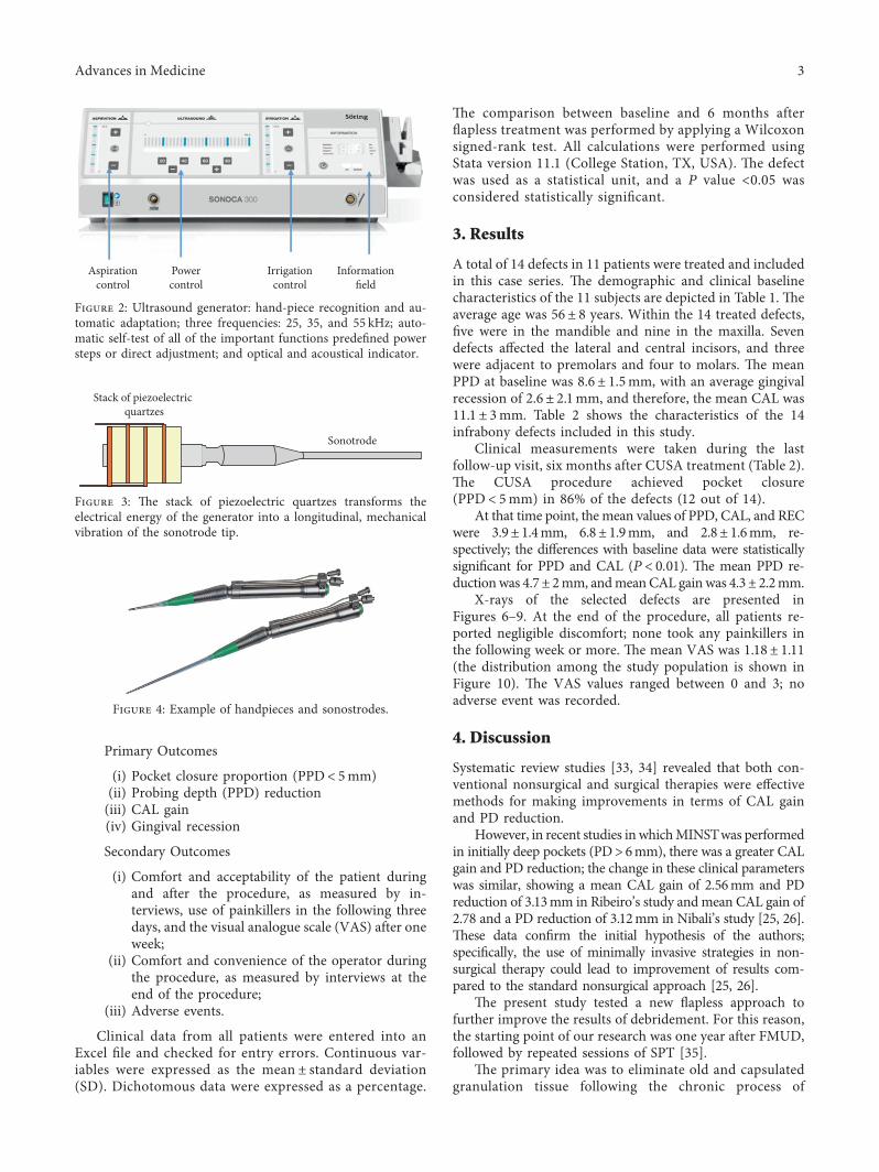
Figure 2: Ultrasound generator: hand-piece recognition and au-tomatic adaptation; three frequencies: 25, 35, and 55 kHz; auto-matic self-test of all of the important functions prede�ned powersteps or direct adjustment; and optical and acoustical indicator.
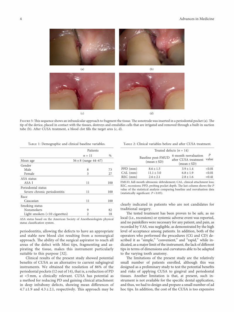
Stack of piezoelectric quartzes
Sonotrode
Figure 3: �e stack of piezoelectric quartzes transforms theelectrical energy of the generator into a longitudinal, mechanicalvibration of the sonotrode tip.
Figure 4: Example of handpieces and sonostrodes.
Advances in Medicine 3
periodontitis, allowing the defects to have an appropriateand stable new blood clot resulting from a nonsurgicalapproach. *e ability of the surgical aspirator to reach allareas of the defect with Mini tips, fragmenting and as-pirating the tissue, makes this instrument particularlysuitable to this purpose [32].
Clinical results of the present study showed potentialbenefits of CUSA as an alternative to current subgingivalinstruments. We obtained the resolution of 86% of theperiodontal pockets (12 out of 14), that is, a reduction of PDat <5mm, a clinically relevant. CUSA has potential asa method for reducing PD and gaining clinical attachmentin deep infrabony defects, showing mean differences of4.7±1.9 and 4.3 ± 2.1, respectively. *is approach may be
clearly indicated in patients who are not candidates fortraditional surgery.
*e tested treatment has been proven to be safe, as nolocal (i.e., recessions) or systemic adverse event was reported,and no painkillers were necessary for any patient, and pain, asrecorded by VAS, was negligible, as demonstrated by the highlevel of acceptance among patients. In addition, both of theoperators who performed the procedures (CG and CD) de-scribed it as “simple,” “convenient,” and “rapid,” while in-dicated, as amajor limit of the instrument, the lack of differenttips in terms of dimensions and curvatures able to be adaptedto the varying tooth anatomy.
*e limitations of the present study are the relativelysmall number of patients enrolled, although this wasdesigned as a preliminary study to test the potential benefitsand risks of applying CUSA to gingival and periodontaltissues. Another limitation is that, at present, such in-strument is not available for the specific dental application,and thus, we had to design and prepare a small number of adhoc tips. In addition, the cost of the CUSA is too expensive
(a) (b)
(c) (d)
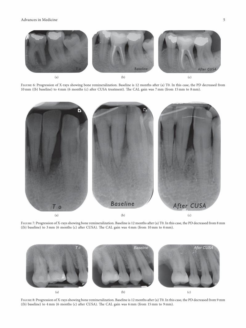
Figure 5:*is sequence shows an infrasulcular approach to fragment the tissue.*e sonotrode was inserted in a periodontal pocket (a).*etip of the device, placed in contact with the tissues, destroys and emulsifies cells that are irrigated and removed through a built-in suctiontube (b). After CUSA treatment, a blood clot fills the target area (c, d).
Table 1: Demographic and clinical baseline variables.
Patientsn � 11 %
Mean age 56± 8 (range 44–67)GenderMale 8 73Female 3 27
ASA statusASA I 11 100
Periodontal statusSevere chronic periodontitis 11 100
RaceCaucasian 11 100
Smoking statusNonsmokers 9 82Light smokers (<10 cigarettes) 2 18
ASA status based on the American Society of Anesthesiologists physicalstatus classification system.
Table 2: Clinical variables before and after CUSA treatment.
Treated defects (n � 14)P
valueBaseline post-FMUD(mean± SD)
6-month reevaluationafter CUSA treatment
(mean± SD)PPD (mm) 8.6± 1.5 3.9± 1.4 <0.01CAL (mm) 11.1± 3.0 6.8± 1.9 <0.01REC (mm) 2.6± 2.1 2.8± 1.6 �0.41FMUD, full-mouth ultrasonic debridement; CAL, clinical attachment loss;REC, recessions; PPD, probing pocket depth. *e last column shows the Pvalue of the statistical analysis comparing baseline and reevaluation data(statistically significant: P< 0.05).
4 Advances in Medicine
(a) (b) (c)
Figure 7: Progression of X-rays showing bone remineralization. Baseline is 12months after (a) T0. In this case, the PD decreased from 8mm((b) baseline) to 3mm (6 months (c) after CUSA). *e CAL gain was 4mm (from 10mm to 6mm).
(a) (b) (c)
Figure 8: Progression of X-rays showing bone remineralization. Baseline is 12months after (a) T0. In this case, the PD decreased from 9mm((b) baseline) to 4mm (6 months (c) after CUSA). *e CAL gain was 6mm (from 15mm to 9mm).
(a) (b) (c)
Figure 6: Progression of X-rays showing bone remineralization. Baseline is 12 months after (a) T0. In this case, the PD decreased from10mm ((b) baseline) to 4mm (6 months (c) after CUSA treatment). *e CAL gain was 7mm (from 15mm to 8mm).
Advances in Medicine 5
compared with ultrasonic devices currently used in theroutine clinical practice.
5. Conclusion
�e �apless approach that was used for treating infrabonydefects achieved successful outcomes in terms of pocketclosure and clinical parameters.
�is approach was identi�ed as a promising method toamplify, and in secondary care, the results that are achievablewith nonsurgical therapy, promoting less morbidity thanany other surgical technique and providing patient satis-faction. �is approach requires future randomized controlstudies to better explain its potential and di�erent appli-cation strategies.
Data Availability
�e data used to support the �ndings of this study areavailable from the corresponding author upon request.
Ethical Approval
�e protocol was approved by the Ethical Committee ofthe University of Milan (Universita degli studi di Milano;number 1/17; date 30/01/2017). All of the proceduresperformed in studies involving human participants were inaccordance with the ethical standards of the institutionalresearch committee and with the 1964 Helsinki Declara-tion and its later amendments or comparable ethicalstandards.
Consent
Informed consent was obtained from all of the individualparticipants who were included in the study.
Conflicts of Interest
All of the authors declare that they have no con�icts ofinterest.
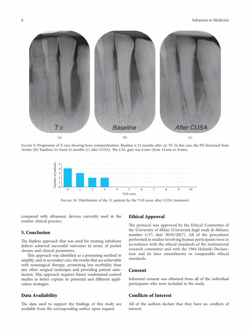
(a) (b) (c)
Figure 9: Progression of X-rays showing bone remineralization. Baseline is 12 months after (a) T0. In this case, the PD decreased from10mm ((b) baseline) to 4mm (6 months (c) after CUSA). �e CAL gain was 6mm (from 14mm to 8mm).
012345
0 1 2 3 4 5 6 7 8 9 10VAS score
Num
ber o
f pat
ient
s
Figure 10: Distribution of the 11 patients by the VAS score after CUSA treatment.
6 Advances in Medicine
References
[1] P. N. Papapanou and J. L. Wennstrom, “*e angular bonydefect as indicator of further alveolar bone loss,” Journal ofClinical Periodontology, vol. 18, no. 5, pp. 317–322, 1991.
[2] G. Matuliene, B. E. Pjetursson, G. E. Salvi et al., “Influence ofresidual pockets on progression of periodontitis and toothloss: results after 11 years of maintenance,” Journal of ClinicalPeriodontology, vol. 35, no. 8, pp. 685–695, 2008.
[3] M. Sanz, M. S. Tonetti, I. Zabalegui et al., “Treatment ofintrabony defects with enamel matrix proteins or barriermembranes: results from a multicenter practice-based clinicaltrial,” Journal of Periodontology, vol. 75, no. 5, pp. 726–733,2004.
[4] I. G. Needleman, H. V. Worthington, E. Giedrys-Leeper, andR. J. Tucker, “Guided tissue regeneration for periodontalinfra-bony defects,” Cochrane Database of Systematic Reviews,vol. 2, 2006.
[5] P. Cortellini and M. S. Tonetti, “Focus on intrabony defects:guided tissue regeneration,” Periodontology, vol. 22, no. 1,pp. 104–132, 2000.
[6] M. Esposito, M. G. Grusovin, N. Papanikolaou, P. Coulthard,and H. V. Worthington, “Enamel matrix derivative(Emdogain®) for periodontal tissue regeneration in intrabonydefects,” Cochrane Database of Systematic Reviews, vol. 4,2009.
[7] S. K. Harrel, “A minimally invasive surgical approach forperiodontal regeneration: surgical technique and observa-tions,” Journal of Periodontology, vol. 70, no. 12, pp. 1547–1557, 1999.
[8] S. K. Harrel, T. G. Wilson, and M. E. Nunn, “Prospectiveassessment of the use of enamel matrix proteins with mini-mally invasive surgery,” Journal of Periodontology, vol. 76,no. 3, pp. 380–384, 2005.
[9] S. K. Harrel, T. G. Wilson, and M. E. Nunn, “Prospectiveassessment of the use of enamel matrix derivative withminimally invasive surgery: 6-year results,” Journal of Peri-odontology, vol. 81, no. 3, pp. 435–441, 2010.
[10] P. Cortellini and M. S. Tonetti, “A minimally invasive surgicaltechnique with an enamel matrix derivative in the re-generative treatment of intra-bony defects: a novel approachto limit morbidity,” Journal of Clinical Periodontology, vol. 34,no. 1, pp. 87–93, 2007.
[11] P. Cortellini and M. S. Tonetti, “Clinical and radiographicoutcomes of the modified minimally invasive surgical tech-nique with and without regenerative materials: a randomized-controlled trial in intra-bony defects,” Journal of ClinicalPeriodontology, vol. 38, no. 4, pp. 365–373, 2011.
[12] P. Cortellini, “Minimally invasive surgical techniques inperiodontal regeneration,” Journal of Evidence-based DentalPractice, vol. 12, no. 3, pp. 89–100, 2012.
[13] L. Trombelli, R. Farina, G. Franceschetti, and G. Calura,“Single-flap approach with buccal access in periodontal re-constructive procedures,” Journal of Periodontology, vol. 80,no. 2, pp. 353–360, 2009.
[14] P. Cortellini, G. Pini Prato, and M. S. Tonetti, “Periodontalregeneration of human infrabony defects. I. Clinical measures,”Journal of Periodontology, vol. 64, no. 4, pp. 254–260, 1993.
[15] P. Cortellini, G. Pini Prato, and M. S. Tonetti, “Periodontalregeneration of human intrabony defects with titaniumreinforced membranes. A controlled clinical trial,” Journal ofPeriodontology, vol. 66, no. 9, pp. 797–803, 1995.
[16] P. Cortellini, G. P. Prato, and M. S. Tonetti, “*e modifiedpapilla preservation technique. A new surgical approach for
interproximal regenerative procedures,” Journal of Peri-odontology, vol. 66, no. 4, pp. 261–266, 1995.
[17] U. M. Wikesjo and R. Nilveus, “Periodontal repair in dogs:effect of wound stabilization on healing,” Journal of Peri-odontology, vol. 61, no. 12, pp. 719–724, 1990.
[18] C. Ghezzi, L. Ferrantino, L. Bernardini, M. Lencioni, andS. Masiero, “Minimally invasive surgical technique in peri-odontal regeneration: a randomized controlled clinical trialpilot study,” International Journal of Periodontics and Re-storative Dentistry, vol. 36, no. 4, pp. 475–482, 2016.
[19] W. J. Linghorne and D. C. O’connell, “Studies in the re-generation and reattachment of supporting structures of theteeth; soft tissue reattachment,” Journal of Dental Research,vol. 29, no. 4, pp. 419–428, 1950.
[20] W. H. Hiatt, R. E. Stallard, E. D. Butler, and B. Badget, “Repairfollowing mucoperiosteal flap surgery with full gingivalretention,” Journal of Periodontology, vol. 39, no. 1, pp. 11–16,1968.
[21] J. M. Haney, R. E. Nilveus, P. J. McMillan, and U. M.Wikesjo,“Periodontal repair in dogs: expanded polytetrafluorethylenebarrier membrane support wound stabilisation and enhancebone regeneration,” Journal of Periodontology, vol. 64, no. 9,pp. 883–890, 1993.
[22] L. Trombelli, A. Simonelli, G. P. Schincaglia, A. Cucchi, andR. Farina, “Single-flap approach for surgical debridement ofdeep intraosseous defects: a randomized controlled trial,”Journal of Periodontology, vol. 83, no. 1, pp. 27–35, 2012.
[23] F. V. Ribeiro, R. C. V. Casarin, F. H. N. Junior, E. A. Sallum,and M. Z. Casati, “*e role of enamel matrix derivativeprotein in minimally invasive surgery in treating intrabonydefects in single-rooted teeth: a randomized clinical trial,”Journal of Periodontology, vol. 82, no. 4, pp. 522–532, 2011.
[24] P. Ower, “Minimally-invasive non-surgical periodontaltherapy,” Dental Update, vol. 40, no. 4, pp. 289-290, 2013.
[25] F. V. Ribeiro, R. C. Casarin, M. A. Palma, F. H. Junior,E. A. Sallum, and M. Z. Casati, “Clinical and patient-centeredoutcomes after minimally invasive non-surgical or surgicalapproaches for the treatment of intrabony defects: a ran-domized clinical trial,” Journal of Periodontology, vol. 82,no. 9, pp. 1256–1266, 2011.
[26] L. Nibali, D. Pometti, T. T. Chen, and Y. K. Tu, “Minimallyinvasive non-surgical approach for the treatment of peri-odontal intrabony defects: a retrospective analysis,” Journal ofClinical Periodontology, vol. 42, no. 9, pp. 853–859, 2015.
[27] L. Nibali, “Intrabony defects and non-surgical treatment,”Primary Dental Journal, vol. 3, no. 3, pp. 48–50, 2014.
[28] M. Aimetti, F. Ferrarotti, G. M. Mariani, and F. Romano, “Anovel flapless approach versus minimally invasive surgery inperiodontal regeneration with enamel matrix derivativeproteins: a 24-month randomized controlled clinical trial,”Clinical Oral Investigations, vol. 21, no. 1, pp. 327–337, 2016.
[29] W. M. El Moghazy, M. S. Hedaya, T. Kaido, H. Egawa,S. Uemoto, and Y. Takada, “Two different methods for donorhepatic transection: cavitron ultrasonic surgical aspirator withbipolar cautery versus cavitron ultrasonic surgical aspiratorwith radiofrequency coagulator–a randomized controlledtrial,” Liver Transplantation, vol. 15, no. 1, pp. 102–105, 2009.
[30] K. Bonner and K. R. Siegel, “Pathology, treatment and man-agement of posterior fossa brain tumors in childhood,” Journalof Neuroscience Nursing, vol. 20, no. 2, pp. 84–93, 1988.
[31] N. S. Qian, Y. H. Liao, S. W. Cai, V. Raut, and J. H. Dong,“Comprehensive application of modern technologies inprecise liver resection,” Hepatobiliary and Pancreatic DiseasesInternational, vol. 12, no. 3, pp. 244–250, 2013.
Advances in Medicine 7
[32] B. J. O’Daly, E. Morris, G. P. Gavin, J. M. O’Byrne, andG. B. McGuinness, “High-power low-frequency ultrasound:a review of tissue dissection and ablation in medicine andsurgery,” Journal of Materials Processing Technology, vol. 200,no. 1–3, pp. 38–58, 2008.
[33] L. J. Heitz-Mayfield, L. Trombelli, F. Heitz, I. Needleman, andD. Moles, “A systematic review of the effect of surgical de-bridement vs non-surgical debridement for the treatment ofchronic periodontitis,” Journal of Clinical Periodontology,vol. 29, no. 3, pp. 92–102, 2002.
[34] L. J. Heitz-Mayfield, “How effective is surgical therapycompared with nonsurgical debridement?,” Periodontology,vol. 37, no. 1, pp. 72–87, 2000.
[35] N. Claffey, B. Loos, B. Gantes, M. Martin, and J. Egelberg,“Probing depth at re-evaluation following initial periodontaltherapy to indicate the initial response to treatment,” Journalof Clinical Periodontology, vol. 16, no. 4, pp. 229–233, 1989.
8 Advances in Medicine

DentistryInternational Journal of
Hindawiwww.hindawi.com Volume 2018
Environmental and Public Health
Journal of
Hindawiwww.hindawi.com Volume 2018
Hindawi Publishing Corporation http://www.hindawi.com Volume 2013Hindawiwww.hindawi.com
The Scientific World Journal
Volume 2018Hindawiwww.hindawi.com Volume 2018
Public Health Advances in
Hindawiwww.hindawi.com Volume 2018
Case Reports in Medicine
Hindawiwww.hindawi.com Volume 2018
International Journal of
Biomaterials
Scienti�caHindawiwww.hindawi.com Volume 2018
PainResearch and TreatmentHindawiwww.hindawi.com Volume 2018
Preventive MedicineAdvances in
Hindawiwww.hindawi.com Volume 2018
Hindawiwww.hindawi.com Volume 2018
Case Reports in Dentistry
Hindawiwww.hindawi.com Volume 2018
Surgery Research and Practice
Hindawiwww.hindawi.com Volume 2018
BioMed Research International Medicine
Advances in
Hindawiwww.hindawi.com Volume 2018
Hindawiwww.hindawi.com Volume 2018
Anesthesiology Research and Practice
Hindawiwww.hindawi.com Volume 2018
Radiology Research and Practice
Hindawiwww.hindawi.com Volume 2018
Computational and Mathematical Methods in Medicine
EndocrinologyInternational Journal of
Hindawiwww.hindawi.com Volume 2018
Hindawiwww.hindawi.com Volume 2018
OrthopedicsAdvances in
Drug DeliveryJournal of
Hindawiwww.hindawi.com Volume 2018
Submit your manuscripts atwww.hindawi.com