Embed Size (px)
Citation preview
Clinical StudyPregnancy-Associated Plasma Protein-A Levels and CoronaryAngiographic Features in Acute Coronary Syndrome Patients
Mohamed Shehata
Department of Cardiology, Faculty of Medicine, Ain Shams University, P.O. Box 11381, Abbassia, Cairo, Egypt
Correspondence should be addressed to Mohamed Shehata; [email protected]
Received 30 September 2013; Revised 16 December 2013; Accepted 24 December 2013; Published 4 February 2014
Academic Editor: Apostolos E. Papalois
Copyright © 2014 Mohamed Shehata. This is an open access article distributed under the Creative Commons Attribution License,which permits unrestricted use, distribution, and reproduction in any medium, provided the original work is properly cited.
Background. Pregnancy-associated plasma protein-A (PAPP-A) is a biomarker of plaque rupture, associated with adverse cardiacevents in acute coronary syndromes (ACSs) patients.Aim. To identify coronary angiographic (CA) features related to PAPP-A levelelevation in ACSs patients. Methods. Forty ACSs patients were enrolled in this prospective cohort study (level of evidence: III-prognostic). Serum samples for PAPP-A quantitation were obtained upon coronary care unit admission. All patients underwentCA and coronary intervention within 6 hours of sampling. Results. Mean age of the study cohort was 57 ± 11 years, (males: 55%,𝑛 = 22). Patients with ST-segment elevation myocardial infarction (35%, 𝑛 = 14) showed significantly higher serum PAPP-A level(11.8 ± 2 𝜇g/mL), compared to non-ST-segment elevation myocardial infarction (15%, 𝑛 = 6) and unstable angina (50%, 𝑛 = 20)patients (11 ± 2.6 𝜇g/mL and 8.7 ± 2.3 𝜇g/mL, resp., 𝑃 < 0.001). Higher PAPP-A levels were significantly associated with complexculprit lesion morphology (11.8 ± 2 𝜇g/mL for type C lesions, 9.7± 2.5 𝜇g/mL and 7.3 ± 3.5 𝜇g/mL for type B and type A lesions,resp., 𝑃 < 0.001), while no relationship to number of diseased coronaries. Conclusion. Higher PAPP-A levels in ACSs patients areassociated with unfavorable coronary anatomy and complex angiographic plaque features.
1. Introduction
The term vulnerable patient had been proposed to definesubjects susceptible to acute coronary events, based on plaquecharacteristics, blood abnormalities andmyocardial vulnera-bility [1]. It is important to identify both vulnerable patientsand vulnerable plaques. Atherosclerotic arteries obtainedat autopsy from patients who died suddenly of cardiaccauses indicated that Pregnancy-associated plasma protein-A (PAPP-A) was abundantly expressed in plaque cells & inextracellular matrix of ruptured & eroded plaques, but not instable plaques [2].
PAPP-A is a zinc-binding matrix metalloproteinase, amember of metzincin super-family, which was originallyidentified in serum of pregnant women to help determine theterm date [3]. It is also used for screening of fetal trisomy inthe first pregnancy trimester [2].
There is growing evidence suggesting that inflammationhas a pivotal role in acute coronary syndromes’ pathogen-esis [4]. PAPP-A probably participates in the inflammatoryreactions of vascular walls, which could lead to structuraldisruption of atherosclerotic plaques [5].
Circulating levels of PAPP-A are significantly elevated inpatients with unstable angina (UA) & myocardial infarction(MI) [2].HighPAPP-A level is a strong independent predictorof ischemic cardiac events & need for revascularization inpatients who present with acute coronary syndromes (ACSs)[6]. Moreover, it is a strong marker of plaque instability [7].
The current study sought to evaluate the diagnostic &short-term prognostic role of measurement of serum PAPP-A level in ACS patients. This was done through a singlesampling process done upon first medical contact, exploringthe correlation with cardiac biochemical markers & coronaryangiographic features.
2. Methods
2.1. Patient Selection. Forty patients who referred to coronarycare unit (CCU) were prospectively enrolled in the currentstudy, between January 2013&April 2013.Theywere classifiedinto patients with UA, ST-segment elevation myocardialinfarction (STEMI), and non-ST-segment elevation myocar-dial infarction (NSTEMI) according to chest pain analysis,electrocardiogram (ECG) findings & serum conventional
Hindawi Publishing CorporationJournal of AngiologyVolume 2014, Article ID 420937, 6 pageshttp://dx.doi.org/10.1155/2014/420937

2 Journal of Angiology
Table 1: Angiographic classification of coronary artery lesions by ACC/AHA [8].
Type A Type B Type CDiscrete(length < 10mm)
Tubular(length 10–20mm)
Diffuse(length > 20mm )
Concentric Eccentric —Readily accessible Moderate tortuosity of proximal segment Excessive tortuosity of proximal segment.Nonangulated segment(<45 degrees)
Moderate angulation(45–90 degrees)
Extremely angulated(>90 degrees)
Smooth contour Irregular contour —Little or no calcification Moderate or heavy calcification Degenerated vein grafts with friable lesionsAbsence of thrombosis Some thrombosis present Total occlusion
No ostial or major side branch involvement Ostial or bifurcation lesions (requiringdouble guidewires) Inability to protect major side branch
Less than totally occlusive Total occlusions < 3 months old Total occlusions > 3 months old/bridgingcollaterals
cardiac biochemical markers’ levels (cardiac Troponin T, cre-atine kinase (CK-total &CK-MB)). Patients with acute sepsis,serious nephropathy or hepatopathy, severe heart failure,malignant tumors, past history of cerebrovascular strokes, ortrauma (surgery) during the last month before enrollment,was excluded. This was in addition to exclusion of pregnantfemales. Before inclusion, informed written consent wasobtained after explanation of the study protocol (TICIPS001),which was approved by our local institutional humanresearch committee as it conforms to the ethical guidelinesof the 1975 Declaration of Helsinki, as revised in 2008.
Patients were subjected to thorough history taking, clini-cal examination, 12 lead ECG, resting transthoracic echocar-diography (TTE) (including assessment of segmental wallmotion abnormalities (SWMA), left ventricle (LV) dimen-sions, and 2D LV ejection fraction (LVEF%) by modifiedSimpson’s method), recording of serum levels of cardiacTroponin T, CK-T, and CK-MB (on admission & serially),recording of serum level of PAPP-A on admission, and inaddition to coronary angiography in a timely tailored fashionaccording to established diagnosis of each patient.
2.2. Transthoracic Echocardiography (TTE). TTE imageswere obtained using General Electric Vivid 7 cardiac ultra-sound machine (General Electric, Horten, Norway). A2.5MHz phased array probe was used, while patients arein left lateral position. Five views were obtained with eachacquisition (parasternal long-axis, parasternal short-axis, api-cal 4-chamber (A4-C), apical 2-chamber (A2-C), and apicallong axis (APLAX) views), with special emphasis on: LVEF%by modified Simpson’s method, left ventricular end-systolic& end-diastolic internal dimensions (LVEDD & LVESD) bym-mode, distribution of SWMA & presence of abnormalvalvular waveforms. TTE was done within 6 hours of CCUadmission.
2.3. Coronary Angiography. Coronary angiography was donewithin the first six hours after initial blood sampling for CK-T, CK-MB, and PAPP-A. Blood sampling was done uponCCUadmission, through a separate peripheral venous access.
Time window between first medical contact & coronaryangiography varied among the included patients, owing todifferent natures of clinical presentations. Patients presentedwith STEMI (evidenced by ECG findings) and underwentcoronary angiography within 2 hours of CCU admission,while patients with UA & NSTEMI had a relatively widertime window before undergoing coronary angiography. Vas-cular access was obtained through femoral artery punctureusing Seldinger’s technique. Standard angiographic viewswere obtained, which included an average six left coronaryartery and two right coronary artery injections, yieldingsufficient data for quantitative angiography. Culprit coronaryartery lesions are classified angiographically according toAmerican College of Cardiology/American Heart Associa-tion (ACC/AHA) classification as shown in Table 1 [8].
2.4. Serum PAPP-A Level Measurement. It was conductedusing enzyme linked immunosorbent assay (ELISA), basedon sandwich principle. Collected blood sample by venipunc-ture was allowed to clot & serum was separated by cen-trifugation at room temperature. The microtiter wells werecoated with polyclonal anti-PAPP-A antibodies. An aliquotof patient’s blood sample was incubated in the coated wellwith assay buffer. After incubation, the unbound materialwas washed off. In the second incubation step, a sandwichcomplex was formedwith a polyclonal anti-PAPP-A antibodyperoxidase conjugate. After adding the substrate solution, theintensity of color change that developed was proportionalto concentration of patient’s serum PAPP-A level. The rangeof assay results used was 0–30𝜇g/mL. The antibody usedwas specific for human PAPP-A with no cross-reactivity toother species. Also, no drug was known to influence theaccuracy of measuring serum PAPP-A level using this assay.However, some studiesmentioned that intravenous (IV) hep-arin administration was associated with high serum PAPP-Alevels [9, 10]. All patients in the present study received IVheparin. However, blood samples for serum PAPP-A levelsquantitation were withdrawn upon CCU admission, that is,before administration of IV heparin.
Journal of Angiology 3
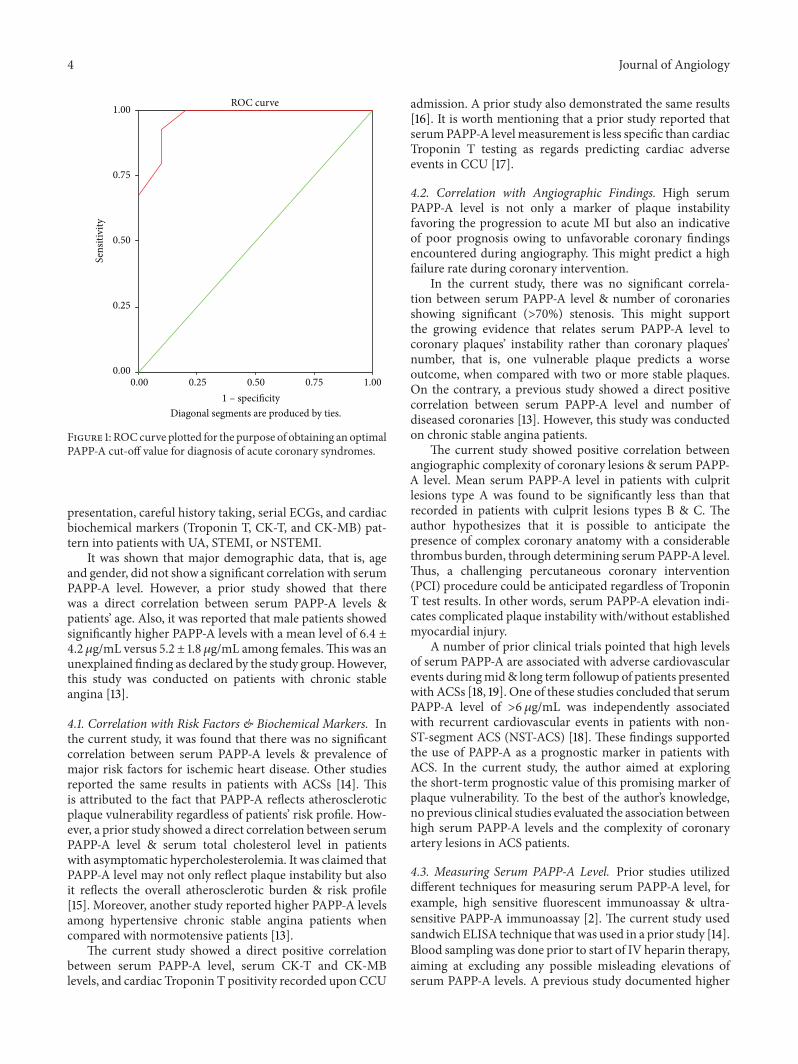
2.5. Statistics. Data were analyzed using Statistical Pack-age for Special Science (SPSS) software computer programversion 15 & described as mean ± standard deviation (SD)for quantitative (numerical) variables and as percentage forqualitative (categorical) variables. Significance level (𝑃 value)&Pearson’s correlation coefficientwere used for assessment ofthe recorded values. Receiver operating characteristic (ROC)curve analysis was constructed resulting in the sensitivity-specificity plot. The optimal cut-off value of serum PAPP-A level, predicting the diagnosis of ACS, was defined byproviding the maximal sum of sensitivity and specificity.
3. Results
The study population (40 patients) included 22 male patients(55%) and 18 female patients (45%)with amean age of 57.08 ±11.11 years. Descriptive data analysis showed that; 26 (65%)patients were diabetic, 17 (42.5%) patients were hypertensive,24 (60%) patients were dyslipidemic, 21 (52.5%) patientswere smokers, and 16 (40%) patients had family history forischemic heart diseases. Upon CCU admission, mean serumCK-T level was 385.3 ± 332.15 IU/L, mean CK-MB level was48 ± 32.92 IU/L, and mean serum PAPP-A level was 10.14 ±2.67 𝜇g/mL.
All patients underwent coronary angiography. Concern-ing number of coronary arteries showing significant (≥70%diameter stenosis) stenosis, it was found that 17 (42.5%)patients had single vessel affection, 10 (25%) patients hadtwo vessels affection, and 13 (32.5%) patients had more thantwo vessels affection. According to ACC/AHA angiographicclassification, it was found that 20 (50%) patients showedatherosclerotic plaques type A, 6 (15%) patients showedatherosclerotic plaques type B, and 14 (35%) patients showedatherosclerotic plaques type C. Patients were categorizedaccording to the characteristics of the culprit lesion only.
Mean serum PAPP-A level among male patients was9.38 ± 3.42 𝜇g/mL, while it was 8.29 ± 3.8 𝜇g/mL amongfemales. There was no significant (𝑃 > 0.05) correlationbetween serum PAPP-A level & patients’ baseline clinicalcharacteristics (age, gender, and risk factors for ischemicheart diseases).
3.1. Biochemical Markers. There was a statistically significantpositive correlation between patients’ serumCK levels (CK-T& CK-MB) & PAPP-A levels measured upon CCU admissionwith a Pearson’s correlation coefficient of 0.604 (CK-T) &0.597 (CK-MB).
Mean serum PAPP-A level among patients with positiveTroponinT test (20, 50%)was recorded to be 11.6± 2.2𝜇g/mL,while it was 7.06± 3.26 𝜇g/mL among patients with negativeTroponin T test. There was a statistically significant positivecorrelation between Troponin T test positivity and serumPAPP-A level (𝑃 < 0.001), being higher in Troponin Tpositive patients.
3.2. Angiographic Findings. Mean serum PAPP-A levelamong patients with single vessel affection was recorded tobe 9.38 ± 3.89 𝜇g/mL, while it was 8.12 ± 3.1 𝜇g/mL amongpatients with two vessels affection & 8.73 ± 3.68 𝜇g/mL
among patients with more than two vessels affection. Resultsshowed no statistically significant correlation between serumPAPP-A level & number of coronary vessels showing signifi-cant stenosis.
Mean serum PAPP-A level among patients with coronaryatherosclerotic plaque type A was recorded to be 7.32 ±3.5 𝜇g/mL while it was 9.75 ± 2.54 𝜇g/mL among patientswith plaque type B & 11.78 ± 2𝜇g/mL among patients withplaque type C. There was a statistically significant positivecorrelation between complexity of coronary artery lesionsand serumPAPP-A level (𝑃 < 0.001), being higher in patientswith plaque types B & C.
Mean serum PAPP-A level among patients with UA (20,50%) was recorded to be 8.72 ± 2.39 𝜇g/mL, while it was11.78 ± 2 𝜇g/mL among patients with STEMI (14, 35%) & 11 ±2.63 𝜇g/mL among patients with NSTEMI (6, 15%). Therewas a statistically significant correlation between the finaldiagnoses of the included patients and serum PAPP-A levels(𝑃 < 0.001), being higher among patients with acute MI(STEMI & NSTEMI).
So, it was found that there was a statistically significantcorrelation between serum PAPP-A level (measured uponCCU admission) and each of the following: initial CK-Tlevel, initial CK-MB level, cardiac Troponin T positivity uponCCU admission, coronary artery lesion complexity, and finaldiagnosis of the ischemic event.
Receiver operating characteristic (ROC) curve analysis(Figure 1) of obtained serum PAPP-A levels (among patientswith Troponin T positive & negative results) reached anoptimal cut-off value of 7.75 𝜇g/mL for prediction of finaldiagnosis of ACS. This cut-off value yielded a sensitivity of80%, specificity of 90%, positive predictive value (PPV) of97%, and negative predictive value (NPV) of 53% (Area underthe curve = 0.97).
4. Discussion
Early diagnosis of ACSs is an important challenging issuethat throws itself on to the top of physicians’ considerations,especially in emergency rooms.
Currently, ACSs are diagnosed using the combination ofpatients’ history, ECG, and biochemical markers of myocar-dial necrosis [11]. Convincing evidence suggests that bothinflammatory as well as thromboticmechanisms are involvedin pathogenesis of ACSs. The availability of a sensitive& specific marker of early plaque instability, whose levelbecomes elevated before or even in the absence of myocardialnecrosis, should improve the diagnostic and therapeuticdecision making [12].
In the current study, PAPP-A as a one of matrix metal-loproteinases that is highly expressed in unstable coronaryatherosclerotic plaques was studied to determine its value inpatients with ACSs (especially those with initial negative car-diac Troponin T results), undergoing coronary angiography& possible subsequent intervention.
This study included 40 patients (22 male patients) withmean age of 57.08 ± 11.11 years who presented to CCUafter emergency room triage, with chest pain of cardiacorigin. They were then categorized according to clinical
4 Journal of Angiology
ROC curve
1.000.750.500.250.00
Sens
itivi
ty1.00
0.75
0.50
0.25
0.00
1 – specificityDiagonal segments are produced by ties.
Figure 1: ROCcurve plotted for the purpose of obtaining an optimalPAPP-A cut-off value for diagnosis of acute coronary syndromes.
presentation, careful history taking, serial ECGs, and cardiacbiochemical markers (Troponin T, CK-T, and CK-MB) pat-tern into patients with UA, STEMI, or NSTEMI.
It was shown that major demographic data, that is, ageand gender, did not show a significant correlation with serumPAPP-A level. However, a prior study showed that therewas a direct correlation between serum PAPP-A levels &patients’ age. Also, it was reported that male patients showedsignificantly higher PAPP-A levels with a mean level of 6.4 ±4.2 𝜇g/mL versus 5.2 ± 1.8 𝜇g/mL among females.This was anunexplained finding as declared by the study group. However,this study was conducted on patients with chronic stableangina [13].
4.1. Correlation with Risk Factors & Biochemical Markers. Inthe current study, it was found that there was no significantcorrelation between serum PAPP-A levels & prevalence ofmajor risk factors for ischemic heart disease. Other studiesreported the same results in patients with ACSs [14]. Thisis attributed to the fact that PAPP-A reflects atheroscleroticplaque vulnerability regardless of patients’ risk profile. How-ever, a prior study showed a direct correlation between serumPAPP-A level & serum total cholesterol level in patientswith asymptomatic hypercholesterolemia. It was claimed thatPAPP-A level may not only reflect plaque instability but alsoit reflects the overall atherosclerotic burden & risk profile[15]. Moreover, another study reported higher PAPP-A levelsamong hypertensive chronic stable angina patients whencompared with normotensive patients [13].
The current study showed a direct positive correlationbetween serum PAPP-A level, serum CK-T and CK-MBlevels, and cardiac Troponin T positivity recorded upon CCU
admission. A prior study also demonstrated the same results[16]. It is worth mentioning that a prior study reported thatserumPAPP-A levelmeasurement is less specific than cardiacTroponin T testing as regards predicting cardiac adverseevents in CCU [17].
4.2. Correlation with Angiographic Findings. High serumPAPP-A level is not only a marker of plaque instabilityfavoring the progression to acute MI but also an indicativeof poor prognosis owing to unfavorable coronary findingsencountered during angiography. This might predict a highfailure rate during coronary intervention.
In the current study, there was no significant correla-tion between serum PAPP-A level & number of coronariesshowing significant (>70%) stenosis. This might supportthe growing evidence that relates serum PAPP-A level tocoronary plaques’ instability rather than coronary plaques’number, that is, one vulnerable plaque predicts a worseoutcome, when compared with two or more stable plaques.On the contrary, a previous study showed a direct positivecorrelation between serum PAPP-A level and number ofdiseased coronaries [13]. However, this study was conductedon chronic stable angina patients.
The current study showed positive correlation betweenangiographic complexity of coronary lesions & serum PAPP-A level. Mean serum PAPP-A level in patients with culpritlesions type A was found to be significantly less than thatrecorded in patients with culprit lesions types B & C. Theauthor hypothesizes that it is possible to anticipate thepresence of complex coronary anatomy with a considerablethrombus burden, through determining serumPAPP-A level.Thus, a challenging percutaneous coronary intervention(PCI) procedure could be anticipated regardless of TroponinT test results. In other words, serum PAPP-A elevation indi-cates complicated plaque instability with/without establishedmyocardial injury.
A number of prior clinical trials pointed that high levelsof serum PAPP-A are associated with adverse cardiovascularevents duringmid& long term followup of patients presentedwith ACSs [18, 19]. One of these studies concluded that serumPAPP-A level of >6𝜇g/mL was independently associatedwith recurrent cardiovascular events in patients with non-ST-segment ACS (NST-ACS) [18]. These findings supportedthe use of PAPP-A as a prognostic marker in patients withACS. In the current study, the author aimed at exploringthe short-term prognostic value of this promising marker ofplaque vulnerability. To the best of the author’s knowledge,no previous clinical studies evaluated the association betweenhigh serum PAPP-A levels and the complexity of coronaryartery lesions in ACS patients.
4.3. Measuring Serum PAPP-A Level. Prior studies utilizeddifferent techniques for measuring serum PAPP-A level, forexample, high sensitive fluorescent immunoassay & ultra-sensitive PAPP-A immunoassay [2]. The current study usedsandwich ELISA technique that was used in a prior study [14].Blood sampling was done prior to start of IV heparin therapy,aiming at excluding any possible misleading elevations ofserum PAPP-A levels. A previous study documented higher

Journal of Angiology 5
serum PAPP-A levels in a group of STEMI patients & inanimal models treated with IV heparin [9]. This is mostprobably related to PAPP-A detachment from blood vessels’walls, induced by heparin.
Until recently, most commercially available assays forPAPP-A serum level measurement were designed to measurehigher concentrations of PAPP-A, that is, during pregnancy.These assays do not meet the required sensitivity to detectserum PAPP-A level in patients with ACSs. Fortunately,ultrasensitive PAPP-A assay has recently become available forthe purpose of diagnosing ACSs [16].
4.4. Clinical Implications. High serum PAPP-A levels mea-sured before PCI in ACS patients call for proper readinessto tackle a complex coronary artery lesion with mostly aconsiderable thrombus burden. This requires having suitablepharmacological agents (e.g., glycoprotein IIb/IIIa inhibitors)& mechanical aids (e.g., thrombus aspiration devices) inhand. Thus, high serum PAPP-A level could help riskstratification of ACS patients upon CCU admission, iden-tifying patients at high risk of in-hospital adverse events.This additionally implies the presence of an experiencedinterventionist capable of dealing with such interventionaldifficulties, expected to be found in coronary angiograms.Moreover, expected higher treatment costs should be takeninto consideration.The current study reached a serum PAPP-A cut-off value (7.75𝜇g/mL) that predicted the final diagnosisof ACS. This could be of special importance in patientswith initial negative Troponin results. Similarly, a priorstudy that included heparin naıve ACS patients stated thatserum PAPP-A levels strongly predicted the diagnosis of ACSwith a PPV & NPV close to those obtained by the currentstudy. In Troponin-negative ACS patients, PAPP-A helpedto reach the correct final diagnosis [10]. This identifies ahigh risk sub-population with high serum PAPP-A level &negative Troponin results.These patients are expected to haveunfavorable angiographic outcomes & technical difficultiesduring PCI, in spite of initial negative Troponin results.Moreover, this subpopulation of patients should be regardedas a high risk category among ACS patients. This can havean impact on oral pharmacological treatment in CCU. Thisincludes, for example, statins intake, as evidenced by a priorstudy that showed that high dose (80mg) statins intake inACS patients undergoing PCI was associated with significantdecrease of serumPAPP-A levels after onemonth [20].On theother hand, a previous study showed that long term regularintake of high dose (four grams) of n-3 fatty acids after acuteMI was associated with higher serum levels of PAPP-A [21].
5. Conclusion
High serum PAPP-A level is associated with unfavorablecoronary anatomy and high probability of lesion complexityin initial coronary angiograms of ACS patients. A cut-offvalue of 7.75𝜇g/mL for serum PAPP-A level predicted thefinal diagnosis of ACS, with a sensitivity of 80% & specificityof 90%.
Limitations of the Study
This is a single-centre study with a small sample size of thecohort. Large scale studies are still needed to confirm theobtained results. Serial serum PAPP-A level sampling wasnot included in the study protocol. Furthermore, followup ofmajor adverse cardiovascular events was also not included.Both items were outside the scope of the present study,which aimed at evaluating the value of serum PAPP-A(single measurement) in augmenting the diagnostic & theinterventional aspects in ACS patients. Further studies withwider scope are needed for evaluation of long term impact ofhigh serum PAPP-A levels in ACS patients.
Conflict of Interests
The author declares that there is no conflict of interestsregarding the publication of this paper.
Acknowledgments
The author wishes to express his gratitude for nursing andtechnical staff of cardiac catheterization laboratory in Cardi-ology Department, Ain Shams University. Special thanks aredue to Dr. Maha Ezz Elden, Head of Emergency Blood WorkLaboratory for her cooperation to accomplish this work.
References
[1] M. Naghavi, P. Libby, E. Falk et al., “From vulnerable plaque tovulnerable patient: a call for newdefinitions and risk assessmentstrategies—part I,” Circulation, vol. 108, no. 14, pp. 1664–1672,2003.
[2] A. Bayes-Genis, C. A. Conover, M. T. Overgaard et al., “Preg-nancy-associated plasma protein A as a marker of acute coro-nary syndromes,”NewEngland Journal ofMedicine, vol. 345, no.14, pp. 1022–1029, 2001.
[3] P. Libby, “Inflammation in atherosclerosis,”Nature, vol. 420, no.6917, pp. 868–874, 2002.
[4] R. Ross, “Atherosclerosis—an inflammatory disease,” New Eng-land Journal of Medicine, vol. 340, no. 2, pp. 115–126, 1999.
[5] L. S. Laursen, M. T. Overgaard, C. G. Nielsen et al., “Sub-strate specificity of the metalloproteinase pregnancy-associatedplasma protein-A (PAPP-A) assessed by mutagenesis and anal-ysis of synthetic peptides: substrate residues distant from thescissile bond are critical for proteolysis,” Biochemical Journal,vol. 367, no. 1, pp. 31–40, 2002.
[6] J. Lund, Q.-P. Qin, T. Ilva et al., “Circulating pregnancy-associ-ated plasma protein a predicts outcome in patients with acutecoronary syndrome but no troponin I elevation,” Circulation,vol. 108, no. 16, pp. 1924–1926, 2003.
[7] C. Heeschen, S. Dimmeler, C. W. Hamm, S. Fichtlscherer, M.L. Simoons, and A. M. Zeiher, “Pregnancy-associated plasmaprotein-A levels in patients with acute coronary syndromes:comparison with markers of systemic inflammation, plateletactivation, and myocardial necrosis,” Journal of the AmericanCollege of Cardiology, vol. 45, no. 2, pp. 229–237, 2005.
[8] S. C. Smith Jr., J. T. Dove, A. K. Jacobs et al., “American Collegeof Cardiology; AmericanHeart Association Task Force on Prac-tice Guidelines. Committee to Revise the 1993 Guidelines forPercutaneous Transluminal Coronary Angioplasty. ACC/AHA

6 Journal of Angiology
guidelines for percutaneous coronary intervention (revision ofthe 1993 PTCA guidelines)—executive summary. A report ofthe American College of Cardiology/American Heart Associ-ation Task Force on Practice Guidelines (committee to revisethe 1993 guidelines for percutaneous transluminal coronaryangioplasty),” Journal of the American College of Cardiology, vol.37, no. 8, pp. 2215–2239, 2001.
[9] C. J. Terkelsen, C. Oxvig, and B. L. Nørgaard, “Temporal courseof pregnancy-associated plasma protein-A in angioplasty-treat-ed ST-segment elevation myocardial infarction patients andpotential significance of concomitant heparin administration,”American Journal of Cardiology, vol. 103, no. 1, pp. 29–35, 2009.
[10] P. Hajek, M. Macek Sr., M. Peskova et al., “High positivepredictive value of PAPP-A for acute coronary syndrome diag-nosis in heparin-naıve patients,” Journal of Thrombosis andThrombolysis, vol. 34, no. 1, pp. 99–105, 2012.
[11] C. W. Hamm, B. U. Goldmann, C. Heeschen, G. Kreymann, J.Berger, and T. Meinertz, “Emergency room triage of patientswith acute chest pain by means of rapid testing for cardiactroponin T or troponin I,”NewEngland Journal ofMedicine, vol.337, no. 23, pp. 1648–1653, 1997.
[12] E. Braunwald, “Unstable angina: an etiologic approach tomanagement,” Circulation, vol. 98, no. 21, pp. 2219–2222, 1998.
[13] J. Cosin-Sales, J. C. Kaski, M. Christiansen et al., “Relationshipamong pregnancy associated plasma protein-A levels, clinicalcharacteristics, and coronary artery disease extent in patientswith chronic stable angina pectoris,” European Heart Journal,vol. 26, no. 20, pp. 2093–2098, 2005.
[14] J.-L. Liu, H. Zhang, X.-J. Xie, L. Chen, andC.-L. Zhao, “Changesof pregnancy-associated plasma protein-A in patients withacute coronary syndrome,”ChineseMedical Journal, vol. 118, no.21, pp. 1827–1829, 2005.
[15] T. Stulc, I.Malbohan, and J.Malık, “Increased levels of pregnan-cy-associated plasma protein-A in patients with hypercholes-terolemia: the effect of atorvastatin treatment,” American HeartJournal, vol. 146, no. 6, article E21, 2003.
[16] J. Khosravi, A. Diamandi, R. G. Krishna, U. Bodani, J. Mistry,and N. Khaja, “Pregnancy associated plasma protein-A: ultra-sensitive immunoassay and determination in coronary heartdisease,” Clinical Biochemistry, vol. 35, no. 7, pp. 531–538, 2002.
[17] O. F. Laterza, S. J. Cameron, D. Chappell, L. J. Sokoll, and G. B.Green, “Evaluation of pregnancy-associated plasma protein Aas a prognostic indicator in acute coronary syndrome patients,”Clinica Chimica Acta, vol. 348, no. 1-2, pp. 163–169, 2004.
[18] M. P. Bonaca, B. M. Scirica, M. S. Sabatine et al., “Prospectiveevaluation of pregnancy-associated plasma protein-a and out-comes in patients with acute coronary syndromes,” Journal ofthe American College of Cardiology, vol. 60, no. 4, pp. 332–338,2012.
[19] W. Y. Mei, Z. M. Du, Q. Zhao et al., “Pregnancy-associatedplasma protein predicts outcomes of percutaneous coronaryintervention in patients with non-ST-elevation acute coronarysyndrome,” Heart Lung, vol. 40, no. 3, pp. e78–e83, 2011.
[20] M. D. Miedema, C. A. Conover, H. MacDonald et al., “Preg-nancy-associated plasma protein-A elevation in patients withacute coronary syndrome and subsequent atorvastatin therapy,”American Journal of Cardiology, vol. 101, no. 1, pp. 35–39, 2008.
[21] H. Aarsetøy, T. Brugger-Andersen, Ø. Hetland, H. Grundt, andD. W. T. Nilsen, “Long term influence of regular intake ofhigh dose n-3 fatty acids onCD40-ligand, pregnancy-associatedplasma protein A and matrix metalloproteinase-9 following
acute myocardial infarction,”Thrombosis and Haemostasis, vol.95, no. 2, pp. 329–336, 2006.

Submit your manuscripts athttp://www.hindawi.com
Stem CellsInternational
Hindawi Publishing Corporationhttp://www.hindawi.com Volume 2014
Hindawi Publishing Corporationhttp://www.hindawi.com Volume 2014
MEDIATORSINFLAMMATION
of
Hindawi Publishing Corporationhttp://www.hindawi.com Volume 2014
Behavioural Neurology
EndocrinologyInternational Journal of
Hindawi Publishing Corporationhttp://www.hindawi.com Volume 2014
Hindawi Publishing Corporationhttp://www.hindawi.com Volume 2014
Disease Markers
Hindawi Publishing Corporationhttp://www.hindawi.com Volume 2014
BioMed Research International
OncologyJournal of
Hindawi Publishing Corporationhttp://www.hindawi.com Volume 2014
Hindawi Publishing Corporationhttp://www.hindawi.com Volume 2014
Oxidative Medicine and Cellular Longevity
Hindawi Publishing Corporationhttp://www.hindawi.com Volume 2014
PPAR Research
The Scientific World JournalHindawi Publishing Corporation http://www.hindawi.com Volume 2014
Immunology ResearchHindawi Publishing Corporationhttp://www.hindawi.com Volume 2014
Journal of
ObesityJournal of
Hindawi Publishing Corporationhttp://www.hindawi.com Volume 2014
Hindawi Publishing Corporationhttp://www.hindawi.com Volume 2014
Computational and Mathematical Methods in Medicine
OphthalmologyJournal of
Hindawi Publishing Corporationhttp://www.hindawi.com Volume 2014
Diabetes ResearchJournal of
Hindawi Publishing Corporationhttp://www.hindawi.com Volume 2014
Hindawi Publishing Corporationhttp://www.hindawi.com Volume 2014
Research and TreatmentAIDS
Hindawi Publishing Corporationhttp://www.hindawi.com Volume 2014
Gastroenterology Research and Practice
Hindawi Publishing Corporationhttp://www.hindawi.com Volume 2014
Parkinson’s Disease
Evidence-Based Complementary and Alternative Medicine
Volume 2014Hindawi Publishing Corporationhttp://www.hindawi.com