Embed Size (px)
Citation preview
Hindawi Publishing CorporationISRN OphthalmologyVolume 2013, Article ID 210565, 8 pageshttp://dx.doi.org/10.1155/2013/210565
Clinical StudyPostkeratoplasty Anterior and Posterior Corneal SurfaceWavefront Analysis: Descemet’s Stripping AutomatedEndothelial Keratoplasty versus Penetrating Keratoplasty
Maria L. Salvetat, Marco Zeppieri, Flavia Miani, and Paolo Brusini
Department of Ophthalmology, Santa Maria della Misericordia University Hospital, Piazzale S. Maria della,Misericordia 15, 33100 Udine, Italy
Correspondence should be addressed to Maria L. Salvetat; [email protected]
Received 26 May 2013; Accepted 12 August 2013
Academic Editors: B. V. Bui, M. Nakazawa, and L. Pierro
Copyright © 2013 Maria L. Salvetat et al. This is an open access article distributed under the Creative Commons AttributionLicense, which permits unrestricted use, distribution, and reproduction in any medium, provided the original work is properlycited.
Purpose. To compare the higher-order aberrations (HOAs) due to the anterior and posterior corneal surfaces in patientsthat underwent either Descemet-stripping-automated-endothelial-keratoplasty (DSAEK) or penetrating keratoplasty (PK) forendothelial dysfunction and age-matched controls. Methods. This retrospective, observational, case series included 28 patientsafter PK, 30 patients after DSAEK, and 30 healthy controls. A Scheimpflug imaging system was used to assess the HOAs due to theanterior and posterior corneal surfaces at 4mm and 6mm optical zones. Total, 3rd and 4th order HOAs were considered. Intra-and intergroup differences were assessed using the Friedman and the Kruskal-Wallis tests, respectively; paired comparisons wereperformed using Duncan’s multiple range test. Results. Total, 3rd and 4th order HOAs due to both corneal surfaces at 4mm and6mm optical zones were significantly higher in the PK group, intermediate in the DSAEK group, and lower in controls (𝑃 < 0.05).The most important HOAs components in both PK and DSAEK groups were trefoil and coma from the anterior corneal surface(𝑃 < 0.05) and trefoil from the posterior corneal surface (𝑃 < 0.05). Conclusions. The optical quality of both corneal surfacesappeared significantly higher after DSAEK than after PK, which can increase the postoperative patient’s quality of vision andsatisfaction.
1. Introduction
Endothelial keratoplasty (EK) is nowadays considered as theprocedure of choice for the treatment of the endothelialdysfunctions [1]. The technique is based on the selectivereplacement of diseased endothelium, while leaving thehealthy recipient anterior cornea structurally intact. EKhas been shown to be a better procedure than penetratingkeratoplasty (PK) due to faster postoperative visual recov-ery, minimal induced topographic changes, lower refractiveerror, higher refraction predictability and stability, absenceof suture-related complications, better corneal structuralintegrity and innervation maintenance, and reduced riskof graft rejection [2–4]. The surgical technique has under-gone modifications and improvements over the years, whichinclude the followingmethods in chronologic order: posteriorlamellar keratoplasty (PLK) [5], deep lamellar endothelial
keratoplasty (DLEK) [6], Descemet stripping endothelial ker-atoplasty (DSEK) [7], Descemet stripping and automatedendothelial keratoplasty (DSAEK) [8–10], andDescemetmem-brane endothelial keratoplasty (DMEK) [11].
The DSAEK technique, which currently tends to bethe preferred EK surgical approach used in many cen-ters, involves the mechanical stripping of the diseased hostendothelium and Descemet’s membrane and replacementwith a donor graft, composed of endothelium, Descemet’smembrane, and a thin layer of posterior stroma, carried outwith an automated microkeratome [8–10].
Although several studies have shown higher postopera-tive visual outcomes after DSAEK than after PK [3, 12, 13],others have reported that the best spectacle-corrected visualacuity (BSCVA) after DSAEK can be lower than after PK[10, 14]. Patient’s age, preoperative corneal haze, interfacehaze, and optical irregularities at the corneal surfaces and/or
2 ISRN Ophthalmology
interface, inducing light scattering and increased irregularastigmatism, are suggested reasons for limited visual out-comes after DSAEK [14–20].
Several studies have shown that the DSAEK procedurecauses minimal changes of the anterior corneal surface, withconsequent optical advantages compared to PK such as lowerregular [3, 10, 12] and irregular astigmatism, also known ashigher-order aberrations (HOAs) [16–20]. Induced irregularastigmatism of the posterior corneal surface has shown to beeither comparable between PK and DSAEK [17, 19, 20] orhigher after DSAEK than after PK [16–18].
The wave-front analysis is an objective method of assess-ing the optical quality of the ocular refractive surfaces, bythe evaluation of the low- and higher-order aberrationsthat can degrade the retinal image [21]. Great amounts ofHOAs, which are not correctable by conventional spectaclesor soft contact lenses, has been shown to reduce the opticalperformance of the eye by inducing halos, glare, monoculardiplopia, decreased contrast sensitivity, and visual acuity [22,23], especially under mesopic or scotopic conditions [24].The cornea is the main contributor of HOAs in the eye and,regardless of cause, corneas with increased wavefront errorshow significant decreases in visual performances that arepupil size dependent [25].
The rotating Scheimpflug imaging system is a relativelynew noncontact method that is able to provide highly repeat-able measurements of the anterior and posterior cornealcurvatures, which can be converted into corneal aberrationsmeasurements by the device software [26, 27].
The aim of our study was to compare the HOAs due tothe anterior and posterior corneal surfaces evaluated witha Scheimpflug-based corneal topographer in patients thatunderwent DSAEK or PK for endothelial dysfunction andage-matched controls with normal corneas, in order to assessthe effect of PK and DSAEK on the optical quality of thecorneal surfaces. Measurements were taken for a 4mm and6mm optical zone, to simulate the photopic and scotopicconditions, respectively.
2. Patients and Methods
This retrospective, observational, and comparative case seriesstudy included 3 groups of subjects: 30 consecutive patientsafter PK, 31 consecutive patients after DSAEK, and 30age-matched healthy subjects with normal corneas (controlgroup). One eye per patient was considered. The study wasin compliance with the tenets of the Helsinki’s Declaration,and informed consent was obtained from all participantsprior to testing. Each participant underwent the followingexaminations on the same day: complete ophthalmologicexamination, including a review of medical history, BSCVAmeasured using the Snellen VA chart, manifest refractionevaluation (including spherical equivalent and cylindricalerror), slit-lamp examination, fundus biomicroscopy with a90D lens, Goldmann applanation tonometry measurement,and imaging with a Scheimpflug-based corneal topographer(Sirius 3D, CSO, Florence, Italy).
Normal subjects were recruited from staff members andvolunteers. PK and DSAEK patients were recruited from the
Cornea Clinic of the Department of Ophthalmology at S.Maria della Misericordia Hospital, Udine, Italy. The studywas in compliance with Institutional Review Board (IRB) andHIPAA requirements and approved by the IRB of S. Mariadella Misericordia Hospital, Udine, Italy.
Normal eyes were defined as no ocular disorders exceptfor refractive errors, normal cornea appearance, normalcorneal topography results, no history of ocular surgery,no previous corneal or conjunctival disease that is likely toaffect the corneal HOAs, and no family history of ocularpathologies.
Inclusion criteria for postoperative patients were pre-vious PK or DSAEK for endothelial dysfunction (at least6 months after complete suture removal); availability ofpostoperative Scheimpflug camera imaging with no missingdata points within the central 6.0mm zone, no intra- orpostoperative complications that can affect the Scheimpflugcamera measurements, no history of ocular surgery otherthan cataract surgery, and willingness to provide informedwritten consent. Exclusion criteria included corneal scars oropacities of the graft, history of postoperative ocular infectionor trauma, graft rejection, history of intraocular surgery otherthan keratoplasty or cataract surgery, presence of ocular orsystemic diseases or medications that could affect the ocularsurface and/or prevent reliable wave-front measurements,and inability to comply with Scheimpflug imaging procedure.
PK [3] and DSAEK [8, 10] techniques have extensivelybeen reported elsewhere and have briefly been described inthe Appendix. All surgeries were performed by a single sur-geon (PB) from June 2008 toMarch 2010 at theDepartment ofOphthalmology of the S. Maria della Misericordia Hospital,Udine, Italy. Corneal diseases requiring keratoplasty wereFuchs dystrophy and pseudophakic bullous keratopathy. Themean duration of the bullous keratopathy was 9.1 ± 9.3months (range 6 to 43 months). Donor corneas in the formof a sclerocorneal button stored in organ culture at 31∘Cwere provided by the “Fondazione Banca degli Occhi delVeneto” (Venezia-Mestre, Italy) Eye Bank. In 6 patients withsignificant lens opacity, 2 PK and 4DSAEK patients, standardphacoemulsification was performed using the phaco choptechnique, followed by implantation of an intraocular lens(IOL) in the bag.
BSCVA in Snellen lines and refraction, reported as meanrefractive spherical equivalents, were measured by a singleoptometrist (LP), who was masked to the type of surgery.
The Sirius 3D rotating Scheimpflug camera (CSO, Flo-rence, Italy) was used for all corneal measurements. Thisnoncontact instrument combines a rotating Scheimpflugcamera with a Placido disk technique, providing high-resolution images of the anterior segment, anterior andposterior corneal topography, and pachymetry of the entirecornea.The system uses a rotating Scheimpflug camera and amonochromatic slit-light source that rotate together aroundthe optical axis of the eye for 180 degrees and acquires 25 to50 images from the anterior segment, allowing the acquisitionof anterior and posterior corneal elevation topographic data.The built-in software provides the conversion of the cornealelevation profile into corneal wave-front data using theZernike vector terms [28] with an expansion of up to the

ISRN Ophthalmology 3
10th order. The root mean square (RMS) of the Zernikevector magnitude is calculated and expressed in 𝜇m. Datafrom an area of up to 10mm in diameter are provided bythe instrument; a graft size of 8.5mm was considered in ourstudy. The automatic release mode was used, which achievescorrect focus and alignment with the corneal apex beforescanning starts. The imaging was performed with the patientseated and correctly positioned in the chinrest and foreheadstrap. The patient was asked to keep both eyes open andto look at a fixation target. The system constantly monitorseye movements; measurements with decentration less than0.6mm are considered valid. The examination quality datawere assessed with a built-in program, and the results withsignificant errors were excluded. Three measurements weretaken by the same experienced examiner (LP) from each eye,and the best scan with the less distorted Scheimpflug imagewas used for analysis. All measurements were collected at thelast visit and at least 6 months after complete suture removalfor postkeratoplasty eyes. The following measurements wereconsidered: simulated keratometric (simK) values in the3mm central zone, RMS of the Zernike vector magnitudeof the total HOAs, and 3rd and 4th order aberrations ofthe anterior and posterior corneal surface within the central4mm and 6mm zones. Total HOAs were defined as the sumof the magnitude of the Zernike vector terms of 3rd to 7thorder. SimK values include diopter power and axis of thesteepest meridian and at 90 degrees (K1 and K2). K1 and K2were averaged to obtain a single corneal curvature value. Thecorneal astigmatism value was defined as the absolute valuefor K2 minus K1.
Data were analyzed using the statistical analysis softwareSPSS for Windows, version 20.0 (SPSS Inc., Chicago, IL,USA). Data were described by medians (standard deviation)and 95% confidence interval (CI). Normality of the datadistribution was assessed with the Kolmogorov-Smirnov test.Intragroup differences were assessed using the Wilcoxonand Friedman tests; intergroup differences were calculatedusing the Kruskal-Wallis tests; theDuncanmultiple range testwas used for multiple comparisons. Correlations were testedusing the Spearman’s rank correlation coefficient. Statisticalsignificance was defined as 𝑃 < 0.05.
3. Results
Scheimpflug imaging was not obtained for 2 patients afterPK and 1 patient after DSAEK and thus were excluded fromthe analysis. A total of 28 patients after PK, 30 patientsafter DSAEK, and 30 healthy controls fulfilled the inclusioncriteria. Detailed demographic, visual, and refractive charac-teristics of the three groups of subjects are listed in Table 1.The wavefront analysis of the corneal HOAs of the 3 groupsis shown in Figure 1.
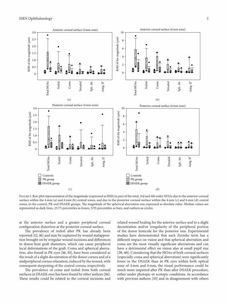
3.1. Anterior Corneal Surface (Figures 1(a) and 1(b)). TotalHOAs, trefoil, and coma were significantly higher in thePK group, intermediate in the DSAEK group, and lower incontrols (𝑃 < 0.05). Tetrafoil appeared significantly lower incontrols (𝑃 < 0.01), comparable between PK and DSAEK
eyes within the 4mm zone (𝑃 > 0.05), and significantlyhigher in the PK eyes than in DSAEK eyes within the6mm zone (𝑃 < 0.05). Spherical aberration and secondaryastigmatism were significantly higher in the PK group thanin the other groups (𝑃 < 0.05).
The most important aberration components were trefoil,coma, and spherical aberration in controls (𝑃 < 0.05), andtrefoil and coma in the DSAEK and PK groups (𝑃 < 0.05).
3.2. Posterior Corneal Surface (Figures 1(c) and 1(d)). TotalHOAs, trefoil, coma, and tetrafoil were significantly higherin the PK eyes, intermediate in the DSAEK eyes, and lowerin controls (𝑃 < 0.05). Spherical aberration and secondaryastigmatism appeared significantly lower in controls (𝑃 <0.01), higher in the PK than in the DSAEK eyes within the4mm zone (𝑃 < 0.05) and comparable between PK andDSAEK groups within the 6mm zone (𝑃 > 0.05).
The trefoil was themost important aberration componentwithin the 4mm zone in the three groups (𝑃 < 0.01).Themost important aberration components within the 6mmzone were trefoil in the control and PK eyes (𝑃 < 0.05), andcoma and trefoil in the DSAEK eyes (𝑃 < 0.05).
The magnitude of the HOAs from the 4mm zone wassignificantly lower than that of theHOAs from the 6mmzonefor both corneal surfaces in all groups (𝑃 < 0.01). In controls,the magnitude of the HOAs of the anterior corneal surfacewas significantly larger than that of the posterior surface (𝑃 <0.01), with exception of that of trefoil, tetrafoil, and secondaryastigmatism within the 4mm zone, which was comparablebetween corneal surfaces (𝑃 > 0.05).
In the PK and DSAEK groups, the magnitude of theHOAs of the corneal anterior surface appeared comparablewith that of the posterior corneal surface (𝑃 > 0.05), exceptfor that of the total HOAs and coma within the 6mm zone,which appeared to be significantly higher on the anteriorsurface (𝑃 < 0.05). Representative aberration color-codedmaps of anterior and posterior corneal surfaces within the6mm central zone in normal, PK andDSAEK eyes are shownin Figure 2. The correlations between the BSCVA and themagnitude of the HOAs of both corneal surfaces were notstatistically significant in any of the 3 groups (𝑃 > 0.05).
4. Discussion
The results of our study showed that the magnitude of thetotal HOAs and of the Zernike vector terms of 3rd and 4thorder from both corneal surfaces was significantly higherin PK and DSAEK eyes than in controls, indicating greatercorneal surface irregularities in grafted eyes. In accordancewith our results, several previous authors have reported agreater amount of HOAs from both corneal surfaces in PKeyes in comparison with normal eyes [16, 17, 19, 20, 29, 30].The asymmetric distortion of both corneal surfaces inducedby differences in curvature, thickness, and diameter betweendonor lenticule and recipient bed, in addition to the woundconfiguration induced by the healing process,may explain theincreased amount of corneal HOAs found after PK.
The magnitude of the anterior corneal HOAs was signif-icantly higher after DSAEK than in controls in our study,

4 ISRN Ophthalmology
Table 1: Demographic and refractive data.
Controls PK group DSAEK group Comparison(𝑛 = 30) (𝑛 = 28) (𝑛 = 30) (𝑃∗)
Patient’s age (years) 66.6 ± 15.7 67.7 ± 14.1 70.5 ± 12.4 0.10(40–87) (21.8–86.2) (43.2–84.8)
Time interval from surgery (months) — 33.3 ± 14.6 32.5 ± 13.1 0.58(19.1–47.3) (18.7–43.1)
Time interval from suture removal (months) — 23.9 ± 13.8 26.0 ± 12.4 0.43(11.2–35.1) (10.4–34.9)
BSCVA (Snellen lines) 0.91 ± 0.2a
0.53 ± 0.3 0.60 ± 0.2 0.001(0.5–1.0) (0.1–1.0) (0.4–1.0)
Spherical-equivalent error (D) 0.05 ± 2.0 −1.45 ± 4.8b
0.33 ± 1.7 0.01(−3.7/2.5) (−14.1/1.6) (−2.1/3.9)
Mean anterior corneal curvature (D) 43.9 ± 1.4 45.4 ± 2.8a
43.1 ± 1.6 0.008(Sim K1 + Sim K2)/2 (41.1–47.5) (42.1–51.4) (39.3–45.8)Anterior corneal astigmatism (D) 0.75 ± 0.5 4.49 ± 2.9
a1.38 ± 0.5 0.0001
(Sim K2− Sim K1) absolute value (0.2–2.5) (1.2–9.8) (0.6–2.7)Results are given as median ± SD (95% confidence interval).∗Kruskal-Wallis test.BSCVA: best spectacles-corrected visual acuity; D: diopters; Sim K: simulated keratometric value.aSignificantly higher than the other groups.bSignificantly lower than the other groups.
which is in disagreement with previous authors that did notfind significant differences between normal and DSAEK eyes[16, 17, 20] and in accordance with others [19]. This maybe explained by the corneal incisions and related woundhealing after the DSAEK procedure. Several other studies[16, 17, 19, 20], however, have reported that the posteriorcorneal HOAs appeared significantly lower in controls thanin DSAEK eyes, due to the insertion of the donor lenticulethat induces evident configuration changes of the cornealposterior surface [31, 32].
The comparison between PK and DSAEK eyes showedthat themagnitude of the total, 3rd and 4th order HOAs fromboth corneal surfaces, within both optical zones of 4mm and6mm, was significantly higher in PK than in DSAEK eyes.These data suggest a higher optical quality of the cornealsurfaces after DSAEK than after PK.
In agreement with our data, several previous authors havereported significantly greater ocular and anterior cornealsurfaceHOAs after PK than after DLEK [29, 33], DSAEK [16–20], andDMEK [20] procedures.The ocular HOAs have beendemonstrated to be significantly higher after DLEK than afterDSAEK,which could be related to the rougher surface createdby the hand dissection of the donor and recipient corneas,inducing higher interface irregularities [34]. Moreover, thecorneal anterior HOAs have been reported to be minimaland comparable after DSAEK and DMEK procedures [20],suggesting that both surgical procedures induce only slightchanges in the anterior corneal configuration.
The comparison regarding the amount of the posteriorcorneal surfaceHOAs between PK andDSAEK surgery is stilla debatable issue. Our data showed greater posterior cornealsurface HOAs after PK than after DSAEK. In disagreementwith our results, studies have reported that the HOAs due tothe posterior corneal surface were either comparable between
the two groups [17, 19, 20] or higher after DSAEK thanafter PK [16, 18]. The different results found in our study incomparison with those reported by previous authors can berelated to differences in the cohort of patients, type of surgicalprocedures, diameter of optical zone considered, and devicesused to measure the corneal wave-front errors. Previousstudies have reported that values provided by the Pentacamsystem for posterior corneal aberrations in normal subjectswere likely to be erroneous [35]. The new Scheimpflug-basedtopographer used in our study may provide a more accurateevaluation of the corneal HOAs.
As found in our study, the contribution of the anteriorsurface to the corneal HOAs tended to be significantly higherthan that of the posterior surface, due to differences in refrac-tion indices [36]. The impact of the posterior corneal surfaceon vision, however, has yet to be sufficiently explained.Although previous authors found that VA correlated signif-icantly with the HOAs due to the anterior corneal surfacebut not with those from the posterior corneal surface innormal and postkeratoplasty eyes [30, 37], others suggestedthe possible influence of posterior corneal curvature onvisual function [38]. A recent study comparing VA andcorneal HOAs between DSAEK and DMEK eyes showed thata significantly higher postoperative BSCVA in the DMEKeyes was associated with comparable corneal HOAs fromthe anterior surface and significantly higher HOAs fromthe posterior surface in the DSAEK eyes [20], suggesting arelationship between visual function and posterior cornealsurface regularity.
In both PK and DSAEK groups, the most importantaberration components were trefoil and coma from theanterior corneal surface and trefoil from the posterior cornealsurface. These data suggest that both surgical proceduresinduce an increased surface irregularity of the entire cornea
ISRN Ophthalmology 5
3.0
2.5
2.0
1.5
1.0
0.5
0
Anterior corneal surface (4mm zone)
Tota
l HO
As
Tref
oil
Com
a
Tetr
afoi
l
Sph.
Ab.
Asti
g. II
∘
RMS
of th
e mag
nitu
de (𝜇
m)
(a)
10
8
6
4
2
0
Anterior corneal surface (6mm zone)
RMS
of th
e mag
nitu
de (𝜇
m)
Tota
l HO
As
Tref
oil
Com
a
Tetr
afoi
l
Sph.
Ab.
Asti
g. II
∘
(b)
3.0
2.5
2.0
1.5
1.0
0.5
0
Tota
l HO
As
Tref
oil
Com
a
Tetr
afoi
l
Sph.
Ab.
Asti
g. II
∘
RMS
of th
e mag
nitu
de (𝜇
m)
DSAEK group
ControlsPK group
Posterior corneal surface (4mm zone)
(c)
10
8
6
4
2
0
RMS
of th
e mag
nitu
de (𝜇
m)
Tota
l HO
As
Tref
oil
Com
a
Tetr
afoi
l
Sph.
Ab.
Asti
g. II
∘
Posterior corneal surface (6mm zone)
DSAEK group
ControlsPK group
(d)
Figure 1: Box-plot representation of themagnitude (expressed as RMS in 𝜇m) of the total, 3rd and 4th orderHOAs due to the anterior cornealsurface within the 4mm (a) and 6mm (b) central zones, and due to the posterior corneal surface within the 4mm (c) and 6mm (d) centralzones, in the control, PK and DSAEK groups. The magnitude of the spherical aberration was expressed as absolute value. Median values arerepresented as dark lines, 25/75 percentiles as boxes, 5/95 percentiles as bars, and outliers as circles.
at the anterior surface and a greater peripheral cornealconfiguration distortion at the posterior corneal surface.
The prevalence of trefoil after PK has already beenreported [12, 16] and may be explained by wound malapposi-tion brought on by irregular wound incisions and differencesin donor-host graft diameters, which can cause peripherallocal deformations of the graft. Coma and spherical aberra-tion, also found in PK eyes [16, 33], have been considered asthe result of a slight decentration of the donor cornea and of amidperipheral cornea relaxation, induced by the wound, withconsequent steepening of the central cornea, respectively.
The prevalence of coma and trefoil from both cornealsurfaces inDSAEK eyes has been found by other authors [16].These results could be related to the corneal incisions and
related wound healing for the anterior surface and to a slightdecentration and/or irregularity of the peripheral portionof the donor lenticule for the posterior one. Experimentalstudies have demonstrated that each Zernike term has adifferent impact on vision and that spherical aberration andcoma are the most visually significant aberrations and canhave a detrimental effect on vision also at small pupil size[39, 40]. Considering that the HOAs of both corneal surfaces(especially coma and spherical aberration) were significantlylower in the DSAEK than in PK eyes within both opticalzone of 4mm and 6mm, the visual performance could bemuch more impaired after PK than after DSAEK procedure,either under photopic or scotopic conditions. In accordancewith previous authors [33] and in disagreement with others

6 ISRN Ophthalmology
DSAEKControls PK
Ant
erio
r cor
neal
surfa
cePo
sterio
r cor
neal
surfa
ce
Figure 2: Representative wavefront maps of anterior and posterior corneal surfaces in normal, PK, and DSAEK eyes. The same color scalewas used for all maps to allow comparisons.
[18, 30], we did not find any correlation between BSCVAand the magnitude of the total, 3th and 4th order HOAsof both corneal surfaces in normal and postkeratoplastyeyes. The lack of significant relationship between the BSCVAand HOAs magnitude can be due to the presence of ocularpathologies other than endothelial dysfunction in DSAEKand PK eyes (especially cataract and macular degeneration)and the low sensitivity of the photopic high-contrast VA tolimited wave-front errors variation, especially when acuity isscored to line as opposed to the letter in normal eyes, as wasthe case in our paper [21, 40].
Our study has several limitations, including that it wasbased on retrospective data, the number of eyes consideredwas relatively small, and the instrument reproducibility wasnot assessed. Previous studies, however, have reported thatanterior and posterior corneal curvature parameters assessedby the rotating Scheimpflug camera were highly repeatable[26]. Another limitation is the fact that only the high-luminance high-contrast BSCVA was evaluated, which hasshown to be less sensitive to HOAs variation than contrastsensitivity and low-contrast visual acuity under mesopicand scotopic conditions [21, 40]. Moreover, the correlationbetween BSCVA and HOAs magnitude was unsurprisinglynot significant considering that both PK andDSAEK patientscould be affected by ocular pathologies other than endothelialdysfunction. Our previous study [41] showed that the totalHOAs from the anterior corneal surface were significantlylower in DALK than in ALTK and PK groups; however,the total HOAs from the posterior corneal surface were
comparable amongst postoperative groups. The aberrationcomponents that were significantly greater included comain the KC and ALTK eyes, trefoil and coma in the DALKeyes, and trefoil in the PK eyes. Further studies are currentlyunderway, which include only patients after PK and DSAEKwithout any other ocular disease (with exception to pseu-dophakia).
In conclusion, the measurement of the corneal wave-front errors can be important in understanding the changesinduced by penetrating or lamellar keratoplasty on thecorneal profile. The results of our study showed that theDSAEK procedure can provide a higher postoperative reg-ularity to both corneal surfaces when compared with PK.This can lead to better postoperative corneal optical quality,thus providing enhanced patient visual performance andsatisfaction. Further studies evaluating contrast sensitivityand low-contrast visual acuity under mesopic and scotopicconditions are needed to better assess the influence of thecorneal wave-front error on the visual quality after PK andDSAEK surgery.
Appendices
A. Surgical Technique
Local anesthesia and akinesiawere achievedwith a peribulbarinjection of 10 cm3 of a 1 : 1 mixture of bupivacaine 0.75% andlidocaine 2%.
ISRN Ophthalmology 7
B. Penetrating Keratoplasty (PK)
A Hanna suction trephine (Moria, Antony, France) was usedto cut a partial depth, circular incision in the recipient cornea,centered at the geometric center of the cornea, with a diam-eter of 8.25mm. Excision of the recipient corneal button wascompleted with curved corneal scissors. A 0.25mmoversizeddonor button was punched from the endothelial side with theHanna punch trephine. Four interrupted sutures and a single16-bite 10-0 nylon suture (Ethicon Inc., Somerville, NJ) wereplaced in all cases.
C. Descemet Stripping Automated EndothelialKeratoplasty (DSAEK)
The donor lamellar graft dissection was performed with ahand-driven microkeratome using the Moria ALTK micro-keratome (model Evolution 3E) equippedwith a 350-micron-deep blade and associated artificial anterior chamber (Moria,Antony, France). After dissection, the anterior corneal cupwas discarded, and the posterior corneal lamellar tissuewas placed in the corneal storage medium Optisol (ChironOphthalmics, Irvine, CA). At the beginning of surgery,the posterior corneal lamellar tissue was transferred to apunching system and was punched from the endothelial sideusing an 8.5mm Hanna punch trephine (Moria, Antony,France). The donor corneal lenticule remained resting onthe donor punching block covered by Optisol solution untiluse. A clear corneal temporal incision was made in thehost with a 2.75mm keratome. The recipient epithelium wasmarked with an 8.5mm Weck trephine (Solan Medtronics,Jacksonville, FL) stained with gentian violet dye to outlinewhere to strip the Descemet membrane and to place thedonor tissue. Two paracenteses were made at the 7- and 10-o’clock positions. A paracentesis was made 2 hours clockwisefrom the corneal incision to allow the positioning of anAC maintenance cannula. The host endothelium and theDescemet membrane were scored in a circular pattern underthe area of the epithelium mark for a diameter of 8.5mm,using a reverse-bent Price-Sinskey hook (Asico, Westmont,IL). The Descemet membrane and the endothelium werestripped using a Price hook and spread on the anteriorsurface of the recipient cornea to make sure a sufficient areahad been removed. The clear corneal incision was widenedto approximately 4.2mm using the keratome. The donorcorneal lenticule was placed on a Busin-glide (Moria USA,Doylestown, Pennsylvania, USA) (endothelial side up) andinserted into the AC using the Price forceps. Unfolding andpositioning of the donor lamella were performed using aircarefully inserted in the CA with a 30-gauge cannula, anda Sinskey hook was used to match the donor within therecipient dissection edges. After the AC was filled with air for7–10 minutes, part of the air was removed and replaced withbalanced salt solution (BSS).
After surgery, all patients underwent patching overnight.DSAEK patients were instructed to lie supine for at least6 hours. Beginning the next morning, 0.1% dexamethasonesodium phosphate and ofloxacin (Alcon Laboratories, Inc.
Fort Worth, TX, USA) eye drops were administered 4 timesdaily for 1 week. The antibiotic drops were discontinued1 week after surgery, and dexamethasone eye drops weretapered for 12 months in all groups. In PK eyes, the runningsuture was removed 12 to 18 months after surgery.
Disclosure
All authors have no financial interest or no disclosure offunding.The paper has not been published elsewhere and hasnot been submitted simultaneously for publication elsewhere.
Conflict of Interests
The authors report no conflict of interests. The authors aloneare responsible for the content and writing of the paper.
References
[1] M. A. Terry, “Endothelial keratoplasty: history, current state,and future directions,” Cornea, vol. 25, no. 8, pp. 873–878, 2006.
[2] M. A. Terry, P. J. Ousley, and D. D. Verdier, “Deep lamel-lar endothelial keratoplasty: visual acuity, astigmatism, andendothelial survival in a large prospective series,” Ophthalmol-ogy, vol. 112, no. 9, pp. 1541–1548, 2005.
[3] I. Bahar, I. Kaiserman, P. McAllum, A. Slomovic, and D.Rootman, “Comparison of posterior lamellar keratoplasty tech-niques to penetrating keratoplasty,”Ophthalmology, vol. 115, no.9, pp. 1525–1533, 2008.
[4] M. O. Price and F. W. Price Jr., “Endothelial keratoplasty—areview,” Clinical and Experimental Ophthalmology, vol. 38, no.2, pp. 128–140, 2010.
[5] G. R. J. Melles, F. A. G. J. Eggink, F. Lander et al., “A surgicaltechnique for posterior lameliar keratoplasty,” Cornea, vol. 17,no. 6, pp. 618–626, 1998.
[6] M. A. Terry and P. J. Ousley, “Replacing the endothelium with-out corneal surface incisions or sutures: the first United Statesclinical series using the deep lamellar endothelial keratoplastyprocedure,” Ophthalmology, vol. 110, no. 4, pp. 755–764, 2003.
[7] G. R. J. Melles, R. H. J. Wijdh, and C. P. Nieuwendaal, “Atechnique to excise the descemet membrane from a recipientcornea (descemetorhexis),” Cornea, vol. 23, no. 3, pp. 286–288,2004.
[8] M. S. Gorovoy, “Descemet-stripping automated endothelialkeratoplasty,” Cornea, vol. 25, no. 8, pp. 886–889, 2006.
[9] M. O. Price and F. W. Price Jr., “Descemet’s strippingwith endothelial keratoplasty. Comparative outcomes withmicrokeratome-dissected andmanually dissected donor tissue,”Ophthalmology, vol. 113, no. 11, pp. 1936–1942, 2006.
[10] F. W. Price Jr. and M. O. Price, “Descemet’s stripping withendothelial keratoplasty in 200 eyes. Early challenges andtechniques to enhance donor adherence,” Journal of Cataractand Refractive Surgery, vol. 32, no. 3, pp. 411–418, 2006.
[11] G. R. J. Melles, T. S. Ong, B. Ververs, and J. van der Wees,“Descemet membrane endothelial keratoplasty (DMEK),”Cornea, vol. 25, no. 8, pp. 987–990, 2006.
[12] I. Bahar, I. Kaiserman, E. Levinger, W. Sansanayudh, A. R.Slomovic, and D. S. Rootman, “Retrospective contralateralstudy comparing descemet stripping automated endothelial
8 ISRN Ophthalmology
keratoplasty with penetrating keratoplasty,” Cornea, vol. 28, no.5, pp. 485–488, 2009.
[13] J. Hjortdal and N. Ehlers, “Descemet’s stripping automatedendothelial keratoplasty and penetrating keratoplasty for Fuchs’endothelial dystrophy,” Acta Ophthalmologica, vol. 87, no. 3, pp.310–314, 2009.
[14] S. B. Koenig, D. J. Covert, W. J. Dupps Jr., and D. M. Meisler,“Visual acuity, refractive error, and endothelial cell density sixmonths after descemet stripping and automated endothelialkeratoplasty (DSAEK),” Cornea, vol. 26, no. 6, pp. 670–674,2007.
[15] A. Kobayashi, Y. Mawatari, H. Yokogawa, and K. Sugiyama, “Invivo laser confocal microscopy after descemet stripping withautomated endothelial keratoplasty,” The American Journal ofOphthalmology, vol. 145, no. 6, pp. 977.e1–985.e1, 2008.
[16] O. Muftuoglu, P. Prasher, R. W. Bowman, J. P. McCulley, and V.V. Mootha, “Corneal higher-order aberrations after descemet’sstripping automated endothelial keratoplasty,” Ophthalmology,vol. 117, no. 5, pp. 878.e6–884.e6, 2010.
[17] T. Yamaguchi, K. Negishi, K. Yamaguchi et al., “Comparison ofanterior and posterior corneal surface irregularity in descemetstripping automated endothelial keratoplasty and penetratingkeratoplasty,” Cornea, vol. 29, no. 10, pp. 1086–1090, 2010.
[18] W. Chamberlain, N. Omid, A. Lin, M. Farid, R. N. Gaster,and R. F. Steinert, “Comparison of corneal surface higher-order aberrations after endothelial keratoplasty, femtosecondlaser-assisted keratoplasty, and conventional penetrating ker-atoplasty,” Cornea, vol. 31, no. 1, pp. 6–13, 2012.
[19] S. Koh, N. Maeda, T. Nakagawa et al., “Characteristic higher-order aberrations of the anterior and posterior corneal surfacesin 3 corneal transplantation techniques,”The American Journalof Ophthalmology, vol. 153, no. 2, pp. 284.e1–290.e1, 2012.
[20] M. Rudolph, K. Laaser, B. O. Bachmann, C. Cursiefen, D.Epstein, and F. E. Kruse, “Corneal higher-order aberrationsafter descemet’s membrane endothelial keratoplasty,” Ophthal-mology, vol. 119, no. 3, pp. 528–535, 2012.
[21] J. S. Pepose andR. A. Applegate, “Making sense out of wavefrontsensing,” The American Journal of Ophthalmology, vol. 139, no.2, pp. 335–343, 2005.
[22] R. A. Applegate, C. Ballentine, H. Gross, E. J. Sarver, and C. A.Sarver, “Visual acuity as a function of Zernike mode and levelof root mean square error,” Optometry and Vision Science, vol.80, no. 2, pp. 97–105, 2003.
[23] V. Fernandez-Sanchez, M. E. Ponce, F. Lara, R. Montes-Mico,J. F. Castejon-Mochon, and N. Lopez-Gil, “Effect of 3rd-orderaberrations on human vision,” Journal of Cataract andRefractiveSurgery, vol. 34, no. 8, pp. 1339–1344, 2008.
[24] K. Pesudovs, J. D. Marsack, W. J. Donnelly III, L. N. Thibos,and R. A. Applegate, “Measuring visual acuity—mesopic orphotopic conditions, and high or low contrast letters?” Journalof Refractive Surgery, vol. 20, no. 5, pp. S508–S514, 2004.
[25] R. A. Applegate, G. Hilmantel, H. C. Howland, E. Y. Tu, T.Starck, and E. J. Zayac, “Corneal first surface optical aberrationsand visual performance,” Journal of Refractive Surgery, vol. 16,no. 5, pp. 507–514, 2000.
[26] H. Shankar, D. Taranath, C. T. Santhirathelagan, and K.Pesudovs, “Anterior segment biometry with the pentacam:comprehensive assessment of repeatability of automated mea-surements,” Journal of Cataract and Refractive Surgery, vol. 34,no. 1, pp. 103–113, 2008.
[27] U. de Sanctis, C. Loiacono, L. Richiardi, D. Turco, B. Mutani,and F. M. Grignolo, “Sensitivity and specificity of posterior
corneal elevation measured by pentacam in discriminatingkeratoconus/subclinical keratoconus,” Ophthalmology, vol. 115,no. 9, pp. 1534–1539, 2008.
[28] C. E. Campbell, “A new method for describing the aberrationsof the eye using Zernike polynomials,” Optometry and VisionScience, vol. 45, pp. 4312–4319, 2004.
[29] H. B. Hindman, R. L.McCally, E.Myrowitz et al., “Evaluation ofdeep lamellar endothelial keratoplasty surgery using scatterom-etry and wavefront analyses,”Ophthalmology, vol. 114, no. 11, pp.2006–2012, 2007.
[30] T. Yamaguch, K. Ohnuma, D. Tomida et al., “The contributionof the posterior surface to the corneal aberrations in eyes afterkeratoplasty,” Investigative Ophthalmology and Visual Science,vol. 52, no. 9, pp. 6222–6229, 2011.
[31] V. Scorcia, S. Matteoni, G. B. Scorcia, G. Scorcia, and M.Busin, “Pentacam assessment of posterior lamellar grafts toexplain hyperopization after descemet’s stripping automatedendothelial keratoplasty,” Ophthalmology, vol. 116, no. 9, pp.1651–1655, 2009.
[32] P. Prasher, O. Muftuoglu, R. W. Bowman, H. D. Cavanagh,J. P. McCulley, and V. V. Mootha, “Corneal power mea-surement with a rotating scheimpflug imaging system afterdescemet-stripping automated endothelial keratoplasty,” Jour-nal of Cataract and Refractive Surgery, vol. 36, no. 8, pp. 1358–1364, 2010.
[33] J. W. McLaren, S. V. Patel, W. M. Bourne, and K. H. Baratz,“Corneal wavefront errors 24 months after deep lamellarendothelial keratoplasty and penetrating keratoplasty,” TheAmerican Journal of Ophthalmology, vol. 147, no. 6, pp. 959.e2–965.e2, 2009.
[34] I. Bahar, W. Sansanayudh, E. Levinger, I. Kaiserman, S. Srini-vasan, and D. Rootman, “Posterior lamellar keratoplasty—comparison of deep lamellar endothelial keratoplasty anddescemet stripping automated endothelial keratoplasty in thesame patients: a patient’s perspective,” The British Journal ofOphthalmology, vol. 93, no. 2, pp. 186–190, 2009.
[35] D. P. Pinero, J. L. Alio, A. Aleson, M. Escaf, and M. Miranda,“Pentacamposterior and anterior corneal aberrations in normaland keratoconic eyes,” Clinical and Experimental Optometry,vol. 92, no. 3, pp. 297–303, 2009.
[36] M.Dubbelman, V. A. D. P. Sicam, andG. L. van derHeijde, “Theshape of the anterior and posterior surface of the aging humancornea,” Vision Research, vol. 46, no. 6-7, pp. 993–1001, 2006.
[37] T. Yamaguchi, K. Negishi, K. Yamaguchi et al., “Effect ofanterior and posterior corneal surface irregularity on visionafter descemet-stripping endothelial keratoplasty,” Journal ofCataract and Refractive Surgery, vol. 35, no. 4, pp. 688–694,2009.
[38] S. Shimmura, H.-. Yang, H. Bissen-Miyajima, J. Shimazaki, andK. Tsubota, “Posterior corneal protrusion after PRK,” Cornea,vol. 16, no. 6, pp. 686–688, 1997.
[39] R. A. Applegate, E. J. Sarver, and V. Khemsara, “Are allaberrations equal?” Journal of Refractive Surgery, vol. 18, no. 5,pp. S556–S562, 2002.
[40] R. A. Applegate, J. D. Marsack, R. Ramos, and E. J. Sarver,“Interaction between aberrations to improve or reduce visualperformance,” Journal of Cataract and Refractive Surgery, vol.29, no. 8, pp. 1487–1495, 2003.
[41] M. L. Salvetat, P. Brusini, E. Pedrotti et al., “Higher-orderaberrations after keratoplasty for keratoconus,” Optometry andVision Science, vol. 90, pp. 293–301, 2013.

Submit your manuscripts athttp://www.hindawi.com
Stem CellsInternational
Hindawi Publishing Corporationhttp://www.hindawi.com Volume 2014
Hindawi Publishing Corporationhttp://www.hindawi.com Volume 2014
MEDIATORSINFLAMMATION
of
Hindawi Publishing Corporationhttp://www.hindawi.com Volume 2014
Behavioural Neurology
EndocrinologyInternational Journal of
Hindawi Publishing Corporationhttp://www.hindawi.com Volume 2014
Hindawi Publishing Corporationhttp://www.hindawi.com Volume 2014
Disease Markers
Hindawi Publishing Corporationhttp://www.hindawi.com Volume 2014
BioMed Research International
OncologyJournal of
Hindawi Publishing Corporationhttp://www.hindawi.com Volume 2014
Hindawi Publishing Corporationhttp://www.hindawi.com Volume 2014
Oxidative Medicine and Cellular Longevity
Hindawi Publishing Corporationhttp://www.hindawi.com Volume 2014
PPAR Research
The Scientific World JournalHindawi Publishing Corporation http://www.hindawi.com Volume 2014
Immunology ResearchHindawi Publishing Corporationhttp://www.hindawi.com Volume 2014
Journal of
ObesityJournal of
Hindawi Publishing Corporationhttp://www.hindawi.com Volume 2014
Hindawi Publishing Corporationhttp://www.hindawi.com Volume 2014
Computational and Mathematical Methods in Medicine
OphthalmologyJournal of
Hindawi Publishing Corporationhttp://www.hindawi.com Volume 2014
Diabetes ResearchJournal of
Hindawi Publishing Corporationhttp://www.hindawi.com Volume 2014
Hindawi Publishing Corporationhttp://www.hindawi.com Volume 2014
Research and TreatmentAIDS
Hindawi Publishing Corporationhttp://www.hindawi.com Volume 2014
Gastroenterology Research and Practice
Hindawi Publishing Corporationhttp://www.hindawi.com Volume 2014
Parkinson’s Disease
Evidence-Based Complementary and Alternative Medicine
Volume 2014Hindawi Publishing Corporationhttp://www.hindawi.com