Embed Size (px)
Citation preview
Clinical StudyInfluence of Insulin Resistance and TNF-𝛼 on theInflammatory Process, Oxidative Stress, and DiseaseActivity in Patients with Rheumatoid Arthritis
Neide Tomimura Costa,1 Tatiana Mayumi Veiga Iriyoda,1
Ana Paula Kallaur,2 Francieli Delongui,3 Daniela Frizon Alfieri,3
Marcell Alysson Batisti Lozovoy,3 Ricardo Braga Amin,1 Vinicius Daher Alvares Delfino,4
Isaias Dichi,4 and Andréa Name Colado Simão3
1Department of Rheumatology, University of Londrina (UEL), Londrina, PR, Brazil2Department of Clinical Analysis, University North of Parana (UNOPAR), Londrina, PR, Brazil3Department of Clinical Pathology, University of Londrina, Robert Koch Avenue No. 60 Bairro Cervejaria,Londrina 86038-440, PR, Brazil4Department of Internal Medicine, University of Londrina, Londrina, PR, Brazil
Correspondence should be addressed to Andrea Name Colado Simao; [email protected]
Received 8 December 2015; Accepted 19 April 2016
Academic Editor: Victor M. Victor
Copyright © 2016 Neide Tomimura Costa et al.This is an open access article distributed under the Creative Commons AttributionLicense, which permits unrestricted use, distribution, and reproduction in any medium, provided the original work is properlycited.
The aim of this study was to evaluate the involvement of TNF-𝛼 and insulin resistance (IR) in the inflammatory process, oxidativestress, and disease activity in patients with rheumatoid arthritis (RA). This cross-sectional study included 270 subjects (controlgroup, 𝑛 = 97) and RA patients (𝑛 = 173). RA patients were divided into four groups: the first group without IR and not usingantitumor necrosis factor-𝛼 (TNF−) (G1, IR−TNF−); the second groupwithout IR and using anti-TNF-𝛼 (G2, IR−TNF+); the thirdgroup with IR and not using anti-TNF-𝛼 (G3, IR+ TNF−); and the fourth group with IR and using anti-TNF-𝛼 (G4, IR+ TNF+). G3and G4 had higher (𝑝 < 0.05) advanced oxidation protein products (AOPPs) and oxidative stress index (OSI) compared to G1. G4group presented higher (𝑝 < 0.05) AOPPs and OSI than G2. TRAP was significantly lower in G3 compared to G1. Plasma TNF-𝛼levels were significantly higher in G4 and G2 compared to G1 (𝑝 < 0.0001) and G3 (𝑝 < 0.0001 and 𝑝 < 0.01, resp.). The presenceof insulin resistance was robustly associated with both oxidative stress and TNF-𝛼 levels. More studies are warranted to verify if IRcan be involved in therapeutic failure with TNF-𝛼 inhibitors. This trial is registered with Brazilian Clinical Trials Registry Registernumber RBR-2jvj92.
1. Introduction
Rheumatoid arthritis (RA) is a chronic inflammatory diseasethat leads to severe joint destruction. In addition, RA patientshave higher risk of developing cardiovascular disease (CVD)and this is related to chronic inflammation [1] and corti-costeroids treatment [2, 3]. Systemic chronic inflammationand proinflammatory cytokines have been proposed asmajorprotagonists in the pathogenesis of insulin resistance (IR),an important factor for CVD [4, 5]. TNF-𝛼 plays a central
role in the pathogenesis of RA [6, 7] and has also beenimplicated in the development of IR [4, 8]. In addition, singleinfusion of the anti-TNF-𝛼 monoclonal antibody decreasedinsulin resistance inRApatients [9]. Abnormalities in glucosemetabolism have been well documented in RA patients andmay also correlate with Disease Activity Score evaluating 28joints (DAS 28) [9].
Oxidative stress has a prominent role in the etiology andpathogenesis of joint tissue injury and chronic inflammationin patients with RA, which may lead to connective tissue
Hindawi Publishing CorporationOxidative Medicine and Cellular LongevityVolume 2016, Article ID 8962763, 9 pageshttp://dx.doi.org/10.1155/2016/8962763
2 Oxidative Medicine and Cellular Longevity
degradation and joint and periarticular deformities [10].Reactive oxygen species (ROS) have been considered anenhancer factor for autoimmune disease risk [11]. ROS areimportant intracellular signaling molecules in the cells ofthe immune system that amplify the synovial inflammatory-proliferative response [12]. Previous studies showed thatelevated levels of lipoperoxidation and decreased antioxidantsystem in RA are positively correlated with DAS 28 andhigh sensitivity C-reactive protein (hsCRP) [13, 14]. Tumornecrosis factor-alpha (TNF-𝛼) can induce higher oxidativestress by initiators of the nuclear factor kappa B activationcascade and is under its transcriptional control, constitutinga positive feedback loop [11]. Moreover, anti-TNF-𝛼 therapycan reduce oxidative stress in patients with RA [15, 16].
Our group has investigated the development of IR andthe metabolic syndrome in chronic inflammatory diseases[17–20] and these reports have found an important role ofoxidative stress in the development andmaintenance of theseconditions. Therefore, it seems that chronic inflammationand oxidative stress contribute to the pathogenesis of bothRA and IR. Furthermore, previous studies have shown that IR[8, 21–23] and oxidative stress [15, 16, 24–26], independently,may impair disease activity in patients with RA.
Therefore, the aim of the present study was to verify theinfluence of insulin resistance and TNF-𝛼 on the inflamma-tory process, oxidative stress, and disease activity in patientswith RA.
2. Patients and Methods
2.1. Subjects. This cross-sectional study included 270 sub-jects, healthy individuals (control group, 𝑛 = 97) and RApatients (𝑛 = 173), aged between 18 and 70 years. Thecontrol group was selected from among blood donors of theUniversityHospital whodid not present autoimmunedisease,and RA patients were selected from among the Ambulatoryof Rheumatology of the University Hospital of Londrina,Parana, Brazil. RA patients were initially divided into twogroups: the first group without IR (IR−, 𝑛 = 91) and thesecond group with IR (IR+, 𝑛 = 82). After that, to verifythe influence of insulin resistance and also of anti-TNF-𝛼 therapy on anthropometric, biochemical, immunological,and oxidative stress parameters in patients with RA, theywere divided into four groups: the first group (control group)without IR andnot using anti-TNF-𝛼 therapy (G1, IR− TNF−,𝑛 = 71); the second group without IR and using anti-TNF-𝛼therapy (G2, IR− TNF+, 𝑛 = 20); the third group with IRand not using anti-TNF-𝛼 therapy (G3, IR+ TNF−, 𝑛 = 63);and the fourth group with IR and using anti-TNF-𝛼 therapy(G4, IR+ TNF+, 𝑛 = 19). RA patients (G2 and G4) wereusing anti-TNF-𝛼 therapy at least for sixmonths. Sex, age, andethnicity were controlled. RA was classified according to the2010 rheumatoid arthritis classification criteria [27].
Disease activity status was determined using DAS 28 [9]and patientswere classified into four different groups, namely,(1) remission group: DAS 28 ≤ 2.6; (2) low disease activitygroup: 2.6<DAS 28≤ 3.2; (3)moderate disease activity group
3.2 < DAS 28 ≤ 5.1; and (4) high disease activity group: DAS28 > 5.1.
None of the subjects was receiving a specific diet. Theindividuals of both groups (control and RA) did not smokeand did not drink alcohol regularly. None of the participantsin the study presented heart, thyroid, renal, hepatic, gastroin-testinal, or oncological diseases, and none were receivingestrogen replacement therapy or drugs for hyperlipidemiaor hyperglycemia or antioxidant supplements. This studywas conducted according to the guidelines laid down inthe Declaration of Helsinki and the Ethical Committee ofthe University of Londrina, Parana, Brazil, approved allprocedures involving human subjects and patients. Writteninformed consent was obtained from all subjects/patients.
2.2. Anthropometric Measurements. Body weight was mea-sured in the morning to the nearest 0.1 kg by using anelectronic scale with individuals wearing light clothing andwithout shoes; height was measured to the nearest 0.1 cmby using a stadiometer. Body mass index was calculated asweight (kg) divided by height (m) squared. Waist circum-ference (WC) was measured on standing subjects midwaybetween the lowest rib and the iliac crest.
2.3. Biochemical, Immunological, andHematological Biomark-ers. After fasting for 12 hours, serum or plasma samples wereobtained and the patients underwent the following laboratoryblood analysis: glucose and uric acid (UA) were evaluatedby a biochemical autoanalyzer (Dimension Dade AR, DadeBehring�, Deerfield, IL, USA), using Dade Behring kits;plasma insulin level and anticyclic citrullinated peptide (anti-CCP) antibody were determined by chemiluminescencemicroparticle immunoassay (Architect, Abbott Laboratory,Abbott Park, IL, USA). The homeostasis model assessment-IR (HOMA-IR) was used as a surrogate measurement ofinsulin resistance [28]. Consider the following: HOMA-IR =insulin fasting (𝜇U/mL) × glucose fasting (nmol/L)/22.5. IRwas considered when HOMA-IR ≥ 2.114 [8]. Serum highsensitivity CRP (hsCRP) and rheumatoid factor (RF) weremeasured using a nephelometric assay (Behring Nephelome-ter II, Dade Behring, Marburg, Germany). TNF-𝛼 levels weremeasured by a sandwich enzyme-linked immunosorbentassay (ELISA) using a commercial immunoassay ELISA(Ready-SET-Go! Set, e-Bioscience, San Diego, California,USA). Erythrocyte sedimentation rate (ESR) was obtainedby automated kinetic-photometricmethod (Ves-Matic CUBE30, DIESSE, Siena, Italy).
2.4. Oxidative Stress Measurements. Samples for evaluatingoxidative stress and total antioxidant capacity were per-formed with EDTA as anticoagulant and antioxidant. Allsamples were centrifuged at 3.000 rpm for 15 minutes andplasma aliquots stored at −70∘C until assayed.
2.5. Tert-Butyl Hydroperoxide-Initiated Chemiluminescence(CL-LOOH). The CL-LOOH in plasma was evaluated asdescribed previously by Gonzalez Flecha et al. [29]. Forchemiluminescence (CL) measurement, reaction mixtures
Oxidative Medicine and Cellular Longevity 3
Table 1: Clinical and laboratory data in patients with rheumatoid arthritis with (IR+) or without (IR−) insulin resistance.
IR− (𝑛 = 91) IR+ (𝑛 = 82) 𝑝
Disease duration (years) 11.0 (5.0–18.3) 8.0 (4.0–20.3) NSRF (IU/mL) 48.3 (0.0–125.0) 26.9 (0.0–118.2) NSAnti-CCP (U/mL) 25.55 (0.13–120.10) 6.65 (0.50–131.40) NSDAS 28 3.51 (2.39–4.49) 3.76 (2.85–4.78) 0.043DAS 28, 𝑛 (%)
Remission (<2.6) 27 (29.7% ) 16 (19.5%)Low (2.6–3.2) 12 (13.2%) 11 (13.4%) 0.001Moderate (3.2–5.1) 42 (46.1%) 39 (47.6%)High (>5.1) 10 (10.0%) 16 (19.5%)
CPR (mg/L) 3.52 (1.31–12.38) 6.35 (2.51–11.08) 0.040ESR (mm) 14.0 (6.0–22.0) 19.5 (9.3–35.5) 0.023
TherapyPrednisone (Y/N) 64/27 54/28 NSAntimalarials (Y/N) 38/53 32/50 NSAnti-TNF-𝛼 (Y/N) 20/71 19/63 NS
Adalimumab 7 6 NSEtanercept 13 13
Methotrexate (Y/N) 57/34 62/20 NSLeflunomide (Y/N) 40/51 35/47 NSChi-square test with Yates correction. Mann-Whitney test. Data are expressed as median (25–75%). Y, yes; N, no; RF, rheumatoid factor; anti-CCP, anti-cycliccitrullinated peptide antibody; DAS 28, Disease Activity Score evaluating 28 joints; CRP, C-reactive protein; ESR, erythrocyte sedimentation rate; and NS, notsignificant.
were placed in 20mL scintillation vials (low-potassium glass)containing final concentrations of plasma (250𝜇L), 30mMKH2PO4/K2HPO4buffer (pH 7.4), and 120mM KCl with
3mM of tert-butyl hydroperoxide in a final volume of 2mL.Tert-butyl hydroperoxide-initiated chemiluminescence wasmeasured in Beckman LS 6000 Liquid Scintillation Counterset to the out-of-coincidence mode, with a response rangefrom 300 to 620 nm. The vials were kept in the dark up tothe moment of assay, and determination was carried out ina dark room at 30∘C. The results are expressed in counts perminute (cpm).
2.6. Determination of Advanced Oxidation Protein Products(AOPPs). AOPPs were determined in the plasma using thesemiautomatedmethod described byWitko-Sarsat et al. [30].AOPPs results of oxidation of amino acid residues such astyrosine, leading to the formation of dityrosine-containingprotein cross-linking products detected by spectrophotom-etry [17, 30]. AOPPs concentrations were expressed as micro-moles per liter (𝜇mol/L) of chloramines-T equivalents.
2.7. Total Radical-Trapping Antioxidant Parameter (TRAP).TRAP was determined as reported by Repetto et al. [31].This method detects hydrosoluble and/or liposoluble plasmaantioxidants bymeasuring the chemiluminescence inhibitiontime induced by 2,2-azobis(2-amidinopropane). The systemwas calibrated with vitamin E analog Trolox, and the valuesof TRAP are expressed in equivalent of 𝜇M Trolox/mg UA.TRAP analysis in conditions associated with hyperuricemia,as in patients with MetS, may be jeopardized because uric
acid concentration is responsible for 60% of plasma totalantioxidant capacity. Thus, a correction of total antioxidantcapacity based on uric acid concentration is needed [32, 33].
2.8. Oxidative Stress Index (OSI). Oxidative stress imbalancewas verified when OSI was calculated as AOPPs (𝜇mol/L)divided by TRAP (𝜇M Trolox/mg UA), which indicates theoxidant-antioxidant ratio as a reflection of the cellular redoxstate.
2.9. Statistical Analysis. Distribution of sex, ethnicity, andtherapy was analyzed by chi-square test with Yates correc-tion. Comparisons between groups were performed usingthe Kruskal-Wallis test with Dunn’s posttest and data wereexpressed as the median (25–75%). The results were consid-ered significant when 𝑝 < 0.05. To determine which factorswere independently associated with IR in RA patients, thevariables that presented 𝑝 < 0.10 in univariate analyses wereincluded in logistic regression model. Logistic regressionanalyses were performed with SPSS v20.0 (IBM, USA).
3. Results
Rheumatoid arthritis patients with or without IR were notstatistically different in relation to disease duration and serumRF and anti-CCP levels and frequency in prednisone andantimalarials and methotrexate and leflunomide use andanti-TNF-𝛼 therapy (Table 1). However, IR+ group had anincreased DAS 28 (𝑝 = 0.043) with enhanced frequency inpatients with high disease activity. In addition, IR+ group
4 Oxidative Medicine and Cellular Longevity
Table 2: Anthropometric, clinical, and laboratorial profile in healthy subjects (controls) and in patients with rheumatoid arthritis (RA) withor without insulin resistance (IR).
Controls(𝑛 = 97)
RA+ IR−(𝑛 = 91)
RA+ IR+(𝑛 = 82)
Control versusRA+ IR−
Control versusRA+ IR+
RA+ IR− versusRA+ IR+
Gender (F/M) 80/17 70/21 70/12 NS NS NSCaucasian/not Caucasian 72/25 58/33 53/29 NS NS NS
Age (years) 51.0(42.5–69.5)
56.0(46.0–63.3)
57.5(48.8–62.3) NS NS NS
BMI (kg/m2) 25.8(23.8–28.0)
25.9(22.8–29.3)
29.4(25.3–33.4) NS <0.0001 <0.0001
WC (cm) 91.5(87.0–97.3)
90.0(82.0–97.3)
98.0(91.0–107.3) NS <0.01 <0.0001
Glucose (mg/dL) 87.0(82.8–95.0)
85.0(80.0–90.0)
96.0(88.9–113.0) NS <0.0001 <0.0001
Insulin (𝜇U/mL) 6.35(4.60–8.03)
6.70(5.30–8.10)
13.95(11.10–16.78) NS <0.0001 <0.0001
HOMA-IR 1.35(1.01–1.69)
1.42(1.07–1.75)
3.41(2.71–4.46) NS <0.0001 <0.0001
CL-LOOH (cpm) 166.7(141.9–179.0)
169.2(150.0–198.9)
166.2(152.6–201.5) NS NS NS
AOPP (𝜇mol/L of chloramines-Tequivalents)
150.4(118.4–209.6)
123.5(100.4–171.3)
173.8(123.9–238.7) <0.05 NS <0.0001
TRAP (𝜇M Trolox/mg UA) 158.9(122.2–200.9)
171.5(146.1–207.9)
155.9(121.0–177.3) NS NS <0.05
OSI 0.228(0.166–0.321)
0.762(0.578–0.952)
1.183(0.753–1.680) <0.0001 <0.0001 <0.001
Kruskal-Wallis test with Dunn’s posttest. Data are expressed as median (25–75%). BMI, body mass index; WC, waist circumference; HOMA-IR, homeostasismodel assessment-insulin resistance; CL-LOOH, tert-butyl hydroperoxide-initiated chemiluminescence; AOPPs, advanced oxidation protein products; TRAP,total radical-trapping antioxidant parameter; and OSI, oxidative stress index.NS: not significant.
showed higher ESR (𝑝 = 0.023) and hsCRP (𝑝 = 0.040)compared to the IR− group (Table 1).
With regard to anthropometric and biochemicalmarkers,IR+ group presented higher BMI (𝑝 < 0.0001, 𝑝 < 0.0001),WC (𝑝 < 0.01; 𝑝 < 0.0001), plasma glucose (𝑝 < 0.0001,𝑝 < 0.0001), and insulin (𝑝 < 0.0001, 𝑝 < 0.0001) levels andHOMA-IR (𝑝 < 0.0001, 𝑝 < 0.0001) compared to the controlgroup and IR− group, respectively (Table 2).
In relation to oxidative stress markers, both IR− andRI+ groups had significantly higher OSI (𝑝 < 0.0001)compared to the control group, whereas IR− group showedlowerAOPPs (𝑝 < 0.05) levels compared to the control group.Higher AOPPs (𝑝 < 0.0001) and OSI (𝑝 < 0.001) and lowerTRAP (𝑝 < 0.05) were verified in the group composed ofIR+ patients in relation to IR− group (Table 2). Plasma TNF-𝛼 levels were significantly higher both in IR− (𝑝 < 0.01) andin IR+ (𝑝 < 0.0001) groups compared to the control group(Figure 1). In addition, RI+ group had higher plasma TNF-𝛼levels than IR− group (𝑝 < 0.05) (Figure 1).
Table 3 shows the differences when the groups weredivided taking into account the presence or absence of IRand anti-TNF-𝛼 therapy. The groups composed of patientswith IR, IR+ TNF− (G3) and IR+ TNF+ (G4), had higher(𝑝 < 0.05) AOPPs and OSI compared to G1 (control group).In addition, G4 group presented higher (𝑝 < 0.05) AOPPsand OSI than IR− TNF+ (G2) group. TRAP was significantly
&
∗#
0
10
20
30
40
50
RA+ IR− RA+ IR+Control
TNF-𝛼
(pg/
mL)
Figure 1: Plasma TNF-𝛼 levels in healthy subjects (controls) andin patients with rheumatoid arthritis with (IR+) or without (IR−)insulin resistance. Kruskal-Wallis test with Dunn’s posttest. ∗IR+versus control, 𝑝 < 0.0001; &IR− versus control, 𝑝 < 0.01; #IR+versus IR−, 𝑝 < 0.05.
lower in IR+ TNF− group (G3) in relation to G1. On theother hand, the groups without insulin resistance, G1 and G2,showed no differences in oxidative stressmarkers (Table 3). Inrelation to the inflammatory profile, ESR showed significantlyhigher (𝑝 < 0.05) levels in G3 and G4 than in G1, and G4
Oxidative Medicine and Cellular Longevity 5
Table 3: Oxidative stress markers, disease activity, and inflammatory parameters in patients with rheumatoid arthritis with (IR+) or without(IR−) insulin resistance and using (TNF+) or not using (TNF−) anti-TNF-𝛼.
G1 (𝑛 = 71) G2 (𝑛 = 20) G3 (𝑛 = 63) G4 (𝑛 = 19)CL-LOOH (cpm) 170.7 (150.0–196.7) 167.4 (147.2–214.4) 165.7 (152.7–204.3) 166.2 (151.8–166.2)AOPP (𝜇mol/L of chloramines-T equivalents) 124.5 (102.6–170.1) 123.2 (99.9–182.8) 173.3∗ (122.4–242.7) 173.8#& (124.4–222.4)TRAP (𝜇M Trolox/mg UA) 175.4 (147.3–210.0) 164.7 (131.8–207.7) 150.8∗ (121.0–178.8) 159.2 (107.5–176.6)OSI 0.73 (0.57–0.92) 0.85 (0.62–1.12) 1.21∗ (0.78–1.79) 1.18#& (0.69–1.53)DAS 28 3.41 (2.23–4.57) 3.83 (3.08–4.89) 3.75 (2.87–4.80) 3.49 (2.78–4.30)CRP (mg/dL) 4.74 (1.26–15.80) 2.75 (1.78–6.76) 6.63 (7.70–11.9) 4.66∗∗ (1.42–8.89)ESR (mm) 14.0 (5.0–22.0) 14.5 (8.3–23.0) 19.0∗ (8.0–32.5) 26.0&# (11.0–44.0)Kruskal-Wallis test with Dunn’s posttest. Data are expressed as median (25–75%). G1, IR− TNF−; G2, IR− TNF+; G3, IR+ TNF−; G4, IR+ TNF+; CL-LOOH,tert-butyl hydroperoxide-initiated chemiluminescence; AOPP, advanced oxidation protein product; TRAP, total radical-trapping antioxidant parameter; OSI,oxidative stress index; DAS 28, Disease Activity Score evaluating 28 joints; CRP, C-reactive protein; and ESR, erythrocyte sedimentation rate.∗G1 versus G3, 𝑝 < 0.05; #G1 versus G4, 𝑝 < 0.05; &G4 versus G2, 𝑝 < 0.05; and ∗∗G4 versus G3.
&∗
$
#
TNF-𝛼
(pg/
mL)
0
50
100
150
200
G2 G3 G4G1
Figure 2: Plasma TNF-𝛼 levels in patients with rheumatoid arthritiswith (IR+) or without (RI−) insulin resistance and using (TNF+)or not using (TNF−) anti-TNF-𝛼. Kruskal-Wallis test with Dunn’sposttest. G1: IR− TNF−; G2: IR− TNF+; G3: IR+ TNF−; and G4:IR+ TNF+. ∗G4 versus G1, 𝑝 < 0.0001; &G4 versus G3, 𝑝 < 0.0001;$G2 versus G1, 𝑝 < 0.0001; and #G2 versus G3, 𝑝 < 0.01.
had also increased ESR (𝑝 < 0.05) levels compared to G2.There were significantly lower (𝑝 < 0.05) hsCRP levels inG4 compared to G3 (Table 3). Plasma TNF-𝛼 levels weresignificantly higher in patients who were using anti-TNF-𝛼therapy, that is, G4 (𝑝 < 0.0001) and G2 (𝑝 < 0.0001),compared to G1 (Figure 2). Also, G4 and G2 had higherplasma TNF-𝛼 levels than G3 (𝑝 < 0.0001 and 𝑝 < 0.01,resp.) (Figure 2).
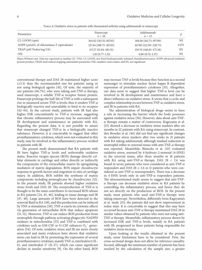
Oxidative stress data according to anti-TNF-𝛼 therapywith etanercept or adalimumab are shown in Table 4. Therewas no significant difference in CL-LOOH, AOPPs, TRAP, orOSI values. However, AOPP levels showed an increase trend(𝑝 = 0.071) in patients using adalimumab and this trend wasindependent of BMI (𝑝 = 0.047, OR: 1.009, CI 95%: 1.000–1.018) (data not shown). In sum, presence of IR was related toincrease inDAS 28 and ESR and hsCRP andTNF-𝛼 levels andAOPPs andOSI and decreased TRAP in patients with RA.Onthe other hand, IR did not have a role in changes related toRF and anti-CCP. In addition, TNF-𝛼 increase is related to IRdevelopment in patients with RA.
4. Discussion
Several reports have shown that IR is related to chronicinflammation [1, 32, 33] and corticosteroid treatment [2, 3].Although previous articles have shown that corticosteroidmay be involved in IR [21, 22, 34], this finding has not beenverified in patients with RA, suggesting that corticosteroidbeneficial anti-inflammatory effects would compensate thedeleterious metabolic action [35, 36]. Penesova et al. [36]showed that low-dose glucocorticoid treatmentwith durationof 2–9 years is relatively safe and did not lead to glucosemetabolism impairment. Independently of whether they hadIR or not, in the present study the patients did not differ in thefrequency they were using prednisone, showing that, in thiscohort of RA patients, corticosteroid use does not seem to bea determinant factor for IR development. Moreover, patientsused less than 7.5mg/d corticosteroid (data not shown),which has been reported as safe [37].
Several reports have shown the association betweenchronic inflammatory disease states and IR [32, 33, 38]. Previ-ous studies demonstrated that TNF-𝛼may have an importantrole in the IR pathogenesis by multiple mechanisms, suchas downregulation of genes that are required for normalinsulin action, direct effects on insulin signaling, induction ofelevated free fatty acids via stimulation of lipolysis, and nega-tive regulation of peroxisome proliferator-activated receptor-𝛾 (PPAR𝛾), an important insulin-sensitizing nuclear receptor[39]. In RA patients with severe and active disease evenin the presence of anti-TNF-𝛼 therapy, high-grade inflam-mation was correlated negatively and independently withcirculating adiponectin concentration [40], an importantanti-inflammatory adipokine related to insulin resistance andmetabolic syndrome [41]. In vitro studies have shown thatTNF-𝛼 induced serine phosphorylation of insulin receptorsubstrate-1 (IRS-1) and inhibited insulin receptor tyrosinekinase, causing a change of the insulin signaling [40]. Inthe present study, patients using anti-TNF-𝛼 therapy, whichis generally indicated to patients who have a severe diseasenot controlled by disease-modifying antirheumatic drugs(DMARDs), showed higher TNF-𝛼 levels. Even with anti-TNF-𝛼 therapy, TNF-𝛼 levels have not reached the val-ues obtained by patients who control disease activity with
6 Oxidative Medicine and Cellular Longevity
Table 4: Oxidative stress in patients with rheumatoid arthritis using adalimumab or etanercept.
Parameters Etanercept𝑛 = 26
Adalimumab𝑛 = 13
𝑝
CL-LOOH (cpm) 164.02 (145.51–187.05) 168.06 (162.71–197.90) NSAOPP (𝜇mol/L of chloramines-T equivalents) 127.44 (108.75–187.05) 167.80 (122.90–228.73) 0.071TRAP (𝜇M Trolox/mg UA) 157.27 (15.84–183.51) 159.70 (148.83–175.58) NSOSI 1.10 (0.77–1.31) 0.86 (0.74–1.37) NSMann-Whitney test. Data are expressed as median (25–75%). CL-LOOH, tert-butyl hydroperoxide-initiated chemiluminescence; AOPP, advanced oxidationprotein product; TRAP, total radical-trapping antioxidant parameter; OSI, oxidative stress index; and NS, not significant.
conventional therapy and DAS 28 maintained higher score(≥3.3) than the recommended one for patients using ornot using biological agents [41]. Of note, the majority ofour patients (66.7%), who were taking anti-TNF-𝛼 therapy,used etanercept, a soluble TNF-𝛼 receptor fusion protein.Etanercept prolongs the half-life of TNF-𝛼with a subsequentrise in measured serum TNF-𝛼 levels; thus it renders TNF-𝛼biologically inactive and unavailable to bind to its receptor[42–44]. In the current study, patients with IR had alsohigher ESR concomitantly to TNF-𝛼 increase, suggestingthat chronic inflammatory process may be associated withIR development and maintenance in patients with RA.Regarding the present data, it is not possible to assurethat etanercept changed TNF-𝛼 in a biologically inactivesubstance. However, it is conceivable to suggest that otherproinflammatory cytokines, which were not evaluated in thisstudy, may be involved in the inflammatory process verifiedin patients with IR.
The present study demonstrated that RA patients withIR have higher TNF-𝛼 levels and unfavorable oxidativestatus. Reactive oxygen species (ROS) damage directly cel-lular elements in cartilage and either directly or indirectlythe components of the extracellular matrix by upregulatingmediators of matrix degradation. ROS impair chondrocyteresponse to growth factors and migration to sites of cartilageinjury. In addition, ROS inhibit the synthesis of matrixcomponents including proteoglycans by chondrocytes [12].In the present study, IR patients showed higher oxidativestress levels and DAS 28. The overproduction of TNF-𝛼 isthought to be the main contributor to increased ROS releasein RA patients [24, 45, 46], leading to tissue damage and IR[47, 48]. Large amounts of ROS have been detected in thesynovial fluid in RA [49], and this production can be inducedby TNF-𝛼 stimulation [50]. TNF-𝛼 exerts its cytotoxic effectsvia generation of intracellular ROS that induce apoptosis[51, 52]. Moreover, TNF-𝛼 can induce ROS production fromneutrophils through pathway activating phagocytic NADPHoxidases in mitochondria [53] and TNF-𝛼 combined withcytokines such as GM-CSF or G-CSF enhances O
2
− gener-ation [54]. Of note, oxidative stress and IR are more closelyassociated and many evidences have shown that oxidativestress can lead to IR by promoting the expression of severalproinflammatory cytokines,mainly TNF-𝛼, interleukin 6 (IL-6), and interleukin 17 (IL-17), which can cause significantdecline in insulin sensitivity [9]. On the other hand, ROS
may increase TNF-𝛼 levels because they function as a secondmessenger to stimulate nuclear factor kappa B dependentexpression of proinflammatory cytokines [55]. Altogether,our data seem to suggest that higher TNF-𝛼 level can beinvolved in IR development and maintenance and have adirect influence on oxidative stress. It seems that a cyclic andcomplex relationship occurs betweenTNF-𝛼, oxidative stress,and IR in patients with RA.
The administration of biological drugs seems to havea role in increasing the barrier which the body possessesagainst oxidative stress [56]. However, data about anti-TNF-𝛼 therapy remain a matter of controversy. Kageyama et al.[15] showed a decrease in oxidative stress markers after sixmonths in 22 patients with RA using etanercept. In contrast,den Broeder et al. [46] did not find any significant changesin oxidative stress markers after two weeks in 21 patientswith RA taking adalimumab, although marked reduction inneutrophil influx to synovial tissue with anti-TNF-𝛼 therapywas reported. Meanwhile, Biniecka et al. [45] evaluatedoxidative stress, assessed by 4-hydroxy-2-nonenal (4-HNE)in the synovial tissue, after three months in 18 patientswith RA using anti-TNF-𝛼 therapy. DAS 28 < 2.6 wasfound in seven patients who were considered as anti-TNF-𝛼responders and DAS 28 ≥ 2.6 in 11 patients who were con-sidered as anti-TNF-𝛼 nonresponders. There was a decreasein 4-HNE levels only in anti-TNF-𝛼 responders patients.The aforementioned study seems to suggest that anti-TNF-𝛼 therapy can decrease oxidative stress in RA patients bycontrolling the inflammatory process, and hence they donot act directly on the production of ROS. In the presentstudy, most patients who used anti-TNF-𝛼 therapy weretaking etanercept. Nevertheless, differently from Kageyama’set al. study [15], the patients did not show improvement inredox state. It is conceivable to suggest that this may haveoccurred because anti-TNF-𝛼 therapy maintained DAS 28 insimilar values obtained by patients who were not using anti-TNF-𝛼 therapy. Meanwhile, inflammatory process shown byincreased ESR and TNF-𝛼 levels, mainly in RA patientswith IR, progressed in these patients being responsible foroxidative stress increase.
Upon looking at the results obtained in the presentstudy, some limitations have to be considered. First, thecross-sectional design does not allow for inference causality.Second, although the minimum number of patients has beenreached by the calculation of the sample size, a greater
Oxidative Medicine and Cellular Longevity 7
number of patients would probably confer more strength tothe statistical results.
This study corroborates with Binieckas et al.’s [45],which suggested that inflammatory state maintenance can beresponsible for oxidative stress found in patients with RA.On the other hand, the data of the present study show thatIR is involved in an unbalanced redox state, which possiblycontributes to maintaining a vicious circle of high-gradeinflammation.
5. Conclusions
This study demonstrates that IR and TNF-𝛼 are importantfactors involved in redox imbalance in patients with RA andit seems to be due to the maintenance of inflammatory stateand disease activity. The data from the present study suggesta complex interaction of TNF-𝛼, oxidative stress, and IR,but the presence of insulin resistance seems to be directlyassociated with both oxidative stress and TNF-𝛼 levels. Thedifferences in oxidative stress markers in RA patients with orwithout IR could contribute to a better design for future drugsand/or nutritional interventional studies in this population.In addition, more studies are warranted to verify if IR can beinvolved in therapeutic failure with TNF-𝛼 inhibitors.
Abbreviations
Anti-CCP: Anticyclic citrullinated peptideAOPPs: Advanced oxidation protein productsCL-LOOH: Tert-butyl hydroperoxide-initiated
chemiluminescencehsCRP: Highly sensitive C-reactive proteinDAS 28: Disease Activity Score evaluating 28 joints4-HNE: 4-Hidroxy-2-nonenalCVD: Cardiovascular diseaseDMARDs: Disease-modifying antirheumatic drugsESR: Erythrocyte sedimentation rateHOMA-IR: Homeostasis model assessment-insulin
resistanceIL-6: Interleukin 6IL-17: Interleukin 17IR: Insulin resistanceOSI: Oxidative stress indexPPAR𝛾: Peroxisome proliferator-activated
receptor-𝛾RA: Rheumatoid arthritisRLU: Relative luminescence unitsRF: Rheumatoid factorROS: Reactive oxygen speciesTRAP: Total radical-trapping antioxidant
parameterTNF-𝛼: Tumor necrosis factor-alphaUA: Uric acidWC: Waist circumference.
Competing Interests
The authors declare that they have no competing interests.
Authors’ Contributions
NeideTomimuraCosta helped to analyze the data and to draftthe paper. Andrea Name Colado Simao performed data anal-ysis and together with Isaias Dichi helped to design the studyand draft the paper and critically revised the paper for impor-tant intellectual content. Vinicius Daher Alvares Delfinohelped to analyze the data and critically revised the paper.Tatiana Mayumi Veiga Iriyoda and Ricardo Braga Aminparticipated in clinical assessments. Francieli Delongui, AnaPaula Kallaur, Marcell Alysson Batisti Lozovoy, and DanielaFrizon Alfieri helped in laboratory analyses. All authors havegiven final approval of the version to be published and agreeto be accountable for all aspects of the work in ensuring thatquestions related to the accuracy or integrity of any part ofthe work are appropriately investigated and resolved.
Acknowledgments
This research was supported by the National Council ofBrazilian Research (CNPq) and Araucaria Foundation fromParana, Brazil.
References
[1] I. D. del Rincon, K. Williams, M. P. Stern, G. L. Freeman, andA. Escalante, “High incidence of cardiovascular events in arheumatoid arthritis cohort not explained by traditional cardiacrisk factors,”Arthritis and Rheumatism, vol. 44, no. 12, pp. 2737–2745, 2001.
[2] S. E. Gabriel, “Cardiovascular morbidity and mortality inrheumatoid arthritis,” The American Journal of Medicine, vol.121, no. 10, pp. S9–S14, 2008.
[3] S. Haque, H. Mirjafari, and I. N. Bruce, “Atherosclerosis inrheumatoid arthritis and systemic lupus erythematosus,” Cur-rent Opinion in Lipidology, vol. 19, no. 4, pp. 338–343, 2008.
[4] C. De Luca and J. M. Olefsky, “Inflammation and insulinresistance,” FEBS Letters, vol. 582, no. 1, pp. 97–105, 2008.
[5] A. M. El-Barbary, E. M. Kassem, M. A. S. El-Sergany, S. A.-M. Essa, and M. A. Eltomey, “Association of anti-modifiedcitrullinated vimentin with subclinical atherosclerosis in earlyrheumatoid arthritis compared with anti-cyclic citrullinatedpeptide,” The Journal of Rheumatology, vol. 38, no. 5, pp. 828–834, 2011.
[6] T. Saxne, M. A. Palladino Jr., D. Heinegard, N. Talal, and F.A. Wollheim, “Detection of tumor necrosis factor 𝛼 but nottumor necrosis factor 𝛽 in rheumatoid arthritis synovial fluidand serum,” Arthritis and Rheumatism, vol. 31, no. 8, pp. 1041–1045, 1988.
[7] I. B. McInnes and G. Schett, “The pathogenesis of rheumatoidarthritis,”TheNew England Journal of Medicine, vol. 365, no. 23,pp. 2205–2219, 2011.
[8] C. P. Chung, A. Oeser, J. F. Solus et al., “Inflammation-associated insulin resistance: differential effects in rheumatoidarthritis and systemic lupus erythematosus define potentialmechanisms,”Arthritis and Rheumatism, vol. 58, no. 7, pp. 2105–2112, 2008.
[9] M. L. L. Prevoo, M. A. van’t Hof, H. H. Kuper, M. A. vanLeeuwen, L. B. A. van de Putte, and P. L. C. M. van Riel,“Modified disease activity scores that include twenty-eight-joint
8 Oxidative Medicine and Cellular Longevity
counts development and validation in a prospective longitu-dinal study of patients with rheumatoid arthritis,” Arthritis &Rheumatism, vol. 38, no. 1, pp. 44–48, 1995.
[10] P. Vasanthi, G. Nalini, and G. Rajasekhar, “Status of oxida-tive stress in rheumatoid arthritis,” International Journal ofRheumatic Diseases, vol. 12, no. 1, pp. 29–33, 2009.
[11] L. I. Filippin, R. Vercelino, N. P. Marroni, and R. M. Xavier,“Redox signalling and the inflammatory response in rheuma-toid arthritis,” Clinical and Experimental Immunology, vol. 152,no. 3, pp. 415–422, 2008.
[12] C. A.Hitchon andH. S. El-Gabalawy, “Oxidation in rheumatoidarthritis,” Arthritis Research and Therapy, vol. 6, no. 6, pp. 265–278, 2004.
[13] S. Taysi, F. Polat, M. Gul, R. Sari, and E. Bakan, “Lipid peroxida-tion, some extracellular antioxidants, and antioxidant enzymesin serum of patients with rheumatoid arthritis,” RheumatologyInternational, vol. 21, no. 5, pp. 200–204, 2002.
[14] S. Z. Hassan, T. A. Gheita, S. A. Kenawy, A. T. Fahim, I. M. El-Sorougy, and M. S. Abdou, “Oxidative stress in systemic lupuserythematosus and rheumatoid arthritis patients: relationshipto disease manifestations and activity,” International Journal ofRheumatic Diseases, vol. 14, no. 4, pp. 325–331, 2011.
[15] Y. Kageyama, M. Takahashi, T. Nagafusa, E. Torikai, andA. Nagano, “Etanercept reduces the oxidative stress markerlevels in patients with rheumatoid arthritis,” RheumatologyInternational, vol. 28, no. 3, pp. 245–251, 2008.
[16] S. Shahmohamadnejad, A. Vaisi-Raygani, Y. Shakiba et al.,“Association between butyrylcholinesterase activity and phe-notypes, paraoxonase192 rs662 gene polymorphism and theirenzymatic activity with severity of rheumatoid arthritis: corre-lationwith systemic inflammatorymarkers and oxidative stress,preliminary report,” Clinical Biochemistry, vol. 48, no. 1-2, pp.63–69, 2015.
[17] M. A. B. Lozovoy, A. N. C. Simao, M. S. N. Hohmann et al.,“Inflammatory biomarkers and oxidative stress measurementsin patients with systemic lupus erythematosus with or withoutmetabolic syndrome,” Lupus, vol. 20, no. 13, pp. 1356–1364, 2011.
[18] M. A. B. Lozovoy, A. N. C. Simao, S. R. Oliveira et al.,“Relationship between iron metabolism, oxidative stress, andinsulin resistance in patients with systemic lupus erythemato-sus,” Scandinavian Journal of Rheumatology, vol. 42, no. 4, pp.303–310, 2013.
[19] S. R. Oliveira, A. N. Colado Simao, A. P. Kallaur et al.,“Disability in patients with multiple sclerosis: influence ofinsulin resistance, adiposity, and oxidative stress,”Nutrition, vol.30, no. 3, pp. 268–273, 2014.
[20] H. K. Morimoto, A. N. C. Simao, E. R. D. D. Almeida etal., “Role of metabolic syndrome and antiretroviral therapyin adiponectin levels and oxidative stress in HIV-1 infectedpatients,” Nutrition, vol. 30, no. 11-12, pp. 1324–1330, 2014.
[21] P. H. Dessein and B. I. Joffe, “Insulin resistance and impairedbeta cell function in rheumatoid arthritis,” Arthritis andRheumatism, vol. 54, no. 9, pp. 2765–2775, 2006.
[22] G. La Montagna, F. Cacciapuoti, R. Buono et al., “Insulinresistance is an independent risk factor for atherosclerosis inrheumatoid arthritis,” Diabetes and Vascular Disease Research,vol. 4, no. 2, pp. 130–135, 2007.
[23] G. Arcaro, “Insulin causes endothelial dysfunction in humans:sites and mechanisms,” Circulation, vol. 105, no. 5, pp. 576–582,2002.
[24] Y. Kageyama, M. Takahashi, T. Ichikawa, E. Torikai, andA. Nagano, “Reduction of oxidative stress marker levels by
anti-TNF-𝛼 antibody, infliximab, in patients with rheumatoidarthritis,” Clinical and Experimental Rheumatology, vol. 26, no.1, pp. 73–80, 2008.
[25] O. Altindag, M. Karakoc, A. Kocyigit, H. Celik, and N. Soran,“Increased DNA damage and oxidative stress in patients withrheumatoid arthritis,” Clinical Biochemistry, vol. 40, no. 3-4, pp.167–171, 2007.
[26] A. Nakajima, Y. Aoki, Y. Shibata et al., “Identification of clinicalparameters associated with serum oxidative stress in patientswith rheumatoid arthritis,” Modern Rheumatology, vol. 24, no.6, pp. 926–930, 2014.
[27] D. Aletaha, T. Neogi, and A. J. Silman, “2010 rheumatoid arthri-tis classification criteria: An American College of Rheumatol-ogy/European League against rheumatism collaborative initia-tive,” Annals of the Rheumatic Diseases, vol. 69, no. 10, pp. 1580–1588, 2010.
[28] S. M. Haffner, H. Miettinen, and M. P. Stern, “The homeostasismodel in the San Antonio Heart Study,” Diabetes Care, vol. 20,no. 7, pp. 1087–1092, 1997.
[29] B. Gonzalez Flecha, S. Llesuy, and A. Boveris, “Hydroperoxide-initiated chemiluminescence: an assay for oxidative stress inbiopsies of heart, liver, and muscle,” Free Radical Biology andMedicine, vol. 10, no. 2, pp. 93–100, 1991.
[30] V. Witko-Sarsat, M. Friedlander, T. N. Khoa et al., “Advancedoxidation protein products as novel mediators of inflammationand monocyte activation in chronic renal failure,” The Journalof Immunology, vol. 161, no. 5, pp. 2524–2532, 1998.
[31] M. Repetto, C. Reides, M. L. Gomez Carretero, M. Costa, G.Griemberg, and S. Llesuy, “Oxidative stress in blood of HIVinfected patients,” Clinica Chimica Acta, vol. 255, no. 2, pp. 107–117, 1996.
[32] G. S. Hotamisligil, “Molecular mechanisms of insulin resistanceand the role of the adipocyte,” International Journal of Obesity,vol. 24, supplement 4, pp. S23–S27, 2000.
[33] C. Popa, M. G. Netea, P. L. C. M. Van Riel, J. W. M. VanDer Meer, and A. F. H. Stalenhoef, “The role of TNF-𝛼 inchronic inflammatory conditions, intermediary metabolism,and cardiovascular risk,” Journal of Lipid Research, vol. 48, no.4, pp. 751–752, 2007.
[34] P. H. Dessein, B. I. Joffe, A. E. Stanwix, B. F. Christian, and M.Veller, “Glucocorticoids and insulin sensitivity in rheumatoidarthritis,” The Journal of Rheumatology, vol. 31, no. 5, pp. 867–874, 2004.
[35] K. L. G. Svenson, G. Lundqvist, L. Wide, and R. Hallgren,“Impaired glucose handling in active rheumatoid arthri-tis: effects of corticosteroids and antirheumatic treatment,”Metabolism, vol. 36, no. 10, pp. 944–948, 1987.
[36] A. Penesova, Z. Radikova, M. Vlcek et al., “Chronic inflamma-tion and low-dose glucocorticoid effects on glucosemetabolismin premenopausal females with rheumatoid arthritis free ofconventional metabolic risk factors,” Physiological Research, vol.62, no. 1, pp. 75–83, 2013.
[37] J. M. Sabio, J. A. Vargas-Hitos, N. Navarrete, C. Hidalgo-Tenorio, and J. Jimenez-Alonso, “Effects of low ormedium-doseof prednisone on insulin resistance in patients with systemiclupus erythematosus,”Clinical and Experimental Rheumatology,vol. 28, no. 4, pp. 483–489, 2010.
[38] L.-S. Tam, B. Tomlinson, T. T. Chu, T. K. Li, and E. K. Li,“Impact of TNF inhibition on insulin resistance and lipids levelsin patients with rheumatoid arthritis,” Clinical Rheumatology,vol. 26, no. 9, pp. 1495–1498, 2007.
Oxidative Medicine and Cellular Longevity 9
[39] D. E. Moller, “Potential role of TNF-𝛼 in the pathogenesis ofinsulin resistance and type 2 diabetes,” Trends in Endocrinologyand Metabolism, vol. 11, no. 6, pp. 212–217, 2000.
[40] T. Hayakawa, Y. Nagai, M. Taniguchi et al., “Tumor necrosisfactor-𝛽 gene NcoI polymorphism decreases insulin resistancein Japanese men,” Metabolism: Clinical and Experimental, vol.49, no. 11, pp. 1506–1509, 2000.
[41] J. A. Singh, D. E. Furst, A. Bharat et al., “2012 update of the2008AmericanCollege of Rheumatology recommendations forthe use of disease-modifying antirheumatic drugs and biologicagents in the treatment of rheumatoid arthritis,” Arthritis Care& Research, vol. 64, no. 5, pp. 625–639, 2012.
[42] J. Lo, L. E. Bernstein, B. Canavan et al., “Effects of TNF-𝛼neutralization on adipocytokines and skeletal muscle adiposityin the metabolic syndrome,” American Journal of Physiology—Endocrinology and Metabolism, vol. 293, no. 1, pp. E102–E109,2007.
[43] K. J. Grattendick, J. M. Nakashima, L. Feng, S. N. Giri, andS. B. Margolin, “Effects of three anti-TNF-𝛼 drugs: etanercept,infliximab and pirfenidone on release of TNF-𝛼 in mediumand TNF-𝛼 associated with the cell in vitro,” InternationalImmunopharmacology, vol. 8, no. 5, pp. 679–687, 2008.
[44] A. Bhatia and R. E. Kast, “Tumor Necrosis Factor (TNF) canparadoxically increase on etanercept treatment, occasionallycontributing to TNF-mediated disease,” Journal of Rheumatol-ogy, vol. 34, no. 2, pp. 447–449, 2007.
[45] M. Biniecka, A. Kennedy, C. T. Ng et al., “Successful tumournecrosis factor (TNF) blocking therapy suppresses oxidativestress and hypoxia-induced mitochondrial mutagenesis ininflammatory arthritis,” Arthritis Research and Therapy, vol. 13,no. 4, article R121, 2011.
[46] A. A. den Broeder, G. J. A. Wanten, W. J. G. Oyen, T. Naber, P.L. C. M. Van Riel, and P. Barrera, “Neutrophil migration andproduction of reactive oxygen species during treatment with afully human anti-tumor necrosis factor-𝛼monoclonal antibodyin patients with rheumatoid arthritis,” Journal of Rheumatology,vol. 30, no. 2, pp. 232–237, 2003.
[47] I. Stagakis, G. Bertsias, S. Karvounaris et al., “Anti-tumornecrosis factor therapy improves insulin resistance, beta cellfunction and insulin signaling in active rheumatoid arthritispatients with high insulin resistance,” Arthritis Research andTherapy, vol. 14, no. 3, article R141, 2012.
[48] A. Stavropoulos-Kalinoglou, G. S. Metsios, V. F. Panoulas, P.Nightingale, Y. Koutedakis, and G. D. Kitas, “Anti-tumournecrosis factor alpha therapy improves insulin sensitivity innormal-weight but not in obese patients with rheumatoidarthritis,” Arthritis Research and Therapy, vol. 14, no. 4, articleR160, 2012.
[49] P. Biemond, A. J. G. Swaak, and J. F. Koster, “Protectivefactors against oxygen free radicals and hydrogen peroxide inrheumatoid arthritis synovial fluid,” Arthritis and Rheumatism,vol. 27, no. 7, pp. 760–765, 1984.
[50] C.-H. Woo, T.-H. Kim, J.-A. Choi et al., “Inhibition of receptorinternalization attenuates the TNF𝛼-induced ROS generationin non-phagocytic cells,” Biochemical and Biophysical ResearchCommunications, vol. 351, no. 4, pp. 972–978, 2006.
[51] K. Schulze-Osthoff, P. H. Krammer, and W. Droge, “Divergentsignalling via APO-1/Fas and the TNF receptor, two homolo-gousmolecules involved in physiological cell death,”TheEMBOJournal, vol. 13, no. 19, pp. 4587–4596, 1994.
[52] K. Hirose, D. L. Longo, J. J. Oppenheim, and K. Matsushima,“Overexpression of mitochondrial manganese superoxide dis-mutase promotes the survival of tumor cells exposed tointerleukin-1, tumor necrosis factor, selected anticancer drugs,and ionizing radiation,” FASEB Journal, vol. 7, no. 2, pp. 361–368,1993.
[53] R. Miesel, M. P. Murphy, and H. Kroger, “Enhanced mitochon-drial radical production in patients with rheumatoid arthritiscorrelates with elevated levels of tumor necrosis factor alpha inplasma,” Free Radical Research, vol. 25, no. 2, pp. 161–169, 1996.
[54] E. Mur, A. Zabernigg, W. Hilbe, W. Eisterer, W. Halder, and J.Thaler, “Oxidative burst of neutrophils in patients with rheuma-toid arthritis: influence of various cytokines and medication,”Clinical and Experimental Rheumatology, vol. 15, no. 3, pp. 233–237, 1997.
[55] G. Bonizzi, J. Piette, M.-P. Merville, and V. Bours, “Cell type-specific role for reactive oxygen species in nuclear factor-kappaB activation by interleukin-1,” Biochemical Pharmacology,vol. 59, no. 1, pp. 7–11, 2000.
[56] S. Coaccioli, A. Panaccione, R. Biondi et al., “Evaluationof oxidative stress in rheumatoid and psoriatic arthritis andpsoriasis,”Clinica Terapeutica, vol. 160, no. 6, pp. 467–472, 2009.

Submit your manuscripts athttp://www.hindawi.com
Stem CellsInternational
Hindawi Publishing Corporationhttp://www.hindawi.com Volume 2014
Hindawi Publishing Corporationhttp://www.hindawi.com Volume 2014
MEDIATORSINFLAMMATION
of
Hindawi Publishing Corporationhttp://www.hindawi.com Volume 2014
Behavioural Neurology
EndocrinologyInternational Journal of
Hindawi Publishing Corporationhttp://www.hindawi.com Volume 2014
Hindawi Publishing Corporationhttp://www.hindawi.com Volume 2014
Disease Markers
Hindawi Publishing Corporationhttp://www.hindawi.com Volume 2014
BioMed Research International
OncologyJournal of
Hindawi Publishing Corporationhttp://www.hindawi.com Volume 2014
Hindawi Publishing Corporationhttp://www.hindawi.com Volume 2014
Oxidative Medicine and Cellular Longevity
Hindawi Publishing Corporationhttp://www.hindawi.com Volume 2014
PPAR Research
The Scientific World JournalHindawi Publishing Corporation http://www.hindawi.com Volume 2014
Immunology ResearchHindawi Publishing Corporationhttp://www.hindawi.com Volume 2014
Journal of
ObesityJournal of
Hindawi Publishing Corporationhttp://www.hindawi.com Volume 2014
Hindawi Publishing Corporationhttp://www.hindawi.com Volume 2014
Computational and Mathematical Methods in Medicine
OphthalmologyJournal of
Hindawi Publishing Corporationhttp://www.hindawi.com Volume 2014
Diabetes ResearchJournal of
Hindawi Publishing Corporationhttp://www.hindawi.com Volume 2014
Hindawi Publishing Corporationhttp://www.hindawi.com Volume 2014
Research and TreatmentAIDS
Hindawi Publishing Corporationhttp://www.hindawi.com Volume 2014
Gastroenterology Research and Practice
Hindawi Publishing Corporationhttp://www.hindawi.com Volume 2014
Parkinson’s Disease
Evidence-Based Complementary and Alternative Medicine
Volume 2014Hindawi Publishing Corporationhttp://www.hindawi.com