Embed Size (px)
Citation preview
Clinical signs, reproduction of attaching/effacing lesions, and enterocyte invasion after oral
inoculation of an O118 enterohaemorrhagicEscherichia coli in neonatal calves
Philippe Stordeura*, Bernard Chinaa, Gerard Charlierb, Stefan Roelsb, Jacques Mainila
aLaboratory of Bacteriology, Faculty of Veterinary Medicine, University of Liège, Sart Tilman B43a, B-4000 Liège, BelgiumbSection of Pathology, Department of Biocontrol, Centre for Research in Veterinary Science and Agrochemistry, Groeselenberg 99, B-1180 Brussels, Belgium
(Received 8 July 1999; accepted 30 September 1999)
ABSTRACT – Attaching and effacing (AE) lesions are produced among others by enteropathogenicEscherichia coli and enterohaemorrhagic E. coli (EHEC), which differs from the former by the productionof cytotoxins active on various cell cultures, the verocytotoxins, or shigacytotoxins. EHEC areassociated with diarrhoea and dysentery in humans and in ruminants, mainly calves from two to eightweeks of age. Clinical signs and/or lesions have been reproduced experimentally with EHEC strainsbelonging to serotypes O5:K4/Nm, O26:K-:H11, O111:Nm, and O157:H7 which are isolated fromcattle and/or humans. The purpose of this work was to develop an experimental model of infection innewborn calves with a bovine EHEC strain isolated from a calf which of died of diarrhoea, andbelonging to the O118:H16 serotype, which is also common to both cattle and humans. The bovineO118:H16 EHEC strain was able to colonize the gut of three newborn calves, and to induce diarrhoeatwenty-four hours after challenge and to produce AE lesions in the small and/or large intestines. AElesions were detected microscopically and ultrastructurally in the small intestine of one calf and in thewhole intestinal track of two calves. Internalization of bacteria and also of pedestal-bacteria complexinside of the enterocyte was observed in two of the three calves. The significance of this stage isunknown but may be related to the invasion of the calf by the bacteria. The challenge strain wasisolated from the mesenteric lymph nodes of the same two calves but not from other organs or fromheart blood. No blood was observed in the faeces of any of the three calves, nor were any lesions in theinternal organs, which may have been related to the production of a verotoxin whose role is stillunknown in cattle. © 2000 Éditions scientifiques et médicales Elsevier SAS
enterohaemorrhagic E.coli (EHEC) / calf / attaching and effacing lesions (AE) / diarrhoea / invasion
1. Introduction
Since 1984 [2], attaching and effacing Escherichia coliis recognised as a cause of diarrhoea and dysentery inyoung calves, mainly from two to eight weeks of age [5,16]. ‘Attaching and effacing’ was the term first used byMoon and collaborators [22] to describe an intestinallesion (AE lesion) caused by specific strains of E. coli:‘effacing’ because of the localized disappearance of thebrush border microvilli; ‘attaching’ because of the inti-
mate attachment of the bacteria to the exposed cytoplas-mic membrane of the enterocyte.
AE lesions are caused by enteropathogenic E. coli(EPEC), Citrobacter rodentium, Hafnia alveii, and entero-haemorrhagic E. coli (EHEC), which differ essentially fromthe EPEC by the production of cytotoxins active on variousepithelial cells (Hela and Hep-2) in culture, the verocyto-toxins (VT1 and VT2 families), also named shigacytotoxins(Stx1 and Stx2 families) [19, 21, 24]. EPEC causes diar-rhoea in various animal species and in humans, whereasEHEC is associated with diarrhoea and dysentery in rumi-
* Correspondence and reprints
Microbes and Infection, 2, 2000, 17−24© 2000 Éditions scientifiques et médicales Elsevier SAS. All rights reserved
Microbes and Infection2000, 17-24
17
nants and in humans. In the latter, EHEC also causes thehaemolytic-uraemic syndrome (HUS) [4, 16, 19, 24, 25].
The pathogenesis of EPEC/EHEC and the production ofthe AE lesions have been intensively studied in vitro oncells in culture with the human EPEC strain E2348/69, anda four-step model has been tentatively proposed [24],consisting of (i) initial adherence to the enterocytemicrovilli; so far fimbrial adhesins have been describedonly for human EPEC [9] and rabbit EPEC [21]; (ii) type IIIsecretion system-mediated signal transduction into theenterocyte resulting in increased levels of phosphorylationand calcium, as well as in cytoskeleton rearrangements.(In vivo, the most dramatic consequence is the effacementof the brush border microvilli); (iii) intimate attachment tothe exposed enterocyte cytoplasmic membrane, mediatedby an outer membrane protein, intimin, with polymerisa-tion of actin filaments underneath the zone of adherenceof the bacteria and enhancement of the cytoskeleton rear-rangements; and (iv) penetration of the bacteria into thecells. The VT/Stx toxins play no role in the development ofthe AE lesions, but cause damage to the intestinal wallvessels resulting in haemorrhages and dysentery inhumans and calves. In humans, they also cause damage tothe arterioles of the kidneys, resulting in the HUS [19, 24,25].
The first step is mediated by genes located on a plasmidor on the chromosome [21, 24]. Genes responsible forsteps (ii) and (iii) are grouped together on the bacterialchromosome forming a pathogenesis island, the locus ofenterocyte effacement, or LEE [20]. The genetic determin-ism of the fourth step is still unknown although chromo-somal mutants deficient only in cell invasion have beendescribed [8]. The genes coding for the VT/Sta are alsolocated on the chromosome, but on phages for several ofthem [19].
The transposition in vivo of these in vitro models hasbeen realized for the first three steps with human andrabbit EPEC by testing mutants [10, 21], but the in vivosignificance of the fourth step, the cellular invasion, is stillunknown. Bovine EPEC and EHEC also possess a LEE [12],and AE lesions and /or clinical signs have been experimen-tally reproduced in young calves and lambs, with bovineEHEC belonging to various serotypes: O5:K4:Nm, O26:K-:H11, O111:Nm, and O157:H7 [1, 7, 18, 23, 29, 32].Bovine EPEC and EHEC belonging to other serotypes have,however, not been tested in vivo yet.
The purpose of this work was to develop an experimen-tal model of infection in newborn calves to confirm thepathogenicity of and follow the production of AE lesionsby a bovine EHEC strain belonging to serotype O118:H16.This model will help to study mutants in variouspathogenicity-associated genes of bovine EHEC in thefuture.
2. Material and methods2.1 Bacterial strain
Strain 340S89 is an O118:H16 E. coli which was iso-lated in 1989 from a two-week-old Friesian calf whichdied of diarrhoea [27]. It tests positive with the gene
probes for intimin (Eae probe) and VT1, but not with thegene probe for VT2 [17] and is able to reproduce AElesions in rabbits [4]. This strain is sensitive to kanamycin,gentamycin, tetracycline, chloramphenicol, and nalidixicacid, but resistant to streptomycin and tellurite.
2.2 Calf infection
Four naturally born calves were isolated immediatelyafter birth in a box which had been washed and desin-fected (Atlantol*, Ecosa, Ghent, Belgium). They received300 millilitres of colostrum which was negative by ELISAand agglutination against strain 340S89. At six hours ofage, three of them were challenged orally with 109 to 1010
CFU of strain 340S89 suspended in sterile saline and thefourth one with saline only. Strain 340S89 was grown onMeat extract Agar slants (Oxoïd, Gent, Belgium) for sixhours aerobically at 37 °C. The bacteria were resuspendedin sterile saline and the optical density at 620 nm wasadjusted to obtain a bacterial concentration between 109
to 1010 per 200 mL. The exact concentration was calcu-lated after inoculation of tenfold dilutions of the suspen-sion onto Gassner agar plates (Belgolabo, Overijse, Bel-gium). This medium was used to select enteric bacteria.Moreover, it provided the opportunity to make a differen-tiate between coliforms thanks to his lactose activity [11].The calves were subsequently fed twice a day with twoliters of UHT whole milk. Clinical investigations and fae-cal sampling were performed every four hours. Faecalsamples were tested by ELISA (trousse ELISA digestive,BioX, Brussels, Belgium) for the presence of K99 entero-toxigenic E. coli, rotavirus, coronavirus, and Crytospo-ridium sp. The excretion of the challenge strain was fol-lowed by bacteriological examination and by PCR.
2.3. Necropsy
The calves were euthanised between 44 and 64 h p.i.(p.i.) by intravenous administration of sodium pentobar-bital. Necropsy was performed on a general routine basisat first and subsequently focused on the abdominal cavity.Samples were taken aseptically from various intestinalsegments (duodenum, jejunum, ileum, caecum, spiralcolon, descending colon, and rectum) and from internalorgans (mesenteric lymph nodes, liver, spleen lungs, kid-neys, heart blood, and brain) for bacteriology, histopathol-ogy, and electron microscopy.
2.4. Bacteriology
The faecal samples were collected in sterile containers.The postmortem faecal samples were taken aseptically(the sections of the gut had been sterilized using burningsteel) with a Pasteur pipette. The samples were thendiluted tenfold. Dilutions 10–4 to 10–6 were inoculatedonto Gassner agar and Gassner agar supplemented withstreptomycin sulphate (Sigma Aldrich, Steinheim, Ger-many,100 µg per mL) and tellurite (Sigma Aldrich, Stein-heim, Germany,10–4 M) plates with a spiral plater system(LED Techno, Hechtel-Eksel, Holland), and incubatedovernight aerobically at 37 °C. After one night of incuba-tion, a calculation of the number of bacteria on the platewas realized and this result was transformed in CFU/g ofintestinal content by a formula.
Original article Stordeur et al.
18 Microbes and Infection2000, 17-24
Each lymph node was investigated before intestineswere opened. The content of this one (taken with a Pasteurpipette after sterilization of the surface using burning steel)was seeded directly onto the selective medium (Gassneragar plates supplemented with streptomycin sulfate andtellurite).
For each faecal sample, five colonies grown on thestreptomycin-tellurite Gassner agar plates at 10–6 concen-tration were compared with strain 340S89 by tube agglu-tination with the O118 immunserum, by determination ofthe antibiotic sensitivity, by PCR, and by pulsed-field gelelectrophoresis (PFGE). In addition, colonies of one platefor each sample were tested by colony hybridization withthe probes for intimin and VT1 after direct transfer ontofilters.
2.5. Tube agglutination and antibiotic sensitivity
The tube agglutination test with an O118 immuneserum was performed following standard procedure [30].The antibiotic sensitivity was performed on Mueller-Hinton II agar (Becton Dickinson Benelux, Erembodegem,Belgium) with the following antibiotic discs according tothe manufacturer’s instructions: tetracyclin (30 µg),chloramphenicol (30 µg), streptomycin (10 µg), gentamy-cin (10 µg), kanamycin (30 µg), nalidixic acid (30 µg),ampicillin (10 µg), and rifampicin (5 µg).
2.6. Colony hybridization, PCR, and PFGE
The colony hybridization assay was performed withgene probes for the intimin (Eae probe) and for VT1(VTprobe) as described by Mainil and collaborators [17]. Theprobes were labelled with [α-32P] dCTP by random prim-ing using the dCTP-labelling beads (Ready to go, Pharma-cia, Uppsala, Sweden). A PCR specific for the eae genewas performed on the faecal samples and on the internalorgans with a positive culture, and a multiplex PCR for theintimin and verotoxin genes was performed on the faecalsamples as described by China and collaborators [3].PGFE was performed on a CHEF mapper apparatus (Bio-
rad laboratories, Hercules, California, USA) using the fol-lowing parameters: voltage of 6V/cm, migration time of21 h, switch time of 2 to 20 s, angle of 120°, rampingfactor of 1 379. The plugs were prepared using the Gen-path group 2 reagent kit (Bio-Rad) and digested for fivehours with Xba1 (Gibco, Paisley, Scotland, UK).
2.7. Histopathology and electron microscopy
Tissues were fixed in a phosphate-bufferedformaldhehyde/glyceraldehyde solution (4%/1%), pro-cessed routinely, and paraffin embedded, and sections of5 µm were obtained. Tissue sections were stained withhematoxylin-eosin and examined for presence of bacteriaand inflammatory lesions. For transmission and scanningelectron microscopy, the samples were fixed in the samesolution. Samples for transmission electron microscopywere embedded in Epon/Spurr (50/50) and ultrafine sec-tions were examined in a Philips 208S electron micro-scope. Samples for scanning electron microscopy werecritical point dried, gold sputtered and examined in aPhilips 501 electron microscope.
3. Results3.1 Clinical signs
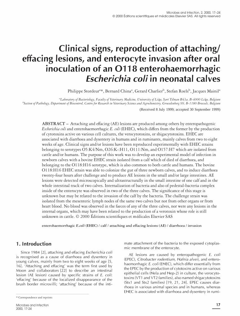
The three calves inoculated with strain 340S89 hadacute serous non-bloody diarrhoea beginning 24 h p.i.,and which lasted for 12 h in two of them (calves 1 and 3)and until the euthanasia in calf 2 (figure 1). A mild hyper-thermia was also noted (39.6 to 39.9 °C), but only calf 2had general clinical signs such as inappetence, prostra-tion, and congestion of mucosae. The control calf hadneither diarrhoea nor general clinical signs.
3.2. Faecal excretion
The challenge E. coli strain 340S89 was detected in thefaecal samples of the three challenged calves frombetween 8 to 20 h p.i. until euthanasia (figure 1), after
Figure 1. Faecal excretion of strain 340S89 (Log CFU/ mL of faeces) and appearance of diarrhoea as a function of p.i. time. The calves wereeuthanazied at 56 hours p.i. (calf 1), 64 hours p.i. (calf 2), and 44 hours p.i. (calf 3), respectively.
Enterohaemorrhagic E. coli infection in calves Original article
Microbes and Infection2000, 17-24
19
growth on streptomycin-tellurite gassner agar plates andidentification by tube agglutination with an O118 immuneserum, by antibiotic sensitivity determination, by DNAcolony hybridization with the Eae and VT1 probes and byPFGE as described in the Material and methods section.The peak of faecal excretion was observed between 20 to24 h p.i.. Strain 340S89 was excreted by most calves atconcentration between 105 and 1010 CFU/g of faeces(figure 1) and represented 65 to 100% of the total coliformpopulation. In the samples, concentrations of 1011 to 1012
CFU/g of faeces were obtained (figure 1) but most prob-ably result from a technical problem.
The PCR results for the eae gene on the faecal sampleswere positive from between 16 to 20 h p.i. until euthana-sia.
The control calf never excreted the challenge strain noran eae PCR-positive E coli. All four calves remainednegative for presence of coronavirus, Cryptosporidiumand K99-enterotoxigenic E. coli. Only calf 2 was foundpositive for excretion of rotavirus as early as 24 h p.i..
3.3. Necropsy
The three calves presented lesions of acute serousenteritis and/or colitis: lesions were localized on the largeintestine (calf 1) or extended throughout the whole intes-tinal tract (calves 2 and 3). No lesions were found in theinternal organs with the exception of the mesentericlymph nodes which were enlarged. The control calfshowed no lesion at all.
3.4. Postmortem bacteriology
E. coli strain 340S89 was detected and identified, as inthe faecal samples, in various intestinal segments of thethree challenged calves (table I) and from the mesentericlymph nodes of calves 2 and 3. Strain 340S89 was presentat concentrations varying between 106 to 109 CFU/g ofintestinal content and represented 60 to 85% of the totalcoliform population. Strain 340S89 was not detected inthe control calf either from the intestine or from theintestinal organs. PCR performed on the mesenteric lymphnodes of calves 2 and 3 were positive, and negative on themesenteric lymph nodes of the control calf.
3.5. Histopathology and electron microscopy
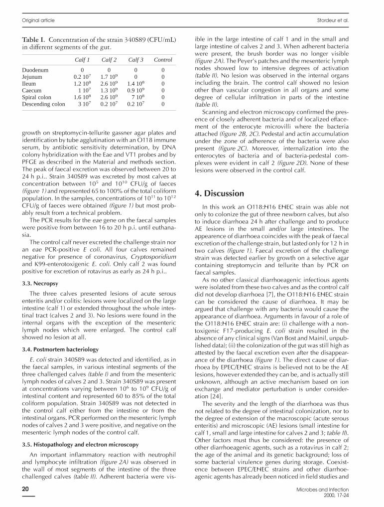
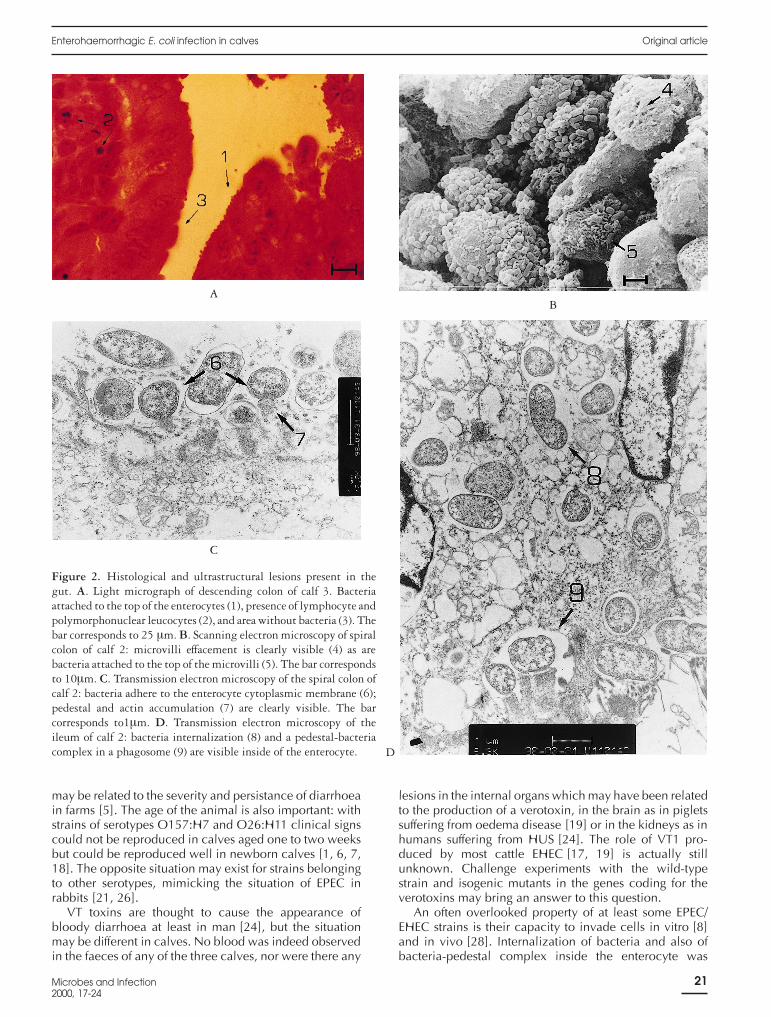
An important inflammatory reaction with neutrophiland lymphocyte infiltration (figure 2A) was observed inthe wall of most segments of the intestine of the threechallenged calves (table II). Adherent bacteria were vis-
ible in the large intestine of calf 1 and in the small andlarge intestine of calves 2 and 3. When adherent bacteriawere present, the brush border was no longer visible(figure 2A). The Peyer’s patches and the mesenteric lymphnodes showed low to intensive degrees of activation(table II). No lesion was observed in the internal organsincluding the brain. The control calf showed no lesionother than vascular congestion in all organs and somedegree of cellular infiltration in parts of the intestine(table II).
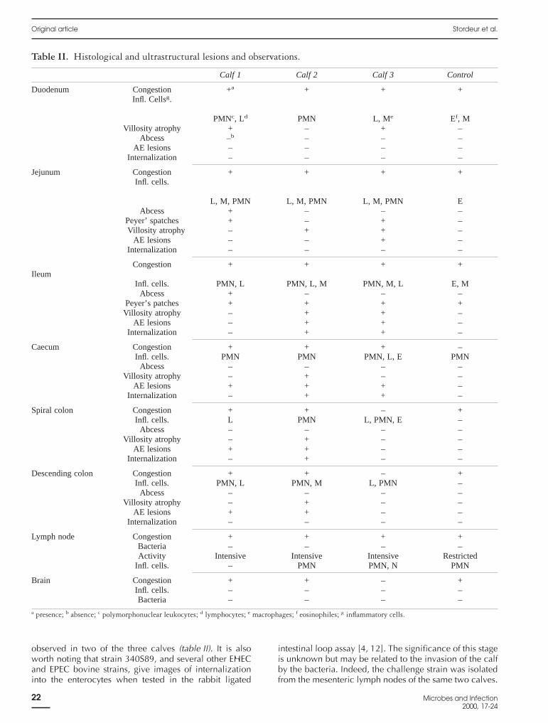
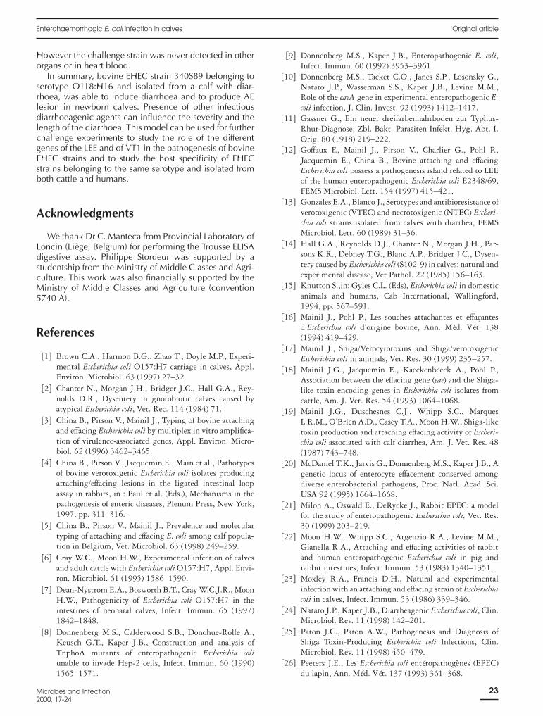
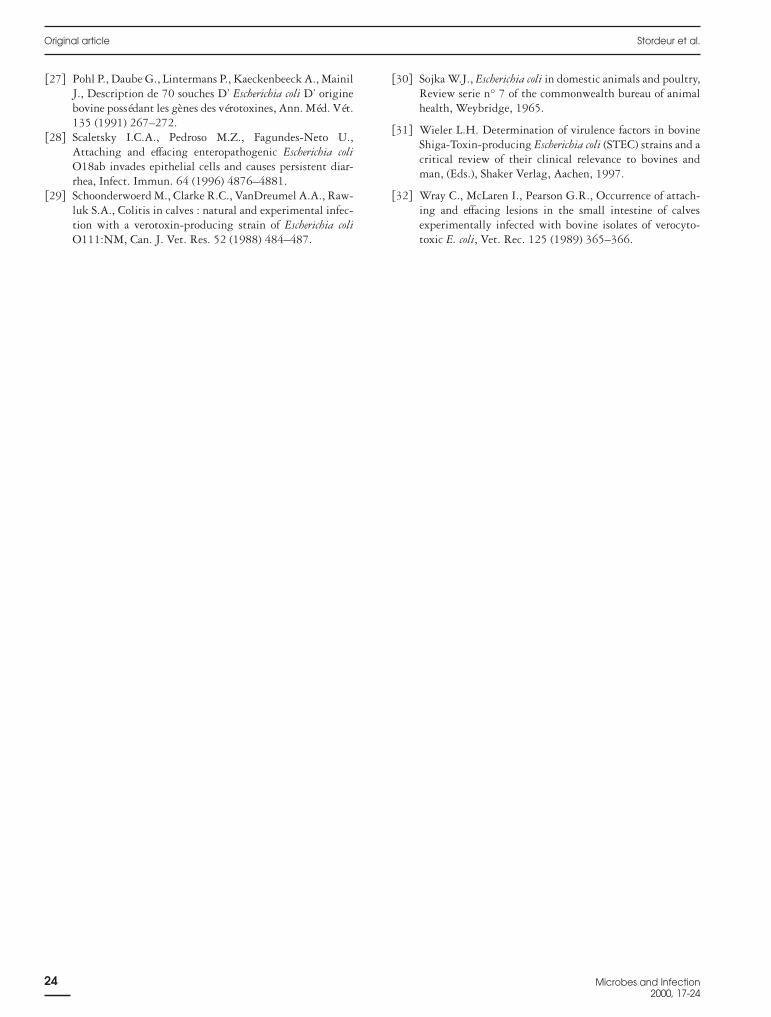
Scanning and electron microscopy confirmed the pres-ence of closely adherent bacteria and of localized efface-ment of the enterocyte microvilli where the bacteriaattached (figure 2B, 2C). Pedestal and actin accumulationunder the zone of adherence of the bacteria were alsopresent (figure 2C). Moreover, internalization into theenterocytes of bacteria and of bacteria-pedestal com-plexes were evident in calf 2 (figure 2D). None of theselesions were observed in the control calf.
4. Discussion
In this work an O118:H16 EHEC strain was able notonly to colonize the gut of three newborn calves, but alsoto induce diarrhoea 24 h after challenge and to produceAE lesions in the small and/or large intestines. Theappearence of diarrhoea coincides with the peak of faecalexcretion of the challenge strain, but lasted only for 12 h intwo calves (figure 1). Faecal excretion of the challengestrain was detected earlier by growth on a selective agarcontaining streptomycin and tellurite than by PCR onfaecal samples.
As no other classical diarrhoeagenic infectious agentswere isolated from these two calves and as the control calfdid not develop diarrhoea [7], the O118:H16 EHEC straincan be considered the cause of diarrhoea. It may beargued that challenge with any bacteria would cause theappearance of diarrhoea. Arguments in favour of a role ofthe O118:H16 EHEC strain are: (i) challenge with a non-toxigenic F17-producing E. coli strain resulted in theabsence of any clinical signs (Van Bost and Mainil, unpub-lished data); (ii) the colonization of the gut was still high asattested by the faecal excretion even after the disappear-ance of the diarrhoea (figure 1). The direct cause of diar-rhoea by EPEC/EHEC strains is believed not to be the AElesions, however extended they can be, and is actually stillunknown, although an active mechanism based on ionexchange and mediator perturbation is under consider-ation [24].
The severity and the length of the diarrhoea was thusnot related to the degree of intestinal colonization, nor tothe degree of extension of the macroscopic (acute serousenteritis) and microscopic (AE) lesions (small intestine forcalf 1, small and large intestine for calves 2 and 3; table II).Other factors must thus be considered: the presence ofother diarrhoeagenic agents, such as a rotavirus in calf 2;the age of the animal and its genetic background; loss ofsome bacterial virulence genes during storage. Coexist-ence between EPEC/EHEC strains and other diarrhoe-agenic agents has already been noticed in field studies and
Table I. Concentration of the strain 340S89 (CFU/mL)in different segments of the gut.
Calf 1 Calf 2 Calf 3 Control
Duodenum 0 0 0 0Jejunum 0.2 107 1.7 109 0 0Ileum 1.2 108 2.6 109 1.4 108 0Caecum 1 107 1.3 109 0.9 109 0Spiral colon 1.6 108 2.6 109 7 108 0Descending colon 3 107 0.2 107 0.2 107 0
Original article Stordeur et al.
20 Microbes and Infection2000, 17-24
may be related to the severity and persistance of diarrhoeain farms [5]. The age of the animal is also important: withstrains of serotypes O157:H7 and O26:H11 clinical signscould not be reproduced in calves aged one to two weeksbut could be reproduced well in newborn calves [1, 6, 7,18]. The opposite situation may exist for strains belongingto other serotypes, mimicking the situation of EPEC inrabbits [21, 26].
VT toxins are thought to cause the appearance ofbloody diarrhoea at least in man [24], but the situationmay be different in calves. No blood was indeed observedin the faeces of any of the three calves, nor were there any
lesions in the internal organs which may have been relatedto the production of a verotoxin, in the brain as in pigletssuffering from oedema disease [19] or in the kidneys as inhumans suffering from HUS [24]. The role of VT1 pro-duced by most cattle EHEC [17, 19] is actually stillunknown. Challenge experiments with the wild-typestrain and isogenic mutants in the genes coding for theverotoxins may bring an answer to this question.
An often overlooked property of at least some EPEC/EHEC strains is their capacity to invade cells in vitro [8]and in vivo [28]. Internalization of bacteria and also ofbacteria-pedestal complex inside the enterocyte was
Figure 2. Histological and ultrastructural lesions present in thegut. A. Light micrograph of descending colon of calf 3. Bacteriaattached to the top of the enterocytes (1), presence of lymphocyte andpolymorphonuclear leucocytes (2), and area without bacteria (3). Thebar corresponds to 25 µm. B. Scanning electron microscopy of spiralcolon of calf 2: microvilli effacement is clearly visible (4) as arebacteria attached to the top of the microvilli (5). The bar correspondsto 10µm. C. Transmission electron microscopy of the spiral colon ofcalf 2: bacteria adhere to the enterocyte cytoplasmic membrane (6);pedestal and actin accumulation (7) are clearly visible. The barcorresponds to1µm. D. Transmission electron microscopy of theileum of calf 2: bacteria internalization (8) and a pedestal-bacteriacomplex in a phagosome (9) are visible inside of the enterocyte.
AB
C
D
Enterohaemorrhagic E. coli infection in calves Original article
Microbes and Infection2000, 17-24
21
observed in two of the three calves (table II). It is alsoworth noting that strain 340S89, and several other EHECand EPEC bovine strains, give images of internalizationinto the enterocytes when tested in the rabbit ligated
intestinal loop assay [4, 12]. The significance of this stageis unknown but may be related to the invasion of the calfby the bacteria. Indeed, the challenge strain was isolatedfrom the mesenteric lymph nodes of the same two calves.
Table II. Histological and ultrastructural lesions and observations.
Calf 1 Calf 2 Calf 3 Control
Duodenum Congestion +a + + +Infl. Cellsg.
PMNc, Ld PMN L, Me Ef, MVillosity atrophy + – + –
Abcess –b – – –AE lesions – – – –
Internalization – – – –
Jejunum Congestion + + + +Infl. cells.
L, M, PMN L, M, PMN L, M, PMN EAbcess + – – –
Peyer’ spatches + – + –Villosity atrophy – + + –
AE lesions – – + –Internalization – – – –
Congestion + + + +Ileum
Infl. cells. PMN, L PMN, L, M PMN, M, L E, MAbcess + – – –
Peyer’s patches + + + +Villosity atrophy – + + –
AE lesions – + + –Internalization – + + –
Caecum Congestion + + + –Infl. cells. PMN PMN PMN, L, E PMN
Abcess – – – –Villosity atrophy – + – –
AE lesions + + + –Internalization – + + –
Spiral colon Congestion + + – +Infl. cells. L PMN L, PMN, E –
Abcess – – – –Villosity atrophy – + – –
AE lesions + + – –Internalization – + – –
Descending colon Congestion + + – +Infl. cells. PMN, L PMN, M L, PMN –
Abcess – – – –Villosity atrophy – + – –
AE lesions + + – –Internalization – – – –
Lymph node Congestion + + + +Bacteria – – – –Activity Intensive Intensive Intensive Restricted
Infl. cells. – PMN PMN, N PMN
Brain Congestion + + – +Infl. cells. – – – –Bacteria – – – –
a presence; b absence; c polymorphonuclear leukocytes; d lymphocytes; e macrophages; f eosinophiles; g inflammatory cells.
Original article Stordeur et al.
22 Microbes and Infection2000, 17-24
However the challenge strain was never detected in otherorgans or in heart blood.
In summary, bovine EHEC strain 340S89 belonging toserotype O118:H16 and isolated from a calf with diar-rhoea, was able to induce diarrhoea and to produce AElesion in newborn calves. Presence of other infectiousdiarrhoeagenic agents can influence the severity and thelength of the diarrhoea. This model can be used for furtherchallenge experiments to study the role of the differentgenes of the LEE and of VT1 in the pathogenesis of bovineEHEC strains and to study the host specificity of EHECstrains belonging to the same serotype and isolated fromboth cattle and humans.
Acknowledgments
We thank Dr C. Manteca from Provincial Laboratory ofLoncin (Liège, Belgium) for performing the Trousse ELISAdigestive assay. Philippe Stordeur was supported by astudentship from the Ministry of Middle Classes and Agri-culture. This work was also financially supported by theMinistry of Middle Classes and Agriculture (convention5740 A).
References
[1] Brown C.A., Harmon B.G., Zhao T., Doyle M.P., Experi-mental Escherichia coli O157:H7 carriage in calves, Appl.Environ. Microbiol. 63 (1997) 27–32.
[2] Chanter N., Morgan J.H., Bridger J.C., Hall G.A., Rey-nolds D.R., Dysentery in gnotobiotic calves caused byatypical Escherichia coli, Vet. Rec. 114 (1984) 71.
[3] China B., Pirson V., Mainil J., Typing of bovine attachingand effacing Escherichia coli by multiplex in vitro amplifica-tion of virulence-associated genes, Appl. Environ. Micro-biol. 62 (1996) 3462–3465.
[4] China B., Pirson V., Jacquemin E., Main et al., Pathotypesof bovine verotoxigenic Escherichia coli isolates producingattaching/effacing lesions in the ligated intestinal loopassay in rabbits, in : Paul et al. (Eds.), Mechanisms in thepathogenesis of enteric diseases, Plenum Press, New York,1997, pp. 311–316.
[5] China B., Pirson V., Mainil J., Prevalence and moleculartyping of attaching and effacing E. coli among calf popula-tion in Belgium, Vet. Microbiol. 63 (1998) 249–259.
[6] Cray W.C., Moon H.W., Experimental infection of calvesand adult cattle with Escherichia coli O157:H7, Appl. Envi-ron. Microbiol. 61 (1995) 1586–1590.
[7] Dean-Nystrom E.A., Bosworth B.T., Cray W.C.J.R., MoonH.W., Pathogenicity of Escherichia coli O157:H7 in theintestines of neonatal calves, Infect. Immun. 65 (1997)1842–1848.
[8] Donnenberg M.S., Calderwood S.B., Donohue-Rolfe A.,Keusch G.T., Kaper J.B., Construction and analysis ofTnphoA mutants of enteropathogenic Escherichia coliunable to invade Hep-2 cells, Infect. Immun. 60 (1990)1565–1571.
[9] Donnenberg M.S., Kaper J.B., Enteropathogenic E. coli,Infect. Immun. 60 (1992) 3953–3961.
[10] Donnenberg M.S., Tacket C.O., Janes S.P., Losonsky G.,Nataro J.P., Wasserman S.S., Kaper J.B., Levine M.M.,Role of the eaeA gene in experimental enteropathogenic E.coli infection, J. Clin. Invest. 92 (1993) 1412–1417.
[11] Gassner G., Ein neuer dreifarbennahrboden zur Typhus-Rhur-Diagnose, Zbl. Bakt. Parasiten Infekt. Hyg. Abt. I.Orig. 80 (1918) 219–222.
[12] Goffaux F., Mainil J., Pirson V., Charlier G., Pohl P.,Jacquemin E., China B., Bovine attaching and effacingEscherichia coli possess a pathogenesis island related to LEEof the human enteropathogenic Escherichia coli E2348/69,FEMS Microbiol. Lett. 154 (1997) 415–421.
[13] Gonzales E.A., Blanco J., Serotypes and antibioresistance ofverotoxigenic (VTEC) and necrotoxigenic (NTEC) Escheri-chia coli strains isolated from calves with diarrhea, FEMSMicrobiol. Lett. 60 (1989) 31–36.
[14] Hall G.A., Reynolds D.J., Chanter N., Morgan J.H., Par-sons K.R., Debney T.G., Bland A.P., Bridger J.C., Dysen-tery caused by Escherichia coli (S102-9) in calves: natural andexperimental disease, Vet Pathol. 22 (1985) 156–163.
[15] Knutton S.,in: Gyles C.L. (Eds), Escherichia coli in domesticanimals and humans, Cab International, Wallingford,1994, pp. 567–591.
[16] Mainil J., Pohl P., Les souches attachantes et effaçantesd’Escherichia coli d’origine bovine, Ann. Méd. Vét. 138(1994) 419–429.
[17] Mainil J., Shiga/Verocytotoxins and Shiga/verotoxigenicEscherichia coli in animals, Vet. Res. 30 (1999) 235–257.
[18] Mainil J.G., Jacquemin E., Kaeckenbeeck A., Pohl P.,Association between the effacing gene (eae) and the Shiga-like toxin encoding genes in Escherichia coli isolates fromcattle, Am. J. Vet. Res. 54 (1993) 1064–1068.
[19] Mainil J.G., Duschesnes C.J., Whipp S.C., MarquesL.R.M., O’Brien A.D., Casey T.A., Moon H.W., Shiga-liketoxin production and attaching effacing activity of Escheri-chia coli associated with calf diarrhea, Am. J. Vet. Res. 48(1987) 743–748.
[20] McDaniel T.K., Jarvis G., Donnenberg M.S., Kaper J.B., Agenetic locus of enterocyte effacement conserved amongdiverse enterobacterial pathogens, Proc. Natl. Acad. Sci.USA 92 (1995) 1664–1668.
[21] Milon A., Oswald E., DeRycke J., Rabbit EPEC: a modelfor the study of enteropathogenic Escherichia coli, Vet. Res.30 (1999) 203–219.
[22] Moon H.W., Whipp S.C., Argenzio R.A., Levine M.M.,Gianella R.A., Attaching and effacing activities of rabbitand human enteropathogenic Escherichia coli in pig andrabbit intestines, Infect. Immun. 53 (1983) 1340–1351.
[23] Moxley R.A., Francis D.H., Natural and experimentalinfection with an attaching and effacing strain of Escherichiacoli in calves, Infect. Immun. 53 (1986) 339–346.
[24] Nataro J.P., Kaper J.B., Diarrheagenic Escherichia coli, Clin.Microbiol. Rev. 11 (1998) 142–201.
[25] Paton J.C., Paton A.W., Pathogenesis and Diagnosis ofShiga Toxin-Producing Escherichia coli Infections, Clin.Microbiol. Rev. 11 (1998) 450–479.
[26] Peeters J.E., Les Escherichia coli entéropathogènes (EPEC)du lapin, Ann. Méd. Vét. 137 (1993) 361–368.
Enterohaemorrhagic E. coli infection in calves Original article
Microbes and Infection2000, 17-24
23
[27] Pohl P., Daube G., Lintermans P., Kaeckenbeeck A., MainilJ., Description de 70 souches D’ Escherichia coli D’ originebovine possédant les gènes des vérotoxines, Ann. Méd. Vét.135 (1991) 267–272.
[28] Scaletsky I.C.A., Pedroso M.Z., Fagundes-Neto U.,Attaching and effacing enteropathogenic Escherichia coliO18ab invades epithelial cells and causes persistent diar-rhea, Infect. Immun. 64 (1996) 4876–4881.
[29] Schoonderwoerd M., Clarke R.C., VanDreumel A.A., Raw-luk S.A., Colitis in calves : natural and experimental infec-tion with a verotoxin-producing strain of Escherichia coliO111:NM, Can. J. Vet. Res. 52 (1988) 484–487.
[30] Sojka W.J., Escherichia coli in domestic animals and poultry,Review serie n° 7 of the commonwealth bureau of animalhealth, Weybridge, 1965.
[31] Wieler L.H. Determination of virulence factors in bovineShiga-Toxin-producing Escherichia coli (STEC) strains and acritical review of their clinical relevance to bovines andman, (Eds.), Shaker Verlag, Aachen, 1997.
[32] Wray C., McLaren I., Pearson G.R., Occurrence of attach-ing and effacing lesions in the small intestine of calvesexperimentally infected with bovine isolates of verocyto-toxic E. coli, Vet. Rec. 125 (1989) 365–366.
Original article Stordeur et al.
24 Microbes and Infection2000, 17-24


![forum.konkurdl.konkur.in/PHD/97/212-E-PHD97-[].pdf · Enterotoxic colibacillosis Septicemic colibacillosis (r Attaching and Effacing Escherichia coli (r Enterohemorrhagic colibacillosis](https://img.pdfslide.us/doc/110x75/5f758831532eb42f2172b12a/forumkonkurdl-pdf-enterotoxic-colibacillosis-septicemic-colibacillosis-r.jpg)