Embed Size (px)
Citation preview
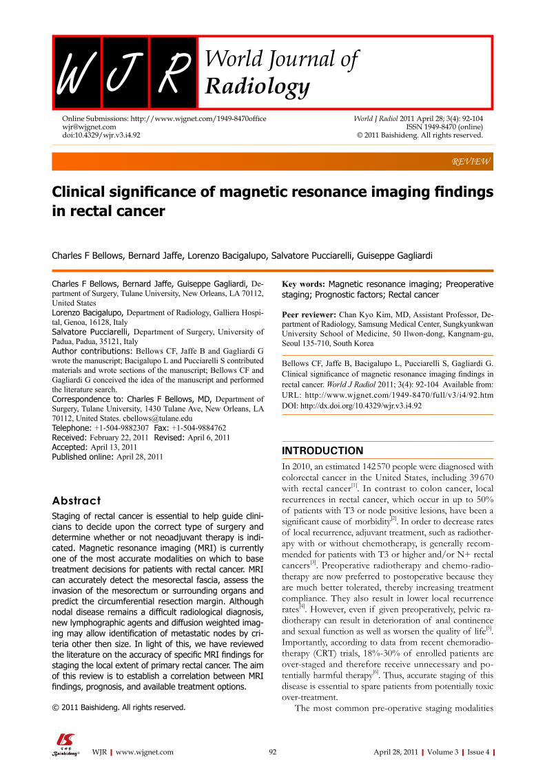
Clinical significance of magnetic resonance imaging findings in rectal cancer
Charles F Bellows, Bernard Jaffe, Lorenzo Bacigalupo, Salvatore Pucciarelli, Guiseppe Gagliardi
Charles F Bellows, Bernard Jaffe, Guiseppe Gagliardi, De-partment of Surgery, Tulane University, New Orleans, LA 70112, United StatesLorenzo Bacigalupo, Department of Radiology, Galliera Hospi-tal, Genoa, 16128, ItalySalvatore Pucciarelli, Department of Surgery, University of Padua, Padua, 35121, ItalyAuthor contributions: Bellows CF, Jaffe B and Gagliardi G wrote the manuscript; Bacigalupo L and Pucciarelli S contributed materials and wrote sections of the manuscript; Bellows CF and Gagliardi G conceived the idea of the manuscript and performed the literature search. Correspondence to: Charles F Bellows, MD, Department of Surgery, Tulane University, 1430 Tulane Ave, New Orleans, LA 70112, United States. [email protected]: +1-504-9882307 Fax: +1-504-9884762Received: February 22, 2011 Revised: April 6, 2011Accepted: April 13, 2011Published online: April 28, 2011
AbstractStaging of rectal cancer is essential to help guide clini-cians to decide upon the correct type of surgery and determine whether or not neoadjuvant therapy is indi-cated. Magnetic resonance imaging (MRI) is currently one of the most accurate modalities on which to base treatment decisions for patients with rectal cancer. MRI can accurately detect the mesorectal fascia, assess the invasion of the mesorectum or surrounding organs and predict the circumferential resection margin. Although nodal disease remains a difficult radiological diagnosis, new lymphographic agents and diffusion weighted imag-ing may allow identification of metastatic nodes by cri-teria other then size. In light of this, we have reviewed the literature on the accuracy of specific MRI findings for staging the local extent of primary rectal cancer. The aim of this review is to establish a correlation between MRI findings, prognosis, and available treatment options.
© 2011 Baishideng. All rights reserved.
Key words: Magnetic resonance imaging; Preoperative staging; Prognostic factors; Rectal cancer
Peer reviewer: Chan Kyo Kim, MD, Assistant Professor, De-partment of Radiology, Samsung Medical Center, Sungkyunkwan University School of Medicine, 50 Ilwon-dong, Kangnam-gu, Seoul 135-710, South Korea
Bellows CF, Jaffe B, Bacigalupo L, Pucciarelli S, Gagliardi G. Clinical significance of magnetic resonance imaging findings in rectal cancer. World J Radiol 2011; 3(4): 92-104 Available from: URL: http://www.wjgnet.com/1949-8470/full/v3/i4/92.htm DOI: http://dx.doi.org/10.4329/wjr.v3.i4.92
INTRODUCTIONIn 2010, an estimated 142 570 people were diagnosed with colorectal cancer in the United States, including 39 670 with rectal cancer[1]. In contrast to colon cancer, local recurrences in rectal cancer, which occur in up to 50% of patients with T3 or node positive lesions, have been a significant cause of morbidity[2]. In order to decrease rates of local recurrence, adjuvant treatment, such as radiother-apy with or without chemotherapy, is generally recom-mended for patients with T3 or higher and/or N+ rectal cancers[3]. Preoperative radiotherapy and chemo-radio-therapy are now preferred to postoperative because they are much better tolerated, thereby increasing treatment compliance. They also result in lower local recurrence rates[4]. However, even if given preoperatively, pelvic ra-diotherapy can result in deterioration of anal continence and sexual function as well as worsen the quality of life[5]. Importantly, according to data from recent chemoradio-therapy (CRT) trials, 18%-30% of enrolled patients are over-staged and therefore receive unnecessary and po-tentially harmful therapy[6]. Thus, accurate staging of this disease is essential to spare patients from potentially toxic over-treatment.
The most common pre-operative staging modalities
REVIEW
World Journal of RadiologyW J R
Online Submissions: http://www.wjgnet.com/[email protected]:10.4329/wjr.v3.i4.92
World J Radiol 2011 April 28; 3(4): 92-104ISSN 1949-8470 (online)
© 2011 Baishideng. All rights reserved.
92 April 28, 2011|Volume 3|Issue 4|WJR|www.wjgnet.com
Bellows CF et al . MRI findings in rectal cancer
for rectal cancer include endorectal ultrasonography (EUS), computed tomography (CT) and magnetic reso-nance imaging (MRI). In two large retrospective studies on rectal cancer patients, over a period of 10 years, the overall accuracy of T and N staging by EUS was shown to be only 69% and 68% respectively[7,8]. Compared to EUS, CT has an even lower accuracy for determining the depth of tumor invasion[9]. In contrast, high defini-tion MRI with phased-array coils has been shown to be more reliable than EUS in staging advanced (stage ≥ Ⅱ) rectal cancer[10]. In fact, MRI has been shown to provide important information about the depth of tumor infiltra-tion within the bowel wall, the relationship between the tumor and mesorectal fascia, and the presence of lymph node and extramural vascular invasion. Information de-termined from MRI can help guide clinicians to decide upon the correct type of surgery, determine whether or not neoadjuvant therapy, such as chemo-radiation, is indicated, and predict patients’ prognosis. This informa-tion can be used to maximize the chance of complete oncological resection, improve survival and the quality of life, and minimize morbidity. The aim of this review is to establish a correlation between MRI findings, prognosis, and available treatment options.
MR IMAGING PROTOCOLSThe introduction of phased-array coils has been a major advance in imaging of rectal cancer allowing high spatial resolution, a large field of coverage[10], and visualization of structures 1-2 mm in diameter[11]. Ideally, for rectal MRI, the field of view should be small (i.e. less than 200 mm), the matrix (resolution in 2D) at least 256 × 256 pixels, and the slices 3 mm or less in thickness.
In the most current MRI protocol (Table 1), the tu-mor is first localized with low-resolution axial and sagit-tal images of the entire pelvis. The field of view is then restricted to the area of the cancer and high-resolution T2 weighted images are obtained perpendicular to the cranio-caudal axis of the rectum at the level of the tumor (Figure 1A). True axial (i.e. perpendicular) images of the tumor are critical because they reduce the overestimation of the tumor depth of invasion noted upon oblique im-aging[12]. Coronal images (parallel to the anus) are impor-tant in identifying the relationship of low rectal tumors to the internal sphincter as well as the external sphinc-ter/levators complex[13] (Figure 1B). T2-weighted sagittal images are often necessary to determine the relationship of the tumor to the peritoneal reflection (Figure 1C)[14]. In some cases, axial diffusion weighted imaging (DWI) may be performed to help in the localization of small tu-mors[15] (Figure 2).
The importance of the imaging protocol for assess-ment of advanced rectal tumors has recently been report-ed. Suzuki and associates demonstrated that by including the imaging parameters listed in sequence 2 and 3 (Table 1), the sensitivity and specificity of assessing invasion of an-terior organs was 80% and 95% respectively, compared to
only 50% and 33% with protocols that employed different imaging parameters[16].
Improving MR image quality can be facilitated with the use of rectal cleaning to limit misinterpretation due to stool residue. Distension of the rectum by air insuffla-tion, gel enema, or intravenous administration of spas-molytic medication also improves evaluation of the rectal wall layers.
MRI FINDINGS IN RECTAL CANCERIn T2 weighted images, rectal cancers typically have a sig-nal-intensity intermediate between that of the perirectal fat, which is bright, and the muscularis propria, which is pitch black. The signal intensity is increased if the tumors contain mucin, but a low signal intensity similar to that of the muscle layer usually indicates a marked desmoplastic reaction of the tumor[17].
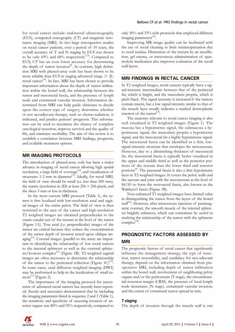
The anatomy relevant to rectal cancer imaging is also well visualized in T2 weighted images (Figure 1). The mucosa has a hypointense signal, the submucosa a hy-perintense signal, the muscularis propria a hypointense signal, and the mesorectal fat a highly hyperintense signal. The mesorectal fascia can be identified as a thin, low-signal intensity structure that envelopes the mesorectum. However, due to a diminishing thickness of mesorectal fat, the mesorectal fascia is typically better visualized in the upper and middle third as well as the posterior por-tions of the rectum than the lower third and anterior portions[9]. The presacral fascia is also a thin hypointense layer in T2 weighted images. It covers the pelvic walls and the sacrum and joins with the mesorectum at the level of S4/S5 to form the rectosacral fascia, also known as the Waldayer’s fascia (Figure 1B).
Non-enhanced T1 weighted images have limited value in distinguishing the tumor from the layers of the bowel wall[14]. However, after intravenous injection of paramag-netic contrast, the smooth muscle of the internal sphinc-ter brightly enhances, which can sometimes be useful in studying the relationship of the tumor with the sphincter complex[18].
PROGNOSTIC FACTORS ASSESSED BY MRIThe prognostic factors of rectal cancer that significantly influence the management strategy, the type of resec-tion, tumor resectability, and candidacy for neo-adjuvant therapy, depend on the information obtained from pre-operative MRI, including depth of tumor infiltration within the bowel wall, involvement of neighboring pelvic organs and/or the peritoneum (T stage), the circumferen-tial resection margin (CRM), the presence of local lymph node metastases (N stage), extramural vascular invasion, and the extent of extramural tumor spread in mm.
T staging The depth of invasion through the muscle wall is one
93 April 28, 2011|Volume 3|Issue 4|WJR|www.wjgnet.com
important element seen on MRI that can help guide clini-cal decision making for patients with rectal cancer. Not only does the incidence of nodal involvement increase with increasing tumor penetration[19,20], but clinical studies have shown that patients with stage Ⅰ (T1-2 N0) rectal cancer do not benefit from neo-adjuvant radiotherapy[21] and may be amenable to a less than radical surgical treat-ment[22]. Patients with clinically staged T3-4 tumors typi-cally require preoperative CRT since it reduces the rates of local recurrence more effectively than either post-operative CRT or preoperative radiotherapy alone[23-25]. However, some problems remain with T stage deter-mination on MR imaging. Overall, the agreement be-tween MRI and histology for T staging has ranged from 66%-94%[18,26-28]. One of the main problems of T staging on MRI is the distinction between T2 and T3 tumors. In fact, investigators have shown that the negative predictive value for invasion beyond the muscularis propria varied from 93% (expert reading) to 76% (general radiologist reading)[26]. This difficulty is attributed to the presence of desmoplastic reactions around the tumor. This reaction
makes it difficult to distinguish between spiculation in the perirectal fat caused by fibrosis alone from that caused by fibrous tissue that contains tumor cells[26]. In contrast, MRI has been shown to be more accurate in imaging the more advanced tumors (T4)[27,29]. According to a meta-analysis, MRI for T4 lesions has a specificity of 96%[30].
CRMThe CRM (lateral, radial) is defined as the surgical cut sur-face of the connective tissues (i.e. lymphovascular, fatty and neural tissue) that circumferentially encase the rectum. It equates to the mesorectal fascia that forms the plane of dissection in rectal cancer surgery. It is assessed by mark-ing the outer surface (i.e. the CRM) with ink, taking serial cuts through the specimen and examining the macroscop-ic and microscopic relations between the tumor and the inked margin (Figure 3A-C). The CRM gives significant information not only about the quality of the performed operation but also prognosis of the disease. Indeed, in a recent study based on the data from a randomized clini-cal trial, Nagtegaal et al[31] demonstrated in a multivariate
94 April 28, 2011|Volume 3|Issue 4|WJR|www.wjgnet.com
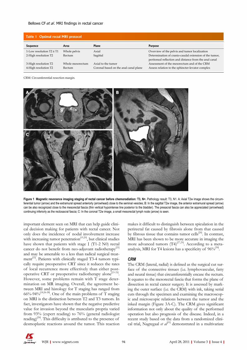
Table 1 Optimal rectal MRI protocol
Sequence Area Plane Purpose
1-Low resolution T2 ± T1 Whole pelvis Axial Overview of the pelvis and tumor localization2-High resolution T2 Rectum Sagittal Determination of cranio-caudal extension of the tumor,
peritoneal reflection and distance from the anal canal 3-High resolution T2 Whole mesorectum Axial to the tumor Assessment of the mesorectum and of the CRM 4-High resolution T2 Rectum Coronal based on the anal canal plane Assess relation to the sphincter-levator complex
CRM: Circumferential resection margin.
C
BA
Figure 1 Magnetic resonance imaging staging of rectal cancer before chemoradiation: T3, N+. Pathology result: T3, N1. A: Axial T2w image shows the circum-ferential tumor (arrow) and the extramural spread anteriorly (arrowhead) close to the seminal vesicles; B: In the sagittal T2w image, the anterior extramural spread (arrow) can be also recognized close to the mesorectal fascia (thin vertical hypointense line posterior to the bladder). The presacral fascia can also be appreciated (arrowhead) continuing inferiorly as the rectosacral fascia; C: In the coronal T2w image, a small mesorectal lymph node (arrow) is seen.
Bellows CF et al . MRI findings in rectal cancer
model that the CRM is more important than the T stage for the prognosis of rectal cancer. The definition of a positive CRM remains a matter of debate. A review of
the literature in 2006 showed that the majority of studies that dealt with CRM status used the ≤ 1 mm definition for positive CRM (91.1%; 7373 of 8094 patients)[32].
95 April 28, 2011|Volume 3|Issue 4|WJR|www.wjgnet.com
DC
BA
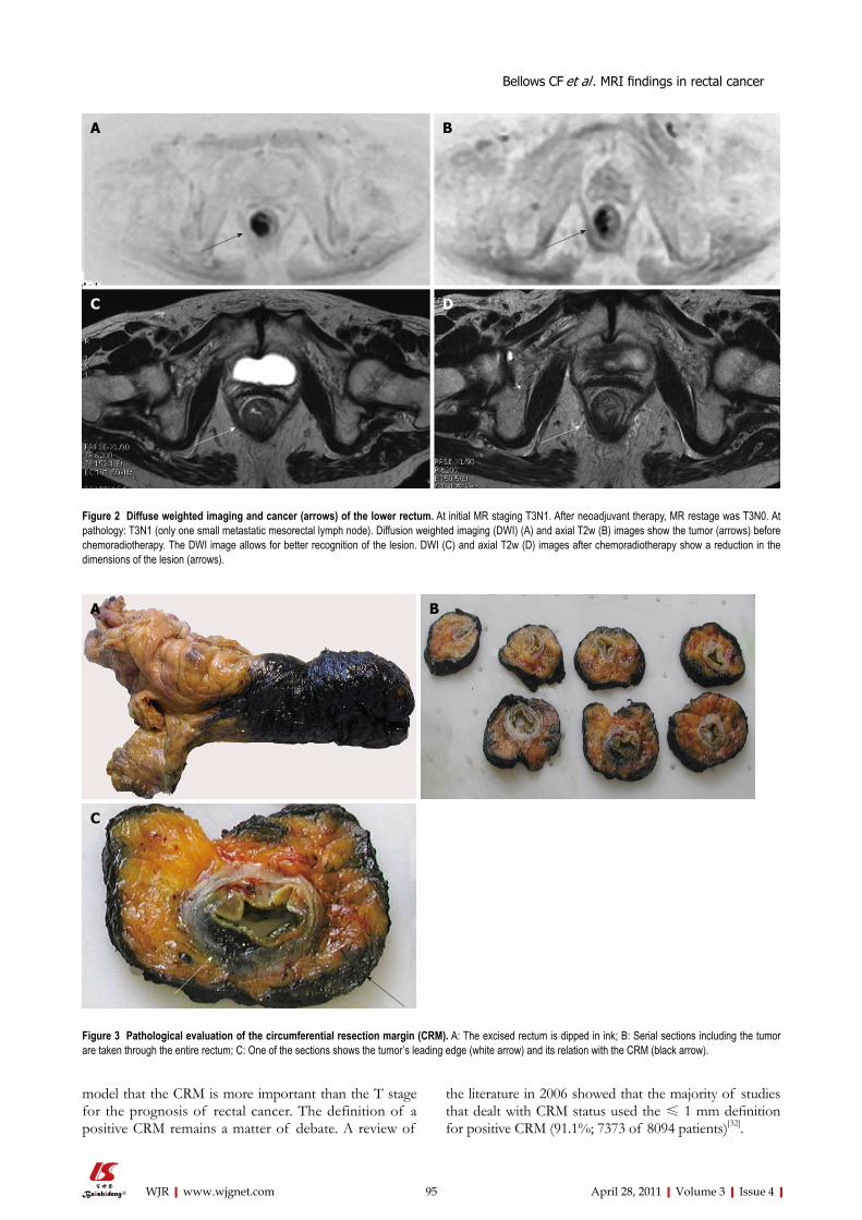
Figure 2 Diffuse weighted imaging and cancer (arrows) of the lower rectum. At initial MR staging T3N1. After neoadjuvant therapy, MR restage was T3N0. At pathology: T3N1 (only one small metastatic mesorectal lymph node). Diffusion weighted imaging (DWI) (A) and axial T2w (B) images show the tumor (arrows) before chemoradiotherapy. The DWI image allows for better recognition of the lesion. DWI (C) and axial T2w (D) images after chemoradiotherapy show a reduction in the dimensions of the lesion (arrows).
C
BA
Figure 3 Pathological evaluation of the circumferential resection margin (CRM). A: The excised rectum is dipped in ink; B: Serial sections including the tumor are taken through the entire rectum; C: One of the sections shows the tumor’s leading edge (white arrow) and its relation with the CRM (black arrow).
Bellows CF et al . MRI findings in rectal cancer
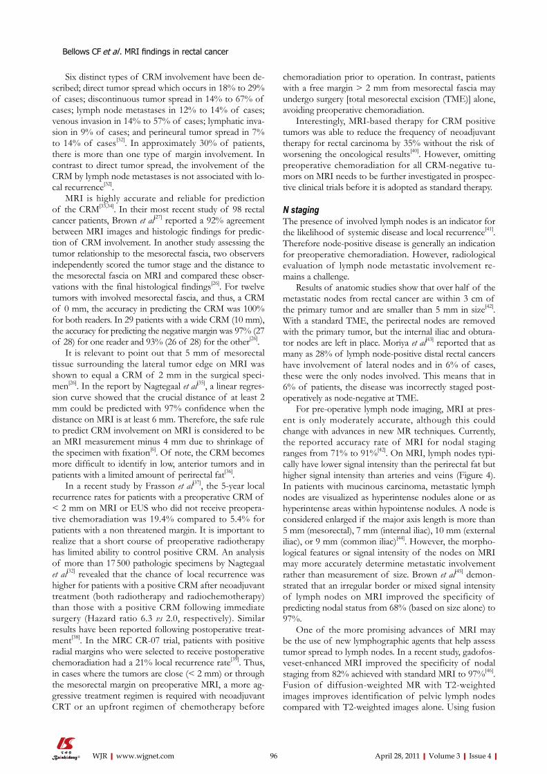
Six distinct types of CRM involvement have been de-scribed; direct tumor spread which occurs in 18% to 29% of cases; discontinuous tumor spread in 14% to 67% of cases; lymph node metastases in 12% to 14% of cases; venous invasion in 14% to 57% of cases; lymphatic inva-sion in 9% of cases; and perineural tumor spread in 7% to 14% of cases[32]. In approximately 30% of patients, there is more than one type of margin involvement. In contrast to direct tumor spread, the involvement of the CRM by lymph node metastases is not associated with lo-cal recurrence[32].
MRI is highly accurate and reliable for prediction of the CRM[33,34]. In their most recent study of 98 rectal cancer patients, Brown et al[27] reported a 92% agreement between MRI images and histologic findings for predic-tion of CRM involvement. In another study assessing the tumor relationship to the mesorectal fascia, two observers independently scored the tumor stage and the distance to the mesorectal fascia on MRI and compared these obser-vations with the final histological findings[26]. For twelve tumors with involved mesorectal fascia, and thus, a CRM of 0 mm, the accuracy in predicting the CRM was 100% for both readers. In 29 patients with a wide CRM (10 mm), the accuracy for predicting the negative margin was 97% (27 of 28) for one reader and 93% (26 of 28) for the other[26].
It is relevant to point out that 5 mm of mesorectal tissue surrounding the lateral tumor edge on MRI was shown to equal a CRM of 2 mm in the surgical speci-men[26]. In the report by Nagtegaal et al[35], a linear regres-sion curve showed that the crucial distance of at least 2 mm could be predicted with 97% confidence when the distance on MRI is at least 6 mm. Therefore, the safe rule to predict CRM involvement on MRI is considered to be an MRI measurement minus 4 mm due to shrinkage of the specimen with fixation[6]. Of note, the CRM becomes more difficult to identify in low, anterior tumors and in patients with a limited amount of perirectal fat[36].
In a recent study by Frasson et al[37], the 5-year local recurrence rates for patients with a preoperative CRM of < 2 mm on MRI or EUS who did not receive preopera-tive chemoradiation was 19.4% compared to 5.4% for patients with a non threatened margin. It is important to realize that a short course of preoperative radiotherapy has limited ability to control positive CRM. An analysis of more than 17 500 pathologic specimens by Nagtegaal et al[32] revealed that the chance of local recurrence was higher for patients with a positive CRM after neoadjuvant treatment (both radiotherapy and radiochemotherapy) than those with a positive CRM following immediate surgery (Hazard ratio 6.3 vs 2.0, respectively). Similar results have been reported following postoperative treat-ment[38]. In the MRC CR-07 trial, patients with positive radial margins who were selected to receive postoperative chemoradiation had a 21% local recurrence rate[39]. Thus, in cases where the tumors are close (< 2 mm) or through the mesorectal margin on preoperative MRI, a more ag-gressive treatment regimen is required with neoadjuvant CRT or an upfront regimen of chemotherapy before
chemoradiation prior to operation. In contrast, patients with a free margin > 2 mm from mesorectal fascia may undergo surgery [total mesorectal excision (TME)] alone, avoiding preoperative chemoradiation.
Interestingly, MRI-based therapy for CRM positive tumors was able to reduce the frequency of neoadjuvant therapy for rectal carcinoma by 35% without the risk of worsening the oncological results[40]. However, omitting preoperative chemoradiation for all CRM-negative tu-mors on MRI needs to be further investigated in prospec-tive clinical trials before it is adopted as standard therapy.
N stagingThe presence of involved lymph nodes is an indicator for the likelihood of systemic disease and local recurrence[41]. Therefore node-positive disease is generally an indication for preoperative chemoradiation. However, radiological evaluation of lymph node metastatic involvement re-mains a challenge.
Results of anatomic studies show that over half of the metastatic nodes from rectal cancer are within 3 cm of the primary tumor and are smaller than 5 mm in size[42]. With a standard TME, the perirectal nodes are removed with the primary tumor, but the internal iliac and obtura-tor nodes are left in place. Moriya et al[43] reported that as many as 28% of lymph node-positive distal rectal cancers have involvement of lateral nodes and in 6% of cases, these were the only nodes involved. This means that in 6% of patients, the disease was incorrectly staged post-operatively as node-negative at TME.
For pre-operative lymph node imaging, MRI at pres-ent is only moderately accurate, although this could change with advances in new MR techniques. Currently, the reported accuracy rate of MRI for nodal staging ranges from 71% to 91%[42]. On MRI, lymph nodes typi-cally have lower signal intensity than the perirectal fat but higher signal intensity than arteries and veins (Figure 4). In patients with mucinous carcinoma, metastatic lymph nodes are visualized as hyperintense nodules alone or as hyperintense areas within hypointense nodules. A node is considered enlarged if the major axis length is more than 5 mm (mesorectal), 7 mm (internal iliac), 10 mm (external iliac), or 9 mm (common iliac)[44]. However, the morpho-logical features or signal intensity of the nodes on MRI may more accurately determine metastatic involvement rather than measurement of size. Brown et al[45] demon-strated that an irregular border or mixed signal intensity of lymph nodes on MRI improved the specificity of predicting nodal status from 68% (based on size alone) to 97%.
One of the more promising advances of MRI may be the use of new lymphographic agents that help assess tumor spread to lymph nodes. In a recent study, gadofos-veset-enhanced MRI improved the specificity of nodal staging from 82% achieved with standard MRI to 97%[46]. Fusion of diffusion-weighted MR with T2-weighted images improves identification of pelvic lymph nodes compared with T2-weighted images alone. Using fusion
96 April 28, 2011|Volume 3|Issue 4|WJR|www.wjgnet.com
Bellows CF et al . MRI findings in rectal cancer
images, 29% additional nodes were detected compared with T2-weighted images alone[47]. The improved nodal identification may aid in treatment planning.
Extramural vascular invasionVenous invasion is defined as the presence of tumor tis-sue within an endothelium lined space, either surrounded by a rim of smooth muscle or containing red blood cells. Talbot et al[48] showed that extramural venous invasion was present in 52% of rectal cancer specimens examined. Of these, the specimens showing invasion in thick-walled veins were significantly associated with distant metastases and death from tumor recurrence.
On MRI, using contiguous 3-mm slices, the presence of tumor signal intensity within a vascular structure is highly suggestive of extramural vascular invasion[49]. Typi-cally, on T2-weighted images, the tumor signal intensity is intermediate (gray) while the veins are serpiginous or tor-tuous linear structures beyond the muscle coat[49]. Larger vessels are typically in a consistent anatomic position and appear black owing to signal void. As tumor invades along the vessel lumen, the vessel expands and ultimately the tumor may disrupt the vessel border, making the ves-sel border appear irregular or nodular[49]. Brown et al[50] found that MRI correctly identified 15 of the 26 rectal cancer patients that had extramural venous invasion documented histologically. In the remaining cases, the subtle microscopic extramural venous invasion could not be resolved on MRI.
Using four criteria (tumor margin, tumor location rela-tive to vessels, vessel size, and vessel border), a 5-point grading system for the MRI-based preoperative assess-ment of extramural vascular invasion has been pro-posed[51]. Initial data suggests it that has been shown to correlate with clinical outcome. On univariable analysis, relapse-free survival at 3 years was 35% for patients with an extramural vascular invasion score on MRI of 3 to 4, compared with 74% for those with a score of 0 to 2 (P < 0.001). Interesting, these scores are similar to relapse-free survival rates noted in patients with histologically positive and negative extramural vascular invasion, respectively (34% vs 73.7%, P < 0.001). Therefore, the stratification of patients into prognostic groups according to MRI extramural vascular invasion score appears to be clinically accurate for assessing the need for preoperative treatment of patients at high risk.
Extramural spread Depth of extramural tumor spread is defined as the measured distance of the tumor beyond the outer longi-tudinal muscle coat. MRI provides valuable information regarding extramural tumor spread[12], except when the tumors are circumferential or have little peri-rectal fat[36].
Pathologists have long recognized that with increasing depth of spread there is an increasing incidence of nodal involvement and extramural vascular invasion[52,53]. More-over, patients with T3 rectal cancers extending less than 5 mm into the perirectal fat have a significantly better
97 April 28, 2011|Volume 3|Issue 4|WJR|www.wjgnet.com
DC
BA
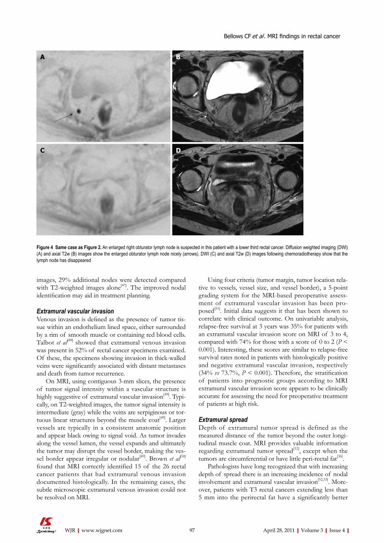
Figure 4 Same case as Figure 2. An enlarged right obturator lymph node is suspected in this patient with a lower third rectal cancer. Diffusion weighted imaging (DWI) (A) and axial T2w (B) images show the enlarged obturator lymph node nicely (arrows). DWI (C) and axial T2w (D) images following chemoradiotherapy show that the lymph node has disappeared
Bellows CF et al . MRI findings in rectal cancer
98 April 28, 2011|Volume 3|Issue 4|WJR|www.wjgnet.com
5-year cancer-related survival rate than do patients with pT3 tumors extending more than 5 mm beyond the rectal wall (85% vs 54%, respectively)[52-54]. Based on these ob-servations, neo-adjuvant therapy has not been routinely recommended for patients with pT3 carcinomas invading minimally (< 5 mm) into the perirectal tissue; instead pa-tients should undergo immediate surgery.
However, the use of < 5 mm as the determinant for the therapeutic decision is controversial and should be used with caution when determining treatment options for patients. Merkel et al[54] reported that tumors with less than 5 mm extramural spread may still have a 38%-43% rate of nodal metastasis. Moreover, if very advanced tu-mors and those with positive margins are excluded this prognostic factor no longer correlates with survival[55].
RE-STAGING AFTER CRTBecause of the increasing use of preoperative CRT, MRI is frequently repeated after treatment to re-stage the tu-mor, assess the response, determine whether it is operable, and establish the extent of surgical resection. However, early studies have questioned the accuracy of MRI in the post-CRT setting with a T-stage correlation of only 47%-54% and an N stage correlation of 64%-68%[46,56-59]. In the study by Kulkarni et al[60], MRI performed 6 wk post CRT overestimated the CRM involvement in 56% of cases, while T stages were over-staged in 38% and N stag-es in 4%. Over-staging was due to lack of discrimination between residual tumor and post-treatment changes, both appearing as a diffuse hypointense signal. Post-treatment changes are due to marked fibrosis of the bowel wall or to peritumoral infiltration of inflammatory cells and prolifer-ating vessels as confirmed by other investigators[46,57].
Recently, improved accuracy of MRI in the post-CRT setting was achieved by lengthening the interval after CRT. In the study by Johnston et al[61], the radiological T-stage determined on MRI obtained 10-11 wk after CRT was the same as the pathological T-stage on the resected speci-men in 14 out of 17 cases (88%) as compared to only a 59% agreement between the MRI and subsequent post-resection histopathology when the MRI was performed 6 wk after treatment. In this study, the pre-operative MRI showed ongoing response to CRT up to 12 wk after CRT, which has important clinical implications regarding the most appropriate time to operate.
Change in the surgical strategy may occur after CRT, es-pecially for patients who seem to exhibit a complete tumor response. For this subset of patients, transanal excision or non-operative treatment in selected circumstances maybe considered with good prognosis[62]. However, predicting the nodal status for these patients using imaging techniques becomes crucial, since the nodes are not removed at lo-cal excision. Importantly, the assessment of tumor spread to lymph nodes may be improved with the use of a novel nanoparticle contrast medium (ultra-small superparamag-netic iron oxide; USPIO)[63,64]. In a recent prospective mul-ticenter study, MRI performed after CRT for rectal cancer
using USPIO was able to improve the negative predictive value of the nodal status to 95%[65]. Unfortunately, this product is not currently commercially available.
SPECIAL CONSIDERATIONAdenocarcinoma of the lower rectumThe lower third of the rectum (less than 5 cm from the anal verge) lies below the level of the peritoneal reflec-tion. The majority of published series have shown that tumors arising in this anatomic location have the worst outcome, with local recurrence rates as high as 30%. This is due, in part, to the fact that compared to the tumors of the upper rectum, the surgical dissection for these low rectal tumors is less straightforward and is associated with a higher rate of perforation through the correct on-cologic plane compared to the tumors of the upper rec-tum. Anatomically, the mesorectum thins out towards the lower third of the rectum and disappears at the level of the internal sphincter. There is less space for the tumor to traverse before it reaches the surgical plane of resec-tion. Consequently, the CRM is more often positive in the surgical specimen for tumors located in the lower rectum, than for those located in the middle and upper rectum[32].
To overcome this shortcoming, a new operation, the “cylindrical” abdominoperineal resection (APR), has been pioneered in Europe[66,67]. In this operation, instead of following the mesorectum all the way to the levator mus-cles, the surgeon stops when the coccyx is visualized. The remaining dissection is performed from the perineum and is facilitated by the prone position. In the standard APR the perineal operator enters the levators anteriorly to the coccyx and the amount of levator muscle and is-chiorectal fat removed around the tumor is not standard-ized. Instead in the cylindrical APR once the levators and the coccyx are encountered the coccyx is excised and the levators are followed laterally to their origin from the lat-eral pelvic sidewalls where they are transected. In the case of anterior tumors, the posterior vaginal wall and part of the prostate are also removed en bloc[66]. One disadvantage of the technique is that it leaves a very large pelvic gap that can not be primarily closed and therefore a muscle flap reconstruction with the gracilis, the rectus abdominis or the gluteus maximus is often required[66]. Compar-ing 27 cylindrical to 99 conventional APRs, West et al[68] found a 70% increase in the amount of tissue removed around the tumor and no violation of the oncologic plane of dissection in the former group as well as a much lower rate of positive CRM 15% vs 40%, respectively. While many series advocate a wide perineal resection, and report low rates of local recurrence, these enhanced perineal resections have not become standard of care and prospective data are lacking[69]. Perineal wound infection, wound breakdown, and neurological dysfunctions are major problems for patients who receive radiation fol-lowed by abdominoperineal excision[69]. Primary closure with a flap overcomes some of these difficulties by bring-ing non-irradiated tissue into the perineal wound.
Bellows CF et al . MRI findings in rectal cancer
99 April 28, 2011|Volume 3|Issue 4|WJR|www.wjgnet.com
In a prospective study of 40 rectal tumors ≤ 5 cm from the dentate line from a single institution, MRI with intravenous contrast medium was universally successful in detecting invasion of the internal and external sphinc-ters[70]. For low-lying rectal tumors that are restricted to the rectal wall or internal sphincter, spare the external sphincter and levator ani, and are not amenable to local excision as determined by preoperative MRI, there are several advantages to performing inter-sphincteric APR. While the standard APR removes the whole sphincter complex in this procedure the dissection is carried out in the inter-sphincteric plane, the external sphincter is left in place and the perineal defect is easily closed by approxi-mating the external sphincter margins. This not only min-imizes the problems with wound healing and postopera-tive pain but also reduces risk of damage to the erigentes nerves, hypogastric plexus, and the neurovascular bundles of Walsh (containing the cavernous nerves). Damage to these nerves causes both sexual and bladder dysfunction in men and women. Overall, this represents an anatomic and well-standardized dissection, which decreases the risk of rectal perforation and positive margins.
Among the sphincter-saving options, the inter-sphinc-teric TME, which removes only the upper half of the internal sphincter, has been recently found to be a valid option for selected tumors of the lower rectum[71]. The procedure is similar to a TME with a manual colo-anal anastomosis. In a TME with a manual colo-anal ansto-mosis the surgeon performs a mucosectomy above the dentate line leaving the internal sphincter intact. In the inter-sphincteric TME the surgeon cuts through the in-ternal sphincter at the level of the dentate line, enters and dissects along the inter-sphinteric plane until establishing a connection with the abdominal operator. The proximal bowel is then manually anastomosed to the dentate line leaving the distal part of the internal sphincter intact. This allows one to achieve negative distal margins for tu-mors down to the ano-rectal junction while still providing good functional and oncologic results[71]. This procedure is indicated for T1-2 tumors that are well or moderately differentiated and for selected T3 tumors that have re-sponded well to CRT.
For some tumors of the lower rectum, a trans-perineal approach overcomes the lack of exposure due to the angled pelvic anatomy and the rectum being surrounded by the levators[72]. In this approach the external sphincter and the perineal body is exposed through a transverse incision be-tween the anus and the vagina or scrotum. This dissection allows the last 2-3 cm of rectum to be directly visualized.
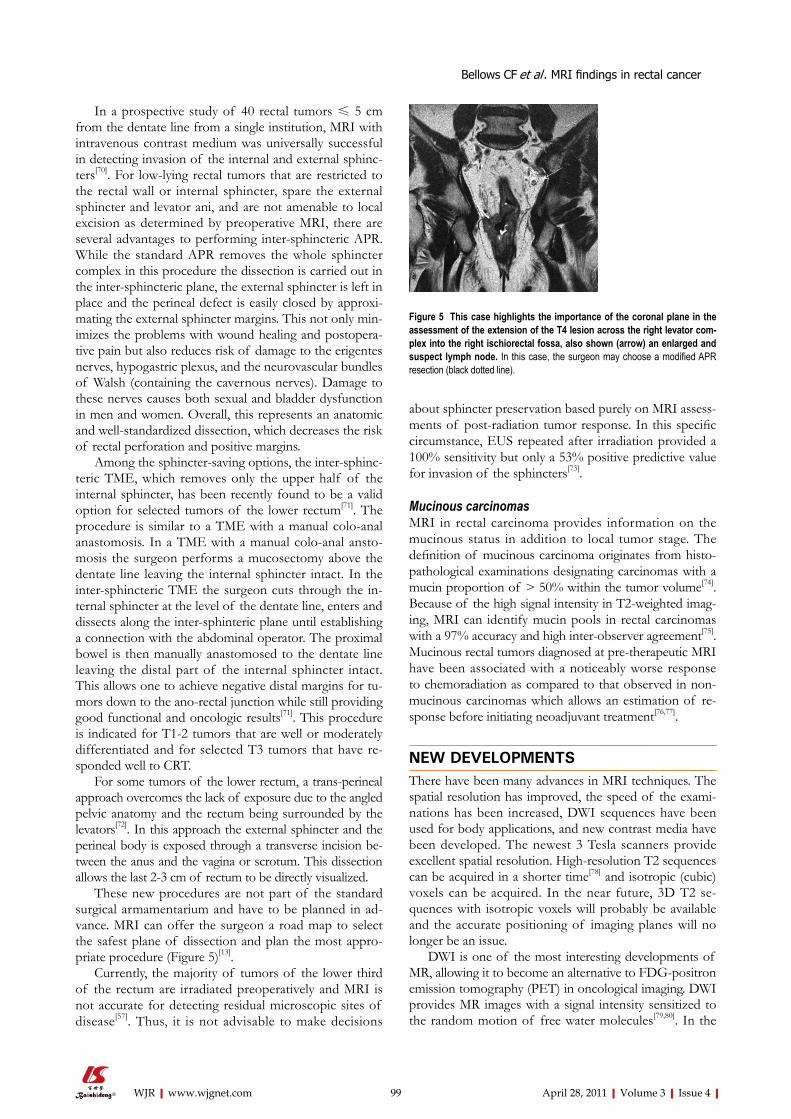
These new procedures are not part of the standard surgical armamentarium and have to be planned in ad-vance. MRI can offer the surgeon a road map to select the safest plane of dissection and plan the most appro-priate procedure (Figure 5)[13].
Currently, the majority of tumors of the lower third of the rectum are irradiated preoperatively and MRI is not accurate for detecting residual microscopic sites of disease[57]. Thus, it is not advisable to make decisions
about sphincter preservation based purely on MRI assess-ments of post-radiation tumor response. In this specific circumstance, EUS repeated after irradiation provided a 100% sensitivity but only a 53% positive predictive value for invasion of the sphincters[73].
Mucinous carcinomasMRI in rectal carcinoma provides information on the mucinous status in addition to local tumor stage. The definition of mucinous carcinoma originates from histo-pathological examinations designating carcinomas with a mucin proportion of > 50% within the tumor volume[74]. Because of the high signal intensity in T2-weighted imag-ing, MRI can identify mucin pools in rectal carcinomas with a 97% accuracy and high inter-observer agreement[75]. Mucinous rectal tumors diagnosed at pre-therapeutic MRI have been associated with a noticeably worse response to chemoradiation as compared to that observed in non-mucinous carcinomas which allows an estimation of re-sponse before initiating neoadjuvant treatment[76,77].
NEW DEVELOPMENTSThere have been many advances in MRI techniques. The spatial resolution has improved, the speed of the exami-nations has been increased, DWI sequences have been used for body applications, and new contrast media have been developed. The newest 3 Tesla scanners provide excellent spatial resolution. High-resolution T2 sequences can be acquired in a shorter time[78] and isotropic (cubic) voxels can be acquired. In the near future, 3D T2 se-quences with isotropic voxels will probably be available and the accurate positioning of imaging planes will no longer be an issue.
DWI is one of the most interesting developments of MR, allowing it to become an alternative to FDG-positron emission tomography (PET) in oncological imaging. DWI provides MR images with a signal intensity sensitized to the random motion of free water molecules[79,80]. In the
Figure 5 This case highlights the importance of the coronal plane in the assessment of the extension of the T4 lesion across the right levator com-plex into the right ischiorectal fossa, also shown (arrow) an enlarged and suspect lymph node. In this case, the surgeon may choose a modified APR resection (black dotted line).
Bellows CF et al . MRI findings in rectal cancer
100 April 28, 2011|Volume 3|Issue 4|WJR|www.wjgnet.com
rectum it is able to distinguish neoplastic from surround-ing normal tissue (Figure 2). As such it may help in the detection of small tumors. However, the major challenge of MRI for rectal cancer is to reliably define the response to neoadjuvant therapy. Predicting a tumor’s response to treatment can be of considerable clinical benefit. Inter-estingly, preliminary results indicate that DWI might be effective in predicting treatment outcomes and for detect-ing the early tumor response[81-83]. In quantitative DWI, the magnetic resonance signal arises from both intracellu-lar and extracellular compartments, and the result is given in terms of the apparent diffusion coefficient (ADC). Changes in the tumor ADC have been shown to corre-late with the development of intra-tumoral fibrosis after chemoradiation[83], histologically proven apoptotic cell death[84] and regression in tumor size after chemotherapy
and chemoradiation[81]. Other authors have supported the use of ADC values in combination with other MR imaging criteria in improving the discrimination between malignant and benign lymph nodes even after chemo-radiation[46,63,85] (Figure 4). The main limitation to DWI imaging today is the variability in ADC values that are obtained with different magnets and imaging protocols. Further studies will be necessary to prove the possible value of DWI on predicting therapy outcome.
There are also alternative imaging techniques. CT has so far had a limited role in the local staging of rectal can-cer. Today, perfusion imaging represents one of the most interesting fields of CT development. Perfusion imaging of large volumes is possible with multi-detector CT scan-ners. This technique has shown promise in predicting the response to neoadjuvant treatment[86]. CT perfusion data
DC
BA
FE
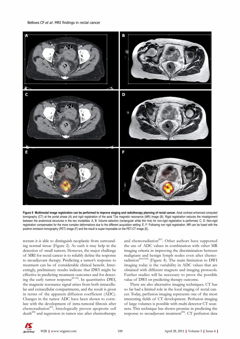
Figure 6 Multimodal image registration can be performed to improve staging and radiotherapy planning of rectal cancer. Axial contrast enhanced computed tomography (CT) at the portal phase (A) and rigid registration of the axial T2w magnetic resonance (MR) image (B). Rigid registration reduces the misalignment between the anatomical structures in the two modalities. A, B: Volume selection (rectangular white thin line) for non-rigid registration is performed; C, D: Non-rigid registration compensates for the more complex deformations due to the different acquisition setting; E, F: Following non rigid registration, MR can be fused with the positron emission tomography (PET) image (F) and the result is super-imposable on the PET-CT image (E).
Bellows CF et al . MRI findings in rectal cancer
101 April 28, 2011|Volume 3|Issue 4|WJR|www.wjgnet.com
cannot currently be obtained with dynamic contrast-en-hanced MR[87] and this represents a strong point in favor of CT.
PET and CT-PET scans are used mainly in the as-sessment of metastatic rectal cancer and local recurrence. Sequential determination of fluorodeoxyglucose uptake on PET/CT has proved useful in differentiating respon-sive from nonresponsive tumors during and at the end of neoadjuvant therapy[88]. However, radionuclide techniques have limitations, such as low spatial resolution and high cost. Large studies are needed to establish the most effec-tive morphologic and functional imaging modalities for post-neoadjuvant therapy restaging of rectal cancer[86].
Over the last several years many strategies have been developed to overcome the limitations of radiotherapy planning using noncontrast-enhanced CT. Radiotherapy guided by MRI is possible using strategies that allow fu-sion and/or co-registration of MR images with those from other imaging techniques[88]. PET-CT, contrast en-hanced CT, and non contrast enhanced CT and MR im-ages can all be fused together to improve the assessment of rectal lesions and radiotherapy planning[89-91] (Figure 6). However, PET-guided radiotherapy has not yet provided a clear advantage. Better delineation of pelvic anatomy and pathology will become progressively more important as radiotherapy protocols are developed that include a boost on the gross tumor volume with documented im-provement in patient outcome[92].
CONCLUSIONRectal cancer is a global disease associated with poor out-comes if not properly staged and treated. The increased use of preoperative chemoradiation and refinement of surgical techniques have led to a greater proportion of patients being considered for curative resection. New surgical options exist for these patients in the form of sphincter saving resection or transanal excision in se-lected circumstances. For the vast majority of rectal car-cinomas, MRI is currently the most accurate modality on which to base treatment decisions for patients with rectal cancer. Traditionally, the decision to apply preoperative treatment for rectal cancer patients has been based on the T- and N-stage. Lately, other MRI findings such as the radial distance of the tumor to the CRM and extramural vascular invasion score have been identified as important risk factors for local failure and survival. We strongly believe that every center that treats patients with rectal cancer should develop a multidisciplinary team featuring a description of the MRI findings and their implementa-tion in the treatment strategy with the aim of increasing resectability, reducing the local recurrence and treatment morbidity, and improving the quality of life.
REFERENCES1 Jemal A, Siegel R, Xu J, Ward E. Cancer statistics, 2010. CA
Cancer J Clin 2010; 60: 277-300
2 Rich T, Gunderson LL, Lew R, Galdibini JJ, Cohen AM, Don-aldson G. Patterns of recurrence of rectal cancer after poten-tially curative surgery. Cancer 1983; 52: 1317-1329
3 NCCN clinical practice guidelines in Oncology. accessed on October 13, 2010. Available from: URL: http://www.nccn.org/professionals/physician_gls/f_guidelines.asp
4 Valentini V, Aristei C, Glimelius B, Minsky BD, Beets-Tan R, Borras JM, Haustermans K, Maingon P, Overgaard J, Pahl-man L, Quirke P, Schmoll HJ, Sebag-Montefiore D, Taylor I, Van Cutsem E, Van de Velde C, Cellini N, Latini P. Multidis-ciplinary Rectal Cancer Management: 2nd European Rectal Cancer Consensus Conference (EURECA-CC2). Radiother Oncol 2009; 92: 148-163
5 Kosaras B, Kirschner DA. Radial component of CNS myelin: junctional subunit structure and supramolecular assembly. J Neurocytol 1990; 19: 187-199
6 Bosset JF. Adjuvant treatment of rectal cancer: improving patient selection. Gastrointest Cancer Res 2008; 2: 37-38
7 Garcia-Aguilar J, Pollack J, Lee SH, Hernandez de Anda E, Mellgren A, Wong WD, Finne CO, Rothenberger DA, Madoff RD. Accuracy of endorectal ultrasonography in preoperative staging of rectal tumors. Dis Colon Rectum 2002; 45: 10-15
8 Kauer WK, Prantl L, Dittler HJ, Siewert JR. The value of en-dosonographic rectal carcinoma staging in routine diagnos-tics: a 10-year analysis. Surg Endosc 2004; 18: 1075-1078
9 Ju H, Xu D, Li D, Chen G, Shao G. Comparison between en-doluminal ultrasonography and spiral computerized tomog-raphy for the preoperative local staging of rectal carcinoma. Biosci Trends 2009; 3: 73-76
10 Gagliardi G, Bayar S, Smith R, Salem RR. Preoperative stag-ing of rectal cancer using magnetic resonance imaging with external phase-arrayed coils. Arch Surg 2002; 137: 447-451
11 Ginsberg GG, Lewis JH, Gallagher JE, Fleischer DE, al-Kawas FH, Nguyen CC, Mundt DJ, Benjamin SB. Diazepam versus midazolam for colonoscopy: a prospective evaluation of predicted versus actual dosing requirements. Gastrointest Endosc 1992; 38: 651-656
12 Brown G, Richards CJ, Newcombe RG, Dallimore NS, Rad-cliffe AG, Carey DP, Bourne MW, Williams GT. Rectal car-cinoma: thin-section MR imaging for staging in 28 patients. Radiology 1999; 211: 215-222
13 Shihab OC, Heald RJ, Rullier E, Brown G, Holm T, Quirke P, Moran BJ. Defining the surgical planes on MRI improves surgery for cancer of the low rectum. Lancet Oncol 2009; 10: 1207-1211
14 Brown G, Daniels IR, Richardson C, Revell P, Peppercorn D, Bourne M. Techniques and trouble-shooting in high spatial resolution thin slice MRI for rectal cancer. Br J Radiol 2005; 78: 245-251
15 Soyer P, Lagadec M, Sirol M, Dray X, Duchat F, Vignaud A, Fargeaudou Y, Placé V, Gault V, Hamzi L, Pocard M, Boudiaf M. Free-breathing diffusion-weighted single-shot echo-planar MR imaging using parallel imaging (GRAPPA 2) and high b value for the detection of primary rectal adenocarcinoma. Cancer Imaging 2010; 10: 32-39
16 Suzuki C, Torkzad MR, Tanaka S, Palmer G, Lindholm J, Holm T, Blomqvist L. The importance of rectal cancer MRI protocols on interpretation accuracy. World J Surg Oncol 2008; 6: 89
17 Akasu T, Iinuma G, Takawa M, Yamamoto S, Muramatsu Y, Moriyama N. Accuracy of high-resolution magnetic reso-nance imaging in preoperative staging of rectal cancer. Ann Surg Oncol 2009; 16: 2787-2794
18 Hoeffel C, Marra MD, Azizi L, Tran Van K, Crema MD, Lewin M, Arrivé L, Tubiana JM. [External phased-array MR imaging preoperative assessment of rectal cancer]. J Radiol 2006; 87: 1821-1830
19 Mellgren A, Sirivongs P, Rothenberger DA, Madoff RD, García-Aguilar J. Is local excision adequate therapy for early rectal cancer? Dis Colon Rectum 2000; 43: 1064-1071; discus-
Bellows CF et al . MRI findings in rectal cancer
102 April 28, 2011|Volume 3|Issue 4|WJR|www.wjgnet.com
sion 1071-107420 Bueno CG, de Barros JJ, Terres JS, Pereira DS Júnior. [Man-
dibular osteoradionecrosis. Clinical study and presentation of two cases]. Quintessencia 1978; 5: 55-73
21 Improved survival with preoperative radiotherapy in resect-able rectal cancer. Swedish Rectal Cancer Trial. N Engl J Med 1997; 336: 980-987
22 Bailey HR, Huval WV, Max E, Smith KW, Butts DR, Zamora LF. Local excision of carcinoma of the rectum for cure. Surgery 1992; 111: 555-561
23 Sauer R, Becker H, Hohenberger W, Rödel C, Wittekind C, Fietkau R, Martus P, Tschmelitsch J, Hager E, Hess CF, Karstens JH, Liersch T, Schmidberger H, Raab R. Preopera-tive versus postoperative chemoradiotherapy for rectal can-cer. N Engl J Med 2004; 351: 1731-1740
24 Bosset JF, Collette L, Calais G, Mineur L, Maingon P, Rados-evic-Jelic L, Daban A, Bardet E, Beny A, Ollier JC. Chemo-therapy with preoperative radiotherapy in rectal cancer. N Engl J Med 2006; 355: 1114-1123
25 Gérard JP, Conroy T, Bonnetain F, Bouché O, Chapet O, Clo-son-Dejardin MT, Untereiner M, Leduc B, Francois E, Maurel J, Seitz JF, Buecher B, Mackiewicz R, Ducreux M, Bedenne L. Preoperative radiotherapy with or without concurrent fluorouracil and leucovorin in T3-4 rectal cancers: results of FFCD 9203. J Clin Oncol 2006; 24: 4620-4625
26 Beets-Tan RG, Beets GL, Vliegen RF, Kessels AG, Van Boven H, De Bruine A, von Meyenfeldt MF, Baeten CG, van En-gelshoven JM. Accuracy of magnetic resonance imaging in prediction of tumour-free resection margin in rectal cancer surgery. Lancet 2001; 357: 497-504
27 Brown G, Radcliffe AG, Newcombe RG, Dallimore NS, Bourne MW, Williams GT. Preoperative assessment of prog-nostic factors in rectal cancer using high-resolution magnetic resonance imaging. Br J Surg 2003; 90: 355-364
28 Videhult P, Smedh K, Lundin P, Kraaz W. Magnetic reso-nance imaging for preoperative staging of rectal cancer in clinical practice: high accuracy in predicting circumferential margin with clinical benefit. Colorectal Dis 2007; 9: 412-419
29 Sebag-Montefiore D. Treatment of T4 tumours: the role of ra-diotherapy. Colorectal Dis 2003; 5: 432-435
30 Bipat S, Glas AS, Slors FJ, Zwinderman AH, Bossuyt PM, Stoker J. Rectal cancer: local staging and assessment of lymph node involvement with endoluminal US, CT, and MR imaging--a meta-analysis. Radiology 2004; 232: 773-783
31 Nagtegaal ID, Gosens MJ, Marijnen CA, Rutten HJ, van de Velde CJ, van Krieken JH. Combinations of tumor and treat-ment parameters are more discriminative for prognosis than the present TNM system in rectal cancer. J Clin Oncol 2007; 25: 1647-1650
32 Nagtegaal ID, Quirke P. What is the role for the circumfer-ential margin in the modern treatment of rectal cancer? J Clin Oncol 2008; 26: 303-312
33 Mulla M, Deb R, Singh R. MRI in T staging of rectal cancer: How effective is it? Indian J Radiol Imaging 2010; 20: 118-121
34 Wieder HA, Rosenberg R, Lordick F, Geinitz H, Beer A, Becker K, Woertler K, Dobritz M, Siewert JR, Rummeny EJ, Stollfuss JC. Rectal cancer: MR imaging before neoadju-vant chemotherapy and radiation therapy for prediction of tumor-free circumferential resection margins and long-term survival. Radiology 2007; 243: 744-751
35 Nagtegaal ID, Marijnen CA, Kranenbarg EK, van de Velde CJ, van Krieken JH. Circumferential margin involvement is still an important predictor of local recurrence in rectal car-cinoma: not one millimeter but two millimeters is the limit. Am J Surg Pathol 2002; 26: 350-357
36 Kim YW, Cha SW, Pyo J, Kim NK, Min BS, Kim MJ, Kim H. Factors related to preoperative assessment of the circumfer-ential resection margin and the extent of mesorectal invasion by magnetic resonance imaging in rectal cancer: a prospec-tive comparison study. World J Surg 2009; 33: 1952-1960
37 Frasson M, Garcia-Granero E, Roda D, Flor-Lorente B, Roselló S, Esclapez P, Faus C, Navarro S, Campos S, Cervantes A. Preoperative chemoradiation may not always be needed for patients with T3 and T2N+ rectal cancer. Cancer 2011; Epub ahead of print
38 Baik SH, Kim NK, Lee YC, Kim H, Lee KY, Sohn SK, Cho CH. Prognostic significance of circumferential resection mar-gin following total mesorectal excision and adjuvant chemo-radiotherapy in patients with rectal cancer. Ann Surg Oncol 2007; 14: 462-469
39 Sebag-Montefiore D, Stephens RJ, Steele R, Monson J, Grieve R, Khanna S, Quirke P, Couture J, de Metz C, Myint AS, Bes-sell E, Griffiths G, Thompson LC, Parmar M. Preoperative radiotherapy versus selective postoperative chemoradio-therapy in patients with rectal cancer (MRC CR07 and NCIC-CTG C016): a multicentre, randomised trial. Lancet 2009; 373: 811-820
40 Strassburg J, Junginger T, Trinh T, Püttcher O, Oberholzer K, Heald RJ, Hermanek P. Magnetic resonance imaging (MRI)-based indication for neoadjuvant treatment of rectal carci-noma and the surrogate endpoint CRM status. Int J Colorectal Dis 2008; 23: 1099-1107
41 Kapiteijn E, Marijnen CA, Nagtegaal ID, Putter H, Steup WH, Wiggers T, Rutten HJ, Pahlman L, Glimelius B, van Krieken JH, Leer JW, van de Velde CJ. Preoperative radio-therapy combined with total mesorectal excision for resect-able rectal cancer. N Engl J Med 2001; 345: 638-646
42 Beets-Tan RG, Beets GL. Rectal cancer: review with empha-sis on MR imaging. Radiology 2004; 232: 335-346
43 Moriya Y, Sugihara K, Akasu T, Fujita S. Importance of ex-tended lymphadenectomy with lateral node dissection for advanced lower rectal cancer. World J Surg 1997; 21: 728-732
44 Grubnic S, Vinnicombe SJ, Norman AR, Husband JE. MR evaluation of normal retroperitoneal and pelvic lymph nodes. Clin Radiol 2002; 57: 193-200; discussion 201-204
45 Brown G, Richards CJ, Bourne MW, Newcombe RG, Rad-cliffe AG, Dallimore NS, Williams GT. Morphologic pre-dictors of lymph node status in rectal cancer with use of high-spatial-resolution MR imaging with histopathologic comparison. Radiology 2003; 227: 371-377
46 Lambregts DM, Beets GL, Maas M, Kessels AG, Bakers FC, Cappendijk VC, Engelen SM, Lahaye MJ, de Bruïne AP, Lam-mering G, Leiner T, Verwoerd JL, Wildberger JE, Beets-Tan RG. Accuracy of gadofosveset-enhanced MRI for nodal stag-ing and restaging in rectal cancer. Ann Surg 2011; 253: 539-545
47 Mir N, Sohaib SA, Collins D, Koh DM. Fusion of high b-value diffusion-weighted and T2-weighted MR images improves identification of lymph nodes in the pelvis. J Med Imaging Ra-diat Oncol 2010; 54: 358-364
48 Talbot IC, Ritchie S, Leighton MH, Hughes AO, Bussey HJ, Morson BC. The clinical significance of invasion of veins by rectal cancer. Br J Surg 1980; 67: 439-442
49 Smith NJ, Shihab O, Arnaout A, Swift RI, Brown G. MRI for detection of extramural vascular invasion in rectal cancer. AJR Am J Roentgenol 2008; 191: 1517-1522
50 Brown G, Daniels IR. Preoperative staging of rectal cancer: the MERCURY research project. Recent Results Cancer Res 2005; 165: 58-74
51 Smith NJ, Barbachano Y, Norman AR, Swift RI, Abulafi AM, Brown G. Prognostic significance of magnetic resonance im-aging-detected extramural vascular invasion in rectal cancer. Br J Surg 2008; 95: 229-236
52 Dukes CE, Bussey HJ. The spread of rectal cancer and its ef-fect on prognosis. Br J Cancer 1958; 12: 309-320
53 Cawthorn SJ, Parums DV, Gibbs NM, A'Hern RP, Caffarey SM, Broughton CI, Marks CG. Extent of mesorectal spread and involvement of lateral resection margin as prognostic fac-tors after surgery for rectal cancer. Lancet 1990; 335: 1055-1059
54 Merkel S, Mansmann U, Papadopoulos T, Wittekind C, Ho-henberger W, Hermanek P. The prognostic inhomogeneity
Bellows CF et al . MRI findings in rectal cancer
103 April 28, 2011|Volume 3|Issue 4|WJR|www.wjgnet.com
of colorectal carcinomas Stage III: a proposal for subdivision of Stage III. Cancer 2001; 92: 2754-2759
55 Picon AI, Moore HG, Sternberg SS, Minsky BD, Paty PB, Blumberg D, Quan SH, Wong WD, Cohen AM, Guillem JG. Prognostic significance of depth of gross or microscopic peri-rectal fat invasion in T3 N0 M0 rectal cancers following sharp mesorectal excision and no adjuvant therapy. Int J Colorectal Dis 2003; 18: 487-492
56 Kim DJ, Kim JH, Lim JS, Yu JS, Chung JJ, Kim MJ, Kim KW. Restaging of Rectal Cancer with MR Imaging after Concurrent Chemotherapy and Radiation Therapy. Radiographics 2010; 30: 503-516
57 Kuo LJ, Chern MC, Tsou MH, Liu MC, Jian JJ, Chen CM, Chung YL, Fang WT. Interpretation of magnetic resonance imaging for locally advanced rectal carcinoma after preopera-tive chemoradiation therapy. Dis Colon Rectum 2005; 48: 23-28
58 Allen SD, Padhani AR, Dzik-Jurasz AS, Glynne-Jones R. Rectal carcinoma: MRI with histologic correlation before and after chemoradiation therapy. AJR Am J Roentgenol 2007; 188: 442-451
59 Vliegen RF, Beets GL, Lammering G, Dresen RC, Rutten HJ, Kessels AG, Oei TK, de Bruïne AP, van Engelshoven JM, Beets-Tan RG. Mesorectal fascia invasion after neoadjuvant chemotherapy and radiation therapy for locally advanced rectal cancer: accuracy of MR imaging for prediction. Radiol-ogy 2008; 246: 454-462
60 Kulkarni T, Gollins S, Maw A, Hobson P, Byrne R, Widdow-son D. Magnetic resonance imaging in rectal cancer down-staged using neoadjuvant chemoradiation: accuracy of pre-diction of tumour stage and circumferential resection margin status. Colorectal Dis 2008; 10: 479-489
61 Johnston DF, Lawrence KM, Sizer BF, Arulampalam TH, Motson RW, Dove E, Lacey N. Locally advanced rectal can-cer: histopathological correlation and predictive accuracy of serial MRI after neoadjuvant chemotherapy. Br J Radiol 2009; 82: 332-336
62 Borschitz T, Wachtlin D, Möhler M, Schmidberger H, Jung-inger T. Neoadjuvant chemoradiation and local excision for T2-3 rectal cancer. Ann Surg Oncol 2008; 15: 712-720
63 Lahaye MJ, Beets GL, Engelen SM, Kessels AG, de Bruïne AP, Kwee HW, van Engelshoven JM, van de Velde CJ, Beets-Tan RG. Locally advanced rectal cancer: MR imaging for restaging after neoadjuvant radiation therapy with concomi-tant chemotherapy. Part II. What are the criteria to predict involved lymph nodes? Radiology 2009; 252: 81-91
64 Engelen SM, Beets-Tan RG, Lahaye MJ, Kessels AG, Beets GL. Location of involved mesorectal and extramesorectal lymph nodes in patients with primary rectal cancer: preop-erative assessment with MR imaging. Eur J Surg Oncol 2008; 34: 776-781
65 Engelen SM, Beets-Tan RG, Lahaye MJ, Lammering G, Jan-sen RL, van Dam RM, Konsten J, Leijtens JW, van de Velde CJ, Beets GL. MRI after chemoradiotherapy of rectal cancer: a useful tool to select patients for local excision. Dis Colon Rec-tum 2010; 53: 979-986
66 Marr R, Birbeck K, Garvican J, Macklin CP, Tiffin NJ, Par-sons WJ, Dixon MF, Mapstone NP, Sebag-Montefiore D, Scott N, Johnston D, Sagar P, Finan P, Quirke P. The modern abdominoperineal excision: the next challenge after total me-sorectal excision. Ann Surg 2005; 242: 74-82
67 Holm T, Ljung A, Häggmark T, Jurell G, Lagergren J. Ex-tended abdominoperineal resection with gluteus maximus flap reconstruction of the pelvic floor for rectal cancer. Br J Surg 2007; 94: 232-238
68 West NP, Finan PJ, Anderin C, Lindholm J, Holm T, Quirke P. Evidence of the oncologic superiority of cylindrical abdomi-noperineal excision for low rectal cancer. J Clin Oncol 2008; 26: 3517-3522
69 Guillem JG, Minsky BD. Extended perineal resection of dis-tal rectal cancers: surgical advance, increased utilization of
neoadjuvant therapies, proper patient selection or all of the above? J Clin Oncol 2008; 26: 3481-3482
70 Holzer B, Urban M, Hölbling N, Feil W, Novi G, Hruby W, Rosen HR, Schiessel R. Magnetic resonance imaging predicts sphincter invasion of low rectal cancer and influences selec-tion of operation. Surgery 2003; 133: 656-661
71 Rullier E, Laurent C, Bretagnol F, Rullier A, Vendrely V, Zerbib F. Sphincter-saving resection for all rectal carcinomas: the end of the 2-cm distal rule. Ann Surg 2005; 241: 465-469
72 Williams NS, Murphy J, Knowles CH. Anterior Perineal PlanE for Ultra-low Anterior Resection of the Rectum (the APPEAR technique): a prospective clinical trial of a new pro-cedure. Ann Surg 2008; 247: 750-758
73 Assenat E, Thézenas S, Samalin E, Bibeau F, Portales F, Azria D, Quenet F, Rouanet P, Saint Aubert B, Senesse P. The value of endoscopic rectal ultrasound in predicting the lateral clear-ance and outcome in patients with lower-third rectal adeno-carcinoma. Endoscopy 2007; 39: 309-313
74 Hamilton SR, Aaltonen LA. World Health Organization Clas-sification of Tumours. Pathology and Genetics of Tumours of the Digestive System. Lyon: IARC Press, 2000: 105-120
75 Kim MJ, Park JS, Park SI, Kim NK, Kim JH, Moon HJ, Park YN, Kim WH. Accuracy in differentiation of mucinous and nonmucinous rectal carcinoma on MR imaging. J Comput As-sist Tomogr 2003; 27: 48-55
76 Grillo-Ruggieri F, Mantello G, Berardi R, Cardinali M, Fenu F, Iovini G, Montisci M, Fabbietti L, Marmorale C, Guerrieri M, Saba V, Bearzi I, Mattioli R, Bonsignori M, Cascinu S. Mucinous rectal adenocarcinoma can be associated to tumor downstaging after preoperative chemoradiotherapy. Dis Co-lon Rectum 2007; 50: 1594-1603
77 Oberholzer K, Menig M, Kreft A, Schneider A, Junginger T, Heintz A, Kreitner KF, Hötker AM, Hansen T, Düber C, Schmidberger H. Rectal Cancer: Mucinous Carcinoma on Magnetic Resonance Imaging Indicates Poor Response to Neoadjuvant Chemoradiation. Int J Radiat Oncol Biol Phys 2011; Epub ahead of print
78 Kim H, Lim JS, Choi JY, Park J, Chung YE, Kim MJ, Choi E, Kim NK, Kim KW. Rectal cancer: comparison of accuracy of local-regional staging with two- and three-dimensional pre-operative 3-T MR imaging. Radiology 2010; 254: 485-492
79 Torricelli P, Pecchi A, Luppi G, Romagnoli R. Gadolinium-enhanced MRI with dynamic evaluation in diagnosing the lo-cal recurrence of rectal cancer. Abdom Imaging 2003; 28: 19-27
80 Kremser C, Judmaier W, Hein P, Griebel J, Lukas P, de Vries A. Preliminary results on the influence of chemoradiation on apparent diffusion coefficients of primary rectal carcinoma measured by magnetic resonance imaging. Strahlenther Onkol 2003; 179: 641-649
81 Dzik-Jurasz A, Domenig C, George M, Wolber J, Padhani A, Brown G, Doran S. Diffusion MRI for prediction of response of rectal cancer to chemoradiation. Lancet 2002; 360: 307-308
82 Barbaro B, Vitale R, Leccisotti L, Vecchio FM, Santoro L, Val-entini V, Coco C, Pacelli F, Crucitti A, Persiani R, Bonomo L. Restaging locally advanced rectal cancer with MR imaging after chemoradiation therapy. Radiographics 2010; 30: 699-716
83 Hein PA, Kremser C, Judmaier W, Griebel J, Pfeiffer KP, Kreczy A, Hug EB, Lukas P, DeVries AF. Diffusion-weight-ed magnetic resonance imaging for monitoring diffusion changes in rectal carcinoma during combined, preoperative chemoradiation: preliminary results of a prospective study. Eur J Radiol 2003; 45: 214-222
84 Chinnaiyan AM, Prasad U, Shankar S, Hamstra DA, Sha-naiah M, Chenevert TL, Ross BD, Rehemtulla A. Combined effect of tumor necrosis factor-related apoptosis-inducing ligand and ionizing radiation in breast cancer therapy. Proc Natl Acad Sci USA 2000; 97: 1754-1759
85 Ono K, Ochiai R, Yoshida T, Kitagawa M, Omagari J, Ko-bayashi H, Yamashita Y. Comparison of diffusion-weighted MRI and 2-[fluorine-18]-fluoro-2-deoxy-D-glucose positron
Bellows CF et al . MRI findings in rectal cancer
104 April 28, 2011|Volume 3|Issue 4|WJR|www.wjgnet.com
emission tomography (FDG-PET) for detecting primary colorectal cancer and regional lymph node metastases. J Magn Reson Imaging 2009; 29: 336-340
86 Bellomi M, Travaini LL. Imaging as a surveillance tool in rec-tal cancer. Expert Rev Med Devices 2010; 7: 99-112
87 Kierkels RG, Backes WH, Janssen MH, Buijsen J, Beets-Tan RG, Lambin P, Lammering G, Oellers MC, Aerts HJ. Compar-ison between perfusion computed tomography and dynamic contrast-enhanced magnetic resonance imaging in rectal can-cer. Int J Radiat Oncol Biol Phys 2010; 77: 400-408
88 Schiepers C, Haustermans K, Geboes K, Filez L, Bormans G, Penninckx F. The effect of preoperative radiation therapy on glucose utilization and cell kinetics in patients with primary rectal carcinoma. Cancer 1999; 85: 803-811
89 Anderson H, Singh N, Miles K. Tumour response evaluation with fluorodeoxyglucose positron emission tomography: re-search technique or clinical tool? Cancer Imaging 2010; 10 Spec
no A: S68-S7290 Bassi MC, Turri L, Sacchetti G, Loi G, Cannillo B, La Mattina
P, Brambilla M, Inglese E, Krengli M. FDG-PET/CT imaging for staging and target volume delineation in preoperative conformal radiotherapy of rectal cancer. Int J Radiat Oncol Biol Phys 2008; 70: 1423-1426
91 Garlaschi A, Bacigalupo LE, Biscaldi E, Chiusano G, Basso C, Rollandi GA. Multi-modal non-rigid image registration of MR and PET-CT for staging and therapy of rectal adenocar-cinoma. Presented as a Scientific Poster at the Radiological Society of North America November 29 - December 4, 2009; Chicago, IL. Abstract Book, 921
92 Roels S, Slagmolen P, Nuyts J, Lee JA, Loeckx D, Maes F, Vandecaveye V, Stroobants S, Ectors N, Penninckx F, Haus-termans K. Biological image-guided radiotherapy in rectal cancer: challenges and pitfalls. Int J Radiat Oncol Biol Phys 2009; 75: 782-790
S- Editor Cheng JX L- Editor O’Neill M E- Editor Zheng XM
Bellows CF et al . MRI findings in rectal cancer