Embed Size (px)
Citation preview
ORIGINAL ARTICLE
Clinical results of implant placement in resorbed ridges usingsimultaneous guided bone regeneration: a multicenter case series
Ioannis Konstantinidis & Tarun Kumar & Udatta Kher &
Panagiotis D. Stanitsas & James E. Hinrichs &
Georgios A. Kotsakis
Received: 6 January 2014 /Accepted: 26 May 2014# Springer-Verlag Berlin Heidelberg 2014
AbstractObjectives The purpose of this case series was to evaluate thenew bone formation following guided bone regeneration(GBR) with a calcium phosphosilicate (CPS), alloplastic boneputty at peri-implant dehiscence defects and to assess survivalrate of implants placed in the augmented sites after 12 monthsof function.Materials and methods Implants were placed in patientsexhibiting Seibert class I ridge defects resulting in peri-implant dehiscence defects. The defects were treated follow-ing GBR principles with the use of a CPS alloplastic bonegraft putty in combination either with a collagen membrane ora titanium mesh. The height of each bony dehiscence wasclinicallymeasured at the time of implant placement and againduring second-stage surgery. The percentage of completedefect coverage, frequency of adverse events, and risk factorsfor residual defect were determined.Results Thirty-six implants were placed in 26 patients.Twenty-seven of the 36 sites employed a collagen membrane
in conjunction with the CPS while the remaining nine sitesutilized a titanium membrane. Mean gain in bone height was3.23±2.04 mm, with 75 % of the peri-implant defects achiev-ing complete regeneration. A negative correlation was identi-fied between patient age and complete coverage of the peri-implant defect (p=0.026). The implant survival rate at12 months was 97.22 %.Conclusion Use of CPS bone putty during delayed implantplacement at peri-implant dehiscence sites either in combina-tion with a collagen membrane or a titanium mesh results inpredictable defect coverage.Clinical relevance The handling characteristics of CPS puttymay simplify GBR protocol. Implants placed in conjunctionwith GBR have a very good survival rate after 1 year offollow-up.
Keywords Calcium phosphosilicate (CPS) . Alloplastic boneputty . Guided bone regeneration (GBR) . Peri-implantdehiscence defects
Introduction
Implant placement frequently has to be performed in deficientridges. As alveolar ridge resorption physiologically occursfollowing each tooth extraction, ridge dimensions may beunfavorable for implant placement in the ideal restorativelydictated location [1, 2]. The use of guided bone regeneration(GBR) for the treatment of ridge deficiencies is considered asafe and predictable treatment modality [3, 4].
However, this therapeutic approach introduces a separatesurgical procedure and an extended healing period that signif-icantly delays the time of implant rehabilitation for the patient.The placement of implants in residual ridges in conjunctionwith simultaneous guided bone regeneration has been sug-gested in order to minimize total treatment time and allow for
I. Konstantinidis (*)Department of Prosthodontics, TU Dresden, Dresden, Germanye-mail: [email protected]
T. KumarBapuji Implant Centre, Bapuji Dental College & Hospital,Davangere, India
U. KherDepartment of Implantology, SDM College of Dental Sciences,Dharwad, India
P. D. StanitsasDepartment of Computer Science and Engineering, University ofMinnesota, Minneapolis, USA
J. E. Hinrichs :G. A. KotsakisAdvanced Education Program in Periodontology, University ofMinnesota, Minneapolis, USA
Clin Oral InvestDOI 10.1007/s00784-014-1268-4
implant placement in compromised sites [5–7]. Various typesof bone grafts have been used for this indication with nobiomaterial showing any significant advantage over anotherin terms of bone regeneration and implant survival [8].
A new generation of putty bone substitutes that displayenhanced handling characteristics has recently received sig-nificant attention. Putty bone substitutes are usually composedof similar active ingredients as particulate ones, but areenriched with a binder/carrier that provides a unique viscousconsistency to the graft. The use of demineralized bone matrixputty and putty xenograft bone substitutes have been success-fully employed in oral surgical procedures such as ridgepreservation and sinus augmentation [9–11].
The use of an alloplastic putty bone substitute has been alsoshown to yield promising results in vivo both in animals andhumans [12–14]. This alloplastic putty bone substitute bioma-terial is composed of calcium phosphosilicate (CPS) particlesembedded in a synthetic absorbable binder composed of glyc-erol and polyethylene glycol with a 70/30 % ratio of activeingredient to binder, respectively. The active ingredient (CPSparticles) has a bimodal particle distribution. The smallerparticles resorb quicker to give the initial burst of calciumand phosphate ions and the larger particles continue to resorbover time, resulting in sustained stimulation in the area. Invitro studies have shown that CPS putty promotes the differ-entiation of osteoprogenitor cells into mature osteoblasts,resulting in bone regeneration in the grafted area [15, 16].
Up to date, no evaluation of the clinical efficacy of the CPSputty in guided bone regeneration has been reported in theliterature.
The purpose of this case series was to clinically evaluatethe new bone formation at peri-implant dehiscence defects byutilizing of a calcium phosphosilicate (CPS), alloplastic boneputty for guided bone regeneration (GBR). The gain in boneheight following GBR was set as the primary outcome. Ad-verse events and survival rate of implants placed in augmentedsites after 12 months of function were recorded as secondaryoutcomes.
Materials and methods
In this case series study, patients that presented in three dif-ferent centers for implant placement were screened accordingto standard clinical protocols of each center. If the treatmentplan included delayed implant placement with simultaneousguided bone regeneration, verbal and written consent wasobtained and the patients were enrolled in the study. Allpatients were treated according to the Declaration of Helsinki(1975), as revised in 2000. Treatment planning and the surgi-cal protocol did not deviate from routine protocols of eachcenter for study purposes. Inclusion criteria for this study wereas follows:
& Seibert class I ridge deficiencies [17]& Bone dehiscence resulting in implant thread exposure
during implant placement in healed ridges& 18–65 years of age& Noncontributory medical history& Tooth extraction at least 6 months prior to the screening
appointment.
Exclusion criteria were as follows:
& Unwillingness to sign the informed consent& History of long-term therapy with medication that may
affect bone healing (oral or IV bisphosphonates, cortico-steroids, NSAIDS, etc.)
& Medical history that contraindicated oral surgicaltreatment
& Use of tobacco& Pregnancy for female participants& Sites where GBR was deemed unnecessary following
implant placement& Circumferential (horizontal) bone defects around the
placed implants
The defects were treated following GBR principles withthe use of a CPS alloplastic bone graft putty in combinationeither with a collagen membrane, or a titanium mesh. Prior toinitiation of the study, a consensus was reached among thethree implant surgeons in regards to the type of measurements.Based on study protocol all surgeons were asked to use thesame type of probe (15 UNC Color-Coded Probe, Hu-Friedy,Chicago, IL) for intraoperative measurements in order tominimize interclinician variability. Each implant surgeonwas given freedom to choose the type of barrier used inconjunction with the CPS putty according to standard clinicalprotocols of each clinic.
Surgical procedure
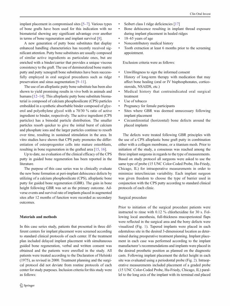
Prior to initiation of the surgical procedure patients wereinstructed to rinse with 0.12 % chlorhexidine for 30 s. Fol-lowing local anesthesia, full-thickness mucoperiosteal flapswere reflected in the surgical area and the bony defects werevisualized (Fig. 1). Tapered implants were placed in eachedentulous site in the desired 3-dimensional location as deter-mined during preoperative treatment planning. Implant place-ment in each case was performed according to the implantmanufacturer’s recommendations and implants were placed inthe desired prosthetic position as planned on the diagnosticcasts. Following implant placement the defect height in eachsite was evaluated using a periodontal probe (Fig. 2). Intraop-erative measurements included placement of a graded probe(15 UNC Color-Coded Probe, Hu-Friedy, Chicago, IL) paral-lel to the long axis of the implant with its terminal end placed
Clin Oral Invest
on the most apical point of the bony dehiscence. The distancefrom the end of the probe to the implant platformwas recordedin each implant site. All measurements were rounded down tothe nearest millimeter. The cortical plate of the ridge aroundthe exposed implant threads was perforated multiple timesprior to grafting to enhance vascularity in the region. Eithera resorbable collagen membrane or a titanium mesh, based onthe clinician’s preference, were contoured to the appropriatedimensions and were tried in (Fig. 3). Subsequently the CPSputty (Novabone putty, Novabone LLC, Alachua, FL) wasdirectly delivered to the site using a preloaded cartridge de-livery system (Fig. 4). The CPS putty was contoured over theimplant using dry gauze under finger pressure and the barrierof choice was positioned over the CPS putty (Fig. 5). Aperiosteal releasing incision was placed at the base of thebuccal flap to aid in coronal advancement, and the flaps weresutured to their original position with primary closure. Allimplants were left to heal in a submerged manner. An appro-priate postoperative antibiotic and analgesic regimen wasprescribed in accordance with the participant’s medical histo-ry. All patients were instructed to rinse with 0.12 % chlorhex-idine for 2 weeks postsurgery. The incidence of membraneexposure and other adverse events (infection, dysesthesia,osteomyelitis) were assessed and recorded during surgicalfollow-up visits using an individualized recall routine for eachpatient.
Patients were scheduled for second-stage surgery at least4 months following implant placement. At the time of implantuncovering, the intraoperative measurement was repeated aspreviously described (Fig. 6).
Implant survival was assessed during implant uncoveringand at 6 months and 1 year postloading. Implant survivalassessment was based on implant mobility, diagnosis of peri-implantitis, radiographic bone levels, and evaluation of sub-jective symptoms such as pain and/or altered sensation [18,19]. Proximal bone levels were assessed on periapical radio-graphs at 12 months postloading using the long-coneparalleling technique with Eggen film holders. The knownimplant length was utilized for calibration in each case aspreviously described by Kotsakis et al. [14]. The bone levelwas measured as the distance between the implant platformand the coronal edge of the first bone-to-implant contact.
Statistical analysis
Gain in defect height as measured clinically was set as theprimary outcome. A nonparametric test (Wilcoxon signed-rank test) was utilized due to its robustness to test the nullhypothesis that GBR with CPS putty does not result in signif-icant gain in bone height around peri-implant dehiscencedefects. Fisher’s exact test was used to evaluate the differencebetween the exposure rates for collagen membranes versus
Fig. 1 Intraoperative clinical image from a representative case. Note thebuccal concavity that is consistent with the preoperative assessment ofSeibert class I defect
Fig. 2 Cortical perforations can also be observed at the buccal aspect ofthe ridge to increase blood supply in the area
Fig. 3 Either a titanium mesh or a collagen membrane was used as abarrier for bone regeneration. The clinical photograph shows the titaniummesh try-in following trimming for adequate adaptation
Fig. 4 The narrow-ended cartridge delivery system simplified graftdelivery in the site
Clin Oral Invest
titanium meshes. Exposure rate was defined as the rate ofmembrane exposures per sites treated for each subgroup,while implant failure rate was recorded as the number ofimplants that did not osseointegrated in each subgroup. Cor-relations among patients’ age, preoperative defect height, typeof membrane, or membrane exposure and the presence orabsence of peri-implant defect at uncovering were investigat-ed using Spearman’s rank correlation coefficient in pairs.Statistical analysis was performed on a site-level. p<0.05was set as the level of statistical significance for all tests.
Results
Implant placement with simultaneous GBR was performed in36 singular edentulous sites in 26 patients (14 ♂, 12 ♀; meanpatient age 42.17±14.39 years). Four different implant sys-tems were used depending on each clinician’s preference, orrestorative dentist’s request. In 24 cases “Tapered Internalimplants” were placed (Biohorizons, Birmingham, AL,USA), in seven cases “Seven” implants were utilized (MISImplants Technologies Inc., Fair Lawn, NJ, USA), in fourcases “CMI IS” implants (NeoBiotech, Seoul, South Korea),
and in one case a “Tapered Screw-Vent” implant (ZimmerDental, Carlsbad, CA, USA).
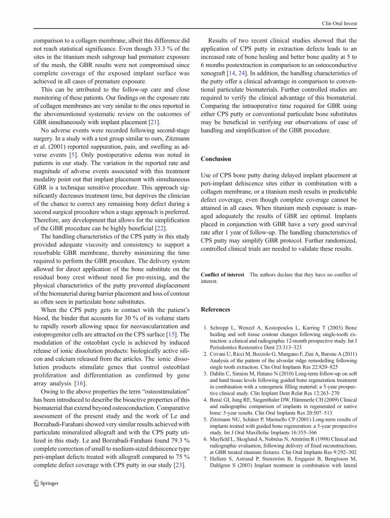
The median time between implant placement and second-stage surgery was 5 months (range 4–7 months). A collagenmembrane was utilized in 27 of the sites while a titanium meshwas employed in the remaining nine sites. Membrane/titaniummesh exposure was noted in 5/36 sites (13.89 %). Titaniummesh and collagen membranes had a 33.33% (3 out of 9 cases)and 7.41 % (2 out of 27 cases) exposure rate, respectively (p=0.158) (Table 1). In two cases when a collagen membrane wasexposed, the exposed portion was trimmed off and the patientwas instructed to rinse with 0.12 % chlorexhidine until the areawas completely epithelized. In both cases, spontaneous softtissue closure was achieved after 2 and 4 weeks, respectively.Titanium mesh exposure was treated by hygiene instructionsaiming in gentle cleaning of the area with an extra soft tooth-brush and rinsing with 0.12 % chlorhexidine once daily. In onecase, spontaneous coverage of the titanium mesh was notedfollowing this regimen. In the remaining cases, the titaniummesh was maintained in place until at least 4 months hadelapsed using the above regimen. The soft-tissue defectsaround the mesh were contained and did not expand afterinitiation of the proposed hygiene regimen. Other than mem-brane exposure, mild to moderate postoperative edema was themost frequent adverse event noted following treatment witheither a collagen membrane, or a titanium mesh in this caseseries. In a few cases, edema co-existed with extra-oral contu-sion in the region. All patients reported mild discomfort for thefirst days following surgery that was well tolerated with non-steroid anti-inflammatory medication.
Clinical evaluation during second-stage surgery revealedosseointegration of 35/36 implants. All implants were followedup for at least 12 months postloading. No more implants lostosseointegration at the 6- and 12-month follow-up visits for acumulative survival rate of 97.22% after 12 months of loading.The implant that failed to osseointegrate was placed in a 45-year-old female with noncontributory medical history. After5 months of uneventful healing, the implant was categorizedas a failure due to clinical mobility at second-stage surgery.
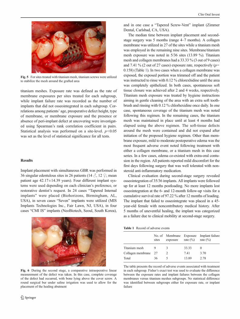
Fig. 5 For sites treated with titaniummesh, titanium screws were utilizedto stabilize the mesh around the grafted area
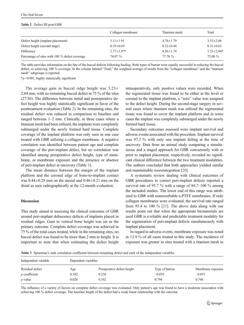
Fig. 6 During the second stage, a comparative intraoperative linearmeasurement of the defect was taken. In this case, complete coverageof the defect had occurred, with bone lying above the cover screw. Around surgical bur under saline irrigation was used to allow for theplacement of the healing abutment
Table 1 Record of adverse events
No. ofsites
Membraneexposure
Exposurerate (%)
Implant failurerate (%)
Titanium mesh 9 3 33.33 0
Collagen membrane 27 2 7.41 3.70
Total 36 5 13.89 2.78
The table presents the record of adverse events associated with treatmentin each subgroup. Fisher’s exact test was used to evaluate the differencebetween the exposure rates and implant failures between the collagenmembranes versus titanium meshes subgroups. No statistical differencewas identified between subgroups either for exposure rate, or implantfailure
Clin Oral Invest
The average gain in buccal ridge height was 3.23±2.04 mm, with no remaining buccal defect in 75 % of the sites(27/36). The difference between initial and postoperative de-fect height was highly statistically significant in favor of theposttreatment evaluation (Table 2). In the remaining sites, theresidual defect was reduced in comparison to baseline andranged between 1–2 mm. Clinically, in three cases where atitaniummesh had been utilized, the implants were completelysubmerged under the newly formed hard tissue. Completecoverage of the implant platform was only seen in one casetreated with GBR utilizing a collagen membrane. A negativecorrelation was identified between patient age and completecoverage of the peri-implant defect, but no correlation wasidentified among preoperative defect height, type of mem-brane, or membrane exposure and the presence or absenceof peri-implant defect at uncovery (Table 3).
The mean distance between the margin of the implantplatform and the coronal edge of bone-to-implant contactwas 0.44±0.28 mm on the mesial and 0.46±0.21 mm on thedistal as seen radiographically at the 12-month evaluation.
Discussion
This study aimed at assessing the clinical outcomes of GBRaround peri-implant dehiscence defects of implants placed inresidual ridges. Gain in vertical bone height was set as theprimary outcome. Complete defect coverage was achieved in75% of the total cases treated, while in the remaining sites, nobuccal defect was found to be more than 2 mm in height. It isimportant to note that when estimating the defect height
intraoperatively, only positive values were recorded. Whenthe regenerated tissue was found to be either at the level orcoronal to the implant platform, a “zero” value was assignedto the defect height. During the second-stage surgery in sev-eral cases where titanium mesh was utilized the regeneratedtissue was found to cover the implant platform and in somecases the implant was completely submerged under the newlyformed hard tissue.
Secondary outcomes assessed were implant survival andadverse events associated with the procedure. Implant survivalwas 97.2 % with only one implant failing at the time ofuncovery. Data from an animal study comparing a simulta-neous and a staged approach for GBR concurrently with orprior to implant placement, respectively, revealed no signifi-cant clinical difference between the two treatment modalities.The authors concluded that both approaches yielded similarand maintainable osseointegration [20].
A systematic review dealing with clinical outcomes ofGBR procedures to correct peri-implant defects reported asurvival rate of 95.7 % with a range of 84.7–100 % amongthe included studies. The lower end of this range was attrib-uted to GBR with nonresorbable e-PTFE membranes. If onlycollagen membranes were evaluated, the survival rate rangedfrom 95.4 to 100 % [21]. The above data along with ourresults point out that when the appropriate biomaterials areused GBR is a reliable and predictable treatment modality forthe regeneration of peri-implant defects simultaneously withimplant placement.
In regard to adverse events, membrane exposure was notedin 13.9 % of all cases treated in this study. The incidence ofexposure was greater in sites treated with a titanium mesh in
Table 2 Defect fill post-GBR
Collagen membrane Titanium mesh Total
Defect height (implant placement) 3.11±1.91 4.78±1.79 3.53±2.06
Defect height (second stage) 0.35±0.69 0.22±0.44 0.31±0.63
Difference 2.77±1.97* 4.56±1.74 3.23±2.04*
Percentage of sites with 100 % defect coverage 74.07 % 77.78 % 75.00 %
The table provides information on the fate of the buccal defects following healing. Both types of barrier were equally successful in reducing the buccaldefect, or achieving 100 % coverage. In the column labeled “Total,” the weighted average of results from the “collagen membrane” and the “titaniummesh” subgroups is reported
*p<0.001, highly statistically significant
Table 3 Spearman’s rank correlation coefficient between remaining defect and each of the independent variables
Independent variable Dependent variables
Residual defect Age Preoperative defect height Type of barrier Membrane exposure
p coefficient 0.382 0.234 −0.035 −0.051p value 0.026 0.162 0.794 0.766
The influence of a variety of factors on complete defect coverage was evaluated. Only patient’s age was found to have a moderate association withachieving 100 % defect coverage. The baseline length of the defect had a weak linear relationship with the outcome
Clin Oral Invest
comparison to a collagen membrane, albeit this difference didnot reach statistical significance. Even though 33.3 % of thesites in the titanium mesh subgroup had premature exposureof the mesh, the GBR results were not compromised sincecomplete coverage of the exposed implant surface wasachieved in all cases of premature exposure.
This can be attributed to the follow-up care and closemonitoring of these patients. Our findings on the exposure rateof collagen membranes are very similar to the ones reported inthe abovementioned systematic review on the outcomes ofGBR simultaneously with implant placement [21].
No adverse events were recorded following second-stagesurgery. In a study with a test group similar to ours, Zitzmannet al. (2001) reported suppuration, pain, and swelling as ad-verse events [5]. Only postoperative edema was noted inpatients in our study. The variation in the reported rate andmagnitude of adverse events associated with this treatmentmodality point out that implant placement with simultaneousGBR is a technique sensitive procedure. This approach sig-nificantly decreases treatment time, but deprives the clinicianof the chance to correct any remaining bony defect during asecond surgical procedure when a stage approach is preferred.Therefore, any development that allows for the simplificationof the GBR procedure can be highly beneficial [22].
The handling characteristics of the CPS putty in this studyprovided adequate viscosity and consistency to support aresorbable GBR membrane, thereby minimizing the timerequired to perform the GBR procedure. The delivery systemallowed for direct application of the bone substitute on theresidual bony crest without need for pre-mixing, and thephysical characteristics of the putty prevented displacementof the biomaterial during barrier placement and loss of contouras often seen in particulate bone substitutes.
When the CPS putty gets in contact with the patient’sblood, the binder that accounts for 30 % of its volume startsto rapidly resorb allowing space for neovascularization andostoprogenitor cells are attracted on the CPS surface [15]. Themodulation of the osteoblast cycle is achieved by inducedrelease of ionic dissolution products: biologically active sili-con and calcium released from the articles. The ionic disso-lution products stimulate genes that control osteoblastproliferation and differentiation as confirmed by genearray analysis [16].
Owing to the above properties the term “osteostimulation”has been introduced to describe the bioactive properties of thisbiomaterial that extend beyond osteoconduction. Comparativeassessment of the present study and the work of Le andBorzabadi-Farahani showed very similar results achievedwithparticulate mineralized allograft and with the CPS putty uti-lized in this study. Le and Borzabadi-Farahani found 79.3 %complete correction of small tomedium-sized dehiscence typeperi-implant defects treated with allograft compared to 75 %complete defect coverage with CPS putty in our study [23].
Results of two recent clinical studies showed that theapplication of CPS putty in extraction defects leads to anincreased rate of bone healing and better bone quality at 5 to6 months postextraction in comparison to an osteoconductivexenograft [14, 24]. In addition, the handling characteristics ofthe putty offer a clinical advantage in comparison to conven-tional particulate biomaterials. Further controlled studies arerequired to verify the clinical advantage of this biomaterial.Comparing the intraoperative time required for GBR usingeither CPS putty or conventional particulate bone substitutesmay be beneficial in verifying our observations of ease ofhandling and simplification of the GBR procedure.
Conclusion
Use of CPS bone putty during delayed implant placement atperi-implant dehiscence sites either in combination with acollagen membrane, or a titanium mesh results in predictabledefect coverage, even though complete coverage cannot beattained in all cases. When titanium mesh exposure is man-aged adequately the results of GBR are optimal. Implantsplaced in conjunction with GBR have a very good survivalrate after 1 year of follow-up. The handling characteristics ofCPS putty may simplify GBR protocol. Further randomized,controlled clinical trials are needed to validate these results.
Conflict of interest The authors declare that they have no conflict ofinterest.
References
1. Schropp L, Wenzel A, Kostopoulos L, Karring T (2003) Bonehealing and soft tissue contour changes following single-tooth ex-traction: a clinical and radiographic 12-month prospective study. Int JPeriodontics Restorative Dent 23:313–323
2. Covani U, Ricci M, Bozzolo G,Mangano F, Zini A, Barone A (2011)Analysis of the pattern of the alveolar ridge remodelling followingsingle tooth extraction. Clin Oral Implants Res 22:820–825
3. Dahlin C, Simion M, Hatano N (2010) Long-term follow-up on softand hard tissue levels following guided bone regeneration treatmentin combination with a xenogeneic filling material: a 5-year prospec-tive clinical study. Clin Implant Dent Relat Res 12:263–270
4. BenićGI, Jung RE, Siegenthaler DW, Hämmerle CH (2009) Clinicaland radiographic comparison of implants in regenerated or nativebone: 5-year results. Clin Oral Implants Res 20:507–513
5. Zitzmann NU, Schärer P, Marinello CP (2001) Long-term results ofimplants treated with guided bone regeneration: a 5-year prospectivestudy. Int J Oral Maxillofac Implants 16:355–366
6. Mayfield L, Skoglund A, Nobréus N, Attström R (1998) Clinical andradiographic evaluation, following delivery of fixed reconstructions,at GBR treated titanium fixtures. Clin Oral Implants Res 9:292–302
7. Hellem S, Astrand P, Stenström B, Engquist B, Bengtsson M,Dahlgren S (2003) Implant treatment in combination with lateral
Clin Oral Invest
augmentation of the alveolar process: a 3-year prospective study. ClinImplant Dent Relat Res 5:233–240
8. Chiapasco M, Zaniboni M (2009) Clinical outcomes of GBR proce-dures to correct peri-implant dehiscences and fenestrations: a system-atic review. Clin Oral Implants Res 4:113–123
9. Bender SA, Rogalski JB, Mills MP, Arnold RM, Cochran DL,Mellonig JT (2005) Evaluation of demineralized bone matrix pasteand putty in periodontal intraosseous defects. J Periodontol 76:768–777
10. Babbush CA (2003) Histologic evaluation of human biopsies afterdental augmentation with a demineralized bone matrix putty. ImplantDent 12:325–332
11. Butz F, Bächle M, Ofer M, Marquardt K, Kohal RJ (2011) Sinusaugmentation with bovine hydroxyapatite/synthetic peptide in a so-dium hyaluronate carrier (PepGen P-15 Putty): a clinical investiga-tion of different healing times. Int J Oral Maxillofac Implants 26:1317–1323
12. Wang Z, Lu B, Chen L, Chang J (2011) Evaluation of anosteostimulative putty in the sheep spine. J Mater Sci Mater Med22:185–191
13. Kotsakis GA, Joachim F, Saroff SA, Mahesh L, Prasad H, Rohrer M(2014) Histomorphometric evaluation of a calcium-phosphosilicatebone substitute in extraction sockets. Int J Periodontics RestorativeDent 34:233–239
14. Kotsakis GA, Salama M, Chrepa V, Hinrichs J, Gaillard P (2014) Arandomized, blinded, controlled clinical study of particulateanorganic bovine bone mineral and calcium phosphosilicate puttybone substitutes for alveolar ridge preservation. Int J Oral MaxillofacImplants 29:141–151
15. Hench LL, Polakjm (2008) A genetic basis for design of biomaterialsfor in situ regeneration. Key Eng Mater 377:151–166
16. Xynos ID, Edgar AI, Buttery LDK, Hench LL, Polak JM (2001)Gene-expression profiling of human osteoblasts following treatment
with the ionic products of bioglass 45S5 dissolution. J BiomedMaterRes 55:151–157
17. Seibert JS (1983) Reconstruction of deformed, partially edentulousridges, using full thickness onlay grafts. Part II. Prosthetic/periodontalinterrelationships. Compend Contin Educ Dent 4:549–562
18. Karoussis IK, Brägger U, Salvi GE, Bürgin W, Lang NP (2004)Effect of implant design on survival and success rates of titaniumoral implants: a 10-year prospective cohort study of the ITI DentalImplant System. Clin Oral Implants Res 15:8–17
19. Weber HP, Lang NP (1990) Tissue integration of non-submergedimplants. 1-year results of a prospective study with 100 ITI hollow-cylinder and hollow-screw implants. Clin Oral Implants Res 1:33–40
20. Artzi Z, Nemcovsky CE, Tal H, Weinberg E, Weinreb M, Prasad H,Rohrer MD, Kozlovsky A (2010) Simultaneous versus two-stageimplant placement and guided bone regeneration in the canine:histomorphometry at 8 and 16 months. J Clin Periodontol 37:1029–1038
21. Chiapasco M, Zaniboni M (2009) Clinical outcomes of GBR proce-dures to correct peri-implant dehiscences and fenestrations: a system-atic review. Clin Oral Implants Res 20:113–123
22. Jung RE, Hälg GA, Thoma DS, Hämmerle CH (2009) A random-ized, controlled clinical trial to evaluate a new membrane for guidedbone regeneration around dental implants. Clin Oral Implants Res 20:162–168
23. Le BT, Borzabadi-Farahani (2013) Simultaneous implant placementand bone grafting with particulate mineralized allograft in sites withbuccal wall defects, a three-year follow-up and review of literature. JCraniomaxillofac. doi:10.1016/j.jcms.2013.07.026
24. Masesh L, Kotsakis G, Venkataraman N, Shukla S, Prasad H (2013)Ridge preservation with the socket-plug technique utilizing analloplastic putty bone substitute or a particulate xenograft: a histo-logical pilot study. J Oral Implantol. doi:10.1563/AAID-JOI-D-13-00025
Clin Oral Invest


![An Innovative Wire Impression Technique of Highly Resorbed ... · determines the retention and comfort of dentures made for patients with unfavorable residual ridges [4]. Minimum](https://img.pdfslide.us/doc/110x75/5e67f0d070ab06249b140b45/an-innovative-wire-impression-technique-of-highly-resorbed-determines-the-retention.jpg)