Embed Size (px)
Citation preview
Implementation: 01.08.2020
Clinical RadiologySpecialty Training Curriculum
Contents
1 Introduction 3
1.1 The purpose of the curriculum 3
1.2 The need for the curriculum 3
1.3 Scope of training 4
1.4 Structure of training 5
1.4.1 Less than full-time training 6
1.5 Capabilities in Practice 6
1.5.1 Generic Capabilities in Practice 6
1.5.2 Specialty-specific Capabilities in Practice 7
1.6 Flexibility of training 8
1.7 Generic professional capabilities and good medical practice 9
2 Content of learning 10
2.1 Generic CiPs 11
2.2 Specialty-specific CiPs 18
2.3 Presentations and conditions 27
2.4 Breadth of training 42
2.4.1 Interventional Radiology 42
2.4.2 Emerging technologies 42
2.4.3 Emerging imaging techniques 42
2.4.4 Academic training 42
2.4.5 Taking time out of programme 43
2.4.6 Acting up as a consultant 43
3 Teaching and learning methods 44
3.1 Work-based experiential learning 44
3.1.1 Optional work-based experiential learning 45
3.2 Formal postgraduate teaching 45
3.3 Independent self-directed learning 45
3.4 External study courses 46
3.5 Learning with peers 46
3.6 Simulation 46
4 Programme of assessment 47
4.1 Purpose of assessment 47
4.2 Programme of assessment 47
4.3 Assessment of CiPs 48
4.4 Critical progression points 51
4.5 Evidence of progress 51
4.5.1 E-portfolio 53
4.5.2 Summative Assessment 53
4.5.3 Formative Assessment 54
4.6 Decisions on progress (ARCP) 56
4.6.1 Appeals 57
4.7 Assessment blueprints 57
5 Supervision and feedback 59
5.1 Feedback 59
5.2 Supervision 59
5.2.1 Educational supervisor 60
5.2.2 Clinical supervisor 61
5.2.3 Trainees 62
5.3 Appraisal 62
6 Appendices 65
6.1 Curriculum development, implementation and review 65
6.1.1 Implementation 65
6.1.2 Intended use 65
6.1.3 Review 65
6.2 Quality management 66
6.3 Equality and diversity 67
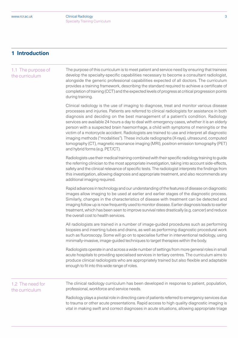
The purpose of this curriculum is to meet patient and service need by ensuring that trainees develop the specialty-specific capabilities necessary to become a consultant radiologist, alongside the generic professional capabilities expected of all doctors. The curriculum provides a training framework, describing the standard required to achieve a certificate of completion of training (CCT) and the expected levels of progress at critical progression points during training.
Clinical radiology is the use of imaging to diagnose, treat and monitor various disease processes and injuries. Patients are referred to clinical radiologists for assistance in both diagnosis and deciding on the best management of a patient’s condition. Radiology services are available 24 hours a day to deal with emergency cases, whether it is an elderly person with a suspected brain haemorrhage, a child with symptoms of meningitis or the victim of a motorcycle accident. Radiologists are trained to use and interpret all diagnostic imaging methods (“modalities”). These include radiographs (X-rays), ultrasound, computed tomography (CT), magnetic resonance imaging (MRI), positron emission tomography (PET) and hybrid forms (e.g. PET/CT).
Radiologists use their medical training combined with their specific radiology training to guide the referring clinician to the most appropriate investigation, taking into account side-effects, safety and the clinical relevance of specific tests. The radiologist interprets the findings from this investigation, allowing diagnosis and appropriate treatment, and also recommends any additional imaging required.
Rapid advances in technology and our understanding of the features of disease on diagnostic images allow imaging to be used at earlier and earlier stages of the diagnostic process. Similarly, changes in the characteristics of disease with treatment can be detected and imaging follow up is now frequently used to monitor disease. Earlier diagnosis leads to earlier treatment, which has been seen to improve survival rates drastically (e.g. cancer) and reduce the overall cost to health services.
All radiologists are trained in a number of image-guided procedures such as performing biopsies and inserting tubes and drains, as well as performing diagnostic procedural work such as fluoroscopy. Some will go on to specialise further in interventional radiology, using minimally-invasive, image-guided techniques to target therapies within the body.
Radiologists operate in and across a wide number of settings from more general roles in small acute hospitals to providing specialised services in tertiary centres. The curriculum aims to produce clinical radiologists who are appropriately trained but also flexible and adaptable enough to fit into this wide range of roles.
The clinical radiology curriculum has been developed in response to patient, population, professional, workforce and service needs.
Radiology plays a pivotal role in directing care of patients referred to emergency services due to trauma or other acute presentations. Rapid access to high quality diagnostic imaging is vital in making swift and correct diagnoses in acute situations, allowing appropriate triage
1 Introduction
1.1 The purpose of the curriculum
1.2 The need for the curriculum
3Clinical Radiology Specialty Training Curriculum
www.rcr.ac.uk
and impacting on transit times through emergency departments and ultimately on inpatient capacity and length of stay.
Patients require access to other essential services, such as for the detection and monitoring of malignant disease and the diagnosis and follow up of major co-morbidities. Image guided biopsy is central to diagnosis and staging for the majority of patients with cancer. The NHS Five Year Forward update plan published in 20171 identified improving cancer services and outcomes for patients as one of the four major priorities for the health service going forward. In line with this, a number of key reviews including the National Cancer Strategy2 and the Health Education England (HEE) Cancer Workforce Strategy3 clearly identified a need for more radiologists to provide diagnostic and interventional services.
Quality assured image based population screening is central to well-established services such as the National Health Breast Screening Programme (NHSBSP) as well as to newer screening services such as CT screening for lung cancer and colon cancer. With an aging population increasing demand for radiology led screening will be an important factor in assuring population health outcomes are sustained.
The curriculum aims to produce clinical radiologists with the ability to provide general and emergency radiology in any NHS environment and specialist skills in one or more areas. This allows provision of acute imaging services vital to supporting the swift and accurate diagnoses of patients reporting to emergency departments, as well as meeting the increasing demand for specialist diagnostic and treatment services.
Patient management is enhanced by multidisciplinary team meetings in which radiologists are pivotal members with leadership roles, with very few decisions made about patient management without radiology input.
Developing and training other practitioners in aspects of diagnostic imaging and intervention requires radiologists with leadership, management and education skills.
This curriculum aims to equip radiologists with the skills to fully engage in these roles.
Specialty training in clinical radiology will normally be a five-year programme that will include exposure to all imaging modalities, body systems and patient groups with the objective of producing clinical radiologists who at the time of CCT will be equipped to deliver a general, acute and emergency service.
Patients who require access to specialist diagnostic and treatment services require radiologists with advanced skills who can deliver specialist imaging in addition to general radiology. Whilst working as a consultant, most clinical radiologists will focus on one or two areas of special interest in order to be able to provide this. The curriculum allows for some focus on areas of special interest at the end of training whilst ensuring that trainees will maintain the skills and flexibility required to adapt to the needs of the local service at the time and in the future.
The curriculum includes the interventional capabilities, such as image-guided biopsies, required by general radiologists but excludes the specialist skills acquired by those who follow the interventional radiology sub-specialty curriculum.
1.3 Scope of training
4Clinical Radiology Specialty Training Curriculum
www.rcr.ac.uk
Clinical radiology training is entered following completion of the foundation training programme (FY1 and FY2) or equivalent, as a minimum. Trainees may have gained additional experience in other programmes (e.g. internal medicine, surgery etc.) before commencing clinical radiology training.
Trainees are required to enrol with the RCR and become trainee members prior to the commencement of their training. Trainees are required to maintain RCR membership, including the full payment of all applicable fees, throughout training for the RCR to be able to recommend them as eligible for award of a CCT.
Trainees will rotate through modality and systems-based attachments in order to gain experience and skills in all of these fields. These attachments include: breast radiology; cardiac radiology; thoracic radiology; gastro-intestinal radiology; molecular imaging and radionuclide radiology; paediatric radiology; musculoskeletal radiology; neuroradiology; head and neck radiology; uro-gynaecological radiology; and core interventional radiology. Higher sub-specialty training in interventional radiology is included in a separate curriculum.
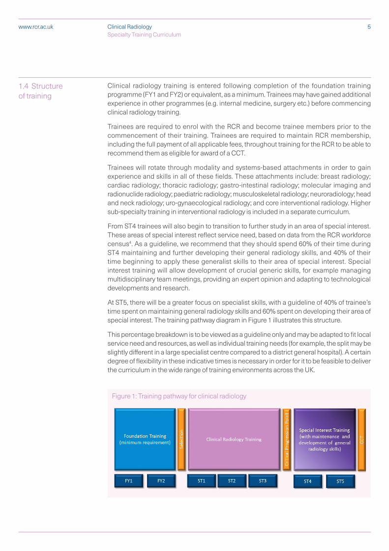
From ST4 trainees will also begin to transition to further study in an area of special interest. These areas of special interest reflect service need, based on data from the RCR workforce census4. As a guideline, we recommend that they should spend 60% of their time during ST4 maintaining and further developing their general radiology skills, and 40% of their time beginning to apply these generalist skills to their area of special interest. Special interest training will allow development of crucial generic skills, for example managing multidisciplinary team meetings, providing an expert opinion and adapting to technological developments and research.
At ST5, there will be a greater focus on specialist skills, with a guideline of 40% of trainee’s time spent on maintaining general radiology skills and 60% spent on developing their area of special interest. The training pathway diagram in Figure 1 illustrates this structure.
This percentage breakdown is to be viewed as a guideline only and may be adapted to fit local service need and resources, as well as individual training needs (for example, the split may be slightly different in a large specialist centre compared to a district general hospital). A certain degree of flexibility in these indicative times is necessary in order for it to be feasible to deliver the curriculum in the wide range of training environments across the UK.
1.4 Structure of training
5Clinical Radiology Specialty Training Curriculum
www.rcr.ac.uk
Figure 1: Training pathway for clinical radiology
Trainees should consider shortage areas aligned to local patient and service needs, with a view to where vacancies lie. Availability of places is mainly dependent on training capacity at present; however we will be working with the four countries to assess and strengthen the systems in place for identifying local workforce needs and how this can inform programme design.
1.4.1 Less than full-time trainingTrainees are entitled to opt for less than full-time training programmes at the discretion of their local deanery and in compliance with current guidance from the GMC. Less than full-time trainees should assume that their clinical training will be of a duration pro-rata with the indicative time for full-time trainees, but this should be reviewed in accordance with the Gold Guide5 (A Reference Guide for Postgraduate Specialty Training in the UK). They should also undertake a pro rata share of the out-of-hours duties (including on-call and other out-of-hours commitments) required of their full-time colleagues in the same programme and at the equivalent stage.
To achieve CCT trainees are expected to demonstrate the capabilities described by the generic and specialty-specific high level outcomes, or ‘capabilities in practice’ (CiPs), as detailed below:
1.5.1 Generic Capabilities in Practice 1. Demonstrate the professional values and behaviours expected of all doctors as outlined
in Good medical practice.
As doctors, consultant radiologists adhere to the principles of ‘Good medical practice’ as stipulated by the GMC.
2. Successfully function within the health service and healthcare systems in the UK.
Like all consultants working within the NHS, radiologists need to understand organisational and management systems so that they can engage positively with them and optimise patient care.
3. Engage in reflection, clinical governance and quality improvement processes to ensure good practice.
Consultant radiologists are expected to stay up to date with their knowledge and skills, and look for ways to improve the quality of their services.
4. Engage in evidence-based practice and safeguard data, including imaging data.
Consultant radiologists require the skills used by all doctors to practise evidence-based medicine.
5. Act as a clinical teacher and supervisor.
Consultant radiologists teach medical students, junior doctors and other healthcare professionals.
6. Work well within a variety of different teams, communicating effectively with colleagues and demonstrating the skills required to lead a team.
1.5 Capabilities in Practice
6Clinical Radiology Specialty Training Curriculum
www.rcr.ac.uk
Clinical radiology relies on a multi-professional team and good communication is an essential component of sound practice, team working and patient centred care. Consultant radiologists must be able to resolve conflict, develop good working relationships and support team development and possess the qualities and behaviours necessary to lead but also to follow, when necessary, in dealing with difficult situations and conflicting attitudes.
1.5.2 Specialty-specific Capabilities in Practice7. Appropriately select and tailor imaging to patient context and the clinical question(s).
Consultant radiologists will discuss clinical cases with referrers and allied imaging professionals and advise on appropriate imaging according to the individual patient, clinical background and the clinical question posed. Imaging investigations have varying health and safety risks to patients and the public that need to be considered. Consultant radiologists weigh up the relative clinical risk/benefit when advising on imaging according to clinical information provided by referrers.
8. Provide timely, accurate and clinically useful reports on imaging studies.
Consultant radiologists provide actionable reports on imaging studies that are performed on patients. They will discuss findings with referrers as required. They will be able to report investigations for common presenting complaints. In addition, they will be able to report more complex investigations as appropriate to their special interest. This may include recommendations regarding onward imaging investigations, imaging follow up and/or other clinical management based on their expert knowledge.
9. Appropriately manage imaging examination lists/procedures according to clinical need and professional expertise.
Consultant radiologists will be able to obtain consent and directly examine a patient in real time with imaging such as ultrasound and perform image-guided procedures.
10. Evaluate image quality and utilise the knowledge of imaging sciences to optimise image quality.
Consultant radiologists need to be able to evaluate image quality and utilise knowledge of imaging physics to maximise the diagnostic certainty of an imaging test.
11. Safely manage the imaging and image-guided intervention needed to support emergency care.
Imaging is required to support the 24/7 emergency service provided by the NHS. Consultant radiologists will be competent in interpreting and performing imaging examinations and/or procedures that are required in the emergency context and where appropriate will suggest use of image-guided intervention or onward referral.
12. Effectively contribute a clinical/imaging opinion to a multidisciplinary team (MDT) meeting.
Imaging is often central to decision making regarding patient management and onward investigation. Consultant radiologists review imaging of cases to be discussed at MDT meetings and present relevant findings pertinent to clinical decision making. They will provide explicit recommendations regarding onward imaging investigations and/or image-guided procedures based on their expert knowledge.
7Clinical Radiology Specialty Training Curriculum
www.rcr.ac.uk
The curriculum supports flexibility and transferability of outcomes across related specialties and disciplines, reflecting key interdependencies between the clinical radiology curriculum and other training programmes, outlined below.
Nuclear medicine
Nuclear medicine physicians are responsible for the administration of unsealed radioactive substances to patients for the purposes of diagnosis, therapy or research. There is significant overlap with the work of radiologists who use radionuclide radiology imaging techniques for diagnosis, and we have undertaken joint stakeholder engagement activity.
Since 2015 the nuclear medicine specialty training curriculum has required trainees to undertake the core component of the clinical radiology curriculum prior to further specialisation in nuclear medicine techniques. This allows for recognition of training in both specialties and ease of transfer between them. Our expectation is that a similar arrangement will continue and we will be working with the nuclear medicine Specialty Advisory Committee on this.
Cardiology
The cardiology specialty training curriculum contains mandatory core and optional advanced level elements of imaging, including cardiac CT, MR and nuclear imaging. The British Society of Cardiac Imaging is a multi-professional body with both radiologists and cardiologists as members, and we routinely consult them on curriculum content.
Breast clinicians
Breast clinicians are doctors who provide a holistic approach to the investigation and management of breast disease. They have skills in clinical examination, interpretation of imaging including mammography and ultrasound, the use of interventional procedures, and the management of benign breast disease. They may work in both symptomatic clinics and the NHS Breast Screening Programme. The speciality is not recognised in UK legislation and therefore has no formal training curriculum, however we have worked with the Association of Breast Clinicians to develop a credential which will draw on aspects of this clinical radiology curriculum.
Radiographers
Radiographers may formally report on certain categories of x rays e.g. trauma radiographs. They work within well-defined and agreed frameworks and require mentorship, training and oversight by clinical radiologists. Radiographer reporting is an integral element of service delivery in many radiology departments in the UK. The RCR is working with HEE and the Society and College of Radiographers on a project to define educational standards for reporting radiographers, which will be informed by this curriculum.
Ultrasound
Sonographers are currently responsible for the provision of the majority of ultrasound services in the UK and a close working relationship with clinical radiology is essential for a sustainable imaging service. The RCR is working with the British Medical Ultrasound Society, Society and College of Radiographers and HEE in developing career pathways for sonographers.
1.6 Flexibility of training
8Clinical Radiology Specialty Training Curriculum
www.rcr.ac.uk
The GMC has developed the Generic professional capabilities (GPC) framework6 with the Academy of Medical Royal Colleges (AoMRC) to describe the fundamental, career-long, generic capabilities required of every doctor. The framework describes the requirement to develop and maintain key professional values and behaviours, knowledge, and skills, using a common language. GPCs also represent a system-wide, regulatory response to the most common concerns about patient safety and fitness to practise within the medical profession. The framework will be relevant at all stages of medical education, training and practice.
Good medical practice (GMP)7 is embedded at the heart of the GPC framework. In describing the principles, duties and responsibilities of doctors, the GPC framework articulates GMP as a series of achievable educational outcomes to enable curriculum design and assessment.
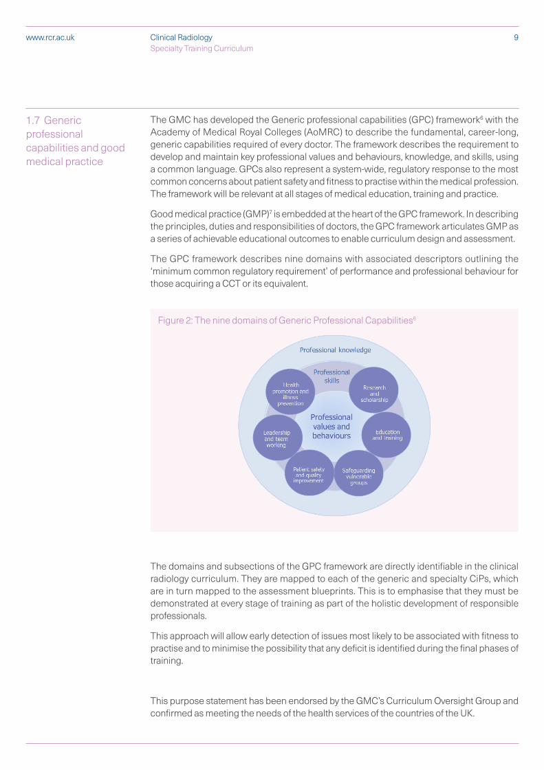
The GPC framework describes nine domains with associated descriptors outlining the ‘minimum common regulatory requirement’ of performance and professional behaviour for those acquiring a CCT or its equivalent.
The domains and subsections of the GPC framework are directly identifiable in the clinical radiology curriculum. They are mapped to each of the generic and specialty CiPs, which are in turn mapped to the assessment blueprints. This is to emphasise that they must be demonstrated at every stage of training as part of the holistic development of responsible professionals.
This approach will allow early detection of issues most likely to be associated with fitness to practise and to minimise the possibility that any deficit is identified during the final phases of training.
This purpose statement has been endorsed by the GMC’s Curriculum Oversight Group and confirmed as meeting the needs of the health services of the countries of the UK.
1.7 Generic professional capabilities and good medical practice
9Clinical Radiology Specialty Training Curriculum
www.rcr.ac.uk
Figure 2: The nine domains of Generic Professional Capabilities6
2 Content of learning
The practice of clinical radiology requires the generic and specialty-specific knowledge, skills, attitudes and procedural competency to diagnose, and sometimes manage, patients referred for imaging to investigate a wide range of symptoms and conditions and perform image-guided procedures. It involves particular emphasis on diagnostic reasoning, communicating uncertainty and working with referrers to ensure appropriate speciality opinion or care is sought when required.
To achieve CCT trainees are expected to demonstrate achievement of the generic and specialty-specific high level outcomes, known as ‘capabilities in practice’ or ‘CiPs’. The CiPs describe the professional capabilities required of a consultant clinical radiologist. Each CiP has a number of descriptors that underpin it, is mapped to the GMC’s Generic Professional Capabilities and accompanied by suggested evidence that may demonstrate progress towards achieving this CiP.
The descriptors are intended to provide guidance to trainees and trainers about the range of clinical contexts which may support achievement of the CiPs, however they are not intended to be prescriptive and do not provide an exhaustive list. Trainees may demonstrate their progress against the CiPs in a variety of different ways, reflecting their strengths, areas of interest and the resources available to them, and should be encouraged to find innovative ways to achieve this. They may also complete activities that provide evidence for more than one CiP.
The level at which trainees meet each CiP is stage dependent and is expected to progress in a spiral fashion throughout training. Trainees will develop at different rates and may be able to demonstrate a higher level of progress in some CiPs compared to others. Excellent trainees may be able to evidence higher achievement at an earlier stage, provide a broader portfolio of evidence, or provide evidence that shows a deeper level of learning. The programme of assessment that forms part of this curriculum outlines the minimum expected levels of achievement at critical progression points in training, where trainees take on significantly more responsibility or where training or patient risk may potentially increase. Sign off will require clinical and educational supervisors to make entrustment decisions on the level of supervision required for each CiP or underlying activity at each critical progression point. More detail is provided in the programme of assessment section of the curriculum.
10Clinical Radiology Specialty Training Curriculum
www.rcr.ac.uk
2.1 Generic CiPs
11Clinical Radiology Specialty Training Curriculum
www.rcr.ac.uk
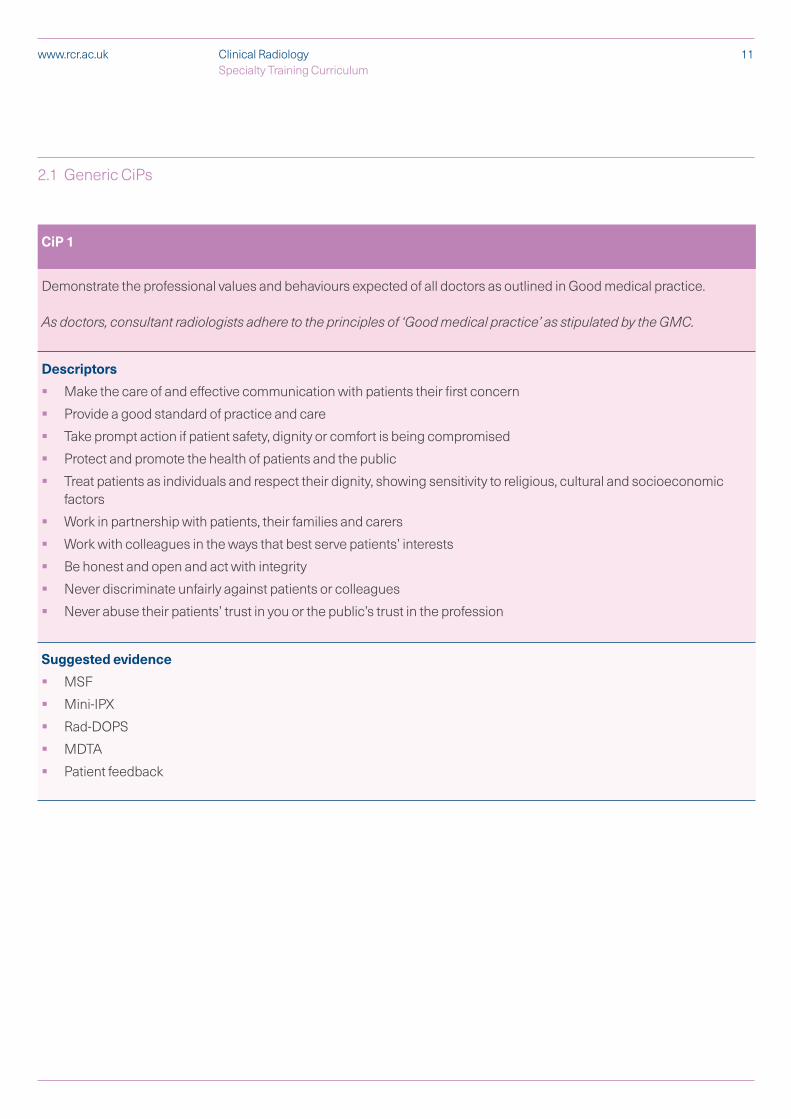
CiP 1
Demonstrate the professional values and behaviours expected of all doctors as outlined in Good medical practice.
As doctors, consultant radiologists adhere to the principles of ‘Good medical practice’ as stipulated by the GMC.
Descriptors
§ Make the care of and effective communication with patients their first concern
§ Provide a good standard of practice and care
§ Take prompt action if patient safety, dignity or comfort is being compromised
§ Protect and promote the health of patients and the public
§ Treat patients as individuals and respect their dignity, showing sensitivity to religious, cultural and socioeconomic factors
§ Work in partnership with patients, their families and carers
§ Work with colleagues in the ways that best serve patients’ interests
§ Be honest and open and act with integrity
§ Never discriminate unfairly against patients or colleagues
§ Never abuse their patients’ trust in you or the public’s trust in the profession
Suggested evidence
§ MSF
§ Mini-IPX
§ Rad-DOPS
§ MDTA
§ Patient feedback
12Clinical Radiology Specialty Training Curriculum
www.rcr.ac.uk
CiP 1
Mapping to GPCs
§ Domain 1: Professional values and behaviours
§ Domain 2: Professional Skills
– Practical skills
– Communication and interpersonal skills
– Dealing with complexity and uncertainty
– Clinical skills: consent
§ Domain 3: Professional knowledge
– Professional requirements
– National legislative requirements
§ Domain 4: Capabilities in health promotion and prevention
§ Domain 5: Capabilities in leadership and teamworking
§ Domain 6: Capabilities in patient safety and quality improvement
– Patient safety
§ Domain 7: Capabilities in safeguarding vulnerable groups
§ Domain 8: Capabilities in education and training
§ Domain 9: Capabilities in research and scholarship
13Clinical Radiology Specialty Training Curriculum
www.rcr.ac.uk
CiP 2
Successfully function within the health service and healthcare systems in the UK.
Like all consultants working within the NHS, radiologists need to understand organisational and management systems so that they can engage positively with them and optimise patient care
Descriptors
§ Understand the structure and organisation of the health service and system including the independent sector and the wider healthcare landscape
§ Understand how services are commissioned, funded and audited
§ Understand how services are deemed to be clinically effective and cost effective
§ Understand how resources are managed, being aware of competing demands and the importance of avoiding waste
§ Understand the concept of health screening and appraise whether a proposed screening test is appropriate in the context of imaging
§ Apply equality and diversity frameworks and ensure that an equal, non-discriminatory approach is adopted in interactions with both patients and colleagues
§ Demonstrate appropriate awareness of, and maintain a professional approach to the use of social media and public communications.
§ Adhere to all relevant professional communication policies
Suggested evidence
§ QIPAT
§ Reflection
§ Leadership/management courses/modules
Mapping to GPCs
§ Domain 2: Professional Skills
– Practical skills
§ Domain 3: Professional knowledge
– The health service and healthcare systems in the four countries
14Clinical Radiology Specialty Training Curriculum
www.rcr.ac.uk
CiP 3
Engage in reflection, clinical governance and quality improvement processes to ensure good practice.
Consultant radiologists are expected to stay up to date with their knowledge and skills, and look for ways to improve the quality of their services.
Descriptors
§ Facilitate and lead on quality improvement and audit projects to improve patient care and experience
§ Promote a culture of openness and accountability including awareness of the duty of candour to patients
§ Appropriately raise concerns including errors
§ Share good practice
§ Advocate clinical quality improvement
§ Engage in clinical governance meetings including peer feedback meetings
§ Demonstrate commitment to continuing professional development by maintaining and/or developing skills relevant to higher training special interest area and/or local service need
§ Appropriately raise concerns regarding negative professional behaviour e.g. bullying
§ Recognise and acknowledge where personal issues impact upon good practice and seek appropriate help
Suggested evidence
§ QIPAT
§ Reflection
§ Evidence of attendance of local governance and/or discrepancy meetings
Mapping to GPCs
§ Domain 1: Professional values and behaviours
§ Domain 2: Professional Skills
– Communication and interpersonal skills
§ Domain 3: Professional knowledge
– Professional requirements
§ Domain 5: Capabilities in leadership and teamworking
§ Domain 6: Capabilities in patient safety and quality improvement
– Patient safety
– Quality Improvement
15Clinical Radiology Specialty Training Curriculum
www.rcr.ac.uk
CiP 4
Engage in evidence-based practice and safeguard data, including imaging data.
Consultant radiologists require the skills used by all doctors to practise evidence-based medicine.
Descriptors
§ Demonstrate an understanding of the principles of research, research methods and the translation of research into clinical practice
§ Identify and critically appraise literature to inform practice
§ Understand and critically appraise new technological developments including radiological applications of Artificial Intelligence (AI)
§ Interpret and communicate research evidence in a meaningful way to patients to support them in making informed decisions about treatment
§ Follow guidelines on ethical conduct in research and consent for research
§ Apply information governance principles to safeguard imaging data in the context of research
§ Adhere to Data Protection Regulations and be familiar with Freedom of Information regulations
§ Understand the role of the Caldicott Guardian within an institution
Suggested evidence
§ Reflection
§ Attendance and participation in a journal club
§ Presentation and/or publication of research
§ Attendance of research meetings and/or courses
§ Postgraduate qualifications e.g. Postgraduate certificate, Masters etc.
§ GCP training
§ Contribution to writing grant applications
§ Contribution to applications to Research Ethics Committees (REC), MHRA etc.
Mapping to GPCs
§ Domain 2: Professional Skills
– Practical skills
§ Domain 3: Professional knowledge
– Professional requirements
– National legislative requirements
§ Domain 9: Capabilities in research and scholarship
16Clinical Radiology Specialty Training Curriculum
www.rcr.ac.uk
CiP 5
Act as a clinical teacher and supervisor.
Consultant radiologists teach medical students, junior doctors and other healthcare professionals.
Descriptors
§ Provide teaching, supervision and assessment of clinical trainees and other healthcare professionals
§ Understand the role of and develop the ability to act as a Clinical Supervisor to the standard required by the GMC
§ Apply information governance principles to safeguard imaging data in context of education
Suggested evidence
§ Teaching observation
§ Reflection
§ Evidence of delivering undergraduate/postgraduate teaching
§ Evidence of teaching and/or assessment design/management/governance
§ Learner feedback forms
§ Postgraduate qualification in medical education
Mapping to GPCs
§ Domain 1: Professional values and behaviours
§ Domain 2: Professional Skills
– Practical skills
– Communication and interpersonal skills
§ Domain 3: Professional knowledge
– Professional requirements
– National legislative requirements
§ Domain 5: Capabilities in leadership and teamworking
§ Domain 6: Capabilities in patient safety and quality improvement
– Patient safety
§ Domain 8: Capabilities in education and training
17Clinical Radiology Specialty Training Curriculum
www.rcr.ac.uk
CiP 6
Work well within a variety of different teams, communicating effectively with colleagues and demonstrating the skills required to lead a team.
Clinical radiology relies on a multi-professional team and good communication is an essential component of sound practice, team working and patient centred care. Consultant radiologists must be able to resolve conflict, develop good working relationships and support team development and possess the qualities and behaviours necessary to lead but also to follow, when necessary, in dealing with difficult situations and conflicting attitudes.
Descriptors
§ Promote and actively participate in multidisciplinary and interprofessional team working, communicate effectively and recognise and respect the roles of all members of the team
§ Effectively lead a multi-professional team allowing all voices to be heard and considered and foster an atmosphere of collaboration
§ Critically appraise performance of colleagues, peers and systems, appropriately escalate concerns and promote an open and transparent culture of learning and development
§ Show awareness of own leadership style and how this impacts on others
§ Demonstrate flexibility in behaviour and ability to adapt techniques and approaches within the multi-professional team to improve engagement in difficult situations
§ Supervise, challenge and mentor colleagues and peers to enhance performance
§ Recognise own limitations and comprehend situations where others are better equipped to lead or where delegation is appropriate
Suggested evidence
§ MSF
§ Mini-IPX
§ Rad-DOPS
§ MDTA
Mapping to GPCs
§ Domain 1: Professional values and behaviours
§ Domain 2: Professional skills
– Practical skills
– Communication and interpersonal skills
– Dealing with complexity and uncertainty
§ Domain 5: Capabilities in leadership and teamworking
§ Domain 6: Capabilities in patient safety and quality improvement
– Patient safety
2.2 Specialty-specific CiPs
18Clinical Radiology Specialty Training Curriculum
www.rcr.ac.uk
CiP 7
Appropriately select and tailor imaging to patient context and the clinical question(s).
Consultant radiologists will discuss clinical cases with referrers and allied imaging professionals and advise on appropriate imaging according to the individual patient, clinical background and the clinical question posed. Imaging investigations have varying health and safety risks to patients and the public that need to be considered. Consultant radiologists weigh up the relative clinical risk/benefit when advising on imaging according to clinical information provided by referrers.
Descriptors
§ Collaborate effectively with referrers to determine the most appropriate imaging pathway for a given presentation
§ Exercise evidence-based practice by utilising current peer-reviewed literature to inform imaging selection for all patient groups
§ Protocol CT and MRI scans appropriately
§ Safeguard patients, including vulnerable groups, and act in accordance with current safety guidelines and legislation in respect of ionising radiation and other imaging techniques/equipment
§ Be able to advise referrers and patients regarding radiation exposure tailored to individual clinical contexts to facilitate informed decision making
Suggested evidence
§ Mini-IPX
§ Rad-DOPS
§ MDTA
19Clinical Radiology Specialty Training Curriculum
www.rcr.ac.uk
CiP 7
Mapping to GPCs
§ Domain 1: Professional values and behaviours
§ Domain 2: Professional Skills
– Practical skills
– Communication and interpersonal skills
– Clinical skills: prescribing medicines safely; using medical devices safely
§ Domain 3: Professional knowledge
– Professional requirements
– National legislative requirements
§ Domain 4: Capabilities in health promotion and illness prevention
§ Domain 5: Capabilities in leadership and teamworking
§ Domain 6: Capabilities in patient safety and quality improvement
– Patient safety
§ Domain 7: Capabilities in safeguarding vulnerable groups
20Clinical Radiology Specialty Training Curriculum
www.rcr.ac.uk
CiP 8
Provide timely, accurate and clinically useful reports on imaging studies.
Consultant radiologists provide actionable reports on imaging studies that are performed on patients. They will discuss findings with referrers as required. They will be able to report investigations for common presenting complaints. In addition, they will be able to report more complex investigations as appropriate to their special interest. This may include recommendations regarding onward imaging investigations, imaging follow up and/or other clinical management based on their expert knowledge.
Descriptors
§ Possess a sound understanding of radiological anatomy, normal variants and artefacts as demonstrated on all of the common imaging modalities.
§ Combining a sound knowledge of radiological anatomy, physiology and pathology, adopt a safe, systematic approach to interpretation of imaging
§ Formulate a clinically useful written report targeted appropriately to the referrer, providing where appropriate a refined differential diagnosis, and demonstrate clinical judgement by providing recommendations for further investigation and/or management
§ Communicate pertinent imaging findings to referrers, and where appropriate to patients, in a time-appropriate manner, including significant, unexpected or incidental findings
§ Demonstrate insight into level of personal expertise and appropriately refer/seek second opinion
§ Identify and appropriately respond to imaging findings that raise safeguarding concerns
§ Demonstrate insight into diagnostic certainty and clearly communicate this within written and verbal reports
Suggested evidence
§ Mini-IPX
§ Rad-DOPS
§ MDTA
§ FRCR Examinations
21Clinical Radiology Specialty Training Curriculum
www.rcr.ac.uk
CiP 8
Mapping to GPCs
§ Domain 1: Professional values and behaviours
§ Domain 2: Professional Skills
– Practical skills
– Communication and interpersonal skills
– Dealing with complexity and uncertainty
– Clinical skills: History taking, diagnosis and medical management
§ Domain 3: Professional knowledge
– National legislative requirements
§ Domain 5: Capabilities in leadership and teamworking
§ Domain 7: Capabilities in safeguarding vulnerable groups
22Clinical Radiology Specialty Training Curriculum
www.rcr.ac.uk
CiP 9
Appropriately manage imaging examination lists/procedures according to clinical need and professional expertise.
Consultant radiologists will be able to obtain consent and directly examine a patient in real time with imaging such as ultrasound and perform image-guided procedures.
Descriptors
§ Explain imaging examinations, risks and findings facilitating informed patient choice
§ Obtain informed consent for relevant imaging examinations and/or procedures from all patients including vulnerable groups, showing sensitivity to issues of equality and diversity
§ Understand and safely prescribe or stop medication relevant to imaging and procedures as appropriate
§ Manage adverse reactions (including anaphylaxis) to administered contrast and drugs
§ Maintain an up to date knowledge of cardiopulmonary resuscitation (CPR) techniques
§ Implement current health and safety and infection control techniques in the context of imaging examinations/procedures
§ Demonstrate insight into level of personal expertise and appropriately refer/seek second opinion
Suggested evidence
§ Rad-DOPS
§ Appropriate evidence of training in management of anaphylaxis and resuscitation
Mapping to GPCs
§ Domain 1: Professional values and behaviours
§ Domain 2: Professional Skills
– Practical skills
– Communication and interpersonal skills
– Dealing with complexity and uncertainty
– Clinical skills: History taking, diagnosis and medical management; consent; humane interventions; prescribing medicines safely; using medical devices safely; infection control and communicable disease
§ Domain 3: Professional knowledge
– Professional requirements
– National legislative requirements
– The health service and healthcare systems in the four countries
§ Domain 6: Capabilities in patient safety and quality improvement
– Patient safety
§ Domain 7: Capabilities in safeguarding vulnerable groups
23Clinical Radiology Specialty Training Curriculum
www.rcr.ac.uk
CiP 10
Evaluate image quality and utilise the knowledge of imaging sciences to optimise image quality.
Consultant radiologists need to be able to evaluate image quality and utilise knowledge of imaging physics to maximise the diagnostic certainty of an imaging test.
Descriptors
§ Evaluate image quality and feed back to the imaging team appropriately to facilitate maintenance of equipment and/or improve practice
§ Appropriately refer to image quality within written reports when there is impact on diagnostic certainty
Suggested evidence
§ Mini-IPX
§ Rad-DOPS
§ FRCR Part 1 Examination
Mapping to GPCs
§ Domain 1: Professional values and behaviours
§ Domain 2: Professional Skills
– Practical skills
– Communication and interpersonal skills
– Dealing with complexity and uncertainty
– Clinical skills: History taking, diagnosis and medical management; using medical devices safely
§ Domain 5: Capabilities in leadership and teamworking
§ Domain 6: Capabilities in patient safety and quality improvement
– Patient safety
– Quality improvement
24Clinical Radiology Specialty Training Curriculum
www.rcr.ac.uk
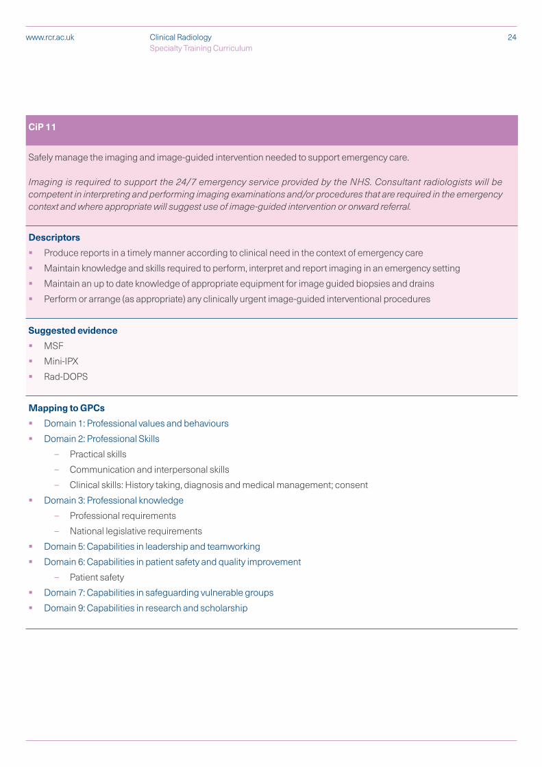
CiP 11
Safely manage the imaging and image-guided intervention needed to support emergency care.
Imaging is required to support the 24/7 emergency service provided by the NHS. Consultant radiologists will be competent in interpreting and performing imaging examinations and/or procedures that are required in the emergency context and where appropriate will suggest use of image-guided intervention or onward referral.
Descriptors
§ Produce reports in a timely manner according to clinical need in the context of emergency care
§ Maintain knowledge and skills required to perform, interpret and report imaging in an emergency setting
§ Maintain an up to date knowledge of appropriate equipment for image guided biopsies and drains
§ Perform or arrange (as appropriate) any clinically urgent image-guided interventional procedures
Suggested evidence
§ MSF
§ Mini-IPX
§ Rad-DOPS
Mapping to GPCs
§ Domain 1: Professional values and behaviours
§ Domain 2: Professional Skills
– Practical skills
– Communication and interpersonal skills
– Clinical skills: History taking, diagnosis and medical management; consent
§ Domain 3: Professional knowledge
– Professional requirements
– National legislative requirements
§ Domain 5: Capabilities in leadership and teamworking
§ Domain 6: Capabilities in patient safety and quality improvement
– Patient safety
§ Domain 7: Capabilities in safeguarding vulnerable groups
§ Domain 9: Capabilities in research and scholarship
25Clinical Radiology Specialty Training Curriculum
www.rcr.ac.uk
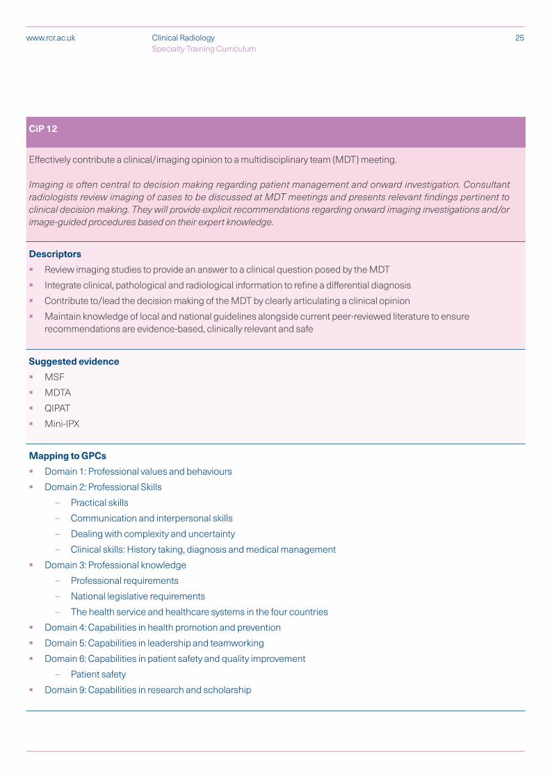
CiP 12
Effectively contribute a clinical/imaging opinion to a multidisciplinary team (MDT) meeting.
Imaging is often central to decision making regarding patient management and onward investigation. Consultant radiologists review imaging of cases to be discussed at MDT meetings and presents relevant findings pertinent to clinical decision making. They will provide explicit recommendations regarding onward imaging investigations and/or image-guided procedures based on their expert knowledge.
Descriptors
§ Review imaging studies to provide an answer to a clinical question posed by the MDT
§ Integrate clinical, pathological and radiological information to refine a differential diagnosis
§ Contribute to/lead the decision making of the MDT by clearly articulating a clinical opinion
§ Maintain knowledge of local and national guidelines alongside current peer-reviewed literature to ensure recommendations are evidence-based, clinically relevant and safe
Suggested evidence
§ MSF
§ MDTA
§ QIPAT
§ Mini-IPX
Mapping to GPCs
§ Domain 1: Professional values and behaviours
§ Domain 2: Professional Skills
– Practical skills
– Communication and interpersonal skills
– Dealing with complexity and uncertainty
– Clinical skills: History taking, diagnosis and medical management
§ Domain 3: Professional knowledge
– Professional requirements
– National legislative requirements
– The health service and healthcare systems in the four countries
§ Domain 4: Capabilities in health promotion and prevention
§ Domain 5: Capabilities in leadership and teamworking
§ Domain 6: Capabilities in patient safety and quality improvement
– Patient safety
§ Domain 9: Capabilities in research and scholarship
26Clinical Radiology Specialty Training Curriculum
www.rcr.ac.uk
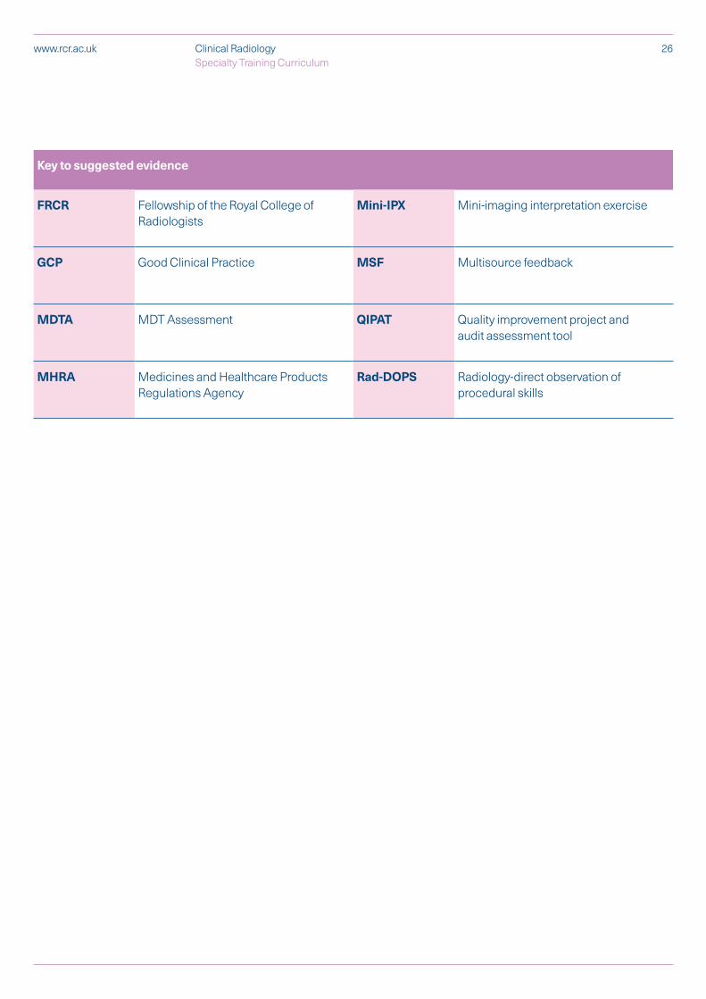
Key to suggested evidence
FRCR Fellowship of the Royal College of Radiologists
Mini-IPX Mini-imaging interpretation exercise
GCP Good Clinical Practice MSF Multisource feedback
MDTA MDT Assessment QIPAT Quality improvement project and audit assessment tool
MHRA Medicines and Healthcare Products Regulations Agency
Rad-DOPS Radiology-direct observation of procedural skills
2.3 Presentations and conditions
Clinical radiology utilises a wide range of imaging modalities and techniques to identify and characterise pathology in the body and can be used to investigate any body system or anatomical region. Clinical radiology trainees are expected to become competent in vetting, protocolling, performing and/or reporting all of the commonly used imaging modalities (e.g. radiographs, fluoroscopy, ultrasound, CT, MRI, radionuclide studies) and to remain up to date with validated new techniques, imaging procedures and protocols.
Any attempt to comprehensively list all clinical presentations, pathological conditions, imaging modalities and techniques would be extensive, but inevitably incomplete, and would rapidly become out of date. Our approach is to provide general guidance and not exhaustive detail. Tables 1-3 outline at a high level the key clinical presentations and conditions presenting to clinical radiology for imaging and the key practical procedures that trainees are expected to have experience of. These tables are not comprehensive; they must be viewed as a guide and interpreted with common sense.
Table 1 describes key clinical presentations and conditions for each of the main body systems. Particular presentations and conditions are listed either because they are common or serious. Clearly some conditions may manifest in a number of body systems and some presentations may be caused by pathology in one or more system, however for conciseness each condition and presentation appears once in the syllabus or on a limited number of occasions.
It is expected that trainees will:
1. be familiar with the normal anatomy and normal variants in each system
2. develop knowledge of the imaging findings of the pathological processes and post treatment appearances affecting each body system including:
– genetic / congenital / developmental conditions
– trauma
– infection
– inflammation
– neoplasia
– connective tissue disorders
– autoimmune disorders
– neurological disorders
– vascular pathology
– haematological diseases
– endocrine diseases
– degenerative diseases
– metabolic disorders
– iatrogenic conditions
– pregnancy associated conditions
– psychiatric associated conditions
By the end of their training all radiology trainees will be expected to advise on the optimum imaging strategy for a given presentation or condition, including selection of the most appropriate modality and protocol for the examination. This should include radionuclide or molecular imaging techniques where appropriate.
27Clinical Radiology Specialty Training Curriculum
www.rcr.ac.uk
All radiologists are required to be trained in a number of basic image guided procedures such as performing biopsies and inserting tubes and drains, as well as performing diagnostic procedural work such as fluoroscopy. It is expected that all trainees will have knowledge of appropriate interventional radiology strategies when investigating the range of common presentations and conditions given in Table 1 and demonstrate the ability to select and use basic interventional radiology techniques. Appropriate adaptations or adjustments should be made to allow trainees with physical disabilities to access this aspect of the curriculum.
28Clinical Radiology Specialty Training Curriculum
www.rcr.ac.uk
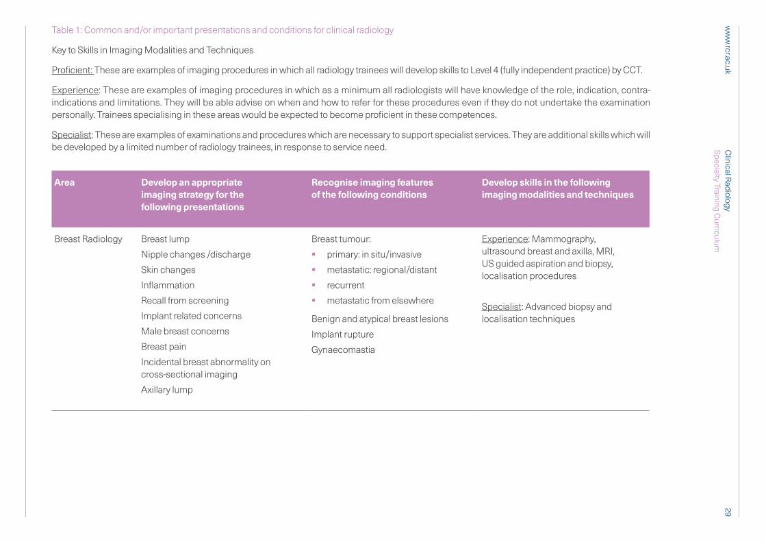
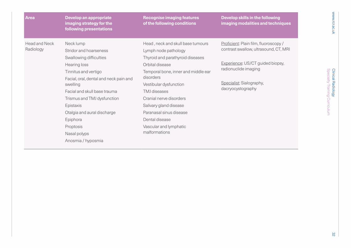
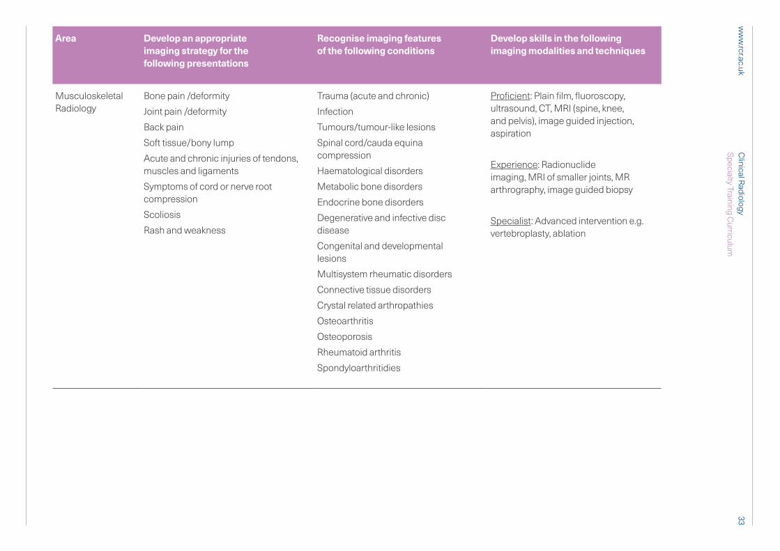
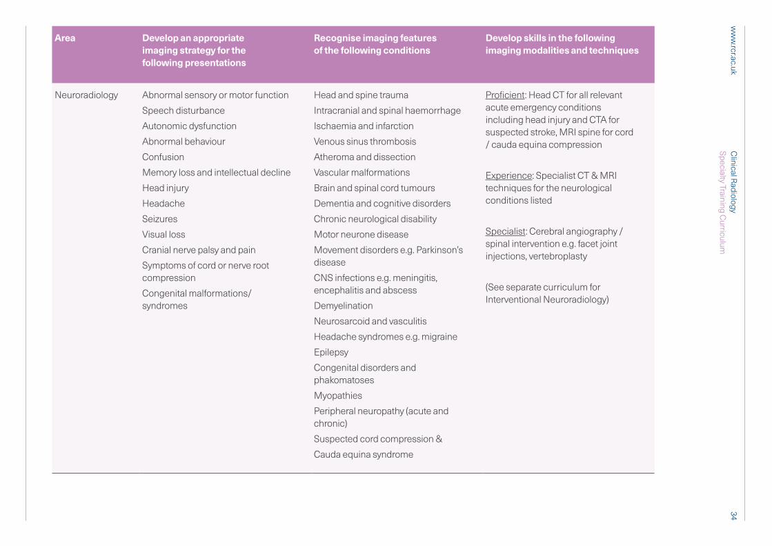
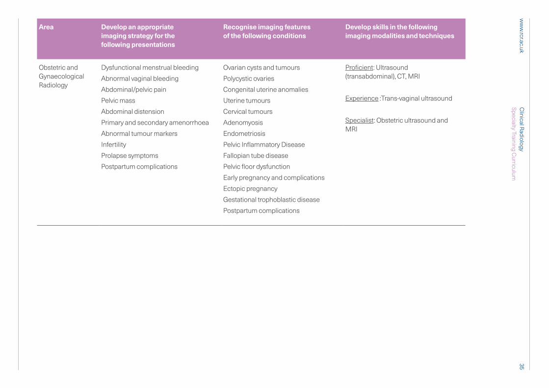
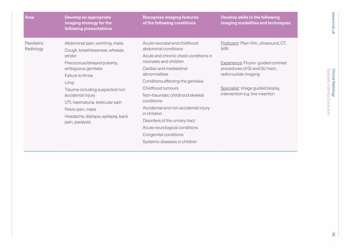
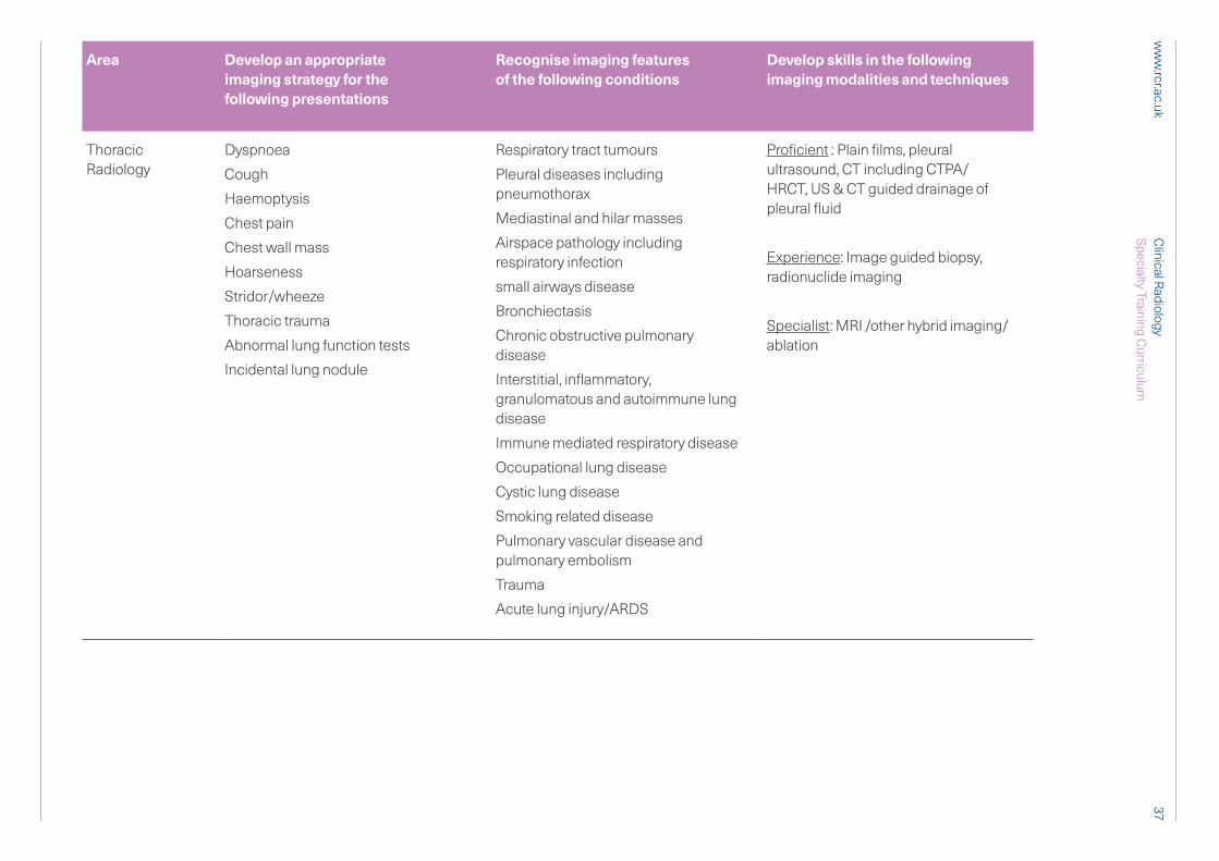
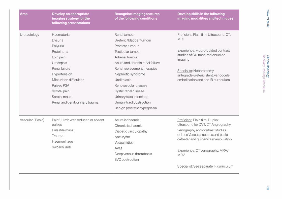
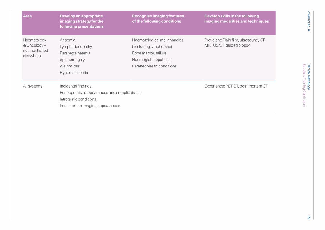
Table 1: Common and/or important presentations and conditions for clinical radiology
Key to Skills in Imaging Modalities and Techniques
Proficient: These are examples of imaging procedures in which all radiology trainees will develop skills to Level 4 (fully independent practice) by CCT.
Experience: These are examples of imaging procedures in which as a minimum all radiologists will have knowledge of the role, indication, contra-indications and limitations. They will be able advise on when and how to refer for these procedures even if they do not undertake the examination personally. Trainees specialising in these areas would be expected to become proficient in these competences.
Specialist: These are examples of examinations and procedures which are necessary to support specialist services. They are additional skills which will be developed by a limited number of radiology trainees, in response to service need.
Area Develop an appropriate imaging strategy for the following presentations
Recognise imaging features of the following conditions
Develop skills in the following imaging modalities and techniques
Breast Radiology Breast lump
Nipple changes /discharge
Skin changes
Inflammation
Recall from screening
Implant related concerns
Male breast concerns
Breast pain
Incidental breast abnormality on cross-sectional imaging
Axillary lump
Breast tumour:
§ primary: in situ/invasive
§ metastatic: regional/distant
§ recurrent
§ metastatic from elsewhere
Benign and atypical breast lesions
Implant rupture
Gynaecomastia
Experience: Mammography, ultrasound breast and axilla, MRI, US guided aspiration and biopsy, localisation procedures
Specialist: Advanced biopsy and localisation techniques
29C
linical Radiology
Specialty Training Curriculum
ww
w.rcr.ac.uk
Area Develop an appropriate imaging strategy for the following presentations
Recognise imaging features of the following conditions
Develop skills in the following imaging modalities and techniques
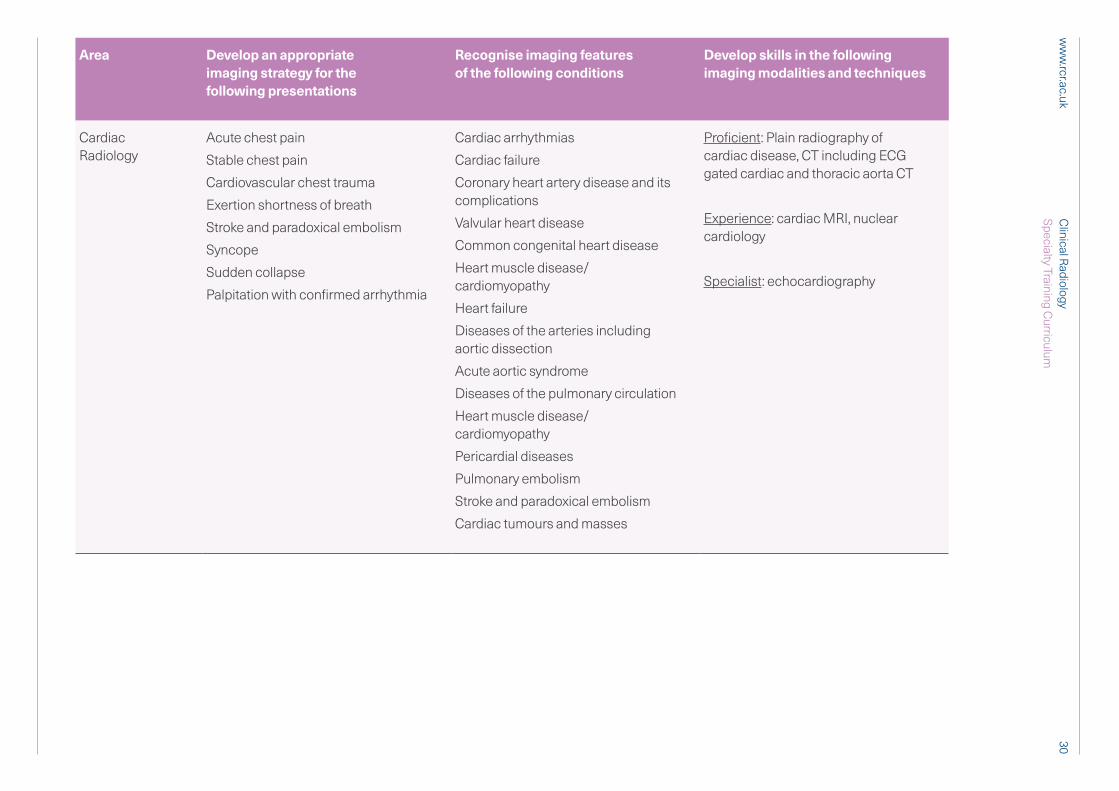
Cardiac Radiology
Acute chest pain
Stable chest pain
Cardiovascular chest trauma
Exertion shortness of breath
Stroke and paradoxical embolism
Syncope
Sudden collapse
Palpitation with confirmed arrhythmia
Cardiac arrhythmias
Cardiac failure
Coronary heart artery disease and its complications
Valvular heart disease
Common congenital heart disease
Heart muscle disease/ cardiomyopathy
Heart failure
Diseases of the arteries including aortic dissection
Acute aortic syndrome
Diseases of the pulmonary circulation
Heart muscle disease/cardiomyopathy
Pericardial diseases
Pulmonary embolism
Stroke and paradoxical embolism
Cardiac tumours and masses
Proficient: Plain radiography of cardiac disease, CT including ECG gated cardiac and thoracic aorta CT
Experience: cardiac MRI, nuclear cardiology
Specialist: echocardiography
30C
linical Radiology
Specialty Training Curriculum
ww
w.rcr.ac.uk
Area Develop an appropriate imaging strategy for the following presentations
Recognise imaging features of the following conditions
Develop skills in the following imaging modalities and techniques
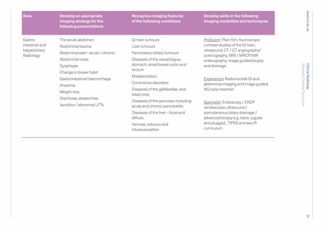
Gastro-Intestinal and hepatobiliary Radiology
The acute abdomen
Abdominal trauma
Abdominal pain - acute / chronic
Abdominal mass
Dysphagia
Change in bowel habit
Gastrointestinal haemorrhage
Anaemia
Weight loss
Diarrhoea, steatorrhea
Jaundice / abnormal LFTs
GI tract tumours
Liver tumours
Pancreatico-biliary tumours
Diseases of the oesophagus, stomach, small bowel colon and rectum
Malabsorption
Continence disorders
Diseases of the gallbladder, and biliary tree
Diseases of the pancreas including acute and chronic pancreatitis
Diseases of the liver – focal and diffuse.
Herniae, volvulus and intussusception
Proficient: Plain film, fluoroscopic contrast studies of the GI tract, ultrasound, CT / CT angiography/colonography, MRI / MRCP/MR enterography, image guided biopsy and drainage
Experience: Radionuclide GI and abdominal imaging and image guided NG tube insertion
Specialist: Endoscopy / ERCP /endoscopic ultrasound / percutaneous biliary drainage / advanced biopsy e.g. trans- jugular and plugged , TIPSS and see IR curriculum
31C
linical Radiology
Specialty Training Curriculum
ww
w.rcr.ac.uk
Area Develop an appropriate imaging strategy for the following presentations
Recognise imaging features of the following conditions
Develop skills in the following imaging modalities and techniques
Head and Neck Radiology
Neck lump
Stridor and hoarseness
Swallowing difficulties
Hearing loss
Tinnitus and vertigo
Facial, oral, dental and neck pain and swelling
Facial and skull base trauma
Trismus and TMJ dysfunction
Epistaxis
Otalgia and aural discharge
Epiphora
Proptosis
Nasal polyps
Anosmia / hyposmia
Head , neck and skull base tumours
Lymph node pathology
Thyroid and parathyroid diseases
Orbital disease
Temporal bone, inner and middle ear disorders
Vestibular dysfunction
TMJ diseases
Cranial nerve disorders
Salivary gland disease
Paranasal sinus disease
Dental disease
Vascular and lymphatic malformations
Proficient: Plain film, fluoroscopy / contrast swallow, ultrasound, CT, MRI
Experience: US/CT guided biopsy, radionuclide imaging
Specialist: Sialography, dacryocystography
32C
linical Radiology
Specialty Training Curriculum
ww
w.rcr.ac.uk
Area Develop an appropriate imaging strategy for the following presentations
Recognise imaging features of the following conditions
Develop skills in the following imaging modalities and techniques
Musculoskeletal Radiology
Bone pain /deformity
Joint pain /deformity
Back pain
Soft tissue/bony lump
Acute and chronic injuries of tendons, muscles and ligaments
Symptoms of cord or nerve root compression
Scoliosis
Rash and weakness
Trauma (acute and chronic)
Infection
Tumours/tumour-like lesions
Spinal cord/cauda equina compression
Haematological disorders
Metabolic bone disorders
Endocrine bone disorders
Degenerative and infective disc disease
Congenital and developmental lesions
Multisystem rheumatic disorders
Connective tissue disorders
Crystal related arthropathies
Osteoarthritis
Osteoporosis
Rheumatoid arthritis
Spondyloarthritidies
Proficient: Plain film, fluoroscopy, ultrasound, CT, MRI (spine, knee, and pelvis), image guided injection, aspiration
Experience: Radionuclide imaging, MRI of smaller joints, MR arthrography, image guided biopsy
Specialist: Advanced intervention e.g. vertebroplasty, ablation
33C
linical Radiology
Specialty Training Curriculum
ww
w.rcr.ac.uk
Area Develop an appropriate imaging strategy for the following presentations
Recognise imaging features of the following conditions
Develop skills in the following imaging modalities and techniques
Neuroradiology Abnormal sensory or motor function
Speech disturbance
Autonomic dysfunction
Abnormal behaviour
Confusion
Memory loss and intellectual decline
Head injury
Headache
Seizures
Visual loss
Cranial nerve palsy and pain
Symptoms of cord or nerve root compression
Congenital malformations/syndromes
Head and spine trauma
Intracranial and spinal haemorrhage
Ischaemia and infarction
Venous sinus thrombosis
Atheroma and dissection
Vascular malformations
Brain and spinal cord tumours
Dementia and cognitive disorders
Chronic neurological disability
Motor neurone disease
Movement disorders e.g. Parkinson’s disease
CNS infections e.g. meningitis, encephalitis and abscess
Demyelination
Neurosarcoid and vasculitis
Headache syndromes e.g. migraine
Epilepsy
Congenital disorders and phakomatoses
Myopathies
Peripheral neuropathy (acute and chronic)
Suspected cord compression &
Cauda equina syndrome
Proficient: Head CT for all relevant acute emergency conditions including head injury and CTA for suspected stroke, MRI spine for cord / cauda equina compression
Experience: Specialist CT & MRI techniques for the neurological conditions listed
Specialist: Cerebral angiography / spinal intervention e.g. facet joint injections, vertebroplasty
(See separate curriculum for Interventional Neuroradiology)
34C
linical Radiology
Specialty Training Curriculum
ww
w.rcr.ac.uk
Area Develop an appropriate imaging strategy for the following presentations
Recognise imaging features of the following conditions
Develop skills in the following imaging modalities and techniques
Obstetric and Gynaecological Radiology
Dysfunctional menstrual bleeding
Abnormal vaginal bleeding
Abdominal/pelvic pain
Pelvic mass
Abdominal distension
Primary and secondary amenorrhoea
Abnormal tumour markers
Infertility
Prolapse symptoms
Postpartum complications
Ovarian cysts and tumours
Polycystic ovaries
Congenital uterine anomalies
Uterine tumours
Cervical tumours
Adenomyosis
Endometriosis
Pelvic Inflammatory Disease
Fallopian tube disease
Pelvic floor dysfunction
Early pregnancy and complications
Ectopic pregnancy
Gestational trophoblastic disease
Postpartum complications
Proficient: Ultrasound (transabdominal), CT, MRI
Experience :Trans-vaginal ultrasound
Specialist: Obstetric ultrasound and MRI
35C
linical Radiology
Specialty Training Curriculum
ww
w.rcr.ac.uk
Area Develop an appropriate imaging strategy for the following presentations
Recognise imaging features of the following conditions
Develop skills in the following imaging modalities and techniques
Paediatric Radiology
Abdominal pain, vomiting, mass
Cough, breathlessness, wheeze, stridor
Precocious/delayed puberty, ambiguous genitalia
Failure to thrive
Limp
Trauma including suspected non accidental injury
UTI, haematuria, testicular pain
Pelvic pain, mass
Headache, diplopia, epilepsy, back pain, paralysis
Acute neonatal and childhood abdominal conditions
Acute and chronic chest conditions in neonates and children
Cardiac and mediastinal abnormalities
Conditions affecting the genitalia
Childhood tumours
Non-traumatic childhood skeletal conditions
Accidental and non accidental injury in children
Disorders of the urinary tract
Acute neurological conditions
Congenital conditions
Systemic diseases in children
Proficient: Plain film, ultrasound, CT, MRI
Experience: Fluoro- guided contrast procedures of GI and GU tract , radionuclide imaging
Specialist: Image guided biopsy, intervention e.g. line insertion
36C
linical Radiology
Specialty Training Curriculum
ww
w.rcr.ac.uk
Area Develop an appropriate imaging strategy for the following presentations
Recognise imaging features of the following conditions
Develop skills in the following imaging modalities and techniques
Thoracic Radiology
Dyspnoea
Cough
Haemoptysis
Chest pain
Chest wall mass
Hoarseness
Stridor/wheeze
Thoracic trauma
Abnormal lung function tests
Incidental lung nodule
Respiratory tract tumours
Pleural diseases including pneumothorax
Mediastinal and hilar masses
Airspace pathology including respiratory infection
small airways disease
Bronchiectasis
Chronic obstructive pulmonary disease
Interstitial, inflammatory, granulomatous and autoimmune lung disease
Immune mediated respiratory disease
Occupational lung disease
Cystic lung disease
Smoking related disease
Pulmonary vascular disease and pulmonary embolism
Trauma
Acute lung injury/ARDS
Proficient : Plain films, pleural ultrasound, CT including CTPA/ HRCT, US & CT guided drainage of pleural fluid
Experience: Image guided biopsy, radionuclide imaging
Specialist: MRI /other hybrid imaging/ablation
37C
linical Radiology
Specialty Training Curriculum
ww
w.rcr.ac.uk
Area Develop an appropriate imaging strategy for the following presentations
Recognise imaging features of the following conditions
Develop skills in the following imaging modalities and techniques
Uroradiology Haematuria
Dysuria
Polyuria
Proteinuria
Loin pain
Urosepsis
Renal failure
Hypertension
Micturition difficulties
Raised PSA
Scrotal pain
Scrotal mass
Renal and genitourinary trauma
Renal tumour
Ureteric/bladder tumour
Prostate tumour
Testicular tumour
Adrenal tumour
Acute and chronic renal failure
Renal replacement therapies
Nephrotic syndrome
Urolithiasis
Renovascular disease
Cystic renal disease
Urinary tract infections
Urinary tract obstruction
Benign prostatic hyperplasia
Proficient: Plain film, Ultrasound, CT, MRI
Experience: Fluoro-guided contrast studies of GU tract , radionuclide imaging
Specialist: Nephrostomy, antegrade ureteric stent, varicocele embolisation and see IR curriculum
Vascular ( Basic) Painful limb with reduced or absent pulses
Pulsatile mass
Trauma
Haemorrhage
Swollen limb
Acute ischaemia
Chronic ischaemia
Diabetic vasculopathy
Aneurysm
Vasculitidies
AVM
Deep venous thrombosis
SVC obstruction
Proficient: Plain film, Duplex ultrasound for DVT, CT Angiography
Venography and contrast studies of lines Vascular access and basic catheter and guidewire manipulation
Experience: CT venography, MRA/MRV
Specialist: See separate IR curriculum
38C
linical Radiology
Specialty Training Curriculum
ww
w.rcr.ac.uk
Area Develop an appropriate imaging strategy for the following presentations
Recognise imaging features of the following conditions
Develop skills in the following imaging modalities and techniques
Haematology & Oncology – not mentioned elsewhere
Anaemia
Lymphadenopathy
Paraproteinaemia
Splenomegaly
Weight loss
Hypercalcaemia
Haematological malignancies
( including lymphomas)
Bone marrow failure
Haemoglobinopathies
Paraneoplastic conditions
Proficient: Plain film, ultrasound, CT, MRI, US/CT guided biopsy
All systems Incidental findings
Post-operative appearances and complications
Iatrogenic conditions
Post mortem imaging appearances
Experience: PET CT, post-mortem CT
39C
linical Radiology
Specialty Training Curriculum
ww
w.rcr.ac.uk
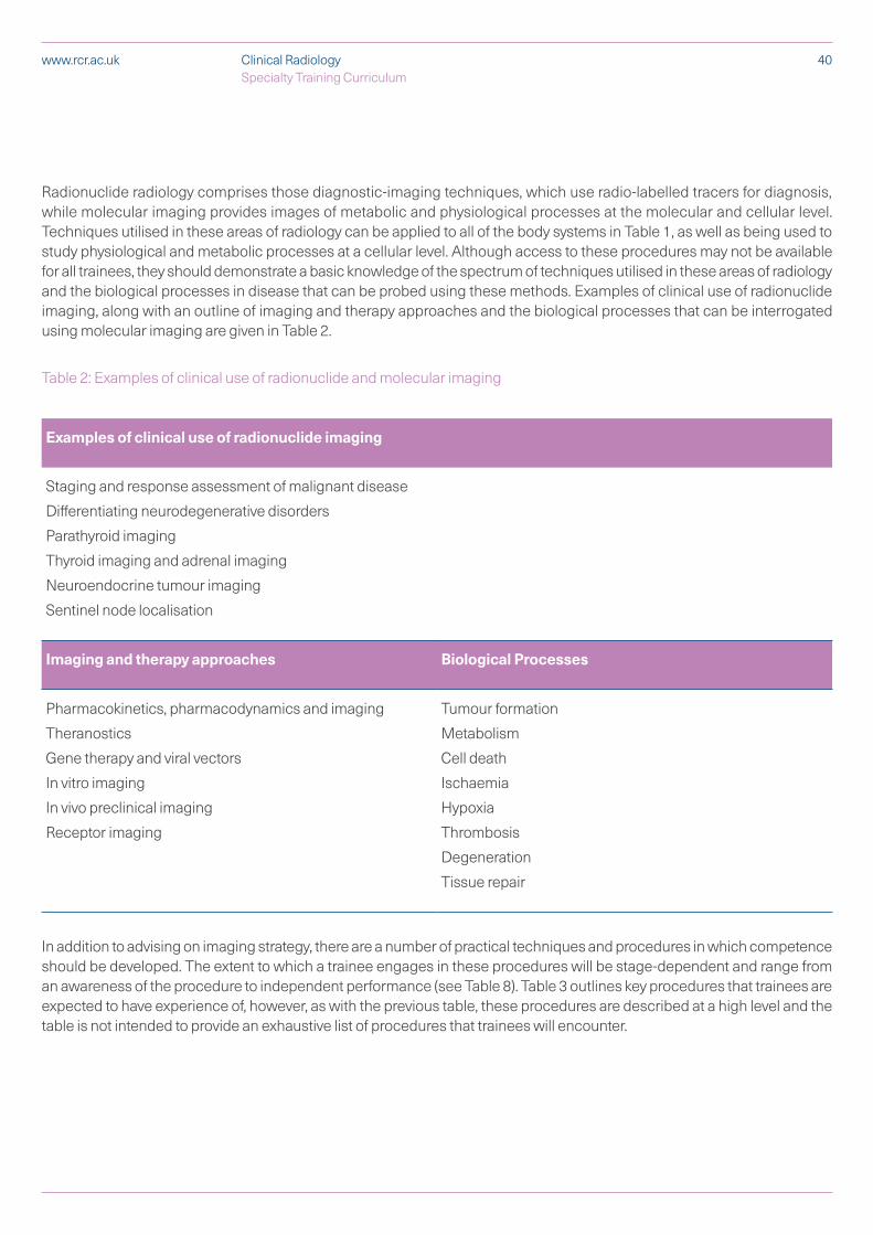
Radionuclide radiology comprises those diagnostic-imaging techniques, which use radio-labelled tracers for diagnosis, while molecular imaging provides images of metabolic and physiological processes at the molecular and cellular level. Techniques utilised in these areas of radiology can be applied to all of the body systems in Table 1, as well as being used to study physiological and metabolic processes at a cellular level. Although access to these procedures may not be available for all trainees, they should demonstrate a basic knowledge of the spectrum of techniques utilised in these areas of radiology and the biological processes in disease that can be probed using these methods. Examples of clinical use of radionuclide imaging, along with an outline of imaging and therapy approaches and the biological processes that can be interrogated using molecular imaging are given in Table 2.
Table 2: Examples of clinical use of radionuclide and molecular imaging
Examples of clinical use of radionuclide imaging
Staging and response assessment of malignant disease
Differentiating neurodegenerative disorders
Parathyroid imaging
Thyroid imaging and adrenal imaging
Neuroendocrine tumour imaging
Sentinel node localisation
Imaging and therapy approaches Biological Processes
Pharmacokinetics, pharmacodynamics and imaging
Theranostics
Gene therapy and viral vectors
In vitro imaging
In vivo preclinical imaging
Receptor imaging
Tumour formation
Metabolism
Cell death
Ischaemia
Hypoxia
Thrombosis
Degeneration
Tissue repair
In addition to advising on imaging strategy, there are a number of practical techniques and procedures in which competence should be developed. The extent to which a trainee engages in these procedures will be stage-dependent and range from an awareness of the procedure to independent performance (see Table 8). Table 3 outlines key procedures that trainees are expected to have experience of, however, as with the previous table, these procedures are described at a high level and the table is not intended to provide an exhaustive list of procedures that trainees will encounter.
40Clinical Radiology Specialty Training Curriculum
www.rcr.ac.uk
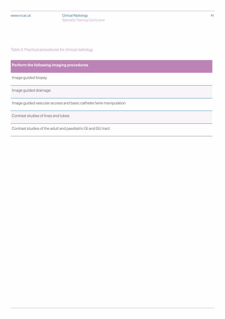
Table 3: Practical procedures for clinical radiology
Perform the following imaging procedures
Image guided biopsy
Image guided drainage
Image guided vascular access and basic catheter/wire manipulation
Contrast studies of lines and tubes
Contrast studies of the adult and paediatric GI and GU tract
41Clinical Radiology Specialty Training Curriculum
www.rcr.ac.uk
2.4 Breadth of training 2.4.1 Interventional RadiologyExposure to interventional radiology in general clinical radiology training is vital to enable continued provision of basic image-guided diagnostic and therapeutic intervention where a formal IR service is not available, or to support a formal IR service. Knowledge of IR techniques, including common indications for their use, is also important to allow appropriate informed clinical discussion in a reporting or MDT setting. Some CR trainees may decide to focus on specific IR techniques to supplement their area of special interest. Although this would not lead to an IR specific CCT, it should be encouraged and supported.
2.4.2 Emerging technologiesTrainees are expected to keep up to date and to embrace and evaluate emerging technologies such as Artificial Intelligence ( AI), Machine Learning, Deep Learning and Radiomics, where these act as an adjunct to imaging analysis and interpretation. AI tools are being developed to assist with diagnostic assessments and trainees should be prepared to adapt these tools into clinical practice once validated. This will require consideration of the following:
§ basic statistics needed to empower radiologists to be able to design and/or interpret a clinical trial in the workplace, involving the testing of AI software or a scientific hypothesis so as to draw meaningful conclusions
§ to be aware of the concepts related to data curation, confidentiality and anonymisation, and regulations concerning use of patient data
§ appreciate the role of image annotation, and how annotated images can be harnessed as data for research and clinical care
§ understand the basic concepts of radiomics
§ understand the meaning, requirements of, and pitfalls inherent in training data, validation data and testing data in developing AI software
§ appreciate AI and machine learning terms including: convolutional neural network, random forest, dice index, use case
2.4.3 Emerging imaging techniques Trainees should be aware of emerging imaging techniques and to undertake training in these techniques where these become available according to their specialist interest. Examples of this include hybrid imaging and CT post mortem scanning. Hybrid imaging in the form of PET/CT is well established in clinical practice and all trainees are expected to have experience of this, but other newer forms of hybrid imaging may evolve into clinical routine. CT post mortem imaging is being performed more frequently and there is likely to be increasing demand for radiologists’ skills in interpreting these examinations in future. While core radiology skills can be applied to image analysis in these emerging techniques, trainees should understand that this is not sufficient alone and that specific training in the emerging technique will be required.
2.4.4 Academic trainingAll trainees are required to demonstrate an understanding of research methodology and critical appraisal linked to clinical practice. There are various ways in which this can be demonstrated. Trainees are required to contribute to a research project during training to gain experience of undertaking research. Alternatively, trainees could, with their educational supervisors, develop a research question and a protocol as a theoretical exercise. All trainees
42Clinical Radiology Specialty Training Curriculum
www.rcr.ac.uk
should develop their critical appraisal skills and regularly appraise and discuss current research papers – for example as part of regular journal clubs.
Trainees may choose to undertake a combined clinical and academic training programme and some trainees may opt to do research leading to a higher degree without being appointed to a formal academic programme. The four nations have different arrangements for academic training and doctors in training should consult their training programme director (TPD) or deanery for further guidance.
2.4.5 Taking time out of programme There are a number of circumstances when a trainee may seek to spend some time out of specialty training, such as undertaking a period of research or taking up a fellowship post. All such requests must be agreed by the postgraduate dean in advance and trainees are advised to discuss their proposals as early as possible. Full guidance on taking time out of programme can be found in the Gold Guide.
2.4.6 Acting up as a consultantA trainee who has passed the Final FRCR Examination may spend up to three months, during the final year of special interest training, “acting-up” as a consultant, provided that a consultant supervisor is identified for the post and satisfactory progress is made. As long as the trainee remains within an approved training programme, the GMC does not need to approve this period of “acting up” and their original CCT date will not be affected. More information on acting up as a consultant can be found in the Gold Guide.
43Clinical Radiology Specialty Training Curriculum
www.rcr.ac.uk
3 Teaching and learning methods
Health Education England (HEE) and its regional offices, NHS Education for Scotland (NES), the Northern Ireland Medical and Dental Training Agency (NIMDTA) and Health Education and Improvement Wales (HEIW) have overall responsibility for the provision of postgraduate medical training in the four nations of the UK. Responsibility for delivering the training needed to meet the curriculum requirements rests with the individual schools of radiology (where they exist) and training programmes under the oversight of the postgraduate deans. This includes mechanisms for addressing under-performance and providing remediation. The GMC’s Promoting Excellence standards set out requirements for the management and delivery of postgraduate medical education and training. The Gold Guide provides further guidance on the management and expectations of training.
Progression through the programme will be determined by the annual review of competence progression (ARCP) process (see section 4.6) and the training requirements for each indicative year of training are summarised in the progression grids for the generic CiPs, specialty-specific CiPs and critical progression points (see sections 4.3 and 4.4). The successful completion of clinical radiology training will be dependent on achieving the expected level in all CiPs and procedural skills. The programme of assessment will be used to monitor and determine progress through the programme. Training will normally take place in a range of district general hospitals and teaching hospitals.
The sequence of training should ensure appropriate progression in experience and responsibility. The training to be provided at each training site is defined to ensure that, during the programme, the entire syllabus is covered and also that unnecessary duplication and educationally unrewarding experiences are avoided.
The curriculum will be delivered through a variety of learning experiences and will allow trainees to achieve the capabilities described through a variety of learning methods. There will be a balance of different modes of learning from formal teaching programmes to experiential learning ‘on the job’. The proportion of time allocated to different learning methods may vary depending on the nature of the attachment within a rotation. Clinical and educational supervisors are encouraged to identify learner-centred educational opportunities in the course of clinical work, maximising the wide variety of learning opportunities in the clinical radiological workplace. Rotations should be constructed to enable trainees to experience the full range of educational and training opportunities available and there will be robust arrangements for quality assurance in place to ensure consistent implementation of the curriculum.
This section identifies the types of situations in which a trainee will learn.
The content of work-based experiential learning is decided by the local faculty but includes active participation in:
§ radiological attachments with gradual reduction in supervision according to increasing competence as judged by trainers (apprenticeship model): Trainees will spend a large proportion of their time involved in supervised radiological practice in a hospital setting. Learning will involve the trainee undertaking an increasing number of radiological tasks
3.1 Work-based experiential learning
44Clinical Radiology Specialty Training Curriculum
www.rcr.ac.uk
in all areas of the imaging department and in other areas where imaging services are provided (e.g. bedside ultrasound).
§ multidisciplinary team meetings: These inter-disciplinary meetings provide excellent learning opportunities.
§ on-call and emergency provision: On-call work should be a positive learning opportunity and this requires appropriate levels of supervision by the consultant on-call with trainees. Whilst the RCR cannot define working arrangements that can be put in place by the employer, guidance that ensures on-call work for trainees can maximise learning is published on the RCR website.
The degree of responsibility taken by the trainee will increase as competency increases. There should be appropriate levels of supervision throughout training with increasing independence and responsibility as learning outcomes are achieved.
3.1.1 Optional work-based experiential learning Time spent within clinical teams related to a special interest area (e.g. with respiratory physicians to support the trainee’s learning of chest radiology) to more fully understand clinical aspects of their work either as a clinical attachment or paired with an appropriate trainee clinician to engage in co-learning, as agreed with clinical supervisors is encouraged.
Formal postgraduate teaching can take a variety of forms and may include:
§ a programme of structured, regular teaching sessions to cohorts of trainees
§ case presentations
§ journal clubs
§ research and audit projects
§ lectures and small group teaching
§ grand Rounds
§ radiological skills demonstrations and teaching
§ joint meetings with clinical specialties
§ attendance at training courses organised on a school or regional basis, which are designed to cover aspects of the training programme outlined in this curriculum
Time will be provided during training for personal study, including longer periods offered as part of study leave. Trainees will use this time in a variety of ways depending upon their stage of learning. Suggested activities include:
§ preparation for assessment and examinations
§ reading, including journals and web-based material
§ maintenance of personal portfolio (self-assessment, reflective learning, personal development plan)
§ audit, quality improvement and research projects
§ achieving personal learning goals beyond normal expectation
3.2 Formal postgraduate teaching
3.3 Independent self-directed learning
45Clinical Radiology Specialty Training Curriculum
www.rcr.ac.uk
Time to be made available for external courses is encouraged, subject to local conditions of service. Examples include management courses and communication courses. Please consult the Gold Guide and deanery guidance on study leave for more information.
There are many opportunities for trainees to learn with and from their peers. Local postgraduate teaching opportunities allow trainees of varied levels of experience to come together for small group sessions. Examination preparation encourages the formation of self-help groups and learning sets.
Simulation is recognised as a useful tool to supplement training in clinical situations. It provides experiential learning and an opportunity to reflect on and learn from mistakes in a safe environment.
A number of different types of simulation are available, which vary considerably in technological complexity, cost and availability. There are many scenarios in radiology where simulation can play a useful role in supporting delivery of this curriculum and each training centre is encouraged to incorporate these techniques into the training programme wherever possible.
3.4 External study courses
3.5 Learning with peers
3.6 Simulation
46Clinical Radiology Specialty Training Curriculum
www.rcr.ac.uk
4 Programme of assessment
4.1 Purpose of assessment
The programme of assessment refers to the integrated framework of exams, assessments in the workplace and judgements made about a learner during their training. The purpose of the programme of assessment is to robustly evidence, ensure and clearly communicate the expected levels of performance at critical progression points, and to demonstrate satisfactory completion of training as required by the curriculum. In order to achieve this, the programme of assessment aims to:
§ enhance learning by providing formative assessment, enabling trainees to receive immediate feedback, understand their own performance and identify areas for development
§ drive learning and enhance the training process by making it clear what is required of trainees and motivating them to ensure they receive suitable training and experience
§ ensure that trainees possess the essential underlying knowledge required for clinical radiology
§ assess trainees’ actual performance in the workplace
§ demonstrate trainees have acquired the GPCs and meet the requirements of GMP
§ provide robust, summative evidence that trainees are meeting the curriculum standards during the training programme
§ inform the ARCP, identifying any requirements for targeted or additional training where necessary and facilitating decisions regarding progression through the training programme
§ identify trainees who may benefit from careers counselling
§ recognise and acknowledge the potential for excellence and where trainees are performing over and above expectations for their stage of training
Accountable, professional judgment is central to ensuring that trainees have demonstrated the CiPs and met the expected levels of performance set out in the curriculum. The programme of assessment details how professional judgements are used and collated to support decisions on progression and satisfactory completion of training.
The programme of assessment is comprised of several different individual types of assessment, covering both summative and formative assessment. Assessment will take place throughout the training programme to allow trainees to continually gather evidence of learning and to provide the formative feedback essential to improving clinical practice. Continuous review and assessment is a fundamental part of clinical radiology training. Radiology trainees are expected to demonstrate improvement and progression during each attachment. It is important that they arrange and undertake assessments in a timely and educationally appropriate manner spread throughout the training year. All assessments, including those conducted in the workplace, are linked to the relevant CiPs (e.g. through the blueprinting of assessment system to the CiPs).
A range of assessments, based on the judgement of many assessors, on multiple occasions, are needed to generate the necessary evidence required for global judgements to be made about satisfactory performance, progression in, and completion of, training. The TPD will
4.2 Programme of assessment
47Clinical Radiology Specialty Training Curriculum
www.rcr.ac.uk
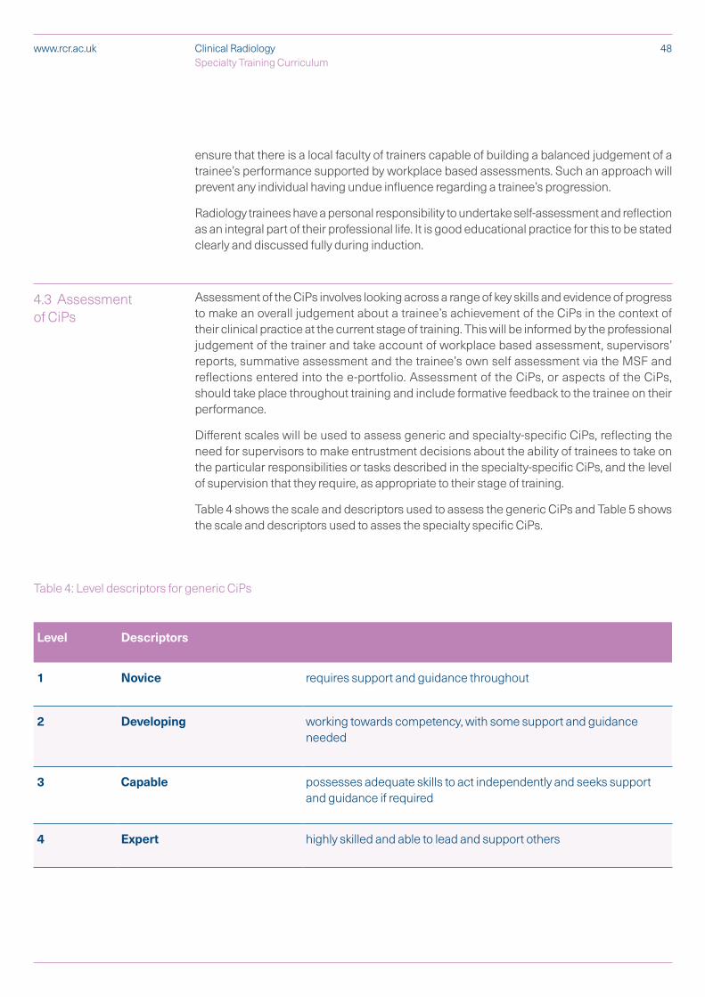
ensure that there is a local faculty of trainers capable of building a balanced judgement of a trainee’s performance supported by workplace based assessments. Such an approach will prevent any individual having undue influence regarding a trainee’s progression.
Radiology trainees have a personal responsibility to undertake self-assessment and reflection as an integral part of their professional life. It is good educational practice for this to be stated clearly and discussed fully during induction.
Assessment of the CiPs involves looking across a range of key skills and evidence of progress to make an overall judgement about a trainee’s achievement of the CiPs in the context of their clinical practice at the current stage of training. This will be informed by the professional judgement of the trainer and take account of workplace based assessment, supervisors’ reports, summative assessment and the trainee’s own self assessment via the MSF and reflections entered into the e-portfolio. Assessment of the CiPs, or aspects of the CiPs, should take place throughout training and include formative feedback to the trainee on their performance.
Different scales will be used to assess generic and specialty-specific CiPs, reflecting the need for supervisors to make entrustment decisions about the ability of trainees to take on the particular responsibilities or tasks described in the specialty-specific CiPs, and the level of supervision that they require, as appropriate to their stage of training.
Table 4 shows the scale and descriptors used to assess the generic CiPs and Table 5 shows the scale and descriptors used to asses the specialty specific CiPs.
4.3 Assessment of CiPs
48Clinical Radiology Specialty Training Curriculum
www.rcr.ac.uk
Table 4: Level descriptors for generic CiPs
Level Descriptors
1 Novice requires support and guidance throughout
2 Developing working towards competency, with some support and guidance needed
3 Capable possesses adequate skills to act independently and seeks support and guidance if required
4 Expert highly skilled and able to lead and support others
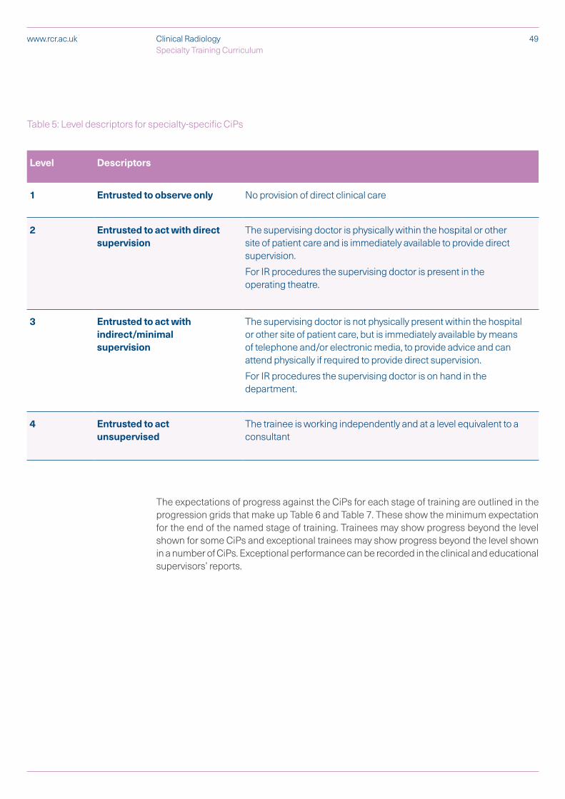
49Clinical Radiology Specialty Training Curriculum
www.rcr.ac.uk
Table 5: Level descriptors for specialty-specific CiPs
Level Descriptors
1 Entrusted to observe only No provision of direct clinical care
2 Entrusted to act with direct supervision
The supervising doctor is physically within the hospital or other site of patient care and is immediately available to provide direct supervision.
For IR procedures the supervising doctor is present in the operating theatre.
3 Entrusted to act with indirect/minimal supervision
The supervising doctor is not physically present within the hospital or other site of patient care, but is immediately available by means of telephone and/or electronic media, to provide advice and can attend physically if required to provide direct supervision.
For IR procedures the supervising doctor is on hand in the department.
4 Entrusted to act unsupervised
The trainee is working independently and at a level equivalent to a consultant
The expectations of progress against the CiPs for each stage of training are outlined in the progression grids that make up Table 6 and Table 7. These show the minimum expectation for the end of the named stage of training. Trainees may show progress beyond the level shown for some CiPs and exceptional trainees may show progress beyond the level shown in a number of CiPs. Exceptional performance can be recorded in the clinical and educational supervisors’ reports.
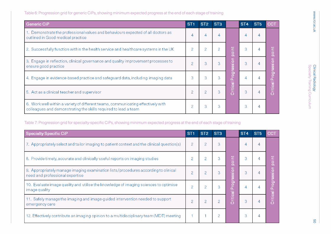
50C
linical Radiology
Specialty Training Curriculum
ww
w.rcr.ac.uk
Table 6: Progression grid for generic CiPs, showing minimum expected progress at the end of each stage of training
Table 7: Progression grid for specialty-specific CiPs, showing minimum expected progress at the end of each stage of training
4.4 Critical progression points
A critical progression point is a point in a curriculum where a learner transitions to higher levels of professional responsibility or enters a new or specialist area of practice, including successful completion of training. These transitions are often associated with an increase in potential risk to patients or those in training, so they need to be carefully managed and decisions to progress need to be based on robust evidence of satisfactory performance.
There are two critical progression points in clinical radiology training. Table 8 outlines the key milestones that trainees should accomplish in order to pass these critical progression points, in addition to achievement of the generic and specialty specific CiPs as appropriate for their stage of training (see Table 6 and Table 7).
The first critical progression point will be where trainees transition to special interest training at the end of ST3. Trainees are expected to have met the required levels for the milestones and procedures outlined in Table 8 by the end of ST3 as a minimum; however individual trainees may achieve the required level for some milestones and procedures at an earlier point in training. Ensuring that trainees have met these requirements by the end of ST3 will allow them to progress to special interest or interventional radiology sub-specialty training with appropriate time to develop their specialist skills while maintaining and further developing their general radiology capabilities.
Trainees will be normally be expected to complete the FRCR 2A examination by the end of ST3. However, passing FRCR 2A alone is not sufficient to begin special interest or IR sub-specialty training. If a trainee has passed FRCR 2A by the end of ST3 but in the opinion of their supervisors has not achieved the other necessary outcomes and milestones to the required level, they should not progress or should only do so with enhanced supervision. Equally there may be a small number of trainees who are performing very well and in whom their supervisors have every confidence but they have not (for a variety of reasons) yet passed the FRCR 2A examination. The ARCP at the end of ST3 will play an important role in these cases in determining individualised, supportive plans for transition to special interest or IR sub-specialty training. Some trainees may require a period of time in a supportive training environment with the supervising physician readily available.
The second critical progression point marks the end of clinical radiology training. Trainees are required to reach level 4 in all generic and specialty-specific CiPs by completion of training.
Radiological practice will be assessed using an integrated package of formative workplace based assessments (WPBAs) and summative examination of knowledge and radiological skills, which will sample across the curriculum. The assessments are supported by structured feedback and are fit for purpose, having undergone evaluation in terms of their feasibility, reliability, validity and reproducibility.
The methods of assessment listed in this section of the curriculum will provide evidence of progress; with the requirements for each stage of training stipulated in the progression grids for the generic CiPs, specialty-specific CiPs and critical progression points (see sections 4.3 and 4.4). Evidence of progress may also be gathered from other sources and trainees are encouraged to demonstrate their progress against the CiPs in a variety of different ways,
4.5 Evidence of progress
51Clinical Radiology Specialty Training Curriculum
www.rcr.ac.uk
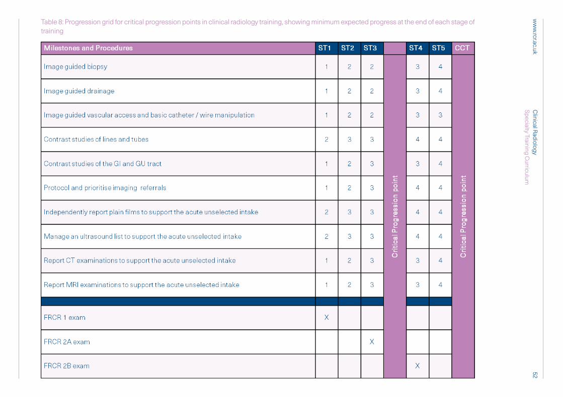
52C
linical Radiology
Specialty Training Curriculum
ww
w.rcr.ac.uk
Table 8: Progression grid for critical progression points in clinical radiology training, showing minimum expected progress at the end of each stage of training
reflecting their strengths, areas of interest and the resources available to them. The trainee will collect evidence to support their self-assessment, and the educational supervisor will use it to reach a global assessment.
4.5.1 E-portfolioOn enrolling with the RCR trainees will be given access to the RCR’s e-portfolio. This is a record of a trainee’s development and progress towards achieving the CiPs. All appraisal meetings, personal development plans and WPBAs should be recorded in the e-portfolio. Trainees are encouraged to reflect on their learning experiences and to record these in the e-portfolio.
The e-portfolio provides a record of objective evidence of capability to work in a range of clinical settings and of satisfactory performance. It will contribute to the educational supervisor’s report and ARCP. Successful completion of the curriculum requires evidence, recorded in the e-portfolio, that the trainee has met all of the generic and specialty-specific CiPs.
It is the trainee’s responsibility to ensure the e-portfolio is kept up to date, arrange assessments and ensure they are recorded, prepare drafts of appraisal forms, maintain their personal development plan, and record their reflections on learning and their progress through the curriculum. It is the supervisor’s responsibility to use the evidence recorded in the e-portfolio (such as outcomes of assessments, reflections and personal development plans) to inform appraisal meetings. They are also expected to update the trainee’s record of progress through the curriculum, write end-of-attachment appraisals and supervisor’s reports.
Deaneries, training programme directors, college tutors and ARCP panels may use the e-portfolio to monitor the progress of trainees for whom they are responsible. The RCR will use summarised, anonymous data from the e-portfolios to support its work in quality assurance.
4.5.2 Summative AssessmentThere are a number of components to summative assessment in clinical radiology training, which together qualify trainees for the award of Fellowship of the Royal College of Radiologists (FRCR). The First FRCR examination consists of two modules: Scientific Basis of Imaging and Anatomy. The second examination is divided into Part A and Part B. The First FRCR Examination (Scientific Basis of Imaging module) and Final FRCR Part A Examination test knowledge through multiple choice and single best answer (SBA) questions. The First FRCR Examination (Anatomy module) tests knowledge by requiring the identification of normal anatomical structures on images. The Final FRCR Part B Examination assesses clinical competence (interpretative, analytical and communication skills). Further guidance for trainees on the structure and content of these exams is available on the RCR website.
Those assessment tools which are not identified individually as summative will contribute to summative judgements about a trainee’s progress as part of the programme of assessment. A suitable number and range of these will ensure reliable assessment of progress and achieve coverage of the curriculum.
53Clinical Radiology Specialty Training Curriculum
www.rcr.ac.uk
54Clinical Radiology Specialty Training Curriculum
www.rcr.ac.uk
4.5.3 Formative AssessmentWorkplace based assessment (WPBA) is the cornerstone of assessment for day-to-day practice. Reflection and feedback is an integral component to all WBPAs to enhance and drive learning. The assessments should be seen as opportunities for identifying strengths and areas for further development; they are not tests that must be passed. Activities to be assessed should be agreed in advance and it is the responsibility of the trainee to arrange this.
In order for trainees to maximise benefit, reflection and feedback should take place as soon as possible after an assessment. Feedback should be of high quality and should include an action plan for future development. Both trainee and trainer should recognise and respect cultural differences when giving and receiving feedback.
A range of assessment tools are available to support WPBA and these are listed below. Minimum numbers of each type WPBA are given, although it is anticipated that trainees may/will undertake many more, as the WPBAs are the vehicles by which the trainee will guarantee one-to-one teaching and ensure appropriate curriculum coverage during their clinical attachments.
LTFT trainees will be expected to undertake the requirements for assessment on a pro rata basis and to spread the balance of workplace based assessments evenly, as set out in the Gold Guide, and ARCP panels must not set expectations beyond this pro-rata number as a basis for decision-making. However, LTFT trainees are also encouraged to undertake more than the minimum number of WPBAs (and at least the same minimum number of WPBAs as full time trainees) in each calendar year on the basis that the numbers are low and WPBAs provide useful learning opportunities.
Multisource feedback (MSF)
This tool is a method of assessing generic skills such as communication, leadership, team working, reliability etc. across the domains of Good Medical Practice. This provides systematic collection and feedback of performance data on a trainee, derived from a number of colleagues. For each assessment, the trainee should nominate at least 15 raters. ‘Raters’ are individuals with whom the trainee works, including supervising consultants, doctors in training more senior than the trainee under assessment and experienced radiographic, nursing or allied health professional colleagues. Raters should be agreed with the educational supervisor at the start of the training year. A minimum of 12 raters must respond to complete the MSF.
The recommended mix of raters/assessors is:
§ 2–4 senior doctors
§ 2–4 doctors in training
§ 2–4 radiographers
§ 2–4 nurses/allied health professionals
§ 2–4 other team members including clerks, secretaries and auxiliary staff
The trainee will not see the individual responses by raters. Feedback is given to the trainee by the educational supervisor.
55Clinical Radiology Specialty Training Curriculum
www.rcr.ac.uk
MSF should usually take place once a year (or at appropriate intervals for LTFT trainees), although the educational supervisor may choose to recommend an additional MSF to investigate a relevant behavioural issue or check progress after an adverse MSF. It is mapped to a self assessment tool with identical domains.
Mini-imaging interpretation exercise (Mini-IPX)
This tool evaluates an observed radiology interpretation/reporting episode. The mini-IPX can be used at any time and in any setting when an assessor is available. Assessors must be trained in giving feedback and understand the role of assessment and a different assessor should be used for each mini-IPX wherever possible, including at least one of consultant level per four month placement. Trainees should agree the timing, problem and assessor, although assessors may also carry out unscheduled assessments. Trainees should receive immediate feedback to aid learning.
Trainees should complete a minimum of six mini-IPXs in each year of training (or equivalent for LTFT trainees). These should be spaced out appropriately, ideally with at least two mini-IPX completed in each post. Mini-IPXs should sample across different clinical radiological problems as summarised in Table 1.
Radiology-direct observation of procedural skills (Rad-DOPS)
A Rad-DOPs is a structured checklist for assessing the interaction of a radiology trainee with the patient when performing a practical procedure. Assessors must be trained both in the procedure and feedback methodology and could include consultants, more senior doctors in training, advanced practitioner radiographers, qualified nurses or allied health professionals. Different assessors should be used for each encounter wherever possible. Trainees should agree the timing, procedure and assessor, although assessors may also carry out unscheduled assessments. Trainees should receive immediate feedback to identify strengths and areas for development.
Trainees should complete a minimum of six Rad-DOPS in each year of training (or equivalent for LTFT trainees), sampling a wide range of different procedures/skills as summarised in Table 3. These should be spaced out appropriately, ideally with at least two Rad-DOPS completed in each post. Rad-DOPS can be undertaken as many times as the trainee and their supervisor feel is necessary and may be used to inform decisions about when a trainee can be regarded as competent to perform a procedure independently.
Teaching observation
The Teaching Observation form is designed to provide structured, formative feedback to trainees on their competence at teaching. It evaluates the competence of a trainee to deliver a teaching episode in a wide variety of settings. Trainees should complete a minimum of two Teaching Observations in each year of training (or equivalent for LTFT trainees), based on any instance of formalised teaching by the trainee, which has been observed by the assessor. The process should be trainee-led (identifying appropriate teaching sessions and assessors).
Quality improvement project and audit assessment tool (QIPAT)
The QIPAT is designed to assess a trainee’s competence in completing an audit or quality improvement project. The assessment can be based on review of audit or quality improvement
56Clinical Radiology Specialty Training Curriculum
www.rcr.ac.uk
documentation or on a presentation at a meeting. If possible, the trainee should be assessed on the same audit or quality improvement project by more than one assessor.
All trainees are expected to complete an audit or quality improvement project for each year within the training programme (or equivalent for LTFT trainees). Trainees should show how they have instigated, collated and presented a piece of work, as well as reflected upon any changes in clinical management as a result of work completed.
MDT Assessment (MDTA)
The MDT Assessment Tool is designed to provide feedback on a trainee’s ability to contribute effectively to multidisciplinary team working and to assume a leadership role in multidisciplinary meetings. As with other WPBAs it is based on the assessor observing a trainee and providing feedback.
MDTAs are optional for trainees in ST1-ST3; however a minimum of two MDTAs per year is expected in ST4 and ST5.
Reflection and log books
The e-portfolio contains a number of documents to support reflection, including blank reflection forms and templates that provide prompts for different types of reflection. Trainees may set any reflections recorded in the e-portfolio to private so that they can only be viewed by the trainee or make them available to their supervisors on their timeline. Further guidance on effective reflection is available on the GMC website.
Trainees are also encouraged to keep log books of practical procedures to document the skills and experience attained and to facilitate reflective learning.
Educational supervisor’s report
The educational supervisor will periodically (at least annually) draw together the results of a trainee’s educational activities to give an overview of their progress in a formal structured educational supervisor’s report. The overall judgment of a trainee will include a triangulated view of the doctor’s performance, which will include their participation in educational activities, appraisals, the assessment process and recording of this in the e-portfolio. The educational supervisor’s report can incorporate commentary or reports from longitudinal observations, such as from supervisors or formative assessments demonstrating progress over time.
Individual progress will be monitored by an annual review, the ARCP. This process is described in the Gold Guide and should be used to integrate and systematically review evidence about a doctor’s performance and progress in a holistic way to facilitate decisions regarding progression through training, as well as identifying any requirements for targeted or additional training where necessary.
Individual deaneries are responsible for organising and conducting ARCPs. The RCR recommends that the postgraduate dean should collaborate with the training programme director (TPD) and the regional specialty adviser (RSA) when overseeing these reviews. The RCR offers every deanery the services of an external RSA to provide “externality” to the ARCP process.
4.6 Decisions on progress (ARCP)
57Clinical Radiology Specialty Training Curriculum
www.rcr.ac.uk
The evidence to be reviewed by ARCP panels should be collected in the trainee’s e-portfolio. We strongly recommend that trainees have an informal e-portfolio review prior to ARCP, either with their educational supervisor or arranged by their TPD. These provide opportunities for early detection of trainees who are failing to gather the required evidence for ARCP.
In order to guide trainees, supervisors and the ARCP panel, the RCR has produced an ARCP decision aid which sets out the requirements for a satisfactory ARCP outcome at the end of each indicative training year. The ARCP decision aid is available on the RCR website.
Satisfactory progression across all domains within the decision aid will lead to progress into the next year of training (Outcome 1). Unsatisfactory progression will be informed by some or all of the following (the decision being undertaken by the ARCP panel): lack of curriculum coverage; inadequate or poor outcomes in workplace based assessments and/or examinations; and areas of concern within the educational supervisor’s report. This will result in one of two outcomes:
§ conditional progress into the next year of training (Outcome 2): A specific action plan will be formulated with the trainee to redress deficiencies in performance. Progress will be re-assessed as appropriate within the next year of training.
§ directed training without progression (Outcome 3): If the trainee is so far short of the objectives for their stage of training such as to prevent them continuing into the next stage of training, directed training is recommended to achieve those objectives. The RCR recommends that repetition of the entire indicative year should only be recommended for exceptional reasons.
4.6.1 Appeals There are formal mechanisms for appealing against decisions taken at all stages of training. Appeals against decisions of the ARCP panel are conducted locally under the supervision of the postgraduate dean. Appeals related to examination results are conducted by the RCR; information can be obtained from the Examinations section of the RCR website. Appeals against a failure to award a CCT may be made to the GMC. It is important to be aware that the relevant regulations specify strict time limits within which appeals must be lodged.
Table 9 shows the possible methods of assessment for each CiP. It is not expected that every method will be used for each competency and additional evidence may be used to help make a judgement on capability.
4.7 Assessment blueprints
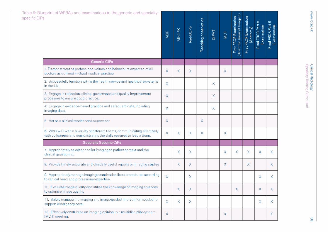
58C
linical Radiology
Specialty Training Curriculum
ww
w.rcr.ac.uk
Table 9: Blueprint of WPBAs and examinations to the generic and specialty-specific CiPs
5 Supervision and feedback
59Clinical Radiology Specialty Training Curriculum
www.rcr.ac.uk
This section of the curriculum describes how trainees will be supervised, how they will receive feedback on performance, and the requirements for trainers.
Access to high quality, supportive, timely and constructive feedback is essential for the professional development of the trainee. Trainee reflection is an important part of the feedback process and exploration of that reflection with the trainer should be a two way dialogue. Effective feedback is known to enhance learning and combining self-reflection to feedback promotes deeper learning. This process should take place throughout training in both formal and informal settings. Opportunities for feedback will arise during appraisal meetings, when trainees are undergoing workplace-based assessments, in the workplace setting, and through discussions with supervisors, trainers, assessors and those within the team. Trainees must develop the ability to seek and respond to feedback on clinical practice from a range of individuals.
Trainers should be supported to deliver valuable and high quality feedback. This can be by providing face to face training to trainers. Trainees would also benefit from such training as they frequently act as assessors to junior doctors, and all involved could also be shown how best to carry out and record reflection.
All elements of work in training posts must be supervised, with the level of supervision varying depending on the experience of the trainee and case mix undertaken. As training progresses the trainee should have the opportunity for increasing autonomy, consistent with safe and effective care for the patient.
Organisations must make sure that each doctor in training has access to a named clinical supervisor and a named educational supervisor. It is preferred that a trainee has a single named educational supervisor for the duration of training. The clinical supervisor will change for each post and will usually be the consultant to whom a radiology trainee is directly responsible for that post.
Educational and clinical supervisors need to be formally recognised by the GMC to carry out their roles. It is essential that training in assessment is provided for trainers and trainees in order to ensure that there is complete understanding of the assessment system, assessment methods, their purposes and use. Training will ensure a shared understanding and a consistency in the use of the WPBA and the application of standards.
Opportunities for feedback to trainees about their performance will arise through the use of the WPBA, regular appraisal meetings with supervisors, other meetings and discussions with supervisors and colleagues, and feedback from ARCP.
The first year in clinical radiology can be a difficult transition for trainees. TPDs and trainers are encouraged to offer advice, a mentor system and a counselling service during the year. The following milestones should be acknowledged:
5.1 Feedback
5.2 Supervision
60Clinical Radiology Specialty Training Curriculum
www.rcr.ac.uk
§ The trainee should meet with their clinical supervisor, educational supervisor and their TPD at the start of their appointment, and again with their clinical and educational supervisors after three-four months. Some individuals may undertake more than one of these roles simultaneously.
§ The trainee’s practice must be closely supervised and patient safety is of paramount importance. Such aspects are monitored by the clinical supervisor for each individual post and documented in the e-portfolio. Formal mechanisms for feeding back any concerns raised by the clinical supervisor, to the trainee, and the educational supervisor and TPD, should be in place. There should be a formal mechanism for counselling trainees who are unsuccessful in the First FRCR Examination.
§ All training in postgraduate radiology should be conducted in GMC-approved locations with appropriate standards of clinical governance and that meet relevant Health and Safety standards for clinical areas.
§ Trainees must work with a level of clinical supervision commensurate with their clinical experience and level of competence. This is the responsibility of the relevant clinical supervisor after discussion with the trainee’s educational supervisor and the designated clinical governance lead. In keeping with the principles of Good Medical Practice, trainees should know that they must limit their clinical practice to within their level of clinical competence and seek help and support without hesitation.
5.2.1 Educational supervisorThe educational supervisor is appropriately trained to be responsible for the overall supervision and management of a doctor’s educational progress during the course of their training or a series of posts. The educational supervisor regularly meets with the doctor in training to help plan their training, review progress and agree learning outcomes. The educational supervisor is responsible for the educational agreement, and for bringing together all relevant evidence to form a summative judgement about progression at the end of the placement or a series of placements.
The educational supervisor is integral to the appraisal process. A trainee appraisal with the educational supervisor will include feedback on performance, review of outcomes of assessments, induction to posts and career advice. The postgraduate deaneries should recognise the active role of educational supervisor in training and offer appropriate support.
Local education providers must ensure that educational supervisors have adequate support and resources to undertake their training role. This will include training in equality and diversity.
The educational supervisor will:
§ ensure that the programme is appropriate for the doctor’s needs
§ be responsible for the radiology trainee’s educational agreement
§ help radiology trainees by reviewing their learning needs in the light of achieved goals
§ carry out and/or collate assessments from clinical supervisors, trainers and other assessors
§ review the trainee’s e-portfolio
§ conduct appraisals and give supportive feedback on the results of MSF
61Clinical Radiology Specialty Training Curriculum
www.rcr.ac.uk
§ complete the structured supervisor’s report at the end of each year of training prior to the ARCP
§ support the trainee through any difficulty
§ tell the clinical director, head of service or medical director and those responsible for training, of serious weaknesses in their trainee’s performance that have not been dealt with
§ tell the radiology trainee the content of any information about them that is given to someone else
§ ensure that all training opportunities meet the requirements of equality and diversity legislation
The educational supervisor, when meeting with the trainee, should discuss issues of clinical governance, risk management and the report of any untoward clinical incidents involving the trainee. If the clinical supervisor should have any concerns about the performance of the trainee, or there were issues of doctor or patient safety, these would be discussed with both the trainee and the educational supervisor. In turn the educational supervisor may consult with the trainee and the TPD/Head of School. These processes, which are integral to trainee development, must not detract from the statutory duty of the employer to deliver effective clinical governance through its management systems.
5.2.2 Clinical supervisor A clinical supervisor will usually be the consultant to whom a radiology trainee is directly responsible for their clinical work and there will be frequent contact between them. They will be appropriately trained to lead on reviewing the trainee’s practice throughout a post and will provide constructive feedback, as well as contributing to the educational supervisor’s report.
Local education providers must ensure that clinical supervisors have adequate support and resources to undertake their training role. This will include training in equality and diversity.
The clinical supervisor is responsible for:
§ ensuring that their radiology trainees are never put in a situation where they are asked to work beyond their competence without appropriate support and supervision. Patient safety must be paramount at all times
§ guaranteeing suitable induction to the radiology department
§ meeting with the radiology trainee at the beginning of each post to discuss what is expected in the post, learning opportunities available and the trainee’s learning needs
§ agreeing how the learning objectives for this period of training will be met and confirming how formative feedback and summative judgements will be made
§ ensuring that the clinical experience available to the trainee is appropriate and properly supervised
§ ensuring that all training opportunities meet the requirements of equality and diversity legislation
§ monitoring, supporting and assessing the radiology trainee’s day-to-day clinical and professional work
§ providing regular feedback on the trainee’s performance
§ undertaking and facilitating WPBA
62Clinical Radiology Specialty Training Curriculum
www.rcr.ac.uk
§ allowing the trainee to give feedback on the experience, quality of training and supervision provided
§ discussing serious concerns with the educational supervisor about a trainee’s performance, health or conduct
§ meeting with the radiology trainee to assess whether they have met the necessary outcomes and completing an end of post review form for each post
5.2.3 Trainees Trainees should make the safety of patients their first priority. Furthermore, trainees should not be practising in clinical scenarios which are beyond their experiences and competences without supervision. Trainees should actively devise individual learning goals in discussion with their trainers and should subsequently identify the appropriate opportunities to achieve said learning goals. Trainees would need to plan their WPBAs accordingly to enable their WPBAs to collectively provide a picture of their development during a training period. Trainees should actively seek guidance from their trainers in order to identify the appropriate learning opportunities and plan the appropriate frequencies and types of WPBAs according to their individual learning needs. It is the responsibility of trainees to seek feedback following learning opportunities and WPBAs. Trainees should self-reflect and self-evaluate regularly with the aid of feedback. Furthermore, trainees should formulate action plans with further learning goals in discussion with their trainers.
A formal process of appraisals and reviews underpins training. This process ensures adequate supervision during training, provides continuity between posts and different supervisors, and is one of the main ways of providing feedback to trainees. Arranging a review is primarily the responsibility of the trainee. A “typical” year of appraisals involving both clinical and educational supervisors is illustrated in Figure 3. All appraisals should be recorded in the e-portfolio.
Annual induction appraisal
When radiology trainees start in a new training year, they must arrange a meeting with their educational supervisor. The induction appraisal is an essential starting point for negotiating educational goals and discussing learning opportunities, the assessment process and use of the e-portfolio. This forms the basis for the educational agreement between the educational supervisor and trainee.
Clinical supervisor: induction appraisal
When radiology trainees start a new post, they must arrange a meeting with their clinical supervisor (this role may be discharged in some cases by the educational supervisor). The appraisal discussions should cover the educational objectives for the clinical attachment and be used to inform the (PDP).
Clinical supervisor: mid-post appraisal
A mid-point meeting during a clinical attachment, although not mandatory is highly recommended. It gives the trainee and clinical supervisor the opportunity to look at the achievements of the trainee and highlights areas for future development, in terms of the PDP and curriculum CiPs.
5.3 Appraisal
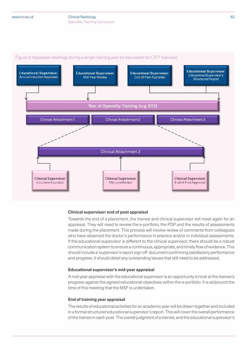
63Clinical Radiology Specialty Training Curriculum
www.rcr.ac.uk
Clinical supervisor: end of post appraisal
Towards the end of a placement, the trainee and clinical supervisor will meet again for an appraisal. They will need to review the e-portfolio, the PDP and the results of assessments made during the placement. This process will involve review of comments from colleagues who have observed the doctor’s performance in practice and/or in individual assessments. If the educational supervisor is different to the clinical supervisor, there should be a robust communication system to ensure a continuous, appropriate, and timely flow of evidence. This should include a ‘supervisor’s report sign off’ document confirming satisfactory performance and progress. It should detail any outstanding issues that still need to be addressed.
Educational supervisor’s mid-year appraisal
A mid-year appraisal with the educational supervisor is an opportunity to look at the trainee’s progress against the agreed educational objectives within the e-portfolio. It is at/around the time of this meeting that the MSF is undertaken.
End of training year appraisal
The results of educational activities for an academic year will be drawn together and included in a formal structured educational supervisor’s report. This will cover the overall performance of the trainee in each post. The overall judgment of a trainee, and the educational supervisor’s
Figure 3: Appraisal meetings during a single training year (or equivalent for LTFT trainees)
64Clinical Radiology Specialty Training Curriculum
www.rcr.ac.uk
recommendations of satisfactory completion of the year of training, will be based on a triangulated view of the doctor’s performance. This will include their participation in educational activities, appraisals, the assessment process and recording of this in the e-portfolio.
The outcome of the final appraisal discussion should be agreed by both the trainee and the educational supervisor and recorded in the structured supervisor’s report in the e-portfolio.
65Clinical Radiology Specialty Training Curriculum
www.rcr.ac.uk
6 Appendices
6.1 Curriculum development, implementation and review
This curriculum was developed by the Clinical Radiology Curriculum Committee. This is a sub-committee of the Specialty Training Board of the Faculty of Clinical Radiology of the Royal College of Radiologists (STB) and reports to it. The members of the Curriculum Committee and STB have broad UK representation across the range of radiological special interest areas and include consultants who are actively involved in teaching and training; trainees; service representatives; and lay persons.
Throughout the development of this curriculum, the Curriculum Committee has consulted widely with a range of stakeholders. These include: Special Interest Group Leads; the Fellowship Examining Board and FRCR Physics and Anatomy Committees; Regional Specialty Advisers; the STB; the RCR’s Junior Radiologist’s Forum; employers’ groups, including NHS Employers; Clinical Directors; professional organisations such as the British Society of Skeletal Radiologists and British Society of Interventional Radiology; patient groups such as the NHS England Patient Experience Team; charities; the RCR lay member network; those representing allied health professions e.g. the Society and College of Radiographers and the Institute of Physics and Engineering in Medicine; other royal colleges including JRCPTB, RCGP, RCEM, RCoA, RCOG, and RCPCH; and the Nuclear Medicine Specialty Advisory Committee, including trainee representatives.
6.1.1 ImplementationThis curriculum will be implemented in August 2020 and all trainees will move to this curriculum, unless they are due to CCT before 1st September 2021. All evidence currently recorded in the e-portfolio will remain and can be used to demonstrate achievement of the new CiPs.
A full suite of training materials is available to support trainees and trainers in the transition to this curriculum, including specific guidance for trainees who are LTFT, out of programme or on statutory leave. Further details can be found on the RCR website, along with documents mapping the competencies in the 2016 curriculum to the CiPs in this curriculum.
6.1.2 Intended useThe curriculum is freely available to trainees and trainers on the RCR and GMC websites. Both trainees and trainers are expected to have a good knowledge of the curriculum and should use it as a guide for their training programme. Clinical and educational supervisors should use the curriculum as the basis of their discussion with trainees, particularly during the appraisal process. Each trainee will engage with the curriculum by maintaining an e-portfolio. The trainee will use the curriculum to develop learning objectives, self-assess accomplishments, and reflect on learning experiences.
6.1.3 ReviewThe STB is responsible for review of the curriculum. Clinical radiology, as a technology supported specialty, is rapidly changing and evolving and as a result the curriculum is kept under constant review to ensure that radiology training and education reflect modern practice. To allow the curriculum to respond appropriately to these changes, there are regular meetings of Special Interest Groups, the Curriculum Committee, the Professional Support
66Clinical Radiology Specialty Training Curriculum
www.rcr.ac.uk
and Standards Board and the Specialty Training Board. These provide opportunities for the curriculum to be discussed, and amendments to be proposed and considered in advance of formal review. Trainers, tutors, regional specialty advisers, programme directors and examiners will also continue to be involved in review through their membership of relevant working parties and committees.
Curriculum evaluation will establish how trainees have responded to the curriculum and ensure that the curriculum facilitates practical delivery of the required training. The curriculum will be evaluated by means of a range of qualitative and quantitative data.
The organisation of training programmes for clinical radiological training and special interest training is the responsibility of Postgraduate Deaneries and their Schools of Radiology or equivalents.
The Deaneries have established appropriate programmes for postgraduate radiological training in their regions. In England and Northern Ireland, Deanery Schools of Radiology have been set up to achieve this. As the term “School of Radiology” is not applicable to all countries and regions, the general term “local faculties for radiological training” is be used.
There may be more than one training programme within a local faculty for radiological training. Each of these is managed by a Training Programme Director (TPD) who is accountable to the Deanery Head of School (or their equivalent). Within each participating local education provider (hospital) within a training programme there is a clinical supervisor who supports the delivery of training and the curriculum within that hospital and is accountable for this to the TPD. The deaneries, through their local faculties and training programmes, will together with the TPDs and Educational/Clinical supervisors co-ordinate local postgraduate radiological training, with terms of reference as follows:
§ allocate trainees into particular posts for core radiological training, and then at ST4 and ST5 levels into posts appropriate to their special interest training needs where possible
§ oversee the quality of training posts provided locally
§ interface with other Deanery Specialty Training faculties (General Practice, Anaesthesia etc.)
§ ensure adequate provision of appropriate educational events
§ ensure curricula implementation across training programmes
§ oversee the workplace-based assessment process and programmes
§ co-ordinate the ARCP process for trainees
§ provide adequate and appropriate career advice
§ provide systems to identify and assist trainee radiologists with training difficulties
§ provide flexible training
§ ensure the appropriate provision of potential to progress into an academic career
Coordination of the curriculum at a national and regional level is the joint responsibility of the Deaneries and the Royal College of Radiologists, with robust arrangements for quality assurance of training. The STB will oversee central evaluation of this curriculum and the e-portfolio. Local management of curriculum delivery and quality management of training programmes is the responsibility of the local faculty of education in consultation with the Royal
6.2 Quality management
67Clinical Radiology Specialty Training Curriculum
www.rcr.ac.uk
College of Radiologists. The need to show how trainees are progressing in their attainment of competences will be a strong driver in ensuring that all the curriculum objectives are met.
Local evaluation is essential for monitoring the quality of teaching, learning and supervision and developing local implementation. It is the responsibility of individual training programmes to carry out this necessary work, which will provide evidence for training programme development, as well as material with which to respond to the findings of the annual GMC survey.
The TPD will typically co-ordinate the evaluation process with administrative support, however anyone involved in the programme will have a contribution to make to its evaluation. Evaluation should be embedded in the training programme as an annual process. The various strands of evidence will need to be summarised, analysed and formulated in time for response to the GMC survey, deanery report and recommendations for programme development.
The Royal College of Radiologists will comply, and ensure compliance, with the requirements of the Equality Act 2010.
We believe that equality of opportunity is fundamental to all radiological practice and to the many and varied ways in which individuals become involved with the RCR, either as members of staff and Officers; as advisers from the medical profession or in a lay capacity; as members of the RCR’s professional bodies or as radiologists in training and examination candidates. Accordingly, it warmly welcomes contributions and applications from as diverse a population as possible, and actively seeks to recruit people to all its activities regardless of protected characteristic.
Deanery quality assurance will ensure that each training programme complies with the equality and diversity standards in postgraduate medical training as set by GMC.
Compliance with anti-discriminatory practice will be assured through:
§ monitoring of recruitment processes;
§ ensuring all RCR representatives have attended appropriate training sessions prior to appointment or within 12 months of taking up post;
§ ensuring trainees have an appropriate, confidential and supportive route to report examples of inappropriate behaviour of a discriminatory nature. Deaneries must also ensure contingency mechanisms are in place if trainees feel unhappy with the response or uncomfortable with the contact individual;
§ monitoring of FRCR examinations;
§ ensuring all assessments discriminate on objective and appropriate criteria and do not unfairly disadvantage trainees with any of the Equality Act 2010 protected characteristics. All efforts shall be made to ensure the participation in training of people with a disability (other than that which would make it impossible to practise safely as a radiologist) through reasonable adjustments.
The RCR takes its obligations under the relevant equal opportunities legislation seriously. This includes ensuring that members of staff involved in the delivery of examinations receive appropriate briefing on the implications of equality and diversity in the treatment of candidates. Those appointed as examiners must demonstrate that they have undergone
6.3 Equality and diversity
68Clinical Radiology Specialty Training Curriculum
www.rcr.ac.uk
appropriate equality and diversity training and that they are willing to abide by good practice in these areas.
The RCR has an Adjustments Procedure for FRCR Examinations published on our website which provides a formal means for candidates to submit a request for an adjustment to be applied in examinations to compensate for disability. All adjustment requests will be considered by the RCR in a fair and consistent way.
References
1. NHS England. Next Steps on the NHS Five Year Forward View. NHS England, 2017.
2. NHS England. Achieving World-class Cancer Outcomes: a Strategy for England 2015-2020. NHS England, 2016.
3. Health Education England. HEE Cancer Workforce Plan Phase 1: Delivering the Cancer Strategy to 2021. Health Education England, 2017
4. The Royal College of Radiologists. Clinical radiology UK workforce census 2018 report. London: The Royal College of Radiologists, 2019.
5. Conference Of Postgraduate Medical Deans (UK). A Reference Guide for Postgraduate Specialty Training in the UK. Conference Of Postgraduate Medical Deans, 2018
6. General Medical Council. Generic professional capabilities framework. General Medical Council, 2017.
7. General Medical Council. Good medical practice. General Medical Council, 2014.
69Clinical Radiology Specilaty Training Curriculum
www.rcr.ac.uk
The Royal College of Radiologists 63 Lincoln’s Inn Fields London WC2A 3JW
+44 (0)20 7405 1282 [email protected] www.rcr.ac.uk
@RCRadiologists
The Royal College of Radiologists. Clinical Radiology
Specialty Training Curriculum. London: The Royal College of Radiologists, 2019.
© The Royal College of Radiologists, July 2019.
For permission to reproduce any of the content contained herein, please email: [email protected]
This material has been produced by The Royal College of Radiologists (RCR) for use internally within the specialties of clinical oncology and clinical radiology in the United Kingdom. It is provided for use by appropriately qualified professionals, and the making of any decision regarding the applicability and suitability of the material in any particular circumstance is subject to the user’s professional judgement.
While every reasonable care has been taken to ensure the accuracy of the material, RCR cannot accept any responsibility for any action taken, or not taken, on the basis of it. As publisher, RCR shall not be liable to any person for any loss or damage, which may arise from the use of any of the material. The RCR does not exclude or limit liability for death or personal injury to the extent only that the same arises as a result of the negligence of RCR, its employees, Officers, members and Fellows, or any other person contributing to the formulation of the material.