Embed Size (px)
Citation preview
CLINICAL PROFILE AND OUTCOME OF DENGUE FEVER AMONG
CHILDREN ADMITTED AT TIRUNELVELI MEDICAL
COLLEGE HOSPITAL
Dissertation submitted to
THE TAMILNADU DR.M.G.R. MEDICAL UNIVERSITY
In partial fulfillment of the regulations
for the award of the degree of
M.D. BRANCH – VII
PEDIATRICS
TIRUNELVELLI MEDICAL COLLEGE & HOSPITAL,
THE TAMILNADU DR.M.G.R. MEDICAL UNIVERSITY,
CHENNAI, INDIA.
APRIL 2012
brought to you by COREView metadata, citation and similar papers at core.ac.uk
provided by ePrints@TNMGRM (Tamil Nadu Dr. M.G.R. Medical University)
CERTIFICATE
This is to certify that the dissertation entitled “CLINICAL PROFILE
AND OUTCOME OF DENGUE FEVER AMONG CHILDREN
ADMITTED AT TVMCH” is the bonafide work of Dr.R.Karthikeyan in
partial fulfillment of the requirements for the degree of Doctor of Medicine in
Paediatrics Examination of the Tamilnadu Dr.M.G.R. Medical University to be
held in April 2012.
PROFESSOR OF PEDIATRICS PROFESSOR & H.O.D
Tirunelveli Medical College & Hospital, Department of Pediatrics,
Tirunelveli-11 Tirunelveli Medical College
& Hospital, Tirunelveli- 11
DEAN
Tirunelveli Medical College & Hospital
Tirunelveli-11
DECLARATION
I, Dr.R.ARTHIKEYAN, solemnly declare that dissertation titled,
“CLINICAL PROFILE AND OUTCOME OF DENGUE FEVER
AMONG CHILDREN IN TVMCH” is a bonafide work done by me at The
Department of Paediatrics, Tirunelvelli Medical College & Hospital during
2009 – 2012 under the guidance and supervision of Dr.T.KATHIR
SUBRAMANIUM M.D., D.C.H., and Dr.S.DEVIKALA M.D., D.C.H.,
Professors, TIRUNELVELLI MEDICAL COLLEGE The Dissertation is
submitted to The Tamilnadu Dr.M.G.R. Medical University, towards partial
3ulfilment of requirement for the award of M.D. Degree (Branch – VII) in
Paediatrics.
Place :
Date :
Dr.R.KARTHIKEYAN
ACKNOWLEDGEMENT
Special acknowledgement to Prof.M.Manoharan, M.S., Dean Tirunelveli
Medical college & Hospital for allowing me to utilise the facilities of this
institution to do this study.
I sincerely thank our HOD, Prof.T.Kathir Subramaniam M.D., D.C.H.,
For granting me permission to conduct this study.
I am most indebted to my teacher and unit chief
Prof.S.Devikala, M.D., D.C.H., for her valuable suggestion and encouragement
throughout study.
I remember with gratitude Prof.Ravichandran, M.D., D.C.H., for the
inspiration and encouragement given to me.
I shall always regard with great gratitude my Assistant
Professors Dr.C.Krishnamurthy, M.D., Dr.T.R.R.Ananthyshri, M.D.,
Dr.A.S.Babukanthakumar, M.D., D.C.H., Dr.T.Viswanathan, M.D., and
Dr.B.Naresh, M.D., for their able guidance and assistance in doing this work.
I profusely thank the department of microbiology for their immense help
in doing dengue serology.
I would like thank Dr.Pethru, M.D., (Community Medicine) for his
valuable work in statistics.
I would like to thank all children and their parents for their consent and
participation in this study.
CONTENTS
INTRODUCTION 1
OBJECTIVE 4
LITERATURE REVIEW 5
METHODOLOGY 49
OBSERVATION AND RESULTS 52
DISCUSSION 61
LIMITATIONS 68
CONCLUSION 69
ANNEXURE
BIBLIOGRAPHY
PROFORMA
MASTER CHART
1
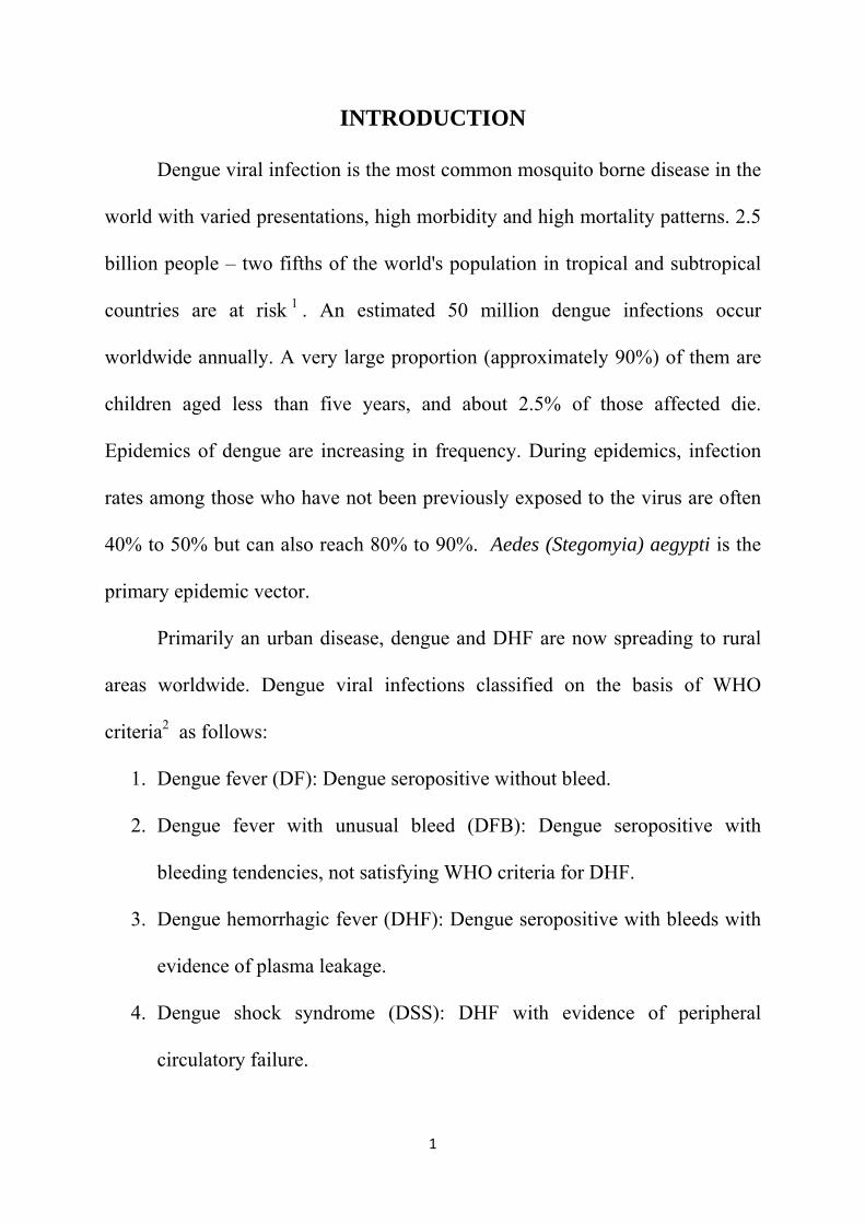
INTRODUCTION
Dengue viral infection is the most common mosquito borne disease in the
world with varied presentations, high morbidity and high mortality patterns. 2.5
billion people – two fifths of the world's population in tropical and subtropical
countries are at risk 1 . An estimated 50 million dengue infections occur
worldwide annually. A very large proportion (approximately 90%) of them are
children aged less than five years, and about 2.5% of those affected die.
Epidemics of dengue are increasing in frequency. During epidemics, infection
rates among those who have not been previously exposed to the virus are often
40% to 50% but can also reach 80% to 90%. Aedes (Stegomyia) aegypti is the
primary epidemic vector.
Primarily an urban disease, dengue and DHF are now spreading to rural
areas worldwide. Dengue viral infections classified on the basis of WHO
criteria2 as follows:
1. Dengue fever (DF): Dengue seropositive without bleed.
2. Dengue fever with unusual bleed (DFB): Dengue seropositive with
bleeding tendencies, not satisfying WHO criteria for DHF.
3. Dengue hemorrhagic fever (DHF): Dengue seropositive with bleeds with
evidence of plasma leakage.
4. Dengue shock syndrome (DSS): DHF with evidence of peripheral
circulatory failure.

2
Dengue generally an acute febrile illness, and sometimes biphasic fever
with severe headache, myalgia, arthralgia, rashes, leucopenia and
thrombocytopenia may also be observed. DHF is characterized by the acute
onset of high fever and is associated with signs and symptoms similar to DF in
the early febrile phase. There are common haemorrhagic manifestation such as
positive tourniquet test (TT), petechiae, easy bruising and/or GI haemorrhage in
severe cases. By the end of the febrile phase, there is a tendency to develop
hypovolemic shock (dengue shock syndrome) due to plasma leakage. The
presence of preceding warning signs such as persistent vomiting, abdominal
pain, lethargy or restlessness, or irritability and oliguria are important for
intervention to prevent shock.
Abnormal haemostasis and plasma leakage are the main patho-
physiological hallmarks of DHF. Thrombocytopenia and rising haematocrit/
haemoconcentration are constant findings before the subsidence of fever/ onset
of shock. DHF occurs most commonly in children with secondary dengue
infection. It has also been documented in primary infections with DENV-1 and
DENV-3 as well as in infants.We live in a country with resource limited settings
confounded by public ignorance about the disease and poor access to health
care. Severity is high in paediatric age group.
No vaccine is available for preventing this disease at present.
Chandrakanta et al3 describe the varied manifestations of dengue viral infection

3
as seen in hospitalized children in northern India. Manjunath J. Kulkarni et al4
in their study describe various clinical manifestations.
The demographic pattern and the trend of illness are vastly changing
every year through the past decade. South India and especially South Tamilnadu
has witnessed several dengue epidemic outbreaks during the past few years.
Early recognition and prompt initiation of appropriate management is vital.
Additional studies about the disease can lead to change in guidelines and
alterations in public health programs. Here, I have made an attempt to study the
clinical profile and outcome of dengue viral infection the time period from Dec
2009 to Dec 2010.
4
OBJECTIVE
To study the clinical profile and outcome of dengue fever in children.
5
LITERATURE REVIEW
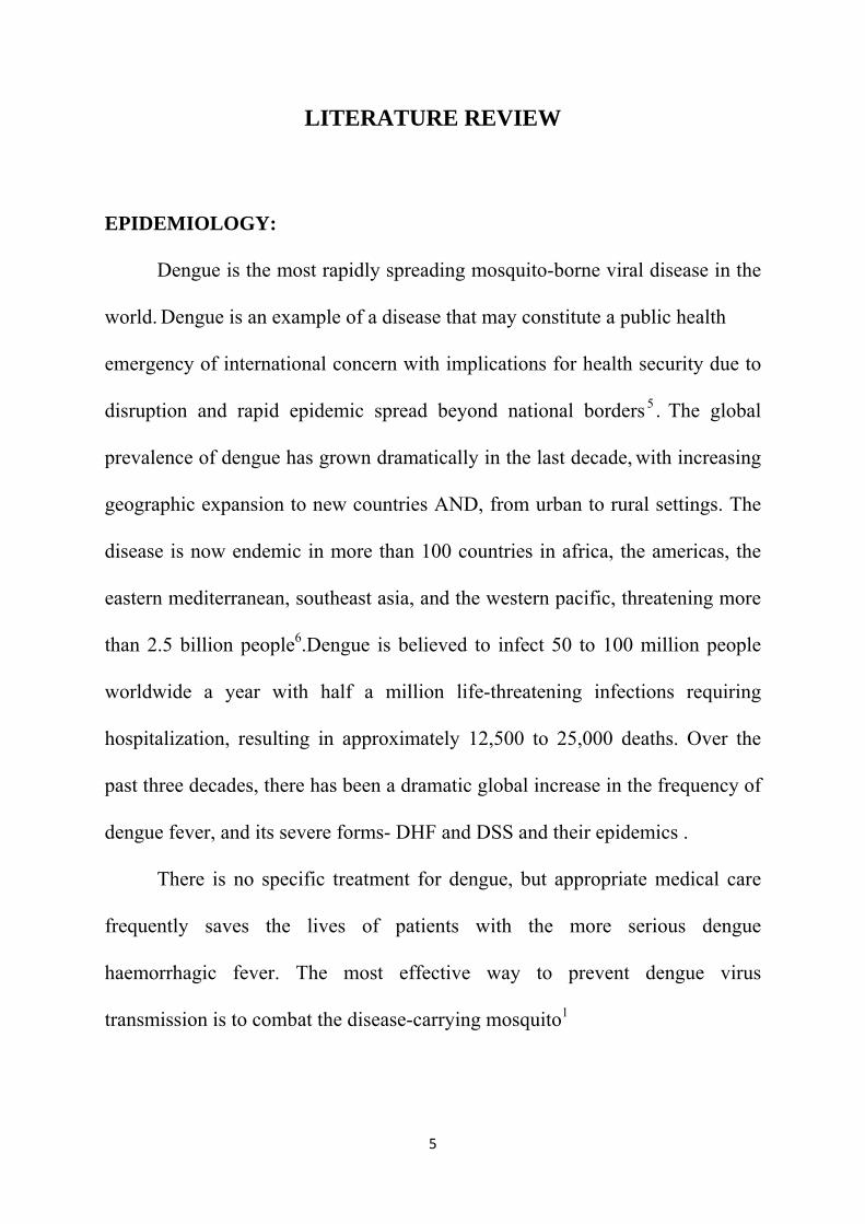
EPIDEMIOLOGY:
Dengue is the most rapidly spreading mosquito-borne viral disease in the
world. Dengue is an example of a disease that may constitute a public health
emergency of international concern with implications for health security due to
disruption and rapid epidemic spread beyond national borders5 . The global
prevalence of dengue has grown dramatically in the last decade, with increasing
geographic expansion to new countries AND, from urban to rural settings. The
disease is now endemic in more than 100 countries in africa, the americas, the
eastern mediterranean, southeast asia, and the western pacific, threatening more
than 2.5 billion people6.Dengue is believed to infect 50 to 100 million people
worldwide a year with half a million life-threatening infections requiring
hospitalization, resulting in approximately 12,500 to 25,000 deaths. Over the
past three decades, there has been a dramatic global increase in the frequency of
dengue fever, and its severe forms- DHF and DSS and their epidemics .
There is no specific treatment for dengue, but appropriate medical care
frequently saves the lives of patients with the more serious dengue
haemorrhagic fever. The most effective way to prevent dengue virus
transmission is to combat the disease-carrying mosquito1
6
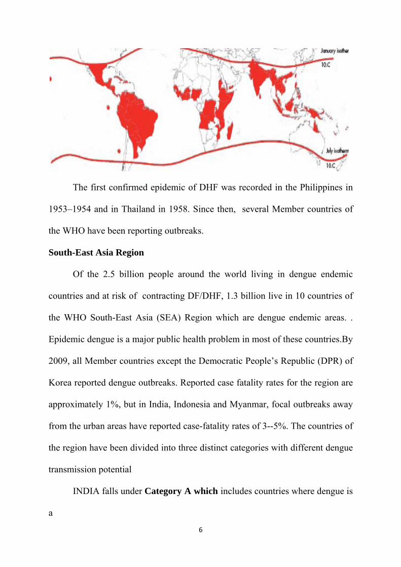
The first confirmed epidemic of DHF was recorded in the Philippines in
1953–1954 and in Thailand in 1958. Since then, several Member countries of
the WHO have been reporting outbreaks.
South-East Asia Region
Of the 2.5 billion people around the world living in dengue endemic
countries and at risk of contracting DF/DHF, 1.3 billion live in 10 countries of
the WHO South-East Asia (SEA) Region which are dengue endemic areas. .
Epidemic dengue is a major public health problem in most of these countries.By
2009, all Member countries except the Democratic People’s Republic (DPR) of
Korea reported dengue outbreaks. Reported case fatality rates for the region are
approximately 1%, but in India, Indonesia and Myanmar, focal outbreaks away
from the urban areas have reported case-fatality rates of 3--5%. The countries of
the region have been divided into three distinct categories with different dengue
transmission potential
INDIA falls under Category A which includes countries where dengue is
a
7
� Major public health problem.
� Leading cause of hospitalization and death among children.
� Hyperendemicity with “all four serotypes” circulating in urban areas.
� Spreading to rural areas.
Cyclic epidemics are increasing in frequency and in-country geographic
expansion is occurring in India.
Dengue prevention and control will be implemented through the Bi-
regional Dengue Strategy (2008--2015) of the WHO South-East Asia and
Western Pacific regions7
The virus
The dengue viruses are members of the genus Flavivirus and family
Flaviviridae. These small (50nm) viruses contain single-strand RNA as
genome. The virion consists of a nucleocapsid with cubic symmetry enclosed in
a lipoprotein envelope. The dengue virus genome is 11,644 nucleotides in
length, and is composed of three structural protein genes encoding the
nucleocaprid or core protein (C), a membrane-associated protein (M), an
envelope protein (E), and seven non-structural protein (NS) genes. Among
nonstructural proteins, envelope glycoprotein, NS1, is of diagnostic and
pathological importance. It is 45 kDa in size and associated with viral
haemagglutination and neutralization activity.8
The dengue viruses form a distinct complex within the genus Flavivirus
based on antigenic and biological characteristics. There are four virus serotypes,

8
which are designated as DENV-1, DENV-2,DENV-3 and DENV-4. Infection
with any one serotype confers lifelong immunity to that virus serotype.Although
all four serotypes are antigenically similar, they are different enough to elicit
cross-protection for only a few months after infection by any one of them.
Secondary infection with another serotype or multiple infections with different
serotypes leads to severe form of dengue (DHF/DSS).
There exists considerable genetic variation within each serotype in the
form of phylogenetically distinct “sub-types” or “genotypes”. Currently, three
sub-types can be identified for DENV-1, six for DENV-2 (one of which is
found in non-human primates), four for DENV-3 and four for DEN-4, with
another DENV-4 being exclusive to non-human primates. Dengue viruses of all
four serotypes have been associated with epidemics of dengue fever (with or
without DHF) with a varying degree of severity.Among them, “Asian”
genotypes of DEN-2 and DEN-3 are frequently associated with severe disease
accompanying secondary dengue infections Intra-host viral diversity
(quasispecies) has also been described in human9
Plasma levels of secreted NS1 (sNS1) correlate with viral titers, being
higher in patients with DHF compared with dengue fever10 . Moreover, elevated
free sNS1 levels within 72 hours of onset of illness identify patients at risk of
developing DHF. Very high levels of NS1 protein are detected in acute phase
samples from patients with secondary dengue infections but not primary
infections. This suggests that NS1 may contribute to formation of circulating
9
immune complexes, which are thought to have an important role in the
pathogenesis of severe dengue infections11 .The dengue virus shares antigenic
epitopes with other flaviviruses such as Japanese encephalitis virus. These
shared epitopes may lead to production of cross reactive antibodies and hence
interfere with serological diagnosis. However,antibodies directed to the prM
protein of dengue viruses are species specific (not cross reactive with those of
other flaviviruses) and may be useful for seroepidemiological studies in dengue
(especially in countries where other flaviviruses are endemic)
Vectors of dengue
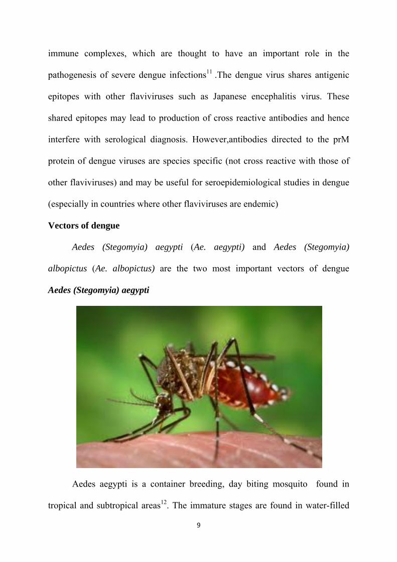
Aedes (Stegomyia) aegypti (Ae. aegypti) and Aedes (Stegomyia)
albopictus (Ae. albopictus) are the two most important vectors of dengue
Aedes (Stegomyia) aegypti
Aedes aegypti is a container breeding, day biting mosquito found in
tropical and subtropical areas12. The immature stages are found in water-filled
10
habitats, mostly in artificial containers closely associated with human dwellings
and often indoors. This maximizes man-vector contact and minimizes contact
with insecticides sprayed outdoors, hence contributing to difficulty in
controlling this vector13. Studies suggest that most female Ae. aegypti may
spend their lifetime in or around the houses where they emerge as adults. This
means that people, rather than mosquitoes, rapidly move the virus within and
between communities . Increased transport, human contact, urbanization and
the proliferation of drinking water supply schemes in rural areas ultimately led
to the species getting entrenched in both urban and rural areas . On account of
the species’ high degree of domestication and strong affinity for human blood, it
achieved high vectorial capacity for transmission of DF/DHF.Significant
increases in the mosquito larval populations are seen during the rainy season.
This may be a reason why epidemics of dengue tend to coincide with the rainy
season10.Furthermore,ambient temperature and relative humidity affect viral
propagation in mosquitoes; rates being highest in climates resembling the rainy
season10.
After biting an infected human, dengue viruses enter an adult female
mosquito. The virus first replicates in the midgut, reaches the haemocoel and
haemolymph, and then gains access to different tissues of the insect. After viral
replication in the salivary glands, the infected mosquito can transmit the virus to
another human11 . Compared with uninfected mosquitoes, infected ones take
11
longer to complete a blood meal. This may contribute to the efficiency of A
.aegypti as a dengue viral vector.
Aedes (Stegomyia) albopictus belongs to the scutellaris group of
subgenus Stegomyia. It is a predominant Asian species. Dengue outbreaks have
also been attributed to Aedes albopictus, Aedes polynesiensis and several
species of the Aedes scutellaris complex. Each of these species has a particular
ecology, behaviour and geographical distribution.
Clinical manifestations
Dengue is one disease entity with different clinical presentations and
often with unpredictable clinical evolution and outcome14.Dengue infection is a
systemic and dynamic disease. It has a wide clinical spectrum that includes both
severe and non-severe clinical manifestations15 . Dengue virus infection may be
asymptomatic or may cause undifferentiated febrile illness (viral syndrome),
dengue fever (DF), or dengue haemorrhagic fever (DHF) including dengue
shock syndrome(DSS)
Undifferentiated fever
Infants, children and adults who have been infected with dengue virus,
especially for the first time (i.e. primary dengue infection), may develop a
simple fever indistinguishable from other viral infections. Maculopapular
rashes may accompany the fever or may appear during defervescence.
Upper respiratory and gastrointestinal symptoms are common

12
Dengue fever
After the incubation period, the illness begins abruptly and is followed by
the three phases -- febrile, critical and recovery .
The course of dengue illness
FEBRILE PHASE
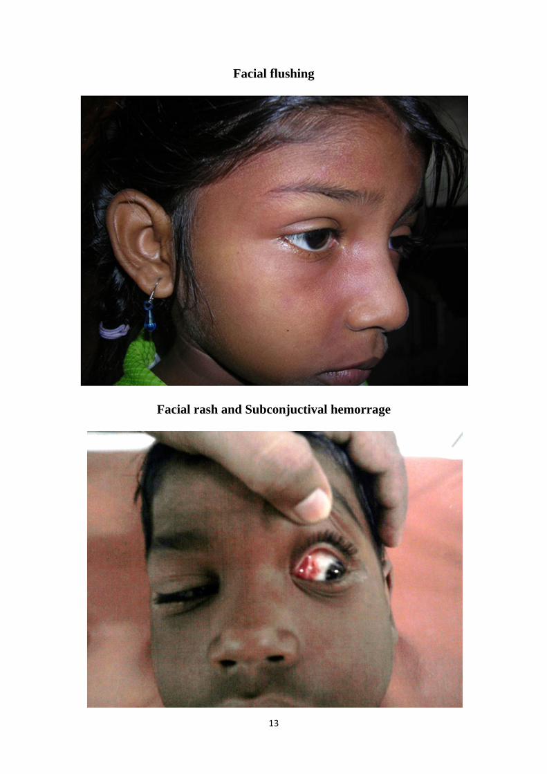
Patients typically develop high-grade fever suddenly. This acute febrile
phase usually lasts 2–7 days and is often accompanied by facial flushing, skin
erythema, generalized body ache, myalgia, arthralgia and headache13. Some
patients may have sore throat, injected pharynx and conjunctival injection.
Anorexia, nausea and vomiting are common. It can be difficult to distinguish
dengue clinically from non-dengue febrile diseases in the early febrile phase. A
positive tourniquet test in this phase increases the probability of dengue 16. In
addition, these clinical features are indistinguishable between severe and non-
severe dengue cases. Therefore monitoring for warning signs and other clinical
13
Facial flushing
Facial rash and Subconjuctival hemorrage

14
parameters is crucial.Mild haemorrhagic manifestations like petechiae and
mucosal membrane bleeding(e.g. nose and gums) may be seen 17 . Massive
vaginal bleeding (in women of childbearing age) and gastrointestinal bleeding
may occur during this phase but is not common 18. The liver is often enlarged
and tender after a few days of fever15. The earliest abnormality in the full blood
count is a progressive decrease in total white cell count, which should alert the
physician to a high probability of dengue.
CRITICAL PHASE:
Around the time of defervescence, when the temperature drops to 37.5–
38ºC or less and remains below this level, usually on days 3–7 of illness, an
increase in capillary permeability in parallel with increasing haematocrit levels
may occur19 This marks the beginning of the critical phase. The period of
clinically significant plasma leakage usually lasts 24–48 hours.
Progressive leukopenia 15 followed by a rapid decrease in platelet count
usually precedes plasma leakage. At this point patients without an increase in
capillary permeability will improve, while those with increased capillary
permeability may become worse as a result of lost plasma volume. The degree
of plasma leakage varies. Pleural effusion and ascites may be clinically
detectable depending on the degree of plasma leakage and the volume of fluid
therapy. Hence chest x-ray and abdominal ultrasound can be useful tools for
15
diagnosis. The degree of increase above the baseline haematocrit often reflects
the severity of plasma leakage.
Shock occurs when a critical volume of plasma is lost through leakage. It
is often preceded by warning signs. The body temperature may be subnormal
when shock occurs. With prolonged shock, the consequent organ hypoperfusion
results in progressive organ impairment, metabolic acidosis and disseminated
intravascular coagulation. This in turn leads to severe haemorrhage causing the
haematocrit to decrease in severe shock. Instead of the leukopenia usually seen
during this phase of dengue, the total white cell count may increase in patients
with severe bleeding. In addition, severe organ impairment such as severe
hepatitis, encephalitis or myocarditis and/or severe bleedingmay also develop
without obvious plasma leakage or shock 20.
Some patients progress to the critical phase of plasma leakage without
defervescence and, in these patients, changes in the full blood count should be
used to guide the onset of the critical phase and plasma leakage.
RECOVERY PHASE:
If the patient survives the 24–48 hour critical phase, a gradual
reabsorption of extravascular compartment fluid takes place in the following
48–72 hours.
General well-being improves, appetite returns, gastrointestinal symptoms
abate, Haemodynamic status stabilizes and diuresis ensues. Some patients may
have a rash of “isles of white in the sea of red”21. Some may experience
16
generalized pruritus. Bradycardia and electrocardiographic changes are common
during this stage.The haematocrit stabilizes or may be lower due to the
dilutional effect of reabsorbed fluid. White blood cell count usually starts to rise
soon after defervescence but the recovery of platelet count is typically later than
that of white blood cell count.Respiratory distress from massive pleural effusion
and ascites will occur at any time if excessive intravenous fluids have been
administered. During the critical and/or recovery phases, excessive fluid therapy
is associated with pulmonary oedema or congestive heart failure.
Febrile, critical and recovery phases in dengue
Dengue hemorrhagic fever
DHF usually follows secondary dengue infections, but may sometimes
follow pimary infections, especially in infants. In such infants, maternally
acquired dengue antibodies are presumed to enhance primary infections 22 .
Clinical deterioration usually occurs during defervescence (often between days
3 and 4). Tachycardia and hypotension characterize the onset of plasma leakage.
When plasma leakage is severe patients may develop othersigns of circulatory
disturbance such as prolonged capillary refill time, narrow pulse pressures, and
shock. Inadequate treatment of such patients often leads to profound shock.In
17
DHF, bleeding may occur from any site and does not correlate with the platelet
counts.
Haemorrhagic manifestations usually occur once the fever has settled.23
Minor degrees of bleeding may manifest as gum bleeding and petechiae. The
commonest site of haemorrhage is the gastrointestinal tract, which manifests as
haematemesis or melaena, followed by epistaxis. Vaginal bleeding is commonly
reported in females21. Convalescence in DHF is usually short and uneventful.
The return of appetite is a good indicator of recovery.
Dengue shock syndrome
Dengue shock syndrome is associated with very high mortality (around
9.3%, increasing to 47% in instances of profound shock)24 . Severe plasma
leakage leading to dengue shock syndrome is associated with cold blotchy skin,
circumoral cyanosis, and circulatory disturbances. Acute abdominal pain and
persisting vomiting are early warning signs of impeding shock. Sudden
hypotension may indicate the onset of profound shock .Prolonged shock is often
accompanied by metabolic acidosis, which may precipitate disseminated
intravascular coagulation or enhance ongoing disseminated intravascular
coagulation, which in turn could lead to massive haemorrhage. Dengue shock
syndrome may be accompanied by encephalopathy due to metabolic or
electrolyte disturbances.

18
COMPLICATIONS:
DHF AND DSS have been associated with increasing frequency in recent
times with the following complications25
19
Differential diagnoses of dengue26
Arboviruses: Chikungunya virus (this has often been mistaken for
dengue in South-East Asia).
Other viral diseases: Measles; rubella and other viral exanthems;
Epstein-Barr Virus (EBV); enteroviruses; influenza; hepatitis A;
Hantavirus.
Bacterial diseases: Meningococcaemia, leptospirosis, typhoid,
melioidosis, rickettsial diseases, scarlet fever.
Parasitic diseases: Malaria.
Pathogenesis of Dengue:
DHF occurs in a small proportion of dengue patients. Although DHF may
occur in patients experiencing dengue virus infection for the first time, most
DHF cases occur in patients with a secondary infection. 27 The association
between occurrence of DHF/DSS and secondary dengue infections implicates
the immune system in the pathogenesis of DHF. Both the innate immunity such
as the complement system and NK cells as well as the adaptive immunity
including humoral and cell mediated immunity are involved in this process28.
Enhancement of immune activation, particularly during a secondary infection,
leads to exaggerated cytokine response resulting in changes in vascular
permeability. In addition, viral products such as NS1 may play a role in
regulating complement activation and vascular permeability.29
20
The plasma leakage is unique in that there is selective leakage of plasma
in the pleural and peritoneal cavities and the period of leakage is short (24–48
hours). Rapid recovery of shock without sequelae and the absence of
inflammation in the pleura and peritoneum indicate functional changes in
vascular integrity rather than structural damage of the endothelium as the
underlying mechanism.Various cytokines with permeability enhancing effect
have been implicated in the pathogenesis of DHF 30 .However, the relative
importance of these cytokines in DHF is still unknown. Studies have shown that
the pattern of cytokine response may be related to the pattern of cross-
recognition of dengue-specific T-cells. Cross-reactive T-cells appear to be
functionally deficit in their cytolytic activity but express enhanced cytokine
production including TNF-a, IFN-g and chemokines31.
Increase in vascular permeability can also be mediated by the activation
of the complement system. Elevated levels of complement fragments have been
documented in DHF.32Some complement fragments such as C3a and C5a are
known to have permeability enhancing effects. In recent studies, the NS1
antigen of dengue virus has been shown to regulate complement activation and
may play a role in the pathogenesis of DHF27
Higher levels of viral load in DHF patients in comparison with DF
patients have been demonstrated in many studies33. The levels of viral protein,
NS1, were also higher in DHF patients34.The degrees of viral load correlate with
measurements of disease severity.

21
High-risk patients
The following host factors contribute to more severe disease and its
complications:
• infants and the elderly,
• obesity,
• pregnant women,
• peptic ulcer disease,
• women who have menstruation or abnormal vaginal bleeding,
• haemolytic diseases such as glucose-6-phosphatase dehydrogenase (G-
6PD) deficiency,
• thalassemia and other haemoglobinopathies,
• congenital heart disease,
• chronic diseases such as diabetes mellitus, hypertension, asthma,
ischaemic heart disease,
• chronic renal failure, liver cirrhosis,
• patients on steroid or NSAID treatment,
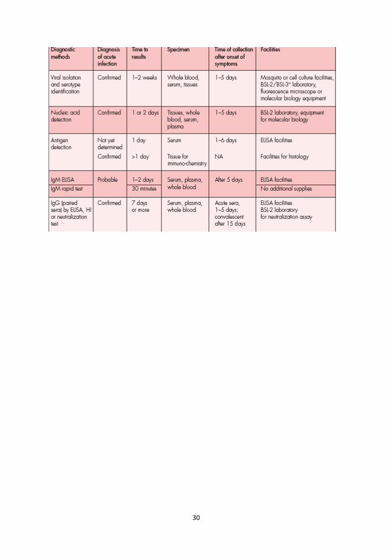
Laboratory Diagnosis
Rapid and accurate dengue diagnosis is of paramount importance for: (i)
epidemiological surveillance;(ii) clinical management; (iii) research; and (iv)
vaccine trials.Clinical management requires early diagnosis of cases,
confirmation of clinical diagnosis and for differential diagnosis from other

22
flaviviruses/infection agents.The following laboratory tests are available to
diagnose dengue fever and DHF:
� Virus isolation – serotypic/genotypic characterization
� Viral nucleic acid detection
� Viral antigen detection
� Immunological response based tests – IgM and IgG antibody assays
� Analysis for haematological parameters
Diagnostic tests and phases of disease
Dengue viraemia in a patient is short, typically occurs 2–3 days prior to
the onset of fever and lasts for four to seven days of illness. During this period
the dengue virus, its nucleic acid and circulating viral antigen can be detected
Antibody response to infection comprises the appearance of different types of
immunoglobulins and IgM and IgG immunoglobulin isotypes are of diagnostic
value in dengue. IgM antibodies are detectable by days 3–5 after the onset of
illness, rise quickly by about two weeks and decline to undetectable levels after
2–3 months. IgG antibodies are detectable at low level by the end of the first
week, increase subsequently and remain for a longer period (for many years).
Because of the late appearance of IgM antibody, i.e. after five days of onset of
fever, serological tests based on this antibody done during the first five days of
clinical illness are usually negative.
During the secondary dengue infection (when the host has previously
been infected by dengue virus), antibody titres rise rapidly. IgG antibodies are
23
detectable at high levels, even in the initial phase, and persist from several
months to a lifelong period. IgM antibody levels are significantly lower in
secondary infection cases. Hence, a ratio of IgM/IgG is commonly used to
differentiate between primary and secondary dengue infections.
Thrombocytopenia is usually observed between the third and eighth day of
illness followed by other haematocrit changes.
Isolation of virus
Isolation of dengue virus from clinical specimens is possible provided the
sample is taken during the first six days of illness and processed without delay.
Specimens that are suitable for virus isolation include: acute phase serum,
plasma or washed buffy coat from the patient, autopsy tissues from fatal cases
(especially liver, spleen, lymph nodes and thymus), and mosquitoes collected
from the affected areas Currently, cell culture is the most widely used method
for dengue virus isolation. The mosquito cell line C6/36 or AP61 are the host
cells of choice for isolation of dengue viruses.
Viral nucleic acid detection
Dengue viral genome, which consists of ribonucleic acid (RNA), can be
detected by reverse transcripatse polymerase chain reaction (RT-PCR) assay.
Nested RT-PCR
Nested RT-PCR assay involves using universal dengue primers targeting
the C/prM region of the viral genome for an initial reverse transcription and
24
amplification step, followed by a nested PCR amplification that is serotype-
specific.
One-step multiplex RT-PCR
This test is an alternative to nested RT-PCR. A combination of the four
serotype - specific reactions are separated by electrophoresis on an agarose gel,
and the amplification products are visualized as bands of different molecular
weights after staining the gel using ethidium bromide dye, and compared with
standard molecular weight markers. In this assay, dengue serotypes are
identified by the size of their bands.
Real-time RT-PCR
The real-time RT-PCR assay is also a one-step assay system using primer
pairs and probes that are specific to each dengue serotype. The use of a
fluorescent probe enables the detection of the reaction products in real time.The
test is very useful for large-scale surveillance.
Isothermal amplification method
The NASBA (nucleic acid sequence-based amplification) assay is an
isothermal RNA-specific amplification assay that does not require thermal
cycling instrumentation.
Compared with virus isolation, the sensitivity of the RT-PCR methods
varies from 80% to 100% and depends upon the region of the genome targeted
by the primers, the approach used to amplify or detect PCR products and the
methods employed for subtyping. The advantages of this technology include
25
high sensitivity and specificity, ease of identifying serotypes and early detection
of the infection. It is, however, an expensive technology.
Recently, Loop Mediated Amplification (LAMP) PCR method has been
developed, which promises an easy-to-do and less expensive instrumentation
alternative for RT-PCR and real-time PCR assays.
Viral antigen detection
The NS1 gene product is a glycoprotein produced by all flaviviruses and
is essential for replication and viability of the virus. The protein is secreted by
mammalian cells but not by insect cells. NS1 antigen appears as early as Day 1
after the onset of the fever and declines to undetectable levels by 5–6 days.
Hence, tests based on this antigen can be used for early diagnosis.ELISA and
dot blot assays directed against the envelop/membrane (EM) antigens and non-
structural protein 1 (NS1) demonstrated that this antigen is present in high
concentrations in the sera of the dengue virus-infected patients during the early
clinical phase of the disease and can be detected in both patients with primary
and secondary dengue infections for up to six days after the onset ofthe illness.
Commercial kits for the detection of NS1 antigens are now available; however,
these kits do not differentiate between the serotypes
Immunological response and serological tests
Five basic serological tests are used for the diagnosis of dengue infection.
26
IgM-capture enzyme-linked immunosorbent assay (MAC-ELISA)
MAC-ELISA has become widely used in the past few years. It is a simple
and rapid test that requires very little sophisticated equipment. MAC-ELISA is
based on detecting the dengue-specific IgM antibodies in the test serum by
capturing them out of solution using anti-human IgM that was previously bound
to the solid phase35. If the patient’s serum has antidengue IgM antibody, it will
bind the dengue antigen that is added in the next step and can be detected by
subsequent addition of an enzyme-labelled anti-dengue antibody, which may be
human or monoclonal antibody. An enzyme-substrate is added to produce a
colour reaction.
The anti-dengue IgM antibody develops a little earlier than IgG, and is
usually detectable by Day5 of the illness, i.e. the antibody is not usually
detectable during the first five days of illness. However,the time of appearance
of IgM antibody varies considerably among patients. IgM antibody titers in
primary infections are significantly higher than in secondary infections,
although it is not uncommon to obtain IgM titers of 320 in the latter cases. In
some primary infections, detectable IgM may persist for more than 90 days, but
in most patients it wanes to an undetectable level by 60 days MAC-ELISA is
slightly less sensitive than the HI test for diagnosing dengue infection. MAC-
ELISA has become an invaluable tool for surveillance of DF, DHF and DSS.It
is especially useful for hospitalized patients who are generally admitted at a late
stage of illness after detectable IgM is already present in the blood.
27
IgG-ELISA
An indirect IgG-ELISA has been developed and compares well with the
HI test.This test can also be used to differentiate primary and secondary dengue
infection
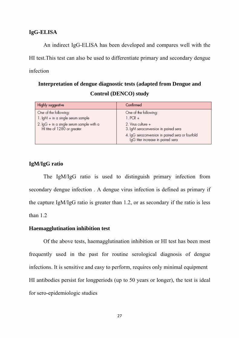
Interpretation of dengue diagnostic tests (adapted from Dengue and
Control (DENCO) study
IgM/IgG ratio
The IgM/IgG ratio is used to distinguish primary infection from
secondary dengue infection . A dengue virus infection is defined as primary if
the capture IgM/IgG ratio is greater than 1.2, or as secondary if the ratio is less
than 1.2
Haemagglutination inhibition test
Of the above tests, haemagglutination inhibition or HI test has been most
frequently used in the past for routine serological diagnosis of dengue
infections. It is sensitive and easy to perform, requires only minimal equipment
HI antibodies persist for longperiods (up to 50 years or longer), the test is ideal
for sero-epidemiologic studies
28
Complement fixation test
The complement fixation or CF test is not widely used for routine dengue
diagnostic serology. It is more difficult to perform and requires highly trained
personnel.
Neutralization test
The neutralization test or NT is the most specific and sensitive
serological test for dengue viruses used for determining the immune protection.
The common protocol used in most dengue laboratories is the serum dilution
plaque reduction neutralization test (PRNT)
Rapid diagnostic test (RDT)
A number of commercial rapid format serological test-kits for anti-
dengue IgM and IgG antibodies have become available in the past few years,
some of these producing results within 15 minutes36.Unfortunately, the accuracy
of most of these tests is uncertain since they have not yet been properly
validated. Rapid tests can yield false positive results due to cross-reaction with
other flaviviruses, malaria parasite, leptospires and immune disorders such as
rheumatoid and lupus.
Haematological tests
Standard haematological parameters such as platelet count and
haematocrit are important and are part of the biological diagnosis of dengue
infection. Therefore, they should be closely monitored.
29
Thrombocytopenia, a drop in platelet count below 100 000 per μl, may be
occasionally observed in dengue fever but is a constant feature in DHF.
Thrombocytopenia is usually found between the third and eighth day of illness
often before or simultaneously with changes in haematocrit.
Haemoconcentration with an increase in the haematocrit of 20% or more
(for the same patient or for a patient of the same age and sex) is considered to
be a definitive evidence of increased vascular permeability and plasma leakage.
Basic metabolic panel findings include the following:
Hyponatremia is the most common electrolyte abnormality observed in
patients with DHF or DSS.
Metabolic acidosis is observed in those with shock, and it must be
corrected rapidly.
Elevated BUN is observed in those with shock.
Liver function test findings include the following:
Mild elevations in transaminase levels may be seen.
Low albumin is a sign of hemoconcentration
30

31
CASE DEFINITION2
DENGUE FEVER:is defined by
DENGUE HEMORRHAGIC FEVER: is defined by

32
DENGUE SHOCK SYNDROME:is defined by
SEVERITY GRADING OF DENGUE:
WHO classification of dengue infections and grading of severity of DHF
33
MANAGEMENT
Disease notification
In dengue-endemic countries, cases of suspected, probable and confirmed
dengue should be notified as soon as possible so that appropriate public health
measures can be initiated . Laboratory confirmation is not necessary before
notification, but should be obtained. In non-endemic countries, usually only
confirmed cases will be notified. Suggested criteria for early notification of
suspected cases are that the patient lives in or has travelled to a dengue-endemic
area, has fever for three days or more, has low or decreasing white cell counts,
and/or has thrombocytopenia ± positive tourniquet test. In dengue-endemic
countries, the later the notification, the more difficult it is to prevent
dengue transmission.
Treatment of Dengue & DHF2:
Febrile Phase:
In the initial phase the treatment of DF & DHF is the same and is as that
of any other viral fever, i.e. symptomatic and supportive.
� Rest.
� Paracetamol (not > than 4 times in 24 hrs) according to age and
weight for fever above 39°C.
� Do not give Aspirin or Brufen. Aspirin can cause gastritis and/or
bleeding. In children, Reye’s syndrome (Encephalopathy) may be a serious
complication.
34
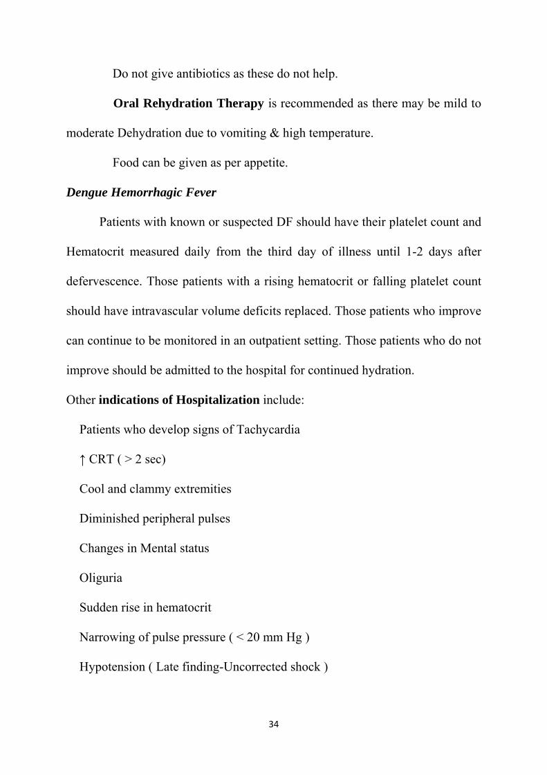
� Do not give antibiotics as these do not help.
� Oral Rehydration Therapy is recommended as there may be mild to
moderate Dehydration due to vomiting & high temperature.
� Food can be given as per appetite.
Dengue Hemorrhagic Fever
Patients with known or suspected DF should have their platelet count and
Hematocrit measured daily from the third day of illness until 1-2 days after
defervescence. Those patients with a rising hematocrit or falling platelet count
should have intravascular volume deficits replaced. Those patients who improve
can continue to be monitored in an outpatient setting. Those patients who do not
improve should be admitted to the hospital for continued hydration.
Other indications of Hospitalization include:
� Patients who develop signs of Tachycardia
� ↑ CRT ( > 2 sec)
� Cool and clammy extremities
� Diminished peripheral pulses
� Changes in Mental status
� Oliguria
� Sudden rise in hematocrit
� Narrowing of pulse pressure ( < 20 mm Hg )
� Hypotension ( Late finding-Uncorrected shock )
35
Intravascular volume deficits should be corrected with isotonic fluids
such as Ringer lactate solution. Boluses of 10-20 ml/kg should be given over 20
minutes and may be repeated. If this fails to correct the deficit, the hematocrit
value should be determined, and, if it is rising, limited clinical information
suggests that a plasma expander may be administered. Starch, dextran 40, or
albumin 5% at a dose of 10-20 ml/kg may be used. If the patient does not
improve after this, blood loss should be considered. Patients with internal or
gastrointestinal bleeding may require transfusion. Patients with coagulopathy
may require fresh frozen plasma.
After patients with dehydration are stabilized, they usually require
intravenous fluids for no more than 24-48 hours. Intravenous fluids should be
stopped when the hematocrit level falls below 40% and adequate intravascular
volume is present. At this time, patients reabsorb extravasated fluid and are at
risk for volume overload if intravenous fluids are continued. Do not interpret a
falling hematocrit value in a clinically improving patient as a sign of internal
bleeding.
36
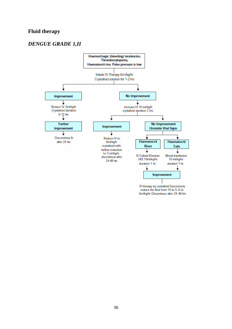
Fluid therapy
DENGUE GRADE 1,II
37
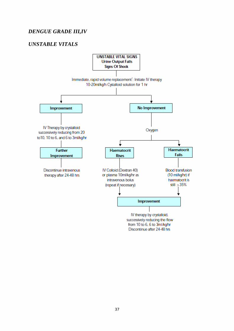
DENGUE GRADE III,IV
UNSTABLE VITALS
38
Fluids Recommended:
Crystalloids :
5% Dextrose in Isotonic NS
5% Dextrose in 1/2 NS
5% Dextrose in RL
Shock Correction NS or RL
Colloids :
Dextran 40
Hemaccel
Plasma
Fluid Replacement
Volume of fluid to be just sufficient to maintain effective circulation
during plasma leakage.
Fluid charts to be made every 1 to 3 hrs and even more frequently in
shock.
Change should not be drastic. e.g. don’t jump from 20 ml to 6ml or vice
versa. Go in a step wise manner.
Indications of Platelet / Whole blood transfusion2
Platelets
Platelet count < 20,000/ cu.mm
Platelet count >20,000 <40,000 with hemorrhagic manifestations.
DIC
39
Whole Blood
Prolonged refractory shock with ↓↓HCT even with adequate fluid
replacement
Severe massive bleeding ( > 10 % of total blood volume).
Indications of FFP
(Fresh frozen Plasma)
Plasma is used when HCT is rising despite fluid replacement
But plasma substitutes are equally good (Dextran 40) & effective
Essential only in cases of massive bleeding with DIC.
Monitoring of patients in DSS
Check vitals every 15-30 minutes until shock is overcome.
Check HCT / Platelets for every 2 hours for the first 6 hours and every 4
hours until stable.
Fluid balance sheet to be maintained. Frequency & volume of urine
output to be recorded. In refractory shock catheter may be needed.
Criteria for Discharge2
Patients who are resuscitated from shock recover rapidly. Patients with
DHF or dengue shock syndrome (DSS) may be discharged from the hospital
when they meet the following criteria:
Afebrile for 24 hours without antipyretics
Good appetite, clinically improved condition
Adequate urine output
40
Stable hematocrit
At least 48 hours have passed since recovery from shock
Absence of respiratory distress
Platelet count greater than 50,000.
Management of high-risk patients
Obese patients have less respiratory reserves and care should be taken to
avoid excessive intravenous fluid infusions. The ideal body weight should
be used to calculate fluid resuscitation and replacement and colloids
should be considered in the early stages of fluid therapy. Once stabilized,
furosemide may be given to induce diuresis.
Infants also have less respiratory reserves and are more susceptible to
liver impairment and electrolyte imbalance. They may have a shorter
duration of plasma leakage and usually respond quickly to fluid
resuscitation. Infants should, therefore, be evaluated more frequently for
oral fluid intake and urine output.
Intravenous insulin is usually required to control the blood sugar levels in
dengue patients with diabetes mellitus. Non-glucose containing
crystalloids should be used.
Patients with hypertension may be on anti-hypertensive therapy that
masks the cardiovascular response in shock. The patient’s own baseline
blood pressure should be considered. A blood pressure that is perceived
to be normal may in fact be low for these patients.
41
Anti-coagulant therapy may have to be stopped temporarily during the
critical period.
Haemolytic diseases and haemoglobiopathies : These patients are at risk
of haemolysis and will require blood transfusion. Caution should
accompany hyper hydration and alkalinisation therapy, which can cause
fluid overload and hypo calcemia.
• Congenital and ischemic heart diseases: Fluid therapy should be more
cautious as they may have less cardiac reserves.
For patients on steroid therapy, continued steroid treatment is
recommended but the route may be changed.
Management of complications
The most common complication is fluid overload
Detection of fluid overload in patients
Early signs and symptoms include puffy eyelids, distended abdomen
(ascites), tachypnoea, mild dyspnoea.
Late signs and symptoms include all of the above, along with moderate to
severe respiratory distress, shortness of breath and wheezing (not due to
asthma) which are also an early sign of interstitial pulmonary oedema and
crepitations. Restlessness/agitation and confusion are signs of hypoxia
and impending respiratory failure.

42
Management of fluid overload
All hypotonic solutions should be stopped. In the early stage of fluid
overload, switch from crystalloid to colloid solutions as bolus fluids. Dextran 40
is effective as 10 ml/kg bolus infusions, but the dose is restricted to 30
ml/kg/day because of its renal effects. Dextran 40 is excreted in the urine and
will affect urine osmolarity. Patients may experience “sticky” urine because of
the hyperoncotic nature of Dextran 40 molecules (osmolarity about twice that of
plasma). Voluven may be effective (osmolarity = 308 mosmole) and the upper
limit is 50ml/kg/day. However, no studies have been done to prove its
effectiveness in cases of DHF/DSS. In the late stage of fluid overload or those
with frank pulmonary oedema, furosemide may be administered if the patient
has stable vital signs. If they are in shock, together with fluid overload 10
ml/kg/h of colloid (dextran) should be given. When the blood pressure is stable,
usually within 10 to 30 minutes of infusion, administer IV 1 mg/kg/dose of
furosemide and continue with dextran infusion until completion. Intravenous
fluid should be reduced to as low as 1 ml/kg/h until discontinuation when
haematocrit decreases to baseline or below (with clinical improvement). The
following pointsshould be noted:
These patients should have a urinary bladder catheter to monitor hourly
urine output.
43
Furosemide should be administered during dextran infusion because the
hyperoncotic nature of dextran will maintain the intravascular volume
while furosemide depletes in the intravascular compartment.
After administration of furosemide, the vital signs should be
monitored every 15 minutes for one hour to note its effects.
If there is no urine output in response to furosemide, check the
intravascular volume status (CVP or lactate). If this is adequate, pre-renal
failure is excluded, implying that the patient is in an acute renal failure
state. These patients may require ventilatory support soon..
In cases with no response to furosemide (no urine obtained), repeated
doses of furosemide and doubling of the dose are recommended. If
oliguric renal failure is established, renal replacement therapy is to be
done as soon as possible. These cases have poor prognosis.
Pleural and/or abdominal tapping may be indicated and can be life-saving
in cases with severe respiratory distress and failure of the above
management. This has to be done with extreme caution because traumatic
bleeding is the most serious complication and leads to death.
Management of encephalopathy
Some DF/DHF patients present unusual manifestations with signs and
symptoms of central nervous system (CNS) involvement, such as convulsion
and/or coma. This has generally been shown to be encephalopathy, not
encephalitis, which may be a result of intracranial haemorrhage or occlusion
44
associated with DIC or hyponatremia. CNS infections documented by virus
isolations from the cerebrospinal fluid (CSF) or brain.
Most of the patients with encephalopathy report hepatic encephalopathy. The
principal treatment of hepatic encephalopathy is to prevent the increase of
intracranial pressure (ICP).Radiological imaging of the brain (CT scan or MRI)
is recommended if available to rule out intracranial haemorrhage. The following
are recommendations for supportive therapy for this condition:
Maintain adequate oxygenation with oxygen therapy. Prevent/reduce ICP
by the following measures:
o give minimal IV fluid to maintain adequate intravascular volume;
ideally the total IV fluid should not be >80% fluid maintenance.
o switch to colloidal solution earlier if haematocrit continues to rise
and a large volume of IV is needed in cases with severe plasma
leakage.
o administer a diuretic if indicated in cases with signs and symptoms
of fluid overload.
o positioning of the patient must be with the head up by 30 degrees.
o early intubation to avoid hypercarbia and to protect the airway.
o may consider steroid to reduce ICP. Dexamethazone 0.15
mg/kg/dose IV to be administered every 6–8 hours.
Decrease ammonia production by the following measures:

45
o give lactulose 5–10 ml every six hours for induction of osmotic
diarrhoea.
o local antibiotic gets rid of bowel flora; it is not necessary if
systemic antibiotics are given.
Maintain blood sugar level at 80–100 mg/dl per cent. Recommend
glucose infusion rate is anywhere between 4–6 mg/kg/hour.
Correct acid-base and electrolyte imbalance, e.g. correct
hypo/hypernatremia, hypo/hyperkalemia, hypocalcemia and acidosis.
Vitamin K1 IV administration; 3 mg for <1-year-old, 5 mg for <5-year-
old and 10 mg for>5-year-old and adult patients.
Anticonvulsants should be given for control of seizures: phenobarbital,
dilantin and diazepam IV as indicated.
Transfuse blood, preferably freshly packed red cells, as indicated. Other
blood components such as as platelets and fresh frozen plasma may not
be given because the fluid overload may cause increased ICP.
Empiric antibiotic therapy may be indicated if there are suspected
superimposed bacterial infections.
H2-blockers or proton pump inhibitor may be given to alleviate
gastrointestinal bleeding.
Avoid unnecessary drugs because most drugs have to be metabolized by
the liver.
46
Consider plasmapheresis or haemodialysis or renal replacement therapy
in cases with clinical deterioration.
VECTOR CONTROL
Environmental management methods37
Environmental modification: This includes any long-lasting physical
transformation of land, water and vegetation aimed at reducing vector
habitats without causing unduly adverse effects on the quality of the
human environment.
Environmental manipulation: This incorporates planned recurrent
activities aimed at producing temporary changes in vector habitats that
involve the management of “essential” and “non-essential” containers,
and the management or removal of “natural” breeding sites.such as
1. improved water supply
2. covering of overhead tanks
3. land filling, levelling
Changes to human habitation or behaviour: These feature the efforts
made to reduce man-vector-virus contact.
Biological control
Biological control is based on the introduction of organisms that prey
upon, reduce populations of the target species. The application of biological
control agents, which are directed against the larval stages of dengue vectors,
While biological control avoids chemical contamination of the environment,
47
Larvivorus fish (Gambusia affinis and Poecilia reticulata) have been
extensively used for the control of An. stephensi and/or Ae. aegypti in large
water bodies or large water containers in many countries. Two species of
endotoxin-producing bacteria, Bacillus thuringiensis serotype H-14 and
Bacillus sphaericus are effective mosquito control agents. Copepods have a
role in dengue vector control,
Chemical control
Chemicals have been used to control Ae. aegypti since the beginning of
the 20th century. Aedes larval habitats were treated with oil and homes were
fumigated with pyrethrins DDT emerged in the early 1960s, organophosphate
insecticides, including fenthion, malathion and fenitrothion, were used for Ae.
aegypti adult control and temephos as a larvicide. Current methods are larvicide
application and space spraying. Larviciding or “focal” control of Ae. aegypti is
usually limited to domestic-use containers that cannot be destroyed, eliminated
or otherwise managed. It is difficult and expensive to apply chemical larvicides
on a long-term basis. Therefore, chemical larvicides are best used in situations
where the disease and vector surveillance indicate the existence of certain
periods of high risk and in localities where outbreaks might occur
48
Personal protection
Protective clothing
Clothing reduces the risk of mosquito bite if the cloth material is
sufficiently thick or loosely fitting. Long sleeves and trousers with stockings
may protect the arms and legs, which are the preferred sites for mosquito bites.
Schoolchildren should adhere to these practices whenever possible Household
insecticidal products, namely mosquito coils and aerosols, are used extensively
for personal protection against mosquitoes. Repellents are common means of
personal protection against mosquitoes and other biting insects .Insecticide-
treated mosquito nets (ITNs) have limited utility in dengue control programmes
since the vector species bites during the day.
49
METHODOLOGY
Type of Study: Cross Sectional Study.
Study Period: The study was conducted from Dec 2009 to Dec 2010.
Settings: Children less than 12 years of age with clinical features of Dengue
(any acute febrile illness with two of the following: myalgia, head ache, retro-
orbital pain, bleeding, altered sensorium, shock or low platelet count) admitted
at Children Medical Ward of Tirunelveli Medical College Hospital during the
study period were registered in the study. Informed consent was obtained and
detailed history was taken. For all cases, the rapid Ig M ELISA test was done at
our hospital. Children positive for Ig M were followed up for clinical profile.
Sample Size: From Dec 2009 to Dec 2010, there were totally 53 cases
according to the inclusion and exclusion criteria.
Inclusion Criteria:
• Children less than 12 years of age with clinical features of dengue (any
acute febrile illness) with two of the following :
o Rash
o Headache
o Myalgia/ Arthralgia
o Bleeding manifestation
o Altered sensorium
o Retro orbital pain
o Shock or low platelet count

50
Exclusion Criteria:
• Clinical Features suggestive of Dengue with other serology positive
cases.
Tools used:
Using Proforma the basic socio demographic details, Clinical features were
collected. Laboratory investigations carried out in these patients include
Haemoglobin, Complete blood count, Dengue IgM serology, Liver function
test, serum amylase. Chest X ray was taken to demonstrate pleural effusion.
Ultrasound abdomen was done to identify ascites, polyserositis and gall bladder
wall thickening. CSF analysis was done in patients with convulsions, meningeal
signs and altered sensorium.
Outcome Measures: Children positive for IgM were followed for the clinical
profile and outcome.
The number of children included based on the above criteria was 53. Children
who were seropositive were classified on the basis of WHO criteria as follows:
1. Dengue fever (DF): dengue seropositive without bleed.
2. Dengue fever with unusual bleed (DFB): dengue seropositive with
bleeding tendencies, not satisfying WHO criteria for DHF
3. Dengue haemorrhagic fever (DHF): Dengue seropositive with bleeds with
evidence of plasma leakage.

51
4. Dengue Shock Syndrome (DSS): DHF with evidence of peripheral
circulatory failure.
Data Analysis:
Data collected were entered in Excel Spreadsheet and analysed using SPSS
Version 16. Simple calculations like Percentages, Proportions and Mean values
were derived. Appropriate statistical tests like Chi – Square test, T test were
used.
52
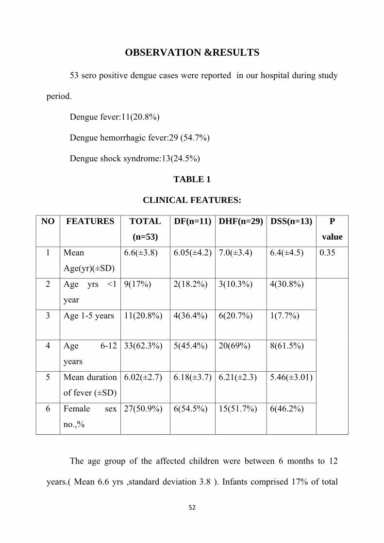
OBSERVATION &RESULTS
53 sero positive dengue cases were reported in our hospital during study
period.
Dengue fever:11(20.8%)
Dengue hemorrhagic fever:29 (54.7%)
Dengue shock syndrome:13(24.5%)
TABLE 1
CLINICAL FEATURES:
NO FEATURES TOTAL
(n=53)
DF(n=11) DHF(n=29) DSS(n=13) P
value
1 Mean
Age(yr)(±SD)
6.6(±3.8) 6.05(±4.2) 7.0(±3.4) 6.4(±4.5) 0.35
2 Age yrs <1
year
9(17%) 2(18.2%) 3(10.3%) 4(30.8%)
3 Age 1-5 years 11(20.8%) 4(36.4%) 6(20.7%) 1(7.7%)
4 Age 6-12
years
33(62.3%) 5(45.4%) 20(69%) 8(61.5%)
5 Mean duration
of fever (±SD)
6.02(±2.7) 6.18(±3.7) 6.21(±2.3) 5.46(±3.01)
6 Female sex
no.,%
27(50.9%) 6(54.5%) 15(51.7%) 6(46.2%)
The age group of the affected children were between 6 months to 12
years.( Mean 6.6 yrs ,standard deviation 3.8 ). Infants comprised 17% of total
53
study group. 20.8% were children between 1 and 5 years of age. 62.3% were
children between 6 and 12 years.DHF and DSS was found to be more common
in 6-12 years age group. But this fact is not statistically significant. Mean
duration of fever was 6.02 days. It was 6.18,6.21,5.46 days in DF, , DHF, DSS
respectively. Males(49.1%) and females(50.9%) were comparatively equally
affected.
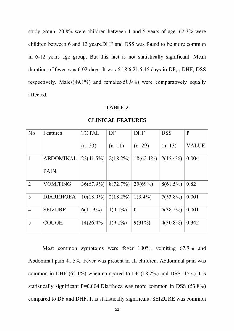
TABLE 2
CLINICAL FEATURES
No Features TOTAL
(n=53)
DF
(n=11)
DHF
(n=29)
DSS
(n=13)
P
VALUE
1 ABDOMINAL
PAIN
22(41.5%) 2(18.2%) 18(62.1%) 2(15.4%) 0.004
2 VOMITING 36(67.9%) 8(72.7%) 20(69%) 8(61.5%) 0.82
3 DIARRHOEA 10(18.9%) 2(18.2%) 1(3.4%) 7(53.8%) 0.001
4 SEIZURE 6(11.3%) 1(9.1%) 0 5(38.5%) 0.001
5 COUGH 14(26.4%) 1(9.1%) 9(31%) 4(30.8%) 0.342
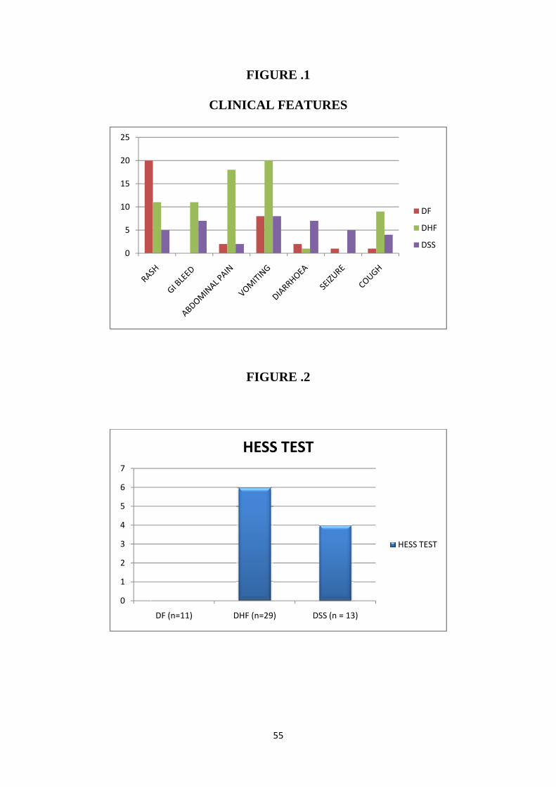
Most common symptoms were fever 100%, vomiting 67.9% and
Abdominal pain 41.5%. Fever was present in all children. Abdominal pain was
common in DHF (62.1%) when compared to DF (18.2%) and DSS (15.4).It is
statistically significant P=0.004.Diarrhoea was more common in DSS (53.8%)
compared to DF and DHF. It is statistically significant. SEIZURE was common
54
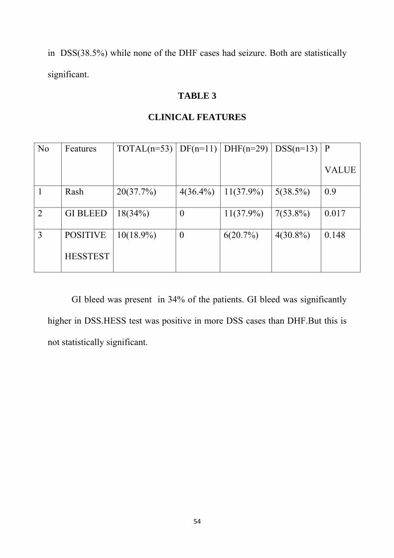
in DSS(38.5%) while none of the DHF cases had seizure. Both are statistically
significant.
TABLE 3
CLINICAL FEATURES
GI bleed was present in 34% of the patients. GI bleed was significantly
higher in DSS.HESS test was positive in more DSS cases than DHF.But this is
not statistically significant.
No Features TOTAL(n=53) DF(n=11) DHF(n=29) DSS(n=13) P
VALUE
1 Rash 20(37.7%) 4(36.4%) 11(37.9%) 5(38.5%) 0.9
2 GI BLEED 18(34%) 0 11(37.9%) 7(53.8%) 0.017
3 POSITIVE
HESSTEST
10(18.9%) 0 6(20.7%) 4(30.8%) 0.148
0
5
10
15
20
25
0
1
2
3
4
5
6
7
DF (n=11)
F
CLINIC
F
DHF
H
55
FIGURE .
CAL FEA
FIGURE .
(n=29)
HESS TES
.1
ATURES
.2
DSS (n = 13
ST
3)
H
DF
DHF
DSS
HESS TEST
56
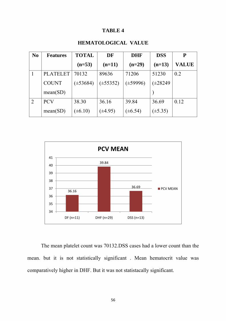
TABLE 4
HEMATOLOGICAL VALUE
No Features TOTAL
(n=53)
DF
(n=11)
DHF
(n=29)
DSS
(n=13)
P
VALUE
1 PLATELET
COUNT
mean(SD)
70132
(±53684)
89636
(±55352)
71206
(±59996)
51230
(±28249
)
0.2
2 PCV
mean(SD)
38.30
(±6.10)
36.16
(±4.95)
39.84
(±6.54)
36.69
(±5.35)
0.12
The mean platelet count was 70132.DSS cases had a lower count than the
mean. but it is not statistically significant . Mean hematocrit value was
comparatively higher in DHF. But it was not statistacally significant.
36.16
39.84
36.69
34
35
36
37
38
39
40
41
DF (n=11) DHF (n=29) DSS (n=13)
PCV MEAN
PCV MEAN
57
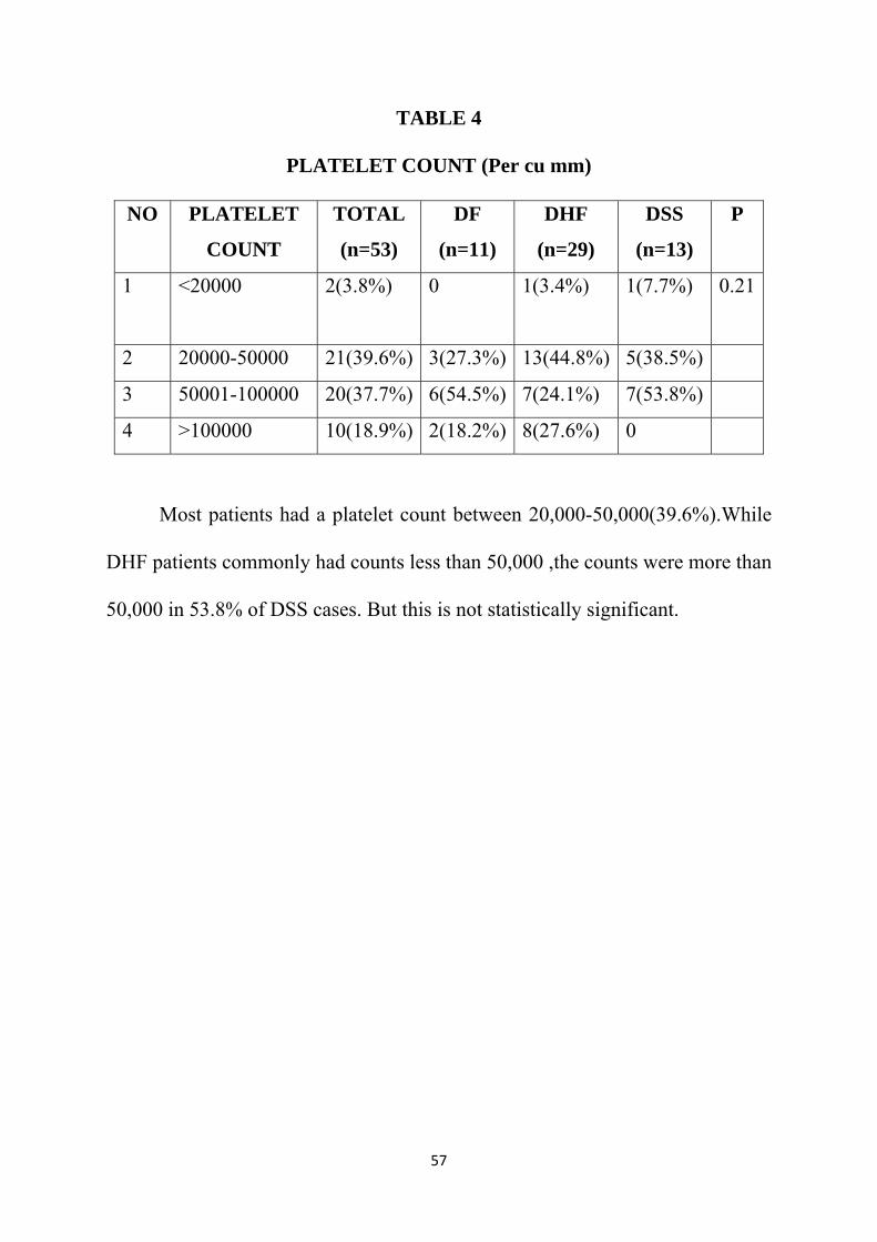
TABLE 4
PLATELET COUNT (Per cu mm)
NO PLATELET
COUNT
TOTAL
(n=53)
DF
(n=11)
DHF
(n=29)
DSS
(n=13)
P
1 <20000 2(3.8%) 0 1(3.4%) 1(7.7%) 0.21
2 20000-50000 21(39.6%) 3(27.3%) 13(44.8%) 5(38.5%)
3 50001-100000 20(37.7%) 6(54.5%) 7(24.1%) 7(53.8%)
4 >100000 10(18.9%) 2(18.2%) 8(27.6%) 0
Most patients had a platelet count between 20,000-50,000(39.6%).While
DHF patients commonly had counts less than 50,000 ,the counts were more than
50,000 in 53.8% of DSS cases. But this is not statistically significant.
No F
1 S
(%
2 S
(%
statisti
Features
GOT
%)
GPT
%)
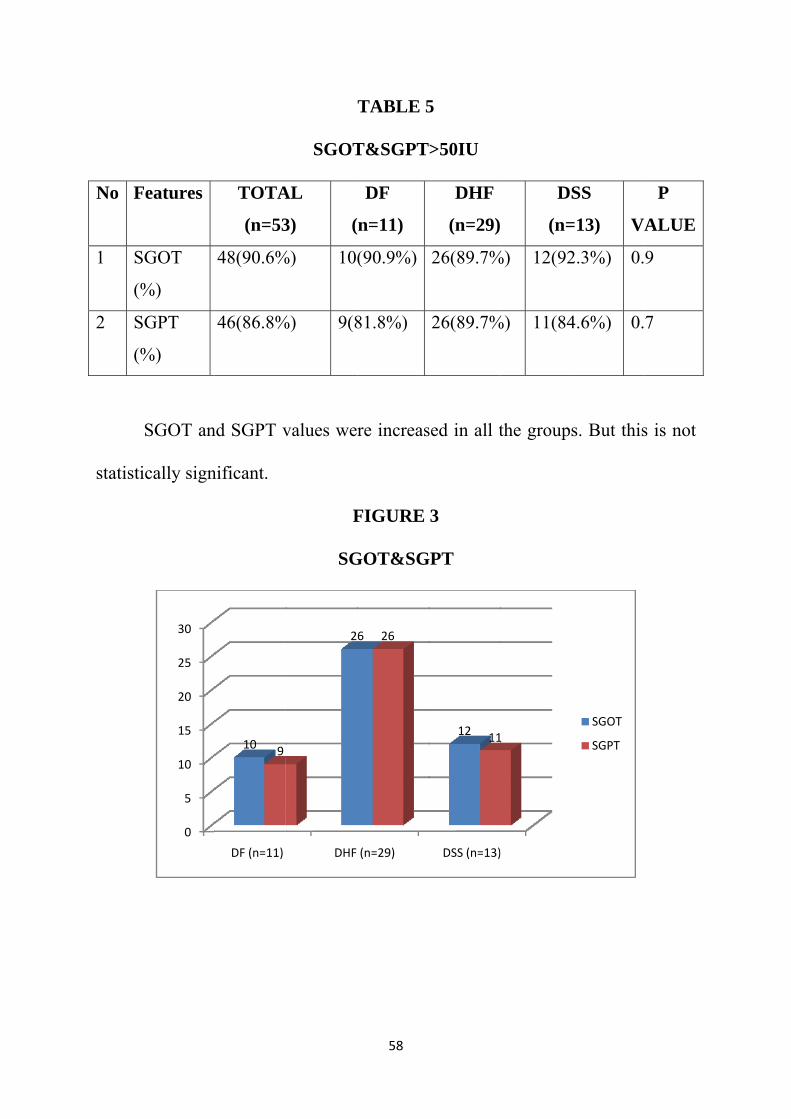
SGOT an
ically sign
0
5
10
15
20
25
30
TOTA
(n=53
48(90.6%
46(86.8%
nd SGPT v
nificant.
DF (n=11)
10 9
SGOT
AL
3) (
%) 10(
%) 9(8
values wer
F
SG
DHF
2
58
TABLE 5
T&SGPT
DF
n=11)
(90.9%)
81.8%)
re increas
FIGURE
GOT&SG
(n=29)
6 26
5
>50IU
DHF
(n=29)
26(89.7%
26(89.7%
sed in all t
3
GPT
DSS (n=13)
12 11
DS
(n=
%) 12(92
%) 11(84
the groups
1
SS
=13) V
2.3%) 0.
4.6%) 0.
s. But this
SGOT
SGPT
P
VALUE
9
7
s is not
No
1 H
2 R
EF
A
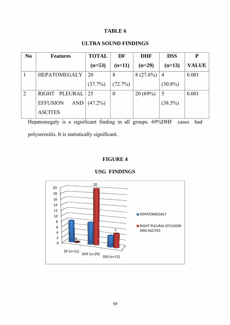
Hepato
polyse
Featu
HEPATOM
RIGHT PL
FFUSION
ASCITES
omegaly i
erositis. It
02468
10
12
14
16
18
20
U
ures
MEGALY
LEURAL
N AND
is a signi
is statistic
DF (n=11)D
0
ULTRA S
TOTA
(n=53)
20
(37.7%)
L
D
25
(47.2%)
ificant fin
cally signi
F
USG
DHF (n=29)D
20
59
TABLE 6
SOUND F
L
)
DF
(n=1
)
8
(72.7%
)
0
nding in a
ificant.
FIGURE
G FINDIN
DSS (n=13)
5
6
FINDING
F
1)
DH
(n=
%)
8 (27
20 (6
all group
4
NGS
HEP
RIGAND
GS
HF
=29) (
7.6%) 4
(30
69%) 5
(38
s. 69%DH
PATOMEGALY
HT PLEURAL ED ASCITES
DSS
n=13)
0.8%)
0
8.5%)
0
HF case
EFFUSION
P
VALUE
0.001
0.001
s had
No
1
2
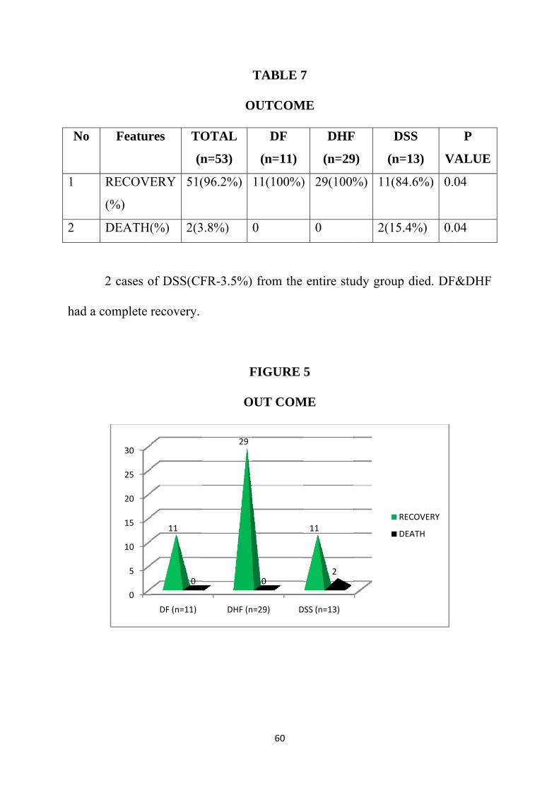
had a c
Featur
RECOVE
(%)
DEATH(
2 cases o
complete r
0
5
10
15
20
25
30
res TO
(n
ERY 51(
(%) 2(3
of DSS(CF
recovery.
DF (n=11)
11
0
O
OTAL
n=53)
(96.2%)
3.8%)
FR-3.5%)
F
O
DHF (n
29
60
TABLE 7
OUTCOM
DF
(n=11)
11(100%)
0
from the
FIGURE
OUT COM
=29) D
0
7
ME
DHF
(n=29
) 29(100%
0
entire stu
5
ME
DSS (n=13)
11
2
F
9)
DS
(n=
%) 11(84
2(15.
udy group
R
D
SS
=13) VA
4.6%) 0.0
.4%) 0.0
died. DF
ECOVERY
DEATH
P
ALUE
04
04
&DHF
61
DISCUSSION
As discussed in the literature, SouthEastAsian region has a varied
clinical profile and outcome compared to the rest of the world. The
demographic pattern and the trend of illness are vastly changing every year
through the past decade. South India and especially SouthTamilnadu has
witnessed several dengue epidemic outbreaks during the past few years. Here I
compare the clinical profile and outcome of Dengue in our hospital with other
Indian and study done other countries.
Totally 53 Sero-positive cases dengue children who presented at
Tirunelveli Medical College hospital during the study period were analysed. As
per the WHO classification, the frequency of Dengue fever was 20.8%, Dengue
hemorrhagic fever 54.7% and Dengue shock syndrome 24.5%, while no cases
presented Dengue fever having unusual bleed.(i.e. bleeding without plasma
leakage) Narayanan et al (Chennai2001)reported DF (72.78%), DHF (18.6%),
DSS (8.4%). Kalyanarooj et al (Indonesia) reported DF (including DFB)
(53%), DHF (including DSS)(47%). Ratageri et al(Hubli2003) reported
DF(18%),DHF (includingDSS) is 82%. In present study, DHF (including DSS)
is 79.2%. Present study is comparable with other studies.
This shows that severe forms of dengue-DHF and DSS have increased
over the decade. It may be due to increasing endemicity, environmental factors
and changing virulence of the viruses.
62
Baseline microvascular permeability in children is greater than that of
adults and this could partly explain why DHF and DSS are more frequently seen
in children38.
AGE:
In the present study Dengue infection was noted in 17% of infants , 1-
5yrs(20.8%)and 62.3% of children 6-12yrs was observed. Manjunath J.
Kulkarni et al Clinico-Epidemiological Profile of Children Hospitalized with
Dengue Indian J Pediatr (2010) 77:1103–1104 study shows Children in 6–12
yrs age group constituted 45.8% of cases forming the most commonly affected
group. Fahad Javaid Siddiqui et al. Study shows older children appeared 5.5
times more likely to be affected than their younger (0 – 5 years) counterparts.
These are comparable with our present study.
SEX:
In the present study, there is no sex predilection for the disease (male
49.1% compared to female 50.1%), while both sexes had equal distribution of
disease severity. Regarding the relationship between gender and severity among
children, Nimmannitya(1987a,b) reported that shock and death occurred more
frequently in females than in males. (Garcia-Rivera et al, 2003;. studies
reporting no significant difference in frequency between male and female.
63
FEVER:
In the present study all children had fever. Mean duration fever was
6.02 days. In DSS mean duration of fever was 5.14 days. Chandrakanta,et al
study shows mean duration of fever 10.7%.
VOMITING:
In the present study 67.9% children had vomiting. Manjunath J. Kulkarni
et al reported vomiting in 35.2% children.Chandrakanta,et al (Changing clinical
manifestations of dengue infection in north India dengue bulletin 2008)shows
vomiting was 41.2%. Ratagiri et al study shows 82%.The percentage is
variable in different studies.
ABDOMINAL PAIN:
In the present study 41.5% children had abdominal pain. Chandrakanta,et
al study shows 25%.Shigeki Hanafusa et al study shows 68%.In the present
study abdominal pain was significantly more common in DHF (62.1%).
GI BLEEDING:
The present study shows 34% incidence of GI bleeding. Chandrakanta,et
al shows 38.8%. Narayanan et al shows high percentage 61% of hematemesis.
In the present study GI bleeding was significantly more common in DSS.
64
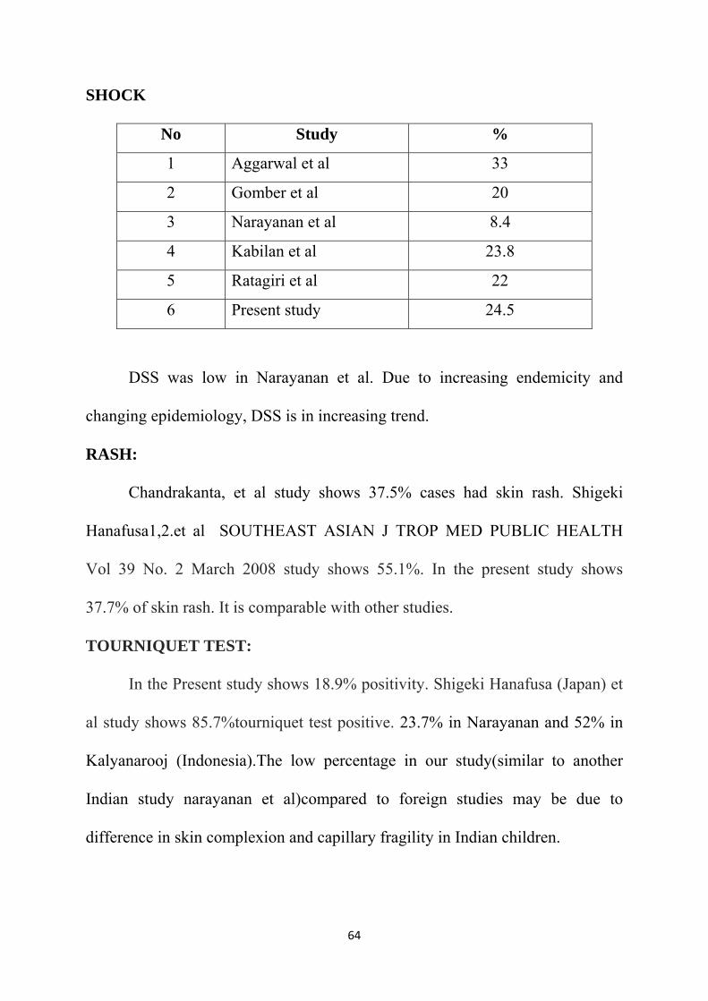
SHOCK
No Study %
1 Aggarwal et al 33
2 Gomber et al 20
3 Narayanan et al 8.4
4 Kabilan et al 23.8
5 Ratagiri et al 22
6 Present study 24.5
DSS was low in Narayanan et al. Due to increasing endemicity and
changing epidemiology, DSS is in increasing trend.
RASH:
Chandrakanta, et al study shows 37.5% cases had skin rash. Shigeki
Hanafusa1,2.et al SOUTHEAST ASIAN J TROP MED PUBLIC HEALTH
Vol 39 No. 2 March 2008 study shows 55.1%. In the present study shows
37.7% of skin rash. It is comparable with other studies.
TOURNIQUET TEST:
In the Present study shows 18.9% positivity. Shigeki Hanafusa (Japan) et
al study shows 85.7%tourniquet test positive. 23.7% in Narayanan and 52% in
Kalyanarooj (Indonesia).The low percentage in our study(similar to another
Indian study narayanan et al)compared to foreign studies may be due to
difference in skin complexion and capillary fragility in Indian children.
65
DIARRHOEA:
Chandrakanta, et al study shows 6.2% of children had loose stools. In the
present study 18.9% children had diarrhea and is statistically significantly
reported in DSS cases .This may be due to the poor socio economic status and
nutritional status in our children,predisposing to early shock in dengue.
SEIZURE:
Chandrakanta et al study shows 45% cases had seizure. In present study
11.3% cases had seizure.Most of the seizure occurred in DSS 38.5% .
COUGH:
Shigeki Hanafusa et al study shows 35.4% children had cough.
Manjunath J. Kulkarni study shows 9.07% . In the present study 26.4% of
children had cough. Cough is a Unique feature of paediatric dengue compared
to adult dengue.
MEAN HEMATOCRIT:
Chandrakanta, et al study shows mean hematocrit value 26.8 %.
Narayanan et al study shows 33.2%. Karthis et al study shows 34.06%. In the
present study, mean hematocrit value is 38.30%.
PLATELET COUNT:
In present study platelet count is as follows
<20000=3.8%
20000-50000=39.6%
50001-100000=37.7%
66
>100000 =18.9%
Maimoona M. Ahmed J Infect Dev Ctries 2010; 4(8):503-510.study
showed
<20000=6.75%
20000-50000=50%
50001-10000=41%
>100000=18.5%
Present study is comparable with this study.
In the present study, there is no correlation between the counts and the
disease severity with 48.2% of DHF occurring in children with counts less than
50,000 while 53.8% of DSS in children with counts more than 50,000.this is
because in dengue, the Development of antibodies potentially cross-reactive to
plasminogen could have a role in causing haemorrhage 39 . The increased
destruction or decreased production of platelets could result in
thrombocytopenia.count may thus not necessarily correlate to bleeding or
disease severity.
SGOT&SGPT:
Maimoona M. Ahmed study shows elevated liver enzyme value. Sharma
et al. from India40 reported elevated transaminases in 90% of patients In the
present Study SGOT is elevated in 90.6% of the cases. SGPT is elevated in
86.8% of the cases. In the present study serum Amylase level elevated .It was
67
72.7%,65.5%,69.2% in DF,DHF,DSS respectively. But it was statistically not
significant.
USG ABDOMEN:
Srikiatkhachom(Indonesia) study shows hepatomegaly 56% . Venkatasai
study also had 21% of hepatomegaly. In present study there was hepatomegaly
37.7% of patient .It was statistically significant.
The present study shows statistically significant right side pleural
effusion and ascites, which is consistent with the pathophysiology of the plasma
leakage in dengue.
MORTALITY
Kabra et al study shows 12-13% of mortality.Agarwal et al study shows
6% of death .4.8% of mortality in gomberstudy. The present study shows 3.8%
of mortality which is consistent with the WHO observed case fatality rates in
india(3-5%).The higher rates could be due to the higher rural population that
reports to our institution.
68
LIMITATIONS OF THE STUDY
1. Confirmation of dengue viral infection was not done. So, all the cases in
the study are PROBABLE DENGUE according to WHO case definition.
2. Viral antibody titers were not done to diagnose primary and secondary
dengue precisely .Only qualitative IgM was done .quantitative analysis
and IgG titre analysis could not be done.
3. Serotypes were not done. So the predominant serotype was not identified.
4. Treatment modalities like type of fluid used, need for inotrope support,
ventilator support, need for blood products were not studied.
69
CONCLUSION
1. Dengue fever is becoming more prevalent in India, especially south India.
Incidence of Dengue shock syndrome is increasing.
2. Children between 6 and 12 yrs were most affected by dengue in my
study.
3. There was no sex predilection.
4. Abdominal pain is a significant symptom in children with bleed(DHF).it
is not a symptom to be ignored .Abdominal Pain in a child with suspected
dengue should alert us to the possibility of GI bleed.
5. Cases initially diagnosed as acute watery diarrhoea, eventually turned out
to be dengue. And diarrhoea children with suspected dengue were
significantly prone for DSS. Hence, high index of suspicion and
aggressive management are the need in such cases.
6. Seizure was significant in DSS cases. Any dengue child throwing
convulsions should hence be promptly evaluated for unrecognised shock.
7. The bleeding in dengue is not purely due to thrombocytopenia. It is due to
multiple etiologies including vascular changes. Refraining from treating
the platelet numbers rather than the patient and strict adherence to
protocols would go a long way in preventing iatrogenic complications
like fluid overload.
8. There is no role for prophylactic platelet transfusion.
70
9. Children who presented to our setup with bleed significantly progressed
to DSS. It is thus an alarming sign.
10. Early recognition, precise assessment and appropriate treatment as per
established WHO protocols should reduce the high mortality rates.
11. There is a probable need for region specific guidelines for better
outcomes.
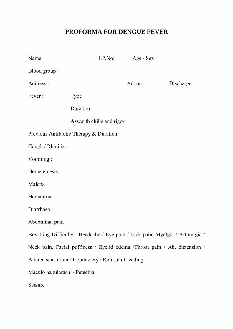
PROFORMA FOR DENGUE FEVER
Name : I.P.No: Age / Sex :
Blood group :
Address : Ad. on Discharge
Fever : Type
Duration
Ass.with chills and rigor
Previous Antibiotic Therapy & Duration
Cough / Rhinitis :
Vomiting :
Hemetemesis
Malena
Hematuria
Diarrhoea
Abdominal pain
Breathing Difficulty : Headache / Eye pain / back pain. Myalgia / Arthralgia /
Neck pain. Facial puffiness / Eyelid edema /Throat pain / Ab. distension /
Altered sensoriam / Irritable cry / Refusal of feeding
Maculo papularash / Petechial
Seizure
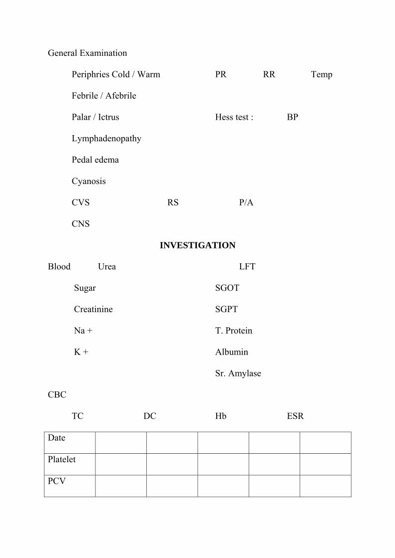
General Examination
Periphries Cold / Warm PR RR Temp
Febrile / Afebrile
Palar / Ictrus Hess test : BP
Lymphadenopathy
Pedal edema
Cyanosis
CVS RS P/A
CNS
INVESTIGATION
Blood Urea LFT
Sugar SGOT
Creatinine SGPT
Na + T. Protein
K + Albumin
Sr. Amylase
CBC
TC DC Hb ESR
Date
Platelet
PCV
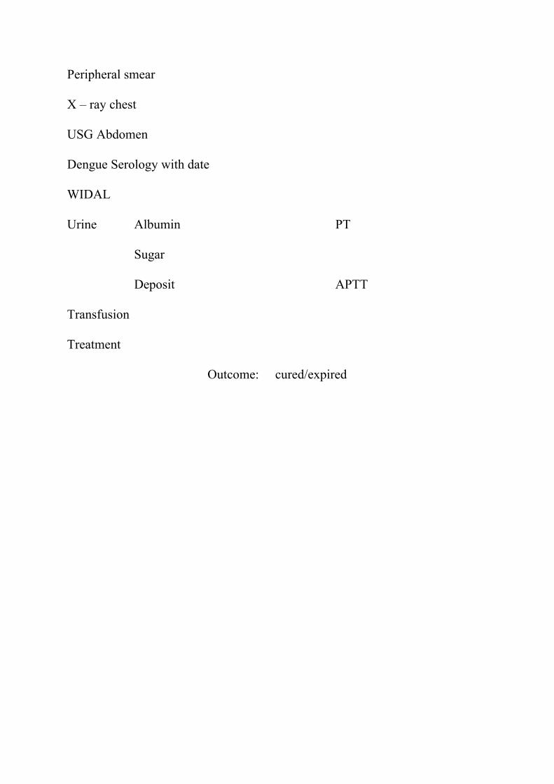
Peripheral smear
X – ray chest
USG Abdomen
Dengue Serology with date
WIDAL
Urine Albumin PT
Sugar
Deposit APTT
Transfusion
Treatment
Outcome: cured/expired
BIBLIOGRAPHY
1 WHO. Dengue and dengue haemorrhagic fever. Factsheet No 117, revised
May 2008. Geneva, World Health Organization, 2008.
(http://www.who.int/mediacentre/factsheets/fs117/en/). 2 World Health Organisation. Prevention and control of dengue and dengue
haemorrhagic fever:comprehensive guidelines. WHO Regional publication,
SEARO, No 29, 1999 3 Chandrakanta et al. Changing clinical manifestations of dengue infection
in North India. Dengue Bulletin : 2008)
4 Manjunath J. Kulkarni et al. Clinico-Epidemiological Profile of Children
Hospitalized with Dengue. Indian J Pediatr (2010) 77:1103–4.
5 The 2005 World Health Assembly resolution WHA 58.3 on revision of
IHA
6 World Health Organization. Report on dengue. 1–5 October 2006, Geneva,
Switzerland. Geneva: WHO, 2007. Document No. TDR/SWG/08
7 WHO. Dengue and dengue haemorrhagic fever. Factsheet No 117, revised
May 2008. Geneva, World Health Organization, 2008
(http://www.who.int/mediacentre/factsheets/fs117/en/)
8 Simmons CP, Halstead SB, Rothman A, Harris E, Screaton G, Rico-Hesse
R, Vaughn D, Holmes E, Guzman M. Working Paper 4.1. Understanding
pathogenesis, immune response and viral factors. In: World Health
Organization. Report on dengue. 1–5 October 2006, Geneva, Switzerland.
Geneva:WHO, 2007. Document No. TDR/SWG/08. p. 54–60
9 Leitmeyer KC. Dengue virus structural differences that correlate with
pathogenesis. Journal of Virology, 1999, 73(6):4738--4747. Lanciotti RS et
al. Molecular evolution and epidemiology of dengue-3 viruses. Journal of
General Virology, 1994, 75(Pt 1):65—75
10 Libraty DH, Young PR, Pickering D, et al. High circulating levels of the
dengue virus nonstructural protein NS1 early in dengue illness correlate
with the development of dengue hemorrhagic fever. J Infect Dis
2002;186:1165–8 (Epub 16 September 2002)
11 Guzman MG, Kouri G. Dengue: an update. Lancet Infect Dis 2002;2:33–
42.
12 Thavara U, Tawatsin A, Chansang C, et al. Larval occurrence, oviposition
behavior and biting activity of potential mosquito vectors of dengue on
Samui Island, Thailand. J Vector Ecol 2001;26 172 -80.
13 Perich MJ, Davila G, Turner A, et al. Behavior of resting Aedes aegypti
(culicidae: diptera) and its relation to ultra-low volume adulticide efficacy
in Panama City, Panama. J Med Entomol 2000;37:541–6.
14 Expert consensus group in latin America (Havana Cuba 2007) (South East
Asia Kualalumpur, Malasiya 2007 (WHO Head quarters) in Geneva,
Switzerland 2008.
15 Rigau-Perez JG et al. Dengue and dengue haemorrhagic fever. Lancet,
1998, 352:971–977.
16 Phuong CXT et al. Evaluation of the World Health Organization standard
tourniquet test in the diagnosis of dengue infection in Vietnam. Tropical
Medicine and International Health, 2002, 7:125–132.
17 Kalayanarooj S et al. Early clinical and laboratory indicators of acute
dengue illness. Journal of Infectious Diseases, 1997, 176:313–321.

18 Balmaseda A et al. Assessment of the World Health Organization scheme
for classification of dengue severity in Nicaragua. American Journal of
Tropical Medicine and Hygiene, 2005, 73:1059–1062.
19 Nimmannitya S et al. Dengue and chikungunya virus infection in man in
Thailand, 1962–64. Observations on hospitalized patients with
haemorrhagic fever. American Journal of Tropical Medicine and Hygiene,
1969, 18(6):954 971.
20 Martinez-Torres E, Polanco-Anaya AC, Pleites-Sandoval EB. Why and
how children with dengue die? Revista cubana de medicina tropical, 2008,
60(1):40–47.
21 Nimmannitya S. Clinical spectrum and management of dengue
haemorrhagic fever. Southeast Asian Journal of Tropical Medicine and
Public Health, 1987, 18(3):392–397
22 Halstead SB, Lan NT, Myint TT, et al. Dengue hemorrhagic fever in
infants: research opportunities ignored. Emerg Infect Dis 2002;8:1474–9.
23 Kalayanarooj S, Chansiriwongs V, Nimmannitya S. Dengue patients at the
Children’s Hospital, Bangkok: 1995–1999. Review. Dengue Bulletin
2002;26:33–43.
24 Kabra SK, Jain Y, Pandey RM, et al. Dengue haemorrhagic fever in
children in the 1996 Delhi epidemic. Trans R Soc Trop Med Hyg
1999;93:294–8.
25 Gulati S., Maheshwari A. Atypical manifestations of dengue. Trop Med Int
Health. 2007 Sep.; 12(9):1087 – 95.65
26 Gibbons RV, Vaughn DW. Dengue: An escalating problem. BMJ. 2002
June 29; 324(7353): 1563–6
27 Burke DS, Nisalak A, Johnson DE. et al. A prospective study of dengue
infections in Bangkok. American Journal of Tropical Medicine and
Hygiene. 1988 Jan; 38(1): 172–80.
Endy TP, Chunsuttiwat S, Nisalak A. et al. Epidemiology of inapparent and
symptomatic acute dengue virus infection: a prospective study of primary
schoolchildren in Kamphaeng Phet, Thailand. Am J Epidemiol. 2002. July
1; 156(1): 40–51
28 Srikiatkhachorn A, Green S. Markers of dengue disease severity. Curr Top
Microbiol Immunol. 2010; 338: 67–82
29 Avirutnan P. et al. Antagonism of the complement component C4 by
flavivirus nonstructural protein NS1. J Exp Med. 2010; 207(4): 793–806
30 Cardier JE, Marino E, Romano E. et al. Proinflammatory factors present in
sera from patients with acute dengue infection induce activation and
apoptosis of human microvascular endothelial cells: possible role of TNF-
alpha in endothelial cell damage in dengue. Cytokine. 2005 Jun 21; 30(6):
359–65
31 Mongkolsapaya J, Duangchinda T, Dejnirattisai W. et al. T cell responses
in dengue hemorrhagic fever: are cross-reactive T cells suboptimal? J
Immunol. 2006 March 15; 176(6): 3821–9.
Dong T, Moran E, Vinh Chau N. et al. High Pro-inflammatory cytokine
secretion and loss of high avidity cross-reactive cytotoxic T-Cells during
the course of secondary dengue virus infection. PloS one. 2007;2(12):
e1192
32 Avirutnan PE, Mehlhop and M.S. Diamond, Complement and its role in
protection and pathogenesis of flavivirus infections. Vaccine. 2008. 26
Suppl 8: p. 100–107.
33 Libraty DH, Endy TP, Houng HS. et al. Differing influences of virus
burden and immune activation on disease severity in secondary dengue-3
virus infections. Journal of Infectious Diseases. 2002 May 1; 185(9):
1213–21.
34 Libraty DH, Young PR, Pickering D. et al. High circulating levels of the
dengue virus non-structural protein NS1 early in dengue illness correlate
with the development of dengue haemorrhagic fever. Journal of Infectious
Diseases. 2002 Oct. 15; 186(8): 1165–8
35 Burke DS, Nisalak A, Johnson DE. et al. A prospective study of dengue
infections in Bangkok. American Journal of Tropical Medicine and
Hygiene. 1988 Jan; 38(1): 172–80
36 Gubler DJ. Serological diagnosis of dengue fever/dengue haemorrhagic
fever. Dengue Bulletin. 1996; 20:23.
37 World Health Organization. Manual on environmental management of
mosquito control. Geneva: WHO,1982. (WHO Offset publication no. 66)
38 Gamble J, Bethell D, Day NPJ, et al. Age-related changes in microvascular
Pebility: A significant factor in the susceptibility of children to shock?.
Clinical Science 2000; 98: 211-6.
39 Chungue E, Poli L, Roche C, Gestas P, Glaziou P, Markoff LJ. Correlation
between detection of plasminogen cross-reactive antibodies and
hemorrhage in dengue virus infection. J Infect Dis 1994; 170: 1304-7.
40 Sharma S and Sharma SK. Clinical profile of DHF in adults during 1996
outbreak in Delhi, India. Dengue Bulletin. 1998; 22: 20-27.
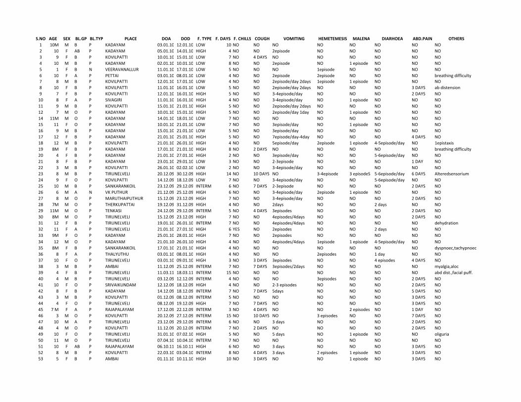
S.NO AGE SEX BL.GP BL.TYP PLACE DOA DOD F. TYPE F. DAYS F. CHILLS COUGH VOMITING HEMETEMESIS MALENA DIARHOEA ABD.PAIN OTHERS
1 10M M B P KADAYAM 03.01.10 12.01.10 LOW 10 NO NO NO NO NO NO NO NO
2 10 F AB P KADAYAM 05.01.10 14.01.10 HIGH 4 NO NO 2episode NO NO NO NO NO
3 9 F B P KOVILPATTI 10.01.10 15.01.10 LOW 7 NO 4 DAYS NO NO NO NO NO NO
4 10 M B P KADAYAM 02.01.10 10.01.10 LOW 8 NO NO 2episode NO 1 episode NO NO NO
5 1 F B N VEERAVANALLUR 11.01.10 17.01.10 LOW 5 NO NO NO 1episode NO NO NO NO
6 10 F A P PETTAI 03.01.10 08.01.10 LOW 4 NO NO 2episode 2episode NO NO NO breathing difficulty
7 8 M B P KOVILPATTI 12.01.10 17.01.10 LOW 4 NO NO 2episode/day 2days 1episode 1 episode NO NO NO
8 10 F B P KOVILPATTI 11.01.10 16.01.10 LOW 5 NO NO 2episode/day 2days NO NO NO 3 DAYS ab distension
9 7 F B P KOVILPATTI 12.01.10 16.01.10 HIGH 5 NO NO 3-4episode/day NO NO NO 2 DAYS NO
10 8 F A P SIVAGIRI 11.01.10 16.01.10 HIGH 4 NO NO 3-4episode/day NO 1 episode NO NO NO
11 9 M B P KOVILPATTI 15.01.10 21.01.10 HIGH 5 NO NO 2episode/day 2days NO NO NO NO NO
12 7 M O P KADAYAM 10.01.10 15.01.10 HIGH 5 NO NO 2episode/day 1day NO 1 episode NO NO NO
14 11M M O P KADAYAM 14.01.10 18.01.10 LOW 7 NO NO NO NO NO NO NO NO
15 11 F O P KADAYAM 10.01.10 21.01.10 LOW 7 NO NO 3episode/day NO 1 episode NO NO NO
16 9 M B P KADAYAM 15.01.10 21.01.10 LOW 5 NO NO 3episode/day NO NO NO NO NO
17 12 F B P KADAYAM 21.01.10 25.01.10 HIGH 5 NO NO 7episode/day-4day NO NO NO 4 DAYS NO
18 12 M B P KOVILPATTI 21.01.10 26.01.10 HIGH 4 NO NO 5episode/day 2episode 1 episode 4-5episode/day NO 1epistaxis
19 8M F B P KADAYAM 17.01.10 21.01.10 HIGH 8 NO 2 DAYS NO NO NO NO NO breathing difficulty
20 4 F B P KADAYAM 21.01.10 27.01.10 HIGH 2 NO NO 3episode/day NO NO 5-6episode/day NO NO
21 8 F B P KADAYAM 23.01.10 29.01.10 LOW 3 NO NO 2-3episode NO NO NO 1 DAY NO
22 3 M B P KOVILPATTI 26.01.10 02.02.10 LOW 2 NO NO 3-4episode/day NO NO NO NO NO
23 8 M B P TIRUNELVELI 20.12.09 30.12.09 HIGH 14 NO 10 DAYS NO 3-4episode 3 episodeS 5-6episode/day 6 DAYS Alteredsensorium
24 9 F O P KOVILPATTI 14.12.09 18.12.09 LOW 7 NO NO 3-4episode/day NO NO 5-6episode/day NO NO
25 10 M B P SANKARANKOIL 23.12.09 29.12.09 INTERM 6 NO 7 DAYS 2-3episode NO NO NO 2 DAYS NO
26 6 M A N VK PUTHUR 21.12.09 25.12.09 HIGH 6 NO NO 3-4episode/day 2episode 1 episode NO NO NO
27 8 M O P MARUTHAIPUTHUR 15.12.09 23.12.09 HIGH 7 NO NO 3-4episode/day NO NO NO 2 DAYS NO
28 7M M O P THERKUPATTAI 19.12.09 31.12.09 HIGH 4 NO NO 2days NO NO 2 days NO NO
29 11M M O P TENKASI 24.12.09 29.12.09 INTERM 5 NO 4 DAYS 3episodes NO NO NO 2 DAYS NO
30 8M M O P TIRUNELVELI 15.12.09 23.12.09 HIGH 7 NO NO 4episodes/4days NO NO NO 2 DAYS NO
31 12 F B P TIRUNELVELI 19.01.10 26.01.10 INTERM 7 NO NO 4episodes/4days NO NO NO NO dehydration
32 11 F A P TIRUNELVELI 21.01.10 27.01.10 HIGH 6 YES NO 2episodes NO NO 2 days NO NO
33 9M F O P KADAYAM 25.01.10 28.01.10 HIGH 7 NO NO 2episodes NO NO NO NO NO
34 12 M O P KADAYAM 21.01.10 26.01.10 HIGH 4 NO NO 4episodes/4days 1episode 1 episode 4-5episode/day NO NO
35 8M F B P SANKARANKOIL 17.01.10 21.01.10 HIGH 4 NO NO NO NO NO NO NO dyspnoec,tachypnoec
36 8 F A P THALYUTHU 03.01.10 08.01.10 HIGH 4 NO NO NO 2episodes NO 1 day NO NO
37 10 F O P TIRUNELVELI 03.01.10 09.01.10 HIGH 3 NO 3 DAYS 3episodes NO NO 4 episodes 4 DAYS NO
38 3 M B P AMBAI 11.12.09 25.12.09 INTERM 7 NO 7 DAYS 3episodes/2days NO NO NO NO myalgia,aloc
39 4 F B P TIRUNELVELI 11.03.11 18.03.11 INTERM 15 NO NO NO NO NO NO NO abd dist.,facial puff.
40 4 M B P TIRUNELVELI 03.12.09 12.12.09 INTERM 4 NO NO NO 3episodes NO NO 2 DAYS NO
41 10 F O P SRIVAIKUNDAM 12.12.09 18.12.09 HIGH 4 NO NO 2-3 episodes NO NO NO 2 DAYS NO
42 8 F B P KADAYAM 14.12.09 18.12.09 INTERM 7 NO 7 DAYS 5days NO NO NO 5 DAYS NO
43 3 M B P KOVILPATTI 01.12.09 08.12.09 INTERM 5 NO NO NO NO NO NO 3 DAYS NO
44 4 F O P TIRUNELVELI 08.12.09 19.12.09 HIGH 7 NO 7 DAYS NO NO NO NO 3 DAYS NO
45 7 M F A P RAJAPALAYAM 17.12.09 22.12.09 INTERM 3 NO 4 DAYS NO NO 2 episodes NO 1 DAY NO
46 3 M O P KOVILPATTI 20.12.09 27.12.09 INTERM 15 NO 10 DAYS NO 3 episodes NO NO 7 DAYS NO
47 10 M A P TIRUNELVELI 23.12.09 29.12.09 INTERM 6 NO NO 3 days NO NO NO 2 DAYS NO
48 4 M O P KOVILPATTI 11.12.09 20.12.09 INTERM 7 NO 2 DAYS NO NO NO NO 2 DAYS NO
49 10 F O P TIRUNELVELI 31.01.10 07.02.10 HIGH 5 NO NO 5 days NO 1 episode NO NO oliguria
50 11 M O P TIRUNELVELI 07.04.10 10.04.10 INTERM 7 NO NO NO NO NO NO NO NO
51 10 F AB P RAJAPALAYAM 06.10.11 16.10.11 HIGH 6 NO NO 3 days NO NO NO 3 DAYS NO
52 8 M B P KOVILPATTI 22.03.10 03.04.10 INTERM 8 NO 4 DAYS 3 days 2 episodes 1 episode NO 3 DAYS NO
53 5 F B P AMBAI 01.11.10 10.11.10 HIGH 10 NO 3 DAYS NO NO 1 episode NO 3 DAYS NO
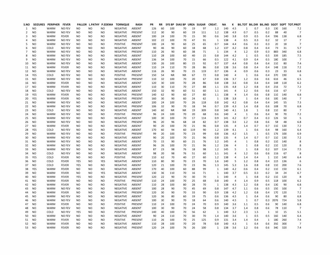
S.NO SEIZURES PERIPHER FEVER PALLOR L.PATHY P.EDEMA TORNIQUE RASH PR RR SYS BP DIAS BP UREA SUGAR CREAT. NA K BIL.TOT BIL.DIR BIL.IND SGOT SGPT TOT.PROT
1 NO WARM NO FEV NO NO NO NEGATIVE ABSENT 126 60 100 70 19 97 1.2 140 4.5 1 0.7 0.3 130 160 7.2
2 NO WARM NO FEV NO NO NO NEGATIVE PRESENT 112 30 90 60 19 111 1.2 138 4.9 0.7 0.5 0.2 88 40 7
3 NO WARM FEVER NO NO NO NEGATIVE ABSENT 100 24 100 70 15 90 0.6 140 3.8 0.9 0.5 0.4 596 138 6.8
4 NO WARM NO FEV NO NO NO NEGATIVE ABSENT 102 22 100 70 47 91 1 138 4 0.5 0.3 0.2 32 17 7
5 NO WARM NO FEV NO NO NO POSITIVE PRESENT 100 24 90 60 25 65 0.7 144 4.4 0.6 0.3 0.3 20 18 7
6 NO COLD NO FEV NO NO NO NEGATIVE ABSENT 90 46 90 60 18 68 1.2 137 4.2 0.8 0.4 0.4 73 31 5.7
7 NO WARM NO FEV NO NO NO NEGATIVE PRESENT 110 26 90 60 38 71 1 134 4 1.2 0.9 0.3 883 340 6.8
8 NO WARM NO FEV NO NO NO NEGATIVE ABSENT 110 28 100 60 40 15 0.8 144 4.2 1 0.5 0.5 339 185 7.5
9 NO WARM NO FEV NO NO NO NEGATIVE ABSENT 136 34 100 70 15 66 0.5 122 4.1 0.9 0.4 0.5 180 100 7
10 NO WARM NO FEV NO NO NO NEGATIVE ABSENT 130 26 100 80 15 92 0.7 137 4.4 0.8 0.4 0.4 110 80 7.4
11 NO WARM FEVER NO NO NO NEGATIVE ABSENT 110 24 110 70 28 86 0.8 138 3.6 0.8 0.4 0.4 148 118 7.2
12 NO WARM NO FEV NO NO NO NEGATIVE ABSENT 116 26 90 70 15 119 0.7 136 4 0.9 0.6 0.3 23 33 6.8
14 YES COLD NO FEV NO NO NO POSITIVE PRESENT 150 54 NR NR 67 72 0.8 140 4 1 0.6 0.4 370 190 6
15 NO WARM NO FEV NO NO NO NEGATIVE PRESENT 110 32 100 70 20 67 0.8 136 3.7 1.2 0.6 0.6 616 46 6.5
16 NO WARM FEVER NO NO NO NEGATIVE ABSENT 120 30 100 70 28 86 0.8 138 3.6 0.8 0.4 0.4 148 118 6.2
17 NO WARM FEVER NO NO NO NEGATIVE ABSENT 110 30 110 70 27 88 1.1 135 4.8 1.2 0.8 0.4 216 72 7.2
18 NO COLD NO FEV NO NO NO NEGATIVE ABSENT 150 32 90 60 51 60 1.1 141 4 1.2 0.6 0.6 116 67 7
19 YES WARM FEVER NO NO NO NEGATIVE PRESENT 140 62 90 60 27 80 1.1 138 4 1.4 0.6 0.8 110 140 6.8
20 YES WARM FEVER NO NO NO NEGATIVE PRESENT 160 30 90 60 24 62 1 138 3.8 1 0.4 0.6 100 140 7
21 NO WARM NO FEV NO NO NO NEGATIVE ABSENT 100 24 100 70 26 118 0.8 142 4.2 0.8 0.4 0.4 145 55 7.3
22 NO WARM NO FEV NO NO NO NEGATIVE PRESENT 106 32 90 70 18 94 0.7 139 4.3 1.4 0.8 0.6 108 70 6.8
23 NO COLD NO FEV NO NO NO NEGATIVE ABSENT 140 60 NR NR 19 309 0.8 140 4.1 1.8 1 0.8 110 80 7
24 NO WARM NO FEV NO NO NO POSITIVE PRESENT 96 24 100 70 20 68 0.6 140 4 1 0.6 0.4 90 70 7.2
25 NO WARM NO FEV NO NO NO NEGATIVE ABSENT 100 30 100 70 17 114 0.9 141 4.2 0.7 0.4 0.3 126 50 5
26 NO WARM NO FEV NO NO NO NEGATIVE PRESENT 96 20 96 68 18 82 0.7 138 3.6 1.2 0.8 0.6 58 48 6.8
27 NO WARM FEVER NO NO NO NEGATIVE ABSENT 90 20 100 70 21 96 0.8 135 4 1.4 0.7 0.7 110 130 7.2
28 YES COLD NO FEV NO NO NO NEGATIVE ABSENT 170 60 94 60 119 90 1.2 139 4.1 1 0.6 0.4 98 160 6.4
29 NO WARM NO FEV NO NO NO POSITIVE PRESENT 99 20 100 70 23 99 0.8 136 4.2 1.5 1 0.5 176 100 6.9
30 NO WARM NO FEV NO NO NO NEGATIVE ABSENT 90 20 100 70 21 96 0.8 135 4 1.4 0.7 0.7 110 130 7.2
31 NO WARM NO FEV NO NO NO NEGATIVE ABSENT 70 15 90 60 21 76 1.2 145 4 1 0.8 0.2 110 140 8
32 NO WARM NO FEV NO NO NO NEGATIVE ABSENT 96 26 100 70 21 96 1.2 136 4 1 0.8 0.2 110 120 8
33 NO WARM NO FEV NO NO NO NEGATIVE ABSENT 87 23 98 76 18 98 1.2 145 5 1 0.8 0.2 107 114 7.5
34 NO COLD NO FEV NO NO NO NEGATIVE ABSENT 98 20 98 76 51 60 1.1 141 4 1.2 0.6 0.6 116 67 8
35 YES COLD FEVER NO NO NO POSITIVE PRESENT 110 62 70 40 27 60 1.2 138 4 1.4 0.4 1 110 140 6.4
36 NO COLD FEVER YES NO YES NEGATIVE ABSENT 110 80 90 70 23 70 1.6 140 5 1.2 0.8 0.4 115 136 6
37 NO COLD FEVER YES NO NO NEGATIVE ABSENT 120 21 100 70 23 96 1.3 145 5.3 1.6 0.8 0.8 110 200 7.6
38 YES WARM FEVER NO NO NO NEGATIVE ABSENT 170 40 100 60 88 99 1 140 4.2 0.6 0.4 0.2 110 150 6.1
39 NO WARM FEVER NO NO YES NEGATIVE ABSENT 130 36 110 70 16 71 1 140 3.7 0.5 0.3 0.2 34 24 6.7
40 NO WARM FEVER YES NO NO NEGATIVE PRESENT 120 22 90 70 30 70 1 140 4 1 0.8 0.2 110 120 8
41 NO WARM FEVER NO NO NO POSITIVE PRESENT 110 24 100 70 25 68 0.8 140 4 1.4 0.9 0.5 118 100 6.2
42 NO WARM FEVER NO NO NO NEGATIVE ABSENT 110 28 100 80 28 70 1 138 4.3 1.2 0.8 0.4 130 90 6.8
43 NO WARM NO FEV NO NO NO NEGATIVE ABSENT 100 28 90 70 45 69 0.8 147 4.7 1.1 0.6 0.5 150 100 7
44 NO WARM FEVER NO NO NO NEGATIVE PRESENT 120 30 90 70 33 90 0.9 138 4.2 1.2 0.8 0.4 170 120 7.2
45 NO WARM NO FEV NO NO NO NEGATIVE ABSENT 120 36 80 60 19 75 0.6 136 4.5 1 0.4 0.6 90 80 6.8
46 NO WARM NO FEV NO NO NO NEGATIVE ABSENT 100 30 90 70 18 64 0.6 140 4.5 1 0.7 0.3 2076 724 5.8
47 NO WARM FEVER NO NO NO POSITIVE PRESENT 110 24 100 70 24 70 0.9 140 3.6 1.1 0.5 0.6 90 140 6.8
48 NO WARM NO FEV NO NO NO NEGATIVE ABSENT 100 30 90 70 24 58 0.8 134 3.7 1.4 0.8 0.6 78 110 7
49 NO COLD NO FEV YES NO NO POSITIVE PRESENT 140 40 100 70 56 62 1 140 3.2 2.3 1.5 1 10 15 5.1
50 NO WARM NO FEV NO NO NO NEGATIVE ABSENT 90 24 110 70 30 70 1.4 140 3.6 1 0.5 0.5 160 140 6.4
51 NO WARM FEVER NO NO NO POSITIVE PRESENT 110 26 100 70 25 125 0.9 131 3.4 1.4 0.4 1 180 260 7.4
52 NO WARM FEVER NO NO NO NEGATIVE ABSENT 110 28 100 70 20 78 0.8 140 4.3 1 0.4 0.6 350 300 7
53 NO WARM FEVER NO NO NO NEGATIVE PRESENT 120 24 100 76 26 100 1 138 3.6 1.2 0.6 0.6 340 320 7.4

S.NO ALBUMIN AMYLASE TC DCP DCL DCE HB ESR30 ESR60 PLATELET PCV X RAY CHEST USG ABDOMEN U.ALB U.SUG U.DEP U.CS B.TRANSFUSION DENG.CAT OUTCOME
1 4.2 190 16700 55 45 0 11.7 10 22 180000 36 NORMAL HEPATOMEGALY NEG NEG NIL NIL NIL DF RECOVERY
2 4.4 108 4000 50 50 0 10.8 14 24 70000 30 NORMAL HEPATOMEGALY NEG NEG NIL NIL 1 PLATELET DF RECOVERY
3 4 88 5200 45 52 3 14.3 2 5 43000 44 R PF R PF ASCITES NEG NEG NIL NIL NIL DHS RECOVERY
4 4 208 9600 92 5 3 11.6 30 62 100000 33 NORMAL R PF ASCITES NEG NEG NIL NIL NIL DHS RECOVERY
5 3.5 180 4500 27 70 3 15 2 7 23000 47 NORMAL R PF ASCITES NEG NEG NIL NIL 1 PLATELET DHS RECOVERY
6 3.7 60 8800 31 56 13 12.6 18 67 62000 37 NORMAL R PF ASCITES NEG NEG NIL NIL NIL DSS RECOVERY
7 4 78 4200 40 60 0 13.1 4 10 36000 41 NORMAL HEPATOMEGALY NEG NEG NIL NIL 3 PLATELET DF RECOVERY
8 4.5 90 5800 32 68 0 12.3 10 25 78000 38 NORMAL R PF ASCITES NEG NEG NIL NIL NIL DHS RECOVERY
9 4 110 25000 53 40 7 10.9 8 20 34000 37 NORMAL R PF ASCITES NEG NEG NIL NIL NIL DHS RECOVERY
10 4.4 70 6300 34 63 3 11 5 12 35000 40 NORMAL HEPATOMEGALY NEG NEG NIL NIL NIL DHS RECOVERY
11 4.2 90 6500 27 56 17 11 5 10 101000 43 NORMAL R PF ASCITES NEG NEG NIL NIL NIL DHS RECOVERY
12 4.3 60 5500 42 58 0 14.7 4 9 32000 46 NORMAL HEPATOMEGALY NEG NEG NIL NIL 2 PLATELET DHS RECOVERY
14 3.5 80 7000 52 44 4 11 10 18 90000 40 R PF HEPATOMEGALY NEG NEG NIL NIL 3 UFFP DSS DEATH
15 3.6 94 6000 40 60 0 12 12 24 63000 30 R PF HEPATOMEGALY NEG NEG NIL NIL NIL DHS RECOVERY
16 3.8 110 8000 38 60 2 12 10 20 110000 43 R PF HEPATOMEGALY NEG NEG NIL NIL NIL DHS RECOVERY
17 4 150 7000 32 66 2 10 8 20 41000 48 NORMAL R PF ASCITES NEG NEG NIL NIL NIL DHS RECOVERY
18 4 114 5000 42 54 4 14.2 20 36 55000 41 NORMAL NORMAL NEG NEG NIL NIL 1UBlood DSS RECOVERY
19 3.8 120 21000 33 63 4 11 10 20 23000 33 NORMAL HEPATOMEGALY NEG NEG NIL NIL 2 PLATELET DSS DEATH
20 4 110 10000 33 64 3 12 8 18 40000 33 PNEUMONIA HEPATOMEGALY NEG NEG NIL NIL NIL DF RECOVERY
21 4.3 140 6200 30 70 0 11 6 14 28000 51 NORMAL R PF ASCITES NEG NEG NIL NIL 1 PLATELET DHS RECOVERY
22 3.8 90 5400 34 62 4 10 12 24 90000 40 NORMAL NORMAL NEG NEG NIL NIL NIL DF RECOVERY
23 4 160 9000 45 55 0 10.5 1 10 20000 31 R PF R PF ASCITES TRACE NEG NIL NIL 1uFFP&3uPLATELET DSS RECOVERY
24 4.2 150 5400 25 70 5 11.8 10 22 76000 36 R PF R PF ASCITES NEG NEG NIL NIL NIL DHS RECOVERY
25 2.3 210 10700 50 42 8 12.2 2 6 23000 36 NORMAL HEPATOMEGALY NEG NEG NIL NIL 1 PLATELET DF RECOVERY
26 3.8 120 3400 24 72 4 12 15 32 103000 37 NORMAL NORMAL NEG NEG NIL NIL NIL DHS RECOVERY
27 4.2 120 3500 46 52 2 14.4 1 4 254000 44 NORMAL R PF ASCITES NEG POS NIL NIL NIL DHS RECOVERY
28 3.3 130 5200 49 49 2 10.7 2 4 26000 33 NORMAL HEPATOMEGALY NEG NEG NIL NIL 3 PLATELET DSS RECOVERY
29 4.1 110 7000 47 52 1 14 1 3 28000 44 NORMAL HEPATOMEGALY NEG NEG NIL NIL NIL DHS RECOVERY
30 4.2 120 5400 22 74 4 14.4 1 4 254000 44 R PF R PF ASCITES NEG POS NIL NIL NIL DHS RECOVERY
31 5 100 5200 49 49 2 14 1 3 89000 41 NORMAL NORMAL NEG NEG NIL NIL NIL DF RECOVERY
32 4 100 7200 39 60 1 16 1 3 85000 41 NORMAL NORMAL NEG NEG NIL NIL NIL DF RECOVERY
33 4 100 6700 21 78 1 14 1 3 200000 40 NORMAL HEPATOMEGALY NEG NEG NIL NIL NIL DF RECOVERY
34 5 112 5800 34 65 1 14.7 1 2 55000 41 NORMAL NORMAL NEG NEG NIL NIL 1u whole blood DSS RECOVERY
35 3.1 150 2800 60 37 3 14 1 4 23000 33 NORMAL NORMAL NEG NEG NIL NIL NIL DSS RECOVERY
36 4 102 20400 78 20 2 14 1 3 62000 37 R PF R PF ASCITES NEG NEG NIL NIL NIL DSS RECOVERY
37 4.3 100 5000 70 26 4 12 1 4 93000 35 NORMAL NORMAL NEG NEG NIL NIL NIL DSS RECOVERY
38 3.1 112 5400 73 24 3 14.2 5 12 95000 44 PNEUMONIA R PF ASCITES NEG NEG NIL NIL NIL DSS RECOVERY
39 3 37 6600 41 53 6 7.5 8 16 80000 26 PNEUMONIA HEPATOMEGALY NEG NEG NIL NIL NIL DF RECOVERY
40 5 110 4800 33 65 2 7 5 8 24000 23 R PF R PF ASCITES NEG NEG NIL NIL 1 UFFP DHS RECOVERY
41 3.2 100 6100 23 75 2 15.7 5 12 22000 47 NORMAL R PF ASCITES NEG NEG NIL NIL 1 PLATELET DHS RECOVERY
42 3.8 104 7000 20 80 0 12 10 20 76000 37 R PF R PF ASCITES NEG NEG NIL NIL NIL DHS RECOVERY
43 4 110 4100 50 48 2 11 22 30 93000 34 NORMAL HEPATOMEGALY NEG NEG NIL NIL NIL DF RECOVERY
44 4.2 140 5200 32 66 2 10 10 20 34000 36 R PF R PF ASCITES NEG NEG NIL NIL NIL DHS RECOVERY
45 3.8 90 6600 17 80 3 10 14 20 58000 32 NORMAL HEPATOMEGALY NEG NEG NIL NIL NIL DHS RECOVERY
46 2 220 15000 70 24 6 9.3 20 30 112000 28 PNEUMONIA HEPATOMEGALY NEG NEG NIL NIL NIL DHS RECOVERY
47 3.8 98 5000 36 60 4 10 12 20 23000 36 NORMAL R PF ASCITES NEG NEG NIL NIL 1 PLATELET DHS RECOVERY
48 3.8 70 3000 30 68 2 11.2 4 8 95000 44 PNEUMONIA R PF ASCITES NEG NEG NIL NIL NIL DHS RECOVERY
49 3.1 40 14000 40 45 15 9 30 40 23000 27 NORMAL R PF ASCITES NEG NEG NIL NIL 2u platelet DSS RECOVERY
50 3.4 123 5100 33 65 2 11.4 5 12 114000 36 NORMAL R PF ASCITES NEG NEG NIL NIL NIL DHS RECOVERY
51 4.2 32 3600 50 46 4 14.8 1 3 20000 45 NORMAL R PF ASCITES NEG NEG NIL NIL 1 PLATELET DHS RECOVERY
52 4 180 8000 40 58 2 11 10 14 60000 39 NORMAL HEPATOMEGALY NEG NEG NIL NIL NIL DHS RECOVERY
53 4.2 160 12500 39 57 4 14.3 3 10 24000 48 NORMAL R PF ASCITES NEG NEG NIL NIL NIL DHS RECOVERY
![Dengue Fever/Severe Dengue Fever/Chikungunya Fever · Dengue fever and severe dengue (dengue hemorrhagic fever [DHF] and dengue shock syndrome [DSS]) are caused by any of four closely](https://img.pdfslide.us/doc/110x75/5e87bf3e7a86e85d3b149cd7/dengue-feversevere-dengue-feverchikungunya-dengue-fever-and-severe-dengue-dengue.jpg)