Embed Size (px)
Citation preview
Clinical Practice Guidelines
DAVID S. LOGERSTEDT, PT, PhD • DAVID SCALZITTI, PT, PhD • MAY ARNA RISBERG, PT, PhDLARS ENGEBRETSEN, MD • KATE E. WEBSTER, PhD • JULIAN FELLER, MD
LYNN SNYDER-MACKLER, PT, ScD • MICHAEL J. AXE, MD • CHRISTINE M. MCDONOUGH, PT, PhD
Knee Stability and Movement Coordination Impairments:
Knee Ligament Sprain Revision 2017
Clinical Practice Guidelines Linked to the International Classification of Functioning,
Disability and Health From the Orthopaedic Section of the American Physical Therapy Association
J Orthop Sports Phys Ther. 2017;47(11):A1-A47. doi:10.2519/jospt.2017.0303
REVIEWERS: Roy D. Altman, MD • Paul Beattie, PT, PhD • John DeWitt, DPT • James M. Elliott, PT, PhD • Amanda Ferland, DPTG. Kelley Fitzgerald, PT, PhD • Sandra Kaplan, PT, PhD • David Killoran, PhD • Joanna Kvist, PT, PhD • Robert Marx, MD, MSc
Leslie Torburn, DPT • James Zachazewski, DPT
For author, coordinator, contributor, and reviewer affiliations, see end of text. ©2017 Orthopaedic Section, American Physical Therapy Association (APTA), Inc, and the Journal of Orthopaedic & Sports Physical Therapy. The Orthopaedic Section, APTA, Inc, and the Journal of Orthopaedic & Sports Physical Therapy consent to the reproduction and distribution of this guideline for educational purposes. Address correspondence to Brenda Johnson, ICF-Based Clinical Practice Guidelines Coordinator, Orthopaedic Section, APTA, Inc, 2920 East Avenue South, Suite 200, La Crosse, WI 54601. E-mail: [email protected]
SUMMARY OF RECOMMENDATIONS . . . . . . . . . . . . . . . . . . . . . . . . . . . . . . A2
INTRODUCTION. . . . . . . . . . . . . . . . . . . . . . . . . . . . . . . . . . . . . . . . . . . . . . . . . . . . . . . . . . . . A3
METHODS . . . . . . . . . . . . . . . . . . . . . . . . . . . . . . . . . . . . . . . . . . . . . . . . . . . . . . . . . . . . . . . . . . . A4
CLINICAL GUIDELINES: Impairment/Function-Based Diagnosis . . . . . . . . . . . . . . . . . . A7
CLINICAL GUIDELINES: Examination . . . . . . . . . . . . . . . . . . . . . . . . . . . . . . . . . . . . . . . . . . . . . . . . . . . . . . . . . . . A17
CLINICAL GUIDELINES: Interventions . . . . . . . . . . . . . . . . . . . . . . . . . . . . . . . . . . . . . . . . . . . . . . . . . . . . . . . . . . A22
AUTHOR/REVIEWER AFFILIATIONS AND CONTACTS . . . . . . A25
REFERENCES . . . . . . . . . . . . . . . . . . . . . . . . . . . . . . . . . . . . . . . . . . . . . . . . . . . . . . . . . . . . . A26
47-11 CPG Knee 3.indd 1 10/18/2017 2:26:08 PM
Jou
rnal
of
Ort
hopa
edic
& S
port
s Ph
ysic
al T
hera
py®
D
ownl
oade
d fr
om w
ww
.josp
t.org
at o
n N
ovem
ber
3, 2
017.
For
per
sona
l use
onl
y. N
o ot
her
uses
with
out p
erm
issi
on.
Cop
yrig
ht ©
201
7 Jo
urna
l of
Ort
hopa
edic
& S
port
s Ph
ysic
al T
hera
py®
. All
righ
ts r
eser
ved.
Knee Ligament Sprain: Clinical Practice Guidelines Revision 2017
a2 | november 2017 | volume 47 | number 11 | journal of orthopaedic & sports physical therapy
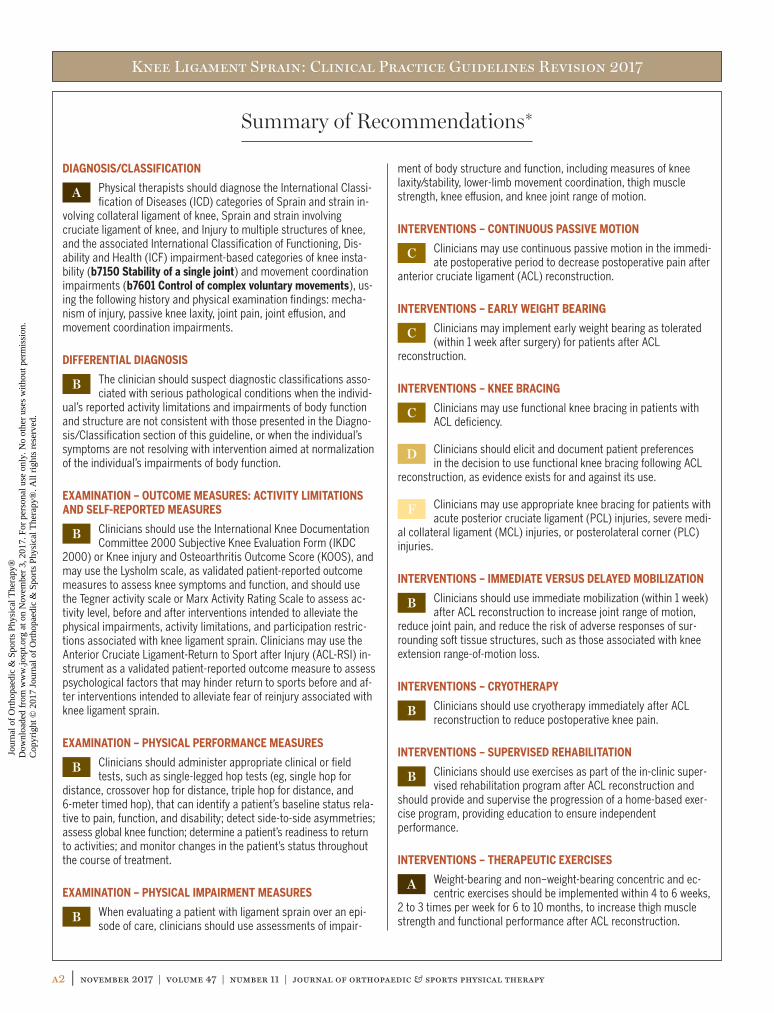
DIAGNOSIS/CLASSIFICATION
A Physical therapists should diagnose the International Classi-fication of Diseases (ICD) categories of Sprain and strain in-
volving collateral ligament of knee, Sprain and strain involving cruciate ligament of knee, and Injury to multiple structures of knee, and the associated International Classification of Functioning, Dis-ability and Health (ICF) impairment-based categories of knee insta-bility (b7150 Stability of a single joint) and movement coordination impairments (b7601 Control of complex voluntary movements), us-ing the following history and physical examination findings: mecha-nism of injury, passive knee laxity, joint pain, joint effusion, and movement coordination impairments.
DIFFERENTIAL DIAGNOSIS
B The clinician should suspect diagnostic classifications asso-ciated with serious pathological conditions when the individ-
ual’s reported activity limitations and impairments of body function and structure are not consistent with those presented in the Diagno-sis/Classification section of this guideline, or when the individual’s symptoms are not resolving with intervention aimed at normalization of the individual’s impairments of body function.
EXAMINATION – OUTCOME MEASURES: ACTIVITY LIMITATIONS AND SELF-REPORTED MEASURES
B Clinicians should use the International Knee Documentation Committee 2000 Subjective Knee Evaluation Form (IKDC
2000) or Knee injury and Osteoarthritis Outcome Score (KOOS), and may use the Lysholm scale, as validated patient-reported outcome measures to assess knee symptoms and function, and should use the Tegner activity scale or Marx Activity Rating Scale to assess ac-tivity level, before and after interventions intended to alleviate the physical impairments, activity limitations, and participation restric-tions associated with knee ligament sprain. Clinicians may use the Anterior Cruciate Ligament-Return to Sport after Injury (ACL-RSI) in-strument as a validated patient-reported outcome measure to assess psychological factors that may hinder return to sports before and af-ter interventions intended to alleviate fear of reinjury associated with knee ligament sprain.
EXAMINATION – PHYSICAL PERFORMANCE MEASURES
B Clinicians should administer appropriate clinical or field tests, such as single-legged hop tests (eg, single hop for
distance, crossover hop for distance, triple hop for distance, and 6-meter timed hop), that can identify a patient’s baseline status rela-tive to pain, function, and disability; detect side-to-side asymmetries; assess global knee function; determine a patient’s readiness to return to activities; and monitor changes in the patient’s status throughout the course of treatment.
EXAMINATION – PHYSICAL IMPAIRMENT MEASURES
B When evaluating a patient with ligament sprain over an epi-sode of care, clinicians should use assessments of impair-
ment of body structure and function, including measures of knee laxity/stability, lower-limb movement coordination, thigh muscle strength, knee effusion, and knee joint range of motion.
INTERVENTIONS – CONTINUOUS PASSIVE MOTION
C Clinicians may use continuous passive motion in the immedi-ate postoperative period to decrease postoperative pain after
anterior cruciate ligament (ACL) reconstruction.
INTERVENTIONS – EARLY WEIGHT BEARING
C Clinicians may implement early weight bearing as tolerated (within 1 week after surgery) for patients after ACL
reconstruction.
INTERVENTIONS – KNEE BRACING
C Clinicians may use functional knee bracing in patients with ACL deficiency.
D Clinicians should elicit and document patient preferences in the decision to use functional knee bracing following ACL
reconstruction, as evidence exists for and against its use.
F Clinicians may use appropriate knee bracing for patients with acute posterior cruciate ligament (PCL) injuries, severe medi-
al collateral ligament (MCL) injuries, or posterolateral corner (PLC) injuries.
INTERVENTIONS – IMMEDIATE VERSUS DELAYED MOBILIZATION
B Clinicians should use immediate mobilization (within 1 week) after ACL reconstruction to increase joint range of motion,
reduce joint pain, and reduce the risk of adverse responses of sur-rounding soft tissue structures, such as those associated with knee extension range-of-motion loss.
INTERVENTIONS – CRYOTHERAPY
B Clinicians should use cryotherapy immediately after ACL reconstruction to reduce postoperative knee pain.
INTERVENTIONS – SUPERVISED REHABILITATION
B Clinicians should use exercises as part of the in-clinic super-vised rehabilitation program after ACL reconstruction and
should provide and supervise the progression of a home-based exer-cise program, providing education to ensure independent performance.
INTERVENTIONS – THERAPEUTIC EXERCISES
A Weight-bearing and non–weight-bearing concentric and ec-centric exercises should be implemented within 4 to 6 weeks,
2 to 3 times per week for 6 to 10 months, to increase thigh muscle strength and functional performance after ACL reconstruction.
Summary of Recommendations*
47-11 CPG Knee 3.indd 2 10/18/2017 2:26:08 PM
Jou
rnal
of
Ort
hopa
edic
& S
port
s Ph
ysic
al T
hera
py®
D
ownl
oade
d fr
om w
ww
.josp
t.org
at o
n N
ovem
ber
3, 2
017.
For
per
sona
l use
onl
y. N
o ot
her
uses
with
out p
erm
issi
on.
Cop
yrig
ht ©
201
7 Jo
urna
l of
Ort
hopa
edic
& S
port
s Ph
ysic
al T
hera
py®
. All
righ
ts r
eser
ved.
Knee Ligament Sprain: Clinical Practice Guidelines Revision 2017
journal of orthopaedic & sports physical therapy | volume 47 | number 11 | november 2017 | a3
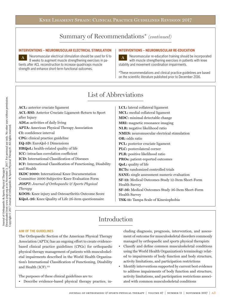
INTERVENTIONS – NEUROMUSCULAR ELECTRICAL STIMULATION
A Neuromuscular electrical stimulation should be used for 6 to 8 weeks to augment muscle strengthening exercises in pa-
tients after ACL reconstruction to increase quadriceps muscle strength and enhance short-term functional outcomes.
INTERVENTIONS – NEUROMUSCULAR RE-EDUCATION
A Neuromuscular re-education training should be incorporated with muscle strengthening exercises in patients with knee
stability and movement coordination impairments.
*These recommendations and clinical practice guidelines are based on the scientific literature published prior to December 2016.
Summary of Recommendations* (continued)
List of Abbreviations
ACL: anterior cruciate ligamentACL-RSI: Anterior Cruciate Ligament-Return to Sport after InjuryADLs: activities of daily livingAPTA: American Physical Therapy AssociationCI: confidence intervalCPG: clinical practice guidelineEQ-5D: EuroQol-5 DimensionsHRQoL: health-related quality of lifeICC: intraclass correlation coefficientICD: International Classification of DiseasesICF: International Classification of Functioning, Disability and HealthIKDC 2000: International Knee Documentation Committee 2000 Subjective Knee Evaluation FormJOSPT: Journal of Orthopaedic & Sports Physical TherapyKOOS: Knee injury and Osteoarthritis Outcome ScoreKQoL-26: Knee Quality of Life 26-item questionnaire
LCL: lateral collateral ligamentMCL: medial collateral ligamentMDC: minimal detectable changeMRI: magnetic resonance imagingNLR: negative likelihood ratioNMES: neuromuscular electrical stimulationOR: odds ratioPCL: posterior cruciate ligamentPLC: posterolateral cornerPLR: positive likelihood ratioPROs: patient-reported outcomesQoL: quality of lifeRCTs: randomized controlled trialsSANE: single assessment numeric evaluationSF-12: Medical Outcomes Study 12-Item Short-Form Health SurveySF-36: Medical Outcomes Study 36-Item Short-Form Health SurveyTSK-11: Tampa Scale of Kinesiophobia
AIM OF THE GUIDELINES
The Orthopaedic Section of the American Physical Therapy Association (APTA) has an ongoing effort to create evidence-based clinical practice guidelines (CPGs) for orthopaedic physical therapy management of patients with musculoskel-etal impairments described in the World Health Organiza-tion’s International Classification of Functioning, Disability and Health (ICF).125
The purposes of these clinical guidelines are to:• Describe evidence-based physical therapy practice, in-
cluding diagnosis, prognosis, intervention, and assess-ment of outcome for musculoskeletal disorders commonly managed by orthopaedic and sports physical therapists
• Classify and define common musculoskeletal conditions using the World Health Organization’s terminology relat-ed to impairments of body function and body structure, activity limitations, and participation restrictions
• Identify interventions supported by current best evidence to address impairments of body function and structure, activity limitations, and participation restrictions associ-ated with common musculoskeletal conditions
Introduction
47-11 CPG Knee 3.indd 3 10/18/2017 2:26:08 PM
Jou
rnal
of
Ort
hopa
edic
& S
port
s Ph
ysic
al T
hera
py®
D
ownl
oade
d fr
om w
ww
.josp
t.org
at o
n N
ovem
ber
3, 2
017.
For
per
sona
l use
onl
y. N
o ot
her
uses
with
out p
erm
issi
on.
Cop
yrig
ht ©
201
7 Jo
urna
l of
Ort
hopa
edic
& S
port
s Ph
ysic
al T
hera
py®
. All
righ
ts r
eser
ved.
Knee Ligament Sprain: Clinical Practice Guidelines Revision 2017
a4 | november 2017 | volume 47 | number 11 | journal of orthopaedic & sports physical therapy
• Identify appropriate outcome measures to assess changes resulting from physical therapy interventions in body function and structure as well as in activity and partici-pation of the individual
• Provide a description to policy makers, using internation-ally accepted terminology, of the practice of orthopaedic physical therapists
• Provide information for payers and claims reviewers re-garding the practice of orthopaedic physical therapy for common musculoskeletal conditions
• Create a reference publication for orthopaedic physical therapy clinicians, academic instructors, clinical instruc-tors, students, interns, residents, and fellows regarding the best current practice of orthopaedic physical therapy
STATEMENT OF INTENTThese guidelines are not intended to be construed or to serve as a standard of medical care. Standards of care are
determined on the basis of all clinical data available for an individual patient and are subject to change as scientific knowledge and technology advance and patterns of care evolve. These parameters of practice should be considered guidelines only. Adherence to them will not ensure a suc-cessful outcome in every patient, nor should they be con-strued as including all proper methods of care or excluding other acceptable methods of care aimed at the same results. The ultimate judgment regarding a particular clinical pro-cedure or treatment plan must be made based on clinician experience and expertise in light of the clinical presentation of the patient, the available evidence, available diagnostic and treatment options, and the patient’s values, expecta-tions, and preferences. However, we suggest that significant departures from accepted guidelines should be documented in the patient’s medical records at the time the relevant clin-ical decision is made.
Introduction (continued)
Content experts were appointed by the Orthopaedic Section of the APTA to conduct a review of the literature and to devel-op an updated “Knee Stability and Movement Coordination Impairments: Knee Ligament Sprain” CPG as indicated by the current state of the evidence in the field. The aims of the revision were to provide a concise summary of the evidence since publication of the original guideline and to develop new recommendations or revise previously published recommen-dations to support evidence-based practice. The authors of this guideline revision worked with research librarians with expertise in systematic reviews to perform a systematic search for concepts associated with ligament injuries and instabilities of the knee for articles published since 2008 related to clas-sification, examination, and intervention strategies consistent with previous guideline development methods related to ICF classification.70 Briefly, the following databases were searched from 2008 to December 2016: MEDLINE (PubMed; 2008 to date), Scopus (Elsevier; 2008 to date), CINAHL (EBSCO; 2008 to date), SPORTDiscus (EBSCO; 2008 to date), Coch-rane Library (Wiley; 2008 to date). (See APPENDIX A for full search strategies and APPENDIX B for search dates and results, available at www.orthopt.org.)
The authors declared relationships and developed a conflict management plan, which included submitting a Conflict of Interest form to the Orthopaedic Section, APTA, Inc. Articles
that were authored by a reviewer were assigned to an alternate reviewer. Funding was provided to the CPG development team for travel and expenses for CPG development training. The CPG development team maintained editorial independence.
Articles contributing to recommendations were reviewed based on specified inclusion and exclusion criteria with the goal of identifying evidence relevant to physical therapist clinical decision making for adult persons with knee stabil-ity and movement coordination impairments/knee ligament sprain. The title and abstract of each article were reviewed independently by 2 members of the CPG development team for inclusion (see APPENDIX C for inclusion and exclusion crite-ria, available at www.orthopt.org). Full-text review was then similarly conducted to obtain the final set of articles for con-tribution to the recommendations. The team leader (D.S.L.) provided the final decision for discrepancies that were not resolved by the review team (see APPENDIX D for a flow chart of articles and APPENDIX E for articles included in recommenda-tions by topic, available at www.orthopt.org). For selected rel-evant topics that were not appropriate for the development of recommendations, such as incidence and imaging, articles were not subject to systematic review and were not included in the flow chart. Evidence tables for this CPG are available on the Clinical Practice Guidelines page of the Orthopaedic Section of the APTA website (www.orthopt.org).
Methods
47-11 CPG Knee 3.indd 4 10/18/2017 2:26:08 PM
Jou
rnal
of
Ort
hopa
edic
& S
port
s Ph
ysic
al T
hera
py®
D
ownl
oade
d fr
om w
ww
.josp
t.org
at o
n N
ovem
ber
3, 2
017.
For
per
sona
l use
onl
y. N
o ot
her
uses
with
out p
erm
issi
on.
Cop
yrig
ht ©
201
7 Jo
urna
l of
Ort
hopa
edic
& S
port
s Ph
ysic
al T
hera
py®
. All
righ
ts r
eser
ved.
Knee Ligament Sprain: Clinical Practice Guidelines Revision 2017
journal of orthopaedic & sports physical therapy | volume 47 | number 11 | november 2017 | a5
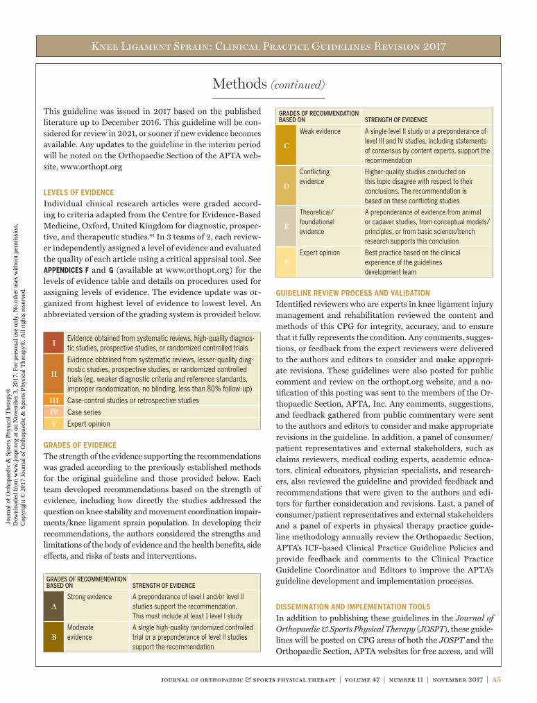
This guideline was issued in 2017 based on the published literature up to December 2016. This guideline will be con-sidered for review in 2021, or sooner if new evidence becomes available. Any updates to the guideline in the interim period will be noted on the Orthopaedic Section of the APTA web-site, www.orthopt.org
LEVELS OF EVIDENCEIndividual clinical research articles were graded accord-ing to criteria adapted from the Centre for Evidence-Based Medicine, Oxford, United Kingdom for diagnostic, prospec-tive, and therapeutic studies.91 In 3 teams of 2, each review-er independently assigned a level of evidence and evaluated the quality of each article using a critical appraisal tool. See APPENDICES F and G (available at www.orthopt.org) for the levels of evidence table and details on procedures used for assigning levels of evidence. The evidence update was or-ganized from highest level of evidence to lowest level. An abbreviated version of the grading system is provided below.
IEvidence obtained from systematic reviews, high-quality diagnos-tic studies, prospective studies, or randomized controlled trials
II
Evidence obtained from systematic reviews, lesser-quality diag-nostic studies, prospective studies, or randomized controlled trials (eg, weaker diagnostic criteria and reference standards, improper randomization, no blinding, less than 80% follow-up)
III Case-control studies or retrospective studies
IV Case series
V Expert opinion
GRADES OF EVIDENCEThe strength of the evidence supporting the recommendations was graded according to the previously established methods for the original guideline and those provided below. Each team developed recommendations based on the strength of evidence, including how directly the studies addressed the question on knee stability and movement coordination impair-ments/knee ligament sprain population. In developing their recommendations, the authors considered the strengths and limitations of the body of evidence and the health benefits, side effects, and risks of tests and interventions.
GRADES OF RECOMMENDATION BASED ON STRENGTH OF EVIDENCE
AStrong evidence A preponderance of level I and/or level II
studies support the recommendation. This must include at least 1 level I study
BModerate evidence
A single high-quality randomized controlled trial or a preponderance of level II studies support the recommendation
GRADES OF RECOMMENDATION BASED ON STRENGTH OF EVIDENCE
C
Weak evidence A single level II study or a preponderance of level III and IV studies, including statements of consensus by content experts, support the recommendation
D
Conflicting evidence
Higher-quality studies conducted on this topic disagree with respect to their conclusions. The recommendation is based on these conflicting studies
E
Theoretical/ foundational evidence
A preponderance of evidence from animal or cadaver studies, from conceptual models/principles, or from basic science/bench research supports this conclusion
FExpert opinion Best practice based on the clinical
experience of the guidelines development team
GUIDELINE REVIEW PROCESS AND VALIDATIONIdentified reviewers who are experts in knee ligament injury management and rehabilitation reviewed the content and methods of this CPG for integrity, accuracy, and to ensure that it fully represents the condition. Any comments, sugges-tions, or feedback from the expert reviewers were delivered to the authors and editors to consider and make appropri-ate revisions. These guidelines were also posted for public comment and review on the orthopt.org website, and a no-tification of this posting was sent to the members of the Or-thopaedic Section, APTA, Inc. Any comments, suggestions, and feedback gathered from public commentary were sent to the authors and editors to consider and make appropriate revisions in the guideline. In addition, a panel of consumer/patient representatives and external stakeholders, such as claims reviewers, medical coding experts, academic educa-tors, clinical educators, physician specialists, and research-ers, also reviewed the guideline and provided feedback and recommendations that were given to the authors and edi-tors for further consideration and revisions. Last, a panel of consumer/patient representatives and external stakeholders and a panel of experts in physical therapy practice guide-line methodology annually review the Orthopaedic Section, APTA’s ICF-based Clinical Practice Guideline Policies and provide feedback and comments to the Clinical Practice Guideline Coordinator and Editors to improve the APTA’s guideline development and implementation processes.
DISSEMINATION AND IMPLEMENTATION TOOLSIn addition to publishing these guidelines in the Journal of Orthopaedic & Sports Physical Therapy (JOSPT), these guide-lines will be posted on CPG areas of both the JOSPT and the Orthopaedic Section, APTA websites for free access, and will
Methods (continued)
47-11 CPG Knee 3.indd 5 10/18/2017 2:26:08 PM
Jou
rnal
of
Ort
hopa
edic
& S
port
s Ph
ysic
al T
hera
py®
D
ownl
oade
d fr
om w
ww
.josp
t.org
at o
n N
ovem
ber
3, 2
017.
For
per
sona
l use
onl
y. N
o ot
her
uses
with
out p
erm
issi
on.
Cop
yrig
ht ©
201
7 Jo
urna
l of
Ort
hopa
edic
& S
port
s Ph
ysic
al T
hera
py®
. All
righ
ts r
eser
ved.
Knee Ligament Sprain: Clinical Practice Guidelines Revision 2017
a6 | november 2017 | volume 47 | number 11 | journal of orthopaedic & sports physical therapy
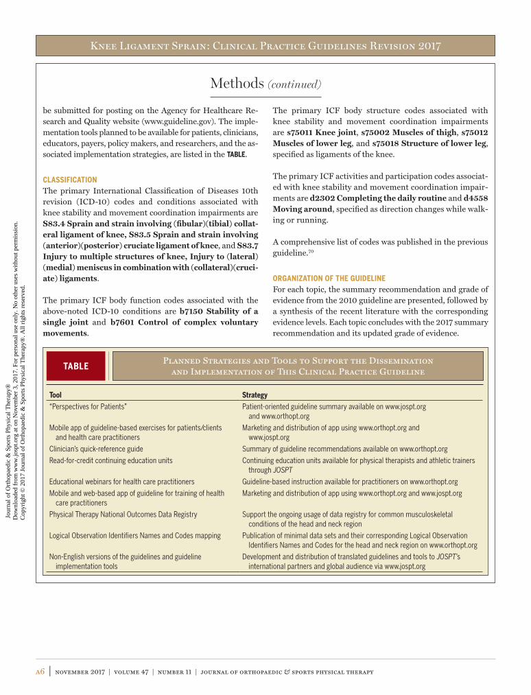
be submitted for posting on the Agency for Healthcare Re-search and Quality website (www.guideline.gov). The imple-mentation tools planned to be available for patients, clinicians, educators, payers, policy makers, and researchers, and the as-sociated implementation strategies, are listed in the TABLE.
CLASSIFICATIONThe primary International Classification of Diseases 10th revision (ICD-10) codes and conditions associated with knee stability and movement coordination impairments are S83.4 Sprain and strain involving (fibular)(tibial) collat-eral ligament of knee, S83.5 Sprain and strain involving (anterior)(posterior) cruciate ligament of knee, and S83.7 Injury to multiple structures of knee, Injury to (lateral)(medial) meniscus in combination with (collateral)(cruci-ate) ligaments.
The primary ICF body function codes associated with the above-noted ICD-10 conditions are b7150 Stability of a single joint and b7601 Control of complex voluntary movements.
The primary ICF body structure codes associated with knee stability and movement coordination impairments are s75011 Knee joint, s75002 Muscles of thigh, s75012 Muscles of lower leg, and s75018 Structure of lower leg, specified as ligaments of the knee.
The primary ICF activities and participation codes associat-ed with knee stability and movement coordination impair-ments are d2302 Completing the daily routine and d4558 Moving around, specified as direction changes while walk-ing or running.
A comprehensive list of codes was published in the previous guideline.70
ORGANIZATION OF THE GUIDELINEFor each topic, the summary recommendation and grade of evidence from the 2010 guideline are presented, followed by a synthesis of the recent literature with the corresponding evidence levels. Each topic concludes with the 2017 summary recommendation and its updated grade of evidence.
Methods (continued)
TABLEPlanned Strategies and Tools to Support the Dissemination
and Implementation of This Clinical Practice Guideline
Tool Strategy
“Perspectives for Patients” Patient-oriented guideline summary available on www.jospt.org and www.orthopt.org
Mobile app of guideline-based exercises for patients/clients and health care practitioners
Marketing and distribution of app using www.orthopt.org and www.jospt.org
Clinician’s quick-reference guide Summary of guideline recommendations available on www.orthopt.org
Read-for-credit continuing education units Continuing education units available for physical therapists and athletic trainers through JOSPT
Educational webinars for health care practitioners Guideline-based instruction available for practitioners on www.orthopt.org
Mobile and web-based app of guideline for training of health care practitioners
Marketing and distribution of app using www.orthopt.org and www.jospt.org
Physical Therapy National Outcomes Data Registry Support the ongoing usage of data registry for common musculoskeletal conditions of the head and neck region
Logical Observation Identifiers Names and Codes mapping Publication of minimal data sets and their corresponding Logical Observation Identifiers Names and Codes for the head and neck region on www.orthopt.org
Non-English versions of the guidelines and guideline implementation tools
Development and distribution of translated guidelines and tools to JOSPT’s international partners and global audience via www.jospt.org
47-11 CPG Knee 3.indd 6 10/18/2017 2:26:08 PM
Jou
rnal
of
Ort
hopa
edic
& S
port
s Ph
ysic
al T
hera
py®
D
ownl
oade
d fr
om w
ww
.josp
t.org
at o
n N
ovem
ber
3, 2
017.
For
per
sona
l use
onl
y. N
o ot
her
uses
with
out p
erm
issi
on.
Cop
yrig
ht ©
201
7 Jo
urna
l of
Ort
hopa
edic
& S
port
s Ph
ysic
al T
hera
py®
. All
righ
ts r
eser
ved.
Knee Ligament Sprain: Clinical Practice Guidelines Revision 2017
journal of orthopaedic & sports physical therapy | volume 47 | number 11 | november 2017 | a7
INCIDENCE2010 SummaryApproximately 80 000 to 250 000 injuries occur to the an-terior cruciate ligament (ACL) per year in the United States, with about 100 000 ACL reconstructions performed annu-ally, the sixth most common orthopaedic procedure in the United States. Approximately 70% of all ACL injuries are noncontact in nature and 30% are contact injuries. The in-cidence of posterior cruciate ligament (PCL) injury is 0.65% to 44% of all ligamentous knee injuries. The most common causes for PCL injury are motor vehicle accidents and ath-letics. The incidence of medial (tibial) collateral ligament (MCL) lesions is 7.9% of all athletic injuries. Injury to the lateral (fibular) collateral ligament (LCL) is the least com-mon of all knee ligament injuries, with an incidence of 4%. Two of the most common multiligament knee injuries involve the MCL and the ACL, and the posterolateral corner (PLC) and the ACL or the PCL. A comprehensive description of the incidence of ligamentous injuries of the knee was published in the 2010 guidelines.70
EVIDENCE UPDATE
IA systematic review of ACL injuries and/or surgery reported that the annual incidence rates of national populations in different countries ranged from
0.01% to 0.05% (median, 0.03%), or 8 to 52 per 100 000 person-years (median, 32 per 100 000 person-years).78 Inci-dence rates for military groups and professional athletes are substantially higher, and rates for amateur athletes are mod-erately higher than national-population incidence rates.78
IOf increasing interest is the rate of second ACL in-jury. A systematic review with meta-analysis by Wig-gins et al123 reported the overall second ACL injury
rate to be 15% (8% to the ipsilateral ACL graft, 7% to the con-tralateral ACL). Patients younger than 25 years had a second ACL injury rate of 21%. Athletes younger than 25 years who returned to sports had a second ACL injury rate of 23%.
IIA systematic review and meta-analysis by Gornitz-ky et al39 reported the overall ACL injury incidence rate to be 0.062 injuries per 1000 exposures in US
high school athletes. Compared to boys, girls had a relative
risk rate of 1.57 injuries per exposure, despite a greater num-ber of ACL injuries in boys. In girls, the highest per-season injury risk levels (incidence rate by number of exposures per season) were seen in soccer (1.11%), basketball (0.88%), and lacrosse (0.53%). In boys, the highest injury risk levels per season were seen in American football (0.80%), lacrosse (0.44%), and soccer (0.30%).
IIIn intermediate follow-up studies, the incidence rate of ipsilateral ACL graft rupture ranged from 3% to 9%, and that of contralateral ACL injury
ranged from 3% to 20.5%.88,126 Female athletes after ACL re-construction and returning to sport are 4.5 times more likely to sustain an ACL injury within 24 months compared to fe-male controls.87,88 A systematic review of studies with a mini-mum of 5 years of follow-up after ACL reconstruction reported an ipsilateral ACL graft rupture rate ranging from 1.8% to 10.4% (pooled, 5.8%) and a contralateral ACL injury rate ranging from 8.2% to 16.0% (pooled, 11.8%).127
IIIIn a case-control study by Webster et al,122 the inci-dence of second ACL injury was 4.5% to the ACL graft (ipsilateral) and 7.5% to the contralateral
ACL. The incidence of second ACL injury was highest in pa-tients who were under 20 years of age at primary surgery (29% to either knee), with an odds ratio (OR) of 6.3 for ipsi-lateral graft rupture and an OR of 3.1 for contralateral ACL injury. Returning to high-risk sports involving cutting and pivoting increased the odds of ipsilateral graft rupture by 3.9-fold and of contralateral ACL rupture by 4.9-fold.
IIIIn a population-based epidemiologic study in Swe-den, young men aged 21 to 30 years had the highest incidence rate of ACL injuries at 225 per 100 000
inhabitants (95% confidence interval [CI]: 220, 229).85 However, girls aged 11 to 20 years were injured at a higher rate (144 per 100 000 inhabitants; 95% CI: 140, 147) than boys of the same ages.85
IIIIn a US-based population epidemiologic study, the overall adjusted incidence rate for ACL ruptures was 68.6 per 100 000 person-years.103 Men had a
higher incidence rate (81.7 per 100 000) compared to women (55.3 per 100 000). Young men aged 19 to 25 years had the
CLINICAL GUIDELINES
Impairment/Function-Based Diagnosis
47-11 CPG Knee 3.indd 7 10/18/2017 2:26:09 PM
Jou
rnal
of
Ort
hopa
edic
& S
port
s Ph
ysic
al T
hera
py®
D
ownl
oade
d fr
om w
ww
.josp
t.org
at o
n N
ovem
ber
3, 2
017.
For
per
sona
l use
onl
y. N
o ot
her
uses
with
out p
erm
issi
on.
Cop
yrig
ht ©
201
7 Jo
urna
l of
Ort
hopa
edic
& S
port
s Ph
ysic
al T
hera
py®
. All
righ
ts r
eser
ved.
Knee Ligament Sprain: Clinical Practice Guidelines Revision 2017
a8 | november 2017 | volume 47 | number 11 | journal of orthopaedic & sports physical therapy
highest incidence rate at 241.0 per 100 000 person-years. The peak incidence rate for women was in the range of 14 to 18 years old (227.6 per 100 000 person-years).
IIIIn an epidemiologic study among high school ath-letes across 9 different sports, the overall ACL injury rate was 6.5 per 100 000 athlete expo-
sures.57 The injury rate ratio for competition was 7.3 (95% CI: 6.1, 8.7) relative to practice. The injury rate ratio for girls was 3.4 (95% CI: 2.6, 4.5) relative to boys in sex-com-parable sports. Girls and boys had an ACL injury rate ratio of 8.8 and 6.5, respectively, for competition relative to practice.
IIIIn the National Collegiate Athletic Association In-jury Surveillance System data update from 2004-2005 through 2012-2013, 60% of ACL injuries in
women were noncontact in nature, as were 59% of ACL in-juries in men.2 In women, the injury rate per 1000 athlete-exposures ranged from 0.02 in ice hockey to 0.24 in gymnastics. In men, the injury rate per 1000 athlete-expo-sures ranged from 0.02 in baseball to 0.17 in football.
IIIIn a descriptive epidemiological study, the incidence of ACL reconstructions increased from 86 687 (32.9 per 100 000 person-years) in 1994 to 129 836 (43.5
per 100 000 person-years) in 2006.73 In women, the incidence increased from 10.36 to 18.06 per 100 000 person-years, and in men from 22.58 to 25.42 per 100 000 person-years. The number of reconstructions increased in patients younger than 20 (12.22 to 17.97 per 100 000 person-years) and in patients older than 40 (1.65 to 7.57 per 100 000 person-years).
IIIIn a descriptive epidemiological study of military cadets, Roach et al99 investigated the incidence of MCL injuries. Of the 128 injuries, 89% were sus-
tained in men and 11% in women. The overall incidence rate was 7.27 per 1000 person-years. In collegiate athletes, the overall incidence rate was 10.14 per 1000 person-years and 0.11 per 1000 athlete-exposures. In intramural athletes, the incidence rate was 0.07 per 1000 athlete-exposures.
2017 SUMMARYThe incidence rates of ACL and MCL injuries are high in physically active individuals. The ACL injury rate remains high in young female athletes compared to male athletes of similar age in comparable sports, most injuries being non-contact injuries. The rate of second ACL injury, to the same and contralateral knee, progressively rises from time of sur-gery, and young female athletes who have returned to sports are particularly vulnerable. No new data exist on the inci-dence of PCL or PLC injuries.
PATHOANATOMICAL FEATURES2010 SummaryNoncontact ACL injuries are likely to happen during decel-eration and acceleration motions with excessive quadriceps contraction and reduced hamstring cocontraction at or near full knee extension.107 Anterior cruciate ligament loading was higher during the application of a quadriceps force when com-bined with knee internal rotation, a valgus load combined with knee internal rotation, or excessive valgus knee loads applied during weight-bearing decelerating activities.107
The most common injury mechanism for a PCL injury was a “dashboard/anterior tibial blow injury” (38.5%), followed by a fall on the flexed knee with the foot in plantar flexion (24.6%), and, last, a sudden violent hyperextension of the knee joint (11.9%).105
The vast majority of MCL injuries involve a sudden application of a valgus torque to the knee,95 typically a direct hit to the later-al aspect of the knee with the foot in contact with the ground.54 The LCL is the main structure responsible for resisting varus forces, particularly in the initial 0° to 30° of knee flexion, and has a role in limiting external rotation of a flexed knee.43
Isolated injury to the PLC can occur from a posterolateral-directed force to the proximal medial tibia with the knee at or near full extension, forcing the knee into hyperextension and varus. Combined PLC injuries can result from knee hy-perextension, external rotation, and varus rotation; complete knee dislocation; or a flexed and externally rotated knee that receives a posteriorly directed force to the tibia.11,74,98
Evidence Update and 2017 SummaryA review of 2 ACL reconstruction registries (Norwegian Knee Ligament Registry and the Kaiser Permanente ACLR Registry) identified sport-specific patterns to knee ligament injuries.41 Soccer accounted for over a third of ACL recon-structions and was used as the reference sport for all other sports. Skiing injuries had 1.13 times (95% CI: 1.01, 1.27) the likelihood of isolated ACL injuries, 2 times the likelihood of PCL injuries, and nearly 2 times the likelihood of MCL and multiligament injuries.41
CLINICAL COURSE2010 SummaryThe summary is new to 2017 guidelines.
Evidence UpdateOutcomes
ISmith et al110 compared outcomes between early and delayed surgery after ACL injury. They report-ed no statistically significant differences between
47-11 CPG Knee 3.indd 8 10/18/2017 2:26:09 PM
Jou
rnal
of
Ort
hopa
edic
& S
port
s Ph
ysic
al T
hera
py®
D
ownl
oade
d fr
om w
ww
.josp
t.org
at o
n N
ovem
ber
3, 2
017.
For
per
sona
l use
onl
y. N
o ot
her
uses
with
out p
erm
issi
on.
Cop
yrig
ht ©
201
7 Jo
urna
l of
Ort
hopa
edic
& S
port
s Ph
ysic
al T
hera
py®
. All
righ
ts r
eser
ved.
Knee Ligament Sprain: Clinical Practice Guidelines Revision 2017
journal of orthopaedic & sports physical therapy | volume 47 | number 11 | november 2017 | a9
cases and the pivot shift test was positive in more than a quarter (26.3%) of the cases. More than 50% of patients con-tinued to report pain; however, 55% had returned to prein-jury sport level and had satisfactory knee function scores (mean Lysholm score, 88.4/100).
IIIA systematic review on the operative management of combined ACL-PCL-PLC injuries examined sev-eral outcome variables.16 All 95 patients had ACL
reconstruction, 72 of those had PLC reconstruction, and 67 of the 72 patients had a PCL reconstruction as well. Fourteen patients with PLC injuries were treated nonoperatively and 9 had anatomical repair. Eighty-eight percent of patients with PLC reconstruction were graded as excellent/good on the objective IKDC (IKDC A/B). Only 33% of those with ana-tomical PLC repair were graded as excellent/good. For those with nonoperative PLC treatment, the mean IKDC 2000 score was 80.5%.
IIISystematic reviews by Geeslin et al34 and Moulton et al79 reported outcomes of acute and chronic in-juries to the PLC. In patients with early surgical
intervention for acute PLC injuries, the mean postoperative Lysholm scores ranged from 87.5 to 90.3 (out of 100) and the mean IKDC 2000 scores ranged from 78.1% to 91.3%. In addition, the overall success rate was 81% based on varus stress examinations. In patients with surgical treatment for chronic PLC injuries, the mean postoperative Lysholm scores ranged from 65.5 to 91.8, and the mean IKDC 2000 scores ranged from 62.6% to 86.0%. Additionally, 90% were catego-rized as successful based on varus stress examinations.
IIIRochecongar et al100 performed a systematic review on combined ACL or PCL and PLC injuries. In pa-tients with combined ACL and PLC surgery, the
mean Lysholm scores improved from 77 (out of 100) preop-eratively to 90 at the last follow-up, and 81.6% of patients were graded as having excellent/good (IKDC A/B) outcomes. In patients with combined PCL and PLC surgery, the mean Lysholm scores improved from 65 preoperatively to 89 at the last follow-up, and 81.0% were graded as excellent/good (IKDC A/B).
Laxity
IPaterno et al,89 in a systematic review, compared sex differences of knee laxity after ACL reconstruction based on autograft type. Women had greater ante-
rior-to-posterior knee laxity after hamstrings autograft ACL reconstruction compared to women after bone-patellar ten-don-bone autograft reconstruction and to men after ACL reconstruction with either autograft. However, the level of evidence for the included trials was low, and no RCTs were available for analysis.
groups for knee laxity or instability, range of motion, muscle strength, patient-reported outcomes (PROs), clinician-re-ported outcomes, return-to-sport levels, or postoperative complications.
IFrobell and colleagues30 conducted a randomized controlled trial (RCT) comparing patients with structured rehabilitation and early ACL reconstruc-
tion to those with structured rehabilitation with an option for later ACL reconstruction. They reported similar mean change scores in Knee injury and Osteoarthritis Outcome Score (KOOS) subscales, the Medical Outcomes Study 36-Item Short-Form Health Survey (SF-36), and Tegner activity scales from baseline to 2 years. In the 5-year follow-up study, they had similar results, with no differences between groups in PROs, activity level, or radiographic incidence of osteoar-thritis in the surgical knee.
IIIn a systematic review assessing outcomes after quadriceps autograft ACL reconstruction, Mulford et al81 reported in 4 comparative studies that liga-
ment stability testing and PROs were similar among quadri-ceps tendon, hamstrings tendon, and bone-patellar tendon-bone autograft ACL reconstruction.
IIA systematic review on the operative management of MCL repair and reconstruction in patients with multiligament knee injuries examined several out-
come variables.64 After MCL repair, the frequency of stable knees via valgus testing ranged from 70% to 100% of cases in 3 studies, the mean Lysholm score ranged from 79 to 90 (out of 100) in 5 studies, and the mean Tegner score ranged from 3.8 to 4.7 (out of 10) in 4 studies. After MCL reconstruc-tion, the frequency of stable knees via valgus testing ranged from 67% to 100% of cases in 3 studies, the mean Lysholm score was 91 in 2 studies, and the mean Tegner score ranged from 5.3 to 5.7 in 2 studies.
IIIIn their systematic review, Magnussen et al72 re-ported on PROs at a minimum of 10 years after ACL reconstruction. They found that the mean ±
SD scores were: Lysholm, 91.7 ± 11.2 (6 studies); Interna-tional Knee Documentation Committee 2000 Subjective Knee Evaluation Form (IKDC 2000), 84.2% ± 15.5% (5 stud-ies); Cincinnati Knee Rating System score, 87.4 ± 14.4 (out of 100) (3 studies); and Tegner score, 5.1 (8 studies).
IIIIn their systematic review, Pujol et al94 examined 12 studies on the natural history of partial ACL inju-ries. They found that the preoperative Lachman
test was positive in 49.7% of cases, whereas the preoperative pivot shift test was negative in all cases. At the follow-up (mean, 5.2 years), the Lachman test was positive in 47.6% of
47-11 CPG Knee 3.indd 9 10/18/2017 2:26:10 PM
Jou
rnal
of
Ort
hopa
edic
& S
port
s Ph
ysic
al T
hera
py®
D
ownl
oade
d fr
om w
ww
.josp
t.org
at o
n N
ovem
ber
3, 2
017.
For
per
sona
l use
onl
y. N
o ot
her
uses
with
out p
erm
issi
on.
Cop
yrig
ht ©
201
7 Jo
urna
l of
Ort
hopa
edic
& S
port
s Ph
ysic
al T
hera
py®
. All
righ
ts r
eser
ved.
Knee Ligament Sprain: Clinical Practice Guidelines Revision 2017
a10 | november 2017 | volume 47 | number 11 | journal of orthopaedic & sports physical therapy
tion compared to healthy controls, and moderately magnified with dynamic postural control tasks (eyes open on unstable platforms or perturbations).
IIIn a systematic review, Relph et al97 examined 6 studies involving patients with ACL-injured knees (either ACL deficiency and/or with ACL recon-
struction) and healthy controls. In studies comparing joint position sense, they reported lower mean angle errors (better joint position sense) in the uninvolved knee compared to the ACL-injured knee, in the healthy control knee compared to the ACL-injured knee, and in the ACL-reconstructed knee compared to the ACL-deficient knee. In studies comparing the threshold to detect passive motion, they reported similar mean angle errors in the uninvolved knee compared to the ACL-injured knee, but lower mean angle errors (better ability to detect passive motion) in the healthy control knee com-pared to the ACL-injured knee.
IIIGokeler et al36 performed a systematic review ex-amining 24 studies on the clinical relevance of proprioceptive-related deficits after ACL injury.
They reported that the level of evidence for all the articles was low. They reported a wide range of associations between thigh muscle strength and proprioception in 5 studies (r = 0.06 to –0.74). In 7 studies between knee joint laxity and proprioception, they reported no to low associations between the 2 variables (r = –0.005 to 0.33). No correlation to moder-ate correlations were reported between hop performance and proprioception in 7 studies (r = –0.11 to –0.56) and between balance and proprioception in 4 studies (r = 0.00 to 0.58). In 15 studies looking at the association between PROs and pro-prioception, no to moderate correlations were reported (r = 0.04 to 0.63). The authors concluded that there were weak to moderate associations between proprioceptive deficits and impairments, performance-based outcomes, and PROs. The evidence was limited in determining that proprioceptive defi-cits were clinically relevant in influencing function.
IIIA systematic review of postural control during sin-gle-leg stance found moderately impaired postural control in both legs in patients after ACL injury
compared to healthy controls.83 Postural control deficits were minimally larger in the injured leg compared to the unin-jured leg in patients after ACL injury.
Functional Performance
IIn a systematic review that examined functional performance tests after ACL reconstruction, Nar-ducci et al82 reported that current performance-
based tests or a test battery have not demonstrated construct or predictive validity for determining clearance for return to sports in athletes 1 year after ACL reconstruction. More
Muscle Function
IIXergia et al128 published a meta-analysis on the in-fluence of graft choice on isokinetic muscle strength using peak torque (Newton meters) 4 to 24 months
after ACL reconstruction. At a speed of 60°/s, patients with bone-patellar tendon-bone grafts had 9% weaker quadriceps and 8% stronger hamstrings compared to patients with ham-string grafts. At a speed of 180°/s, patients with bone-patellar tendon-bone grafts had 7% weaker quadriceps and 9% stron-ger hamstrings compared to patients with hamstring grafts.
IIHart et al47 published a systematic review on quad-riceps activation (the ability to contract all motor units in the quadriceps muscles during a volitional
contraction) after knee injuries. If an individual was unable to maximally contract motor units, the application of an ex-ternal stimulation produced a contraction greater than the volitional contraction. The quotient between the volitional contraction and the externally produced contraction from the stimulation is referred to as the quadriceps activation ratio. Ten studies examined quadriceps activation in patients with ACL-deficient knees. The mean percent of quadriceps activa-tion for the injured side was 87.3% (95% CI: 85.4%, 89.3%), and for the noninjured side was 91.0% (95% CI: 89.3%, 92.7%). The prevalence of poor quadriceps activation (crite-rion of 95% activation) was 57.1% on the injured side, 34.2% on the noninjured side, and 21% bilaterally. Four clinical tri-als examined quadriceps activation in patients after ACL re-construction. The mean quadriceps activation for the injured side was 86.5% (95% CI: 78.1%, 94.9%), and for the nonin-jured side was 84.0% (95% CI: 74.8%, 93.2%).
IIIA systematic review of 61 articles was performed on the utility of strength deficits as return-to-sport cri-teria.90 Forty-nine articles reported strength deficits
in quadriceps and hamstrings muscles and 6 articles reported strength deficits in hip muscles after ACL reconstruction. Quadriceps and hamstrings strength decreased from surgery to 6 months after surgery. Quadriceps strength deficits can persist up to 5 years after ACL reconstruction. Knee extensor strength deficits were associated with bone-patellar tendon-bone autografts and knee flexor strength deficits were associ-ated with hamstrings autografts.
Balance and Proprioception
IA systematic review by Howells et al50 examined 10 studies on postural control after ACL reconstruc-tion. The participants were in their mid-to-late
twenties and the testing was performed from 1 week to 96 months after ACL reconstruction. The majority of the studies were cross-sectional. The authors found that static postural control (single-leg stance on fixed platform with eyes open and closed) was moderately impaired after ACL reconstruc-
47-11 CPG Knee 3.indd 10 10/18/2017 2:26:10 PM
Jou
rnal
of
Ort
hopa
edic
& S
port
s Ph
ysic
al T
hera
py®
D
ownl
oade
d fr
om w
ww
.josp
t.org
at o
n N
ovem
ber
3, 2
017.
For
per
sona
l use
onl
y. N
o ot
her
uses
with
out p
erm
issi
on.
Cop
yrig
ht ©
201
7 Jo
urna
l of
Ort
hopa
edic
& S
port
s Ph
ysic
al T
hera
py®
. All
righ
ts r
eser
ved.
Knee Ligament Sprain: Clinical Practice Guidelines Revision 2017
journal of orthopaedic & sports physical therapy | volume 47 | number 11 | november 2017 | a11
habilitation. Higher motivation and confidence and lower fear increased the likelihood of returning to preinjury activity levels and returning more quickly. However, the authors re-ported that all studies had a moderate to high risk of bias.
Return to Sport
IA 2014 systematic review on return to sport after ACL reconstruction of 7556 participants by Ardern et al7 found that 81% of athletes were able to return
to some level of sport, 65% of athletes returned to their pre-injury sport level, and 55% of athletes returned to competi-tive sport. Limb-to-limb symmetry with hop performance, younger age, male sex, and risk appraisal increased the odds of returning to preinjury sport. Interestingly, athletes with bone-patellar tendon-bone autografts had greater odds of returning to preinjury sports, whereas athletes with ham-strings autografts had greater odds of returning to competi-tive sports, although most of the studies included in this comparison were not randomized trials.
IIICzuppon et al21 examined individual factors related to return to sport after ACL reconstruction. Higher postoperative quadriceps strength, less knee effu-
sion, lower pain, fewer episodes of knee instability, and great-er tibial rotational range of motion were associated with return to sports. Additionally, lower kinesiophobia, higher athletic confidence, and higher preoperative knee self-effica-cy and self-motivation were associated with return to sports. While the level of evidence was weak, fewer impairments and better psychological responses had an effect on the ability to return to sport.
IIIA systematic review of 15 articles by Undheim et al117 reported on the muscle strength limb symme-try index as a return-to-sport criterion. Eight arti-
cles reported specific limb symmetry index scores ranging from 70% to 90%, as a return-to-sport criterion, while the other 7 reported either returning to “normal,” “adequate,” or “good” or to preoperative levels of strength. However, it is unclear whether deficits in quadriceps strength can be used to predict return to sport.
2017 SummaryThe clinical course for most patients after ligament injury and surgery is satisfactory, with no differences between graft type or timing of surgery. Rates of return to any sport are good, but there are substantially lower rates for return to preinjury levels or competitive sports. Physical impairments, performance-based tests, PROs, and psychological responses may influence return-to-sport rates. Other important factors include psychological responses, including fear of movement/reinjury, athletic confidence, self-efficacy, and emotions, after ACL reconstruction.
research is needed to establish a battery of different perfor-mance-based tests to identify athletes’ readiness to return to sport. Before athletes begin participating in high-demand activities, additional sessions of intensive rehabilitation may be recommended to improve knee function performance and movement symmetry.
Psychological Factors
IA systematic review of 8 articles by Everhart et al26 evaluated psychological predictors of outcomes after ACL reconstruction. They reported that fear
of movement and pain catastrophizing were not associated with knee function in the early rehabilitation phase. Pa-tients with higher motivation were more compliant with home exercises and demonstrated greater effort during re-habilitation. High optimism was associated with higher self-reported knee function. Confidence increased the likeli-hood of returning to preinjury activity levels and returning more quickly.
II te Wierike et al114 performed a systematic review of 24 studies to evaluate psychological responses in the cognitive, affective, and behavioral domains on
recovery among individuals after ACL reconstruction. They reported that a high internal health locus of control and higher self-efficacy prior to surgery resulted in better out-comes after ACL reconstruction. High internal locus of con-trol and self-efficacy can facilitate recovery after ACL injury. During the rehabilitation process, athletes had fewer nega-tive feelings, more positive emotions about returning to sport, and less pain. Fear of reinjury negatively influenced recovery. Athletes with positive coping strategies (ie, relax-ation, imagery, self-efficacy training, modeling) and good adherence to rehabilitation also had better recovery. Athletes who returned to sports after ACL injury were more seasoned and had lower levels of fear of reinjury compared to those who did not return to sports. Furthermore, psychological in-terventions can facilitate an athlete’s rehabilitation.
IIA systematic review evaluated 11 studies and 15 psy-chological factors associated with returning to sport after a sports injury.8 The review reported
that athletes who returned to sport after ACL reconstruction had significantly higher preoperative motivation and more positive psychological response than those who did not re-turn. In athletes who returned, those with more positive per-ception of their return had greater intrinsic motivation and a greater sense of autonomy, competence, and relatedness need satisfaction (supported and encouraged by team coach and team during the injury process). Positive emotions in-creased and negative emotions decreased as rehabilitation progressed and upon return to sport. Fear significantly in-creased when competition was resumed as compared to re-
47-11 CPG Knee 3.indd 11 10/18/2017 2:26:11 PM
Jou
rnal
of
Ort
hopa
edic
& S
port
s Ph
ysic
al T
hera
py®
D
ownl
oade
d fr
om w
ww
.josp
t.org
at o
n N
ovem
ber
3, 2
017.
For
per
sona
l use
onl
y. N
o ot
her
uses
with
out p
erm
issi
on.
Cop
yrig
ht ©
201
7 Jo
urna
l of
Ort
hopa
edic
& S
port
s Ph
ysic
al T
hera
py®
. All
righ
ts r
eser
ved.
Knee Ligament Sprain: Clinical Practice Guidelines Revision 2017
a12 | november 2017 | volume 47 | number 11 | journal of orthopaedic & sports physical therapy
Conflicting evidence exists regarding the magnitude of the posterior slope of the tibial plateau as an ACL injury fac-tor. A lack of evidence exists regarding biomechanical and neuromuscular risk factors for noncontact ACL injuries in male athletes.
DIAGNOSIS/CLASSIFICATION2010 SummaryThe ICD diagnosis of a sprain of the ACL and the associ-ated ICF diagnosis of knee stability and movement coor-dination impairments are made with a reasonable level of certainty when the patient presents with the following clini-cal findings12,52,55,71,104:• Mechanism of injury consisting of deceleration and accel-
eration motions with noncontact valgus load at or near full knee extension
• Hearing or feeling a “pop” at time of injury• Hemarthrosis within 0 to 12 hours following injury• History of giving way• Positive Lachman test with “soft” end feel or increased
anterior tibial translation (sensitivity, 85%; 95% CI: 83%, 87% and specificity, 94%; 95% CI: 92%, 95%)
• Positive pivot shift test (sensitivity, 24%; 95% CI: 21%, 27% and specificity, 98%; 95% CI: 96%, 99%)
Anterior cruciate ligament sprain–associated ICF diagnosis of knee stability and movement coordination impairments:• 6-meter single-limb timed hop test result that is less than
80% of the uninvolved limb• Maximum voluntary isometric quadriceps strength in-
dex that is less than 80% using the burst superimposition technique
• Reported history of giving-way episodes with 2 or more activities of daily living (ADLs)
The ICD diagnosis of a sprain of the PCL and the associ-ated ICF diagnosis of knee stability and movement coor-dination impairments are made with a reasonable level of certainty when the patient presents with the following clini-cal findings55,56,74,111:• Posterior-directed force on the proximal tibia (dashboard/
anterior tibial blow injury), a fall on the flexed knee, or a sudden violent hyperextension of the knee joint
• Localized posterior knee pain with kneeling or decelerating• Positive posterior drawer test at 90° with a nondiscrete end
feel or an increased posterior tibial translation (sensitivity, 90%; 95% CI not available and specificity, 99%; 95% CI not available)
• Posterior sag (subluxation) of the proximal tibia posteriorly relative to the anterior aspect of the femoral condyles (sen-sitivity, 79%; 95% CI: 57%, 91% and specificity, 100%; 95% CI: 85%, 100%)
RISK FACTORS2010 Recommendation
BClinicians should consider the shoe-surface interac-tion, increased body mass index, narrow femoral notch width, increased joint laxity, preovulatory
phase of the menstrual cycle in females, combined loading pattern, and strong quadriceps activation during eccentric contractions as predisposing factors for the risk of sustaining a noncontact ACL injury. The vast majority of PCL, collateral, and multiple ligament injuries are the result of contact inju-ries. Thus, a lack of evidence exists regarding risk factor stratification for these injuries.
Evidence Update
IIIn a comprehensive systematic review of risk factors in male athletes, dry weather, artificial turf com-pared to natural grass, and a greater posterior slope
of the lateral tibial plateau may increase the risk of noncon-tact ACL injuries.3 Limited evidence exists on the neuromus-cular and biomechanical variables as risk factors for noncontact ACL injuries in male athletes.3
IIA systematic review and meta-analysis by Worde-man et al124 examined the in vivo evidence of medial and lateral tibial plateau slopes as risk factors for
ACL injury. While the current evidence suggests a potential association between greater posterior slope of either the lat-eral or medial tibial plateau in individuals with ACL-injured knees compared to healthy controls, inconsistencies exist on actual values that are considered “at risk,” and the variability in tibial plateau slope values in controls is high.
IVSmith et al108,109 performed 2 systematic reviews on risk factors for ACL injury. The first review focused on anatomic and neuromuscular risk. They re-
viewed 30 case-control and prospective cohort studies. They reported that an increased risk of sustaining an ACL injury was associated with female sex, narrow intercondylar femoral notch size, lesser concavity depth of the medial tibial plateau, greater posterior tibial plateau slope, and greater knee joint anterior/posterior laxity.108 The second review focused on hormonal, genetic, cognitive function, previous injury, and extrinsic risk factors. They reviewed 21 studies and reported that prior ACL reconstruction and familial predisposition were associated with ACL injury risk.109
2017 SummaryDry weather conditions and artificial turf surface are po-tential risk factors for noncontact ACL injuries. Female sex, narrow intercondylar femoral notch size, lesser concavity depth of the medial tibial plateau, greater anterior/poste-rior tibiofemoral joint laxity, prior ACL reconstruction, and familial predisposition are associated with ACL injury risk.
47-11 CPG Knee 3.indd 12 10/18/2017 2:26:11 PM
Jou
rnal
of
Ort
hopa
edic
& S
port
s Ph
ysic
al T
hera
py®
D
ownl
oade
d fr
om w
ww
.josp
t.org
at o
n N
ovem
ber
3, 2
017.
For
per
sona
l use
onl
y. N
o ot
her
uses
with
out p
erm
issi
on.
Cop
yrig
ht ©
201
7 Jo
urna
l of
Ort
hopa
edic
& S
port
s Ph
ysic
al T
hera
py®
. All
righ
ts r
eser
ved.
Knee Ligament Sprain: Clinical Practice Guidelines Revision 2017
journal of orthopaedic & sports physical therapy | volume 47 | number 11 | november 2017 | a13
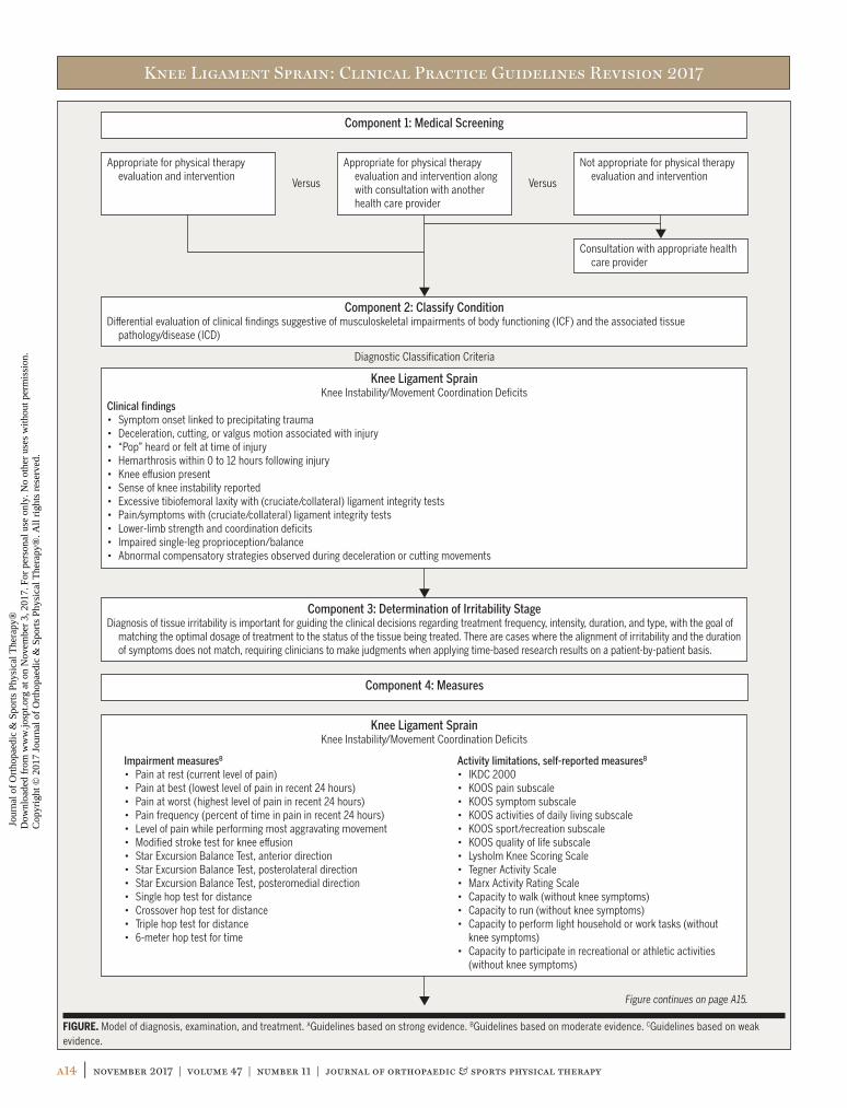
sis, and treatment planning for patients with knee stability and movement coordination impairments associated with knee ligament sprain uses the following components: (1) medical screening, (2) classification of condition through evaluation of clinical findings suggestive of musculoskeletal impairments of body functioning (ICF) and associated tis-sue pathology/disease (ICD), (3) determination of irritabil-ity stage, (4) determination of evaluative outcome measure instruments, and (5) intervention strategies for patients with ligament sprain. This model is depicted in the FIGURE.
Component 1Medical screening incorporates the findings of the history and physical examination to determine whether the patient’s symptoms originate from a condition that requires referral to another health care provider. The Ottawa knee rules, dis-cussed earlier, are an example of tools that may be helpful in this decision-making process. In addition to these conditions, clinicians should screen for the presence of psychosocial is-sues that may affect prognostication and treatment decision making for rehabilitation. Psychological stress negatively in-fluences recovery. Fear of reinjury is a frequently cited reason why athletes do not return to sport or reduce their level of physical activity.5,6 Low internal health locus of control (the belief in one’s ability to control one’s life), lower self-efficacy, and depressive symptoms prior to surgery result in worse outcomes after ACL reconstruction.32,114 Athletes who did not return to sport after ACL reconstruction had significantly lower preoperative motivation and more negative psychologi-cal response than those who did return.9 Accordingly, iden-tifying cognitive behavioral tendencies during the patient’s evaluation can direct the therapist to employ specific patient education strategies to optimize patient outcomes after phys-ical therapy interventions and potentially provide indications for referring the patient for consultation with another medi-cal or mental health practitioner.13
Component 2Differential evaluation of musculoskeletal clinical findings is used to determine the most relevant physical impairments associated with the patient’s reported activity limitations and medical diagnosis.59 Clusters of these clinical findings are described as impairment patterns in the physical therapy literature and are labeled according to the key impairment(s) of body function associated with that cluster. The ICD-10 and primary and secondary ICF codes associated with ligament sprain are provided in the 2010 ICF-based ligament sprain CPG.70 These impairment patterns are useful in driving the interventions, which focus on normalizing the key impair-ments of body function, which in turn improves the move-ment and function of the patient and lessens or alleviates the activity limitations commonly reported by the patients who meet the diagnostic criteria of that specific pattern. Key clini-
The ICD diagnosis of a sprain of the MCL and the associ-ated ICF diagnosis of knee stability and movement coor-dination impairments are made with a reasonable level of certainty when the patient presents with the following clini-cal findings58,92,95:• Trauma by a force applied to the lateral aspect of the lower
extremity• Rotational trauma• Medial knee pain with valgus stress test performed at 30°
of knee flexion (sensitivity, 78%; 95% CI: 64%, 92% and specificity, 67%; 95% CI: 57%, 76%)
• Increased separation between the femur and tibia (laxity) with a valgus stress test performed at 30° of knee flexion (sensitivity, 91%; 95% CI: 81%, 100% and specificity, 49%; 95% CI: 39%, 59%)
• Tenderness over the MCL and its attachments reproduces familiar pain
The ICD diagnosis of a sprain of the LCL and the associ-ated ICF diagnosis of knee stability and movement coor-dination impairments are made with a reasonable level of certainty when the patient presents with the following clini-cal findings18:• Varus trauma• Localized swelling over the LCL• Tenderness over the LCL and its attachments reproduces
familiar pain• Lateral knee pain with varus stress test performed at 0°
and 30° of knee flexion• Increased separation between the femur and tibia (laxity)
with varus stress test applied at 0° and 30° of knee flexion
Evidence UpdateNone.
2017 Recommendation
APhysical therapists should diagnose the ICD catego-ries of Sprain and strain involving collateral liga-ment of knee, Sprain and strain involving cruciate
ligament of knee, and Injury to multiple structures of knee, and the associated ICF impairment-based categories of knee instability (b7150 Stability of a single joint) and movement coordination impairments (b7601 Control of complex vol-untary movements), using the following history and physi-cal examination findings: mechanism of injury, passive knee laxity, joint pain, joint effusion, and movement coordination impairments.
Decision Tree ModelA pathoanatomical/medical diagnosis of ligament sprain can provide valuable information in describing tissue patholo-gy and may assist in preoperative planning and predicting prognosis. The proposed model for examination, diagno-
47-11 CPG Knee 3.indd 13 10/18/2017 2:26:11 PM
Jou
rnal
of
Ort
hopa
edic
& S
port
s Ph
ysic
al T
hera
py®
D
ownl
oade
d fr
om w
ww
.josp
t.org
at o
n N
ovem
ber
3, 2
017.
For
per
sona
l use
onl
y. N
o ot
her
uses
with
out p
erm
issi
on.
Cop
yrig
ht ©
201
7 Jo
urna
l of
Ort
hopa
edic
& S
port
s Ph
ysic
al T
hera
py®
. All
righ
ts r
eser
ved.
Knee Ligament Sprain: Clinical Practice Guidelines Revision 2017
a14 | november 2017 | volume 47 | number 11 | journal of orthopaedic & sports physical therapy
Appropriate for physical therapy evaluation and intervention
Appropriate for physical therapy evaluation and intervention along with consultation with another health care provider
Not appropriate for physical therapy evaluation and intervention
Consultation with appropriate health care provider
Component 1: Medical Screening
Component 4: Measures
Component 2: Classify ConditionDi�erential evaluation of clinical findings suggestive of musculoskeletal impairments of body functioning (ICF) and the associated tissue
pathology/disease (ICD)
Component 3: Determination of Irritability StageDiagnosis of tissue irritability is important for guiding the clinical decisions regarding treatment frequency, intensity, duration, and type, with the goal of
matching the optimal dosage of treatment to the status of the tissue being treated. There are cases where the alignment of irritability and the duration of symptoms does not match, requiring clinicians to make judgments when applying time-based research results on a patient-by-patient basis.
Knee Ligament SprainKnee Instability/Movement Coordination Deficits
Clinical findings• Symptom onset linked to precipitating trauma• Deceleration, cutting, or valgus motion associated with injury• “Pop” heard or felt at time of injury• Hemarthrosis within 0 to 12 hours following injury• Knee e�usion present• Sense of knee instability reported• Excessive tibiofemoral laxity with (cruciate/collateral) ligament integrity tests• Pain/symptoms with (cruciate/collateral) ligament integrity tests• Lower-limb strength and coordination deficits• Impaired single-leg proprioception/balance• Abnormal compensatory strategies observed during deceleration or cutting movements
Knee Ligament SprainKnee Instability/Movement Coordination Deficits
Versus Versus
Diagnostic Classification Criteria
Activity limitations, self-reported measuresB
• IKDC 2000 • KOOS pain subscale• KOOS symptom subscale• KOOS activities of daily living subscale• KOOS sport/recreation subscale• KOOS quality of life subscale• Lysholm Knee Scoring Scale• Tegner Activity Scale• Marx Activity Rating Scale• Capacity to walk (without knee symptoms)• Capacity to run (without knee symptoms)• Capacity to perform light household or work tasks (without
knee symptoms)• Capacity to participate in recreational or athletic activities
(without knee symptoms)
Impairment measuresB
• Pain at rest (current level of pain)• Pain at best (lowest level of pain in recent 24 hours)• Pain at worst (highest level of pain in recent 24 hours)• Pain frequency (percent of time in pain in recent 24 hours)• Level of pain while performing most aggravating movement• Modified stroke test for knee e�usion• Star Excursion Balance Test, anterior direction• Star Excursion Balance Test, posterolateral direction• Star Excursion Balance Test, posteromedial direction• Single hop test for distance• Crossover hop test for distance• Triple hop test for distance• 6-meter hop test for time
FIGURE. Model of diagnosis, examination, and treatment. AGuidelines based on strong evidence. BGuidelines based on moderate evidence. CGuidelines based on weak evidence.
Figure continues on page A15.
47-11 CPG Knee 3.indd 14 10/18/2017 2:26:12 PM
Jou
rnal
of
Ort
hopa
edic
& S
port
s Ph
ysic
al T
hera
py®
D
ownl
oade
d fr
om w
ww
.josp
t.org
at o
n N
ovem
ber
3, 2
017.
For
per
sona
l use
onl
y. N
o ot
her
uses
with
out p
erm
issi
on.
Cop
yrig
ht ©
201
7 Jo
urna
l of
Ort
hopa
edic
& S
port
s Ph
ysic
al T
hera
py®
. All
righ
ts r
eser
ved.
Knee Ligament Sprain: Clinical Practice Guidelines Revision 2017
journal of orthopaedic & sports physical therapy | volume 47 | number 11 | november 2017 | a15
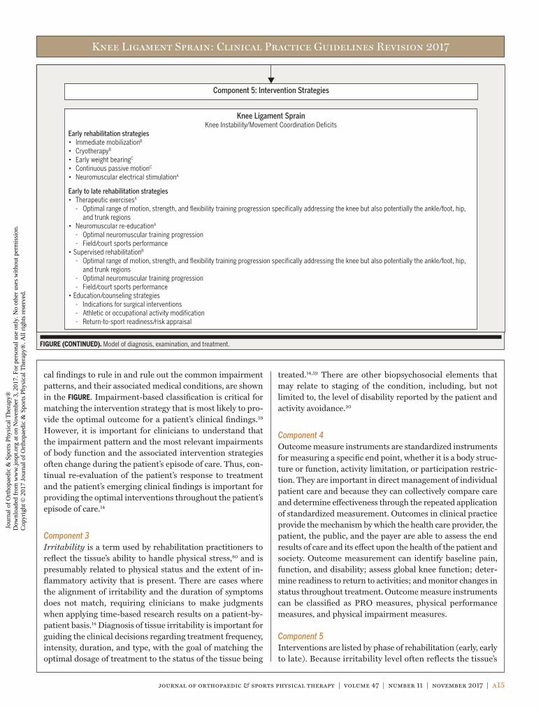
FIGURE (CONTINUED). Model of diagnosis, examination, and treatment.
Component 5: Intervention Strategies
Knee Ligament SprainKnee Instability/Movement Coordination Deficits
Early rehabilitation strategies• Immediate mobilizationB
• CryotherapyB
• Early weight bearingC
• Continuous passive motionC • Neuromuscular electrical stimulationA
Early to late rehabilitation strategies • Therapeutic exercisesA
- Optimal range of motion, strength, and flexibility training progression specifically addressing the knee but also potentially the ankle/foot, hip, and trunk regions
• Neuromuscular re-educationA
- Optimal neuromuscular training progression - Field/court sports performance• Supervised rehabilitationB - Optimal range of motion, strength, and flexibility training progression specifically addressing the knee but also potentially the ankle/foot, hip,
and trunk regions - Optimal neuromuscular training progression - Field/court sports performance• Education/counseling strategies - Indications for surgical interventions - Athletic or occupational activity modification - Return-to-sport readiness/risk appraisal
treated.14,59 There are other biopsychosocial elements that may relate to staging of the condition, including, but not limited to, the level of disability reported by the patient and activity avoidance.20
Component 4Outcome measure instruments are standardized instruments for measuring a specific end point, whether it is a body struc-ture or function, activity limitation, or participation restric-tion. They are important in direct management of individual patient care and because they can collectively compare care and determine effectiveness through the repeated application of standardized measurement. Outcomes in clinical practice provide the mechanism by which the health care provider, the patient, the public, and the payer are able to assess the end results of care and its effect upon the health of the patient and society. Outcome measurement can identify baseline pain, function, and disability; assess global knee function; deter-mine readiness to return to activities; and monitor changes in status throughout treatment. Outcome measure instruments can be classified as PRO measures, physical performance measures, and physical impairment measures.
Component 5Interventions are listed by phase of rehabilitation (early, early to late). Because irritability level often reflects the tissue’s
cal findings to rule in and rule out the common impairment patterns, and their associated medical conditions, are shown in the FIGURE. Impairment-based classification is critical for matching the intervention strategy that is most likely to pro-vide the optimal outcome for a patient’s clinical findings.59 However, it is important for clinicians to understand that the impairment pattern and the most relevant impairments of body function and the associated intervention strategies often change during the patient’s episode of care. Thus, con-tinual re-evaluation of the patient’s response to treatment and the patient’s emerging clinical findings is important for providing the optimal interventions throughout the patient’s episode of care.14
Component 3Irritability is a term used by rehabilitation practitioners to reflect the tissue’s ability to handle physical stress,80 and is presumably related to physical status and the extent of in-flammatory activity that is present. There are cases where the alignment of irritability and the duration of symptoms does not match, requiring clinicians to make judgments when applying time-based research results on a patient-by-patient basis.14 Diagnosis of tissue irritability is important for guiding the clinical decisions regarding treatment frequency, intensity, duration, and type, with the goal of matching the optimal dosage of treatment to the status of the tissue being
47-11 CPG Knee 3.indd 15 10/18/2017 2:26:12 PM
Jou
rnal
of
Ort
hopa
edic
& S
port
s Ph
ysic
al T
hera
py®
D
ownl
oade
d fr
om w
ww
.josp
t.org
at o
n N
ovem
ber
3, 2
017.
For
per
sona
l use
onl
y. N
o ot
her
uses
with
out p
erm
issi
on.
Cop
yrig
ht ©
201
7 Jo
urna
l of
Ort
hopa
edic
& S
port
s Ph
ysic
al T
hera
py®
. All
righ
ts r
eser
ved.
Knee Ligament Sprain: Clinical Practice Guidelines Revision 2017
a16 | november 2017 | volume 47 | number 11 | journal of orthopaedic & sports physical therapy
sistent with those presented in the Diagnosis/Classification section of this guideline, or when the individual’s symptoms are not resolving with intervention aimed at normalization of the individual’s impairments of body function.
IMAGING STUDIES2010 and 2017 SummaryWhen a patient reports a history of knee trauma, the thera-pist needs to be alert for the presence of knee fracture. The Ottawa knee rule has been developed and validated to as-sist clinicians in determining when to order radiographs in individuals with acute knee injury, which recommends knee radiographs for patients with any of the following criteria: age 55 or older, isolated tenderness of patella (no bone ten-derness of knee other than patella), tenderness of head of the fibula, inability to flex the knee to 90°, inability to bear weight both immediately and in the emergency department for 4 steps regardless of limping.10,112 Clinical examination by well-trained clinicians appears to be as accurate as magnetic resonance imaging (MRI) in regard to the diagnosis of cru-ciate or meniscal lesions.62,71 A lower threshold of suspicion of a meniscal tear is warranted in middle-aged and elderly patients.44,71 Magnetic resonance imaging may be reserved for more complicated or confusing cases62 and may assist an orthopaedic surgeon in aiding in preoperative planning and predicting the prognosis.62,71
ability to accept physical stress, clinicians should match the most appropriate intervention strategies to the irritability level of the patient’s condition.14,59 Additionally, clinicians should attend to influences from psychosocial factors5,6,9 in patients with conditions in all stages of recovery.
DIFFERENTIAL DIAGNOSIS2010 Recommendation
BClinicians should consider diagnostic classifications associated with serious pathological conditions or psychosocial factors when the patient’s reported
activity limitations or impairments of body function and structure are not consistent with those presented in the Diagnosis/Classification section of this guideline, or when the patient’s symptoms are not resolving with interventions aimed at normalization of the patient’s impairments of body function.
Evidence UpdateNone.
2017 Recommendation
BClinicians should suspect diagnostic classifications associated with serious pathological conditions when the individual’s reported activity limitations
and impairments of body function and structure are not con-
47-11 CPG Knee 3.indd 16 10/18/2017 2:26:12 PM
Jou
rnal
of
Ort
hopa
edic
& S
port
s Ph
ysic
al T
hera
py®
D
ownl
oade
d fr
om w
ww
.josp
t.org
at o
n N
ovem
ber
3, 2
017.
For
per
sona
l use
onl
y. N
o ot
her
uses
with
out p
erm
issi
on.
Cop
yrig
ht ©
201
7 Jo
urna
l of
Ort
hopa
edic
& S
port
s Ph
ysic
al T
hera
py®
. All
righ
ts r
eser
ved.
Knee Ligament Sprain: Clinical Practice Guidelines Revision 2017
journal of orthopaedic & sports physical therapy | volume 47 | number 11 | november 2017 | a17
OUTCOME MEASURES – ACTIVITY LIMITATIONS: SELF-REPORTED MEASURES2010 Recommendation
AClinicians should use a validated PRO measure with a general health questionnaire, along with a validated activity scale, for patients with knee sta-
bility and movement coordination impairments. These tools are useful for identifying a patient’s baseline status relative to pain, function, and disability and for monitoring changes in the patient’s status throughout the course of treatment.
Evidence UpdateGeneral Health Questionnaires
IWebster and Feller121 reported on a large patient cohort comparing results on the SF-36 to those from the Medical Outcomes Study 12-Item Short-
Form Health Survey (SF-12). A cohort of 1474 patients after ACL reconstruction completed the SF-36 and the SF-12 from the subscales of the SF-36. The authors reported small but statistically significant mean differences between subscales of the SF-36 and SF-12, except for the bodily pain subscale. They reported that a large majority of patients after ACL re-construction have average to above-average scores compared to the US general population. For the physical component summary score, the authors reported moderate correlations between the SF-36 and SF-12 with self-reported outcome measures (r = 0.47-0.62).
IA longitudinal analysis of a multicenter cohort of 1411 patients who completed the SF-36 preoperatively and at 2 years and/or 6 years after ACL reconstruc-
tion24 found that higher physical component summary scores were predicted by higher preoperative physical component summary scores, younger age, lower preoperative body mass index, having greater than 50% lateral meniscus excision, and no treatment to the lateral meniscus tear. Lower physical com-ponent summary scores were predicted by shorter follow-up, ACL revision reconstruction, preoperative smoking, less educa-tion, and degenerative changes of the lateral tibial plateau.
IIMånsson et al76 examined the predictability of pre-operative factors on SF-36 and KOOS question-naires. They reported that sex, preoperative Tegner
level, knee flexion deficit, and age explained 29% of the vari-ance in SF-36 general health, while Tegner level, single-legged hop performance, and knee flexion range-of-motion deficit explained 21% of the variance in SF-36 role-emotion-
al. Similarly, preoperative Tegner level explained 14% of the variance in KOOS sport and recreation, and preoperative Tegner level and knee flexion range-of-motion deficit ex-plained 18% of the variance in KOOS quality of life (QoL). Preoperative Tegner level and knee flexion range-of-motion deficit are good predictors of health-related quality of life (HRQoL) after ACL reconstruction.
IIThomeé et al115 examined preoperative self-efficacy of knee function as a predictor of activity level and knee symptoms and function 1 year after ACL re-
construction. Preoperative knee self-efficacy was a significant predictor of Tegner activity level and physical activity scale scores. Preoperative future knee self-efficacy was a significant predictor of KOOS sport and recreation and KOOS QoL.
IIThis study reported on the development and valida-tion of a new self-report questionnaire, the Knee Quality of Life 26-item (KQoL-26) questionnaire,
for patients with a suspected ligamentous or meniscal inju-ry.33 The questionnaire contains 26 items with 3 subscales of knee-related QoL: physical functioning, activity limitations, and emotional functioning. The KQoL-26 was found to have good evidence for internal reliability (Cronbach α = .91-.94), test-retest reliability (estimates of 0.80 to 0.93), construct validity (correlations with other knee scales, including the Lysholm knee scoring scale, EuroQol-5 Dimensions [EQ-5D] questionnaire, SF-36, and knee symptom questions), and responsiveness (higher effect sizes and responsiveness statistics than the EQ-5D and SF-36).
IIIMånsson et al75 compared the results of the SF-36 in patients 2 to 7 years after ACL reconstruction with a sex- and age-matched control group and compared
the results between sex and graft type. They reported that pa-tients in the long term after ACL reconstruction have good health relative to the general population. Additionally, there were no differences between men and women in HRQoL.
Knee Injury and Osteoarthritis Outcome Score
IISalavati and colleagues101 reported on the reliability and validity of the KOOS in competitive athletes after ACL reconstruction. In this population, the
KOOS was found to have good internal reliability (Cronbach α = .74-.96), test-retest reliability (intraclass correlation co-efficient [ICC]>0.75), and construct validity (correlations with SF-36 ranging from r = 0.72 to 0.79).
CLINICAL GUIDELINES
Examination
47-11 CPG Knee 3.indd 17 10/18/2017 2:26:12 PM
Jou
rnal
of
Ort
hopa
edic
& S
port
s Ph
ysic
al T
hera
py®
D
ownl
oade
d fr
om w
ww
.josp
t.org
at o
n N
ovem
ber
3, 2
017.
For
per
sona
l use
onl
y. N
o ot
her
uses
with
out p
erm
issi
on.
Cop
yrig
ht ©
201
7 Jo
urna
l of
Ort
hopa
edic
& S
port
s Ph
ysic
al T
hera
py®
. All
righ
ts r
eser
ved.
Knee Ligament Sprain: Clinical Practice Guidelines Revision 2017
a18 | november 2017 | volume 47 | number 11 | journal of orthopaedic & sports physical therapy
IIA systematic review by Filbay et al27 reported on HRQoL after ACL reconstruction. They included 14 studies on various HRQoL measures. Pooled
KOOS QoL subscores of 74.5 (95% CI: 68.3, 80.7) were lower than previously reported healthy population norms with no knee symptoms (90; 95% CI: 83.7, 96.3) and general popula-tion norms (82.4; 95% CI: 79.9, 84.9). The authors reported that meniscus injuries, revision surgery, and severe radio-graphic osteoarthritis were factors associated with poor KOOS QoL subscores.
IIA systematic review by Filbay et al28 reported on HRQoL in patients with ACL-deficient knees great-er than 5 years after the index injury. They included
11 studies on various HRQoL measures. The KOOS-QoL sub-scores ranged from a mean ± SD of 54 ± 17 to 77 ± 22 and were lower than previously reported general population norms (81 ± 24). No differences were reported between pooled KOOS QoL subscores for groups with ACL-deficient compared to ACL-reconstructed (2.9; 95% CI: –3.3, 9.1) knees.
IIGranan et al40 reported that from the Norwegian Knee Ligament Registry, patients who had an ACL reconstruction revision surgery had clinically signifi-
cantly lower scores on the KOOS sport and recreation and QoL subscales compared to those who did not have revision surgery. When adjusted for age, sex, and preoperative KOOS scores, the risk of a subsequent revision surgery was 3.7 (95% CI: 2.2, 6.0) times higher in patients with a 2-year postsurgery KOOS QoL score less than 44, compared to those with a score greater than or equal to 44. Additionally, for every 10-point drop in KOOS QoL subscore, there was a 33.6% (95% CI: 21.2%, 47.55%) higher risk for ACL reconstruction revision surgery.
IIIA Rasch model was used to assess the internal con-struct validity of the KOOS in 200 consecutive ques-tionnaires from patients 20 weeks after ACL
reconstruction.19 Fit to the Rasch model was achieved only for the sport and recreation subscale and the QoL subscale. The KOOS was found to have fair to good evidence for internal consistency (Cronbach α = .59 for knee symptoms to .91 for function in ADLs). The subdomains for pain, symptoms, and ADLs do not adequately assess patients 20 weeks after ACL reconstruction, whereas the sport and recreation and QoL sub-domains can be used to assess change in these constructs.
IIIThe KOOS has been cross-culturally adapted for use in both the Persian and Arabic languages. In Iranian patients with ACL, meniscus, and com-
bined meniscus and ACL injuries, the Persian version showed acceptable test-retest reliability on all subscales (ICC>0.70) except the sport and recreation subscale (ICC = 0.61), and adequate construct validity against the SF-36.102 The Arabic
version showed acceptable test-retest reliability (ICC>0.7) for all subscales, as well as good construct validity against sub-scales of the RAND-36 (Arabic version of the SF-36) and visual analog scale scores of pain in Egyptian patients with ACL, meniscus, and combined knee injuries.4
IKDC 2000 Subjective Knee Evaluation Form
IIShelbourne and colleagues106 evaluated the rela-tionship between the single assessment numeric evaluation (SANE) rating score and the IKDC
2000 and the modified Cincinnati Knee Rating System yearly in patients after ACL reconstruction or knee arthroscopy. In patients after ACL reconstruction, the correlation between the SANE rating score was 0.66 with the IKDC 2000 and the modified Cincinnati Knee Rating System, and 0.74 for both scales in patients after knee arthroscopy.
IIIvan Meer et al118 evaluated the IKDC 2000 and the KOOS to determine which questionnaire may be more useful early and within 1 year after ACL re-
construction. Both questionnaires were found to have good evidence for test-retest reliability (ICC>0.81 for both scales; the minimal detectable change [MDC] for KOOS subscales ranged from 18.3 to 35.2 and was 12.2 for the IKDC 2000), content validity (high percentage of relevant items), and no floor effects. The KOOS pain and ADL subscales had high ceiling effects, whereas the IKDC 2000 scores did not exhibit a ceiling effect. The authors concluded that the IKDC 2000 questionnaire was more useful for young, active individuals early and within 1 year after ACL reconstruction.
IIIHambly and Griva45 identified that after ACL re-construction, the IKDC 2000 outperformed the KOOS for the whole cohort and for sex subgroups.
For the IKDC 2000 in patients less than 12 months after ACL reconstruction, 15 of the 18 items and 10 of the 18 items were deemed important by more than half and by more than 75% of the patients, respectively. For the KOOS in patients less than 12 months after ACL reconstruction, 33 of the 42 items and 14 of the 42 items were deemed important by more than half and by more than 75% of the patients, respectively. How-ever, the KOOS sport and recreation and QoL subscales had significantly higher mean importance rankings than the IKDC 2000 for the whole cohort and for sex subgroups.
IIIDella Villa et al23 reported that higher knee func-tion (IKDC 2000) scores 12 months after ACL re-construction and quicker recovery were associated
with younger patients with higher preinjury Tegner levels or those at the professional sports level without concomitant capsular injuries and without postoperative knee bracing. Higher knee function was also related to more on-field train-ing sessions and better muscle strength recovery.
47-11 CPG Knee 3.indd 18 10/18/2017 2:26:12 PM
Jou
rnal
of
Ort
hopa
edic
& S
port
s Ph
ysic
al T
hera
py®
D
ownl
oade
d fr
om w
ww
.josp
t.org
at o
n N
ovem
ber
3, 2
017.
For
per
sona
l use
onl
y. N
o ot
her
uses
with
out p
erm
issi
on.
Cop
yrig
ht ©
201
7 Jo
urna
l of
Ort
hopa
edic
& S
port
s Ph
ysic
al T
hera
py®
. All
righ
ts r
eser
ved.
Knee Ligament Sprain: Clinical Practice Guidelines Revision 2017
journal of orthopaedic & sports physical therapy | volume 47 | number 11 | november 2017 | a19
good, with a standard response mean of 1.26. No ceiling (1.6%) or floor (0.8%) effects were seen.
Psychological Questionnaires
IIIn a systematic review, Ardern et al8 evaluated 11 studies and 15 psychological factors associated with returning to sport after a sports injury. They report-
ed that athletes who returned to sport after ACL reconstruc-tion had significantly higher preoperative motivation and more positive psychological response than those who did not return. In those who returned, those with a more positive per-ception of their return had greater intrinsic motivation and a greater sense of autonomy, competence, and relatedness need satisfaction. Positive emotions increased and negative emo-tions decreased as rehabilitation progressed and upon return to sport. Fear significantly increased when competition was resumed, as compared to rehabilitation. However, the authors reported that all studies had a moderate to high risk of bias.
IIIThe Anterior Cruciate Ligament-Return to Sport after Injury (ACL-RSI) scale has been cross-cultur-ally adapted for use in the Swedish, French, and
Turkish languages. In Swedish patients after ACL reconstruc-tion, the Swedish ACL-RSI (scored on a scale from 1 to 10) showed good face validity (relevant and easy to understand for the patient population), good evidence for internal consistency (Cronbach α = .95) and construct validity (relationships be-tween returning to sport and previous activity levels, and with other knee scales including the Tampa Scale of Kinesiopho-bia-11 [TSK-11], Knee Self-Efficacy Scale, KOOS, Anterior Cruciate Ligament-Quality of Life, and the Multidimensional Health Locus of Control, C-form), acceptable floor and ceiling effects, and good reproducibility (ICC2,1 = 0.89; standard error of the measurement, 0.7; and MDC for individual score of 1.9 and MDC for the group score of 0.3).65 In French-speaking patients after ACL reconstruction, the French version of the ACL-RSI showed good evidence for internal consistency (Cronbach α = .96), good discriminant validity, good construct validity (correlations against the IKDC 2000, KOOS subscales, and Lysholm scale), acceptable floor and ceiling effects, and good reproducibility (ICC = 0.90).15 In patients after ACL re-construction, the Turkish translation of the ACL-RSI showed evidence for good internal consistency (Cronbach α = .88), fair construct validity (r = 0.36-0.58 against the IKDC 2000, KOOS subscales, Lysholm scale, and TSK-11), good discrimi-nant validity, acceptable floor and ceiling effects, and excellent test-retest reliability (ICC = 0.92).46
IIIFlanigan et al29 evaluated the factors for not return-ing to sport after primary or revision ACL recon-struction. They reported that 46% of patients had
returned to preinjury activity levels, whereas 54% did not re-turn. In those who did not return, 68% reported persistent knee
Lysholm and Tegner Scales
IBriggs et al17 reported the reliability, validity, and responsiveness of the Lysholm score and Tegner scale for patients with ACL injury. The Lysholm
score was found to have acceptable evidence for test-retest reliability (ICC = 0.9 and MDC of 8.9), construct validity (correlations with other knee scales, including the IKDC 2000 and SF-12), and responsiveness (moderate to large ef-fect sizes and standardized response means on all domains, except for locking). The Tegner scale was found to have ac-ceptable evidence for test-retest reliability (ICC = 0.8 and MDC of 1), low construct validity (low correlations with the IKDC 2000 and SF-12), and responsiveness (large effect sizes and standardized response means).
IIIThe Tegner scale and the Marx Activity Rating Scale have been cross-culturally adapted for use in the Persian language.84 In Iranian patients with
ACL injury, the Persian version showed acceptable test-retest reliability on the Tegner scale (ICC = 0.81) and on the Marx Activity Rating Scale (ICC = 0.78), and acceptable evidence for internal reliability on the Marx Activity Rating Scale (Cronbach α = .87). The MDC for the Tegner scale was 0.75 and for the Marx Activity Rating Scale was 1.70. There was low construct validity of both the Tegner scale and Marx Ac-tivity Rating Scale (low correlations with the KOOS and SF-12), likely because the Tegner scale and Marx Activity Rating Scale measure activity level, whereas the SF-12 measures general health and the KOOS measures knee-specific symp-toms and function.
IIIThe Tegner rating scale has been cross-culturally adapted for use in the Chinese language.51 In Chinese patients with ACL injury, the simplified Chinese ver-
sion showed high test-retest reliability for healthy controls (ICC = 0.97) and for patients post–ACL reconstruction (ICC = 0.95-0.99), and acceptable reliability for patients pre–ACL reconstruction (ICC = 0.71). The MDC for the Tegner scale was 0.43 for healthy controls, 2.12 for preoperative patients, 0.89 for patients 2 to 3 months post–ACL reconstruction, and 0.44 for patients 3 to 12 months post–ACL reconstruction. No ceil-ing or floor effects were seen for any group.
IIIThe Lysholm scale has been cross-culturally adapt-ed for use in the Chinese language.119 In Chinese patients with ACL injury, the Chinese version
showed high test-retest reliability for patients with ACL in-jury (ICC = 0.94) and acceptable internal reliability (Cron-bach α = .73). There was good construct validity of the Chinese Lysholm scale, with high correlations with the IKDC 2000 (r = 0.73-0.81) and moderate correlations with the physical component summary score of the SF-36 (r = 0.51-0.71). The responsiveness of the Chinese Lysholm scale was
47-11 CPG Knee 3.indd 19 10/18/2017 2:26:12 PM
Jou
rnal
of
Ort
hopa
edic
& S
port
s Ph
ysic
al T
hera
py®
D
ownl
oade
d fr
om w
ww
.josp
t.org
at o
n N
ovem
ber
3, 2
017.
For
per
sona
l use
onl
y. N
o ot
her
uses
with
out p
erm
issi
on.
Cop
yrig
ht ©
201
7 Jo
urna
l of
Ort
hopa
edic
& S
port
s Ph
ysic
al T
hera
py®
. All
righ
ts r
eser
ved.
Knee Ligament Sprain: Clinical Practice Guidelines Revision 2017
a20 | november 2017 | volume 47 | number 11 | journal of orthopaedic & sports physical therapy
metry indexes for isokinetic knee extensor strength tests (de-pending on graft type and isokinetic speed) ranged from 65% to 86% 6 months after ACL reconstruction, 84% to 91% 12 months after ACL reconstruction, and 91% to 100% 24 months after ACL reconstruction. Limb symmetry indexes for iso-kinetic knee flexor strength tests (depending on graft type and isokinetic speed) ranged from 84% to 96% 6 months after ACL reconstruction, 87% to 99% 12 months after ACL reconstruc-tion, and 88% to 100% 24 months after ACL reconstruction.
IIHegedus et al48 performed a systematic review of 29 articles examining the measurement properties of performance-based tests for the knee. They report-
ed poor to fair methodological quality of studies examining reliability, poor to good quality of studies examining hypoth-esis testing and construct validity, and primarily good quality of studies examining criterion validity. Methodological qual-ity examining responsiveness was poor to good, while no studies examined measurement error.
IIAfter an ACL injury and nonoperative rehabilita-tion, a cutoff score greater than 88% on the single-leg single hop for distance test can be used to
identify with high probability that the patient will have nor-mal knee function based on the 15th percentile from the age- and sex-matched IKDC 2000 normative data 1 year later.42 Single-leg hop tests conducted 6 months after ACL recon-struction can predict the likelihood of successful and unsuc-cessful outcome 1 year after ACL reconstruction.68 Athletes demonstrating less than the 88% cutoff score on the 6-meter timed hop test at 6 months may benefit from targeted train-ing to improve limb symmetry in an attempt to normalize function. Athletes with minimal side-to-side differences on the crossover hop test at 6 months will likely have normal knee function 1 year after reconstruction if they continue with their current training regimen. Preoperative single-leg hop tests are not able to predict postoperative outcomes.68 Single-leg hop tests can identify important limb asymmetries and predict self-reported knee function in patients with knee stability and movement coordination impairments.
IIThe triple hop for distance limb symmetry index was a significant predictor of the IKDC 2000 jump-related question (14h) and KOOS jump-related
question (sport and recreation item 3) 2 years after ACL reconstruction.96
2017 Recommendation
BClinicians should administer appropriate clinical or field tests, such as single-leg hop tests (eg, single hop for distance, crossover hop for distance, triple
hop for distance, and 6-meter timed hop), that can identify a patient’s baseline status relative to pain, function, and dis-
symptoms and 52% reported fear of reinjury as factors for not returning to sport. Additionally, 29% reported non–knee-relat-ed life events, such as children, occupation, education, or other health issues, as other factors for not returning to sport.
IIIGeorge et al35 investigated whether shortened ver-sions of the TSK-11 and Pain Catastrophizing Scale may be used with patients after ACL reconstruc-
tion. They reported that the TSK-11 could be shortened to a 3-item fear-of-injury scale (items 1, 2, and 10) in the early postoperative phase (less than 12 weeks after surgery), but the TSK-11 is not recommended in the late postoperative phase (greater than 6 months after surgery). The shortened version of the Pain Catastrophizing Scale is not recommend-ed in the early postoperative phase, but may be shortened to a 7-item helplessness-and-magnification scale (items 1, 3, 4, 5, 6, 7, and 13) in the late postoperative phase.
2017 Recommendations
BClinicians should use the IKDC 2000 or KOOS and may use the Lysholm scale as validated PRO mea-sures to assess knee symptoms and function, and
should use the Tegner scale or Marx Activity Rating Scale to assess activity level, before and after interventions intended to alleviate the physical impairments, activity limitations, and participation restrictions associated with knee ligament sprain. Clinicians may use the ACL-RSI as a validated PRO measure to assess psychological factors that may hinder re-turn to sports before and after interventions intended to al-leviate fear of reinjury associated with knee ligament sprain.
PHYSICAL PERFORMANCE MEASURESRefer to the 2010 knee stability and movement coordination impairment CPG for a list of activity limitation measures and their measurement properties.70
2010 Recommendation
CClinicians should utilize easily reproducible physical performance measures, such as single-limb hop tests, to assess activity limitations and participation restric-
tions associated with their patients' knee stability and move-ment coordination impairments to assess the changes in the patient’s level of function over the episode of care and to classify and screen knee stability and movement coordination.
Evidence Update
IIA systematic review by Abrams et al1 examined per-formance-based tests after ACL reconstruction. They reported that limb symmetry indexes for hop
tests ranged from 76% to 90% 6 months after ACL reconstruc-tion, 88% to 95% 12 months after ACL reconstruction, and 92% to 99% 24 months after ACL reconstruction. Limb sym-
47-11 CPG Knee 3.indd 20 10/18/2017 2:26:12 PM
Jou
rnal
of
Ort
hopa
edic
& S
port
s Ph
ysic
al T
hera
py®
D
ownl
oade
d fr
om w
ww
.josp
t.org
at o
n N
ovem
ber
3, 2
017.
For
per
sona
l use
onl
y. N
o ot
her
uses
with
out p
erm
issi
on.
Cop
yrig
ht ©
201
7 Jo
urna
l of
Ort
hopa
edic
& S
port
s Ph
ysic
al T
hera
py®
. All
righ
ts r
eser
ved.
Knee Ligament Sprain: Clinical Practice Guidelines Revision 2017
journal of orthopaedic & sports physical therapy | volume 47 | number 11 | november 2017 | a21
IIA test battery of performance-based measures and PROs was developed to enhance decision making regarding clearance for return to sports.38 Return-to-
sport criteria were defined as isokinetic and hop test symmetry indexes greater than 90%, Landing Error Scoring System score less than 5, ACL-RSI score greater than 56, and IKDC 2000 scores at or above the 15th percentile of age- and sex-matched normative data. Six months after ACL reconstruction, only 7.1% of patients passed all criteria. This study was a preliminary study to develop a test battery on return-to-sport criteria.
IIA systematic review of 9 studies evaluated the reli-ability of diagnostic clinical tests for ACL injury.22 The anterior drawer test had moderate to excellent inter-
rater reliability (κ = 0.96) in low-quality studies. The Lachman test had poor to excellent interrater reliability (κ = 0.19-0.93) and poor to moderate intrarater reliability (κ = 0.29-0.51).
IIA systematic review by Kopkow et al63 reported that the quadriceps active test was the most specific test for diagnosing a PCL tear (sensitivity, 0.53-0.98;
specificity, 0.96-1.00; PLR = 11.97-98.44; NLR = 0.04-0.50) and that the posterior sag sign was the most sensitive (sensi-tivity, 0.46-1.00; specificity, 1.00; PLR = 88.35; NLR = 0.28). However, the methodological quality of the studies reviewed was insufficient and the studies had a high risk of bias.
IILeblanc et al66 performed a systematic review of 8 studies examining the diagnostic accuracy of the Lachman test. Pooled analysis for sensitivity during
awake evaluations of the Lachman test for complete tear of the ACL was 96%, whereas nonpooled analysis for specificity was 78.1%.
IIIHigher preoperative quadriceps strength index (OR = 1.02), young age (OR = 0.92), and male sex (OR = 2.45) were associated with higher quadriceps
strength 6 months after ACL reconstruction.116 Higher knee pain (OR = 0.17) was associated with lower odds of higher quadriceps strength 6 months after ACL reconstruction. A cutoff score greater than 70.2% on the preoperative quadri-ceps strength index can be used to identify with moderate probability (PLR = 2) that the patient will have a quadriceps strength index of at least 85% 6 months after ACL reconstruction.
2017 Recommendation
BWhen evaluating a patient with ligament sprain over an episode of care, clinicians should use as-sessments of impairment of body structure and
function, including measures of knee laxity/stability, lower-limb movement coordination, thigh muscle strength, knee effusion, and knee joint range of motion.
ability; detect side-to-side asymmetries; assess global knee function; determine a patient’s readiness to return to activi-ties; and monitor changes in the patient’s status throughout the course of treatment.
PHYSICAL IMPAIRMENT MEASURESRefer to the 2010 knee stability and movement coordination impairment CPG for a list of physical impairment measures and their measurement properties.70
Evidence Update
IA systematic review by Swain et al113 reported small to large likelihood ratios for the Lachman test (pos-itive likelihood ratio [PLR] = 1.39-40.81, negative
likelihood ratio [NLR] = 0.02-0.52), anterior drawer test (PLR = 1.94-87.88, NLR = 0.23-0.74), and the pivot shift test (PLR = 4.37-16.42, NLR = 0.38-0.84) across studies. A PLR is the likelihood that a given positive clinical test result would be expected in a patient with an ACL tear, compared to the likelihood that the same positive test result would be expect-ed in a patient without an ACL tear, whereas an NLR is the likelihood that a given negative clinical test result would be expected in a patient without an ACL tear, compared to the likelihood that the same negative result would be expected in a patient with an ACL tear. Likelihood threshold values can be categorized as low (PLR>2, NLR<0.2), moderate (PLR = 2-5, NLR = 0.1-0.2), or high (PLR>5, NLR<0.1).
IIPreoperative quadriceps strength deficits are pre-dictive of impaired self-reported knee function 6 months and 2 years after ACL reconstruction.25,69
IIGreater than 6 months after ACL reconstruction, a cutoff score greater than 3.10 Nm·kg–1 on quadri-ceps peak torque per body mass can be used to
identify with high probability (8.15 times higher odds) that the patient will have high self-reported knee function (IKDC 2000 greater than 90%) at the same time point.93 A cutoff score greater than 96.5% on the quadriceps limb symmetry index can be used to identify with moderate probability (2.78 times higher odds) that the patient will have high self-report-ed knee function.
IIAthletes with no knee effusion, no knee instability, and who reported over 93% on the IKDC 2000 1 year after ACL reconstruction were more than 14
times more likely to return to sports 1 year after ACL recon-struction (PLR = 14.54).67 Athletes meeting only 1 criterion (no knee effusion, no knee instability, or greater than 93% on the IKDC 2000) 1 year after ACL reconstruction were more than 6 times more likely to not return to sports 1 year after ACL reconstruction (NLR = 0.16).67
47-11 CPG Knee 3.indd 21 10/18/2017 2:26:12 PM
Jou
rnal
of
Ort
hopa
edic
& S
port
s Ph
ysic
al T
hera
py®
D
ownl
oade
d fr
om w
ww
.josp
t.org
at o
n N
ovem
ber
3, 2
017.
For
per
sona
l use
onl
y. N
o ot
her
uses
with
out p
erm
issi
on.
Cop
yrig
ht ©
201
7 Jo
urna
l of
Ort
hopa
edic
& S
port
s Ph
ysic
al T
hera
py®
. All
righ
ts r
eser
ved.
Knee Ligament Sprain: Clinical Practice Guidelines Revision 2017
a22 | november 2017 | volume 47 | number 11 | journal of orthopaedic & sports physical therapy
A systematic search of the literature did not reveal high-quality reviews or articles to alter the 2010 recommenda-tions for continuous passive motion, early weight bearing, knee bracing, or immediate versus delayed mobilization. However, edits were made to these 2010 recommendations to improve the clarity of the recommendations. Additional-ly, in the 2010 guidelines, “accelerated” rehabilitation was a separate category. Early restoration of knee extension range of motion and early weight-bearing activity have now been in practice for over 20 years and are the current standard of care after isolated ACL reconstruction, thus “accelerated” rehabilitation is included in the Immediate Versus Delayed Mobilization section in these 2017 guidelines. Likewise, in the 2010 guidelines, eccentric strengthening was a sepa-rate category. However, eccentric strengthening, a form of therapeutic exercise, is now placed under the Therapeutic Exercise section.
CONTINUOUS PASSIVE MOTION2010 and 2017 Recommendation
CClinicians may use continuous passive motion in the immediate postoperative period to decrease postoperative pain after ACL reconstruction.
EARLY WEIGHT BEARING2010 Recommendation
CEarly weight bearing can be used for patients fol-lowing ACL reconstruction without incurring det-rimental effects on stability or function.
Evidence UpdateNone.
2017 Recommendation
CClinicians may implement early weight bearing as tolerated (within 1 week after surgery) for patients after ACL reconstruction.
KNEE BRACING2010 Recommendation
CThe use of functional knee bracing appears to be more beneficial than not using a brace in patients with ACL deficiency.
BThe use of immediate postoperative knee bracing appears to be no more beneficial than not using a brace in patients following ACL reconstruction.
DConflicting evidence exists for the use of functional knee bracing in patients following ACL reconstruction.
FKnee bracing can be used for patients with acute PCL injuries, severe MCL injuries, or PLC injuries.
Evidence Update
IIA systematic review by Kinikli et al61 included 11 studies on the effectiveness of knee bracing after ACL reconstruction. They evaluated 7 RCTs, 2 pro-
spective controlled trials, 1 clinic trial, and 1 crossover study. For knee laxity between braced and nonbraced groups, the braced group had statistically significant but not clinically significantly less knee laxity after ACL reconstruction. For muscle strength, no differences were found between the braced and nonbraced groups. For knee joint range of mo-tion, knee pain, and self-reported knee function, the braced and the nonbraced groups were similar in outcomes at the majority of follow-up periods and in the majority of studies.
2017 Recommendation
CClinicians may use functional knee bracing in pa-tients with ACL deficiency.
DClinicians should elicit and document patient pref-erences in the decision to use functional knee brac-ing after ACL reconstruction, as evidence exists for
and against its use.
FClinicians may use appropriate knee bracing for patients with acute PCL injuries, severe MCL inju-ries, or PLC injuries.
IMMEDIATE VERSUS DELAYED MOBILIZATION2010 Recommendation
BClinicians should consider the use of immediate mobilization following ACL reconstruction to in-crease range of motion, reduce pain, and limit ad-
verse changes to soft tissue structures.
CLINICAL GUIDELINES
Interventions
47-11 CPG Knee 3.indd 22 10/18/2017 2:26:12 PM
Jou
rnal
of
Ort
hopa
edic
& S
port
s Ph
ysic
al T
hera
py®
D
ownl
oade
d fr
om w
ww
.josp
t.org
at o
n N
ovem
ber
3, 2
017.
For
per
sona
l use
onl
y. N
o ot
her
uses
with
out p
erm
issi
on.
Cop
yrig
ht ©
201
7 Jo
urna
l of
Ort
hopa
edic
& S
port
s Ph
ysic
al T
hera
py®
. All
righ
ts r
eser
ved.
Knee Ligament Sprain: Clinical Practice Guidelines Revision 2017
journal of orthopaedic & sports physical therapy | volume 47 | number 11 | november 2017 | a23
Evidence UpdateNone.
2017 Recommendation
BClinicians should use immediate mobilization (within 1 week) after ACL reconstruction to in-crease joint range of motion, reduce joint pain, and
reduce the risk of adverse responses of surrounding soft tis-sue structures, such as those associated with knee extension range-of-motion loss.
CRYOTHERAPY2010 Recommendation
CClinicians should consider the use of cryotherapy to reduce postoperative knee pain immediately post–ACL reconstruction.
Evidence Update
IIA systematic review by Martimbianco et al77 con-cluded that cryotherapy after arthroscopic ACL reconstruction significantly reduced immediate
postsurgery pain and did not increase the risk of adverse events in the short term (up to 48 hours after surgery). The limited evidence currently available from RCTs is insuffi-cient to draw definitive conclusions on the effectiveness of cryotherapy for other outcomes such as edema, knee func-tion, postoperative blood loss, duration of hospital stay, range of motion, postoperative analgesic medication use, patient satisfaction, or QoL.
2017 Recommendation
BClinicians should use cryotherapy immediately af-ter ACL reconstruction to reduce postoperative knee pain.
SUPERVISED REHABILITATION2010 Recommendation
BClinicians should use exercises as part of the in-clinic program, supplemented by a prescribed home-based program supervised by a physical ther-
apist, in patients with knee stability and movement coordina-tion impairments.
Evidence Update
IIPapalia and colleagues,86 in a systematic review, evaluated 10 studies comparing outcomes between home-based and supervised outpatient rehabilita-
tion after ACL reconstruction. The systematic review includ-ed 7 RCTs that evaluated range of motion and PROs (Lysholm scale, Tegner scale, or modified Cincinnati Knee Rating System)
as their primary outcome variables. In intermediate follow-ups, PRO scores were high. However, activity level (Tegner) scores ranged from 4 to 5. Only Hohmann et al,49 in this systematic review, reported on performance-based out-comes, on which both groups demonstrated limb asymme-tries. Therefore, caution is warranted in interpreting the results of this systematic review. Mid-range Tegner scores suggest that patients were not competitive or pivoting-type athletes, and limb asymmetries on performance-based tests suggest that patients were not fully rehabilitated at last follow-up.
2017 Recommendation
BClinicians should use exercises as part of the in-clinic supervised rehabilitation program after ACL reconstruction and should provide and supervise
the progression of a home-based exercise program, providing education to ensure independent performance.
THERAPEUTIC EXERCISES2010 Recommendation
AClinicians should consider the use of non–weight-bearing (open-chain) exercises in conjunction with weight-bearing (closed-chain) exercises in
patients with knee stability and movement coordination impairments.
Evidence Update
IIOne moderate-quality systematic review by Gokeler et al37 included 8 RCTs and 2 prospective cohort studies on the effect of various rehabilitation strate-
gies on quadriceps function after ACL reconstruction. Seven studies reported an increase in quadriceps strength when tested isokinetically. Five studies reported an increase in ac-tivity level based on Tegner scores throughout rehabilitation. Cincinnati Knee Rating System scores were increased after neuromuscular training and strength training.
2017 Recommendation
AWeight-bearing and non–weight-bearing concen-tric and eccentric exercises should be implemented within 4 to 6 weeks, 2 to 3 times per week for 6 to
10 months, to increase thigh muscle strength and functional performance after ACL reconstruction.
NEUROMUSCULAR ELECTRICAL STIMULATION2010 Recommendation
BNeuromuscular electrical stimulation (NMES) can be used with patients following ACL reconstruction to increase quadriceps muscle strength.
47-11 CPG Knee 3.indd 23 10/18/2017 2:26:13 PM
Jou
rnal
of
Ort
hopa
edic
& S
port
s Ph
ysic
al T
hera
py®
D
ownl
oade
d fr
om w
ww
.josp
t.org
at o
n N
ovem
ber
3, 2
017.
For
per
sona
l use
onl
y. N
o ot
her
uses
with
out p
erm
issi
on.
Cop
yrig
ht ©
201
7 Jo
urna
l of
Ort
hopa
edic
& S
port
s Ph
ysic
al T
hera
py®
. All
righ
ts r
eser
ved.
Knee Ligament Sprain: Clinical Practice Guidelines Revision 2017
a24 | november 2017 | volume 47 | number 11 | journal of orthopaedic & sports physical therapy
Evidence Update
IIn a high-quality systematic review, Kim et al60 investigated the effects of NMES on quadriceps muscle strength, performance-based measures,
and patient-reported measures after ACL reconstruction. The NMES was administered within the first 3 weeks after ACL reconstruction and for a duration of 3 to 11 weeks. They found that, in 10 of 11 comparisons of quadriceps strength outcomes, the effect sizes were small to very large (0.08-3.81), favoring NMES combined with exercise when compared to exercise only or another comparable treat-ment. In 3 comparisons of performance-based measures, the effects were small to moderate (0.07-0.64, with 95% CIs crossing zero), favoring NMES combined with exercise when compared to exercise only. In 2 comparisons of pa-tient-reported measures, the effects were moderate (0.66-0.72), favoring NMES compared to another comparable treatment. They concluded from 8 RCTs that NMES com-bined with exercise may be more effective in improving quadriceps strength than exercise alone after ACL recon-struction, whereas its effect on functional performance and patient-oriented outcomes is inconclusive. A variety of NMES parameters and applications of NMES were used in the reviewed studies.
IIImoto et al53 conducted a systematic review of 17 RCTs on the effectiveness of NMES after ACL inju-ries. In 3 studies, the mean difference of isometric
quadriceps strength 6 to 8 weeks after ACL reconstruction was 32.7 Nm, favoring NMES compared to other treatments (95% CI: 25.48, 39.92). The evidence in this moderate-qual-ity review concluded that conventional rehabilitation aug-mented with NMES can improve quadriceps muscle strength 6 to 8 weeks after ACL reconstruction.
2017 Recommendation
ANeuromuscular electrical stimulation should be used for 6 to 8 weeks to augment muscle strength-ening exercises in patients after ACL reconstruc-
tion to increase quadriceps muscle strength and enhance short-term functional outcomes.
NEUROMUSCULAR RE-EDUCATION2010 Recommendation
BClinicians should consider the use of neuromuscu-lar re-education as a supplementary program to strength training in patients with knee stability and
movement coordination impairments.
Evidence Update
IOne moderate-quality systematic review by Gokeler et al37 included 8 RCTs and 2 prospective cohort studies on the effect of rehabilitation on quadriceps
muscle strength and patient-reported measures after ACL re-construction. Seven studies reported an increase in quadriceps strength, and 2 studies reported that training on an eccentric ergometer significantly improved quadriceps strength more than concentric training. Five studies reported an increase in Tegner activity score throughout rehabilitation, but 4 of 5 stud-ies found no differences between groups at the final follow-up time point. Four studies reported that neuromuscular training and strength training can increase patient-reported measures 6 months to 2 years after ACL reconstruction. The evidence from this review indicates that neuromuscular training incor-porating motor learning principles should be added to strength training to optimize patient outcomes.
IIA moderate-quality systematic review conducted by Zech and colleagues129 included 13 RCTs and 2 con-trolled clinical trials, with 3 studies focused on ACL
injuries. They concluded that neuromuscular interventions after ACL injuries can be effective for the nonoperative management of ACL injuries, in terms of function and joint stability.
IIIThe results of a systematic review looked at the ef-fectiveness of electromyographic biofeedback on the quadriceps after ACL reconstruction.120 In 1 study,
biofeedback appeared to benefit short-term postsurgical quad-riceps strength. However, this review is very limited, as only 2 studies evaluated biofeedback after ACL reconstruction, and only 1 provided analysis on current surgical techniques.
2017 Recommendation
ANeuromuscular re-education training should be incorporated with muscle strengthening exercises in patients with knee stability and movement coor-
dination impairments.
47-11 CPG Knee 3.indd 24 10/18/2017 2:26:13 PM
Jou
rnal
of
Ort
hopa
edic
& S
port
s Ph
ysic
al T
hera
py®
D
ownl
oade
d fr
om w
ww
.josp
t.org
at o
n N
ovem
ber
3, 2
017.
For
per
sona
l use
onl
y. N
o ot
her
uses
with
out p
erm
issi
on.
Cop
yrig
ht ©
201
7 Jo
urna
l of
Ort
hopa
edic
& S
port
s Ph
ysic
al T
hera
py®
. All
righ
ts r
eser
ved.
Knee Ligament Sprain: Clinical Practice Guidelines Revision 2017
journal of orthopaedic & sports physical therapy | volume 47 | number 11 | november 2017 | a25
AUTHORSDavid S. Logerstedt, PT, PhDAssistant ProfessorDepartment of Physical TherapyUniversity of the SciencesPhiladelphia, [email protected]
David Scalzitti, PT, PhDAssistant ProfessorDepartment of Physical TherapyGeorge Washington UniversityWashington, [email protected]
May Arna Risberg, PT, PhDChairNorwegian Research Center for Active
RehabilitationOslo, NorwayandProfessorDepartment of Sports MedicineNorwegian School of Sport SciencesOslo, [email protected]
Lars Engebretsen, MDOslo Sports Trauma Research CenterNorwegian School of Sport SciencesOslo, NorwayandDepartment of OrthopaedicsOslo University HospitalOslo, [email protected]
Kate E. Webster, PhDAssociate ProfessorSchool of Allied HealthLa Trobe UniversityMelbourne, [email protected]
Julian Feller, MDOrthoSport Victoria Research UnitDeakin University and Epworth
HealthcareMelbourne, [email protected]
Lynn Snyder-Mackler, PT, ScDAlumni Distinguished ProfessorDepartment of Physical TherapyandFaculty Athletics RepresentativeUniversity of DelawareNewark, [email protected]
Michael J. Axe, MDFirst State OrthopaedicsNewark, DE
Christine M. McDonough, PT, PhDICF-Based Clinical Practice Guidelines
EditorOrthopaedic Section, APTA, IncLa Crosse, WIandAdjunct Clinical Assistant ProfessorDepartment of Orthopaedic SurgeryGeisel School of Medicine at DartmouthDartmouth-Hitchcock Medical CenterLebanon, NHandResearch Assistant ProfessorHealth and Disability Research InstituteBoston University School of Public
HealthBoston, [email protected]
REVIEWERSRoy D. Altman, MDProfessor of MedicineDivision of Rheumatology and
ImmunologyDavid Geffen School of Medicine at
UCLALos Angeles, [email protected]
Paul Beattie, PT, PhDClinical ProfessorDivision of Rehabilitative SciencesUniversity of South CarolinaColumbia, [email protected]
John DeWitt, DPTDirectorPhysical Therapy Sports and
Orthopaedic ResidenciesThe Ohio State UniversityColumbus, [email protected]
James M. Elliott, PT, PhDAssistant ProfessorNorthwestern University Physical
Therapy and Human Movement Sciences
Feinberg School of MedicineNorthwestern UniversityChicago, [email protected]
Amanda Ferland, DPTClinical FacultyIntech Rehabilitation Group/Division of
Biokinesiology and Physical TherapyOrthopaedic Physical Therapy
ResidencyUniversity of Southern California
Los Angeles, CAandSpine Rehabilitation FellowshipBeijing, [email protected]
G. Kelley Fitzgerald, PT, PhDProfessor and Associate Dean of
Graduate StudiesSchool of Health and Rehabilitation
SciencesandDirectorPhysical Therapy Clinical and
Translational Research CenterUniversity of PittsburghPittsburgh, [email protected]
Sandra Kaplan, PT, PhDClinical Practice Guidelines CoordinatorPediatric Section, APTA, IncAlexandria, VAandProfessorDoctoral Programs in Physical TherapyRutgers UniversityNewark, [email protected]
David Killoran, PhDPatient/Consumer RepresentativeICF-Based Clinical Practice GuidelinesOrthopaedic Section, APTA, IncLa Crosse, WIandProfessor EmeritusLoyola Marymount UniversityLos Angeles, [email protected]
Joanna Kvist, PT, PhDProfessorDepartment of Medical and Health
SciencesDivision of PhysiotherapyLinköping UniversityLinköping, [email protected]
Robert Marx, MD, MScDepartment of OrthopaedicsHospital for Special SurgeryNew York, [email protected]
Leslie Torburn, DPTPrincipal and ConsultantSilhouette Consulting, IncSacramento, [email protected]
James Zachazewski, DPTClinical Director
Department of Physical and Occupational Therapy
Massachusetts General HospitalBoston, MAandClinical Content Lead, Health
ProfessionsPartners eCareBoston, MAandAdjunct Assistant Clinical ProfessorProgram in Physical TherapyMGH Institute of Health ProfessionsCharlestown, [email protected]
GUIDELINES EDITORSChristine M. McDonough, PT, PhDICF-Based Clinical Practice Guidelines
EditorOrthopaedic Section, APTA, IncLa Crosse, WIandAdjunct Clinical Assistant ProfessorDepartment of Orthopaedic SurgeryGeisel School of Medicine at DartmouthDartmouth-Hitchcock Medical CenterLebanon, NHandResearch Assistant ProfessorHealth and Disability Research InstituteBoston University School of Public
HealthBoston, [email protected]
Guy G. Simoneau, PT, PhD, FAPTAICF-Based Clinical Practice Guidelines
EditorOrthopaedic Section, APTA, IncLa Crosse, WIandProfessorDepartment of Physical TherapyMarquette UniversityMilwaukee, [email protected]
Joseph J. Godges, DPT, MAICF-Based Clinical Practice Guidelines
EditorOrthopaedic Section, APTA, IncLa Crosse, WIandAdjunct Associate ProfessorClinical Physical TherapyDivision of Biokinesiology and Physical
TherapyHerman Ostrow School of DentistryUniversity of Southern CaliforniaLos Angeles, [email protected]
AFFILIATIONS AND CONTACTS
ACKNOWLEDGMENTS: The authors would like to acknowledge the contributions of George Washington University Himmelfarb Health Sciences Library librarian Tom Harrod, for his guidance and assistance in the design and implementation of the literature search. The authors would also like to acknowledge the assistance in screening articles provided by Nicholas Ienni, Doctor of Physical Therapy student.
47-11 CPG Knee 3.indd 25 10/18/2017 2:26:13 PM
Jou
rnal
of
Ort
hopa
edic
& S
port
s Ph
ysic
al T
hera
py®
D
ownl
oade
d fr
om w
ww
.josp
t.org
at o
n N
ovem
ber
3, 2
017.
For
per
sona
l use
onl
y. N
o ot
her
uses
with
out p
erm
issi
on.
Cop
yrig
ht ©
201
7 Jo
urna
l of
Ort
hopa
edic
& S
port
s Ph
ysic
al T
hera
py®
. All
righ
ts r
eser
ved.
Knee Ligament Sprain: Clinical Practice Guidelines Revision 2017
a26 | november 2017 | volume 47 | number 11 | journal of orthopaedic & sports physical therapy
1. Abrams GD, Harris JD, Gupta AK, et al. Functional performance test-ing after anterior cruciate ligament reconstruction: a systematic review. Orthop J Sports Med. 2014;2:2325967113518305. https://doi.org/10.1177/2325967113518305
2. Agel J, Rockwood T, Klossner D. Collegiate ACL injury rates across 15 sports: National Collegiate Athletic Association Injury Surveillance System data update (2004-2005 through 2012-2013). Clin J Sport Med. 2016;26:518-523. https://doi.org/10.1097/JSM.0000000000000290
3. Alentorn-Geli E, Mendiguchía J, Samuelsson K, et al. Prevention of anterior cruciate ligament injuries in sports—part I: systematic review of risk fac-tors in male athletes. Knee Surg Sports Traumatol Arthrosc. 2014;22:3-15. https://doi.org/10.1007/s00167-013-2725-3
4. Almangoush A, Herrington L, Attia I, et al. Cross-cultural adaptation, reliability, internal consistency and validation of the Arabic version of the Knee injury and Osteoarthritis Outcome Score (KOOS) for Egyptian people with knee injuries. Osteoarthritis Cartilage. 2013;21:1855-1864. https://doi.org/10.1016/j.joca.2013.09.010
5. Ardern CL, Österberg A, Tagesson S, Gauffin H, Webster KE, Kvist J. The impact of psychological readiness to return to sport and recreational activities after anterior cruciate ligament reconstruction. Br J Sports Med. 2014;48:1613-1619. https://doi.org/10.1136/bjsports-2014-093842
6. Ardern CL, Taylor NF, Feller JA, Webster KE. Fear of re-injury in people who have returned to sport following anterior cruciate ligament reconstruction surgery. J Sci Med Sport. 2012;15:488-495. https://doi.org/10.1016/j.jsams.2012.03.015
7. Ardern CL, Taylor NF, Feller JA, Webster KE. Fifty-five per cent return to competitive sport following anterior cruciate ligament reconstruction surgery: an updated systematic review and meta-analysis including aspects of physical functioning and contextual factors. Br J Sports Med. 2014;48:1543-1552. https://doi.org/10.1136/bjsports-2013-093398
8. Ardern CL, Taylor NF, Feller JA, Webster KE. A systematic review of the psychological factors associated with returning to sport following injury. Br J Sports Med. 2013;47:1120-1126. https://doi.org/10.1136/bjsports-2012-091203
9. Ardern CL, Taylor NF, Feller JA, Whitehead TS, Webster KE. Psychological responses matter in returning to preinjury level of sport after anterior cruciate ligament reconstruction surgery. Am J Sports Med. 2013;41:1549-1558. https://doi.org/10.1177/0363546513489284
10. Bachmann LM, Haberzeth S, Steurer J, ter Riet G. The accu-racy of the Ottawa knee rule to rule out knee fractures: a sys-tematic review. Ann Intern Med. 2004;140:121-124. https://doi.org/10.7326/0003-4819-140-5-200403020-00013
11. Bahk MS, Cosgarea AJ. Physical examination and imaging of the lateral collateral ligament and posterolateral corner of the knee. Sports Med Arthrosc. 2006;14:12-19.
12. Benjaminse A, Gokeler A, van der Schans CP. Clinical diagnosis of an anterior cruciate ligament rupture: a meta-analysis. J Orthop Sports Phys Ther. 2006;36:267-288. https://doi.org/10.2519/jospt.2006.2011
13. Bergbom S, Boersma K, Overmeer T, Linton SJ. Relationship among pain catastrophizing, depressed mood, and outcomes across physical therapy treatments. Phys Ther. 2011;91:754-764. https://doi.org/10.2522/ptj.20100136
14. Blanpied PR, Gross AR, Elliott JM, et al. Neck pain: revision 2017. J Orthop Sports Phys Ther. 2017;47:A1-A83. https://doi.org/10.2519/jospt.2017.0302
15. Bohu Y, Klouche S, Lefevre N, Webster K, Herman S. Translation, cross-cultural adaptation and validation of the French version of the Anterior Cruciate Ligament-Return to Sport after Injury (ACL-RSI) scale. Knee Surg
Sports Traumatol Arthrosc. 2015;23:1192-1196. https://doi.org/10.1007/s00167-014-2942-4
16. Bonanzinga T, Zaffagnini S, Grassi A, Marcheggiani Muccioli GM, Neri MP, Marcacci M. Management of combined anterior cruciate ligament-postero-lateral corner tears: a systematic review. Am J Sports Med. 2014;42:1496-1503. https://doi.org/10.1177/0363546513507555
17. Briggs KK, Lysholm J, Tegner Y, Rodkey WG, Kocher MS, Steadman JR. The reliability, validity, and responsiveness of the Lysholm score and Tegner activity scale for anterior cruciate ligament injuries of the knee: 25 years later. Am J Sports Med. 2009;37:890-897. https://doi.org/10.1177/0363546508330143
18. Brukner P, Khan K. Clinical Sports Medicine. 3rd ed. Sydney, Australia: McGraw-Hill; 2006.
19. Comins J, Brodersen J, Krogsgaard M, Beyer N. Rasch analysis of the Knee injury and Osteoarthritis Outcome Score (KOOS): a statistical re-evaluation. Scand J Med Sci Sports. 2008;18:336-345. https://doi.org/10.1111/j.1600-0838.2007.00724.x
20. Cook CE. Orthopedic Manual Therapy: An Evidence-Based Approach. 2nd ed. Upper Saddle River, NJ: Pearson Education; 2012.
21. Czuppon S, Racette BA, Klein SE, Harris-Hayes M. Variables associated with return to sport following anterior cruciate ligament reconstruction: a systematic review. Br J Sports Med. 2014;48:356-364. https://doi.org/10.1136/bjsports-2012-091786
22. Décary S, Ouellet P, Vendittoli PA, Desmeules F. Reliability of physical examination tests for the diagnosis of knee disorders: evidence from a systematic review. Man Ther. 2016;26:172-182. https://doi.org/10.1016/j.math.2016.09.007
23. Della Villa F, Ricci M, Perdisa F, et al. Anterior cruciate ligament recon-struction and rehabilitation: predictors of functional outcome. Joints. 2015;3:179-185. https://doi.org/10.11138/jts/2015.3.4.179
24. Dunn WR, Wolf BR, Harrell FE, Jr., et al. Baseline predictors of health-related quality of life after anterior cruciate ligament reconstruction: a longitudinal analysis of a multicenter cohort at two and six years. J Bone Joint Surg Am. 2015;97:551-557. https://doi.org/10.2106/JBJS.N.00248
25. Eitzen I, Holm I, Risberg MA. Preoperative quadriceps strength is a significant predictor of knee function two years after anterior cruciate ligament reconstruction. Br J Sports Med. 2009;43:371-376. https://doi.org/10.1136/bjsm.2008.057059
26. Everhart JS, Best TM, Flanigan DC. Psychological predictors of anterior cruciate ligament reconstruction outcomes: a systematic review. Knee Surg Sports Traumatol Arthrosc. 2015;23:752-762. https://doi.org/10.1007/s00167-013-2699-1
27. Filbay SR, Ackerman IN, Russell TG, Macri EM, Crossley KM. Health-related quality of life after anterior cruciate ligament reconstruction: a systematic review. Am J Sports Med. 2014;42:1247-1255. https://doi.org/10.1177/0363546513512774
28. Filbay SR, Culvenor AG, Ackerman IN, Russell TG, Crossley KM. Quality of life in anterior cruciate ligament-deficient individuals: a systematic review and meta-analysis. Br J Sports Med. 2015;49:1033-1041. https://doi.org/10.1136/bjsports-2015-094864
29. Flanigan DC, Everhart JS, Pedroza A, Smith T, Kaeding CC. Fear of reinjury (kinesiophobia) and persistent knee symptoms are common factors for lack of return to sport after anterior cruciate ligament recon-struction. Arthroscopy. 2013;29:1322-1329. https://doi.org/10.1016/j.arthro.2013.05.015
30. Frobell RB, Roos EM, Roos HP, Ranstam J, Lohmander LS. A randomized trial of treatment for acute anterior cruciate ligament tears. N Engl J Med. 2010;363:331-342. https://doi.org/10.1056/NEJMoa0907797
REFERENCES
47-11 CPG Knee 3.indd 26 10/18/2017 2:26:13 PM
Jou
rnal
of
Ort
hopa
edic
& S
port
s Ph
ysic
al T
hera
py®
D
ownl
oade
d fr
om w
ww
.josp
t.org
at o
n N
ovem
ber
3, 2
017.
For
per
sona
l use
onl
y. N
o ot
her
uses
with
out p
erm
issi
on.
Cop
yrig
ht ©
201
7 Jo
urna
l of
Ort
hopa
edic
& S
port
s Ph
ysic
al T
hera
py®
. All
righ
ts r
eser
ved.
Knee Ligament Sprain: Clinical Practice Guidelines Revision 2017
journal of orthopaedic & sports physical therapy | volume 47 | number 11 | november 2017 | a27
31. Frobell RB, Roos HP, Roos EM, Roemer FW, Ranstam J, Lohmander LS. Treatment for acute anterior cruciate ligament tear: five year outcome of randomised trial. BMJ. 2013;346:f232. https://doi.org/10.1136/bmj.f232
32. Garcia GH, Wu HH, Park MJ, et al. Depression symptomatology and anteri-or cruciate ligament injury: incidence and effect on functional outcome—a prospective cohort study. Am J Sports Med. 2016;44:572-579. https://doi.org/10.1177/0363546515612466
33. Garratt AM, Brealey S, Robling M, et al. Development of the Knee Quality of Life (KQoL-26) 26-item questionnaire: data quality, reliability, validity and responsiveness. Health Qual Life Outcomes. 2008;6:48. https://doi.org/10.1186/1477-7525-6-48
34. Geeslin AG, Moulton SG, LaPrade RF. A systematic review of the out-comes of posterolateral corner knee injuries, part 1: surgical treatment of acute injuries. Am J Sports Med. 2016;44:1336-1342. https://doi.org/10.1177/0363546515592828
35. George SZ, Lentz TA, Zeppieri G, Lee D, Chmielewski TL. Analysis of short-ened versions of the Tampa Scale for Kinesiophobia and Pain Catastro-phizing Scale for patients after anterior cruciate ligament reconstruction. Clin J Pain. 2012;28:73-80. https://doi.org/10.1097/AJP.0b013e31822363f4
36. Gokeler A, Benjaminse A, Hewett TE, et al. Proprioceptive deficits after ACL injury: are they clinically relevant? Br J Sports Med. 2012;46:180-192. https://doi.org/10.1136/bjsm.2010.082578
37. Gokeler A, Bisschop M, Benjaminse A, Myer GD, Eppinga P, Otten E. Quad-riceps function following ACL reconstruction and rehabilitation: implica-tions for optimisation of current practices. Knee Surg Sports Traumatol Arthrosc. 2014;22:1163-1174. https://doi.org/10.1007/s00167-013-2577-x
38. Gokeler A, Welling W, Zaffagnini S, Seil R, Padua D. Development of a test battery to enhance safe return to sports after anterior cruciate ligament reconstruction. Knee Surg Sports Traumatol Arthrosc. 2017;25:192-199. https://doi.org/10.1007/s00167-016-4246-3
39. Gornitzky AL, Lott A, Yellin JL, Fabricant PD, Lawrence JT, Ganley TJ. Sport-specific yearly risk and incidence of anterior cruciate ligament tears in high school athletes: a systematic review and meta-analysis. Am J Sports Med. 2016;44:2716-2723. https://doi.org/10.1177/0363546515617742
40. Granan LP, Baste V, Engebretsen L, Inacio MC. Associations between inadequate knee function detected by KOOS and prospective graft failure in an anterior cruciate ligament-reconstructed knee. Knee Surg Sports Traumatol Arthrosc. 2015;23:1135-1140. https://doi.org/10.1007/s00167-014-2925-5
41. Granan LP, Inacio MC, Maletis GB, Funahashi TT, Engebretsen L. Sport-specific injury pattern recorded during anterior cruciate ligament reconstruction. Am J Sports Med. 2013;41:2814-2818. https://doi.org/10.1177/0363546513501791
42. Grindem H, Logerstedt D, Eitzen I, et al. Single-legged hop tests as predic-tors of self-reported knee function in nonoperatively treated individuals with anterior cruciate ligament injury. Am J Sports Med. 2011;39:2347-2354. https://doi.org/10.1177/0363546511417085
43. Grood ES, Noyes FR, Butler DL, Suntay WJ. Ligamentous and capsular restraints preventing straight medial and lateral laxity in intact human cadaver knees. J Bone Joint Surg Am. 1981;63:1257-1269.
44. Habata T, Uematsu K, Hattori K, Takakura Y, Fujisawa Y. Clinical features of the posterior horn tear in the medial meniscus. Arch Orthop Trauma Surg. 2004;124:642-645. https://doi.org/10.1007/s00402-004-0659-4
45. Hambly K, Griva K. IKDC or KOOS: which one captures symptoms and disabilities most important to patients who have undergone initial anterior cruciate ligament reconstruction? Am J Sports Med. 2010;38:1395-1404. https://doi.org/10.1177/0363546509359678
46. Harput G, Tok D, Ulusoy B, et al. Translation and cross-cultural adaptation of the anterior cruciate ligament-return to sport after injury (ACL-RSI)
scale into Turkish. Knee Surg Sports Traumatol Arthrosc. 2017;25:159-164. https://doi.org/10.1007/s00167-016-4288-6
47. Hart JM, Pietrosimone B, Hertel J, Ingersoll CD. Quadriceps activation following knee injuries: a systematic review. J Athl Train. 2010;45:87-97. https://doi.org/10.4085/1062-6050-45.1.87
48. Hegedus EJ, McDonough S, Bleakley C, Cook CE, Baxter GD. Clinician-friendly lower extremity physical performance measures in athletes: a systematic review of measurement properties and correlation with injury, part 1. The tests for knee function including the hop tests. Br J Sports Med. 2015;49:642-648. https://doi.org/10.1136/bjsports-2014-094094
49. Hohmann E, Tetsworth K, Bryant A. Physiotherapy-guided versus home-based, unsupervised rehabilitation in isolated anterior cruciate injuries following surgical reconstruction. Knee Surg Sports Traumatol Arthrosc. 2011;19:1158-1167. https://doi.org/10.1007/s00167-010-1386-8
50. Howells BE, Ardern CL, Webster KE. Is postural control restored following anterior cruciate ligament reconstruction? A systematic review. Knee Surg Sports Traumatol Arthrosc. 2011;19:1168-1177. https://doi.org/10.1007/s00167-011-1444-x
51. Huang H, Zhang D, Jiang Y, et al. Translation, validation and cross-cultural adaptation of a simplified-Chinese version of the Tegner Activity Score in Chinese patients with anterior cruciate ligament injury. PLoS One. 2016;11:e0155463. https://doi.org/10.1371/journal.pone.0155463
52. Hurd WJ, Axe MJ, Snyder-Mackler L. A 10-year prospective trial of a pa-tient management algorithm and screening examination for highly active individuals with anterior cruciate ligament injury: part 2, determinants of dynamic knee stability. Am J Sports Med. 2008;36:48-56. https://doi.org/10.1177/0363546507308191
53. Imoto AM, Peccin S, Almeida GJ, Saconato H, Atallah AN. Effectiveness of electrical stimulation on rehabilitation after ligament and meniscal inju-ries: a systematic review. São Paulo Med J. 2011;129:414-423. https://doi.org/10.1590/S1516-31802011000600008
54. Indelicato PA. Isolated medial collateral ligament injuries in the knee. J Am Acad Orthop Surg. 1995;3:9-14.
55. Jackson JL, O’Malley PG, Kroenke K. Evaluation of acute knee pain in primary care. Ann Intern Med. 2003;139:575-588. https://doi.org/10.7326/0003-4819-139-7-200310070-00010
56. Janousek AT, Jones DG, Clatworthy M, Higgins LD, Fu FH. Posterior cruci-ate ligament injuries of the knee joint. Sports Med. 1999;28:429-441. https://doi.org/10.2165/00007256-199928060-00005
57. Joseph AM, Collins CL, Henke NM, Yard EE, Fields SK, Comstock RD. A multisport epidemiologic comparison of anterior cruciate ligament injuries in high school athletics. J Athl Train. 2013;48:810-817. https://doi.org/10.4085/1062-6050-48.6.03
58. Kastelein M, Wagemakers HP, Luijsterburg PA, Verhaar JA, Koes BW, Bier-ma-Zeinstra SM. Assessing medial collateral ligament knee lesions in gen-eral practice. Am J Med. 2008;121:982-988.e2. https://doi.org/10.1016/j.amjmed.2008.05.041
59. Kelley MJ, Shaffer MA, Kuhn JE, et al. Shoulder pain and mobility deficits: adhesive capsulitis. J Orthop Sports Phys Ther. 2013;43:A1-A31. https://doi.org/10.2519/jospt.2013.0302
60. Kim KM, Croy T, Hertel J, Saliba S. Effects of neuromuscular electrical stimulation after anterior cruciate ligament reconstruction on quadriceps strength, function, and patient-oriented outcomes: a systematic review. J Orthop Sports Phys Ther. 2010;40:383-391. https://doi.org/10.2519/jospt.2010.3184
61. Kinikli Gİ, Callaghan MJ, Parkes MJ, Yüksel İ. Bracing after anterior cruci-ate ligament reconstruction: systematic review and meta-analysis. Turkiye Klinikleri Spor Bilimleri. 2014;6:28-38.
47-11 CPG Knee 3.indd 27 10/18/2017 2:26:13 PM
Jou
rnal
of
Ort
hopa
edic
& S
port
s Ph
ysic
al T
hera
py®
D
ownl
oade
d fr
om w
ww
.josp
t.org
at o
n N
ovem
ber
3, 2
017.
For
per
sona
l use
onl
y. N
o ot
her
uses
with
out p
erm
issi
on.
Cop
yrig
ht ©
201
7 Jo
urna
l of
Ort
hopa
edic
& S
port
s Ph
ysic
al T
hera
py®
. All
righ
ts r
eser
ved.
Knee Ligament Sprain: Clinical Practice Guidelines Revision 2017
a28 | november 2017 | volume 47 | number 11 | journal of orthopaedic & sports physical therapy
62. Kocabey Y, Tetik O, Isbell WM, Atay ÖA, Johnson DL. The value of clini-cal examination versus magnetic resonance imaging in the diagnosis of meniscal tears and anterior cruciate ligament rupture. Arthroscopy. 2004;20:696-700. https://doi.org/10.1016/j.arthro.2004.06.008
63. Kopkow C, Freiberg A, Kirschner S, Seidler A, Schmitt J. Physical exami-nation tests for the diagnosis of posterior cruciate ligament rupture: a systematic review. J Orthop Sports Phys Ther. 2013;43:804-813. https://doi.org/10.2519/jospt.2013.4906
64. Kovachevich R, Shah JP, Arens AM, Stuart MJ, Dahm DL, Levy BA. Operative management of the medial collateral ligament in the multi-ligament injured knee: an evidence-based systematic review. Knee Surg Sports Traumatol Arthrosc. 2009;17:823-829. https://doi.org/10.1007/s00167-009-0810-4
65. Kvist J, Österberg A, Gauffin H, Tagesson S, Webster K, Ardern C. Transla-tion and measurement properties of the Swedish version of ACL-Return to Sports after Injury questionnaire. Scand J Med Sci Sports. 2013;23:568-575. https://doi.org/10.1111/j.1600-0838.2011.01438.x
66. Leblanc MC, Kowalczuk M, Andruszkiewicz N, et al. Diagnostic accuracy of physical examination for anterior knee instability: a systematic review. Knee Surg Sports Traumatol Arthrosc. 2015;23:2805-2813. https://doi.org/10.1007/s00167-015-3563-2
67. Lentz TA, Zeppieri G, Jr., Tillman SM, et al. Return to preinjury sports participation following anterior cruciate ligament reconstruction: con-tributions of demographic, knee impairment, and self-report measures. J Orthop Sports Phys Ther. 2012;42:893-901. https://doi.org/10.2519/jospt.2012.4077
68. Logerstedt D, Grindem H, Lynch A, et al. Single-legged hop tests as predictors of self-reported knee function after anterior cruciate ligament reconstruction: the Delaware-Oslo ACL Cohort study. Am J Sports Med. 2012;40:2348-2356. https://doi.org/10.1177/0363546512457551
69. Logerstedt D, Lynch A, Axe MJ, Snyder-Mackler L. Pre-operative quad-riceps strength predicts IKDC2000 scores 6 months after anterior cruciate ligament reconstruction. Knee. 2013;20:208-212. https://doi.org/10.1016/j.knee.2012.07.011
70. Logerstedt DS, Snyder-Mackler L, Ritter RC, Axe MJ, Godges JJ. Knee stability and movement coordination impairments: knee ligament sprain. J Orthop Sports Phys Ther. 2010;40:A1-A37. https://doi.org/10.2519/jospt.2010.0303
71. Madhusudhan TR, Kumar TM, Bastawrous SS, Sinha A. Clinical ex-amination, MRI and arthroscopy in meniscal and ligamentous knee injuries – a prospective study. J Orthop Surg Res. 2008;3:19. https://doi.org/10.1186/1749-799X-3-19
72. Magnussen RA, Verlage M, Flanigan DC, Kaeding CC, Spindler KP. Patient-reported outcomes and their predictors at minimum 10 years after ante-rior cruciate ligament reconstruction: a systematic review of prospectively collected data. Orthop J Sports Med. 2015;3:2325967115573706. https://doi.org/10.1177/2325967115573706
73. Mall NA, Chalmers PN, Moric M, et al. Incidence and trends of anterior cruciate ligament reconstruction in the United States. Am J Sports Med. 2014;42:2363-2370. https://doi.org/10.1177/0363546514542796
74. Malone AA, Dowd GS, Saifuddin A. Injuries of the posterior cruciate liga-ment and posterolateral corner of the knee. Injury. 2006;37:485-501. https://doi.org/10.1016/j.injury.2005.08.003
75. Månsson O, Kartus J, Sernert N. Health-related quality of life after anterior cruciate ligament reconstruction. Knee Surg Sports Traumatol Arthrosc. 2011;19:479-487. https://doi.org/10.1007/s00167-010-1303-1
76. Månsson O, Kartus J, Sernert N. Pre-operative factors predicting good outcome in terms of health-related quality of life after ACL re-construction. Scand J Med Sci Sports. 2013;23:15-22. https://doi.
org/10.1111/j.1600-0838.2011.01426.x
77. Martimbianco AL, da Silva BN, de Carvalho AP, Silva V, Torloni MR, Peccin MS. Effectiveness and safety of cryotherapy after arthroscopic anterior cruciate ligament reconstruction. A systematic review of the literature. Phys Ther Sport. 2014;15:261-268. https://doi.org/10.1016/j.ptsp.2014.02.008
78. Moses B, Orchard J, Orchard J. Systematic review: annual incidence of ACL injury and surgery in various populations. Res Sports Med. 2012;20:157-179.
79. Moulton SG, Geeslin AG, LaPrade RF. A systematic review of the out-comes of posterolateral corner knee injuries, part 2: surgical treatment of chronic injuries. Am J Sports Med. 2016;44:1616-1623. https://doi.org/10.1177/0363546515593950
80. Mueller MJ, Maluf KS. Tissue adaptation to physical stress: a proposed “Physical Stress Theory” to guide physical therapist practice, education, and research. Phys Ther. 2002;82:383-403. https://doi.org/10.1093/ptj/82.4.383
81. Mulford JS, Hutchinson SE, Hang JR. Outcomes for primary anterior cruci-ate reconstruction with the quadriceps autograft: a systematic review. Knee Surg Sports Traumatol Arthrosc. 2013;21:1882-1888. https://doi.org/10.1007/s00167-012-2212-2
82. Narducci E, Waltz A, Gorski K, Leppla L, Donaldson M. The clinical utility of functional performance tests within one-year post-ACL reconstruction: a systematic review. Int J Sports Phys Ther. 2011;6:333-342.
83. Negahban H, Mazaheri M, Kingma I, van Dieën JH. A systematic review of postural control during single-leg stance in patients with untreated anterior cruciate ligament injury. Knee Surg Sports Traumatol Arthrosc. 2014;22:1491-1504. https://doi.org/10.1007/s00167-013-2501-4
84. Negahban H, Mostafaee N, Sohani SM, et al. Reliability and validity of the Tegner and Marx activity rating scales in Iranian patients with anterior cruciate ligament injury. Disabil Rehabil. 2011;33:2305-2310. https://doi. org/10.3109/09638288.2011.570409
85. Nordenvall R, Bahmanyar S, Adami J, Stenros C, Wredmark T, Felländer-Tsai L. A population-based nationwide study of cruciate ligament injury in Sweden, 2001-2009: incidence, treatment, and sex differences. Am J Sports Med. 2012;40:1808-1813. https://doi.org/10.1177/0363546512449306
86. Papalia R, Vasta S, Tecame A, D’Adamio S, Maffulli N, Denaro V. Home-based vs supervised rehabilitation programs following knee surgery: a systematic review. Br Med Bull. 2013;108:55-72. https://doi.org/10.1093/bmb/ldt014
87. Paterno MV. Incidence and predictors of second anterior cruciate liga-ment injury after primary reconstruction and return to sport. J Athl Train. 2015;50:1097-1099. https://doi.org/10.4085/1062-6050-50.10.07
88. Paterno MV, Rauh MJ, Schmitt LC, Ford KR, Hewett TE. Incidence of second ACL injuries 2 years after primary ACL reconstruction and return to sport. Am J Sports Med. 2014;42:1567-1573. https://doi.org/10.1177/0363546514530088
89. Paterno MV, Weed AM, Hewett TE. A between sex comparison of anterior-posterior knee laxity after anterior cruciate ligament reconstruction with patellar tendon or hamstrings autograft: a systematic review. Sports Med. 2012;42:135-152. https://doi.org/10.2165/11596940-000000000-00000
90. Petersen W, Taheri P, Forkel P, Zantop T. Return to play following ACL recon-struction: a systematic review about strength deficits. Arch Orthop Trau-ma Surg. 2014;134:1417-1428. https://doi.org/10.1007/s00402-014-1992-x
91. Phillips B, Ball C, Sackett D, et al. Oxford Centre for Evidence-based Medi-cine - Levels of Evidence (March 2009). Available at: http://www.cebm.net/index.aspx?o=1025. Accessed August 4, 2009.
47-11 CPG Knee 3.indd 28 10/18/2017 2:26:13 PM
Jou
rnal
of
Ort
hopa
edic
& S
port
s Ph
ysic
al T
hera
py®
D
ownl
oade
d fr
om w
ww
.josp
t.org
at o
n N
ovem
ber
3, 2
017.
For
per
sona
l use
onl
y. N
o ot
her
uses
with
out p
erm
issi
on.
Cop
yrig
ht ©
201
7 Jo
urna
l of
Ort
hopa
edic
& S
port
s Ph
ysic
al T
hera
py®
. All
righ
ts r
eser
ved.
Knee Ligament Sprain: Clinical Practice Guidelines Revision 2017
journal of orthopaedic & sports physical therapy | volume 47 | number 11 | november 2017 | a29
92. Phisitkul P, James SL, Wolf BR, Amendola A. MCL injuries of the knee: cur-rent concepts review. Iowa Orthop J. 2006;26:77-90.
93. Pietrosimone B, Lepley AS, Harkey MS, et al. Quadriceps strength predicts self-reported function post-ACL reconstruction. Med Sci Sports Exerc. 2016;48:1671-1677. https://doi.org/10.1249/MSS.0000000000000946
94. Pujol N, Colombet P, Cucurulo T, et al. Natural history of partial anterior cruciate ligament tears: a systematic literature review. Orthop Traumatol Surg Res. 2012;98:S160-S164. https://doi.org/10.1016/j.otsr.2012.09.013
95. Reider B. Medial collateral ligament injuries in athletes. Sports Med. 1996;21:147-156. https://doi.org/10.2165/00007256-199621020-00005
96. Reinke EK, Spindler KP, Lorring D, et al. Hop tests correlate with IKDC and KOOS at minimum of 2 years after primary ACL reconstruction. Knee Surg Sports Traumatol Arthrosc. 2011;19:1806-1816. https://doi.org/10.1007/s00167-011-1473-5
97. Relph N, Herrington L, Tyson S. The effects of ACL injury on knee proprio-ception: a meta-analysis. Physiotherapy. 2014;100:187-195. https://doi.org/10.1016/j.physio.2013.11.002
98. Ricchetti ET, Sennett BJ, Huffman GR. Acute and chronic management of posterolateral corner injuries of the knee. Orthopedics. 2008;31:479-488; quiz 489-490.
99. Roach CJ, Haley CA, Cameron KL, Pallis M, Svoboda SJ, Owens BD. The epidemiology of medial collateral ligament sprains in young athletes. Am J Sports Med. 2014;42:1103-1109. https://doi.org/10.1177/0363546514524524
100. Rochecongar G, Plaweski S, Azar M, et al. Management of combined anterior or posterior cruciate ligament and posterolateral corner injuries: a systematic review. Orthop Traumatol Surg Res. 2014;100:S371-S378. https://doi.org/10.1016/j.otsr.2014.09.010
101. Salavati M, Akhbari B, Mohammadi F, Mazaheri M, Khorrami M. Knee injury and Osteoarthritis Outcome Score (KOOS); reliability and validity in competitive athletes after anterior cruciate ligament reconstruction. Osteoarthritis Cartilage. 2011;19:406-410. https://doi.org/10.1016/j.joca.2011.01.010
102. Salavati M, Mazaheri M, Negahban H, et al. Validation of a Persian-version of Knee injury and Osteoarthritis Outcome Score (KOOS) in Iranians with knee injuries. Osteoarthritis Cartilage. 2008;16:1178-1182. https://doi.org/10.1016/j.joca.2008.03.004
103. Sanders TL, Maradit Kremers H, Bryan AJ, et al. Incidence of anterior cruciate ligament tears and reconstruction: a 21-year population-based study. Am J Sports Med. 2016;44:1502-1507. https://doi.org/10.1177/0363546516629944
104. Scholten RJ, Opstelten W, van der Plas CG, Bijl D, Deville WL, Bouter LM. Accuracy of physical diagnostic tests for assessing ruptures of the ante-rior cruciate ligament: a meta-analysis. J Fam Pract. 2003;52:689-694.
105. Schulz MS, Russe K, Weiler A, Eichhorn HJ, Strobel MJ. Epidemiol-ogy of posterior cruciate ligament injuries. Arch Orthop Trauma Surg. 2003;123:186-191. https://doi.org/10.1007/s00402-002-0471-y
106. Shelbourne KD, Barnes AF, Gray T. Correlation of a single assessment nu-meric evaluation (SANE) rating with modified Cincinnati Knee Rating Sys-tem and IKDC subjective total scores for patients after ACL reconstruction or knee arthroscopy. Am J Sports Med. 2012;40:2487-2491. https://doi.org/10.1177/0363546512458576
107. Shimokochi Y, Shultz SJ. Mechanisms of noncontact anterior cruci-ate ligament injury. J Athl Train. 2008;43:396-408. https://doi.org/10.4085/1062-6050-43.4.396
108. Smith HC, Vacek P, Johnson RJ, et al. Risk factors for anterior cruci-ate ligament injury: a review of the literature—part 1: neuromus-cular and anatomic risk. Sports Health. 2012;4:69-78. https://doi.
org/10.1177/1941738111428281
109. Smith HC, Vacek P, Johnson RJ, et al. Risk factors for anterior cruciate ligament injury: a review of the literature—part 2: hormonal, genetic, cog-nitive function, previous injury, and extrinsic risk factors. Sports Health. 2012;4:155-161. https://doi.org/10.1177/1941738111428282
110. Smith TO, Davies L, Hing CB. Early versus delayed surgery for anterior cruciate ligament reconstruction: a systematic review and meta-analysis. Knee Surg Sports Traumatol Arthrosc. 2010;18:304-311. https://doi.org/10.1007/s00167-009-0965-z
111. Solomon DH, Simel DL, Bates DW, Katz JN, Schaffer JL. The rational clini-cal examination. Does this patient have a torn meniscus or ligament of the knee? Value of the physical examination. JAMA. 2001;286:1610-1620. https://doi.org/10.1001/jama.286.13.1610
112. Stiell IG, Greenberg GH, Wells GA, et al. Derivation of a decision rule for the use of radiography in acute knee injuries. Ann Emerg Med. 1995;26:405-413. https://doi.org/10.1016/S0196-0644(95)70106-0
113. Swain MS, Henschke N, Kamper SJ, Downie AS, Koes BW, Maher CG. Accuracy of clinical tests in the diagnosis of anterior cruciate ligament injury: a systematic review. Chiropr Man Therap. 2014;22:25. https://doi.org/10.1186/s12998-014-0025-8
114. te Wierike SC, van der Sluis A, van den Akker-Scheek I, Elferink-Gemser MT, Visscher C. Psychosocial factors influencing the recovery of athletes with anterior cruciate ligament injury: a systematic review. Scand J Med Sci Sports. 2013;23:527-540. https://doi.org/10.1111/sms.12010
115. Thomeé P, Währborg P, Börjesson M, Thomeé R, Eriksson BI, Karlsson J. Self-efficacy of knee function as a pre-operative predictor of out-come 1 year after anterior cruciate ligament reconstruction. Knee Surg Sports Traumatol Arthrosc. 2008;16:118-127. https://doi.org/10.1007/s00167-007-0433-6
116. Ueda Y, Matsushita T, Araki D, et al. Factors affecting quadriceps strength recovery after anterior cruciate ligament reconstruction with hamstring autografts in athletes. Knee Surg Sports Traumatol Arthrosc. 2017;25:3213-3219. https://doi.org/10.1007/s00167-016-4296-6
117. Undheim MB, Cosgrave C, King E, et al. Isokinetic muscle strength and readiness to return to sport following anterior cruciate ligament recon-struction: is there an association? A systematic review and a protocol recommendation. Br J Sports Med. 2015;49:1305-1310. https://doi.org/10.1136/bjsports-2014-093962
118. van Meer BL, Meuffels DE, Vissers MM, et al. Knee injury and Osteoarthri-tis Outcome Score or International Knee Documentation Committee Sub-jective Knee Form: which questionnaire is most useful to monitor patients with an anterior cruciate ligament rupture in the short term? Arthroscopy. 2013;29:701-715. https://doi.org/10.1016/j.arthro.2012.12.015
119. Wang W, Liu L, Chang X, Jia ZY, Zhao JZ, Xu WD. Cross-cultural translation of the Lysholm knee score in Chinese and its validation in patients with anterior cruciate ligament injury. BMC Musculoskelet Disord. 2016;17:436. https://doi.org/10.1186/s12891-016-1283-5
120. Wasielewski NJ, Parker TM, Kotsko KM. Evaluation of electromyographic biofeedback for the quadriceps femoris: a systematic review. J Athl Train. 2011;46:543-554. https://doi.org/10.4085/1062-6050-46.5.543
121. Webster KE, Feller JA. Use of the short form health surveys as an out-come measure for anterior cruciate ligament reconstruction. Knee Surg Sports Traumatol Arthrosc. 2014;22:1142-1148. https://doi.org/10.1007/s00167-013-2520-1
122. Webster KE, Feller JA, Leigh WB, Richmond AK. Younger patients are at increased risk for graft rupture and contralateral injury after anterior cru-ciate ligament reconstruction. Am J Sports Med. 2014;42:641-647. https://doi.org/10.1177/0363546513517540
123. Wiggins AJ, Grandhi RK, Schneider DK, Stanfield D, Webster KE, Myer GD.
47-11 CPG Knee 3.indd 29 10/18/2017 2:26:13 PM
Jou
rnal
of
Ort
hopa
edic
& S
port
s Ph
ysic
al T
hera
py®
D
ownl
oade
d fr
om w
ww
.josp
t.org
at o
n N
ovem
ber
3, 2
017.
For
per
sona
l use
onl
y. N
o ot
her
uses
with
out p
erm
issi
on.
Cop
yrig
ht ©
201
7 Jo
urna
l of
Ort
hopa
edic
& S
port
s Ph
ysic
al T
hera
py®
. All
righ
ts r
eser
ved.
Knee Ligament Sprain: Clinical Practice Guidelines Revision 2017
a30 | november 2017 | volume 47 | number 11 | journal of orthopaedic & sports physical therapy
Risk of secondary injury in younger athletes after anterior cruciate liga-ment reconstruction: a systematic review and meta-analysis. Am J Sports Med. 2016;44:1861-1876. https://doi.org/10.1177/0363546515621554
124. Wordeman SC, Quatman CE, Kaeding CC, Hewett TE. In vivo evidence for tibial plateau slope as a risk factor for anterior cruciate ligament injury: a systematic review and meta-analysis. Am J Sports Med. 2012;40:1673-1681. https://doi.org/10.1177/0363546512442307
125. World Health Organization. International Classification of Functioning, Dis-ability and Health: ICF. Geneva, Switzerland: World Health Organization; 2009.
126. Wright RW, Dunn WR, Amendola A, et al. Risk of tearing the intact anterior cruciate ligament in the contralateral knee and rupturing the anterior cruciate ligament graft during the first 2 years after anterior cruciate ligament reconstruction: a prospective MOON Cohort study. Am J Sports Med. 2007;35:1131-1134. https://doi.org/10.1177/0363546507301318
127. Wright RW, Magnussen RA, Dunn WR, Spindler KP. Ipsilateral graft and
contralateral ACL rupture at five years or more following ACL reconstruc-tion: a systematic review. J Bone Joint Surg Am. 2011;93:1159-1165. https://doi.org/10.2106/JBJS.J.00898
128. Xergia SA, McClelland JA, Kvist J, Vasiliadis HS, Georgoulis AD. The influ-ence of graft choice on isokinetic muscle strength 4-24 months after anterior cruciate ligament reconstruction. 2011;19:768-780. https://doi.org/10.1007/s00167-010-1357-0
129. Zech A, Hübscher M, Vogt L, Banzer W, Hänsel F, Pfeifer K. Neuromus-cular training for rehabilitation of sports injuries: a systematic review. Med Sci Sports Exerc. 2009;41:1831-1841. https://doi.org/10.1249/MSS.0b013e3181a3cf0d
@ MORE INFORMATIONWWW.JOSPT.ORG
47-11 CPG Knee 3.indd 30 10/18/2017 2:26:13 PM
Jou
rnal
of
Ort
hopa
edic
& S
port
s Ph
ysic
al T
hera
py®
D
ownl
oade
d fr
om w
ww
.josp
t.org
at o
n N
ovem
ber
3, 2
017.
For
per
sona
l use
onl
y. N
o ot
her
uses
with
out p
erm
issi
on.
Cop
yrig
ht ©
201
7 Jo
urna
l of
Ort
hopa
edic
& S
port
s Ph
ysic
al T
hera
py®
. All
righ
ts r
eser
ved.
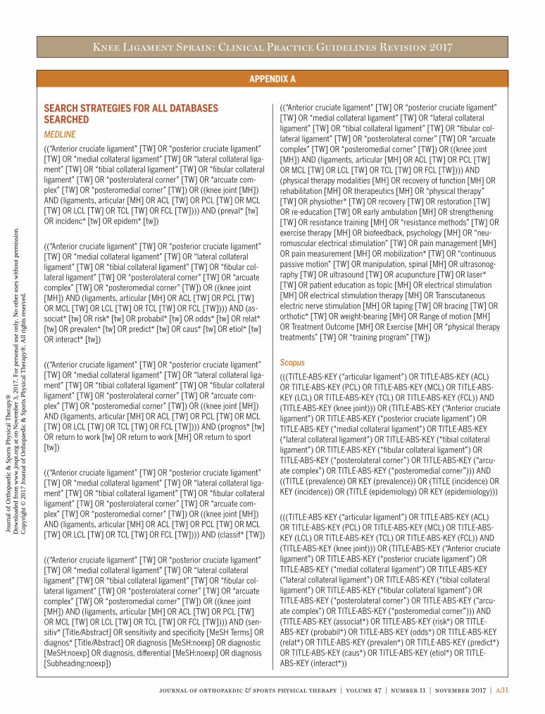
Knee Ligament Sprain: Clinical Practice Guidelines Revision 2017
journal of orthopaedic & sports physical therapy | volume 47 | number 11 | november 2017 | a31
APPENDIX A
SEARCH STRATEGIES FOR ALL DATABASES SEARCHEDMEDLINE
((“Anterior cruciate ligament” [TW] OR “posterior cruciate ligament” [TW] OR “medial collateral ligament” [TW] OR “lateral collateral liga-ment” [TW] OR “tibial collateral ligament” [TW] OR “fibular collateral ligament” [TW] OR “posterolateral corner” [TW] OR “arcuate com-plex” [TW] OR “posteromedial corner” [TW]) OR ((knee joint [MH]) AND (ligaments, articular [MH] OR ACL [TW] OR PCL [TW] OR MCL [TW] OR LCL [TW] OR TCL [TW] OR FCL [TW]))) AND (preval* [tw] OR incidenc* [tw] OR epidem* [tw])
((“Anterior cruciate ligament” [TW] OR “posterior cruciate ligament” [TW] OR “medial collateral ligament” [TW] OR “lateral collateral ligament” [TW] OR “tibial collateral ligament” [TW] OR “fibular col-lateral ligament” [TW] OR “posterolateral corner” [TW] OR “arcuate complex” [TW] OR “posteromedial corner” [TW]) OR ((knee joint [MH]) AND (ligaments, articular [MH] OR ACL [TW] OR PCL [TW] OR MCL [TW] OR LCL [TW] OR TCL [TW] OR FCL [TW]))) AND (as-sociat* [tw] OR risk* [tw] OR probabil* [tw] OR odds* [tw] OR relat* [tw] OR prevalen* [tw] OR predict* [tw] OR caus* [tw] OR etiol* [tw] OR interact* [tw])
((“Anterior cruciate ligament” [TW] OR “posterior cruciate ligament” [TW] OR “medial collateral ligament” [TW] OR “lateral collateral liga-ment” [TW] OR “tibial collateral ligament” [TW] OR “fibular collateral ligament” [TW] OR “posterolateral corner” [TW] OR “arcuate com-plex” [TW] OR “posteromedial corner” [TW]) OR ((knee joint [MH]) AND (ligaments, articular [MH] OR ACL [TW] OR PCL [TW] OR MCL [TW] OR LCL [TW] OR TCL [TW] OR FCL [TW]))) AND (prognos* [tw] OR return to work [tw] OR return to work [MH] OR return to sport [tw])
((“Anterior cruciate ligament” [TW] OR “posterior cruciate ligament” [TW] OR “medial collateral ligament” [TW] OR “lateral collateral liga-ment” [TW] OR “tibial collateral ligament” [TW] OR “fibular collateral ligament” [TW] OR “posterolateral corner” [TW] OR “arcuate com-plex” [TW] OR “posteromedial corner” [TW]) OR ((knee joint [MH]) AND (ligaments, articular [MH] OR ACL [TW] OR PCL [TW] OR MCL [TW] OR LCL [TW] OR TCL [TW] OR FCL [TW]))) AND (classif* [TW])
((“Anterior cruciate ligament” [TW] OR “posterior cruciate ligament” [TW] OR “medial collateral ligament” [TW] OR “lateral collateral ligament” [TW] OR “tibial collateral ligament” [TW] OR “fibular col-lateral ligament” [TW] OR “posterolateral corner” [TW] OR “arcuate complex” [TW] OR “posteromedial corner” [TW]) OR ((knee joint [MH]) AND (ligaments, articular [MH] OR ACL [TW] OR PCL [TW] OR MCL [TW] OR LCL [TW] OR TCL [TW] OR FCL [TW]))) AND (sen-sitiv* [Title/Abstract] OR sensitivity and specificity [MeSH Terms] OR diagnos* [Title/Abstract] OR diagnosis [MeSH:noexp] OR diagnostic [MeSH:noexp] OR diagnosis, differential [MeSH:noexp] OR diagnosis [Subheading:noexp])
((“Anterior cruciate ligament” [TW] OR “posterior cruciate ligament” [TW] OR “medial collateral ligament” [TW] OR “lateral collateral ligament” [TW] OR “tibial collateral ligament” [TW] OR “fibular col-lateral ligament” [TW] OR “posterolateral corner” [TW] OR “arcuate complex” [TW] OR “posteromedial corner” [TW]) OR ((knee joint [MH]) AND (ligaments, articular [MH] OR ACL [TW] OR PCL [TW] OR MCL [TW] OR LCL [TW] OR TCL [TW] OR FCL [TW]))) AND (physical therapy modalities [MH] OR recovery of function [MH] OR rehabilitation [MH] OR therapeutics [MH] OR “physical therapy” [TW] OR physiother* [TW] OR recovery [TW] OR restoration [TW] OR re-education [TW] OR early ambulation [MH] OR strengthening [TW] OR resistance training [MH] OR “resistance methods” [TW] OR exercise therapy [MH] OR biofeedback, psychology [MH] OR “neu-romuscular electrical stimulation” [TW] OR pain management [MH] OR pain measurement [MH] OR mobilization* [TW] OR “continuous passive motion” [TW] OR manipulation, spinal [MH] OR ultrasonog-raphy [TW] OR ultrasound [TW] OR acupuncture [TW] OR laser* [TW] OR patient education as topic [MH] OR electrical stimulation [MH] OR electrical stimulation therapy [MH] OR Transcutaneous electric nerve stimulation [MH] OR taping [TW] OR bracing [TW] OR orthotic* [TW] OR weight-bearing [MH] OR Range of motion [MH] OR Treatment Outcome [MH] OR Exercise [MH] OR “physical therapy treatments” [TW] OR “training program” [TW])
Scopus
(((TITLE-ABS-KEY (“articular ligament”) OR TITLE-ABS-KEY (ACL) OR TITLE-ABS-KEY (PCL) OR TITLE-ABS-KEY (MCL) OR TITLE-ABS-KEY (LCL) OR TITLE-ABS-KEY (TCL) OR TITLE-ABS-KEY (FCL)) AND (TITLE-ABS-KEY (knee joint))) OR (TITLE-ABS-KEY (“Anterior cruciate ligament”) OR TITLE-ABS-KEY (“posterior cruciate ligament”) OR TITLE-ABS-KEY (“medial collateral ligament”) OR TITLE-ABS-KEY (“lateral collateral ligament”) OR TITLE-ABS-KEY (“tibial collateral ligament”) OR TITLE-ABS-KEY (“fibular collateral ligament”) OR TITLE-ABS-KEY (“posterolateral corner”) OR TITLE-ABS-KEY (“arcu-ate complex”) OR TITLE-ABS-KEY (“posteromedial corner”))) AND ((TITLE (prevalence) OR KEY (prevalence)) OR (TITLE (incidence) OR KEY (incidence)) OR (TITLE (epidemiology) OR KEY (epidemiology)))
(((TITLE-ABS-KEY (“articular ligament”) OR TITLE-ABS-KEY (ACL) OR TITLE-ABS-KEY (PCL) OR TITLE-ABS-KEY (MCL) OR TITLE-ABS-KEY (LCL) OR TITLE-ABS-KEY (TCL) OR TITLE-ABS-KEY (FCL)) AND (TITLE-ABS-KEY (knee joint))) OR (TITLE-ABS-KEY (“Anterior cruciate ligament”) OR TITLE-ABS-KEY (“posterior cruciate ligament”) OR TITLE-ABS-KEY (“medial collateral ligament”) OR TITLE-ABS-KEY (“lateral collateral ligament”) OR TITLE-ABS-KEY (“tibial collateral ligament”) OR TITLE-ABS-KEY (“fibular collateral ligament”) OR TITLE-ABS-KEY (“posterolateral corner”) OR TITLE-ABS-KEY (“arcu-ate complex”) OR TITLE-ABS-KEY (“posteromedial corner”))) AND (TITLE-ABS-KEY (associat*) OR TITLE-ABS-KEY (risk*) OR TITLE-ABS-KEY (probabil*) OR TITLE-ABS-KEY (odds*) OR TITLE-ABS-KEY (relat*) OR TITLE-ABS-KEY (prevalen*) OR TITLE-ABS-KEY (predict*) OR TITLE-ABS-KEY (caus*) OR TITLE-ABS-KEY (etiol*) OR TITLE-ABS-KEY (interact*))
47-11 CPG Knee 3.indd 31 10/18/2017 2:26:13 PM
Jou
rnal
of
Ort
hopa
edic
& S
port
s Ph
ysic
al T
hera
py®
D
ownl
oade
d fr
om w
ww
.josp
t.org
at o
n N
ovem
ber
3, 2
017.
For
per
sona
l use
onl
y. N
o ot
her
uses
with
out p
erm
issi
on.
Cop
yrig
ht ©
201
7 Jo
urna
l of
Ort
hopa
edic
& S
port
s Ph
ysic
al T
hera
py®
. All
righ
ts r
eser
ved.
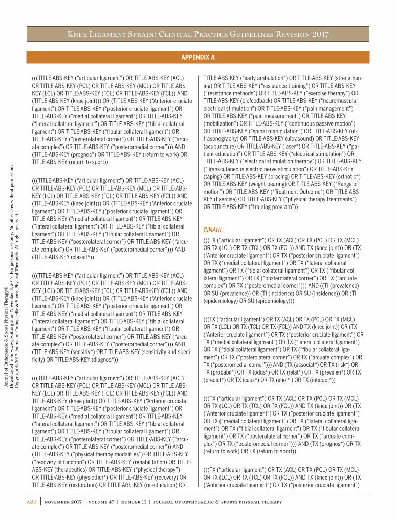
Knee Ligament Sprain: Clinical Practice Guidelines Revision 2017
a32 | november 2017 | volume 47 | number 11 | journal of orthopaedic & sports physical therapy
APPENDIX A
(((TITLE-ABS-KEY (“articular ligament”) OR TITLE-ABS-KEY (ACL) OR TITLE-ABS-KEY (PCL) OR TITLE-ABS-KEY (MCL) OR TITLE-ABS-KEY (LCL) OR TITLE-ABS-KEY (TCL) OR TITLE-ABS-KEY (FCL)) AND (TITLE-ABS-KEY (knee joint))) OR (TITLE-ABS-KEY (“Anterior cruciate ligament”) OR TITLE-ABS-KEY (“posterior cruciate ligament”) OR TITLE-ABS-KEY (“medial collateral ligament”) OR TITLE-ABS-KEY (“lateral collateral ligament”) OR TITLE-ABS-KEY (“tibial collateral ligament”) OR TITLE-ABS-KEY (“fibular collateral ligament”) OR TITLE-ABS-KEY (“posterolateral corner”) OR TITLE-ABS-KEY (“arcu-ate complex”) OR TITLE-ABS-KEY (“posteromedial corner”))) AND (TITLE-ABS-KEY (prognos*) OR TITLE-ABS-KEY (return to work) OR TITLE-ABS-KEY (return to sport))
(((TITLE-ABS-KEY (“articular ligament”) OR TITLE-ABS-KEY (ACL) OR TITLE-ABS-KEY (PCL) OR TITLE-ABS-KEY (MCL) OR TITLE-ABS-KEY (LCL) OR TITLE-ABS-KEY (TCL) OR TITLE-ABS-KEY (FCL)) AND (TITLE-ABS-KEY (knee joint))) OR (TITLE-ABS-KEY (“Anterior cruciate ligament”) OR TITLE-ABS-KEY (“posterior cruciate ligament”) OR TITLE-ABS-KEY (“medial collateral ligament”) OR TITLE-ABS-KEY (“lateral collateral ligament”) OR TITLE-ABS-KEY (“tibial collateral ligament”) OR TITLE-ABS-KEY (“fibular collateral ligament”) OR TITLE-ABS-KEY (“posterolateral corner”) OR TITLE-ABS-KEY (“arcu-ate complex”) OR TITLE-ABS-KEY (“posteromedial corner”))) AND (TITLE-ABS-KEY (classif*))
(((TITLE-ABS-KEY (“articular ligament”) OR TITLE-ABS-KEY (ACL) OR TITLE-ABS-KEY (PCL) OR TITLE-ABS-KEY (MCL) OR TITLE-ABS-KEY (LCL) OR TITLE-ABS-KEY (TCL) OR TITLE-ABS-KEY (FCL)) AND (TITLE-ABS-KEY (knee joint))) OR (TITLE-ABS-KEY (“Anterior cruciate ligament”) OR TITLE-ABS-KEY (“posterior cruciate ligament”) OR TITLE-ABS-KEY (“medial collateral ligament”) OR TITLE-ABS-KEY (“lateral collateral ligament”) OR TITLE-ABS-KEY (“tibial collateral ligament”) OR TITLE-ABS-KEY (“fibular collateral ligament”) OR TITLE-ABS-KEY (“posterolateral corner”) OR TITLE-ABS-KEY (“arcu-ate complex”) OR TITLE-ABS-KEY (“posteromedial corner”))) AND (TITLE-ABS-KEY (sensitiv*) OR TITLE-ABS-KEY (sensitivity and speci-ficity) OR TITLE-ABS-KEY (diagnos*))
(((TITLE-ABS-KEY (“articular ligament”) OR TITLE-ABS-KEY (ACL) OR TITLE-ABS-KEY (PCL) OR TITLE-ABS-KEY (MCL) OR TITLE-ABS-KEY (LCL) OR TITLE-ABS-KEY (TCL) OR TITLE-ABS-KEY (FCL)) AND TITLE-ABS-KEY (knee joint)) OR TITLE-ABS-KEY (“Anterior cruciate ligament”) OR TITLE-ABS-KEY (“posterior cruciate ligament”) OR TITLE-ABS-KEY (“medial collateral ligament”) OR TITLE-ABS-KEY (“lateral collateral ligament”) OR TITLE-ABS-KEY (“tibial collateral ligament”) OR TITLE-ABS-KEY (“fibular collateral ligament”) OR TITLE-ABS-KEY (“posterolateral corner”) OR TITLE-ABS-KEY (“arcu-ate complex”) OR TITLE-ABS-KEY (“posteromedial corner”)) AND (TITLE-ABS-KEY (“physical therapy modalities”) OR TITLE-ABS-KEY (“recovery of function”) OR TITLE-ABS-KEY (rehabilitation) OR TITLE-ABS-KEY (therapeutics) OR TITLE-ABS-KEY (“physical therapy”) OR TITLE-ABS-KEY (physiother*) OR TITLE-ABS-KEY (recovery) OR TITLE-ABS-KEY (restoration) OR TITLE-ABS-KEY (re-education) OR
TITLE-ABS-KEY (“early ambulation”) OR TITLE-ABS-KEY (strengthen-ing) OR TITLE-ABS-KEY (“resistance training”) OR TITLE-ABS-KEY (“resistance methods”) OR TITLE-ABS-KEY (“exercise therapy”) OR TITLE-ABS-KEY (biofeedback) OR TITLE-ABS-KEY (“neuromuscular electrical stimulation”) OR TITLE-ABS-KEY (“pain management”) OR TITLE-ABS-KEY (“pain measurement”) OR TITLE-ABS-KEY (mobilization*) OR TITLE-ABS-KEY (“continuous passive motion”) OR TITLE-ABS-KEY (“spinal manipulation”) OR TITLE-ABS-KEY (ul-trasonography) OR TITLE-ABS-KEY (ultrasound) OR TITLE-ABS-KEY (acupuncture) OR TITLE-ABS-KEY (laser*) OR TITLE-ABS-KEY (“pa-tient education”) OR TITLE-ABS-KEY (“electrical stimulation”) OR TITLE-ABS-KEY (“electrical stimulation therapy”) OR TITLE-ABS-KEY (“Transcutaneous electric nerve stimulation”) OR TITLE-ABS-KEY (taping) OR TITLE-ABS-KEY (bracing) OR TITLE-ABS-KEY (orthotic*) OR TITLE-ABS-KEY (weight-bearing) OR TITLE-ABS-KEY (“Range of motion”) OR TITLE-ABS-KEY (“Treatment Outcome”) OR TITLE-ABS-KEY (Exercise) OR TITLE-ABS-KEY (“physical therapy treatments”) OR TITLE-ABS-KEY (“training program”))
CINAHL
(((TX (“articular ligament”) OR TX (ACL) OR TX (PCL) OR TX (MCL) OR TX (LCL) OR TX (TCL) OR TX (FCL)) AND TX (knee joint)) OR (TX (“Anterior cruciate ligament”) OR TX (“posterior cruciate ligament”) OR TX (“medial collateral ligament”) OR TX (“lateral collateral ligament”) OR TX (“tibial collateral ligament”) OR TX (“fibular col-lateral ligament”) OR TX (“posterolateral corner”) OR TX (“arcuate complex”) OR TX (“posteromedial corner”))) AND ((TI (prevalence) OR SU (prevalence)) OR (TI (incidence) OR SU (incidence)) OR (TI (epidemiology) OR SU (epidemiology)))
(((TX (“articular ligament”) OR TX (ACL) OR TX (PCL) OR TX (MCL) OR TX (LCL) OR TX (TCL) OR TX (FCL)) AND TX (knee joint)) OR (TX (“Anterior cruciate ligament”) OR TX (“posterior cruciate ligament”) OR TX (“medial collateral ligament”) OR TX (“lateral collateral ligament”) OR TX (“tibial collateral ligament”) OR TX (“fibular collateral liga-ment”) OR TX (“posterolateral corner”) OR TX (“arcuate complex”) OR TX (“posteromedial corner”))) AND (TX (associat*) OR TX (risk*) OR TX (probabil*) OR TX (odds*) OR TX (relat*) OR TX (prevalen*) OR TX (predict*) OR TX (caus*) OR TX (etiol* ) OR TX (interact*))
(((TX (“articular ligament”) OR TX (ACL) OR TX (PCL) OR TX (MCL) OR TX (LCL) OR TX (TCL) OR TX (FCL)) AND TX (knee joint)) OR (TX (“Anterior cruciate ligament”) OR TX (“posterior cruciate ligament”) OR TX (“medial collateral ligament”) OR TX (“lateral collateral liga-ment”) OR TX (“tibial collateral ligament”) OR TX (“fibular collateral ligament”) OR TX (“posterolateral corner”) OR TX (“arcuate com-plex”) OR TX (“posteromedial corner”))) AND (TX (prognos*) OR TX (return to work) OR TX (return to sport))
(((TX (“articular ligament”) OR TX (ACL) OR TX (PCL) OR TX (MCL) OR TX (LCL) OR TX (TCL) OR TX (FCL)) AND TX (knee joint)) OR (TX (“Anterior cruciate ligament”) OR TX (“posterior cruciate ligament”)
47-11 CPG Knee 3.indd 32 10/18/2017 2:26:13 PM
Jou
rnal
of
Ort
hopa
edic
& S
port
s Ph
ysic
al T
hera
py®
D
ownl
oade
d fr
om w
ww
.josp
t.org
at o
n N
ovem
ber
3, 2
017.
For
per
sona
l use
onl
y. N
o ot
her
uses
with
out p
erm
issi
on.
Cop
yrig
ht ©
201
7 Jo
urna
l of
Ort
hopa
edic
& S
port
s Ph
ysic
al T
hera
py®
. All
righ
ts r
eser
ved.
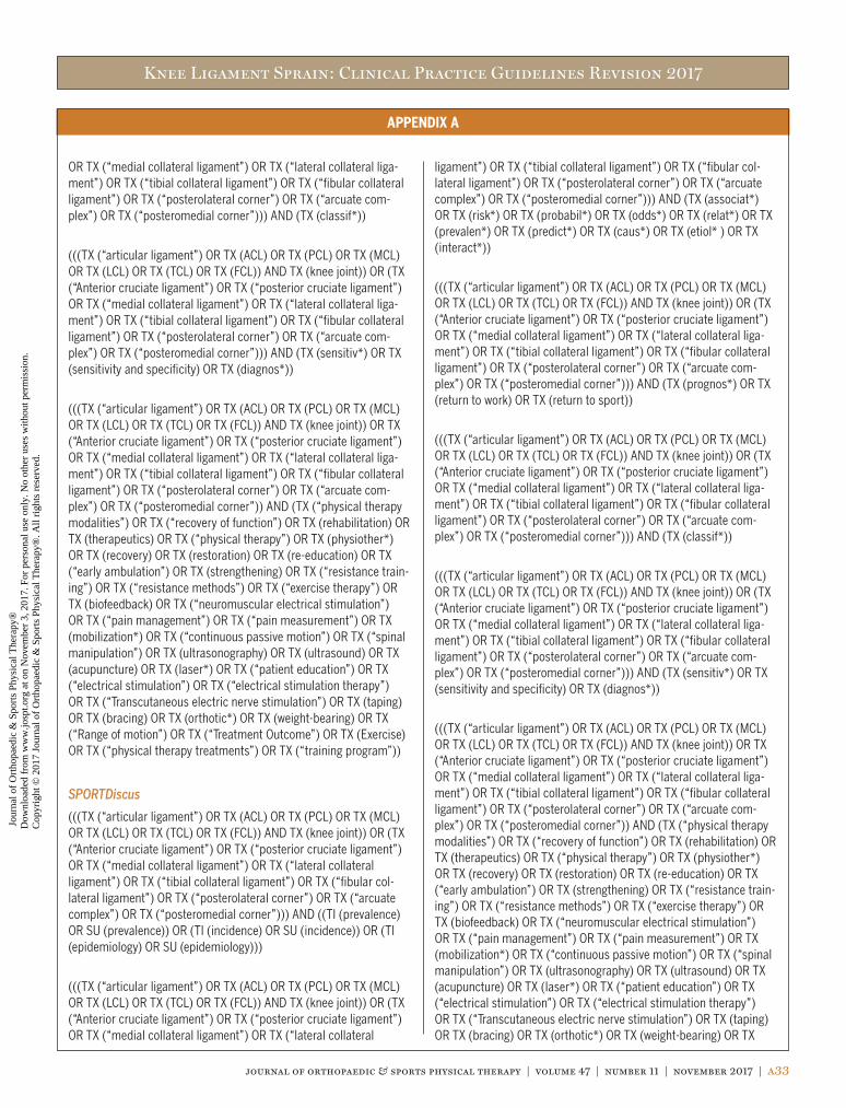
Knee Ligament Sprain: Clinical Practice Guidelines Revision 2017
journal of orthopaedic & sports physical therapy | volume 47 | number 11 | november 2017 | a33
APPENDIX A
OR TX (“medial collateral ligament”) OR TX (“lateral collateral liga-ment”) OR TX (“tibial collateral ligament”) OR TX (“fibular collateral ligament”) OR TX (“posterolateral corner”) OR TX (“arcuate com-plex”) OR TX (“posteromedial corner”))) AND (TX (classif*))
(((TX (“articular ligament”) OR TX (ACL) OR TX (PCL) OR TX (MCL) OR TX (LCL) OR TX (TCL) OR TX (FCL)) AND TX (knee joint)) OR (TX (“Anterior cruciate ligament”) OR TX (“posterior cruciate ligament”) OR TX (“medial collateral ligament”) OR TX (“lateral collateral liga-ment”) OR TX (“tibial collateral ligament”) OR TX (“fibular collateral ligament”) OR TX (“posterolateral corner”) OR TX (“arcuate com-plex”) OR TX (“posteromedial corner”))) AND (TX (sensitiv*) OR TX (sensitivity and specificity) OR TX (diagnos*))
(((TX (“articular ligament”) OR TX (ACL) OR TX (PCL) OR TX (MCL) OR TX (LCL) OR TX (TCL) OR TX (FCL)) AND TX (knee joint)) OR TX (“Anterior cruciate ligament”) OR TX (“posterior cruciate ligament”) OR TX (“medial collateral ligament”) OR TX (“lateral collateral liga-ment”) OR TX (“tibial collateral ligament”) OR TX (“fibular collateral ligament”) OR TX (“posterolateral corner”) OR TX (“arcuate com-plex”) OR TX (“posteromedial corner”)) AND (TX (“physical therapy modalities”) OR TX (“recovery of function”) OR TX (rehabilitation) OR TX (therapeutics) OR TX (“physical therapy”) OR TX (physiother*) OR TX (recovery) OR TX (restoration) OR TX (re-education) OR TX (“early ambulation”) OR TX (strengthening) OR TX (“resistance train-ing”) OR TX (“resistance methods”) OR TX (“exercise therapy”) OR TX (biofeedback) OR TX (“neuromuscular electrical stimulation”) OR TX (“pain management”) OR TX (“pain measurement”) OR TX (mobilization*) OR TX (“continuous passive motion”) OR TX (“spinal manipulation”) OR TX (ultrasonography) OR TX (ultrasound) OR TX (acupuncture) OR TX (laser*) OR TX (“patient education”) OR TX (“electrical stimulation”) OR TX (“electrical stimulation therapy”) OR TX (“Transcutaneous electric nerve stimulation”) OR TX (taping) OR TX (bracing) OR TX (orthotic*) OR TX (weight-bearing) OR TX (“Range of motion”) OR TX (“Treatment Outcome”) OR TX (Exercise) OR TX (“physical therapy treatments”) OR TX (“training program”))
SPORTDiscus
(((TX (“articular ligament”) OR TX (ACL) OR TX (PCL) OR TX (MCL) OR TX (LCL) OR TX (TCL) OR TX (FCL)) AND TX (knee joint)) OR (TX (“Anterior cruciate ligament”) OR TX (“posterior cruciate ligament”) OR TX (“medial collateral ligament”) OR TX (“lateral collateral ligament”) OR TX (“tibial collateral ligament”) OR TX (“fibular col-lateral ligament”) OR TX (“posterolateral corner”) OR TX (“arcuate complex”) OR TX (“posteromedial corner”))) AND ((TI (prevalence) OR SU (prevalence)) OR (TI (incidence) OR SU (incidence)) OR (TI (epidemiology) OR SU (epidemiology)))
(((TX (“articular ligament”) OR TX (ACL) OR TX (PCL) OR TX (MCL) OR TX (LCL) OR TX (TCL) OR TX (FCL)) AND TX (knee joint)) OR (TX (“Anterior cruciate ligament”) OR TX (“posterior cruciate ligament”) OR TX (“medial collateral ligament”) OR TX (“lateral collateral
ligament”) OR TX (“tibial collateral ligament”) OR TX (“fibular col-lateral ligament”) OR TX (“posterolateral corner”) OR TX (“arcuate complex”) OR TX (“posteromedial corner”))) AND (TX (associat*) OR TX (risk*) OR TX (probabil*) OR TX (odds*) OR TX (relat*) OR TX (prevalen*) OR TX (predict*) OR TX (caus*) OR TX (etiol* ) OR TX (interact*))
(((TX (“articular ligament”) OR TX (ACL) OR TX (PCL) OR TX (MCL) OR TX (LCL) OR TX (TCL) OR TX (FCL)) AND TX (knee joint)) OR (TX (“Anterior cruciate ligament”) OR TX (“posterior cruciate ligament”) OR TX (“medial collateral ligament”) OR TX (“lateral collateral liga-ment”) OR TX (“tibial collateral ligament”) OR TX (“fibular collateral ligament”) OR TX (“posterolateral corner”) OR TX (“arcuate com-plex”) OR TX (“posteromedial corner”))) AND (TX (prognos*) OR TX (return to work) OR TX (return to sport))
(((TX (“articular ligament”) OR TX (ACL) OR TX (PCL) OR TX (MCL) OR TX (LCL) OR TX (TCL) OR TX (FCL)) AND TX (knee joint)) OR (TX (“Anterior cruciate ligament”) OR TX (“posterior cruciate ligament”) OR TX (“medial collateral ligament”) OR TX (“lateral collateral liga-ment”) OR TX (“tibial collateral ligament”) OR TX (“fibular collateral ligament”) OR TX (“posterolateral corner”) OR TX (“arcuate com-plex”) OR TX (“posteromedial corner”))) AND (TX (classif*))
(((TX (“articular ligament”) OR TX (ACL) OR TX (PCL) OR TX (MCL) OR TX (LCL) OR TX (TCL) OR TX (FCL)) AND TX (knee joint)) OR (TX (“Anterior cruciate ligament”) OR TX (“posterior cruciate ligament”) OR TX (“medial collateral ligament”) OR TX (“lateral collateral liga-ment”) OR TX (“tibial collateral ligament”) OR TX (“fibular collateral ligament”) OR TX (“posterolateral corner”) OR TX (“arcuate com-plex”) OR TX (“posteromedial corner”))) AND (TX (sensitiv*) OR TX (sensitivity and specificity) OR TX (diagnos*))
(((TX (“articular ligament”) OR TX (ACL) OR TX (PCL) OR TX (MCL) OR TX (LCL) OR TX (TCL) OR TX (FCL)) AND TX (knee joint)) OR TX (“Anterior cruciate ligament”) OR TX (“posterior cruciate ligament”) OR TX (“medial collateral ligament”) OR TX (“lateral collateral liga-ment”) OR TX (“tibial collateral ligament”) OR TX (“fibular collateral ligament”) OR TX (“posterolateral corner”) OR TX (“arcuate com-plex”) OR TX (“posteromedial corner”)) AND (TX (“physical therapy modalities”) OR TX (“recovery of function”) OR TX (rehabilitation) OR TX (therapeutics) OR TX (“physical therapy”) OR TX (physiother*) OR TX (recovery) OR TX (restoration) OR TX (re-education) OR TX (“early ambulation”) OR TX (strengthening) OR TX (“resistance train-ing”) OR TX (“resistance methods”) OR TX (“exercise therapy”) OR TX (biofeedback) OR TX (“neuromuscular electrical stimulation”) OR TX (“pain management”) OR TX (“pain measurement”) OR TX (mobilization*) OR TX (“continuous passive motion”) OR TX (“spinal manipulation”) OR TX (ultrasonography) OR TX (ultrasound) OR TX (acupuncture) OR TX (laser*) OR TX (“patient education”) OR TX (“electrical stimulation”) OR TX (“electrical stimulation therapy”) OR TX (“Transcutaneous electric nerve stimulation”) OR TX (taping) OR TX (bracing) OR TX (orthotic*) OR TX (weight-bearing) OR TX
47-11 CPG Knee 3.indd 33 10/18/2017 2:26:14 PM
Jou
rnal
of
Ort
hopa
edic
& S
port
s Ph
ysic
al T
hera
py®
D
ownl
oade
d fr
om w
ww
.josp
t.org
at o
n N
ovem
ber
3, 2
017.
For
per
sona
l use
onl
y. N
o ot
her
uses
with
out p
erm
issi
on.
Cop
yrig
ht ©
201
7 Jo
urna
l of
Ort
hopa
edic
& S
port
s Ph
ysic
al T
hera
py®
. All
righ
ts r
eser
ved.
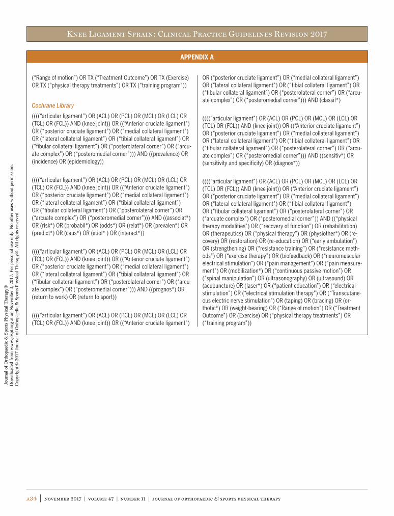
Knee Ligament Sprain: Clinical Practice Guidelines Revision 2017
a34 | november 2017 | volume 47 | number 11 | journal of orthopaedic & sports physical therapy
APPENDIX A
(“Range of motion”) OR TX (“Treatment Outcome”) OR TX (Exercise) OR TX (“physical therapy treatments”) OR TX (“training program”))
Cochrane Library
((((“articular ligament”) OR (ACL) OR (PCL) OR (MCL) OR (LCL) OR (TCL) OR (FCL)) AND (knee joint)) OR ((“Anterior cruciate ligament”) OR (“posterior cruciate ligament”) OR (“medial collateral ligament”) OR (“lateral collateral ligament”) OR (“tibial collateral ligament”) OR (“fibular collateral ligament”) OR (“posterolateral corner”) OR (“arcu-ate complex”) OR (“posteromedial corner”))) AND ((prevalence) OR (incidence) OR (epidemiology))
((((“articular ligament”) OR (ACL) OR (PCL) OR (MCL) OR (LCL) OR (TCL) OR (FCL)) AND (knee joint)) OR ((“Anterior cruciate ligament”) OR (“posterior cruciate ligament”) OR (“medial collateral ligament”) OR (“lateral collateral ligament”) OR (“tibial collateral ligament”) OR (“fibular collateral ligament”) OR (“posterolateral corner”) OR (“arcuate complex”) OR (“posteromedial corner”))) AND ((associat*) OR (risk*) OR (probabil*) OR (odds*) OR (relat*) OR (prevalen*) OR (predict*) OR (caus*) OR (etiol* ) OR (interact*))
((((“articular ligament”) OR (ACL) OR (PCL) OR (MCL) OR (LCL) OR (TCL) OR (FCL)) AND (knee joint)) OR ((“Anterior cruciate ligament”) OR (“posterior cruciate ligament”) OR (“medial collateral ligament”) OR (“lateral collateral ligament”) OR (“tibial collateral ligament”) OR (“fibular collateral ligament”) OR (“posterolateral corner”) OR (“arcu-ate complex”) OR (“posteromedial corner”))) AND ((prognos*) OR (return to work) OR (return to sport))
((((“articular ligament”) OR (ACL) OR (PCL) OR (MCL) OR (LCL) OR (TCL) OR (FCL)) AND (knee joint)) OR ((“Anterior cruciate ligament”)
OR (“posterior cruciate ligament”) OR (“medial collateral ligament”) OR (“lateral collateral ligament”) OR (“tibial collateral ligament”) OR (“fibular collateral ligament”) OR (“posterolateral corner”) OR (“arcu-ate complex”) OR (“posteromedial corner”))) AND (classif*)
((((“articular ligament”) OR (ACL) OR (PCL) OR (MCL) OR (LCL) OR (TCL) OR (FCL)) AND (knee joint)) OR ((“Anterior cruciate ligament”) OR (“posterior cruciate ligament”) OR (“medial collateral ligament”) OR (“lateral collateral ligament”) OR (“tibial collateral ligament”) OR (“fibular collateral ligament”) OR (“posterolateral corner”) OR (“arcu-ate complex”) OR (“posteromedial corner”))) AND ((sensitiv*) OR (sensitivity and specificity) OR (diagnos*))
((((“articular ligament”) OR (ACL) OR (PCL) OR (MCL) OR (LCL) OR (TCL) OR (FCL)) AND (knee joint)) OR (“Anterior cruciate ligament”) OR (“posterior cruciate ligament”) OR (“medial collateral ligament”) OR (“lateral collateral ligament”) OR (“tibial collateral ligament”) OR (“fibular collateral ligament”) OR (“posterolateral corner”) OR (“arcuate complex”) OR (“posteromedial corner”)) AND ((“physical therapy modalities”) OR (“recovery of function”) OR (rehabilitation) OR (therapeutics) OR (“physical therapy”) OR (physiother*) OR (re-covery) OR (restoration) OR (re-education) OR (“early ambulation”) OR (strengthening) OR (“resistance training”) OR (“resistance meth-ods”) OR (“exercise therapy”) OR (biofeedback) OR (“neuromuscular electrical stimulation”) OR (“pain management”) OR (“pain measure-ment”) OR (mobilization*) OR (“continuous passive motion”) OR (“spinal manipulation”) OR (ultrasonography) OR (ultrasound) OR (acupuncture) OR (laser*) OR (“patient education”) OR (“electrical stimulation”) OR (“electrical stimulation therapy”) OR (“Transcutane-ous electric nerve stimulation”) OR (taping) OR (bracing) OR (or-thotic*) OR (weight-bearing) OR (“Range of motion”) OR (“Treatment Outcome”) OR (Exercise) OR (“physical therapy treatments”) OR (“training program”))
47-11 CPG Knee 3.indd 34 10/18/2017 2:26:14 PM
Jou
rnal
of
Ort
hopa
edic
& S
port
s Ph
ysic
al T
hera
py®
D
ownl
oade
d fr
om w
ww
.josp
t.org
at o
n N
ovem
ber
3, 2
017.
For
per
sona
l use
onl
y. N
o ot
her
uses
with
out p
erm
issi
on.
Cop
yrig
ht ©
201
7 Jo
urna
l of
Ort
hopa
edic
& S
port
s Ph
ysic
al T
hera
py®
. All
righ
ts r
eser
ved.
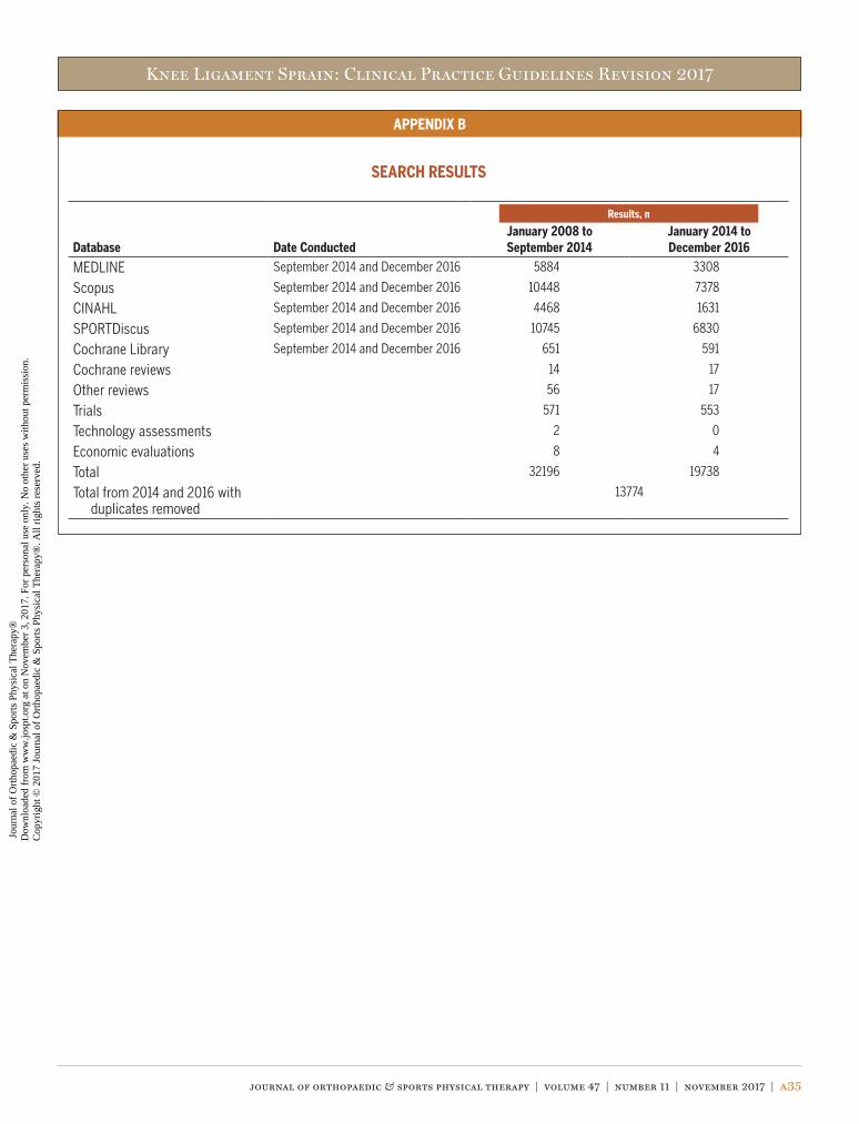
Knee Ligament Sprain: Clinical Practice Guidelines Revision 2017
journal of orthopaedic & sports physical therapy | volume 47 | number 11 | november 2017 | a35
APPENDIX B
SEARCH RESULTS
Database Date ConductedJanuary 2008 to September 2014
January 2014 to December 2016
MEDLINE September 2014 and December 2016 5884 3308
Scopus September 2014 and December 2016 10448 7378
CINAHL September 2014 and December 2016 4468 1631
SPORTDiscus September 2014 and December 2016 10745 6830
Cochrane Library September 2014 and December 2016 651 591
Cochrane reviews 14 17
Other reviews 56 17
Trials 571 553
Technology assessments 2 0
Economic evaluations 8 4
Total 32196 19738
Total from 2014 and 2016 with duplicates removed
13774
Results, n
47-11 CPG Knee 3.indd 35 10/18/2017 2:26:14 PM
Jou
rnal
of
Ort
hopa
edic
& S
port
s Ph
ysic
al T
hera
py®
D
ownl
oade
d fr
om w
ww
.josp
t.org
at o
n N
ovem
ber
3, 2
017.
For
per
sona
l use
onl
y. N
o ot
her
uses
with
out p
erm
issi
on.
Cop
yrig
ht ©
201
7 Jo
urna
l of
Ort
hopa
edic
& S
port
s Ph
ysic
al T
hera
py®
. All
righ
ts r
eser
ved.
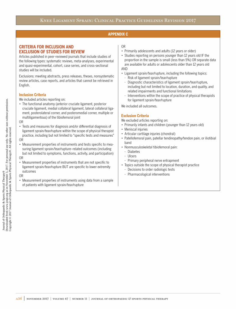
Knee Ligament Sprain: Clinical Practice Guidelines Revision 2017
a36 | november 2017 | volume 47 | number 11 | journal of orthopaedic & sports physical therapy
APPENDIX C
CRITERIA FOR INCLUSION AND EXCLUSION OF STUDIES FOR REVIEWArticles published in peer-reviewed journals that include studies of the following types: systematic reviews, meta-analyses, experimental and quasi-experimental, cohort, case series, and cross-sectional studies will be included.
Exclusions: meeting abstracts, press releases, theses, nonsystematic review articles, case reports, and articles that cannot be retrieved in English.
Inclusion CriteriaWe included articles reporting on:• The functional anatomy (anterior cruciate ligament, posterior
cruciate ligament, medial collateral ligament, lateral collateral liga-ment, posterolateral corner, and posteromedial corner, multiple or multiligamentous) of the tibiofemoral joint
OR• Tests and measures for diagnosis and/or differential diagnosis of
ligament sprain/tear/rupture within the scope of physical therapist practice, including but not limited to “specific tests and measures”
OR• Measurement properties of instruments and tests specific to mea-
suring ligament sprain/tear/rupture–related outcomes (including but not limited to symptoms, functions, activity, and participation)
OR• Measurement properties of instruments that are not specific to
ligament sprain/tear/rupture BUT are specific to lower extremity outcomes
OR• Measurement properties of instruments using data from a sample
of patients with ligament sprain/tear/rupture
OR• Primarily adolescents and adults (12 years or older)• Studies reporting on persons younger than 12 years old IF the
proportion in the sample is small (less than 5%) OR separate data are available for adults or adolescents older than 12 years old
AND• Ligament sprain/tear/rupture, including the following topics:
- Risk of ligament sprain/tear/rupture- Diagnostic characteristics of ligament sprain/tear/rupture,
including but not limited to location, duration, and quality, and related impairments and functional limitations
- Interventions within the scope of practice of physical therapists for ligament sprain/tear/rupture
We included all outcomes.
Exclusion CriteriaWe excluded articles reporting on:• Primarily infants and children (younger than 12 years old)• Meniscal injuries• Articular cartilage injuries (chondral)• Patellofemoral pain, patellar tendinopathy/tendon pain, or iliotibial
band• Nonmusculoskeletal tibiofemoral pain:
- Diabetes- Ulcers- Primary peripheral nerve entrapment
• Topics outside the scope of physical therapist practice- Decisions to order radiologic tests- Pharmacological interventions
47-11 CPG Knee 3.indd 36 10/18/2017 2:26:14 PM
Jou
rnal
of
Ort
hopa
edic
& S
port
s Ph
ysic
al T
hera
py®
D
ownl
oade
d fr
om w
ww
.josp
t.org
at o
n N
ovem
ber
3, 2
017.
For
per
sona
l use
onl
y. N
o ot
her
uses
with
out p
erm
issi
on.
Cop
yrig
ht ©
201
7 Jo
urna
l of
Ort
hopa
edic
& S
port
s Ph
ysic
al T
hera
py®
. All
righ
ts r
eser
ved.
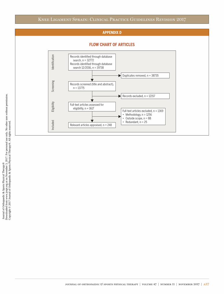
Knee Ligament Sprain: Clinical Practice Guidelines Revision 2017
journal of orthopaedic & sports physical therapy | volume 47 | number 11 | november 2017 | a37
APPENDIX D
FLOW CHART OF ARTICLES
Records identified through database search, n = 32772
Records identified through database search 12/2016, n = 19738
Records screened (title and abstract), n = 13775
Duplicates removed, n = 38735
Full-text articles assessed for eligibility, n = 1617
Records excluded, n = 12157
Relevant articles appraised, n = 248
Full-text articles excluded, n = 1369• Methodology, n = 1256• Outside scope, n = 88• Redundant, n = 25
Iden
tifica
tion
Elig
ibili
tySc
reen
ing
Incl
uded
47-11 CPG Knee 3.indd 37 10/18/2017 2:26:14 PM
Jou
rnal
of
Ort
hopa
edic
& S
port
s Ph
ysic
al T
hera
py®
D
ownl
oade
d fr
om w
ww
.josp
t.org
at o
n N
ovem
ber
3, 2
017.
For
per
sona
l use
onl
y. N
o ot
her
uses
with
out p
erm
issi
on.
Cop
yrig
ht ©
201
7 Jo
urna
l of
Ort
hopa
edic
& S
port
s Ph
ysic
al T
hera
py®
. All
righ
ts r
eser
ved.
Knee Ligament Sprain: Clinical Practice Guidelines Revision 2017
a38 | november 2017 | volume 47 | number 11 | journal of orthopaedic & sports physical therapy
APPENDIX E
ARTICLES INCLUDED IN RECOMMENDATIONS
DIAGNOSIS/CLASSIFICATION: DIFFERENTIAL DIAGNOSIS
ExaminationOutcome Measures – Activity Limitations: Self-ReportedAlmangoush A, Herrington L, Attia I, et al. Cross-cultural adaptation,
reliability, internal consistency and validation of the Arabic ver-sion of the Knee injury and Osteoarthritis Outcome Score (KOOS) for Egyptian people with knee injuries. Osteoarthritis Cartilage. 2013;21:1855-1864. https://doi.org/10.1016/j.joca.2013.09.010
Ardern CL, Taylor NF, Feller JA, Webster KE. A systematic review of the psychological factors associated with returning to sport fol-lowing injury. Br J Sports Med. 2013;47:1120-1126. https://doi.org/10.1136/bjsports-2012-091203
Bohu Y, Klouche S, Lefevre N, Webster K, Herman S. Translation, cross-cultural adaptation and validation of the French version of the Anterior Cruciate Ligament-Return to Sport after Injury (ACL-RSI) scale. Knee Surg Sports Traumatol Arthrosc. 2015;23:1192-1196. https://doi.org/10.1007/s00167-014-2942-4
Briggs KK, Lysholm J, Tegner Y, Rodkey WG, Kocher MS, Steadman JR. The reliability, validity, and responsiveness of the Lysholm score and Tegner activity scale for anterior cruciate ligament in-juries of the knee: 25 years later. Am J Sports Med. 2009;37:890-897. https://doi.org/10.1177/0363546508330143
Comins J, Brodersen J, Krogsgaard M, Beyer N. Rasch analysis of the Knee injury and Osteoarthritis Outcome Score (KOOS): a statisti-cal re-evaluation. Scand J Med Sci Sports. 2008;18:336-345. https://doi.org/10.1111/j.1600-0838.2007.00724.x
Della Villa F, Ricci M, Perdisa F, et al. Anterior cruciate ligament re-construction and rehabilitation: predictors of functional outcome. Joints. 2015;3:179-185. https://doi.org/10.11138/jts/2015.3.4.179
Dunn WR, Wolf BR, Harrell FE, Jr., et al. Baseline predictors of health-related quality of life after anterior cruciate ligament reconstruc-tion: a longitudinal analysis of a multicenter cohort at two and six years. J Bone Joint Surg Am. 2015;97:551-557. https://doi.org/10.2106/JBJS.N.00248
Filbay SR, Ackerman IN, Russell TG, Macri EM, Crossley KM. Health-related quality of life after anterior cruciate ligament reconstruc-tion: a systematic review. Am J Sports Med. 2014;42:1247-1255. https://doi.org/10.1177/0363546513512774
Flanigan DC, Everhart JS, Pedroza A, Smith T, Kaeding CC. Fear of reinjury (kinesiophobia) and persistent knee symptoms are com-mon factors for lack of return to sport after anterior cruciate liga-ment reconstruction. Arthroscopy. 2013;29:1322-1329. https://doi.org/10.1016/j.arthro.2013.05.015
Garratt AM, Brealey S, Robling M, et al. Development of the Knee Quality of Life (KQoL-26) 26-item questionnaire: data quality, reli-ability, validity and responsiveness. Health Qual Life Outcomes. 2008;6:48. https://doi.org/10.1186/1477-7525-6-48
George SZ, Lentz TA, Zeppieri G, Lee D, Chmielewski TL. Analysis of shortened versions of the Tampa Scale for Kinesiophobia and
Pain Catastrophizing Scale for patients after anterior cruciate ligament reconstruction. Clin J Pain. 2012;28:73-80. https://doi.org/10.1097/AJP.0b013e31822363f4
Granan LP, Baste V, Engebretsen L, Inacio MC. Associations between inadequate knee function detected by KOOS and prospective graft failure in an anterior cruciate ligament-reconstructed knee. Knee Surg Sports Traumatol Arthrosc. 2015;23:1135-1140. https://doi.org/10.1007/s00167-014-2925-5
Hambly K, Griva K. IKDC or KOOS: which one captures symp-toms and disabilities most important to patients who have undergone initial anterior cruciate ligament reconstruc-tion? Am J Sports Med. 2010;38:1395-1404. https://doi.org/10.1177/0363546509359678
Harput G, Tok D, Ulusoy B, et al. Translation and cross-cultural adaptation of the anterior cruciate ligament-return to sport after injury (ACL-RSI) scale into Turkish. Knee Surg Sports Traumatol Arthrosc. 2017;25:159-164. https://doi.org/10.1007/s00167-016-4288-6
Huang H, Zhang D, Jiang Y, et al. Translation, validation and cross-cultural adaptation of a simplified-Chinese version of the Tegner Activity Score in Chinese patients with anterior cruciate ligament injury. PLoS One. 2016;11:e0155463. https://doi.org/10.1371/journal.pone.0155463
Kvist J, Österberg A, Gauffin H, Tagesson S, Webster K, Ardern C. Translation and measurement properties of the Swed-ish version of ACL-Return to Sports after Injury question-naire. Scand J Med Sci Sports. 2013;23:568-575. https://doi.org/10.1111/j.1600-0838.2011.01438.x
Månsson O, Kartus J, Sernert N. Health-related quality of life after anterior cruciate ligament reconstruction. Knee Surg Sports Traumatol Arthrosc. 2011;19:479-487. https://doi.org/10.1007/s00167-010-1303-1
Månsson O, Kartus J, Sernert N. Pre-operative factors predicting good outcome in terms of health-related quality of life after ACL reconstruction. Scand J Med Sci Sports. 2013;23:15-22. https://doi.org/10.1111/j.1600-0838.2011.01426.x
Negahban H, Mostafaee N, Sohani SM, et al. Reliability and validity of the Tegner and Marx activity rating scales in Iranian patients with anterior cruciate ligament injury. Disabil Rehabil. 2011;33:2305-2310. https://doi.org/10.3109/09638288.2011.570409
Salavati M, Akhbari B, Mohammadi F, Mazaheri M, Khorrami M. Knee injury and Osteoarthritis Outcome Score (KOOS); reliability and validity in competitive athletes after anterior cruciate ligament reconstruction. Osteoarthritis Cartilage. 2011;19:406-410. https://doi.org/10.1016/j.joca.2011.01.010
Shelbourne KD, Barnes AF, Gray T. Correlation of a single assess-ment numeric evaluation (SANE) rating with modified Cincinnati Knee Rating System and IKDC subjective total scores for patients after ACL reconstruction or knee arthroscopy. Am J Sports Med. 2012;40:2487-2491. https://doi.org/10.1177/0363546512458576
Thomeé P, Währborg P, Börjesson M, Thomeé R, Eriksson BI, Karls-
47-11 CPG Knee 3.indd 38 10/18/2017 2:26:14 PM
Jou
rnal
of
Ort
hopa
edic
& S
port
s Ph
ysic
al T
hera
py®
D
ownl
oade
d fr
om w
ww
.josp
t.org
at o
n N
ovem
ber
3, 2
017.
For
per
sona
l use
onl
y. N
o ot
her
uses
with
out p
erm
issi
on.
Cop
yrig
ht ©
201
7 Jo
urna
l of
Ort
hopa
edic
& S
port
s Ph
ysic
al T
hera
py®
. All
righ
ts r
eser
ved.
Knee Ligament Sprain: Clinical Practice Guidelines Revision 2017
journal of orthopaedic & sports physical therapy | volume 47 | number 11 | november 2017 | a39
APPENDIX E
son J. Self-efficacy of knee function as a pre-operative predictor of outcome 1 year after anterior cruciate ligament reconstruction. Knee Surg Sports Traumatol Arthrosc. 2008;16:118-127. https://doi.org/10.1007/s00167-007-0433-6
van Meer BL, Meuffels DE, Vissers MM, et al. Knee injury and Osteo-arthritis Outcome Score or International Knee Documentation Committee Subjective Knee Form: which questionnaire is most useful to monitor patients with an anterior cruciate ligament rup-ture in the short term? Arthroscopy. 2013;29:701-715. https://doi.org/10.1016/j.arthro.2012.12.015
Wang W, Liu L, Chang X, Jia ZY, Zhao JZ, Xu WD. Cross-cultural trans-lation of the Lysholm knee score in Chinese and its validation in patients with anterior cruciate ligament injury. BMC Musculoskel-et Disord. 2016;17:436. https://doi.org/10.1186/s12891-016-1283-5
Webster KE, Feller JA. Use of the short form health surveys as an outcome measure for anterior cruciate ligament reconstruction. Knee Surg Sports Traumatol Arthrosc. 2014;22:1142-1148. https://doi.org/10.1007/s00167-013-2520-1
Physical Performance MeasuresAbrams GD, Harris JD, Gupta AK, et al. Functional performance test-
ing after anterior cruciate ligament reconstruction: a systematic review. Orthop J Sports Med. 2014;2:2325967113518305. https://doi.org/10.1177/2325967113518305
Grindem H, Logerstedt D, Eitzen I, et al. Single-legged hop tests as predictors of self-reported knee function in nonop-eratively treated individuals with anterior cruciate ligament injury. Am J Sports Med. 2011;39:2347-2354. https://doi.org/10.1177/0363546511417085
Hegedus EJ, McDonough S, Bleakley C, Cook CE, Baxter GD. Clini-cian-friendly lower extremity physical performance measures in athletes: a systematic review of measurement properties and correlation with injury, part 1. The tests for knee function includ-ing the hop tests. Br J Sports Med. 2015;49:642-648. https://doi.org/10.1136/bjsports-2014-094094
Logerstedt D, Grindem H, Lynch A, et al. Single-legged hop tests as predictors of self-reported knee function after anterior cruci-ate ligament reconstruction: the Delaware-Oslo ACL Cohort study. Am J Sports Med. 2012;40:2348-2356. https://doi.org/10.1177/0363546512457551
Reinke EK, Spindler KP, Lorring D, et al. Hop tests correlate with IKDC and KOOS at minimum of 2 years after primary ACL reconstruc-tion. Knee Surg Sports Traumatol Arthrosc. 2011;19:1806-1816. https://doi.org/10.1007/s00167-011-1473-5
Physical Impairment MeasuresAbrams GD, Harris JD, Gupta AK, et al. Functional performance test-
ing after anterior cruciate ligament reconstruction: a systematic review. Orthop J Sports Med. 2014;2:2325967113518305. https://doi.org/10.1177/2325967113518305
Décary S, Ouellet P, Vendittoli PA, Desmeules F. Reliability of physical examination tests for the diagnosis of knee disorders: evidence
from a systematic review. Man Ther. 2016;26:172-182. https://doi.org/10.1016/j.math.2016.09.007
Eitzen I, Holm I, Risberg MA. Preoperative quadriceps strength is a significant predictor of knee function two years after anterior cru-ciate ligament reconstruction. Br J Sports Med. 2009;43:371-376. https://doi.org/10.1136/bjsm.2008.057059
Gokeler A, Welling W, Zaffagnini S, Seil R, Padua D. Develop-ment of a test battery to enhance safe return to sports after anterior cruciate ligament reconstruction. Knee Surg Sports Traumatol Arthrosc. 2017;25:192-199. https://doi.org/10.1007/s00167-016-4246-3
Kopkow C, Freiberg A, Kirschner S, Seidler A, Schmitt J. Physical examination tests for the diagnosis of posterior cruciate liga-ment rupture: a systematic review. J Orthop Sports Phys Ther. 2013;43:804-813. https://doi.org/10.2519/jospt.2013.4906
Leblanc MC, Kowalczuk M, Andruszkiewicz N, et al. Diagnostic accuracy of physical examination for anterior knee instabil-ity: a systematic review. Knee Surg Sports Traumatol Arthrosc. 2015;23:2805-2813. https://doi.org/10.1007/s00167-015-3563-2
Lentz TA, Zeppieri G, Jr., Tillman SM, et al. Return to preinjury sports participation following anterior cruciate ligament reconstruction: contributions of demographic, knee impairment, and self-report measures. J Orthop Sports Phys Ther. 2012;42:893-901. https://doi.org/10.2519/jospt.2012.4077
Logerstedt D, Lynch A, Axe MJ, Snyder-Mackler L. Pre-operative quadriceps strength predicts IKDC2000 scores 6 months after anterior cruciate ligament reconstruction. Knee. 2013;20:208-212. https://doi.org/10.1016/j.knee.2012.07.011
Pietrosimone B, Lepley AS, Harkey MS, et al. Quadriceps strength predicts self-reported function post-ACL reconstruction. Med Sci Sports Exerc. 2016;48:1671-1677. https://doi.org/10.1249/MSS.0000000000000946
Swain MS, Henschke N, Kamper SJ, Downie AS, Koes BW, Maher CG. Accuracy of clinical tests in the diagnosis of anterior cruci-ate ligament injury: a systematic review. Chiropr Man Therap. 2014;22:25. https://doi.org/10.1186/s12998-014-0025-8
Ueda Y, Matsushita T, Araki D, et al. Factors affecting quadriceps strength recovery after anterior cruciate ligament reconstruc-tion with hamstring autografts in athletes. Knee Surg Sports Traumatol Arthrosc. 2017;25:3213-3219. https://doi.org/10.1007/s00167-016-4296-6
INTERVENTIONS
Continuous Passive MotionEarly Weight Bearing: Knee BracingKinikli Gİ, Callaghan MJ, Parkes MJ, Yüksel İ. Bracing after anterior
cruciate ligament reconstruction: systematic review and meta-analysis. Turkiye Klinikleri Spor Bilimleri. 2014;6:28-38.
Immediate Versus Delayed MobilizationCryotherapyMartimbianco AL, da Silva BN, de Carvalho AP, Silva V, Torloni
47-11 CPG Knee 3.indd 39 10/18/2017 2:26:14 PM
Jou
rnal
of
Ort
hopa
edic
& S
port
s Ph
ysic
al T
hera
py®
D
ownl
oade
d fr
om w
ww
.josp
t.org
at o
n N
ovem
ber
3, 2
017.
For
per
sona
l use
onl
y. N
o ot
her
uses
with
out p
erm
issi
on.
Cop
yrig
ht ©
201
7 Jo
urna
l of
Ort
hopa
edic
& S
port
s Ph
ysic
al T
hera
py®
. All
righ
ts r
eser
ved.
Knee Ligament Sprain: Clinical Practice Guidelines Revision 2017
a40 | november 2017 | volume 47 | number 11 | journal of orthopaedic & sports physical therapy
APPENDIX E
MR, Peccin MS. Effectiveness and safety of cryotherapy after arthroscopic anterior cruciate ligament reconstruction. A system-atic review of the literature. Phys Ther Sport. 2014;15:261-268. https://doi.org/10.1016/j.ptsp.2014.02.008
Supervised RehabilitationHohmann E, Tetsworth K, Bryant A. Physiotherapy-guided versus
home-based, unsupervised rehabilitation in isolated anterior cru-ciate injuries following surgical reconstruction. Knee Surg Sports Traumatol Arthrosc. 2011;19:1158-1167. https://doi.org/10.1007/s00167-010-1386-8
Papalia R, Vasta S, Tecame A, D’Adamio S, Maffulli N, Denaro V. Home-based vs supervised rehabilitation programs following knee surgery: a systematic review. Br Med Bull. 2013;108:55-72. https://doi.org/10.1093/bmb/ldt014
Therapeutic ExercisesGokeler A, Bisschop M, Benjaminse A, Myer GD, Eppinga P, Otten E.
Quadriceps function following ACL reconstruction and rehabilita-tion: implications for optimisation of current practices. Knee Surg Sports Traumatol Arthrosc. 2014;22:1163-1174. https://doi.org/10.1007/s00167-013-2577-x
Neuromuscular Electrical StimulationImoto AM, Peccin S, Almeida GJ, Saconato H, Atallah AN. Ef-
fectiveness of electrical stimulation on rehabilitation after ligament and meniscal injuries: a systematic review. São Paulo Med J. 2011;129:414-423. https://doi.org/10.1590/S1516-31802011000600008
Kim KM, Croy T, Hertel J, Saliba S. Effects of neuromuscular electri-cal stimulation after anterior cruciate ligament reconstruction on quadriceps strength, function, and patient-oriented outcomes: a systematic review. J Orthop Sports Phys Ther. 2010;40:383-391. https://doi.org/10.2519/jospt.2010.3184
Neuromuscular Re-educationGokeler A, Bisschop M, Benjaminse A, Myer GD, Eppinga P, Otten E.
Quadriceps function following ACL reconstruction and rehabilita-tion: implications for optimisation of current practices. Knee Surg Sports Traumatol Arthrosc. 2014;22:1163-1174. https://doi.org/10.1007/s00167-013-2577-x
Wasielewski NJ, Parker TM, Kotsko KM. Evaluation of electro-myographic biofeedback for the quadriceps femoris: a sys-tematic review. J Athl Train. 2011;46:543-554. https://doi.org/10.4085/1062-6050-46.5.543
Zech A, Hübscher M, Vogt L, Banzer W, Hänsel F, Pfeifer K. Neuro-muscular training for rehabilitation of sports injuries: a system-atic review. Med Sci Sports Exerc. 2009;41:1831-1841. https://doi.org/10.1249/MSS.0b013e3181a3cf0d
47-11 CPG Knee 3.indd 40 10/18/2017 2:26:14 PM
Jou
rnal
of
Ort
hopa
edic
& S
port
s Ph
ysic
al T
hera
py®
D
ownl
oade
d fr
om w
ww
.josp
t.org
at o
n N
ovem
ber
3, 2
017.
For
per
sona
l use
onl
y. N
o ot
her
uses
with
out p
erm
issi
on.
Cop
yrig
ht ©
201
7 Jo
urna
l of
Ort
hopa
edic
& S
port
s Ph
ysic
al T
hera
py®
. All
righ
ts r
eser
ved.
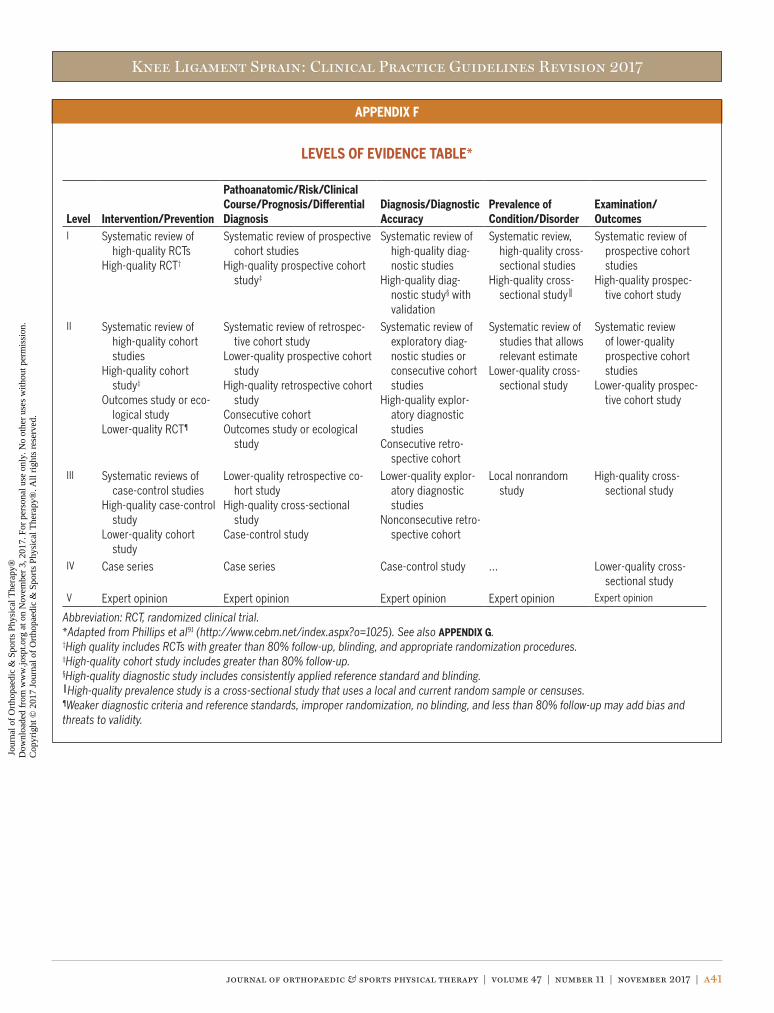
Knee Ligament Sprain: Clinical Practice Guidelines Revision 2017
journal of orthopaedic & sports physical therapy | volume 47 | number 11 | november 2017 | a41
APPENDIX F
LEVELS OF EVIDENCE TABLE*
Level Intervention/Prevention
Pathoanatomic/Risk/Clinical Course/Prognosis/Differential Diagnosis
Diagnosis/Diagnostic Accuracy
Prevalence of Condition/Disorder
Examination/Outcomes
I Systematic review of high-quality RCTs
High-quality RCT†
Systematic review of prospective cohort studies
High-quality prospective cohort study‡
Systematic review of high-quality diag-nostic studies
High-quality diag-nostic study§ with validation
Systematic review, high-quality cross-sectional studies
High-quality cross-sectional study║
Systematic review of prospective cohort studies
High-quality prospec-tive cohort study
II Systematic review of high-quality cohort studies
High-quality cohort study‡
Outcomes study or eco-logical study
Lower-quality RCT¶
Systematic review of retrospec-tive cohort study
Lower-quality prospective cohort study
High-quality retrospective cohort study
Consecutive cohortOutcomes study or ecological
study
Systematic review of exploratory diag-nostic studies or consecutive cohort studies
High-quality explor-atory diagnostic studies
Consecutive retro-spective cohort
Systematic review of studies that allows relevant estimate
Lower-quality cross-sectional study
Systematic review of lower-quality prospective cohort studies
Lower-quality prospec-tive cohort study
III Systematic reviews of case-control studies
High-quality case-control study
Lower-quality cohort study
Lower-quality retrospective co-hort study
High-quality cross-sectional study
Case-control study
Lower-quality explor-atory diagnostic studies
Nonconsecutive retro-spective cohort
Local nonrandom study
High-quality cross-sectional study
IV Case series Case series Case-control study … Lower-quality cross-sectional study
V Expert opinion Expert opinion Expert opinion Expert opinion Expert opinion
Abbreviation: RCT, randomized clinical trial.*Adapted from Phillips et al91 (http://www.cebm.net/index.aspx?o=1025). See also APPENDIX G.†High quality includes RCTs with greater than 80% follow-up, blinding, and appropriate randomization procedures.‡High-quality cohort study includes greater than 80% follow-up.§High-quality diagnostic study includes consistently applied reference standard and blinding.║High-quality prevalence study is a cross-sectional study that uses a local and current random sample or censuses.¶Weaker diagnostic criteria and reference standards, improper randomization, no blinding, and less than 80% follow-up may add bias and threats to validity.
47-11 CPG Knee 3.indd 41 10/18/2017 2:26:14 PM
Jou
rnal
of
Ort
hopa
edic
& S
port
s Ph
ysic
al T
hera
py®
D
ownl
oade
d fr
om w
ww
.josp
t.org
at o
n N
ovem
ber
3, 2
017.
For
per
sona
l use
onl
y. N
o ot
her
uses
with
out p
erm
issi
on.
Cop
yrig
ht ©
201
7 Jo
urna
l of
Ort
hopa
edic
& S
port
s Ph
ysic
al T
hera
py®
. All
righ
ts r
eser
ved.
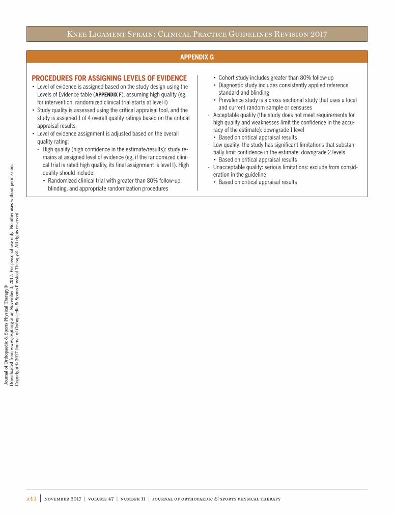
Knee Ligament Sprain: Clinical Practice Guidelines Revision 2017
a42 | november 2017 | volume 47 | number 11 | journal of orthopaedic & sports physical therapy
APPENDIX G
PROCEDURES FOR ASSIGNING LEVELS OF EVIDENCE• Level of evidence is assigned based on the study design using the
Levels of Evidence table (APPENDIX F), assuming high quality (eg, for intervention, randomized clinical trial starts at level I)
• Study quality is assessed using the critical appraisal tool, and the study is assigned 1 of 4 overall quality ratings based on the critical appraisal results
• Level of evidence assignment is adjusted based on the overall quality rating:- High quality (high confidence in the estimate/results): study re-
mains at assigned level of evidence (eg, if the randomized clini-cal trial is rated high quality, its final assignment is level I). High quality should include:• Randomized clinical trial with greater than 80% follow-up,
blinding, and appropriate randomization procedures
• Cohort study includes greater than 80% follow-up• Diagnostic study includes consistently applied reference
standard and blinding• Prevalence study is a cross-sectional study that uses a local
and current random sample or censuses- Acceptable quality (the study does not meet requirements for
high quality and weaknesses limit the confidence in the accu-racy of the estimate): downgrade 1 level• Based on critical appraisal results
- Low quality: the study has significant limitations that substan-tially limit confidence in the estimate: downgrade 2 levels• Based on critical appraisal results
- Unacceptable quality: serious limitations: exclude from consid-eration in the guideline• Based on critical appraisal results
47-11 CPG Knee 3.indd 42 10/18/2017 2:26:14 PM
Jou
rnal
of
Ort
hopa
edic
& S
port
s Ph
ysic
al T
hera
py®
D
ownl
oade
d fr
om w
ww
.josp
t.org
at o
n N
ovem
ber
3, 2
017.
For
per
sona
l use
onl
y. N
o ot
her
uses
with
out p
erm
issi
on.
Cop
yrig
ht ©
201
7 Jo
urna
l of
Ort
hopa
edic
& S
port
s Ph
ysic
al T
hera
py®
. All
righ
ts r
eser
ved.
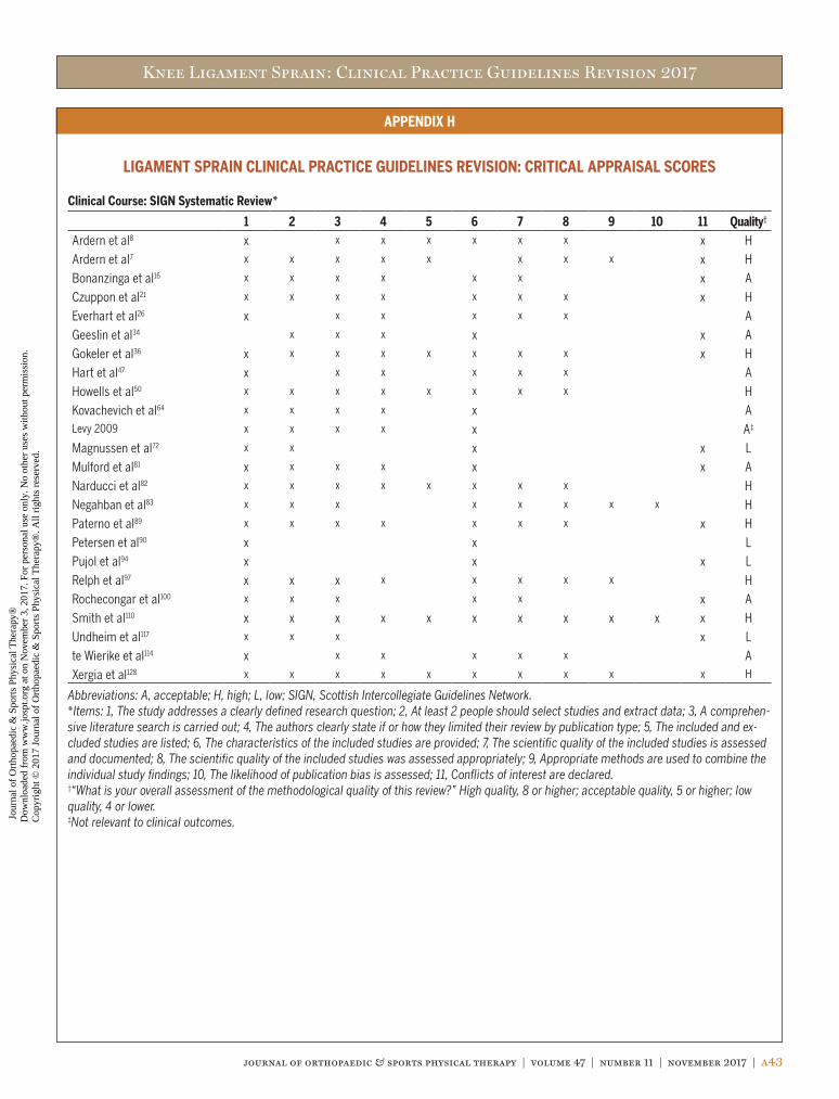
Knee Ligament Sprain: Clinical Practice Guidelines Revision 2017
journal of orthopaedic & sports physical therapy | volume 47 | number 11 | november 2017 | a43
APPENDIX H
LIGAMENT SPRAIN CLINICAL PRACTICE GUIDELINES REVISION: CRITICAL APPRAISAL SCORES
Clinical Course: SIGN Systematic Review*
1 2 3 4 5 6 7 8 9 10 11 Quality†
Ardern et al8 x x x x x x x x H
Ardern et al7 x x x x x x x x x H
Bonanzinga et al16 x x x x x x x A
Czuppon et al21 x x x x x x x x H
Everhart et al26 x x x x x x A
Geeslin et al34 x x x x x A
Gokeler et al36 x x x x x x x x x H
Hart et al47 x x x x x x A
Howells et al50 x x x x x x x x H
Kovachevich et al64 x x x x x ALevy 2009 x x x x x A‡
Magnussen et al72 x x x x L
Mulford et al81 x x x x x x A
Narducci et al82 x x x x x x x x H
Negahban et al83 x x x x x x x x H
Paterno et al89 x x x x x x x x H
Petersen et al90 x x L
Pujol et al94 x x x L
Relph et al97 x x x x x x x x H
Rochecongar et al100 x x x x x x A
Smith et al110 x x x x x x x x x x x H
Undheim et al117 x x x x L
te Wierike et al114 x x x x x x A
Xergia et al128 x x x x x x x x x x H
Abbreviations: A, acceptable; H, high; L, low; SIGN, Scottish Intercollegiate Guidelines Network.*Items: 1, The study addresses a clearly defined research question; 2, At least 2 people should select studies and extract data; 3, A comprehen-sive literature search is carried out; 4, The authors clearly state if or how they limited their review by publication type; 5, The included and ex-cluded studies are listed; 6, The characteristics of the included studies are provided; 7, The scientific quality of the included studies is assessed and documented; 8, The scientific quality of the included studies was assessed appropriately; 9, Appropriate methods are used to combine the individual study findings; 10, The likelihood of publication bias is assessed; 11, Conflicts of interest are declared.†“What is your overall assessment of the methodological quality of this review?” High quality, 8 or higher; acceptable quality, 5 or higher; low quality, 4 or lower.‡Not relevant to clinical outcomes.
47-11 CPG Knee 3.indd 43 10/18/2017 2:26:15 PM
Jou
rnal
of
Ort
hopa
edic
& S
port
s Ph
ysic
al T
hera
py®
D
ownl
oade
d fr
om w
ww
.josp
t.org
at o
n N
ovem
ber
3, 2
017.
For
per
sona
l use
onl
y. N
o ot
her
uses
with
out p
erm
issi
on.
Cop
yrig
ht ©
201
7 Jo
urna
l of
Ort
hopa
edic
& S
port
s Ph
ysic
al T
hera
py®
. All
righ
ts r
eser
ved.
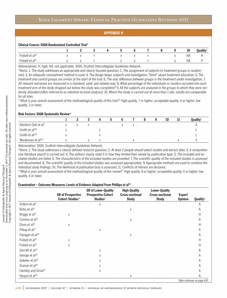
Knee Ligament Sprain: Clinical Practice Guidelines Revision 2017
a44 | november 2017 | volume 47 | number 11 | journal of orthopaedic & sports physical therapy
APPENDIX H
Clinical Course: SIGN Randomized Controlled Trial*
1 2 3 4 5 6 7 8 9 10 Quality†
Frobell et al30 x x x x x x x NA H
Frobell et al31 x x x x x x x NA H
Abbreviations: H, high; NA, not applicable; SIGN, Scottish Intercollegiate Guidelines Network.*Items: 1, The study addresses an appropriate and clearly focused question; 2, The assignment of subjects to treatment groups is random-ized; 3, An adequate concealment method is used; 4, The design keeps subjects and investigators “blind” about treatment allocation; 5, The treatment and control groups are similar at the start of the trial; 6, The only difference between groups is the treatment under investigation; 7, All relevant outcomes are measured in a standard, valid, and reliable way; 8, What percentage of the individuals or clusters recruited into each treatment arm of the study dropped out before the study was completed? 9, All the subjects are analyzed in the groups to which they were ran-domly allocated (often referred to as intention-to-treat analysis); 10, Where the study is carried out at more than 1 site, results are comparable for all sites.†“What is your overall assessment of the methodological quality of this trial?” High quality, 7 or higher; acceptable quality, 4 or higher; low quality, 3 or lower.
Risk Factors: SIGN Systematic Review*
1 2 3 4 5 6 7 8 9 10 11 Quality†
Alentorn-Geli et al3 x x x x x A
Smith et al108 x x L
Smith et al109 x x L
Wordeman et al124 x x x x x A
Abbreviation: SIGN, Scottish Intercollegiate Guidelines Network.*Items: 1, The study addresses a clearly defined research question; 2, At least 2 people should select studies and extract data; 3, A comprehen-sive literature search is carried out; 4, The authors clearly state if or how they limited their review by publication type; 5, The included and ex-cluded studies are listed; 6, The characteristics of the included studies are provided; 7, The scientific quality of the included studies is assessed and documented; 8, The scientific quality of the included studies was assessed appropriately; 9, Appropriate methods are used to combine the individual study findings; 10, The likelihood of publication bias is assessed; 11, Conflicts of interest are declared.†“What is your overall assessment of the methodological quality of this review?” High quality, 8 or higher; acceptable quality, 5 or higher; low quality, 4 or lower.
Examination – Outcome Measures: Levels of Evidence Adapted From Phillips et al91
SR of Prospective Cohort Studies*
SR of Lower-Quality Prospective Cohort
Studies†
High-Quality Cross-sectional
Study
Lower-Quality Cross-sectional
StudyExpert
Opinion Quality‡
Ardern et al9 x A
Bohu et al15 x A
Briggs et al17 x H
Comins et al19 x A
Dunn et al24 x H
Filbay et al27 x A
Flanigan et al29 x A
Frobell et al30 x H
Frobell et al31 x H
Garratt et al33 x A
George et al35 x A
Gokeler et al36 x A
Granan et al40 x A
Hambly and Griva45 x A
Harput et al46 x ATable continues on page A45.
47-11 CPG Knee 3.indd 44 10/18/2017 2:26:15 PM
Jou
rnal
of
Ort
hopa
edic
& S
port
s Ph
ysic
al T
hera
py®
D
ownl
oade
d fr
om w
ww
.josp
t.org
at o
n N
ovem
ber
3, 2
017.
For
per
sona
l use
onl
y. N
o ot
her
uses
with
out p
erm
issi
on.
Cop
yrig
ht ©
201
7 Jo
urna
l of
Ort
hopa
edic
& S
port
s Ph
ysic
al T
hera
py®
. All
righ
ts r
eser
ved.
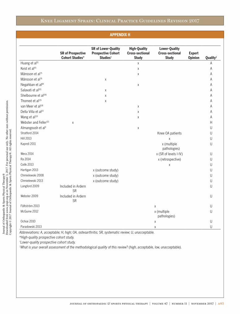
Knee Ligament Sprain: Clinical Practice Guidelines Revision 2017
journal of orthopaedic & sports physical therapy | volume 47 | number 11 | november 2017 | a45
APPENDIX H
SR of Prospective Cohort Studies*
SR of Lower-Quality Prospective Cohort
Studies†
High-Quality Cross-sectional
Study
Lower-Quality Cross-sectional
StudyExpert
Opinion Quality‡
Huang et al51 x A
Kvist et al65 x A
Månsson et al75 x A
Månsson et al76 x A
Negahban et al84 x A
Salavati et al101 x A
Shelbourne et al106 x A
Thomeé et al115 x A
van Meer et al118 x A
Della Villa et al23 x A
Wang et al119 x A
Webster and Feller121 x H
Almangoush et al4 x UStratford 2014 Knee OA patients UHill 2013 x UKapreli 2011 x (multiple
pathologies)U
Wera 2014 x (SR of levels I-IV) URa 2014 x (retrospective) UCelik 2013 x UHartigan 2013 x (outcome study) UChmielewski 2008 x (outcome study) UChmielewski 2013 x (outcome study) ULangford 2009 Included in Ardern
SRU
Webster 2009 Included in Ardern SR
U
Fältström 2013 x UMcGuine 2012 x (multiple
pathologies)U
Ochiai 2010 x UParadowski 2013 x U
Abbreviations: A, acceptable; H, high; OA, osteoarthritis; SR, systematic review; U, unacceptable.*High-quality prospective cohort study.†Lower-quality prospective cohort study.‡What is your overall assessment of the methodological quality of this review? (high, acceptable, low, unacceptable).
47-11 CPG Knee 3.indd 45 10/18/2017 2:26:15 PM
Jou
rnal
of
Ort
hopa
edic
& S
port
s Ph
ysic
al T
hera
py®
D
ownl
oade
d fr
om w
ww
.josp
t.org
at o
n N
ovem
ber
3, 2
017.
For
per
sona
l use
onl
y. N
o ot
her
uses
with
out p
erm
issi
on.
Cop
yrig
ht ©
201
7 Jo
urna
l of
Ort
hopa
edic
& S
port
s Ph
ysic
al T
hera
py®
. All
righ
ts r
eser
ved.
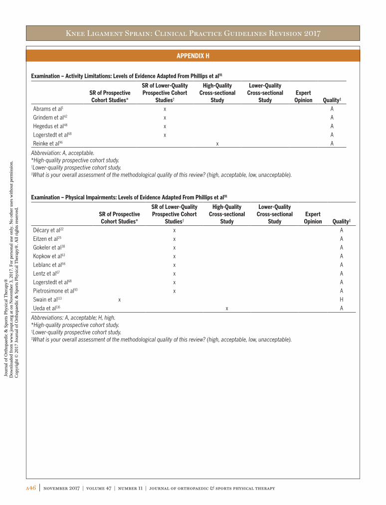
Knee Ligament Sprain: Clinical Practice Guidelines Revision 2017
a46 | november 2017 | volume 47 | number 11 | journal of orthopaedic & sports physical therapy
APPENDIX H
Examination – Activity Limitations: Levels of Evidence Adapted From Phillips et al91
SR of Prospective Cohort Studies*
SR of Lower-Quality Prospective Cohort
Studies†
High-Quality Cross-sectional
Study
Lower-Quality Cross-sectional
StudyExpert
Opinion Quality‡
Abrams et al1 x A
Grindem et al42 x A
Hegedus et al48 x A
Logerstedt et al68 x A
Reinke et al96 x A
Abbreviation: A, acceptable.*High-quality prospective cohort study.†Lower-quality prospective cohort study.‡What is your overall assessment of the methodological quality of this review? (high, acceptable, low, unacceptable).
Examination – Physical Impairments: Levels of Evidence Adapted From Phillips et al91
SR of Prospective Cohort Studies*
SR of Lower-Quality Prospective Cohort
Studies†
High-Quality Cross-sectional
Study
Lower-Quality Cross-sectional
StudyExpert
Opinion Quality‡
Décary et al22 x A
Eitzen et al25 x A
Gokeler et al38 x A
Kopkow et al63 x A
Leblanc et al66 x A
Lentz et al67 x A
Logerstedt et al68 x A
Pietrosimone et al93 x A
Swain et al113 x H
Ueda et al116 x A
Abbreviations: A, acceptable; H, high.*High-quality prospective cohort study.†Lower-quality prospective cohort study.‡What is your overall assessment of the methodological quality of this review? (high, acceptable, low, unacceptable).
47-11 CPG Knee 3.indd 46 10/18/2017 2:26:15 PM
Jou
rnal
of
Ort
hopa
edic
& S
port
s Ph
ysic
al T
hera
py®
D
ownl
oade
d fr
om w
ww
.josp
t.org
at o
n N
ovem
ber
3, 2
017.
For
per
sona
l use
onl
y. N
o ot
her
uses
with
out p
erm
issi
on.
Cop
yrig
ht ©
201
7 Jo
urna
l of
Ort
hopa
edic
& S
port
s Ph
ysic
al T
hera
py®
. All
righ
ts r
eser
ved.
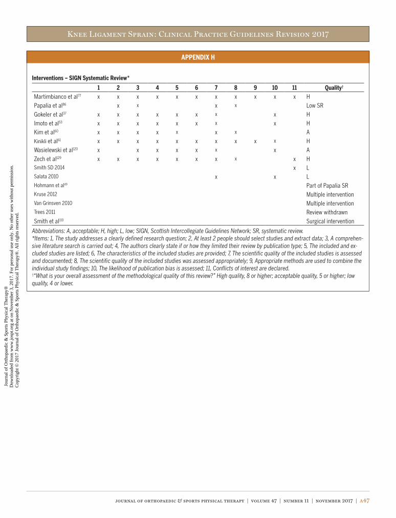
Knee Ligament Sprain: Clinical Practice Guidelines Revision 2017
journal of orthopaedic & sports physical therapy | volume 47 | number 11 | november 2017 | a47
APPENDIX H
Interventions – SIGN Systematic Review*
1 2 3 4 5 6 7 8 9 10 11 Quality†
Martimbianco et al77 x x x x x x x x x x x H
Papalia et al86 x x x x Low SR
Gokeler et al37 x x x x x x x x H
Imoto et al53 x x x x x x x x H
Kim et al60 x x x x x x x A
Kinikli et al61 x x x x x x x x x x H
Wasielewski et al120 x x x x x x x A
Zech et al129 x x x x x x x x x HSmith SD 2014 x LSalata 2010 x x LHohmann et al49 Part of Papalia SRKruse 2012 Multiple interventionVan Grinsven 2010 Multiple interventionTrees 2011 Review withdrawn
Smith et al110 Surgical intervention
Abbreviations: A, acceptable; H, high; L, low; SIGN, Scottish Intercollegiate Guidelines Network; SR, systematic review.*Items: 1, The study addresses a clearly defined research question; 2, At least 2 people should select studies and extract data; 3, A comprehen-sive literature search is carried out; 4, The authors clearly state if or how they limited their review by publication type; 5, The included and ex-cluded studies are listed; 6, The characteristics of the included studies are provided; 7, The scientific quality of the included studies is assessed and documented; 8, The scientific quality of the included studies was assessed appropriately; 9, Appropriate methods are used to combine the individual study findings; 10, The likelihood of publication bias is assessed; 11, Conflicts of interest are declared.†“What is your overall assessment of the methodological quality of this review?” High quality, 8 or higher; acceptable quality, 5 or higher; low quality, 4 or lower.
47-11 CPG Knee 3.indd 47 10/18/2017 2:26:15 PM
Jou
rnal
of
Ort
hopa
edic
& S
port
s Ph
ysic
al T
hera
py®
D
ownl
oade
d fr
om w
ww
.josp
t.org
at o
n N
ovem
ber
3, 2
017.
For
per
sona
l use
onl
y. N
o ot
her
uses
with
out p
erm
issi
on.
Cop
yrig
ht ©
201
7 Jo
urna
l of
Ort
hopa
edic
& S
port
s Ph
ysic
al T
hera
py®
. All
righ
ts r
eser
ved.
This article has been cited by:
1. 2017. Knee Ligament Sprain Guidelines: Revision 2017: Using the Evidence to Guide Physical Therapist Practice. Journalof Orthopaedic & Sports Physical Therapy 47:11, 822-823. [Abstract] [Full Text] [PDF] [PDF Plus]
Jou
rnal
of
Ort
hopa
edic
& S
port
s Ph
ysic
al T
hera
py®
D
ownl
oade
d fr
om w
ww
.josp
t.org
at o
n N
ovem
ber
3, 2
017.
For
per
sona
l use
onl
y. N
o ot
her
uses
with
out p
erm
issi
on.
Cop
yrig
ht ©
201
7 Jo
urna
l of
Ort
hopa
edic
& S
port
s Ph
ysic
al T
hera
py®
. All
righ
ts r
eser
ved.