Embed Size (px)
Citation preview
C O N T I N U I N G E D U C A T I O N
Clinical Myocardial Perfusion PET/CT*
Marcelo F. Di Carli1–3, Sharmila Dorbala1–3, Jolene Meserve1, Georges El Fakhri1, Arkadiusz Sitek1, andStephen C. Moore1
1Division of Nuclear Medicine/PET, Department of Radiology, Brigham and Women’s Hospital, Harvard Medical School, Boston,Massachusetts; 2Division of Cardiovascular Imaging, Department of Radiology, Brigham and Women’s Hospital, Harvard MedicalSchool, Boston, Massachusetts; and 3Division of Cardiology, Department of Medicine, Brigham and Women’s Hospital, HarvardMedical School, Boston, Massachusetts
The field of nuclear cardiology is witnessing growing interest inthe use of cardiac PET for the evaluation of patients with coro-nary artery disease (CAD). The available evidence suggeststhat myocardial perfusion PET provides an accurate means fordiagnosing obstructive CAD, which appears superior to SPECTespecially in the obese and in those undergoing pharmacologicstress. The ability to record changes in left ventricular functionfrom rest to peak stress and to quantify myocardial perfusion(in mL/min/g of tissue) provides an added advantage overSPECT for evaluating multivessel CAD. There is growing andconsistent evidence that gated myocardial perfusion PET alsoprovides clinically useful risk stratification. Although the intro-duction of hybrid PET/CT technology offers the exciting possibil-ity of assessing the extent of anatomic CAD (CT coronaryangiography) and its functional consequences (ischemic burden)in the same setting, there are technical challenges in the imple-mentation of CT-based transmission imaging for attenuationcorrection. Nonetheless, this integrated platform for assessinganatomy and biology offers a great potential for translating ad-vances in molecularly targeted imaging into humans.
Key Words: myocardial perfusion imaging; PET/CT; CT angiog-raphy; cardiac PET
J Nucl Med 2007; 48:783–793DOI: 10.2967/jnumed.106.032789
PET has contributed significantly to advance our un-derstanding of heart physiology and pathophysiology for.25 y. Initially, it emerged as a powerful investigative toolthat allowed in vivo quantification of physiologic processes,including myocardial perfusion and metabolism, neuronaland receptor function, and, more recently, molecularly tar-
geted oncologic imaging. Despite its success in researchapplications, the limited availability of this technology, itsincreased cost, and the limited data supporting its use andreimbursement have all contributed to the relatively limitedclinical acceptance of this imaging technology. Fortunately,there are now clear signs that change is under way. Indeed,the exponential growth in the number of PET/CT systems—attributable primarily to the technology’s widely acceptedrole in clinical oncology—along with the Food and DrugAdministration’s approval of PET radiopharmaceuticals forcardiac imaging, changes in reimbursement, and the increasingdocumentation of PET’s clinical efficacy have all contributedto help advance its clinical role in cardiovascular medicine.
The emergence of integrated PET/CT technology as thedominant configuration of clinical scanners also holds greatpromise for cardiac imaging as it provides a potential op-portunity to delineate the anatomic extent and physiologicseverity of coronary atherosclerosis in a single setting.However, the recent rapid growth of PET/CT is now openinga considerable gap between the most sophisticated users ofthe technology and those with a more limited knowledge baseand fewer training opportunities; this includes cardiologists,as well as nuclear medicine specialists, and radiologists, whofrequently lack clinical experience in performing and inter-preting these cardiovascular procedures. The objective of thisreview is to provide trainees and practicing imaging special-ists with a practical review of how to perform and interpretmyocardial perfusion imaging with PET/CT in the clinicalsetting.
RADIOPHARMACEUTICALS
Although several tracers have been used for evaluatingmyocardial perfusion with PET, the most widely used inclinical practice are 82Rb and 13N-ammonia.
82Rb82Rb is a potassium analog that is a generator product with
a physical half-life of 76 s and kinetic properties similar tothose of 201Tl (1). Because of the distinct advantage of notrequiring an on-site cyclotron, 82Rb is the most widely usedradionuclide for assessment of myocardial perfusion with
Received Jan. 16, 2007; revision accepted Mar. 20, 2007.For correspondence or reprints contact: Marcelo F. Di Carli, MD, Division of
Nuclear Medicine/PET, Brigham and Women’s Hospital, 75 Francis St.,Boston, MA 02115.
E-mail: [email protected]*NOTE: FOR CE CREDIT, YOU CAN ACCESS THIS ACTIVITY THROUGH
THE SNM WEB SITE (http://www.snm.org/ce_online) THROUGH MAY 2008.Marcelo F. Di Carli reports having an affiliation with GE Healthcare, Bracco
Diagnostics, Bristol–Myers Squibb Imaging, Siemens Medical Solutions, andAstellas Pharma US, Inc. No other potential conflict of interest relevant to thisarticle was reported.
COPYRIGHT ª 2007 by the Society of Nuclear Medicine, Inc.
CARDIAC PET/CT • Di Carli et al. 783
by on December 26, 2018. For personal use only. jnm.snmjournals.org Downloaded from
PET. Its parent radionuclide is 82Sr, which has a physicalhalf-life of 26 d. Consequently, the 82Sr/82Rb generator isreplaced every 4 wk. The generator can be eluted with.90% yield every 10 min. The short physical half-life of82Rb and the rapid reconstitution of the generator allow fastsequential perfusion imaging and laboratory throughput,thereby maximizing clinical efficiency.
After intravenous injection, 82Rb rapidly crosses the cap-illary membrane (1). Myocardial uptake of 82Rb requiresactive transport via the sodium/potassium adenosine triphos-phate transporter, which is dependent on coronary blood flow(2). The single-capillary transit extraction fraction of 82Rbexceeds 50%. However, like other nondiffusible tracers, itsnet extraction fraction decreases in a nonlinear fashion withincreasing myocardial blood flow (2–4).
The maximum kinetic energy of positrons emitted during82Rb decay is significantly higher than that of 18F or 13N.Consequently, the spatial uncertainty in the location of thedecaying nucleus—which depends on the distance traveledby the positrons before their annihilation (positron range)—is greater for 82Rb (2.6-mm full width at half maximum[FWHM]) than for 18F (0.2-mm FWHM) or 13N (0.7-mmFWHM). Although 82Rb imaging yields excellent imagequality with current PET technology, its longer positronrange and its short half-life, which requires significant imagesmoothing to suppress noise, both mitigate somewhat theimproved spatial resolution of PET.
13N-Ammonia13N-ammonia is a cyclotron product and has a physical
half-life of 9.96 min. After injection, 13N-ammonia rapidlydisappears from the circulation, permitting the acquisitionof images of excellent quality. Although the sequestrationof 13N-ammonia in the lungs is usually minimal, it may beincreased in patients with depressed left ventricular (LV)systolic function or chronic pulmonary disease and, occa-sionally, in smokers. This may, in turn, adversely affect thequality of the images. In these cases, it may be necessary toincrease the time between injection and image acquisitionto optimize the contrast between myocardial and back-ground activity.
In arterial blood, 13N-ammonia consists of neutral ammo-nia (NH3) in equilibrium with its charged ammonium (NH4)ion. The neutral NH3 molecule readily diffuses across plasmaand cell membranes, leading to virtually complete extractionfrom the vascular pool. Inside the myocyte, it reequilibrateswith its ammonium form, which is trapped in glutamine viathe enzyme glutamine synthase (5,6). Once inside the myo-cyte, 13N-ammonia is incorporated into the glutamine pooland becomes metabolically trapped (6). Only a small fractiondiffuses back into the intravascular space (6). Its retention bythe myocardium has a nonlinear and inverse relationship withblood flow. In healthy humans, the fraction of 13N-ammoniaretained by the myocardium during its first pass is 0.83 whenblood flow is 1 mL/min/g, and decreases to 0.60 as flowincreases to 3 mL/min/g. As with other nondiffusible tracers,
the net tissue extraction (the product of the retained fractionduring first pass and myocardial blood flow) decreases asmyocardial blood flow increases. For 13N-ammonia, therelation between net tissue extraction and blood flow is linearfor values of blood flow up to 2.5 mL/min/g. At high flowrates, ‘‘metabolic trapping’’ of 13N-ammonia becomes therate-limiting factor affecting tracer retention. This leads tounderestimation of blood flow at high flow rates (7). There-fore, to quantify myocardial blood flow using 13N-ammonia,it becomes necessary to correct for flow-dependent changesin net tissue extraction.
Myocardial retention of 13N-ammonia is heterogeneous,with retention in the lateral left ventricular (LV) wall beingabout 10% lower than that of other segments, even in healthysubjects. 13N-Ammonia images may also be degraded byoccasional intense liver activity, which can interfere with theevaluation of the inferior wall. Although the sequestration of13N-ammonia in the lungs is usually minimal, it may beincreased in patients with depressed LV systolic function orchronic pulmonary disease and, occasionally, in smokers (8).In these cases, it may be necessary to increase the timebetween injection and image acquisition to optimize thecontrast between myocardial and background activity.
13N-Ammonia allows the acquisition of ungated and gatedimages of good quality. These studies take full advantage ofthe superior resolution of PET relative to SPECT, as the half-life of 13N is sufficiently long and its average positron range isvery short. Gated 13N-ammonia imaging can provide accu-rate assessments of both regional and global cardiac function(9). However, this imaging agent is not well suited for peakstress gated imaging because of the 3- to 4-min time intervalbetween radiotracer injection and the start of imaging and therelatively long (;20 min) acquisition time.
IMAGING PROTOCOLS
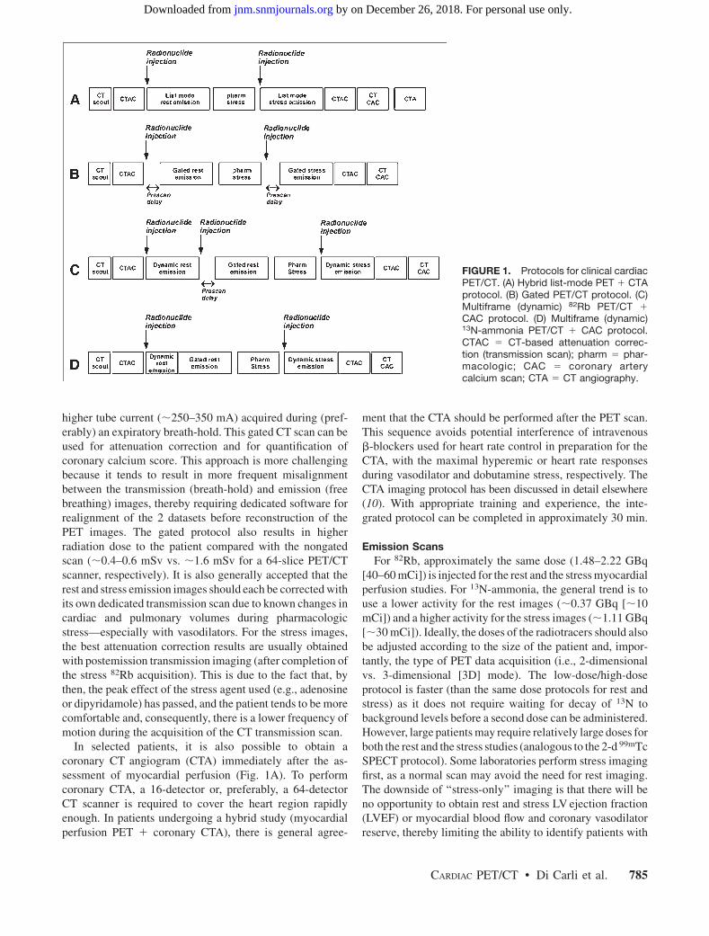
Figure 1 illustrates common protocols used for imagingmyocardial perfusion with PET/CT, in which the CT scan isused for attenuation correction.
CT Scans
Patient positioning is performed with a CT scout image ortopogram. This is followed by a low-dose CT scan coveringthe heart region. It is important to understand that acquisitionparameters for CT-based transmission imaging vary withthe configuration of the CT scanner (e.g., 8-, 16-, and 64-detector CT) and clinical protocol. However, the general scansettings used in most clinics for CT transmission imaging,independent of the manufacturer, include (a) a slow rotationspeed (e.g., 1 s/revolution), combined with a relatively highpitch (e.g., 0.5–0.6:1); (b) a nongated scan; (c) a high tubepotential (e.g., 140 kVp) and a low tube current (;10–20mA); and (d) a CT acquisition obtained during tidal expira-tion breath-hold or shallow breathing. It is also possible to usea prospectively gated CT scan, in which the x-ray tube is‘‘turned on’’ only during the end-diastolic phase of thecardiac cycle—typically 75%–80% of the R–R interval, with
784 THE JOURNAL OF NUCLEAR MEDICINE • Vol. 48 • No. 5 • May 2007
by on December 26, 2018. For personal use only. jnm.snmjournals.org Downloaded from
higher tube current (;250–350 mA) acquired during (pref-erably) an expiratory breath-hold. This gated CT scan can beused for attenuation correction and for quantification ofcoronary calcium score. This approach is more challengingbecause it tends to result in more frequent misalignmentbetween the transmission (breath-hold) and emission (freebreathing) images, thereby requiring dedicated software forrealignment of the 2 datasets before reconstruction of thePET images. The gated protocol also results in higherradiation dose to the patient compared with the nongatedscan (;0.4–0.6 mSv vs. ;1.6 mSv for a 64-slice PET/CTscanner, respectively). It is also generally accepted that therest and stress emission images should each be corrected withits own dedicated transmission scan due to known changes incardiac and pulmonary volumes during pharmacologicstress—especially with vasodilators. For the stress images,the best attenuation correction results are usually obtainedwith postemission transmission imaging (after completion ofthe stress 82Rb acquisition). This is due to the fact that, bythen, the peak effect of the stress agent used (e.g., adenosineor dipyridamole) has passed, and the patient tends to be morecomfortable and, consequently, there is a lower frequency ofmotion during the acquisition of the CT transmission scan.
In selected patients, it is also possible to obtain acoronary CT angiogram (CTA) immediately after the as-sessment of myocardial perfusion (Fig. 1A). To performcoronary CTA, a 16-detector or, preferably, a 64-detectorCT scanner is required to cover the heart region rapidlyenough. In patients undergoing a hybrid study (myocardialperfusion PET 1 coronary CTA), there is general agree-
ment that the CTA should be performed after the PET scan.This sequence avoids potential interference of intravenousb-blockers used for heart rate control in preparation for theCTA, with the maximal hyperemic or heart rate responsesduring vasodilator and dobutamine stress, respectively. TheCTA imaging protocol has been discussed in detail elsewhere(10). With appropriate training and experience, the inte-grated protocol can be completed in approximately 30 min.
Emission Scans
For 82Rb, approximately the same dose (1.48–2.22 GBq[40–60 mCi]) is injected for the rest and the stress myocardialperfusion studies. For 13N-ammonia, the general trend is touse a lower activity for the rest images (;0.37 GBq [;10mCi]) and a higher activity for the stress images (;1.11 GBq[;30 mCi]). Ideally, the doses of the radiotracers should alsobe adjusted according to the size of the patient and, impor-tantly, the type of PET data acquisition (i.e., 2-dimensionalvs. 3-dimensional [3D] mode). The low-dose/high-doseprotocol is faster (than the same dose protocols for rest andstress) as it does not require waiting for decay of 13N tobackground levels before a second dose can be administered.However, large patients may require relatively large doses forboth the rest and the stress studies (analogous to the 2-d 99mTcSPECT protocol). Some laboratories perform stress imagingfirst, as a normal scan may avoid the need for rest imaging.The downside of ‘‘stress-only’’ imaging is that there will beno opportunity to obtain rest and stress LV ejection fraction(LVEF) or myocardial blood flow and coronary vasodilatorreserve, thereby limiting the ability to identify patients with
FIGURE 1. Protocols for clinical cardiacPET/CT. (A) Hybrid list-mode PET 1 CTAprotocol. (B) Gated PET/CT protocol. (C)Multiframe (dynamic) 82Rb PET/CT 1
CAC protocol. (D) Multiframe (dynamic)13N-ammonia PET/CT 1 CAC protocol.CTAC 5 CT-based attenuation correc-tion (transmission scan); pharm 5 phar-macologic; CAC 5 coronary arterycalcium scan; CTA 5 CT angiography.
CARDIAC PET/CT • Di Carli et al. 785
by on December 26, 2018. For personal use only. jnm.snmjournals.org Downloaded from
extensive coronary artery disease (CAD) and ‘‘balanced’’ischemia.
There are several ways in which the emission images canbe acquired. These include:
• ECG Gated Imaging. This is the most common clinicalapproach. Imaging begins 90–120 s after 82Rb injection,or 3–5 min after 13N-ammonia injection, to allow forclearance of radioactivity from the lungs and bloodpool; the scan duration is approximately 5 or 20 minfor 82Rb or 13N-ammonia, respectively (Fig. 1B).The number of gated frames is usually set to 8 or 16,with rejection of ectopic beats outside the acceptancewindow.
• Multiframe or Dynamic Imaging. Imaging begins withthe bolus (short infusion) of 82Rb or 13N-ammonia andcontinues for 7–8 min or 20 min, respectively (Fig. 1C).The advantage of this approach is that it allowsquantification of myocardial blood flow (in mL/min/g)—for example, by fitting regional tissue and bloodtime–activity curves to a suitable kinetic model. How-ever, its main disadvantage is that one needs to perform aseparate radionuclide injection to obtain ECG-gatedimages from which to assess cardiac function, espe-cially when using 82Rb. Using 13N-ammonia, one canacquire a short multiframe or dynamic scan (4 min) thatcan be followed with a separate ;15-min ECG-gatedscan to assess myocardial perfusion and LVEF, withoutthe need for an additional radionuclide injectionbecause of its longer physical half-life (;10 min)(Fig. 1D). To measure the LV blood time–activity curvenoninvasively, one must acquire many dynamic PETimage frames while the tracer bolus passes through theright ventricle (RV), the lungs, and the LV. During thisinterval, it is common for a relatively large amount ofactivity (;0.74–1.11 GBq [;20–30 mCi]) to be locatedentirely within the PET scanner’s axial field of view.Because of possible counting-rate limitations undersuch conditions—particularly in the 3D (septaless) scanmode—great care must be taken to ensure the accuracyof the PET system’s corrections for random and scattercoincidences as well as dead time. Although quantita-tive inaccuracies seen at high counting rates may bemitigated by injecting less activity, this could yield toomuch image noise in the later (tissue) phase of thedynamic study, so a careful compromise needs to bedetermined for each given PET system and scan mode.
• List-Mode Imaging. This is the ideal approach becausea single injection and data acquisition allows multipleimage reconstructions (i.e., summed, ECG gated, andmultiframe or dynamic) for a comprehensive physio-logic examination of the heart. With this approach,image acquisition commences with the bolus injectionof the radionuclide and continues for 7–8 min or 20min for 82Rb and 13N-ammonia, respectively (Fig.1A). List-mode imaging requires significant computer
power to perform the multiple reconstructions, espe-cially for the 3D image acquisition mode.
Stress testing can be performed with pharmacologicmeans (e.g., adenosine, dipyridamole, or dobutamine)—most common—or with exercise (11). The latter is easierwith 13N-ammonia (half-life of ;10 min) than with 82Rb(half-life of 76 s).
QUALITY ASSURANCE FOR CARDIAC PET/CT
Performing good-quality cardiac PET/CT is technicallydemanding and, thus, familiarity with key quality controlsteps is crucial to optimize clinical results. These qualitycontrol measures include routine inspection of the transmis-sion and emission data and the transmission-emission align-ment. A description of routine maintenance, calibration, andquality control of the PET/CT scanner is beyond the scope ofthis review; however, the reader is encouraged to reviewexcellent recent discussions on the subject (12,13).
Count Density
The level of statistical noise in the emission and transmis-sion images should be checked to see whether enough countshave been acquired. This is crucial with 82Rb imagingbecause of the short half-life of the isotope. Although theimage quality of CT transmission images may be suboptimalin heavy patients, this does not appear to compromise thequality of the attenuation correction because the attenuationmap is integrated along the PET lines of response to obtainattenuation-correction factors. Also, of course, many moretransmitted photons are used for CT, even with a low (10–20mA) tube current, than for a transmission-source (e.g., 68Ge)scan on a conventional PET system. Hence, it is not necessaryto increase the CT dose in heavy patients. The most commonsources of count-poor emission studies include large patientsize, inadequate radionuclide dose or delivery (e.g., problemswith intravenous access), or inadequate scan duration.
Ratio of Heart to Blood-Pool Counts
Acquisition of emission images before complete clearanceof radiotracer from the blood pool may potentially degradeimage quality. The optimal prescan delay for acquisition of13N-ammonia images is ;3 min and for 82Rb images is ;90 sin healthy subjects. The most common factors that prolongcirculation time of the radionuclide include severe LV sys-tolic dysfunction (LVEF , 30%, either chronic or acutebecause of severe ischemia during stress), RV systolic dys-function, and primary lung disease. In those clinical settings,increased prescan delay is usually required to improve imagequality (14). As discussed earlier, multiframe (i.e., dynamic)or list-mode imaging protocols are very helpful, as assump-tions with regard to prescan delay are not required.
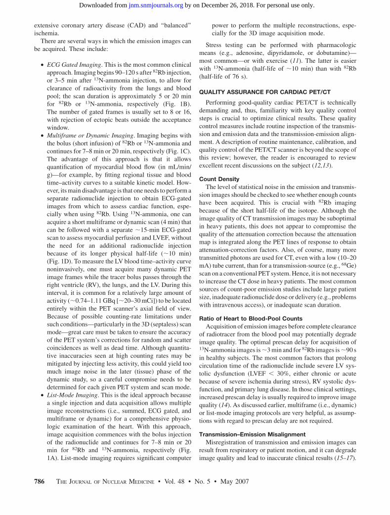
Transmission–Emission Misalignment
Misregistration of transmission and emission images canresult from respiratory or patient motion, and it can degradeimage quality and lead to inaccurate clinical results (15–17).
786 THE JOURNAL OF NUCLEAR MEDICINE • Vol. 48 • No. 5 • May 2007
by on December 26, 2018. For personal use only. jnm.snmjournals.org Downloaded from
Because of the circular arrays of scintillation detectors inPET/CT cameras, patient movement during imaging affectsall projections and can be very difficult to detect by directinspection of the rotating projections, as assessed withSPECT. Because transmission and emission imaging issequential, patient or respiratory (e.g., deep inspiration)motion during the emission images will most likely lead totransmission–emission misalignment and potential attenuation-correction artifacts (Fig. 2). The extent and direction of thismisalignment will determine whether artifacts will beapparent in the attenuation-corrected images. Most com-mercial PET/CT systems now include software tools tocorrect for transmission–emission misalignments. This toolallows correction of the sinograms before tomographicreconstruction and rereconstruction with the correct atten-uation map.
Image Reconstruction Artifacts
Artifacts resulting from filtered backprojection recon-struction algorithms, image truncation, and CT transmissionartifacts, such as beam hardening, may be observed withcardiac PET/CT. As seen in SPECT myocardial perfusionimaging, excessive subdiaphragmatic activity from highcounts in the liver or bowel may result in decreased countsin the adjacent inferior LV wall when filtered backprojectionis used for image reconstruction. This artifact may be
particularly problematic if seen only on the stress imagesbecause it may cause an apparent reversible perfusion defect.Fortunately, artifacts from filtered backprojection are notcommon with PETas iterative methods are generally used forimage reconstruction. Streak artifacts may be seen in largepatients with arms-down imaging (a result of beam hardeningfrom the large bones) or with metal implants in the field ofview. In PET/CT systems that allow transmission imagingwith an external radioactive source, this approach mayobviate the limitations of CT-based transmission imaging.Preliminary evidence suggests that a segmented reconstruc-tion algorithm for the CT scan may help overcome artifactsfrom metallic implants (18). Residual radiotracer activity inthe intravenous line within the field of view may be an ad-ditional source of image reconstruction artifacts that can beminimized by flushing the intravenous tubing.
DIAGNOSTIC ACCURACY
Table 1 summarizes the published studies documentingthe diagnostic accuracy of myocardial perfusion PET fordetecting obstructive CAD. The average weighted sensitiv-ity for detecting at least one coronary artery with .50%stenosis is 90% (range, 83%–100%), whereas the averagespecificity is 89% (range, 73%–100%). The correspondingaverage positive and negative predictive values (PPV and
FIGURE 2. Transmission–emission misalignment. (A) Misaligned CT transmission and 82Rb images (right) and resultinganterolateral perfusion defect on stress–rest 82Rb PET (left). Perfusion defect results from applying incorrect attenuationcoefficients during tomographic reconstruction to area of LV myocardium overlying lung field on CT transmission scan (arrows). (B)Correction of emission–transmission misalignment (right) and resulting normal perfusion study.
CARDIAC PET/CT • Di Carli et al. 787
by on December 26, 2018. For personal use only. jnm.snmjournals.org Downloaded from
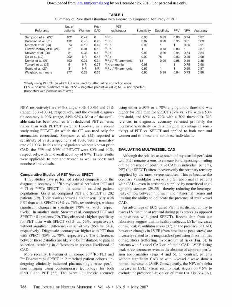
NPV, respectively) are 94% (range, 80%–100%) and 73%(range, 36%–100%), respectively, and the overall diagnos-tic accuracy is 90% (range, 84%–98%). Most of the avail-able data has been obtained with dedicated PET cameras,rather than with PET/CT systems. However, in a recentstudy using PET/CT (in which the CT was used only forattenuation correction), Sampson et al. (22) reported asensitivity of 93%, a specificity of 83%, with a normalcyrate of 100%. In this study of patients without known priorCAD, the PPV and NPV of PET/CT were 80% and 94%,respectively, with an overall accuracy of 87%. These resultswere applicable to men and women as well as obese andnonobese individuals.
Comparative Studies of PET Versus SPECT
Three studies have performed a direct comparison of thediagnostic accuracy of 82Rb myocardial perfusion PET and201Tl or 99mTc SPECT in the same or matched patientpopulations. Go et al. compared PET and SPECT in 202patients (19). Their results showed a higher sensitivity withPET than with SPECT (93% vs. 76%, respectively), withoutsignificant changes in specificity (78% vs. 80%, respec-tively). In another study, Stewart et al. compared PET andSPECT in 81 patients (20). They observed a higher specificityfor PET than with SPECT (83% vs. 53%, respectively),without significant differences in sensitivity (86% vs. 84%,respectively). Diagnostic accuracy was higher with PET thanwith SPECT (89% vs. 78%, respectively). The differencesbetween these 2 studies are likely to be attributable to patientselection, resulting in differences in prescan likelihood ofCAD.
More recently, Bateman et al. compared 82Rb PET and99mTc-sestamibi SPECT in 2 matched patient cohorts un-dergoing clinically indicated pharmacologic-stress perfu-sion imaging using contemporary technology for bothSPECT and PET (21). The overall diagnostic accuracy
using either a 50% or a 70% angiographic threshold washigher for PET than for SPECT (87% vs. 71% with a 50%threshold, and 89% vs. 79% with a 70% threshold). Dif-ferences in diagnostic accuracy reflected primarily theincreased specificity (with a marginal advantage in sensi-tivity) of PET vs. SPECT and applied to both men andwomen and to obese and nonobese individuals.
EVALUATING MULTIVESSEL CAD
Although the relative assessment of myocardial perfusionwith PET remains a sensitive means for diagnosing or rulingout the presence of obstructive CAD in individual patients,PET (like SPECT) often uncovers only the coronary territorysupplied by the most severe stenoses. This is because thecoronary vasodilator reserve is often abnormal in patientswith CAD—even in territories supplied by noncritical angi-ographic stenoses (29,30)—thereby reducing the heteroge-neity of flow between ‘‘normal’’ and ‘‘abnormal’’ zones andlimiting the ability to delineate the presence of multivesselCAD.
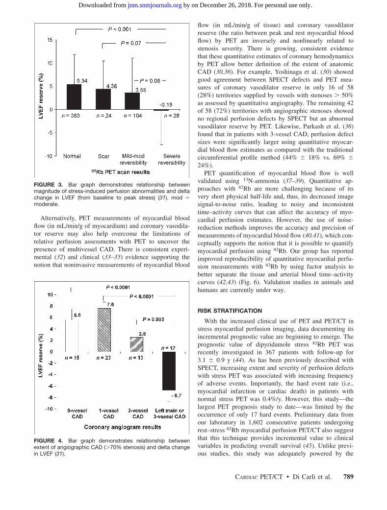
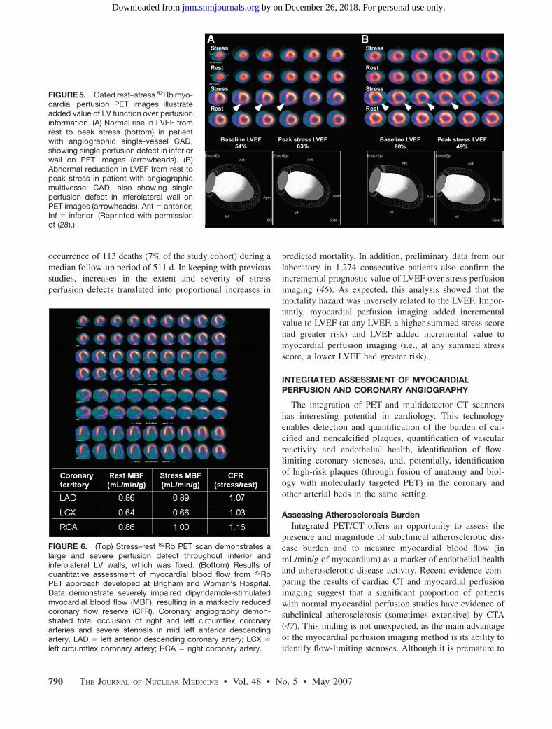
An advantage of ECG-gated PET is its distinct ability toassess LV function at rest and during peak stress (as opposedto poststress with gated SPECT). Recent data from ourlaboratory suggest that in healthy subjects, LVEF increasesduring peak vasodilator stress (31). In the presence of CAD,however, changes in LVEF (from baseline to peak stress) areinversely related to the magnitude of perfusion abnormalitiesduring stress (reflecting myocardium at risk) (Fig. 3). Inpatients with 3-vessel CAD or left main CAD, LVEF duringpeak stress decreases even in the absence of apparent perfu-sion abnormalities (Figs. 4 and 5). In contrast, patientswithout significant CAD or with 1-vessel disease show anormal increase in LVEF. Consequently, the NPV of a deltaincrease in LVEF (from rest to peak stress) of $5% toexclude the presence 3-vessel or left main CAD is 97% (31).
TABLE 1Summary of Published Literature with Regard to Diagnostic Accuracy of PET
Reference
No. of
patients Women
Prior
CAD
PET
radiotracer Sensitivity Specificity PPV NPV Accuracy
Sampson et al. (22)* 102 0.42 0 82Rb 0.93 0.83 0.80 0.94 0.87
Bateman et al. (21) 112 0.46 0.25 82Rb 0.87 0.93 0.95 0.81 0.89Marwick et al. (23) 74 0.19 0.49 82Rb 0.90 1 1 0.36 0.91
Grover-McKay et al. (24) 31 0.01 0.13 82Rb 1 0.73 0.80 1 0.87
Stewart et al. (20) 81 0.36 0.42 82Rb 0.83 0.86 0.94 0.64 0.84
Go et al. (19) 202 NR 0.47 82Rb 0.93 78 0.93 0.80 0.90Demer et al. (25) 193 0.26 0.34 82Rb /13N-ammonia 83 0.95 0.98 0.60 0.85
Tamaki et al. (26) 51 NR 0.75 13N-ammonia 0.98 1 1 0.75 0.98
Gould et al. (27) 31 NR NR 82Rb /13N-ammonia 0.95 1 1 0.90 0.97
Weighted summary 877 0.29 0.35 0.90 0.89 0.94 0.73 0.90
*Study using PET/CT (in which CT was used for attenuation correction only).
PPV 5 positive predictive value; NPV 5 negative predictive value; NR 5 not reported.(Reprinted with permission of (28).)
788 THE JOURNAL OF NUCLEAR MEDICINE • Vol. 48 • No. 5 • May 2007
by on December 26, 2018. For personal use only. jnm.snmjournals.org Downloaded from
Alternatively, PET measurements of myocardial bloodflow (in mL/min/g of myocardium) and coronary vasodila-tor reserve may also help overcome the limitations ofrelative perfusion assessments with PET to uncover thepresence of multivessel CAD. There is consistent experi-mental (32) and clinical (33–35) evidence supporting thenotion that noninvasive measurements of myocardial blood
flow (in mL/min/g of tissue) and coronary vasodilatorreserve (the ratio between peak and rest myocardial bloodflow) by PET are inversely and nonlinearly related tostenosis severity. There is growing, consistent evidencethat these quantitative estimates of coronary hemodynamicsby PET allow better definition of the extent of anatomicCAD (30,36). For example, Yoshinaga et al. (30) showedgood agreement between SPECT defects and PET mea-sures of coronary vasodilator reserve in only 16 of 58(28%) territories supplied by vessels with stenoses . 50%as assessed by quantitative angiography. The remaining 42of 58 (72%) territories with angiographic stenoses showedno regional perfusion defects by SPECT but an abnormalvasodilator reserve by PET. Likewise, Parkash et al. (36)found that in patients with 3-vessel CAD, perfusion defectsizes were significantly larger using quantitative myocar-dial blood flow estimates as compared with the traditionalcircumferential profile method (44% 6 18% vs. 69% 6
24%).PET quantification of myocardial blood flow is well
validated using 13N-ammonia (37–39). Quantitative ap-proaches with 82Rb are more challenging because of itsvery short physical half-life and, thus, its decreased imagesignal-to-noise ratio, leading to noisy and inconsistenttime–activity curves that can affect the accuracy of myo-cardial perfusion estimates. However, the use of noise-reduction methods improves the accuracy and precision ofmeasurements of myocardial blood flow (40,41), which con-ceptually supports the notion that it is possible to quantifymyocardial perfusion using 82Rb. Our group has reportedimproved reproducibility of quantitative myocardial perfu-sion measurements with 82Rb by using factor analysis tobetter separate the tissue and arterial blood time–activitycurves (42,43) (Fig. 6). Validation studies in animals andhumans are currently under way.
RISK STRATIFICATION
With the increased clinical use of PET and PET/CT instress myocardial perfusion imaging, data documenting itsincremental prognostic value are beginning to emerge. Theprognostic value of dipyridamole stress 82Rb PET wasrecently investigated in 367 patients with follow-up for3.1 6 0.9 y (44). As has been previously described withSPECT, increasing extent and severity of perfusion defectswith stress PET was associated with increasing frequencyof adverse events. Importantly, the hard event rate (i.e.,myocardial infarction or cardiac death) in patients withnormal stress PET was 0.4%/y. However, this study—thelargest PET prognosis study to date—was limited by theoccurrence of only 17 hard events. Preliminary data fromour laboratory in 1,602 consecutive patients undergoingrest–stress 82Rb myocardial perfusion PET/CT also suggestthat this technique provides incremental value to clinicalvariables in predicting overall survival (45). Unlike previ-ous studies, this study was adequately powered by the
FIGURE 3. Bar graph demonstrates relationship betweenmagnitude of stress-induced perfusion abnormalities and deltachange in LVEF (from baseline to peak stress) (31). mod 5
moderate.
FIGURE 4. Bar graph demonstrates relationship betweenextent of angiographic CAD (.70% stenosis) and delta changein LVEF (31).
CARDIAC PET/CT • Di Carli et al. 789
by on December 26, 2018. For personal use only. jnm.snmjournals.org Downloaded from
occurrence of 113 deaths (7% of the study cohort) during amedian follow-up period of 511 d. In keeping with previousstudies, increases in the extent and severity of stressperfusion defects translated into proportional increases in
predicted mortality. In addition, preliminary data from ourlaboratory in 1,274 consecutive patients also confirm theincremental prognostic value of LVEF over stress perfusionimaging (46). As expected, this analysis showed that themortality hazard was inversely related to the LVEF. Impor-tantly, myocardial perfusion imaging added incrementalvalue to LVEF (at any LVEF, a higher summed stress scorehad greater risk) and LVEF added incremental value tomyocardial perfusion imaging (i.e., at any summed stressscore, a lower LVEF had greater risk).
INTEGRATED ASSESSMENT OF MYOCARDIALPERFUSION AND CORONARY ANGIOGRAPHY
The integration of PET and multidetector CT scannershas interesting potential in cardiology. This technologyenables detection and quantification of the burden of cal-cified and noncalcified plaques, quantification of vascularreactivity and endothelial health, identification of flow-limiting coronary stenoses, and, potentially, identificationof high-risk plaques (through fusion of anatomy and biol-ogy with molecularly targeted PET) in the coronary andother arterial beds in the same setting.
Assessing Atherosclerosis Burden
Integrated PET/CT offers an opportunity to assess thepresence and magnitude of subclinical atherosclerotic dis-ease burden and to measure myocardial blood flow (inmL/min/g of myocardium) as a marker of endothelial healthand atherosclerotic disease activity. Recent evidence com-paring the results of cardiac CT and myocardial perfusionimaging suggest that a significant proportion of patientswith normal myocardial perfusion studies have evidence ofsubclinical atherosclerosis (sometimes extensive) by CTA(47). This finding is not unexpected, as the main advantageof the myocardial perfusion imaging method is its ability toidentify flow-limiting stenoses. Although it is premature to
FIGURE 5. Gated rest–stress 82Rb myo-cardial perfusion PET images illustrateadded value of LV function over perfusioninformation. (A) Normal rise in LVEF fromrest to peak stress (bottom) in patientwith angiographic single-vessel CAD,showing single perfusion defect in inferiorwall on PET images (arrowheads). (B)Abnormal reduction in LVEF from rest topeak stress in patient with angiographicmultivessel CAD, also showing singleperfusion defect in inferolateral wall onPET images (arrowheads). Ant 5 anterior;Inf 5 inferior. (Reprinted with permissionof (28).)
FIGURE 6. (Top) Stress–rest 82Rb PET scan demonstrates alarge and severe perfusion defect throughout inferior andinferolateral LV walls, which was fixed. (Bottom) Results ofquantitative assessment of myocardial blood flow from 82RbPET approach developed at Brigham and Women’s Hospital.Data demonstrate severely impaired dipyridamole-stimulatedmyocardial blood flow (MBF), resulting in a markedly reducedcoronary flow reserve (CFR). Coronary angiography demon-strated total occlusion of right and left circumflex coronaryarteries and severe stenosis in mid left anterior descendingartery. LAD 5 left anterior descending coronary artery; LCX 5
left circumflex coronary artery; RCA 5 right coronary artery.
790 THE JOURNAL OF NUCLEAR MEDICINE • Vol. 48 • No. 5 • May 2007
by on December 26, 2018. For personal use only. jnm.snmjournals.org Downloaded from
predict the clinical significance of this observation, it pro-vides intriguing evidence for future investigations of thepotential complementary role of CTA and perfusion imag-ing for individualizing risk stratification and patient man-agement. Whereas myocardial perfusion imaging willlikely continue to define the need for revascularization,the objective assessment of atherosclerotic burden (bothcalcified and noncalcified plaques) by CTA might be able to
play a role in individualizing the intensity and goals ofmedical therapy.
Guiding Management of CAD
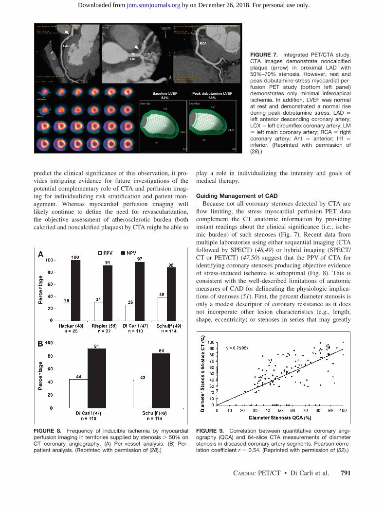
Because not all coronary stenoses detected by CTA areflow limiting, the stress myocardial perfusion PET datacomplement the CT anatomic information by providinginstant readings about the clinical significance (i.e., ische-mic burden) of such stenoses (Fig. 7). Recent data frommultiple laboratories using either sequential imaging (CTAfollowed by SPECT) (48,49) or hybrid imaging (SPECT/CT or PET/CT) (47,50) suggest that the PPV of CTA foridentifying coronary stenoses producing objective evidenceof stress-induced ischemia is suboptimal (Fig. 8). This isconsistent with the well-described limitations of anatomicmeasures of CAD for delineating the physiologic implica-tions of stenoses (51). First, the percent diameter stenosis isonly a modest descriptor of coronary resistance as it doesnot incorporate other lesion characteristics (e.g., length,shape, eccentricity) or stenoses in series that may greatly
FIGURE 7. Integrated PET/CTA study.CTA images demonstrate noncalcifiedplaque (arrow) in proximal LAD with50%–70% stenosis. However, rest andpeak dobutamine stress myocardial per-fusion PET study (bottom left panel)demonstrates only minimal inferoapicalischemia. In addition, LVEF was normalat rest and demonstrated a normal riseduring peak dobutamine stress. LAD 5
left anterior descending coronary artery;LCX 5 left circumflex coronary artery; LM5 left main coronary artery; RCA 5 rightcoronary artery; Ant 5 anterior; Inf 5
inferior. (Reprinted with permission of(28).)
FIGURE 8. Frequency of inducible ischemia by myocardialperfusion imaging in territories supplied by stenosis . 50% onCT coronary angiography. (A) Per-vessel analysis. (B) Per-patient analysis. (Reprinted with permission of (28).)
FIGURE 9. Correlation between quantitative coronary angi-ography (QCA) and 64-slice CTA measurements of diameterstenosis in diseased coronary artery segments. Pearson corre-lation coefficient r 5 0.54. (Reprinted with permission of (52).)
CARDIAC PET/CT • Di Carli et al. 791
by on December 26, 2018. For personal use only. jnm.snmjournals.org Downloaded from
affect the impedance to blood flow. Second, vasomotor toneand coronary collateral flow, both of which are known toaffect myocardial perfusion, are not assessed by simplemeasures of stenosis severity. Finally, as described earlier,CTA is limited in its ability to accurately define the severityof stenosis (a surrogate of physiologic significance) (Fig.9). In contrast, myocardial perfusion imaging provides asimple and accurate integrated measure of the effect of allof these parameters on coronary resistance and tissue per-fusion, thereby optimizing selection of patients who mayultimately benefit from revascularization. This finding, ifconfirmed in larger studies, would suggest that additionalnoninvasive testing would be required after CTA beforeconsideration for invasive catheterization. Furthermore, theuse of CTA alone in patients with atherosclerosis withoutmyocardial perfusion imaging would potentially result inan enormous increase in the costs of care and resource usedue to unnecessary downstream catheterization and revas-cularization procedures (53).
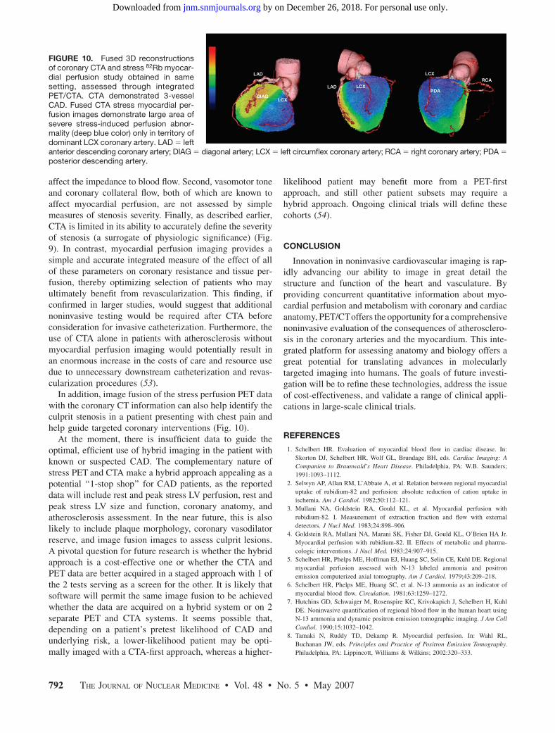
In addition, image fusion of the stress perfusion PET datawith the coronary CT information can also help identify theculprit stenosis in a patient presenting with chest pain andhelp guide targeted coronary interventions (Fig. 10).
At the moment, there is insufficient data to guide theoptimal, efficient use of hybrid imaging in the patient withknown or suspected CAD. The complementary nature ofstress PET and CTA make a hybrid approach appealing as apotential ‘‘1-stop shop’’ for CAD patients, as the reporteddata will include rest and peak stress LV perfusion, rest andpeak stress LV size and function, coronary anatomy, andatherosclerosis assessment. In the near future, this is alsolikely to include plaque morphology, coronary vasodilatorreserve, and image fusion images to assess culprit lesions.A pivotal question for future research is whether the hybridapproach is a cost-effective one or whether the CTA andPET data are better acquired in a staged approach with 1 ofthe 2 tests serving as a screen for the other. It is likely thatsoftware will permit the same image fusion to be achievedwhether the data are acquired on a hybrid system or on 2separate PET and CTA systems. It seems possible that,depending on a patient’s pretest likelihood of CAD andunderlying risk, a lower-likelihood patient may be opti-mally imaged with a CTA-first approach, whereas a higher-
likelihood patient may benefit more from a PET-firstapproach, and still other patient subsets may require ahybrid approach. Ongoing clinical trials will define thesecohorts (54).
CONCLUSION
Innovation in noninvasive cardiovascular imaging is rap-idly advancing our ability to image in great detail thestructure and function of the heart and vasculature. Byproviding concurrent quantitative information about myo-cardial perfusion and metabolism with coronary and cardiacanatomy, PET/CToffers the opportunity for a comprehensivenoninvasive evaluation of the consequences of atherosclero-sis in the coronary arteries and the myocardium. This inte-grated platform for assessing anatomy and biology offers agreat potential for translating advances in molecularlytargeted imaging into humans. The goals of future investi-gation will be to refine these technologies, address the issueof cost-effectiveness, and validate a range of clinical appli-cations in large-scale clinical trials.
REFERENCES
1. Schelbert HR. Evaluation of myocardial blood flow in cardiac disease. In:
Skorton DJ, Schelbert HR, Wolf GL, Brundage BH, eds. Cardiac Imaging: A
Companion to Braunwald’s Heart Disease. Philadelphia, PA: W.B. Saunders;
1991:1093–1112.
2. Selwyn AP, Allan RM, L’Abbate A, et al. Relation between regional myocardial
uptake of rubidium-82 and perfusion: absolute reduction of cation uptake in
ischemia. Am J Cardiol. 1982;50:112–121.
3. Mullani NA, Goldstein RA, Gould KL, et al. Myocardial perfusion with
rubidium-82. I. Measurement of extraction fraction and flow with external
detectors. J Nucl Med. 1983;24:898–906.
4. Goldstein RA, Mullani NA, Marani SK, Fisher DJ, Gould KL, O’Brien HA Jr.
Myocardial perfusion with rubidium-82. II. Effects of metabolic and pharma-
cologic interventions. J Nucl Med. 1983;24:907–915.
5. Schelbert HR, Phelps ME, Hoffman EJ, Huang SC, Selin CE, Kuhl DE. Regional
myocardial perfusion assessed with N-13 labeled ammonia and positron
emission computerized axial tomography. Am J Cardiol. 1979;43:209–218.
6. Schelbert HR, Phelps ME, Huang SC, et al. N-13 ammonia as an indicator of
myocardial blood flow. Circulation. 1981;63:1259–1272.
7. Hutchins GD, Schwaiger M, Rosenspire KC, Krivokapich J, Schelbert H, Kuhl
DE. Noninvasive quantification of regional blood flow in the human heart using
N-13 ammonia and dynamic positron emission tomographic imaging. J Am Coll
Cardiol. 1990;15:1032–1042.
8. Tamaki N, Ruddy TD, Dekamp R. Myocardial perfusion. In: Wahl RL,
Buchanan JW, eds. Principles and Practice of Positron Emission Tomography.
Philadelphia, PA: Lippincott, Williams & Wilkins; 2002:320–333.
FIGURE 10. Fused 3D reconstructionsof coronary CTA and stress 82Rb myocar-dial perfusion study obtained in samesetting, assessed through integratedPET/CTA. CTA demonstrated 3-vesselCAD. Fused CTA stress myocardial per-fusion images demonstrate large area ofsevere stress-induced perfusion abnor-mality (deep blue color) only in territory ofdominant LCX coronary artery. LAD 5 leftanterior descending coronary artery; DIAG 5 diagonal artery; LCX 5 left circumflex coronary artery; RCA 5 right coronary artery; PDA 5
posterior descending artery.
792 THE JOURNAL OF NUCLEAR MEDICINE • Vol. 48 • No. 5 • May 2007
by on December 26, 2018. For personal use only. jnm.snmjournals.org Downloaded from
9. Hickey KT, Sciacca RR, Bokhari S, et al. Assessment of cardiac wall motion and
ejection fraction with gated PET using N-13 ammonia. Clin Nucl Med. 2004;
29:243–248.
10. Hoffmann U, Ferencik M, Cury RC, Pena AJ. Coronary CT angiography. J Nucl
Med. 2006;47:797–806.
11. Chow BJ, Beanlands RS, Lee A, et al. Treadmill exercise produces larger
perfusion defects than dipyridamole stress N-13 ammonia positron emission
tomography. J Am Coll Cardiol. 2006;47:411–416.
12. DiFilippo FP. Instrumentation and principles of imaging: PET. In: Di Carli MF,
Lipton MJ, eds. Cardiac PET and PET/CT Imaging. New York, NY: Springer-
Verlag. In press.
13. Townsend DW, Besozzi MC, Carney JPC. Integrated PET/CT. In: Di Carli MF,
Lipton MJ, eds. Cardiac PET and PET/CT Imaging. New York, NY: Springer-
Verlag. In press.
14. Bacharach SL, Bax JJ, Case J, et al. PET myocardial glucose metabolism and
perfusion imaging. Part 1. Guidelines for data acquisition and patient prepara-
tion. J Nucl Cardiol. 2003;10:543–556.
15. Bettinardi V, Gilardi MC, Lucignani G, et al. A procedure for patient
repositioning and compensation for misalignment between transmission and
emission data in PET heart studies. J Nucl Med. 1993;34:137–142.
16. Loghin C, Sdringola S, Gould KL. Common artifacts in PET myocardial
perfusion images due to attenuation-emission misregistration: clinical signifi-
cance, causes, and solutions. J Nucl Med. 2004;45:1029–1039.
17. McCord ME, Bacharach SL, Bonow RO, Dilsizian V, Cuocolo A, Freedman N.
Misalignment between PET transmission and emission scans: its effect on myo-
cardial imaging. J Nucl Med. 1992;33:1209–1214; discussion 1214–1215.
18. Mirzaei S, Guerchaft M, Bonnier C, Knoll P, Doat M, Braeutigam P. Use of
segmented CT transmission map to avoid metal artifacts in PET images by a
PET-CT device [abstract]. BMC Nucl Med. 2005;5(1):3.
19. Go RT, Marwick TH, MacIntyre WJ, et al. A prospective comparison of
rubidium-82 PET and thallium-201 SPECT myocardial perfusion imaging
utilizing a single dipyridamole stress in the diagnosis of coronary artery disease
[see comments]. J Nucl Med. 1990;31:1899–1905.
20. Stewart RE, Schwaiger M, Molina E, et al. Comparison of rubidium-82 positron
emission tomography and thallium-201 SPECT imaging for detection of
coronary artery disease. Am J Cardiol. 1991;67:1303–1310.
21. Bateman TM, Heller GV, McGhie AI, et al. Diagnostic accuracy of rest/stress
ECG-gated Rb-82 myocardial perfusion PET: comparison with ECG-gated
Tc-99m sestamibi SPECT. J Nucl Cardiol. 2006;13:24–33.
22. Sampson UK, Limaye A, Dorbala S, et al. Diagnostic accuracy of rubidium-82
myocardial perfusion imaging with hybrid positron emission tomography/
computed tomography (PET-CT) in the detection of coronary artery disease.
J Am Coll Cardiol. 2007;49:1052–1058.
23. Marwick TH, Nemec JJ, Stewart WJ, Salcedo EE. Diagnosis of coronary artery
disease using exercise echocardiography and positron emission tomography:
comparison and analysis of discrepant results. J Am Soc Echocardiogr. 1992;5:
231–238.
24. Grover-McKay M, Ratib O, Schwaiger M, et al. Detection of coronary artery
disease with positron emission tomography and rubidium 82. Am Heart J.
1992;123:646–652.
25. Demer LL, Gould KL, Goldstein RA, et al. Assessment of coronary artery
disease severity by positron emission tomography: comparison with quantitative
arteriography in 193 patients. Circulation. 1989;79:825–835.
26. Tamaki N, Yonekura Y, Senda M, et al. Value and limitation of stress thallium-
201 single photon emission computed tomography: comparison with nitrogen-13
ammonia positron tomography. J Nucl Med. 1988;29:1181–1188.
27. Gould KL, Goldstein RA, Mullani NA, et al. Noninvasive assessment of
coronary stenoses by myocardial perfusion imaging during pharmacologic
coronary vasodilation. VIII. Clinical feasibility of positron cardiac imaging
without a cyclotron using generator-produced rubidium-82. J Am Coll Cardiol.
1986;7:775–789.
28. Di Carli MF, Hachamovitch R. New technology for noninvasive imaging of
coronary artery disease. Circulation. 2007;115:1464–1480.
29. Uren NG, Crake T, Lefroy DC, de Silva R, Davies GJ, Maseri A. Reduced
coronary vasodilator function in infarcted and normal myocardium after myo-
cardial infarction [see comments]. N Engl J Med. 1994;331:222–227.
30. Yoshinaga K, Katoh C, Noriyasu K, et al. Reduction of coronary flow reserve in
areas with and without ischemia on stress perfusion imaging in patients with
coronary artery disease: a study using oxygen 15-labeled water PET. J Nucl
Cardiol. 2003;10:275–283.
31. Dorbala S, Vangala D, Sampson U, Limaye A, Kwong R, Di Carli MF. Value of
left ventricular ejection fraction reserve in evaluating the magnitude of myo-
cardium at risk and the extent of angiographic coronary artery disease: a 82Rb
PET/CT study. J Nucl Med. 2007;349–358.
32. Gould KL, Lipscomb K. Effects of coronary stenoses on coronary flow reserve
and resistance. Am J Cardiol. 1974;34:48–55.
33. Di Carli M, Czernin J, Hoh CK, et al. Relation among stenosis severity,
myocardial blood flow, and flow reserve in patients with coronary artery disease.
Circulation. 1995;91:1944–1951.
34. Uren NG, Melin JA, De Bruyne B, Wijns W, Baudhuin T, Camici PG. Relation
between myocardial blood flow and the severity of coronary-artery stenosis.
N Engl J Med. 1994;330:1782–1788.
35. Beanlands RS, Muzik O, Melon P, et al. Noninvasive quantification of regional
myocardial flow reserve in patients with coronary atherosclerosis using nitrogen-
13 ammonia positron emission tomography: determination of extent of altered
vascular reactivity. J Am Coll Cardiol. 1995;26:1465–1475.
36. Parkash R, deKemp RA, Ruddy TD, et al. Potential utility of rubidium 82 PET
quantification in patients with 3-vessel coronary artery disease. J Nucl Cardiol.
2004;11:440–449.
37. Kuhle WG, Porenta G, Huang SC, et al. Quantification of regional myocardial
blood flow using 13N-ammonia and reoriented dynamic positron emission
tomographic imaging. Circulation. 1992;86:1004–1017.
38. Nitzsche EU, Choi Y, Czernin J, Hoh CK, Huang SC, Schelbert HR. Noninvasive
quantification of myocardial blood flow in humans: a direct comparison of the
[13N]ammonia and the [15O]water techniques. Circulation. 1996;93:2000–2006.
39. Muzik O, Beanlands RS, Hutchins GD, Mangner TJ, Nguyen N, Schwaiger M.
Validation of nitrogen-13-ammonia tracer kinetic model for quantification of
myocardial blood flow using PET. J Nucl Med. 1993;34:83–91.
40. Lin JW, Laine AF, Akinboboye O, Bergmann SR. Use of wavelet transforms in
analysis of time-activity data from cardiac PET. J Nucl Med. 2001;42:194–200.
41. Lin JW, Laine AF, Bergmann SR. Improving PET-based physiological quanti-
fication through methods of wavelet denoising. IEEE Trans Biomed Eng. 2001;
48:202–212.
42. El Fakhri G, Sitek A, Guerin B, Kijewski MF, Di Carli MF, Moore SC.
Quantitative dynamic cardiac 82Rb PET using generalized factor and compart-
ment analyses. J Nucl Med. 2005;46:1264–1271.
43. El Fakhri G, Guerin G, Sitek A, et al. Absolute myocardial blood flow
quantification in Rb-82 cardiac PET using generalized factor and compartment
analysis: a reproducibility study. Eur J Nucl Med Mol Imaging. 2006;33:S217.
44. Yoshinaga K, Chow BJ, Williams K, et al. What is the prognostic value of
myocardial perfusion imaging using rubidium-82 positron emission tomogra-
phy? J Am Coll Cardiol. 2006;48:1029–1039.
45. Dorbala S, Hachamovitch R, Kwong R, Curillova Z, Di Carli MF. Incremental
prognostic value of rubidium-82 myocardial perfusion PET-CT imaging in
patients with known or suspected CAD. J Am Coll Cardiol. 2007;49:109A.
46. Dorbala S, Hachamovitch R, Kwong R, Curillova Z, Di Carli MF. Incremental
prognostic value of left ventricular ejection fraction assessment over myocardial
perfusion imaging: a rubidium-82 PET/CT study. J Am Coll Cardiol. 2007;
49:109A.
47. Di Carli MF, Dorbala S, Limaye A, et al. Clinical value of hybrid PET/CT
cardiac imaging: complementary roles of multi-detector CT coronary angiog-
raphy and stress PET perfusion imaging [abstract]. J Am Coll Cardiol. 2006;
47:115A.
48. Hacker M, Jakobs T, Matthiesen F, et al. Comparison of spiral multidetector CT
angiography and myocardial perfusion imaging in the noninvasive detection of
functionally relevant coronary artery lesions: first clinical experiences. J Nucl
Med. 2005;46:1294–1300.
49. Schuijf JD, Wijns W, Jukema JW, et al. Relationship between noninvasive
coronary angiography with multi-slice computed tomography and myocardial
perfusion imaging. J Am Coll Cardiol. 2006;48:2508–2514.
50. Rispler S, Roguin A, Keidar Z, et al. Integrated SPECT/CT for the assessment of
hemodynamically significant coronary artery lesions. J Am Coll Cardiol. 2007;
49:1059–1067.
51. Gould KL. Identifying and measuring severity of coronary artery stenosis:
quantitative coronary arteriography and positron emission tomography. Circu-
lation. 1988;78:237–245.
52. Leber AW, Knez A, von Ziegler F, et al. Quantification of obstructive and
nonobstructive coronary lesions by 64-slice CT: a comparative study with
quantitative coronary angiography and intravascular ultrasound. J Am Coll
Cardiol. 2005;46:147–154.
53. Shaw LJ, Hachamovitch R, Berman DS, et al. The economic consequences of
available diagnostic and prognostic strategies for the evaluation of stable angina
patients: an observational assessment of the value of precatheterization
ischemia—Economics of Noninvasive Diagnosis (END) Multicenter Study
Group. J Am Coll Cardiol. 1999;33:661–669.
54. Di Carli MF, Hachamovitch R. The study of myocardial perfusion and coronary
anatomy imaging roles in CAD (SPARC). Available at: http://www.sparctrial.
org. Accessed April 2, 2007.
CARDIAC PET/CT • Di Carli et al. 793
by on December 26, 2018. For personal use only. jnm.snmjournals.org Downloaded from
Doi: 10.2967/jnumed.106.0327892007;48:783-793.J Nucl Med.
Marcelo F. Di Carli, Sharmila Dorbala, Jolene Meserve, Georges El Fakhri, Arkadiusz Sitek and Stephen C. Moore Clinical Myocardial Perfusion PET/CT
http://jnm.snmjournals.org/content/48/5/783This article and updated information are available at:
http://jnm.snmjournals.org/site/subscriptions/online.xhtml
Information about subscriptions to JNM can be found at:
http://jnm.snmjournals.org/site/misc/permission.xhtmlInformation about reproducing figures, tables, or other portions of this article can be found online at:
(Print ISSN: 0161-5505, Online ISSN: 2159-662X)1850 Samuel Morse Drive, Reston, VA 20190.SNMMI | Society of Nuclear Medicine and Molecular Imaging
is published monthly.The Journal of Nuclear Medicine
© Copyright 2007 SNMMI; all rights reserved.
by on December 26, 2018. For personal use only. jnm.snmjournals.org Downloaded from