Embed Size (px)
Citation preview
UNIVERSITÀ DEGLI STUDI DI PADOVA
Sede Amministrativa: Università degli Studi di Padova
Dipartimento di Scienze Cardiache, Toraciche e Vascolari
SCUOLA DI DOTTORATO DI RICERCA IN Scienze Mediche, Cliniche e Sperimentali
Indirizzo: Scienze Cardiovascolari XXV ciclo
Clinical, morphological and molecular characterization of cancer phenotypes associated with chronic obstructive pulmonary disease (COPD): new prospective of target
therapies
Direttore della Scuola : Ch.mo Prof. Gaetano Thiene
Coordinatore d’indirizzo: Ch.mo Prof. Gaetano Thiene
Supervisore: Ch.ma Prof.ssa Fiorella Calabrese Ch.mo Prof. Federico Rea
DOTTORANDO: DOTT. MARCO SCHIAVON
ANNO ACCADEMICO: 2011-2012

INDEX ABSTRACT............................................................................................................................page 1 RIASSUNTO..........................................................................................................................page 5 1 BACKGROUND..................................................................................................................page 9
1.1 Definition of COPD..............................................................................................page 9
1.2 Epidemiology......................................................................................................page 10
1.3 Clinical aspects...................................................................................................page 11
1.3.1 Lung symptoms.....................................................................................page 11
1.3.2 Comorbidity and systemic involvement................................................page 14
1.4 Diagnosis............................................................................................................page 15
1.4.1 History and physical examination........................................................page 15
1.4.2 Spirometry............................................................................................page 16
1.4.3 Evaluation of gravity disease..............................................................page 16
1.4.4 Additional investigations......................................................................page 18
1.5 Morphological aspects........................................................................................page 20
1.6 Pathogenesis of the disease.................................................................................page 22
1.5.1 Aetiology..............................................................................................page 22
1.5.2 Pathogenetic mechanisms....................................................................page 25
1.7 Lung cancer.........................................................................................................page 29
1.8 COPD and Lung cancer......................................................................................page 32
1.8.1 Risk Factors.........................................................................................page 33
1.8.2 The inflammatory theory......................................................................page 35
1.8.3 Pathway axys IL17- R23......................................................................page 37
2 AIM OF THE RESEARCH...............................................................................................page 39
3 MATERIALS AMD METHODS......................................................................................page 41
3.1 Study population.................................................................................................page 41
3.2 Surgical procedure and lung sampling...............................................................page 45
3.3 Histological analysis...........................................................................................page 46
3.3.1 Pathological diagnosis.........................................................................page 47
3.3.2 Morphological evaluations..................................................................page 47
3.3.3 Characterization of the peri-and intra-tumor IL-17 cytokine pattern…..page 48
3.4 Genetic analysis..................................................................................................page 48
3.5 Electronic database.............................................................................................page 49
3.6 Statistical analysis................................................................................................page 49
4 RESULTS...........................................................................................................................page 51
4.1 Population characteristics..................................................................................page 51
4.2 Histological evaluation.......................................................................................page 54
4.3 Genetic analyses.................................................................................................page 59
4.4 Characterization of the peri-and intra-tumor IL-17 cytokine pattern................page 61
5 DISCUSSION....................................................................................................................page 63
6 REFERENCES...................................................................................................................page 71
7 PRODUCTS OF THE RESEARCH..................................................................................page 75
1
ABSTRACT BACKGROUND
Chronic obstructive pulmonary disease (COPD) and lung cancer are two catastrophic diseases,
representing leading causes of morbidity and mortality worldwide.
Although the treatment has greatly improved both diseases continue to show increasing
frequency and above all an unpredictable progression.
Several studies have firmly established a strict connection between COPD and lung cancer
highlighting also the importance of the inflammatory response as a risk factor for both diseases.
The inflammatory paradigm is undoubtedly one of the most fascinating theories to connect
COPD and lung cancer and it has acquired new impetus by the recent discoveries in the COPD
pathogenesis. Emerging evidence in this context has emphasized the role of adaptive immune
responses, possibly with an autoimmune component due to the recognition of pulmonary self-
antigens modified by cigarette smoking and to the failure of mechanisms regulating
immunological tolerance. In this context, COPD-associated cancers might have specific
pathogenetic and morphological features, differently from tumours arising in non-COPD
patients, due to the synergic effect of cigarette smoke and chronic inflammation.
AIM OF THE RESEARCH
This research project focuses on the study of lung cancer in patients with COPD compared to
smokers without COPD and never smoker patients in order to identify eventual distinct clinical,
morphological and molecular phenotypes.
MATERIALS AND METHODS
From 2010 to 2012, we prospectively enrolled patients with peripheral non small lung cancer
submitted to anatomical lung resection (lobectomy, bilobectomy or pneumonectomy) associated
with systematic lymphadenectomy. Patients with central airway cancer, secondary lung tumours
or previously submitted to inductive treatment were excluded from the study.
According to respiratory functional tests and smoking history patients were then divided in 3
groups: COPD patients, smokers without COPD and never-smoker subjects with normal lung
2
function (FEV1/FVC ratio >70%). Each patient underwent a full clinical and instrumental
assessment.
Morphological studies included detailed analysis of growth pattern (according to the latest
revision of adenocarcinoma classification), cell proliferation (Ki67/MIB1 expression),
parameters of intra-and peri-tumoral remodelling (inflammation, fibrosis and necrosis) and
tumoural detection of interleukin-17 (IL-17) cytokine. Genetic analysis of EGFR and KRAS
mutations was also performed in all cases.
RESULTS
In the study period, 66 patients who met the inclusion/exclusion criteria were initially
enrolled:16 COPD, 32 smokers without COPD and 18 never smokers.
As the selection criteria affected the predominant histologic profile with a clear predominance of
the adenocarcinoma histotype (63% in COPD patients, 71% in smokers and 56% in never-
smokers), we performed our investigations only in patients with this histology to obtain results
not affected by different histotypes. Therefore the study group was composed of 43 patients (10
COPD, 23 smokers and 10 never-smokers), whose main demographic and functional parameters
were comparable except for male/female ratio, reversed in never-smokers, and for lung function,
reduced in COPD patients, as expected.
Given the specific aim the comparison of different clinical, morphological and molecular data
was mainly performed within the category of smoking patients (COPD patients and smokers
without COPD), while never smokers represented control group.
From a clinical point of view the most important differences concern the number of peripheral
blood basophils and standard uptake value of positron emission tomography–computed
tomography (SUV of PET-CT). COPD patients showed a significant higher number of basophils
and lower SUV of PET-CT than smokers without COPD.
Concerning the histological evaluation adenocarcinoma of COPD patients showed a more
frequent lepid pattern, less evident solid aspect and lower MIB1/Ki67 index than
adenocarcinoma of smokers without COPD. A significant more extensive necrosis was found in
adenocarcinoma of COPD and smokers without COPD compared to never-smokers. Finally
although not statistically significant a stronger IL17 tissue expression was observed in COPD
cases compared to smokers without COPD.
As regards molecular data the most interesting finding was a trend of less frequency of KRAS
mutation in adenocarcinoma of COPD patients.
3
CONCLUSIONS
Adenocarcinoma in COPD patients presents clinical, molecular and morphological features of
lower aggressiveness (higher number of basophils, low SUV of PET-CT, increased lepidic
component, reduced solid pattern, lower cell proliferation and less frequent K-RAS mutation)
compared to that of smokers without COPD.
Alternative mechanisms of carcinogenesis may be involved in the development/progression of
lung cancer in COPD patients. Given the importance of inflammation in the pathogenesis of the
disease other mechanisms, such as IL-17 pathway, mainly driving inflammatory mediated
carcinogenesis might be crucial.
Additional knowledge of these mechanisms would be of considerable help in the fight against
lung cancer especially concerning therapeutic perspectives, providing a rational basis for the
development of targeted and more effective treatments.

4
5
RIASSUNTO
INTRODUZIONE
La BPCO e il tumore polmonare sono due malattie catastrofiche e rappresentano alcune tra le
principali cause di morbilità e mortalità in tutto il mondo.
Sebbene il trattamento di queste patologie è notevolmente migliorato negli ultimi anni, esse
continuano a presentare una crescente incidenza e soprattutto un andamento clinico non
prevedibile a priori.
Lo stesso termine "tumore polmonare non a piccole cellule (NSCLC)" comprende un gruppo di
malattie neoplastiche con caratteristiche cliniche e molecolari estremamente eterogenee.
Diversi studi hanno ormai fermamente stabilito la stretta connessione tra la BPCO e il cancro del
polmone evidenziando anche l'importanza della risposta infiammatoria agli stimoli nocivi, in
particolare il fumo di sigaretta, come fattore di rischio fondamentale per entrambe le malattie. La
teoria infiammatoria è senza dubbio uno dei paradigmi più affascinanti per collegare la BPCO e
il tumore polmonare e ha acquisito un nuovo impulso dalle più recenti scoperte nella patogenesi
della BPCO.
Infatti, sono emerse in questo campo evidenze importanti che hanno sottolineato il ruolo
fondamentale di risposte immunitarie adattative, anche con una componente autoimmune dovuta
sia al riconoscimento di auto-antigeni polmonari modificati dal fumo di sigaretta sia al fallimento
dei meccanismi che regolano la tolleranza immunologica. In questo contesto, le neoplasie a
insorgenza in pazienti con BPCO, per effetto sinergico del fumo e di una specifica
infiammazione cronica, potrebbero possedere specifiche caratteristiche patogenetiche e
morfologiche, differenti da tumori di altre popolazioni non affette da BPCO.
SCOPO DELLA RICERCA
Questo progetto di ricerca si concentra sullo studio del cancro del polmone nei pazienti con
BPCO comparandolo a due gruppi di controllo, composti da fumatori sani e pazienti non
fumatori, al fine di individuare distinti fenotipi neoplastici dal punto di vista bioumorale,
morfologico e molecolare.
6
MATERIALI E METODI
Dal 2010 al 2012, sono stati arruolati nello studio pazienti con NSCLC in sede periferica
sottoposti a resezione polmonare anatomica (lobectomia, bilobectomia o pneumonectomia)
associata a linfadenectomia sistematica. I pazienti con neoplasia a carico delle vie aeree centrali,
con tumore polmonare secondario o precedentemente sottoposti a trattamento chemio-
radioterapico sono stati esclusi dal progetto. Ogni paziente è stato sottoposto ad una completa
valutazione clinica e strumentale, che ha compreso i test di funzionalità polmonare polmonari (i
criteri GOLD sono stati utilizzati per identificare i pazienti con BPCO), radiografia del
torace/TAC torace/18FDG PET-TC analisi del sangue.
I pazienti sono stati poi divisi in 3 gruppi in base alle prove funzionali respiratorie e alla storia di
fumo: pazienti con BPCO, soggetti fumatori e pazienti non fumatori con funzione polmonare
normale (rapporto FEV1/FVC> 70%).
Lo studio istologico della neoplasia è stato caratterizzato da: stadiazione pTNM, analisi
morfometrica del pattern di crescita (secondo l'ultima revisione della classificazione del cancro
del polmone), proliferazione cellulare (mediante valutazione dell’espressione di Ki67/MIB1), i
parametri di rimodellamento intra-e peri-tumorale (infiammazione, fibrosi, necrosi) e la
caratterizzazione del pattern citochinico di IL-17 a livello peri-e intra-tumorale.
Infine è stata eseguita l'analisi genetica delle mutazioni dei geni EGFR e KRAS.
RISULTATI
Nel periodo di studio sono stati inizialmente arruolati 66 pazienti che rispettavano i criteri di
inclusione/esclusione, di cui 16 BPCO, 32 fumatori senza BPCO e 18 non fumatori. Poiché i
criteri di selezione hanno profondamente condizionato il profilo istologico predominante, con
una netta prevalenza dell’istotipo adenocarcinoma (63% nella BPCO, 71% nei fumatori e 56%
nei non fumatori), abbiamo deciso di condurre la valutazione neoplastica funzionale,
morfologica, molecolare solo nell’ istologia prevalente.
Pertanto, il gruppo di studio è risultato composto da 43 pazienti (10 BPCO, 23 fumatori, 10 non
fumatori), che presentavano comparabili dati demografici e funzionali ad eccezione del rapporto
maschio/ femmina, invertito nei non fumatori, per la funzione polmonare, ridotta nei pazienti con
BPCO. Dato l'obiettivo specifico, il confronto dei differenti dati clinici, morfologici e molecolari
è stato svolto prevalentemente all'interno della categoria dei soggetti fumatori (pazienti affetti da
BPCO e fumatori senza BPCO), mentre i pazienti con storia negativa di fumo hanno
rappresentato il gruppo di controllo.
7
Da un punto di vista clinico, le più rimarcabili differenze sono emerse a livello del numero di
basofili nel sangue periferico e del valore di standard uptake value (SUV) all’indagine PET-TC.
Infatti i pazienti con BPCO hanno mostrato un numero significativamente superiore di basofili e
un SUV inferiore rispetto ai soggetti fumatori senza BPCO.
Per quanto riguarda la valutazione istologica, gli adenocarcinomi nei pazienti con BPCO
hanno presentato un aumento del pattern lepidico, con riduzione della componente solida e una
più bassa espressione del Ki67/MIB1 rispetto ai tumori dello stesso istotipo insorti in soggetti
fumatori senza BPCO. Si è evidenziata una maggiore rappresentazione della componente
necrotica negli adenocarcinomi dei pazienti fumatori, con o senza BPCO, rispetto al gruppo dei
non fumatori. Infine un forte ma non significativo aumento di IL-17 è stato osservata nei casi con
BPCO rispetto ai fumatori.
L’analisi molecolare ha permesso di osservare, come dato più rilevante, un trend di ridotta
frequenza di mutazione di KRAS negli adenocarcinomi dei pazienti con BPCO rispetto alle
neoplasie del gruppo dei fumatori.
CONCLUSIONI
Gli adenocarcinomi correlati alla BPCO sono emersi presentare caratteristiche cliniche,
morfologiche e molecolari di minore aggressività (aumento del numero di basofili, ridotto
SUVmax alla PET-TC, aumento della componente lepidica, ridotti pattern solido e proliferazione
cellulare e meno frequente mutazione di K-RAS) rispetto alle neoplasie insorte in pazienti
fumatori senza BPCO. Vie alternative di carcinogenesi potrebbero essere coinvolte nello
sviluppo/progressione del tumore polmonare dei pazienti con BPCO. Data l'importanza
dell'infiammazione nella patogenesi di questa malattia polmonare, altri meccanismi, quale il
pathway di IL-17, potrebbero essere cruciali per lo sviluppo cancerogenetico principalmente
mediato dall’infiammazione. La conoscenza di questi meccanismi potrebbe essere di notevole
aiuto nella lotta contro il tumore polmonare soprattutto per quanto riguarda nuove prospettive
terapeutiche, fornendo le basi per sviluppare trattamenti mirati e con maggiore efficacia.

8
9
BACKGROUND
1.1. DEFINITION OF COPD
Chronic Obstructive Pulmonary Disease (COPD) is an inflammatory disorder of the lung,
characterized by airflow limitation (bronchial obstruction),that is not fully reversible [1].
The airflow limitation is usually progressive and is associated with an abnormal inflammatory
response of the lung to noxious particles or gases, primarily from cigarette smoking.
In the past COPD was known with different names. Bonet described a condition of “voluminous
lungs” in 1679. In 1769, Giovanni Morgagni reported 19 cases where the lungs were “turgid”
particularly from air. The first description and illustration of the enlarged airspaces in
emphysema was provided by Ruysh in 1721. Matthew Baillie illustrated an emphysematous lung
in 1789 and described the destructive character of the condition. In 1814 Badham used the word
“catarrh” to describe the cough and mucus hypersecretion of chronic bronchitis that was first
reported as a disabling disorder. He recognised that chronic bronchitis was a disabling disorder.
René Laennec, the physician who invented the stethoscope, used the term “emphysema (1837) to
describe lungs that did not collapse as usual because they were full of air and the airways were
filled with mucus. In 1842, John Hutchinson invented the spirometer, which allowed the
measurement of vital capacity of the lungs. However, his spirometer could only measure volume,
not airflow. Tiffeneau in 1947, and Gaensler in 1950 and 1951, described the principles of
measuring airflow [2].
The terms chronic bronchitis and emphysema were formally defined at the CIBA guest
symposium of physicians in 1959. The term COPD was first used by William Briscoe in 1965
and has gradually overtaken other terms to become now the preferred name for this disease [3].
In 1977 Fletcher and Peto described COPD as an obstructive and hypersecretory chronic disorder
of the airways, strictly related to cigarette smoke [4].
10
The 1980 and 90s saw a surge in the use of medication to manage the symptoms of COPD and
restore pulmonary function. A major push in COPD education meant that smoking cessation and
clean air awareness became prime focuses of self-care treatment.
Today it is known that a healthy lifestyle can help people with COPD to manage and improve
their symptoms. Healthcare professionals stress the importance of diet, nutrition, and physical
exercise as part of a COPD rehabilitation program. Over the years, physicians have done much to
help understand the causes, diagnosis, and progression of COPD.
1.2 EPIDEMIOLOGY
COPD is a major cause of morbidity and mortality worldwide, resulting in a significant
economic and social costs and growing [1].
It affects about 10% of the general population but the prevalence in heavy smokers may reach
50%.
The epidemiology varies considerably between countries and between population groups within
the same nation and this difference may be related both to different exposure and individual
susceptibility to risk factors and different methods and criteria for diagnostic characterization.
The BOLD Study (Burden of Obstructive Lung Disease) [5], a prevalence study conducted in
2007 on 9425 subjects in 12 different locations of the world, reported the following data:
- Higher prevalence in males.
- Prevalence of disease progressively increase with age in both males and females.
- Increased prevalence in parallel with the increase in the number of pack-years (number
of cigarettes per day multiplied by years of smoking divided by 20).
11
- Substantial prevalence of COPD even in non-smokers: 11.3%.
Most of the epidemiological data collected in Western countries shows that the diagnosis of
COPD is found in less than 6% of the population, but it is reasonable to assume that the disease
still remains under-diagnosed.
On the one hand, the slow evolution of the natural history and lack of specificity of symptoms,
especially in the early stages, are the cause of the delay with which patients come to medical
attention. Secondly the population at risk is large, with absence of valid screening programs
[6,7].
Since the disease was responsible for 4% of the total deaths, the WHO in 2001 placed this
disease in fifth place among the leading causes of death in industrialized countries and in sixth
place in the developing countries. It is expected that the prevalence and impact of the disease
will increase in the coming decades, in parallel with the most exposure to recognized risk factors
and aging population. By 2020, according to the BOLD Study, it is estimated that COPD will
become the third leading cause of death in industrialized countries.
1.3 CLINICAL ASPECTS
1.3.1 LUNG SYMPTOMS
In the lungs, COPD is mainly characterized by the presence of progressive stress dyspnea that
can evolve to respiratory failure associated with chronic bronchitis.
The obstruction of the small airways, responsible for the loss of lung function, has a slow and
progressive development and typically occurs late, whereas the contribution of this part of the
bronchial tree to expiratory flow is 10-15% of the total resistance [8]. Obstruction of the small
airways and increased lung compliance determine an increase of exhalation duration,
appearance of hyperinflation and increase in the residual volume. In addition to increase the
functional dead space, hyperinflation causes a flattening of the diaphragm, thereby affecting its
12
contractile efficiency, and reduces the movements of the chest wall. This condition is clinically
manifested with stress dyspnea and limitation of exercise tolerance [9].
The effort made by patients suffering from emphysema during exhalation, causes a pink colour
in their faces, hence the term commonly used to refer to them, “Pink Puffers” [10]. With the
progression of the disease, respiratory effort increases in association with expansion of the
residual volume and an increase in the muscular work required for each breath. The result is a
reduction in pulmonary and alveolar ventilation responsible for blood gas alterations such as
chronic hypoxemia (PaO2 <55 mmHg) and hypercapnia (PaCO2> 45 mmHg) with a final result
of pulmonary acidosis more or less offset by the retention of bicarbonate in the kidney [11].
Chronic bronchitis
Clinical condition characterized by the presence of cough and sputum production for at least
three months a year for two consecutive years. These symptoms may occur in the natural history
of COPD at different times and with varying severity. Hypersecretion of mucus in the proximal
airways, responsible for chronic bronchitis, however, does not correlate with the decline in lung
function (FEV) in patients with COPD [12].
Patients with advanced COPD that have primarily chronic bronchitis rather than emphysema
were commonly referred to as “blue bloaters” due to the bluish colour of the skin and lips
(cyanosis) [9].
13
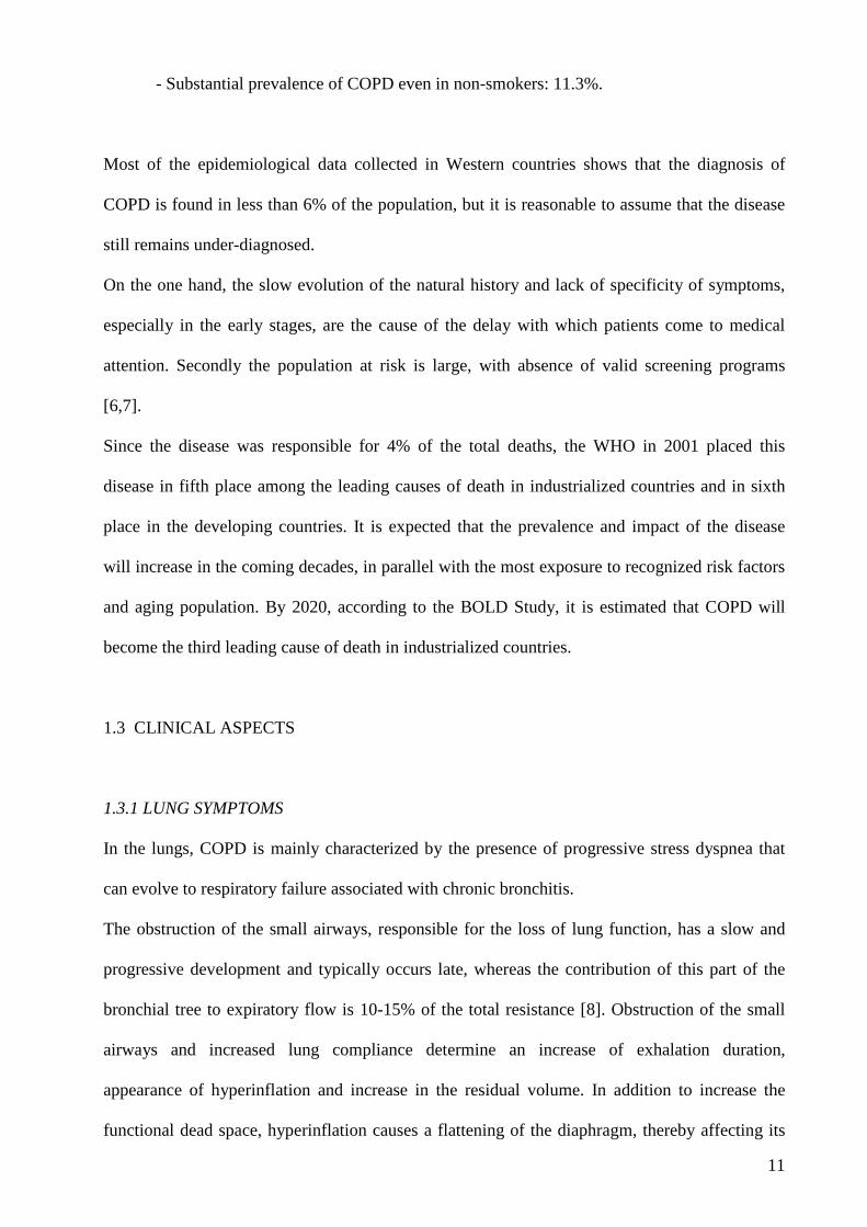
TABLE 1.1 Clinical features of the principal COPD patterns, from Travis WD et al., atlas of nontumour pathology “Non-neoplastic disorders of the lower respiratory tract” (8).
Pulmonary Hypertension
The inefficient alveolar ventilation is associated with a reduction of vascular bed in the
parenchyma which is on the base of hypoxic vasoconstriction. Even the destruction of alveolar
septa in emphysema contributes to reduce the extension of the pulmonary capillary. An
endothelial dysfunction is also present in pulmonary vessels, which correlates with the airways
inflammation. The structural changes in the pulmonary arterioles occur progressively such as
hypertrophy of the intima and in the second place of the tunica muscularis, and determine the
persistent increase in resistance in the pulmonary microcirculation [13].
These alterations lead to the development of pulmonary hypertension, which may appear in the
advanced stages of COPD (FEV <25% predicted), and it is responsible for right ventricular
hypertrophy and, ultimately, for cor pulmonaris.
Exacerbations
Exacerbations have a significant impact on morbidity, disease progression, disability and socio-
medical costs [14]. Bacterial or viral infections, often overlapping with other factors such as
environmental pollutants are the principal cause of exacerbation. From a clinical point of view,
exacerbations may lead to an increased cough, change in sputum characteristics, that becomes
14
more abundant and/or purulent, appearance of wheezing / whistling expiratory and worsening of
dyspnea.
During the course of the disease, the frequency and severity of exacerbations tends to increase,
resulting in accelerated decline in lung function.
1.3.2 COMORBIDITY AND SYSTEMIC INVOLVEMENT
Comorbidities contribute to the overall severity in individual patients (Global Initiative for
Chronic Obstructive Lung Disease, Revision 2011) [1]. Moreover, COPD also produces
significant systemic consequences mainly due to the development of the systemic inflammation.
Cardiovascular disease
These are mainly acute cardiovascular events due to atherosclerosis: endothelial dysfunction,
that occurs in the pulmonary vessels, becomes progressively a systemic condition, along with the
spread of the inflammatory reaction. CRP (C-reactive protein) is a key marker of systemic
inflammatory response and the serum level correlates with the risk of acute cardiovascular events
(CAD), such as myocardial infarction, unstable angina, and others. In COPD, CRP levels
correlate with the severity of bronchial obstruction (ie, with the decline of FEV) and the risk of
cardiac and pulmonary complications [15,16].
Metabolic syndrome
It is a complex disorder and an emerging clinical challenge, recognised clinically by the findings
of abdominal obesity, elevated triglycerides, atherogenic dyslipidaemia, elevated blood pressure,
high blood glucose and/or insulin resistance, for which the relative risk is increased (RR = 1.8)
especially in women [17]. Patients with COPD often have one or more component of the
metabolic syndrome and osteoporosis (70% of patients) which are, at least in part, independent
from treatment with steroids and/or the decreased physical activity.
15
COPD patients have an increased risk of fractures, especially at vertebral level, and it has a
direct effect on lung function with reduced vital capacity (estimated a loss of 7% for each
vertebral fracture) [18].
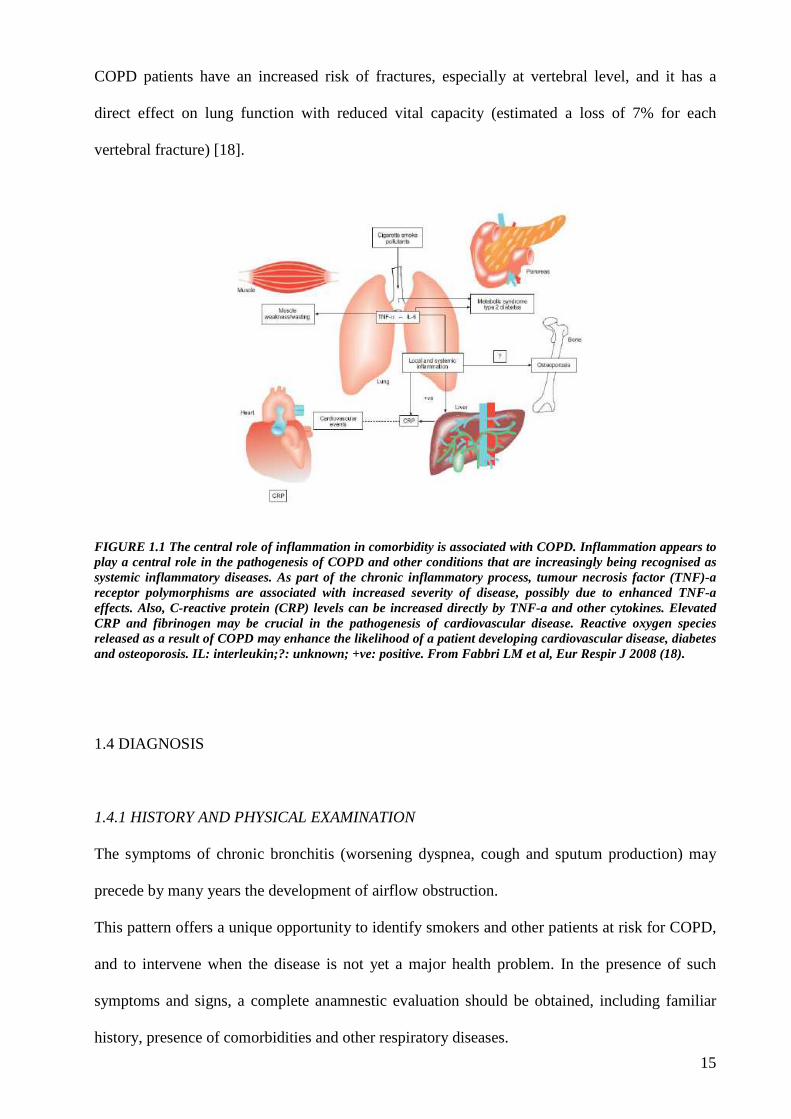
FIGURE 1.1 The central role of inflammation in comorbidity is associated with COPD. Inflammation appears to play a central role in the pathogenesis of COPD and other conditions that are increasingly being recognised as systemic inflammatory diseases. As part of the chronic inflammatory process, tumour necrosis factor (TNF)-a receptor polymorphisms are associated with increased severity of disease, possibly due to enhanced TNF-a effects. Also, C-reactive protein (CRP) levels can be increased directly by TNF-a and other cytokines. Elevated CRP and fibrinogen may be crucial in the pathogenesis of cardiovascular disease. Reactive oxygen species released as a result of COPD may enhance the likelihood of a patient developing cardiovascular disease, diabetes and osteoporosis. IL: interleukin;?: unknown; +ve: positive. From Fabbri LM et al, Eur Respir J 2008 (18).
1.4 DIAGNOSIS
1.4.1 HISTORY AND PHYSICAL EXAMINATION
The symptoms of chronic bronchitis (worsening dyspnea, cough and sputum production) may
precede by many years the development of airflow obstruction.
This pattern offers a unique opportunity to identify smokers and other patients at risk for COPD,
and to intervene when the disease is not yet a major health problem. In the presence of such
symptoms and signs, a complete anamnestic evaluation should be obtained, including familiar
history, presence of comorbidities and other respiratory diseases.
16
Particular interest must be reserved to risk factors exposure, especially cigarette smoking, other
environmental factors and genetic predisposition or working.
1.4.2 SPIROMETRY
This is the most reproducible and objective measure available of airflow limitation. The
spirometry should be performed after adequate dose of bronchodilator with short duration of
action, that reduce the variability of the test. A value of FEV / FVC < 0.70 after bronchodilator
allows confirmation of persistent airway obstruction.
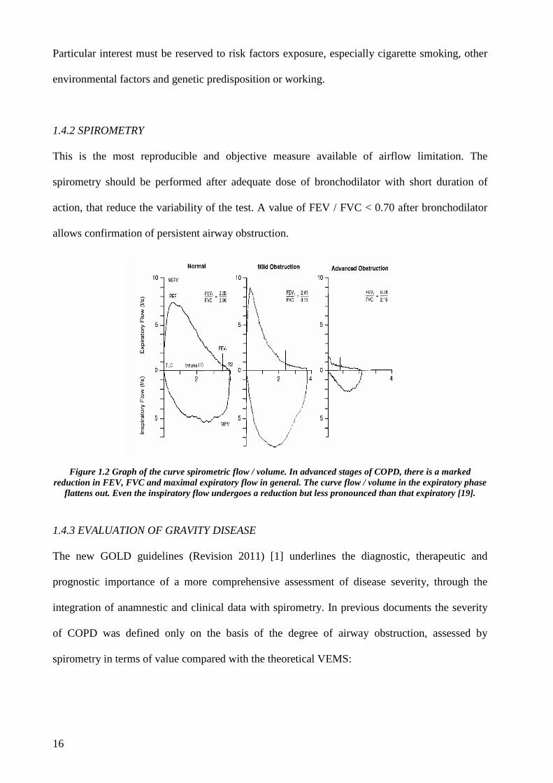
Figure 1.2 Graph of the curve spirometric flow / volume. In advanced stages of COPD, there is a marked
reduction in FEV, FVC and maximal expiratory flow in general. The curve flow / volume in the expiratory phase flattens out. Even the inspiratory flow undergoes a reduction but less pronounced than that expiratory [19].
1.4.3 EVALUATION OF GRAVITY DISEASE
The new GOLD guidelines (Revision 2011) [1] underlines the diagnostic, therapeutic and
prognostic importance of a more comprehensive assessment of disease severity, through the
integration of anamnestic and clinical data with spirometry. In previous documents the severity
of COPD was defined only on the basis of the degree of airway obstruction, assessed by
spirometry in terms of value compared with the theoretical VEMS:
17
- GOLD 1: Mild, FEV> 80% predicted
- GOLD 2: Moderate, 50% <FEV <80% predicted
- GOLD 3: Severe, 30% <FEV <50% predicted
- GOLD 4: Very Severe, FEV <30% predicted FEV.
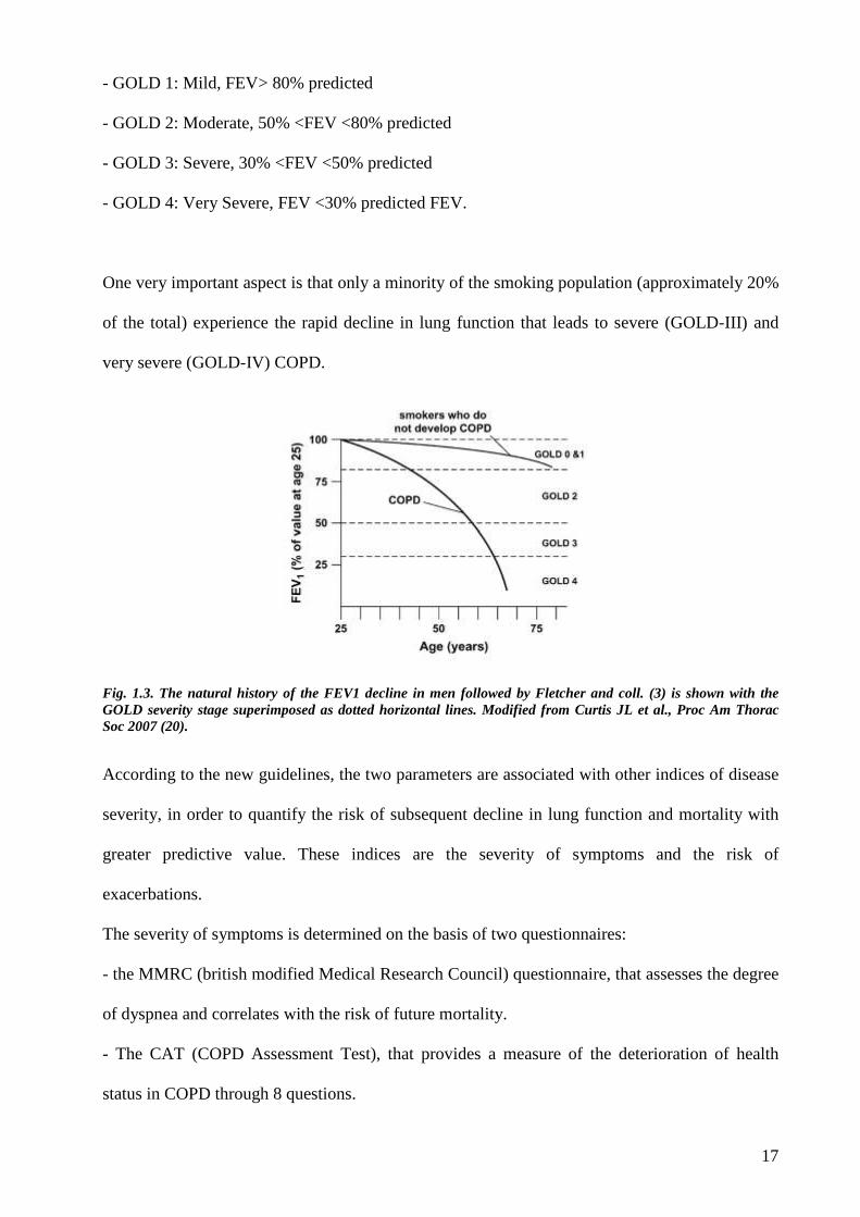
One very important aspect is that only a minority of the smoking population (approximately 20%
of the total) experience the rapid decline in lung function that leads to severe (GOLD-III) and
very severe (GOLD-IV) COPD.
Fig. 1.3. The natural history of the FEV1 decline in men followed by Fletcher and coll. (3) is shown with the GOLD severity stage superimposed as dotted horizontal lines. Modified from Curtis JL et al., Proc Am Thorac Soc 2007 (20).
According to the new guidelines, the two parameters are associated with other indices of disease
severity, in order to quantify the risk of subsequent decline in lung function and mortality with
greater predictive value. These indices are the severity of symptoms and the risk of
exacerbations.
The severity of symptoms is determined on the basis of two questionnaires:
- the MMRC (british modified Medical Research Council) questionnaire, that assesses the degree
of dyspnea and correlates with the risk of future mortality.
- The CAT (COPD Assessment Test), that provides a measure of the deterioration of health
status in COPD through 8 questions.
18
The risk of subsequent exacerbations is based on the history of similar events recently treated.
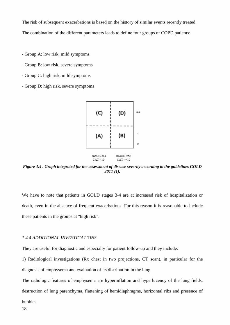
The combination of the different parameters leads to define four groups of COPD patients:
- Group A: low risk, mild symptoms
- Group B: low risk, severe symptoms
- Group C: high risk, mild symptoms
- Group D: high risk, severe symptoms
Figure 1.4 . Graph integrated for the assessment of disease severity according to the guidelines GOLD 2011 (1).
We have to note that patients in GOLD stages 3-4 are at increased risk of hospitalization or
death, even in the absence of frequent exacerbations. For this reason it is reasonable to include
these patients in the groups at "high risk".
1.4.4 ADDITIONAL INVESTIGATIONS
They are useful for diagnostic and especially for patient follow-up and they include:
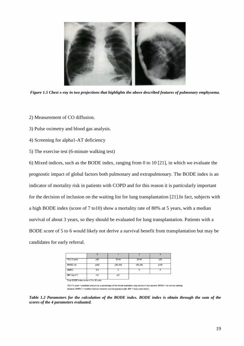
1) Radiological investigations (Rx chest in two projections, CT scan), in particular for the
diagnosis of emphysema and evaluation of its distribution in the lung.
The radiologic features of emphysema are hyperinflation and hyperlucency of the lung fields,
destruction of lung parenchyma, flattening of hemidiaphragms, horizontal ribs and presence of
bubbles.
19
Figure 1.5 Chest x-ray in two projections that highlights the above described features of pulmonary emphysema.
2) Measurement of CO diffusion.
3) Pulse oximetry and blood gas analysis.
4) Screening for alpha1-AT deficiency
5) The exercise test (6-minute walking test)
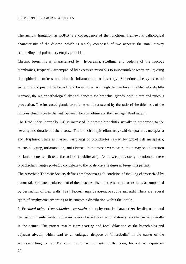
6) Mixed indices, such as the BODE index, ranging from 0 to 10 [21], in which we evaluate the
prognostic impact of global factors both pulmonary and extrapulmonary. The BODE index is an
indicator of mortality risk in patients with COPD and for this reason it is particularly important
for the decision of inclusion on the waiting list for lung transplantation [21].In fact, subjects with
a high BODE index (score of 7 to10) show a mortality rate of 80% at 5 years, with a median
survival of about 3 years, so they should be evaluated for lung transplantation. Patients with a
BODE score of 5 to 6 would likely not derive a survival benefit from transplantation but may be
candidates for early referral.
Table 1.2 Parameters for the calculation of the BODE index. BODE index is obtain through the sum of the scores of the 4 parameters evaluated.
20
1.5 MORPHOLOGICAL ASPECTS
The airflow limitation in COPD is a consequence of the functional framework pathological
characteristic of the disease, which is mainly composed of two aspects: the small airway
remodeling and pulmonary emphysema [1].
Chronic bronchitis is characterized by hyperemia, swelling, and oedema of the mucous
membranes, frequently accompanied by excessive mucinous to mucopurulent secretions layering
the epithelial surfaces and chronic inflammation at histology. Sometimes, heavy casts of
secretions and pus fill the bronchi and bronchioles. Although the numbers of goblet cells slightly
increase, the major pathological changes concern the bronchial glands, both in size and mucous
production. The increased glandular volume can be assessed by the ratio of the thickness of the
mucous gland layer to the wall between the epithelium and the cartilage (Reid index).
The Reid index (normally 0.4) is increased in chronic bronchitis, usually in proportion to the
severity and duration of the disease. The bronchial epithelium may exhibit squamous metaplasia
and dysplasia. There is marked narrowing of bronchioles caused by goblet cell metaplasia,
mucus plugging, inflammation, and fibrosis. In the most severe cases, there may be obliteration
of lumen due to fibrosis (bronchiolitis obliterans). As it was previously mentioned, these
bronchiolar changes probably contribute to the obstructive features in bronchitis patients.
The American Thoracic Society defines emphysema as “a condition of the lung characterized by
abnormal, permanent enlargement of the airspaces distal to the terminal bronchiole, accompanied
by destruction of their walls” [22]. Fibrosis may be absent or subtle and mild. There are several
types of emphysema according to its anatomic distribution within the lobule.
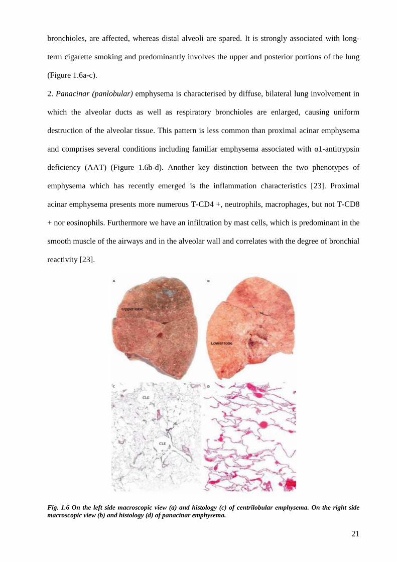
1. Proximal acinar (centrilobular, centriacinar) emphysema is characterized by distension and
destruction mainly limited to the respiratory bronchioles, with relatively less change peripherally
in the acinus. This pattern results from scarring and focal dilatation of the bronchioles and
adjacent alveoli, which lead to an enlarged airspace or “microbulla” in the center of the
secondary lung lobule. The central or proximal parts of the acini, formed by respiratory
21
bronchioles, are affected, whereas distal alveoli are spared. It is strongly associated with long-
term cigarette smoking and predominantly involves the upper and posterior portions of the lung
(Figure 1.6a-c).
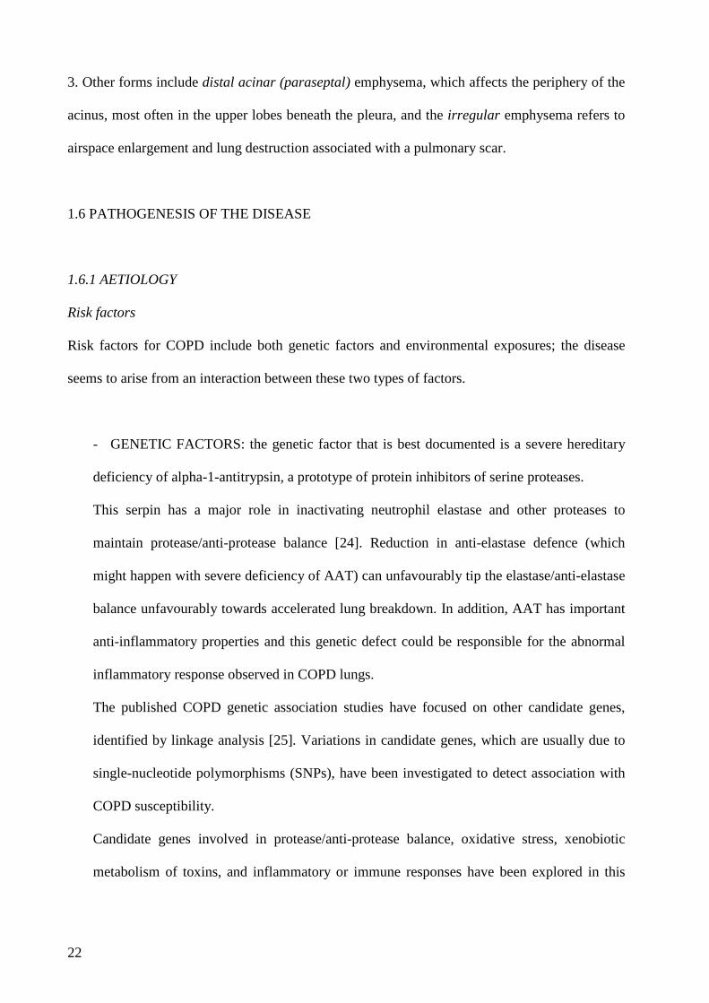
2. Panacinar (panlobular) emphysema is characterised by diffuse, bilateral lung involvement in
which the alveolar ducts as well as respiratory bronchioles are enlarged, causing uniform
destruction of the alveolar tissue. This pattern is less common than proximal acinar emphysema
and comprises several conditions including familiar emphysema associated with α1-antitrypsin
deficiency (AAT) (Figure 1.6b-d). Another key distinction between the two phenotypes of
emphysema which has recently emerged is the inflammation characteristics [23]. Proximal
acinar emphysema presents more numerous T-CD4 +, neutrophils, macrophages, but not T-CD8
+ nor eosinophils. Furthermore we have an infiltration by mast cells, which is predominant in the
smooth muscle of the airways and in the alveolar wall and correlates with the degree of bronchial
reactivity [23].
Fig. 1.6 On the left side macroscopic view (a) and histology (c) of centrilobular emphysema. On the right side macroscopic view (b) and histology (d) of panacinar emphysema.
22
3. Other forms include distal acinar (paraseptal) emphysema, which affects the periphery of the
acinus, most often in the upper lobes beneath the pleura, and the irregular emphysema refers to
airspace enlargement and lung destruction associated with a pulmonary scar.
1.6 PATHOGENESIS OF THE DISEASE
1.6.1 AETIOLOGY
Risk factors
Risk factors for COPD include both genetic factors and environmental exposures; the disease
seems to arise from an interaction between these two types of factors.
- GENETIC FACTORS: the genetic factor that is best documented is a severe hereditary
deficiency of alpha-1-antitrypsin, a prototype of protein inhibitors of serine proteases.
This serpin has a major role in inactivating neutrophil elastase and other proteases to
maintain protease/anti-protease balance [24]. Reduction in anti-elastase defence (which
might happen with severe deficiency of AAT) can unfavourably tip the elastase/anti-elastase
balance unfavourably towards accelerated lung breakdown. In addition, AAT has important
anti-inflammatory properties and this genetic defect could be responsible for the abnormal
inflammatory response observed in COPD lungs.
The published COPD genetic association studies have focused on other candidate genes,
identified by linkage analysis [25]. Variations in candidate genes, which are usually due to
single-nucleotide polymorphisms (SNPs), have been investigated to detect association with
COPD susceptibility.
Candidate genes involved in protease/anti-protease balance, oxidative stress, xenobiotic
metabolism of toxins, and inflammatory or immune responses have been explored in this
23
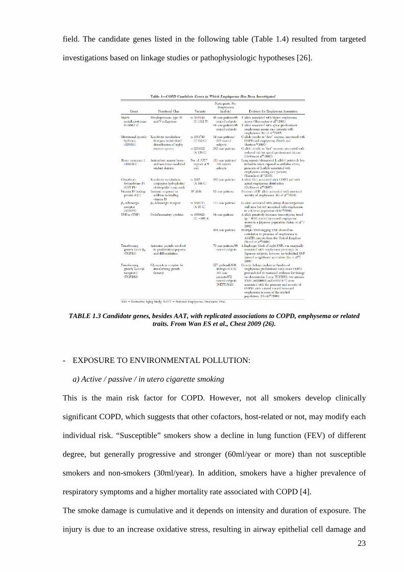
field. The candidate genes listed in the following table (Table 1.4) resulted from targeted
investigations based on linkage studies or pathophysiologic hypotheses [26].
TABLE 1.3 Candidate genes, besides AAT, with replicated associations to COPD, emphysema or related traits. From Wan ES et al., Chest 2009 (26).
- EXPOSURE TO ENVIRONMENTAL POLLUTION:
a) Active / passive / in utero cigarette smoking
This is the main risk factor for COPD. However, not all smokers develop clinically
significant COPD, which suggests that other cofactors, host-related or not, may modify each
individual risk. “Susceptible” smokers show a decline in lung function (FEV) of different
degree, but generally progressive and stronger (60ml/year or more) than not susceptible
smokers and non-smokers (30ml/year). In addition, smokers have a higher prevalence of
respiratory symptoms and a higher mortality rate associated with COPD [4].
The smoke damage is cumulative and it depends on intensity and duration of exposure. The
injury is due to an increase oxidative stress, resulting in airway epithelial cell damage and
24
activation of pro-inflammatory signals, responsible for recruitment of innate immunity cells
and the perpetuation of tissue aggression [27,28].
b) Occupational dusts and chemicals.
Occupational exposures include organic and inorganic dusts and chemical agents and fumes.
A statement published by the American Thoracic Society concluded that occupational
exposures account for 10 to 20% of either symptoms or functional impairment consistent
with COPD [29].
c) Indoor and outdoor air pollution.
There is evidence that indoor pollution from burning biomass fuels for cooking and heating
is an important cause of COPD in many developing countries [30,31], but the exact
pathogenetic mechanisms have not yet been elucidated.
Other recognized risk factors are:
- Altered growth and development of respiratory system: a low weight birth is associated
with reduced values of lung function indices (FEV) in adulthood, implying a state of
incomplete development of the respiratory system [32]. In addition, numerous studies have
demonstrated the effect of pediatric recurrent respiratory infections on the risk of subsequent
decline of FEV.
- Sex: historically COPD hits the males, as they are more exposed to cigarette smoke. The
incidence and prevalence of the disease in women is growing, with the increase in smoking
in recent decades [33].
- Age and ageing: epidemiological studies show that prevalence and impact of the disease
increased with age [5]. Nevertheless, it is unclear whether aging is itself a risk factor for the
disease or whether this association is an expression of cumulative and prolonged exposure to
traditional risk factors.
25
- Respiratory And Systemic Infections: tuberculosis is a risk factor for the development of
COPD. On the other hand it is seen that HIV infection accelerates the onset of emphysema
smoke-related [34].
- Socio-Economic Status: There is a strong evidence of an inverse relationship between
socioeconomic status and the risk of developing COPD, but the factors responsible for this
association are not clear [35].
- Asma: A longitudinal study (Tucson Epidemiological Study of Airway Obstructive
Disease) has shown that adults with asthma have a RR = 12 to develop COPD than non-
asthmatics, adjusted for smoking status [36].
- Chronic Bronchitis: the study of Fletcher and colleagues showed that chronic bronchitis
was not associated with decline in lung function [4]. Subsequent studies, however, have re-
evaluated the role of mucus hypersecretion in the decline of FEV and the existence of an
increased risk of developing COPD in young adults who smoke and who are suffering from
chronic bronchitis [36].
1.6.2 PATHOGENETIC MECHANISM
Despite advances in care to date there are no therapies which halt or reverse the progressive and
accelerated decline in lung function of this condition. Better means of preventing and treating
COPD are urgently needed and a better understanding of the pathological bases is a necessary
requirement.
The discovery of the AAT deficiency in the 60s led to the hypothesis that an imbalance between
proteases and antiproteases in the lung could be the cause of lung damage in all COPD patients
[37]. Smoking induces an increased number of neutrophils and macrophages in the lung and the
release of proteolytic enzymes from these cells. The released proteases, not fully inhibited by
antiproteases, lead to proteolysis of lung connective tissue (more specifically of elastin) and
emphysema. However, the pathogenetic view of COPD has expanded significantly from what
was understood in past decades. Moving beyond the original protease/antiprotease hypothesis, T-
26
lymphocytes have been identified as a key component of the inflammatory response, thus
introducing the concept that adaptive immunity may be centrally involved in the pathogenesis of
the disease [38]. Indeed, recent studies have convincingly shown that emphysema can be
produced in experimental models, not only by manipulation of proteolitic pathways, but also
targeting CD8 T-cell responses [39]. Distinctive immune features, such as lymphoid follicles
rich in B and T cells are found in lung tissue of patients with COPD, at least in the severe stages
of the disease [40]. Altered T and B cell responses are also seen in the peripheral blood of
patients with COPD, indicating that immunological alteration extends beyond the lung.
Despite the well documented presence of inflammation in COPD, it is unclear what steps are
involved in the genesis and progression of the disease. The new concept emerging from the most
recent observations, to which substantial contribution was gives by our group, is that persistence
of the immune activation in COPD could be due to an autoimmune reaction secondary to the
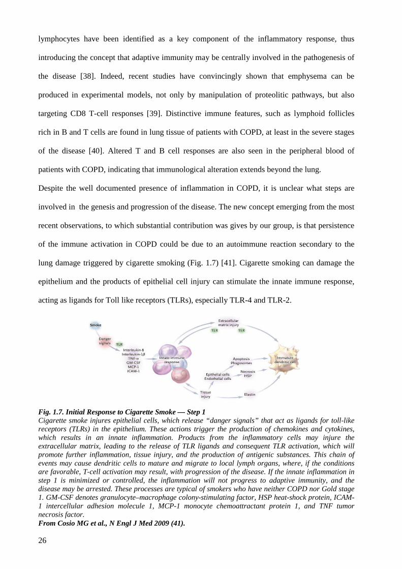
lung damage triggered by cigarette smoking (Fig. 1.7) [41]. Cigarette smoking can damage the
epithelium and the products of epithelial cell injury can stimulate the innate immune response,
acting as ligands for Toll like receptors (TLRs), especially TLR-4 and TLR-2.
Fig. 1.7. Initial Response to Cigarette Smoke — Step 1 Cigarette smoke injures epithelial cells, which release “danger signals” that act as ligands for toll-like receptors (TLRs) in the epithelium. These actions trigger the production of chemokines and cytokines, which results in an innate inflammation. Products from the inflammatory cells may injure the extracellular matrix, leading to the release of TLR ligands and consequent TLR activation, which will promote further inflammation, tissue injury, and the production of antigenic substances. This chain of events may cause dendritic cells to mature and migrate to local lymph organs, where, if the conditions are favorable, T-cell activation may result, with progression of the disease. If the innate inflammation in step 1 is minimized or controlled, the inflammation will not progress to adaptive immunity, and the disease may be arrested. These processes are typical of smokers who have neither COPD nor Gold stage 1. GM-CSF denotes granulocyte–macrophage colony-stimulating factor, HSP heat-shock protein, ICAM-1 intercellular adhesion molecule 1, MCP-1 monocyte chemoattractant protein 1, and TNF tumor necrosis factor. From Cosio MG et al., N Engl J Med 2009 (41).
27
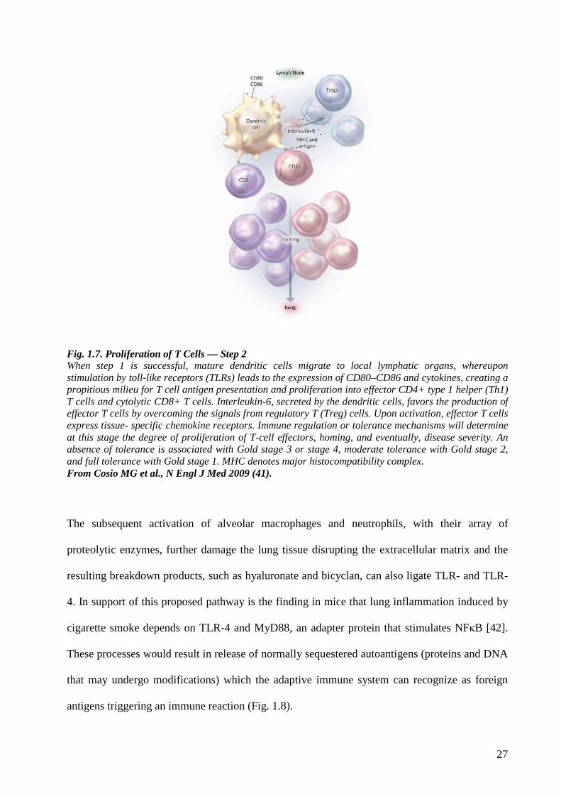
Fig. 1.7. Proliferation of T Cells — Step 2 When step 1 is successful, mature dendritic cells migrate to local lymphatic organs, whereupon stimulation by toll-like receptors (TLRs) leads to the expression of CD80–CD86 and cytokines, creating a propitious milieu for T cell antigen presentation and proliferation into effector CD4+ type 1 helper (Th1) T cells and cytolytic CD8+ T cells. Interleukin-6, secreted by the dendritic cells, favors the production of effector T cells by overcoming the signals from regulatory T (Treg) cells. Upon activation, effector T cells express tissue- specific chemokine receptors. Immune regulation or tolerance mechanisms will determine at this stage the degree of proliferation of T-cell effectors, homing, and eventually, disease severity. An absence of tolerance is associated with Gold stage 3 or stage 4, moderate tolerance with Gold stage 2, and full tolerance with Gold stage 1. MHC denotes major histocompatibility complex. From Cosio MG et al., N Engl J Med 2009 (41).
The subsequent activation of alveolar macrophages and neutrophils, with their array of
proteolytic enzymes, further damage the lung tissue disrupting the extracellular matrix and the
resulting breakdown products, such as hyaluronate and bicyclan, can also ligate TLR- and TLR-
4. In support of this proposed pathway is the finding in mice that lung inflammation induced by
cigarette smoke depends on TLR-4 and MyD88, an adapter protein that stimulates NFκB [42].
These processes would result in release of normally sequestered autoantigens (proteins and DNA
that may undergo modifications) which the adaptive immune system can recognize as foreign
antigens triggering an immune reaction (Fig. 1.8).
28
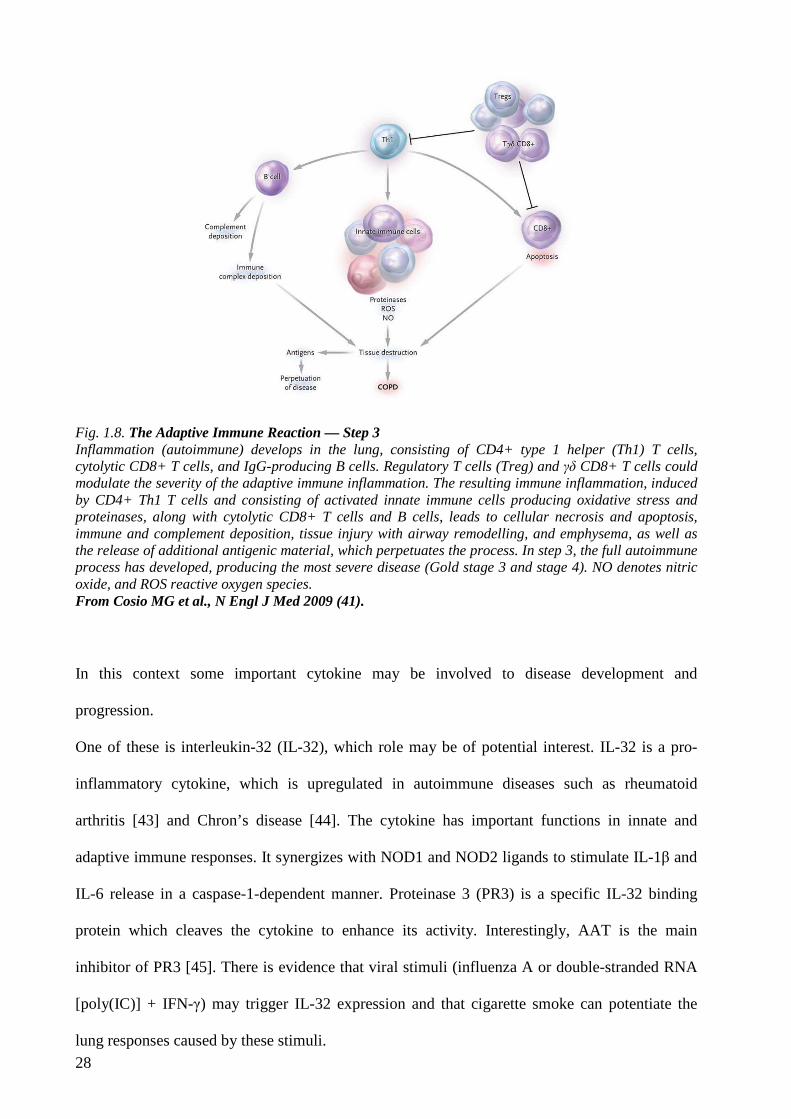
Fig. 1.8. The Adaptive Immune Reaction — Step 3 Inflammation (autoimmune) develops in the lung, consisting of CD4+ type 1 helper (Th1) T cells, cytolytic CD8+ T cells, and IgG-producing B cells. Regulatory T cells (Treg) and γδ CD8+ T cells could modulate the severity of the adaptive immune inflammation. The resulting immune inflammation, induced by CD4+ Th1 T cells and consisting of activated innate immune cells producing oxidative stress and proteinases, along with cytolytic CD8+ T cells and B cells, leads to cellular necrosis and apoptosis, immune and complement deposition, tissue injury with airway remodelling, and emphysema, as well as the release of additional antigenic material, which perpetuates the process. In step 3, the full autoimmune process has developed, producing the most severe disease (Gold stage 3 and stage 4). NO denotes nitric oxide, and ROS reactive oxygen species. From Cosio MG et al., N Engl J Med 2009 (41).
In this context some important cytokine may be involved to disease development and
progression.
One of these is interleukin-32 (IL-32), which role may be of potential interest. IL-32 is a pro-
inflammatory cytokine, which is upregulated in autoimmune diseases such as rheumatoid
arthritis [43] and Chron’s disease [44]. The cytokine has important functions in innate and
adaptive immune responses. It synergizes with NOD1 and NOD2 ligands to stimulate IL-1β and
IL-6 release in a caspase-1-dependent manner. Proteinase 3 (PR3) is a specific IL-32 binding
protein which cleaves the cytokine to enhance its activity. Interestingly, AAT is the main
inhibitor of PR3 [45]. There is evidence that viral stimuli (influenza A or double-stranded RNA
[poly(IC)] + IFN-γ) may trigger IL-32 expression and that cigarette smoke can potentiate the
lung responses caused by these stimuli.
29
Of interest, we recently reported an increased expression of IL-32 in lung tissue of patients with
COPD, where it was co-localized with TNFa and correlated with the degree of airflow
obstruction, suggesting that IL-32 could be a potentially useful biomarker to evaluate the
progression of the disease [46].
1.7 LUNG CANCER
Lung cancer is currently the leading cause of cancer death in the world with a 5-year survival of
about 15%, despite the use of increasingly aggressive therapies [47]. Currently, lung cancer has a
maximum incidence between 55 and 65 years and it is responsible for 32% of total deaths for
cancer in men and 25% in women [47]. This high mortality closely linked to the characteristics
of the tumor and to the delay in diagnosis of this disease which presents a rather low percentage
of patients (approximately 20%) with early stage cancer.
The World Health Organization has included in the new classification of lung cancer 4 main
histological types [48]; however, under therapeutic and prognostic purposes the most useful and
widespread differentiation of lung cancer involves two major groups, defined as small cell lung
carcinoma (SCLC) and non-small cell lung cancer (NSCLC).
Small cell lung cancer is a extremely malignant tumor, with a very high mortality rate and
molecular pathogenic pattern completely different from NSCLC.
The NSCLC category can be divided in different histological types, among which the most
important are the squamous cell carcinoma and the adenocarcinoma.
The adenocarcinoma classification was recently revised [49] with the abolition of the
bronchioloalveolar carcinoma and mixed subtype adenocarcinoma. By contrary, invasive
adenocarcinomas are classified by predominant pattern after using comprehensive histologic
subtyping with lepidic, acinar, papillary, and solid patterns.
Adenocarcinoma incidence is increased in the last two decades, in parallel with the decrease in
the incidence of squamous form [47]. In Europe, they account for 30% of bronchopulmonary
30
carcinomas while in North America they already are the most common histological type (about
50%). The increased incidence is probably related to the use of filter cigarettes and the
consequent reduction in the size of the particulates and increase of nitrates. It has also been
described the association with pre-existing lung scars caused by eg tuberculosis or pulmonary
infarcts.
Worldwide, tobacco smoking is associated with more of 90% of cases of lung cancer [47] . In
more developed countries, the incidence and mortality rates are generally declining, reflecting
previous trends in smoking prevalence. However, in less developed countries lung cancer rates
are predicted to continue to increase due to endemic tobacco use [50] . There is a clear role for
genetics since only 15% of lifetime smokers develop lung cancer and 10% of lung cancers occur
in never-smokers especially in women [51] and in Asiatic women in particular [52] .
One important hypothesis in these categories of patients is the presence of a genetic background
that can predispose to lung cancer in the absence of external environmental pollutants [52].
In this field the two most important recognized oncogenes in tumour developing and growing are
EGFR e KRAS.
Epidermal growth factor receptor (EGFR or ErbB-1) is a tyrosine kinase receptor belonging to
the ErbB family; it is widely expressed in NSCLC (40-80% of NSCLC) and plays a significant
role in carcinogenesis through improper activation of EGFR Tirosin-Kinase (TK) domaine which
results in increased malignant cell survival, proliferation, invasion and metastasis. The TK
activity of EGFR may be dysregulated by various oncogenic mechanisms, including the presence
of harbored mutations in the TK domain of EGFR [53-55]. Overall, the frequency of EGFR
mutations is 5–20%, depending on the populations studied [56].
RAS is one of the important molecules in the downstream of EGFR signaling pathway. Wild-
type forms of ras proteins have GTPase activity and their activation is strictly dependent on the
bond with alternative forms of GDP (inactive) or GTP (active), allowing tight control of signal
transduction [57]. These proteins acquire oncogenic function through point mutations at codons
12, 13, or 61 of exon 2 that abolish the GTPase function giving therefore a constitutionally active
31
conformation and deregulating thereby cell proliferation. For these reasons mutated Ras genes,
especially K-ras, observed among 20~30% NSCLC patients, have been implicated in the
pathogenesis and prognosis of lung cancer [58].
In 1991 Slebos and after ten years Ahrendt found that k-ras mutations are found almost
exclusively in patients with lung adenocarcinoma and smoking history. In addition, Feng
reported that exposure to this carcinogen induces alterations at the codon 12 of K-ras in the
human bronchial epithelium, suggesting a direct molecular mechanism of smoking in human
lung carcinogenesis. Moreover NSCLC patients with K-ras mutations are associated with
unfavorable prognosis [59].
Concerning lung cancer treatment, surgery actually represents the cornerstone of therapy for
NSCLC even if global resectability still ranges from 10 to 25% [60]. With strict pre-operative
selection criteria, the healing rate in patients undergoing complete resection for NSCLC in early
stage (I-II) ranges between 40 and 70% [61]. However, even in these cases, the possibility of
loco-regional and/or distance recurrences is not an uncommon event, representing the main
factor affecting long-term survival.
For this reason, the use of integrated approaches with adjuvant regimen of chemo- and / or
radiotherapy has been recently introduced even in early stages in order to reduce this risk [62].
These integrated approaches have enabled a further increase in 5 year overall survival, but
according to most authors, new therapeutic protocols based on traditional chemotherapy does not
seem to offer further opportunities for improvement of results [63].
In addition to their carcinogenic activity, EGFR and KRAS have emerged as two of the most
relevant targets for cancer treatment.
Gefitinib and erlotinib are small-molecule reversible tyrosine kinase inhibitors (TKIs) that
selectively target EGFR. The presence of somatic mutations in the tyrosine kinase domain of
EGFR (exon 18-21) involving the ATP-binding pocket of the receptor was shown to represent
the most important predictive marker [53-55]. For these patients, the response rate to gefitinib
32
and erlotinib is approximately 75%, suggesting that these mutations, at least in part, drive
malignant transformation [56,64].
In contrast, somatic mutations in exon 2 (codon 12-13) of KRAS have been associated with
primary resistance to EGFR inhibitors. EGFR and KRAS mutations seem to be mutually
exclusive, which is consistent with the idea that different alterations of the same pathway are
involved in lung carcinogenesis [65].
Regarding lung cancer long-term results, TNM staging system [66], is currently the most
important and accurate prognostic instrument. However, it still presents significant difficulties in
predicting the effectively outcome of individual patients. Some authors has recently proposed the
use of PET-CT scan, and in particular the maximum standard uptake value (SUVmax), as new
tool to predict tumor biologic aggressiveness [67]. It is therefore understandable how a greater
knowledge of molecular and histological cancer phenotypes associated to patient characteristics,
able to predict individual outcomes and treatments, is therefore desirable.
1.8 COPD AND LUNG CANCER
It is becoming increasingly evident that COPD and lung cancer are strictly related.
Indeed COPD has recently established as important risk factor for developing lung cancer, even
independently from the effects of smoking. Indeed, it has been shown that the risk of lung cancer
is increased in smokers with COPD up to 6-fold compared to smokers with comparable cigarette
exposure, but without COPD [68,69]. It has also been shown that the presence of COPD
increased lung cancer mortality even among nonsmokers [70].
In addition, lung cancer is also a leading cause of morbidity and mortality in patients with COPD
as 33% of patients died of lung cancer over a 14.5-year follow-up [71]. Furthermore 50–70% of
patients diagnosed with lung cancer have spirometric evidence of COPD [72]]. As a practical
consequence of the epidemiological associations between COPD and lung cancer, an important
question is whether the relationship between lung cancer and COPD is subtype specific. A small
33
case – control study [73] showed that airflow obstruction is primarily a risk factor for squamous
cell lung cancer (odds ratio = 3.49, 95% CI = 1.63 to 18.5; P = .006), whereas symptoms of
chronic bronchitis without COPD is a risk factor (risk greater than fourfold) for adenocarcinoma
of the lung. In a subset analysis, having concurrent bronchitic symptoms and COPD was
associated with a more than threefold increased risk for squamous cell carcinoma. Despite these
striking epidemiological associations, the mechanisms by which COPD can be linked to lung
cancer are poorly understood.
1.8.1 Risk factors
Primarily, the main risk factor for the onset of both diseases is cigarette smoking, but they
probably also share a common familial component and environmental risk factors other than
smoking. This supposition is reinforced by the fact that only a fraction of smokers (around 15%)
will develop lung cancer and/or COPD in their lifetime [1,74] which suggests a different
individual susceptibility to the risk of lung cancer and/or COPD or time of disease onset.
Some common elements in the pathogenesis of COPD and lung cancer include oxidants, familiar
and genetic predisposition (p53, Rb, K-ras), peptides and endopeptidases (bombesin-like),
dysregulation of growth factor expression and among others inflammation.
While the mechanism of cigarette smoking damage in COPD has been previously treated, for
lung carcinogenesis, the action is probably related to DNA mutations induction.
Carcinogenesis is a complex process characterised by the accumulation of multiple independent
genetic alterations, often involving overexpression of oncogenes and loss of tumour suppressor
genes. These genetic alterations disrupt the normal regulation of cell signalling pathways,
essential for the control of cell growth, differentiation and apoptosis [75]. The observation of
familial aggregation of emphysema dates back more than 200 years, and studies conducted 30
years ago reveal a familial aggregation of lung cancer associated with COPD that is not
explained by α1-antitrypsin genotype or smoking history [76,77]. Familial aggregation is well
34
documented for multiple cancers, although it has been difficult to separate the contributions of
genetic differences and environmental exposures in the development of lung cancer.
A number of linkage studies [78] have investigated genes involved in both lung function and
COPD and candidate susceptibility genes involved in COPD and lung cancer. These studies have
led to the identification of markers on chromosome 6q for lung cancer and abnormal lung
function and on chromosome 12 for lung cancer, COPD, and lung function, as well as numerous
other candidate susceptibility genes involved in detoxification, immune regulation, matrix
remodeling, DNA repair, cellular proliferation, and tumor suppression [78]. Among the genetic
connections between COPD and lung cancer, it is particularly interesting to note that both the Z
and the S alleles of the α1-antitrypsin gene are more common in patients with lung cancer
compared with the general population, as is a polymorphism in the neutrophil elastase gene
[79,80], suggesting that an imbalance between neutrophil elastase and α 1-antitrypsin may
contribute to the development of both COPD and lung cancer [81].
The role of epigenetic modifications in the common pathogenesis of COPD and lung cancer also
deserves attention.
Although there is growing evidence implicating DNA methylation, histone deacetylation, and
protein phosphorylation in lung cancer pathogenesis [82,83], this knowledge is only now being
applied to COPD, alone or when associated with lung cancer [84].
It is plausible that several candidate risk genes and pathways identified by lung cancer studies
may be shared by these two diseases and could constitute potential targets for the newly
developed drugs (eg, demethylating agents and histone deacetylase – inhibiting agents) that
modify epigenetic alterations.
The inflammatory paradigm is undoubtedly one of the most fascinating theories to connect
COPD and lung cancer and it has acquired new impetus by the recent discoveries in the COPD
pathogenesis.
35
1.8.2 The inflammatory theory
As described above, COPD is characterized by the accumulation of macrophages, CD4+ and
CD8+ T cells, dendritic cells, B cells and neutrophils, particularly in smaller airways and lung
parenchyma, and the severity of COPD is associated with the degree of infiltration by these
inflammatory cells [1].
Moreover, chronic inflammation may play a significant role in the pathogenesis of lung cancer as
a tumour promoter. A causal relation between inflammation and cancer was initially proposed by
Galen and later by Virchow [85], who noticed the infiltration of leucocytes in malignant tissues
and suggested that cancers arise at regions of chronic inflammation. The hypothesized
carcinogenetic mechanism in COPD patients states that cigarette smoke initially creates a direct
parenchimal damages that up-regulates the cytokines production, such as interleukin (IL)-1b, and
the Th-1 cytokines, which can promote the inflammatory response through typical COPD T-
lymphocytes and finally resulting in an overproduction of cytokines, such as IL-6, IL-8, and IL-
10. Some of the latter mediators can inhibit apoptosis, interfere with cellular repair, and promote
angiogenesis. All in all, chronic inflammation may play a pathogenic role in lung cancer by
amplifying the initial mutagenic damage and enhancing both tumour growth and metastasis. IL-
8, for example, has been shown to be pro-oncogenic, such as releasing BCL-2, but also
suppresses oncogenes, such as p53, hence minimizing apoptosis while inducing cell
transformation. In addition, among mechanisms that regulate the progression or suppression of
tumor growth, the role of cellular senescence is becoming increasing appreciated. Cellular
senescence permanently arrests cell growth in tissues at risk for malignant transformation,
particularly those that experience prolonged inflammation, such as the airways and the lung
parenchyma in smokers with COPD. On the other hand, senescent cells undergo profound
modifications which can have deleterious effects on the tissue microenvinronment. The most
significant of these alterations is the acquisition of a senescence-associated secretory phenotype
(SASP) that promotes the secretion of several proinflammatory cytokines (particularly IL-6 and
IL-8), growth factors and metalloproteinases, which could eventually promote tumor progression
36
and invasiveness. Of note, senescent cells have the ability to influence the populations of
macrophages and lymphocytes infiltrating nascent tumors, shifting the balance from cell
populations associated with tumor suppression (M1 macrophages, Th1 T-lymphocytes) to those
that instead promote tumor progression (M2 macrophages, Th2 and T regulatory lymphocytes).
The mechanism of perpetuation of damage typical of COPD could also explain in these patients
the risk of lung cancer irrespective of active smoking [70]. In fact, after quitting smoking, lung
cancer risk remains increased in patients with COPD, though this risk is superior in those who
continue to smoke [70]. The close connection between COPD, inflammation, and lung cancer is
even more apparent in light of recent findings that point to a possible relationship between
inhaled corticosteroids and reduced lung cancer risk in COPD patients [86,87].
Complementary to this is the observation that the incidence of lung cancer is associated with
specific stages of COPD severity. Lung cancer is assigned as the cause of death in 33% of
patients with mild-to-moderate COPD with a decreased in patients with more severe disease
[88,89].
According to this data, in our experience we did not observed cases of lung malignancy in
patients with end-stage COPD candidates to lung transplantation, differently from other end-
stage chronic inflammatory diseases such as scleroderma, sarcoidosis and especially pulmonary
fibrosis.
For these reasons our research group has proposed a pathogenetic hypothesis that, in agreement
with recent autoimmune theory, attributes to the peculiar inflammatory phenotype of COPD
advanced stages a protective and surveillance role against neoplastic development.
As previously reported, the T regulatory cells and the associated cytokine pattern play a strategic
role in the interaction between COPD and lung cancer, although a complete understanding of the
process is still far away.
In this field, the analysis of TH17 cells and the IL17 cytokine could really represent the link
between these two important diseases.
37
1.8.3 Pathway axys IL17- R23
Th17 cells are a critical component of the adaptive immune response and have also been
implicated in chronic inflammatory diseases and autoimmune diseases [90], although their
primary function appears to be the clearance of pathogens that are not adequately handled by
Th1 and Th2 cells, especially extracellular bacteria and fungi [91]. The cytokine
microenvironment influences the differentiation of naive T cells into Th17 cells. The Th17
lineage depends on the presence of TGF-b1 and IL-6, found in elevated concentrations
particularly in chronic obstructive pulmonary disease (COPD) [92].
In effect, an increase in Th17 cells was observed in patients with COPD compared with current
smokers without COPD and healthy subjects. Human Th17 cells differ from other cell subsets in
their potency to induce proinflammatory cytokines in bronchial epithelial cells, airway
fibroblasts and smooth muscle cells and above all they secrete the cytokine IL-17 [93], which
plays an important proinflammatory role in COPD.
The IL-17 family comprises of six members (IL-17A–17F) [94] and five receptors (IL-17RA–
17RE) [95].
IL-17RA is the largest member of the IL-17R family and at least four ligands are mediated
through this subunit [96].
The expression of IL-17RA is up-regulated by cytokines such as IL-15 (produced by a range of
non-T-cells, including macrophages and IL- 21 [97] on CD8+ T-cells, and so may play an
important role in COPD.
IL-23 plays a key role in the maintaining and expanding the Th17 cell lineage over time to
release IL-17 [98] and is secreted from APCs such as dendritic cells. IL23 is released in response
to inflammatory signals and therefore is continually expressed in chronic inflammation.
IL-17 induces epithelial cells to produce antimicrobial peptides (such as b defensins) and
numerous chemokines, such as TNF-a, IL-1b, IL-6, GM-CSF, granulocyte colony-stimulating
factor and IL-8 [99]. All these findings clearly indicate that IL-17 is able to generate an
38
intriguing crosstalk between the adaptive and innate immune systems, regulating an efficient
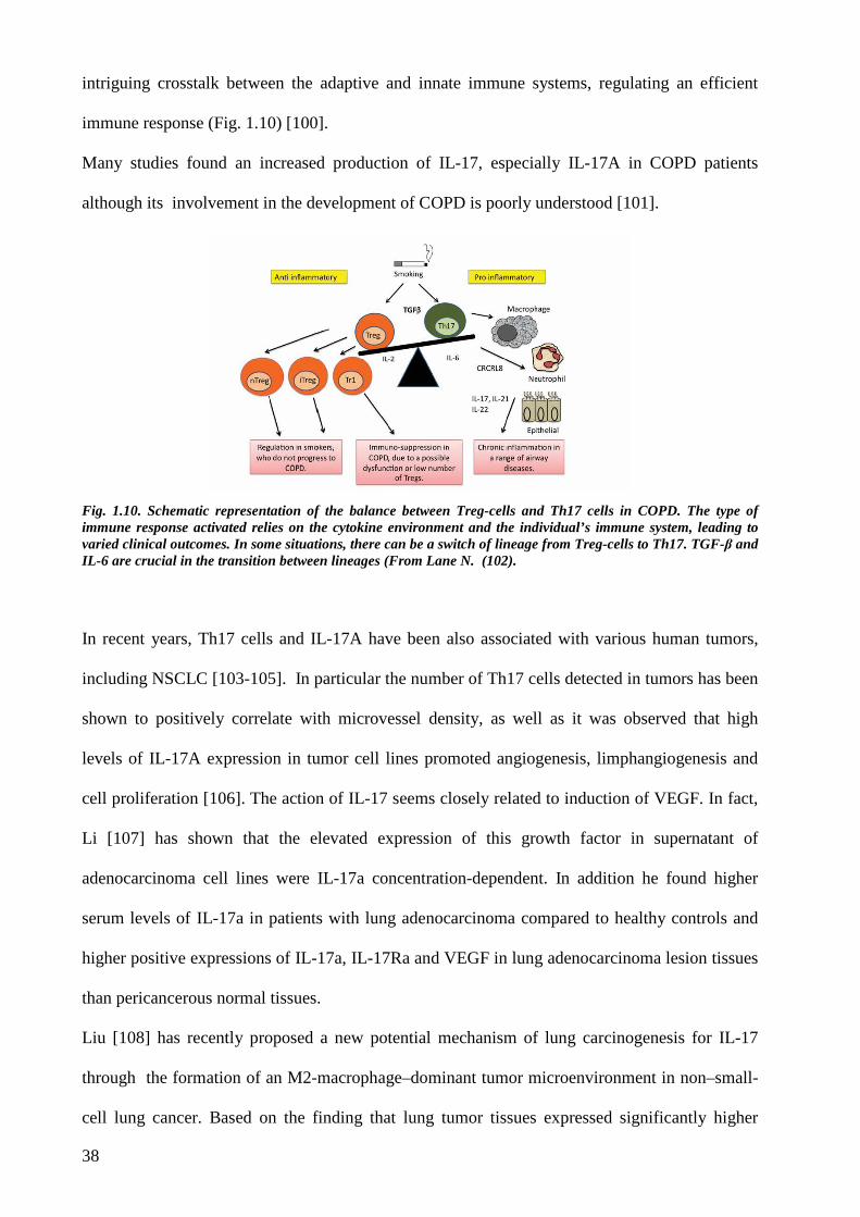
immune response (Fig. 1.10) [100].
Many studies found an increased production of IL-17, especially IL-17A in COPD patients
although its involvement in the development of COPD is poorly understood [101].
Fig. 1.10. Schematic representation of the balance between Treg-cells and Th17 cells in COPD. The type of immune response activated relies on the cytokine environment and the individual’s immune system, leading to varied clinical outcomes. In some situations, there can be a switch of lineage from Treg-cells to Th17. TGF-β and IL-6 are crucial in the transition between lineages (From Lane N. (102).
In recent years, Th17 cells and IL-17A have been also associated with various human tumors,
including NSCLC [103-105]. In particular the number of Th17 cells detected in tumors has been
shown to positively correlate with microvessel density, as well as it was observed that high
levels of IL-17A expression in tumor cell lines promoted angiogenesis, limphangiogenesis and
cell proliferation [106]. The action of IL-17 seems closely related to induction of VEGF. In fact,
Li [107] has shown that the elevated expression of this growth factor in supernatant of
adenocarcinoma cell lines were IL-17a concentration-dependent. In addition he found higher
serum levels of IL-17a in patients with lung adenocarcinoma compared to healthy controls and
higher positive expressions of IL-17a, IL-17Ra and VEGF in lung adenocarcinoma lesion tissues
than pericancerous normal tissues.
Liu [108] has recently proposed a new potential mechanism of lung carcinogenesis for IL-17
through the formation of an M2-macrophage–dominant tumor microenvironment in non–small-
cell lung cancer. Based on the finding that lung tumor tissues expressed significantly higher
39
levels of IL-17 and prostaglandin E2 (PGE2) than normal lung tissues, he speculated on the
possibility that IL-17 served as a chemoattractant to recruit macrophages into the lung tumors
while PGE2 induced differentiation of M2 macrophages.
Based on these fundamental premises, lung cancer associated with COPD seems to possess
specific pathogenetic and morphological features, differently from cancers resulting from simple
smoke damage.
Therefore, a better understanding of these phenotypes could lead to important consequences of
great relevance in therapy and care of patients affected by COPD and lung cancer.

40

41
2. AIM OF THE RESEARCH
This research project focuses on the study of lung cancer in patients with COPD compared to
control groups, composed by healthy smokers and non-smoking patients in order to identify
eventual distinct biohumoral, morphological and molecular phenotypes.

42
43
3. MATERIALS AND METHODS
3.1 STUDY POPULATION
From January 2010 to December 2012 we prospectively enrolled in the study patients submitted
to anatomical surgical resection for non small cell lung cancer (NSCLC) in the Thoracic Surgery
Unit of Padua and Strasbourg.
Inclusion criteria were the follows:
- Patients undergoing major resections (lobectomy or pneumonectomy) associated with
hilar-mediastinal lymphadenectomy; this decision was motivated from the need to have
sufficient non neoplastic lung parenchyma to study the peri-neoplastic tissue remodeling.
- Patients with peripheral lung nodule.
- Patients with suspected or known primary NSCLC.
Exclusion criteria were the follows:
- Patients with inflammatory lesion or with metastatic pulmonary cancer.
- Patients with NSCLC involving central airways in order to avoid distortions of lung
architecture or presence of local and/or systemic inflammatory reactions.
- Patients with chest wall involvement.
- Patients previously treated by chemo and / or radiation therapy.
- Past history of asthma or allergic rhinitis and acute upper respiratory tract infections.
- For COPD patients, presence of exacerbations during the month preceding surgery.
All patients included in the research project were subjected full clinical/biohumoral and
functional analysis.
44
Clinical study
For each subject a detailed medical history with particular attention to respiratory symptoms,
smoking history, concomitant medications and comorbidities was collected.
Venous blood samples, preserved in Ethylene Diamine Tetraacetic acid, were sent to the central
laboratory to evaluate the levels of erythrocyte sedimentation rate and C-reactive protein as
known systemic inflammation signs.
All subjects underwent pulmonary function tests (PFTs) that were performed and evaluated by
an expert pneumologist following the most recent guidelines [109].
Briefly, the following parameters will be measured: vital capacity (CV), inspiratory capacity
(IC), forced vital capacity (FVC), forced expiratory volume in the first second (FEV1), FEV1 to
FVC ratio (FEV1/FVC), functional residual capacity (FRC), residual volume (RV) and total lung
capacity (TLC). To assess the reversibility of the airway obstruction in subjects with a baseline
FEV1/FVC <70 %, measurements will be repeated 15 minutes after the inhalation of 400 µg of
salbutamol. The diffusing capacity for carbon-monoxide (DLCO) will be assessed using the
single breath technique.
Arterial haemogasanalysis was performed to investigate gas exchange (oxygen partial pressure-
PaO2, carbon dioxide partial pressure-PaCO2 and haemoglobin saturation-SHb).
A complete radiological assessment, including chest-x ray, Chest and Abdomen CT scan was
performed in every patients both for oncological and research reasons.
In addition patients routinely underwent 18FDG PET-CT scan with measurement of primary lung
tumor maximal standard uptake value (SUVmax), that is SUV on the highest image pixel in the
parenchymal tumor regions.
In all patients bronchoscopic examination with bronchoalveolar lavage (BAL) was carried out.
BAL, blood and fresh cancer tissue were preserved in frozen tissue bank for future
immunohistochemical and molecular studies.
The collection and preservation of biological materials for frozen bank followed the guidelines
of Regione Veneto and Azienda Ospedaliera di Padova.
45
Patients enrolled in the study were then divided in 3 groups according to respiratory functional
tests and smoking history: COPD patients, smoker patients without COPD and never-smoker
subjects (with normal lung function).
COPD subjects were identified by the presence of fixed airflow limitation (forced expiratory
volume in the first second (FEV1) to forced vital capacity (FVC) ratio < 70%). They will be
subsequently staged on the basis of FEV1 values discriminating smokers with severe COPD
(FEV1<50% predicted) from those with mild/moderate COPD (FEV1>50% predicted).
3.2 SURGICAL PROCEDURE AND LUNG SAMPLING
As previously reported, every patient underwent an anatomical resection associated with
systematic limphadenectomy.
Immediately after the surgical procedure, samples from the nodule and at least three from non-
tumour subpleuric area (2 cm3) were preserved in RNAlater® and then stored in liquid nitrogen
for molecular studies. The rest of lung parenchyma was sent to the Pathological Anatomy
Laboratory and perfused using 4% formaldehyde via bronchial tree with constant pressure of 25
cm of water (Fig. 3.1).
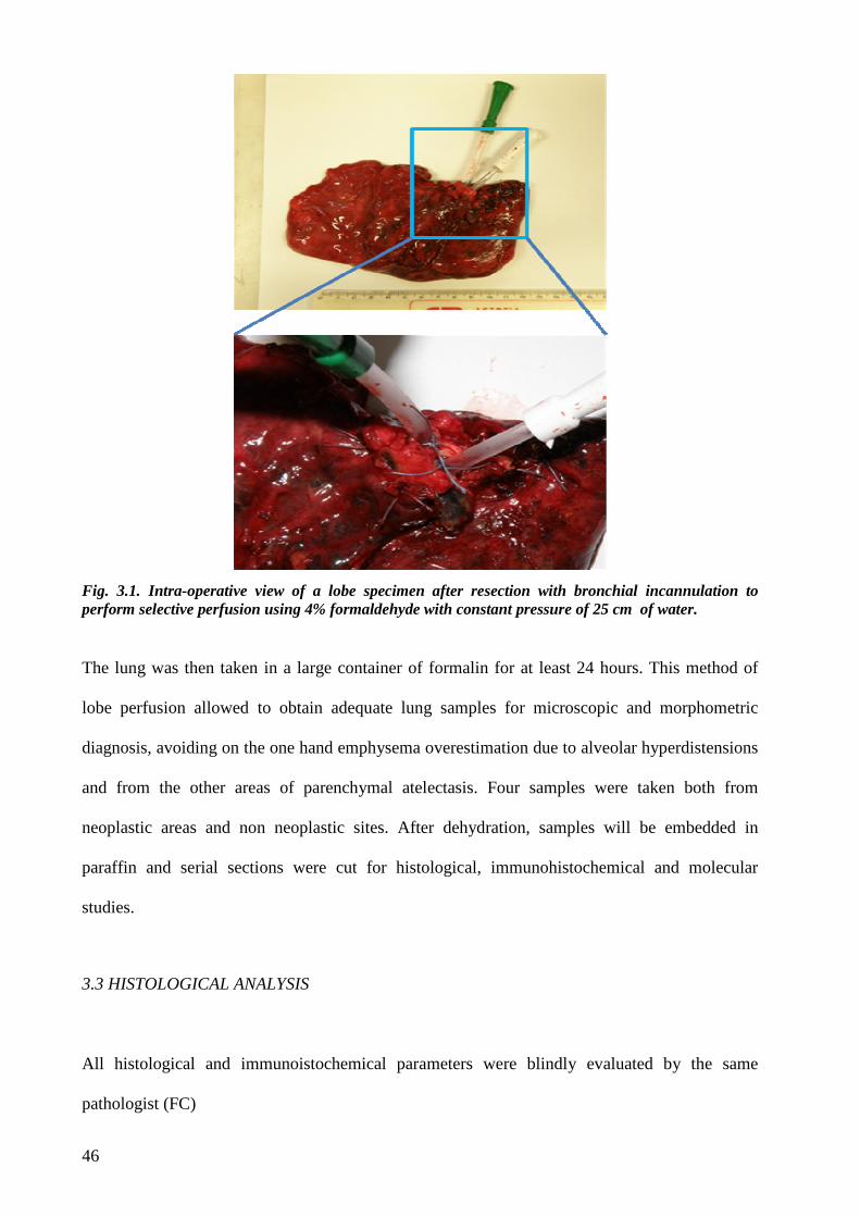
46
Fig. 3.1. Intra-operative view of a lobe specimen after resection with bronchial incannulation to perform selective perfusion using 4% formaldehyde with constant pressure of 25 cm of water.
The lung was then taken in a large container of formalin for at least 24 hours. This method of
lobe perfusion allowed to obtain adequate lung samples for microscopic and morphometric
diagnosis, avoiding on the one hand emphysema overestimation due to alveolar hyperdistensions
and from the other areas of parenchymal atelectasis. Four samples were taken both from
neoplastic areas and non neoplastic sites. After dehydration, samples will be embedded in
paraffin and serial sections were cut for histological, immunohistochemical and molecular
studies.
3.3 HISTOLOGICAL ANALYSIS
All histological and immunoistochemical parameters were blindly evaluated by the same
pathologist (FC)
47
3.3.1 Pathological diagnosis
Tissue sections were stained with haematoxylin and eosin and Masson’s stains. Histo-
pathological evaluation of the tumor were done by an expert Pathologist according to the latest
revision of lung cancer classification [48]: in particular, in case of adenocarcinoma, a precise
quantification of the different histological pattern (lepidic, acinar, papillary and solid) percentage
was done [49].
pTNM staging was conducted in accordance with the 7th edition of 2009 [66].
3.3.2 Morphological evaluations
In all neoplastic lesions were evaluated and quantified the following parameters:
- Inflammation, necrosis and fibrosis were quantified by an expert pathologist in
peritumour and intratumour areas and the amount was expressed both semiquantitatively
using a score system from 0 to 3 (no positivity, 0; mild, 1; moderate, 2; strong, 3) both as
percentage of extension:1: <30% of neoplastic and perineoplastic areas; 2: from 30 to 60%
and 3>60%.
- Four µm-thick sections were cut and processed for immunohistochemical analysis of ki-
67. Briefly after dewaxing and hydration, sections were incubated in citrate buffer 5 mM at
pH 6.0 in a microwave oven for 30 min for antigen retrieval. Afterwards, sections were
treated with blocking serum (Ultratech HRP Kit, Immunotech, Beckman Coulter, USA) and
incubated for 60 min with the primary monoclonal antibody anti-ki-67 (MIB-1, Immunotech,
Marseille, France) at a concentration of 1:50. Sections were subsequently incubated with a
secondary biotinylated antibody for 10 min and then with streptavidin-biotin complex
conjugated to horseradish peroxidase for 10 min (Ultratech HRP Kit, Immunotech, Beckman
Coulter, USA). Immunoreactivity was visualized with diaminobenzidine (DAB, Dako,
Glostrup, Denmark). Finally, the sections were counterstained with Mayer’s hematoxylin.
Negative controls for nonspecific binding were processed omitting the primary antibodies and
48
revealed no signal. Ki-67 positivity was evaluated by using an assisted computerized
morphometric analysis counting at least 100 cells in the most representative areas. Data were
expressed as number of positive cells/total cell count % and ki-67 quantification represented
the ki-67 labeling index.
3.3.3 Characterization of the peri-and intra-tumor IL-17 cytokine pattern
For immunohistochemistry, deparaffinized sections were treated with 3% hydrogen peroxide in
phosphate-buffered saline (PBS) for 10 minutes and incubated for 30 minutes at 90°C for antigen
retrieval. Next, the specimens were individually incubated for 12 hours at 4°C with the
polyclonal antibody diluted in 2% bovine serum albumin before use anti–IL-17 (1:100)
(AbCAM). The sections were washed and incubated with the streptavidinperoxidase conjugate
(Dako) for 30 minutes.
The slides were washed with HRP labeled polymer anti-rabbit (Dako) for 30’ and
immunoreactivity was visualized with 3-3’-diaminobenzidine (Dako). Negative controls for
nonspecific binding were processed omitting the primary antibody and revealed no signal. The
sections were counterstained with hematoxylin.
Data were expressed using a combined score from staining intensity ( graded with score 1: mild,
score 2: moderate, and score 3: strong ) and percentage of positive cells.
3.4 GENETIC ANALYSES
All paraffin-embedded samples were evaluated by a pathologist in order to assess the tumour
tissue quality and quantity. Molecular analysis was performed when the tumour tissue comprised
more than 50% of the entire sample. DNA was extracted using a QIAmp DNA kit (Qiagen,
Milan, Italy), according to the manufacturer’s instructions. Agarose gel electrophoresis was used
to control its quality and quantification was performed by spectrophotometry.
49
A total of 100 ng of genomic DNA was used for polymerase chain reaction (PCR) to amplify
exons 18, 19, 20, 21 of EGFR gene and exon 2 of KRAS gene by using primers, sequences and
amplification conditions according to previous studies (8, 17). PCR products were purified using
a pre-sequencing kit (Amersham Biosciences, Little Chalfont Buckinghamshire, UK).
Subsequently, they were sequenced with both forward and reverse primers using BigDye
Terminator v1.1. Cycle Sequencing Kit (Applied Biosystems, Foster City, CA, USA).
Automated sequencing was carried out using an ABI 3130xl DNA sequence (Applied
Biosystems).
3.5 ELECTRONIC DATABASE
A multidisciplinary electronic database (Excel, Microsoft Office Professional 2007) was built
following the privacy rules.The database contains all the clinical, radiological, functional,
pathological and molecular data and was used for statistical analysis.
The database did not include any personal identification label and was held in secure password
protected storage under the responsibility of the Unit Coordinators (Prof. Rea, Prof. Saetta and
Prof. Calabrese) in accordance with the requirements of the Health Information.
The study, conforming to the Declaration of Helsinki, was approved by our local ethic
committee and patients signed an informed consent
3.6 STATISTICAL ANALYSIS
All cases were coded and the measurements were made without knowledge of clinical data.
Normality of distribution for quantitative variables was assessed by means of Shapiro-Wilcox
statistics. Non normal variables were log-transformed. Group data were expressed as mean and
SEM, or as median and IQR when appropriate. To compare means from two different groups we

50
used the unpaired 2-tail Student’s t test or one-way analysis of variance (ANOVA) for normal
data; for non-normal data Mann-Whitney’s U test or Kruskall-Wallis test were performed.
Categorical data were analyzed by the chi-square test.
Statistical analysis was performed using SAA statistical software version 9.1 (SAS institute,
Carry, NC, USA). P values lower than 0.05 were considered statistically significant.
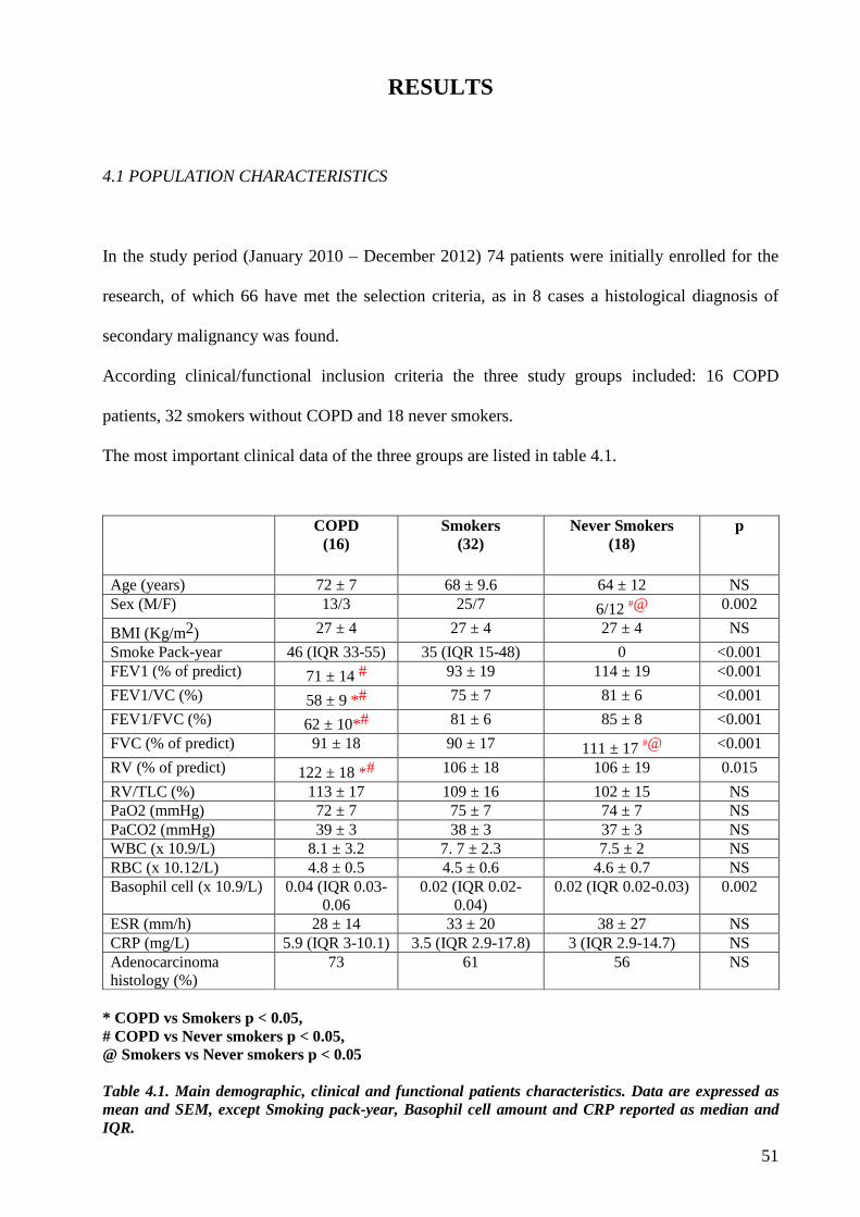
51
RESULTS
4.1 POPULATION CHARACTERISTICS
In the study period (January 2010 – December 2012) 74 patients were initially enrolled for the
research, of which 66 have met the selection criteria, as in 8 cases a histological diagnosis of
secondary malignancy was found.
According clinical/functional inclusion criteria the three study groups included: 16 COPD
patients, 32 smokers without COPD and 18 never smokers.
The most important clinical data of the three groups are listed in table 4.1.
COPD
(16) Smokers
(32) Never Smokers
(18) p
Age (years) 72 ± 7 68 ± 9.6 64 ± 12 NS Sex (M/F) 13/3 25/7 6/12 #@ 0.002
BMI (Kg/m2) 27 ± 4 27 ± 4 27 ± 4 NS
Smoke Pack-year 46 (IQR 33-55) 35 (IQR 15-48) 0 <0.001 FEV1 (% of predict) 71 ± 14 # 93 ± 19 114 ± 19 <0.001
FEV1/VC (%) 58 ± 9 *# 75 ± 7 81 ± 6 <0.001
FEV1/FVC (%) 62 ± 10*# 81 ± 6 85 ± 8 <0.001
FVC (% of predict) 91 ± 18 90 ± 17 111 ± 17 #@ <0.001
RV (% of predict) 122 ± 18 *# 106 ± 18 106 ± 19 0.015
RV/TLC (%) 113 ± 17 109 ± 16 102 ± 15 NS PaO2 (mmHg) 72 ± 7 75 ± 7 74 ± 7 NS PaCO2 (mmHg) 39 ± 3 38 ± 3 37 ± 3 NS WBC (x 10.9/L) 8.1 ± 3.2 7. 7 ± 2.3 7.5 ± 2 NS RBC (x 10.12/L) 4.8 ± 0.5 4.5 ± 0.6 4.6 ± 0.7 NS Basophil cell (x 10.9/L) 0.04 (IQR 0.03-
0.06 0.02 (IQR 0.02-
0.04) 0.02 (IQR 0.02-0.03) 0.002
ESR (mm/h) 28 ± 14 33 ± 20 38 ± 27 NS CRP (mg/L) 5.9 (IQR 3-10.1) 3.5 (IQR 2.9-17.8) 3 (IQR 2.9-14.7) NS Adenocarcinoma histology (%)
73 61 56 NS
* COPD vs Smokers p < 0.05, # COPD vs Never smokers p < 0.05, @ Smokers vs Never smokers p < 0.05 Table 4.1. Main demographic, clinical and functional patients characteristics. Data are expressed as mean and SEM, except Smoking pack-year, Basophil cell amount and CRP reported as median and IQR.
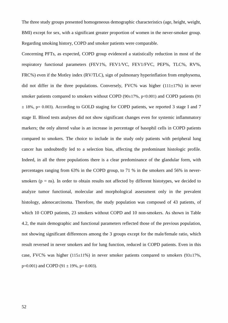
52
The three study groups presented homogeneous demographic characteristics (age, height, weight,
BMI) except for sex, with a significant greater proportion of women in the never-smoker group.
Regarding smoking history, COPD and smoker patients were comparable.
Concerning PFTs, as expected, COPD group evidenced a statistically reduction in most of the
respiratory functional parameters (FEV1%, FEV1/VC, FEV1/FVC, PEF%, TLC%, RV%,
FRC%) even if the Motley index (RV/TLC), sign of pulmonary hyperinflation from emphysema,
did not differ in the three populations. Conversely, FVC% was higher (111±17%) in never
smoker patients compared to smokers without COPD (90±17%, p<0.001) and COPD patients (91
± 18%, p= 0.003). According to GOLD staging for COPD patients, we reported 3 stage I and 7
stage II. Blood tests analyses did not show significant changes even for systemic inflammatory
markers; the only altered value is an increase in percentage of basophil cells in COPD patients
compared to smokers. The choice to include in the study only patients with peripheral lung
cancer has undoubtedly led to a selection bias, affecting the predominant histologic profile.
Indeed, in all the three populations there is a clear predominance of the glandular form, with
percentages ranging from 63% in the COPD group, to 71 % in the smokers and 56% in never-
smokers (p = ns). In order to obtain results not affected by different histotypes, we decided to
analyze tumor functional, molecular and morphological assessment only in the prevalent
histology, adenocarcinoma. Therefore, the study population was composed of 43 patients, of
which 10 COPD patients, 23 smokers without COPD and 10 non-smokers. As shown in Table
4.2, the main demographic and functional parameters reflected those of the previous population,
not showing significant differences among the 3 groups except for the male/female ratio, which
result reversed in never smokers and for lung function, reduced in COPD patients. Even in this
case, FVC% was higher (115±11%) in never smoker patients compared to smokers (93±17%,
p=0.001) and COPD (91 ± 19%, p= 0.003).
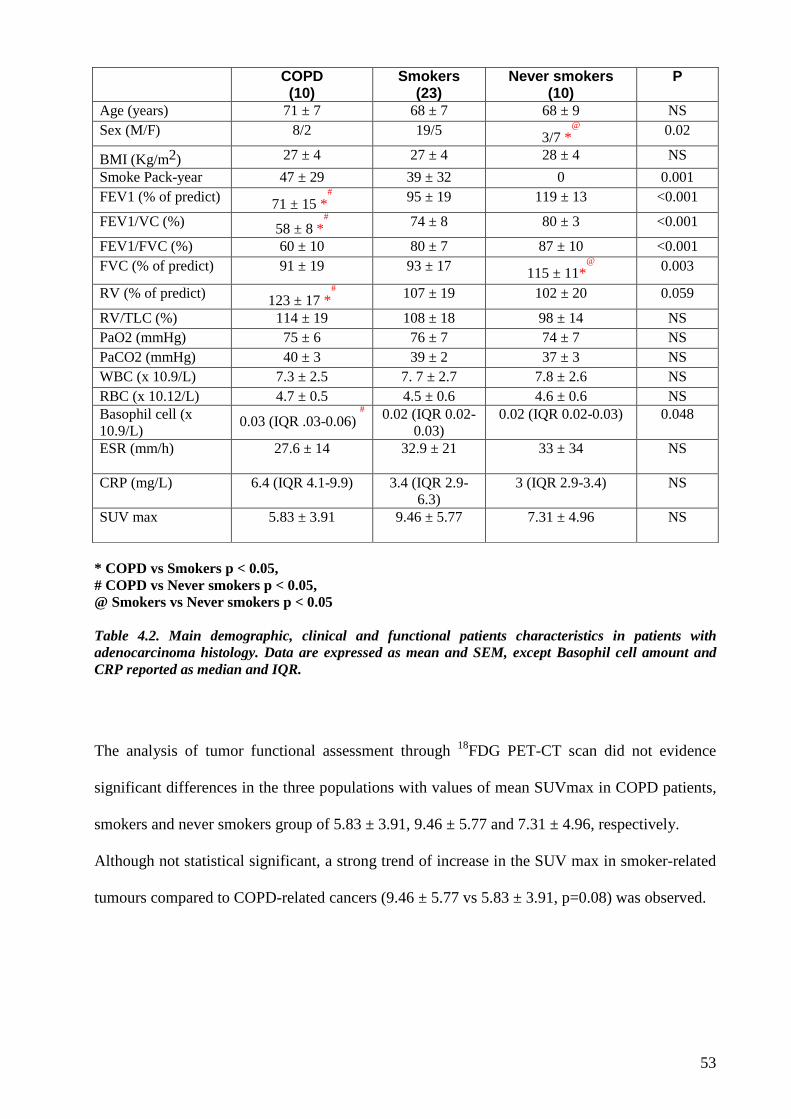
53
COPD (10)
Smokers (23)
Never smokers (10)
P
Age (years) 71 ± 7 68 ± 7 68 ± 9 NS Sex (M/F) 8/2 19/5 3/7 *
@
0.02
BMI (Kg/m2) 27 ± 4 27 ± 4 28 ± 4 NS
Smoke Pack-year 47 ± 29 39 ± 32 0 0.001 FEV1 (% of predict) 71 ± 15 *
#
95 ± 19 119 ± 13 <0.001
FEV1/VC (%) 58 ± 8 *#
74 ± 8 80 ± 3 <0.001
FEV1/FVC (%) 60 ± 10 80 ± 7 87 ± 10 <0.001 FVC (% of predict) 91 ± 19 93 ± 17 115 ± 11*
@
0.003
RV (% of predict) 123 ± 17 *#
107 ± 19 102 ± 20 0.059
RV/TLC (%) 114 ± 19 108 ± 18 98 ± 14 NS PaO2 (mmHg) 75 ± 6 76 ± 7 74 ± 7 NS PaCO2 (mmHg) 40 ± 3 39 ± 2 37 ± 3 NS WBC (x 10.9/L) 7.3 ± 2.5 7. 7 ± 2.7 7.8 ± 2.6 NS RBC (x 10.12/L) 4.7 ± 0.5 4.5 ± 0.6 4.6 ± 0.6 NS Basophil cell (x 10.9/L)
0.03 (IQR .03-0.06) #
0.02 (IQR 0.02-0.03)
0.02 (IQR 0.02-0.03) 0.048
ESR (mm/h) 27.6 ± 14 32.9 ± 21 33 ± 34 NS
CRP (mg/L) 6.4 (IQR 4.1-9.9) 3.4 (IQR 2.9-6.3)
3 (IQR 2.9-3.4) NS
SUV max 5.83 ± 3.91 9.46 ± 5.77 7.31 ± 4.96 NS
* COPD vs Smokers p < 0.05, # COPD vs Never smokers p < 0.05, @ Smokers vs Never smokers p < 0.05 Table 4.2. Main demographic, clinical and functional patients characteristics in patients with adenocarcinoma histology. Data are expressed as mean and SEM, except Basophil cell amount and CRP reported as median and IQR.
The analysis of tumor functional assessment through 18FDG PET-CT scan did not evidence
significant differences in the three populations with values of mean SUVmax in COPD patients,
smokers and never smokers group of 5.83 ± 3.91, 9.46 ± 5.77 and 7.31 ± 4.96, respectively.
Although not statistical significant, a strong trend of increase in the SUV max in smoker-related
tumours compared to COPD-related cancers (9.46 ± 5.77 vs 5.83 ± 3.91, p=0.08) was observed.
54
4.2 HISTOLOGIC EVALUATION
According to WHO guidelines, a diagnosis of adenocarcinoma histology was found in all
patients. In one case of never smokers group an additional well-differentiated stage I
neuroendocrine tumor was observed in tissue specimen.
The maximal lung tumour diameter was measured and the mean values for each group was
3.11±1.3 cm (COPD patients), 3.2±0.1 cm (smokers) and 3.1±1.1 cm (never smokers), without
statistical difference.
Pathological staging of all patients was reported in table 4.3 and revealed a comparable
stratification between the groups with a majority of early stages (p=n.s.).
Stage IIIA was due to mediastinal nodal involvement not evidenced at pre-operative
investigation, except in one never smoker patient that was staged T4 for an ipsilateral lung
nodule. The smoker patient without COPD with stage IV underwent to left upper lobectomy for
two lung metastasis of a previous resected adenocarcinoma.
COPD Smokers Never smokers
Stage IA 4 (40%) 5 (22%) 4 (40%)
Stage IB 3 (30%) 10 (43%) 1 (10%)
Stage IIA 1 (10%) 2 (9%) 3 (30%)
Stage IIB 1 (10%) 2 (9%) 0
Stage IIIA 1 (10%) 3 (13%) 2 (20%)
Stage IIIB 0 0 0
Stage IV 0 1 (4%) 0
Table 4.3. Stage distribution in study population without significant differences in distribution.
The evaluation of the histological analysis was reported in Table 4.4.
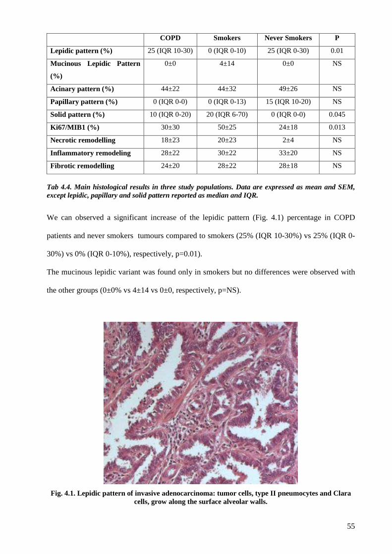
55
COPD Smokers Never Smokers P
Lepidic pattern (%) 25 (IQR 10-30) 0 (IQR 0-10) 25 (IQR 0-30) 0.01
Mucinous Lepidic Pattern
(%)
0±0 4±14 0±0 NS
Acinary pattern (%) 44±22 44±32 49±26 NS
Papillary pattern (%) 0 (IQR 0-0) 0 (IQR 0-13) 15 (IQR 10-20) NS
Solid pattern (%) 10 (IQR 0-20) 20 (IQR 6-70) 0 (IQR 0-0) 0.045
Ki67/MIB1 (%) 30±30 50±25 24±18 0.013
Necrotic remodelling 18±23 20±23 2±4 NS
Inflammatory remodeling 28±22 30±22 33±20 NS
Fibrotic remodelling 24±20 28±22 28±18 NS
Tab 4.4. Main histological results in three study populations. Data are expressed as mean and SEM, except lepidic, papillary and solid pattern reported as median and IQR.
We can observed a significant increase of the lepidic pattern (Fig. 4.1) percentage in COPD
patients and never smokers tumours compared to smokers (25% (IQR 10-30%) vs 25% (IQR 0-
30%) vs 0% (IQR 0-10%), respectively, p=0.01).
The mucinous lepidic variant was found only in smokers but no differences were observed with
the other groups (0±0% vs 4±14 vs 0±0, respectively, p=NS).
Fig. 4.1. Lepidic pattern of invasive adenocarcinoma: tumor cells, type II pneumocytes and Clara cells, grow along the surface alveolar walls.
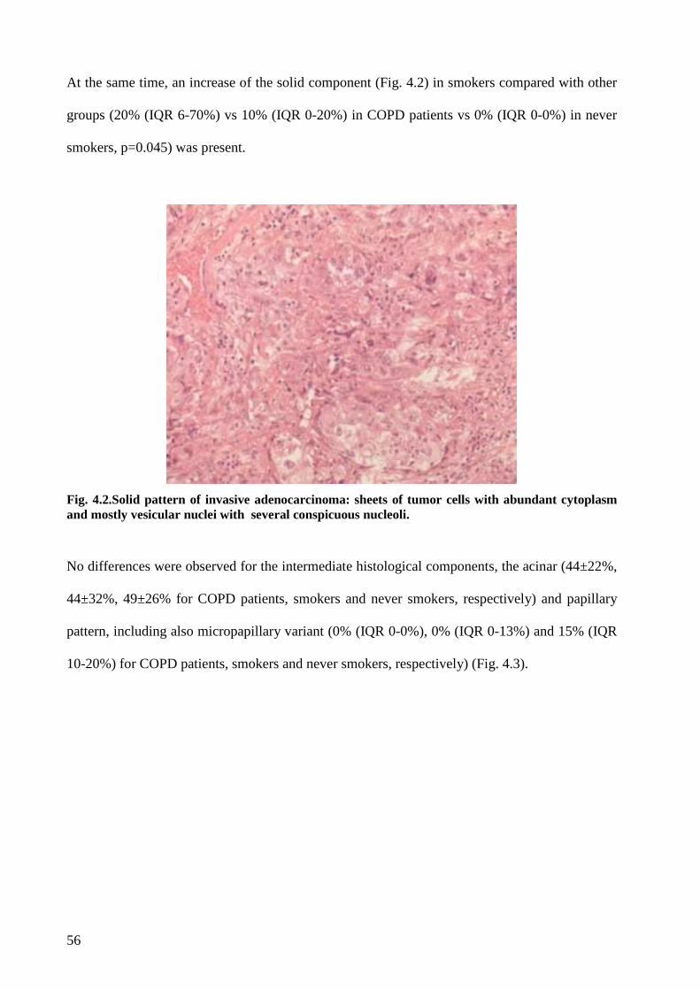
56
At the same time, an increase of the solid component (Fig. 4.2) in smokers compared with other
groups (20% (IQR 6-70%) vs 10% (IQR 0-20%) in COPD patients vs 0% (IQR 0-0%) in never
smokers, p=0.045) was present.
Fig. 4.2.Solid pattern of invasive adenocarcinoma: sheets of tumor cells with abundant cytoplasm and mostly vesicular nuclei with several conspicuous nucleoli.
No differences were observed for the intermediate histological components, the acinar (44±22%,
44±32%, 49±26% for COPD patients, smokers and never smokers, respectively) and papillary
pattern, including also micropapillary variant (0% (IQR 0-0%), 0% (IQR 0-13%) and 15% (IQR
10-20%) for COPD patients, smokers and never smokers, respectively) (Fig. 4.3).
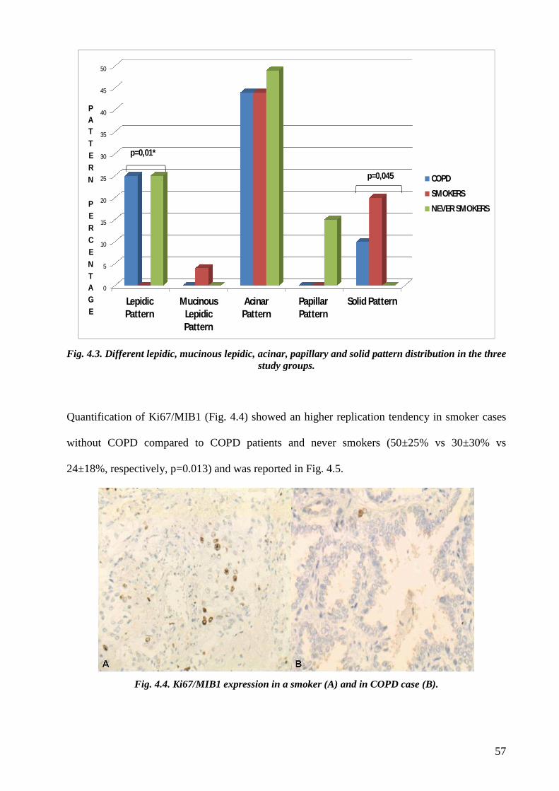
57
0
5
10
15
20
25
30
35
40
45
50
LepidicPattern
MucinousLepidicPattern
AcinarPattern
PapillarPattern
Solid Pattern
PATTERN
PERCENTAGE
COPD
SMOKERS
NEVER SMOKERS
p=0,045
p=0,01*
Fig. 4.3. Different lepidic, mucinous lepidic, acinar, papillary and solid pattern distribution in the three study groups.
Quantification of Ki67/MIB1 (Fig. 4.4) showed an higher replication tendency in smoker cases
without COPD compared to COPD patients and never smokers (50±25% vs 30±30% vs
24±18%, respectively, p=0.013) and was reported in Fig. 4.5.
Fig. 4.4. Ki67/MIB1 expression in a smoker (A) and in COPD case (B).
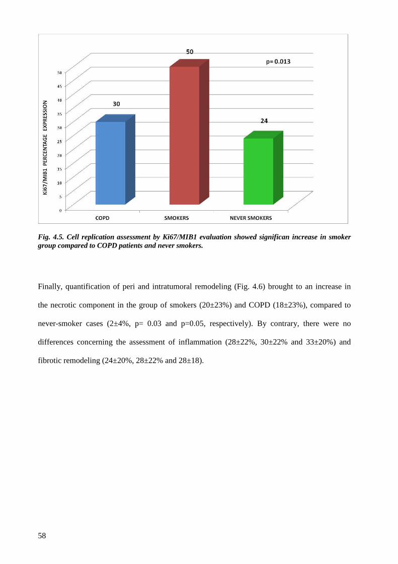
58
Fig. 4.5. Cell replication assessment by Ki67/MIB1 evaluation showed significan increase in smoker group compared to COPD patients and never smokers.
Finally, quantification of peri and intratumoral remodeling (Fig. 4.6) brought to an increase in
the necrotic component in the group of smokers (20±23%) and COPD (18±23%), compared to
never-smoker cases (2±4%, p= 0.03 and p=0.05, respectively). By contrary, there were no
differences concerning the assessment of inflammation (28±22%, 30±22% and 33±20%) and
fibrotic remodeling (24±20%, 28±22% and 28±18).
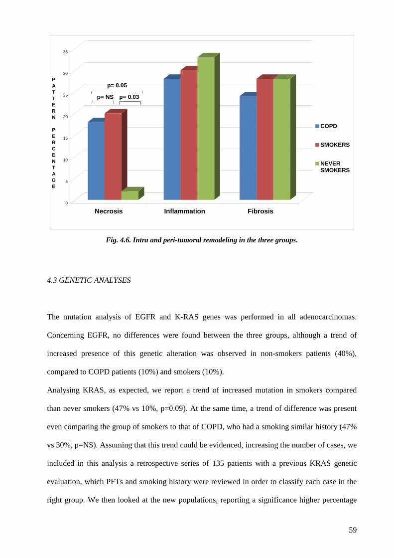
59
0
5
10
15
20
25
30
35
PATTERN PERCENTAGE
Necrosis Inflammation Fibrosis
COPD
SMOKERS
NEVERSMOKERS
p= NS p= 0.03
p= 0.05
Fig. 4.6. Intra and peri-tumoral remodeling in the three groups.
4.3 GENETIC ANALYSES
The mutation analysis of EGFR and K-RAS genes was performed in all adenocarcinomas.
Concerning EGFR, no differences were found between the three groups, although a trend of
increased presence of this genetic alteration was observed in non-smokers patients (40%),
compared to COPD patients (10%) and smokers (10%).
Analysing KRAS, as expected, we report a trend of increased mutation in smokers compared
than never smokers (47% vs 10%, p=0.09). At the same time, a trend of difference was present
even comparing the group of smokers to that of COPD, who had a smoking similar history (47%
vs 30%, p=NS). Assuming that this trend could be evidenced, increasing the number of cases, we
included in this analysis a retrospective series of 135 patients with a previous KRAS genetic
evaluation, which PFTs and smoking history were reviewed in order to classify each case in the
right group. We then looked at the new populations, reporting a significance higher percentage
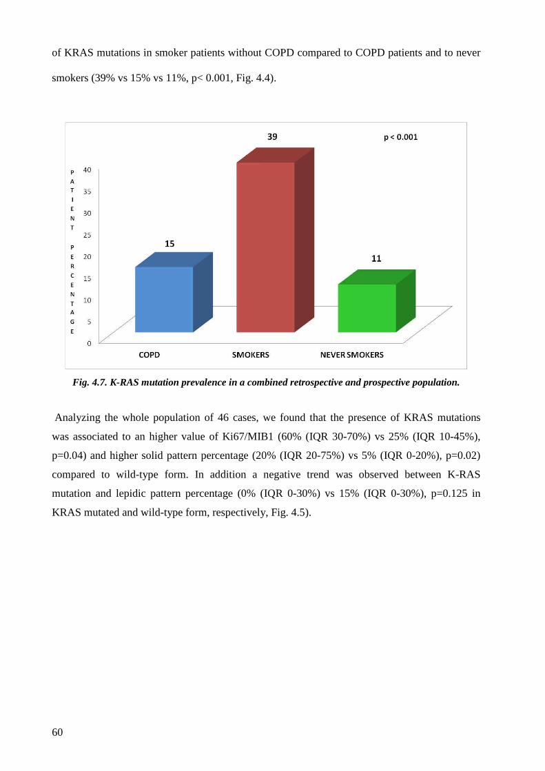
60
of KRAS mutations in smoker patients without COPD compared to COPD patients and to never
smokers (39% vs 15% vs 11%, p< 0.001, Fig. 4.4).
Fig. 4.7. K-RAS mutation prevalence in a combined retrospective and prospective population.
Analyzing the whole population of 46 cases, we found that the presence of KRAS mutations
was associated to an higher value of Ki67/MIB1 (60% (IQR 30-70%) vs 25% (IQR 10-45%),
p=0.04) and higher solid pattern percentage (20% (IQR 20-75%) vs 5% (IQR 0-20%), p=0.02)
compared to wild-type form. In addition a negative trend was observed between K-RAS
mutation and lepidic pattern percentage (0% (IQR 0-30%) vs 15% (IQR 0-30%), p=0.125 in
KRAS mutated and wild-type form, respectively, Fig. 4.5).
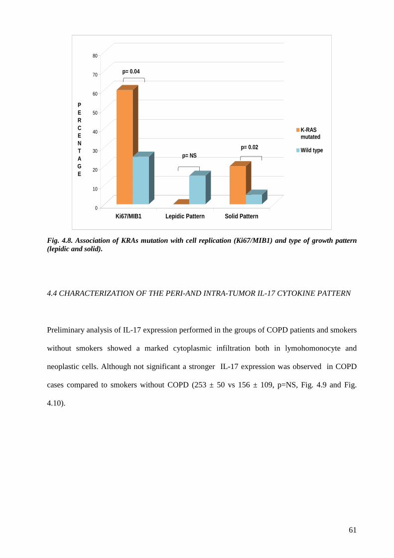
61
0
10
20
30
40
50
60
70
80
PERCENTAGE
Ki67/MIB1 Lepidic Pattern Solid Pattern
K-RASmutated
Wild typep= 0.02
p= 0.04
p= NS
Fig. 4.8. Association of KRAs mutation with cell replication (Ki67/MIB1) and type of growth pattern (lepidic and solid).
4.4 CHARACTERIZATION OF THE PERI-AND INTRA-TUMOR IL-17 CYTOKINE PATTERN
Preliminary analysis of IL-17 expression performed in the groups of COPD patients and smokers
without smokers showed a marked cytoplasmic infiltration both in lymohomonocyte and
neoplastic cells. Although not significant a stronger IL-17 expression was observed in COPD
cases compared to smokers without COPD (253 ± 50 vs 156 ± 109, p=NS, Fig. 4.9 and Fig.
4.10).
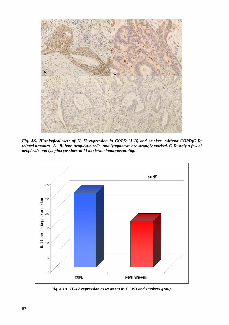
62
Fig. 4.9. Histological view of IL-17 expression in COPD (A-B) and smoker without COPD(C-D) related tumours. A –B: both neoplastic cells and lymphocyte are strongly marked. C-D: only a few of neoplastic and lymphocyte show mild-moderate immunostaining.
0
50
100
150
200
250
300
IL-1
7 p
erce
nta
ge
exp
ress
ion
COPD Never Smokers
p= NS
Fig. 4.10. IL-17 expression assessment in COPD and smokers group.
63
5. DISCUSSION
Lung cancer is the most prevalent tumour worldwide and a common cause of cancer-related
death [47]. Evidence now demonstrates that pathological subtype classification of “non small-
cell lung cancer” (NSCLC), a heterogeneous group of diseases, is central in selecting optimal
treatment [110,111]. However, even if surgical and oncologic treatments have greatly improved
in recent years overall based on better pathological and molecular diagnosis, lung cancer
continues to present increasing frequency, poor long-term results and an unpredictable disease
progression.
The co-presence of other diseases in individual patients may strongly influence the
carcinogenetic process, making it not only dependent on the classical risk factors. In this context,
the relationship that exists between COPD and lung cancer is undoubtedly important. Several
epidemiological studies have now firmly established the connection between the two diseases
highlighting how COPD is by far the greatest risk factor for lung cancer amongst smokers [68].
Skillrud et al first assessed the risk of lung cancer in patient with COPD compared to a matched
case-control study and estimated that the cumulative probability of developing lung cancer
within 10 years was 8.8% for those with COPD and 2% for patients with normal function [112].
This indicates that ~1% of patients with COPD develop lung cancer each year, while only 0.2%
of patients with normal function develop lung cancer (five-fold increased risk of lung cancer).
Several cross-sectional studies, together with recently published prospective studies show
smokers with COPD (or reduced FEV1) have up to 4-to 6-fold increased risk of lung cancer
when compared with smokers with normal function.
Furthermore 50-70% of patients diagnosed with lung cancer have spirometric evidence of COPD
[113].
64
While the main risk factor for the onset of both diseases is represented by cigarette smoke
exposure, they also share a common genetic predisposition which justifies the incidence of these
diseases in only a fraction of smokers [74].
This increased risk of lung cancer among smokers with COPD is probably due to activation of
common signaling pathways for both diseases by tobacco smoke as well as by smoking-induced
chronic inflammation which is a risk factor for both diseases. A causal relation between
inflammation and cancer has already been proposed by Galen and later by Virchow [85], who
observed inflammatory cell infiltration in malignant tissues and suggested that cancers arise at
regions of chronic inflammation. The longer the inflammation persists, the higher the risk of
associated carcinogenesis [114].
The importance of this inflammatory response has recently been highlighted, inviting a broader
look at the possible involvement of the immune system in the pathogenetic events of COPD.
Emerging evidences in this context have emphasized the role of adaptive immune responses,
possibly with an autoimmune component due to the recognition of pulmonary self-antigens
modified by cigarette smoking and to the failure of mechanisms regulating immunological
tolerance [41]. Thus it is not surprising that COPD is associated with increased risk of
developing lung cancer due to the synergic effect of cigarette smoke and chronic inflammation.
The histologic subtype more frequently associated with smoker COPD is squamous cell
carcinoma however no other specific clinical, morphological and/or molecular details have been
described in lung cancer associated with COPD.
In our research study we speculated that lung cancer developed in COPD patients has a different
clinical/morphological/molecular phenotype compared to lung cancer of smokers without COPD
and never smoker subjects. The comparison was mainly performed within the category of
smoking patients: COPD versus smokers without COPD, with the main objective to assess the
impact of COPD on the tumor phenotype. Never smokers were an important control group in our
research study even if it should be noted that tumours arising in thi category of patients have
peculiar genetic substrates and mechanisms. The findings of EGFR mutation balanced by the
65
low KRAS–mutation frequency in never-smoking cases enrolled in the project is proof of these
characteristics.
The decision to perform the analysis only in adenocarcinomas was surely influenced by the high
percentage of this histologic type determined by the selection bias of inclusion/exclusion criteria
and by the need to obtain results not affected by different histotypes. However, this choice
reflects the epidemiology trend of lung cancer with adenocarcinoma incidence that has now
surpassed that of squamous cell carcinomas in both sexes, possibly due to changes in cigarette
smoke composition and/or inhalation [115].
A fundamental point of our prospective study is represented by the fact that the study population
included COPD and well matched smoker patients without COPD both for age, sex and smoking
history, well known factors which may influence lung carcinogenesis. Among PFTs, excluding
those specific for COPD (FEV1%, FEV1/VC, FEV1/FVC, PEF%, TLC%, RV%, FRC%), an
approximately equal decrease of FVC percentage in patients with a smoking history (smokers or
COPD) was observed. Functional impairment in these patients may be the spirometric expression
of parenchymal damage caused by smoke exposure. With regard to biohumoral, and in particular
to inflammatory indices, except for basophils, we did not observe significant difference between
COPD and smoker patients. The raised number of blood basophils in COPD patients could
reflect an anti-tumour immune response to circulating antigen. There are only few papers that
focus on the significance of an increased number of basophils and cancer and even fewer for
lung cancer. Basophils mediators are reported to attract eosinophils in the site of nematode
infestation and arm them for more potent killing of the nematodes [116]. It is possible that the
link between the numbers of circulating basophils and eosinophils in lung cancer patients lies in
a similar interaction in relation to the tumour, since eosinophil infiltration of the tumours was
associated with both blood eosinophilia and better prognosis [117].
Tumour functional assessment was evaluated through 18FDG PET-scan which has been imposed
as a strategic diagnostic and staging tool for lung cancer. 18FDG PET-scan has recently been
proposed as a prognostic examination in particular through the SUV index since it seems closely
66
related to tumour biological aggressiveness [118]. An important meta-analysis performed by
Berghmans et al [67] in 2008 took into consideration 13 studies including 1474 patients with
different stages of NSCLC. They reported that in 11 studies a high SUV was identified as a poor
prognostic factor for survival although two studies found no significant correlation; in addition
they estimated an overall combined hazard risk (HR) for the 13 reports of 2.27. Nair et al.
recently published a clinical study where they analyzed the biologic basis for the use of FDG as a
biomarker, founding that four of eight single genes associated with FDG uptake (LY6E,
RNF149, MCM6, and FAP) were also associated with survival [119]. A lower SUV was detected
in our COPD lung cancer. This is likely due to a less proliferative index of this neoplasia
compared to lung cancer of smokers without COPD. Different works in the literature have
reported a significant relation between tumour proliferation detected by MIB1/Ki67 and SUV,
highlightening the significant value of FDG as an additional prognostic instrument [120]. The
histological analysis of neoplastic patterns associated in our study groups revealed interesting
data, emphasising significant differences.
From a morphological point of view adenocarcinoma of COPD patients showed less frequent
solid and more frequent lepidic pattern and lower Ki67 proliferation. The more extensive
necrosis present both in COPD patients and smokers with lung cancer seems to be the expression
of direct smoking cellular damage. All these aspects would characterize this form as a less
aggressive neoplastic phenotype. Regarding lepidic pattern, the mucinous variant was found only
in the smokers and analysed separately for the other lepidic cases. Indeed the new classification
of adenocarcinoma [49] has now divided the mucinous from the nonmucinous form, as multiple
studies [121-123] indicated that tumours formerly classified as mucinous BAC have major
clinical, radiologic, pathologic, and genetic differences from the tumours formerly classified as
nonmucinous BAC. In particular, these tumours show a very strong correlation with KRAS
mutation, whereas nonmucinous adenocarcinomas are more likely to show EGFR mutation and
only occasionally KRAS mutation [124]. In addition, the neoplasms formerly termed mucinous
BAC and now classified as invasive mucinous adenocarcinoma, has been recognized to have
67
invasive components in the majority of cases [125], leading to a different clinical prognosis
compared to nonmucinous variants.
Concerning the molecular data the finding of less frequent K-RAS mutation in lung COPD
patients was remarkable. This finding reinforces the concept that tumour related to COPD
presented different biological behaviour. The impact of KRAS alterations on prognosis has
already been studied in lung cancer and two different meta-analyses suggested an association of
KRAS with poorer survival [126-127].
Our group in 2010 [128] also confirmed the impact of KRAS mutations on prognosis in a
clinically selected group of advanced adenocarcinomas, highlighting the role of KRAS as an
independent prognostic marker.
The analysis of the pathway axis of IL 17 seems to be an important link between COPD and lung
cancer providing a possible pathogenetic substrate for both diseases. In our study we observed
the increased cytokine expression in lung cancer of COPD cases.
A key role for this cytokine has already been emphasised in the context of autoimmune diseases
[129], and more recently in COPD patients, where it seems to support the maintenance of
chronic inflammation [99].
New evidences of this axis in the neoplastic field have been reported remarking its crucial role
also in lung [130]. The relationship between IL-17 and cancer, whether beneficial or
antagonistic, continues to be a controversial issue. The majority of studies reported an important
influence of this cytokine on the induction of angiogenesis favoring vascular endothelial growth
factor–VEGF-secretion and on neoplastic cell growth. [107] through the alteration of
immunosurveillance component [108].
There are still no evidence in the literature that describe the presence and the role of this cytokine
in adenocarcinoma associated to COPD. In our study, the small number of subjects certainly
influenced the observed results and probably the lack of a significant difference. However, what
seems to emerge is that the IL-17 pathway, recently demonstrated to be involved in the complex
68
immunological background of COPD, could play a key role in cancer development/progression
of patients with COPD compared to smokers without COPD.
In conclusion the scenario that emerged from our research study is the presence in COPD
patients of a different cancer to that of matched case smokers without COPD. Clinical,
biohumoral, morphological and molecular findings of COPD lung cancer showed stigmata of
lower aggressiveness and invasiveness lung cancer phenotype.
It might be speculated that alternative mechanisms of carcinogenesis are involved in the
development/progression of COPD related lung cancer. Given the importance of inflammation in
the pathogenesis of COPD the involvement of other pathways, such as IL-17, mainly driving
inflammatory mediated carcinogenesis could be suspected.
The main limitation of our research is represented by the low number of patients especially in the
COPD group. Moreover the absence of COPD advanced stages does not allow assessment of the
presence of any differences in inflammatory and neoplastic phenotype within the GOLD staging.
Finally, the short follow-up of these patients, considering also the early disease stages included
in the study, does not allow us to perform any survival analysis.
Future research
The availability of large amount of materials properly stored in a dedicated tissue archive will be
able to expand the knowledge of neoplastic phenotypes associated with study populations.
In this field, the characterization of mmunophenotype inflammatory cell infiltration both in
peritumoral inflammatory infiltrate and in distant areas with particular attention to lymphocytic,
macrophagic and cytokine patterns will be of great importance.
The presence of liquid samples as blood and BAL will also lead to the research for possible
peripheral biomarkers of disease that can provide predictive and prognostic information related
to the neoplastic process. Finally, the analysis of medium-term results of survival and local or
distant recurrence could support the preliminary data obtained in this study proposing a different
lung cancer phenotype in COPD patients which show sign of less aggressiveness.
69
New knowledge of mechanisms at the basis of COPD lung cancer would be of considerable help
in the fight against lung cancer both for individualized follow-up and for therapeutic
perspectives, providing a rationale basis to develop targeted and more effective treatments to
prevent or, at least, slow down cancer evolution (anti-inflammatory immunomodulatory or
biological therapies).

70
71
6. REFERENCES 1) Global Strategy for the Diagnosis, Management and Prevention of COPD, Global Initiative
for Chronic Obstructive Lung Disease (GOLD) 2011. Ù
2) Petty TL. The history of COPD. Int J Chron Obstruct Pulmon Dis. 2006; 1(1):3-14.
3) Fishman AP. One hundred years of chronic obstructive pulmonary disease. Am J Respir Crit
Care Med. 2005; 171(9):941-948.
4) Fletcher C, Peto R. The natural history of chronic airflow obstruction. Br Med J. 1977;
1(6077):1645-1648.
5) Buist AS, McBurnie MA, Vollmer WM, Gillespie S, Burney P, Mannino DM, Menezes
AM, Sullivan SD, Lee TA, Weiss KB, Jensen RL, Marks GB, Gulsvik A, Nizankowska-
Mogilnicka E; BOLD Collaborative Research Group. International variation in the prevalence of
COPD (the BOLD Study): a population-based prevalence study. Lancet. 2007;370:741-50.
6) Halbert RJ, Natoli JL, Gano A, Badamgarav E, Buist AS, Mannino DM. Global burden of
COPD: systematic review and meta-analysis. Eur Respir J. 2006;28:523-32.
7) Van den Boom G, van Schayck CP, van Möllen MP, Tirimanna PR, den Otter JJ, van
Grunsven PM, Buitendijk MJ, van Herwaarden CL, van Weel C. Active detection of chronic
obstructive pulmonary disease and asthma in the general population. Results and economic
consequences of the DIMCA program. Am J Respir Crit Care Med. 1998;158:1730-8.
8) Mead J. The lung's "quiet zone". N Engl J Med. 1970;282:1318-9.
9) O'Donnell DE, Laveneziana P. Dyspnea and activity limitation in COPD: mechanical factors.
COPD. 2007;4:225-36.
10) Chung C, Delaney J and Hodgins R. “Respirology” Toronto Notes (2008) pp.R9.
72
11) Rodríguez-Roisin R, Drakulovic M, Rodríguez DA, Roca J, Barberà JA, Wagner PD.
Ventilation-perfusion imbalance and chronic obstructive pulmonary disease staging severity. J
Appl Physiol. 2009;106:1902-8.
12) Peto R, Speizer FE, Cochrane AL, Moore F, Fletcher CM, Tinker CM, Higgins IT, Gray RG,
Richards SM, Gilliland J, Norman-Smith B. The relevance in adults of air-flow obstruction, but
not of mucus hypersecretion, to mortality from chronic lung disease. Results from 20 years of
prospective observation. Am Rev Respir Dis. 1983;128:491-500.
13) Peinado VI, Pizarro S, Barberà JA. Pulmonary vascular involvement in COPD. Chest.
2008;134:808-14.
14) Celli BR, Barnes PJ. Exacerbations of chronic obstructive pulmonary disease. Eur Respir J.
2007 Jun;29(6):1224-38.
15) Vestbo J. Systemic inflammation and progression of COPD. Thorax. 2007;62:469-70.
16) Dahl M, Vestbo J, Lange P, Bojesen SE, Tybjaerg-Hansen A, Nordestgaard BG. C-reactive
protein as a predictor of prognosis in chronic obstructive pulmonary disease. Am J Respir Crit
Care Med. 2007;175:250-5.
17) Fabbri LM, Luppi F, Beghé B, Rabe KF. Complex chronic comorbidities of COPD. Eur
Respir J. 2008; 31(1):204-212.
18) Sin DD, Man JP, Man SF. The risk of osteoporosis in Caucasian men and women with
obstructive airways disease. Am J Med. 2003;114:10-4.
19) Fishman’s Pulmonary Disease and Disorders. McGraw-Hill 2008. Fourth Edition. p 712.
20) Curtis JL, Freeman CM, Hogg JC. The immunopathogenesis of chronic obstructive
pulmonary disease: insights from recent research. Proc Am Thorac Soc. 2007; 4(7):512-521.
21) Celli BR, Cote CG, Marin JM, Casanova C, Montes de Oca M, Mendez RA, Pinto Plata V,
Cabral HJ. The body-mass index, airflow obstruction, dyspnea, and exercise capacity index in
chronic obstructive pulmonary disease. N Engl J Med. 2004;350:1005-12.
73
22) Travis WD et al. Atlas of nontumour pathology “Non-Neoplastic Disorders of the Lower
Respiratory Tract” (2002); pp.435-457.
23) Ballarin A, Bazzan E, Zenteno RH, Turato G, Baraldo S, Zanovello D, Mutti E, Hogg JC,
Saetta M, Cosio MG. Mast Cell Infiltration Discriminates between Histopathological Phenotypes
of Chronic Obstructive Pulmonary Disease. Am J Respir Crit Care Med. 2012;186:233-9.
24) Baugh RJ, Travis J. Human leukocyte granule elastase: rapid isolation and characterization.
Biochemistry. 1976; 15(4):836-841.
25) Hersh CP, DeMeo DL, Silverman EK. National Emphysema Treatment Trial state of the art:
genetics of emphysema. Proc Am Thorac Soc. 2008; 5(4):486-493.
26) Wan ES, Silverman EK. Genetics of COPD and emphysema. Chest. 2009; 136(3):859-866.
27) Viegi G, Paoletti P, Vellutini M, Carrozzi L, Di Pede F, Baldacci S, Modena P, Pedreschi
M, Di Pede C, Giuntini C. Effects of daily cigarette consumption on respiratory symptoms and
lung function in a general population sample of north-Italian men. Respiration. 1991;58:282-6.
28) Sethi JM, Rochester CL. Smoking and chronic obstructive pulmonary disease. Clin Chest
Med. 2000;21:67-86.
29) Balmes J, Becklake M, Blanc P, Henneberger P, Kreiss K, Mapp C, Milton D, Schwartz D,
Toren K, Viegi G. Environmental and Occupational Health Assembly, American Thoracic
Society. American Thoracic Society Statement: Occupational contribution to the burden of
airway disease. Am J Respir Crit Care Med. 2003; 167(5):787-797.
30) Bruce N, Perez-Padilla R, Albalak R. Indoor air pollution in developing countries: a major
environmental and public health challenge. Bull World Health Organ. 2000; 78(9):1078-1092.
31) Rivera RM, Cosio MG, Ghezzo H, Salazar M, Pérez-Padilla R. Comparison of lung
morphology in COPD secondary to cigarette and biomass smoke. Int J Tuberc Lung Dis. 2008;
12(8):972-977.
74
32) Lawlor DA, Ebrahim S, Davey Smith G. Association of birth weight with adult lung
function: findings from the British Women's Heart and Health Study and a meta-analysis.
Thorax. 2005;60:851-8.
33) Eisner MD, Anthonisen N, Coultas D, Kuenzli N, Perez-Padilla R, Postma D, Romieu I,
Silverman EK, Balmes JR; Committee on Nonsmoking COPD, Environmental and Occupational
Health Assembly. An official American Thoracic Society public policy statement: Novel risk
factors and the global burden of chronic obstructive pulmonary disease. Am J Respir Crit Care
Med. 2010;182:693-718.
34) Crothers K, Huang L, Goulet JL, Goetz MB, Brown ST, Rodriguez-Barradas MC, Oursler
KK, Rimland D, Gibert CL, Butt AA, Justice AC. HIV infection and risk for incident pulmonary
diseases in the combination antiretroviral therapy era. Am J Respir Crit Care Med.
2011;183:388-95.
35) Prescott E, Lange P, Vestbo J. Socioeconomic status, lung function and admission to
hospital for COPD: results from the Copenhagen City Heart Study. Eur Respir J. 1999;13:1109-
14.
36) Silva GE, Sherrill DL, Guerra S, Barbee RA. Asthma as a risk factor for COPD in a
longitudinal study. Chest. 2004;126:59-65.
37) Vestbo J, Prescott E, Lange P. Association of chronic mucus hypersecretion with FEV1
decline and chronic obstructive pulmonary disease morbidity. Copenhagen City Heart Study
Group. Am J Respir Crit Care Med. 1996;153:1530-5.
38) Abboud RT, Vimalanathan S. Pathogenesis of COPD. Part I. The role of
proteaseantiprotease imbalance in emphysema. Int J Tuberc Lung Dis. 2008; 12(4):361-367.
39) Baraldo S, Saetta M. To reg or not to reg: that is the question in COPD. Eur Respir J.
2008;31:486-8.
40) Renda T, Baraldo S, Pelaia G, Bazzan E, Turato G, Papi A, Maestrelli P, Maselli R,
Vatrella A, Fabbri LM, Zuin R, Marsico SA, Saetta M. Increased activation of p38 MAPK in
COPD. Eur Respir J. 2008;31:62-9.

75
41) Cosio MG, Saetta M, Agusti A. Immunologic aspects of chronic obstructive pulmonary
disease. N Engl J Med. 2009; 360(23):2445-2454.
42) Turato G, Barbato A, Baraldo S, Zanin ME, Bazzan E, Lokar-Oliani K, Calabrese F,
Panizzolo C, Snijders D, Maestrelli P, Zuin R, Fabbri LM, Saetta M. Nonatopic children with
multitrigger wheezing have airway pathology comparable to atopic asthma. Am J Respir Crit
Care Med. 2008;178:476-82.
43) Joosten LA, Netea MG, Kim SH, Yoon DY, Oppers-Walgreen B, Radstake TR, Barrera P,
van de Loo FA, Dinarello CA, van den Berg WB. IL-32, a proinflammatory cytokine in
rheumatoid arthritis. Proc Natl Acad Sci USA. 2006; 103:3298–3303.
44) Shioya M, Nishida A, Yagi Y, Ogawa A, Tsujikawa T, Kim-Mitsuyama S, Takayanagi A,
Shimizu N, Fujiyama Y, Andoh A. Epithelial overexpression of interleukin-32alpha in
inflammatory bowel disease. Clin Exp Immunol. 2007; 149:480–486.
45) Baraldo S, Lokar-Oliani K, Turato G, Zuin R, Saetta M. The Role of Lymphocytes in the
Pathogenesis of Asthma and COPD. Curr Med Chem. 2007;14:2250-6.
46) Zanini A, Chetta A, Saetta M, Baraldo S, D'Ippolito R, Castagnaro A, Neri M and Olivieri
D. Chymase-positive mast cells play a role in the vascular component of airway remodeling in
asthma. J Allergy Clin Immunol. 2007;120:329-33.
47) Jemal A, Siegel R, Ward E. Cancer statistics, 2009. CA Cancer J Clin. 2009; 59(4):225.
48) Travis WD, Brambilla E, Mulller –Hermellink HK, Harris CC. Tumors of the Lung, Pleura,
Thymus and Heart. WHO. 2004.
49) Travis WD, Brambilla E, Noguchi M, Nicholson AG, Geisinger KR, Yatabe Y, Beer DG,
Powell CA, Riely GJ, Van Schil PE, Garg K, Austin JH, Asamura H, Rusch VW, Hirsch FR,
Scagliotti G, Mitsudomi T, Huber RM, Ishikawa Y, Jett J, Sanchez-Cespedes M, Sculier JP,
Takahashi T, Tsuboi M, Vansteenkiste J, Wistuba I, Yang PC, Aberle D, Brambilla C, Flieder D,
Franklin W, Gazdar A, Gould M, Hasleton P, Henderson D, Johnson B, Johnson D, Kerr K,
Kuriyama K, Lee JS, Miller VA, Petersen I, Roggli V, Rosell R, Saijo N, Thunnissen E, Tsao M,
Yankelewitz D. International association for the study of lung cancer/american thoracic
76
society/european respiratory society international multidisciplinary classification of lung
adenocarcinoma. J Thorac Oncol. 2011;6(2):244-85.
50) Youlden DR, Cramb SM, Baade PD: The international epidemiology of lung cancer:
geographical distribution and secular trends. J Thorac Oncol 2008; 3: 819–831.
51) Egleston BL, Meireles SI, Flieder DB, Clapper ML: Population-based trends in lung cancer
incidence in women. Semin Oncol 2009; 36: 506–515.
52) Scagliotti GV, Longo M, Novello S: Nonsmall cell lung cancer in never smokers. Curr
Opin Oncol 2009; 21: 99–104.
53) Lynch TJ, Bell DW, Sordella R, Gurubhagavatula S, Okimoto RA, Brannigan BW, Harris
PL, Haserlat SM, Supko JG, Haluska FG, Louis DN, Christiani DC, Settleman J, Haber DA.
Activating Mutations in the Epidermal Growth Factor Receptor Underlyng Responsiveness of
Non.Small-Cell Lung Cancer to Gefitinib. The New England Journal of Medicine 2004; 350; 21:
2129-2138.
54) Paez JG, Jänne PA, Lee JC, Tracy S, Greulich H, Gabriel S, Herman P, Kaye FJ, Lindeman
N, Boggon TJ, Naoki K, Sasaki H, Fujii Y, Eck MJ, Sellers WR, Johnson BE, Meyerson M.
EGFR Mutations in Lung Cancer: correlations with Clinical Response to Gefitinib Theraphy.
Science 2004; 304: 1497-1500.
55) Pao W, Miller V, Zakowski M, Doherty J, Politi K, Sarkaria I, Singh B, Heelan R, Rusch
V, Fulton L, Mardis E, Kupfer D, Wilson R, Kris M, Varmus H. EGF receptor gene mutations
are common in lung cancers from “never smokers” and are associated with sensitivity of tumors
to gefitinib and erlotinib. PNAS 2004; 101: 13306-13311.
56) Riely GJ, Pao W, Pham D, Li AR, Rizvi N, Venkatraman ES, Zakowski MF, Kris MG,
Ladanyi M, Miller VA. Clinical course of patients with non-small cell lung cancer and epidermal
growth factor receptor exon 19 and exon 21 mutations treated with gefitinib or erlotinib. Clin
Cancer Res 2006; 12: 839–844.
57) Kalemkerian GP, Pass HI. Present Concepts in the Molecular Biology of Lung Cancer in
Shields TW. General Thoracic Surgery, 6th Edition.©2005 Lippincott Williams & Wilkins.
58) Bos JL. Ras oncogenes in human cancer: a review. Cancer Res. 1989; 49(17): 4682-9.
77
59) Cho JY, Kim JH, Lee YH, et al. Correlation between K-ras gene mutation and prognosis of
patients with nonsmall cell lung carcinoma. Cancer. 1997; 79(3): 462-7.
60) Bidoli P, Robustelli della Cuna e Pastorino U: Neoplasie del polmone. In: Medicina
Oncologica. Bonadonna G, Robustelli della Cuna, Valagusa. MASSON, Milano, 2003.
61) Schrump DS, Altorki NA, Henschke CL, Cartier D, Turrisi AT, Gutierrez M. Non-Small
Cell Lung Cancer. In: CANCER Principles and Practice of Oncology. De Vita V, Hellman S,
Rosemberg SA eds. Lippincott Williams and Wilkins Publ, Philadelphia, 2005, pp 753-810.
62) Singhal S, Shrager JB, Kaiser LR. Multimodality Therapy for Non-Small-Cell Lung Cancer
in Shields TW. General Thoracic Surgery, 6th Edition.©2005 Lippincott Williams & Wilkins.
63) Schiller JH, Harrington D, Belani CP, Langer C, Sandler A, Krook J, Zhu J, Johnson DH;
Eastern Cooperative Oncology Group. Comparison of four chemotherapy regimens for advanced
non-small-cell lung cancer. N Engl J Med. 2002. 346, 92-98.
64) Jackman DM, Yeap BY, Sequist LV, Lindeman N, Holmes AJ, Joshi VA, Bell DW,
Huberman MS, Halmos B, Rabin MS, Haber DA, Lynch TJ, Meyerson M, Johnson BE, Jänne
PA. Exon 19 deletion mutations of epidermal growth factor receptor are associated with
prolonged survival in non-small cell lung cancer patients treated with gefitinib or erlotinib. Clin
Cancer Res. 2006; 12: 3908–3914.
65) Suzuki M, Shigematsu H, Iizasa T, Hiroshima K, Nakatani Y, Minna JD, Gazdar AF,
Fujisawa T. Exclusive mutation in epidermal growth factor receptor gene, HER-2, and KRAS,
and synchronous methylation of nonsmall cell lung cancer. Cancer 2006;106:2200 –2207.
66) Vallières E, Shepherd FA, Crowley J. The IASLC Lung Cancer Staging Project: proposals
regarding the relevance of TNM in the pathologic staging of small cell lung cancer in the
forthcoming (seventh) edition of the TNM classification for lung cancer. J Thorac Oncol. 2009
Sep;4(9):1049-59.
67) Berghmans T, Dusart M, Paesmans M, Hossein-Foucher C, Buvat I, Castaigne C,
Scherpereel A, Mascaux C, Moreau M, Roelandts M, Alard S, Meert AP, Patz EF Jr, Lafitte JJ,
Sculier JP; European Lung Cancer Working Party for the IASLC Lung Cancer Staging Project.
Primary tumor standardized uptake value (SUVmax) measured on fluorodeoxyglucose positron
78
emission tomography (FDG-PET) is of prognostic value for survival in non-small cell lung
cancer (NSCLC): a systematic review and meta-analysis (MA) by the European Lung Cancer
Working Party for the IASLC Lung Cancer Staging Project. J Thorac Oncol. 2008 Jan;3(1):6-12.
68) Punturieri A, Szabo E, Croxton TL, Shapiro SD, Dubinett SM: Lung cancer and chronic
obstructive pulmonary disease: needs and opportunities for integrated research. J Natl Cancer
Inst. 2009; 101: 554–559.
69) Ben-Zaken CS, Pare PD, Man SF, Sin DD. The growing burden of chronic obstructive
pulmonary disease and lung cancer in women: examining sex differences in cigarette smoke
metabolism. Am J Respir Crit Care Med. 2007; 176: 113–120.
70) Turner MC, Chen Y, Krewski D, Calle EE, Thun MJ. Chronic obstructive pulmonary
disease is associated with lung cancer mortality in a prospective study of never smokers. Am J
Respir Crit Care Med. 2007;176:285–90.
71) Anthonisen NR, Skeans MA, Wise RA, Manfreda J, Kanner RE, Connett JE. The effects of
a smoking cessation intervention on 14.5-year mortality: a randomized clinical trial. Ann Intern
Med. 2005; 142: 233–239.
72) Yao H, Rahman I. Current concepts on the role of inflammation in COPD and lung cancer.
Curr Opin Pharmacol. 2009; 9: 375– 383.
73) Papi A, Casoni G, Caramori G, Guzzinati I, Boschetto P, Ravenna F, Calia N, Petruzzelli S,
Corbetta L, Cavallesco G, Forini E, Saetta M, Ciaccia A, Fabbri LM. COPD increases the risk of
squamous histological subtype in smokers who develop non-small cell lung carcinoma. Thorax.
2004 ; 59(8): 679 – 681.
74) Spitz MR, Wei Q, Dong Q, Amos CI, Wu X. Genetic susceptibility to lung cancer: the role
of DNA damage and repair. Cancer Epidemiol Biomarkers Prev. 2003;12:689–98.
75) Ocak S, Sos ML, Thomas RK, Massion PP. High-throughput molecular analysis in lung
cancer: insights into biology and potential clinical applications. Eur Respir J. 2009;34:489–506.
76) Cohen BH, Ball WC Jr, Bias WB, Brashears S, Chase GA, Diamond EL, Hsu SH, Kreiss P,
Levy DA, Menkes HA, Permutt S, Tockman MS. A genetic-epidemiologic study of chronic
79
obstructive pulmonary disease. I. Study design and preliminary observations. Johns Hopkins
Med J. 1975 ; 137 ( 3 ): 95 – 104.
77) Cohen BH, Diamond EL, Graves CG, Kreiss P, Levy DA, Menkes HA, Permutt S, Quaskey
S, Tockman MS. A common familial component in lung cancer and chronic obstructive
pulmonary disease. Lancet. 1977; 2( 8037 ): 523 – 526.
78) Schwartz AG, Ruckdeschel JC. Familial lung cancer: genetic susceptibility and relationship
to chronic obstructive pulmonary disease. Am J Respir Crit Care Med. 2006 ; 173 ( 1 ): 16 – 22.
79) Yang P, Wentzlaff KA, Katzmann JA, Marks RS, Allen MS, Lesnick TG, Lindor NM,
Myers JL, Wiegert E, Midthun DE, Thibodeau SN, Krowka MJ.. Alpha1-antitrypsin defi ciency
allele carriers among lung cancer patients. Cancer Epidemiol Biomarkers Prev. 1999; 8( 5 ): 461
– 465.
80) Yang P, Bamlet WR, Sun Z, Ebbert JO, Aubry MC, Krowka MJ, Taylor WR, Marks RS,
Deschamps C, Swensen SJ, Wieben ED, Cunningham JM, Melton LJ, de Andrade M. Alpha1-
antitrypsin and neutrophil elastase imbalance and lung cancer risk. Chest. 2005; 128 (1): 445 –
452.
81) Sun Z, Yang P. Role of imbalance between neutrophil elastase and alpha 1-antitrypsin in
cancer development and progression. Lancet Oncol. 2004; 5 ( 3 ): 182 – 190.
82) Bowman RV, Yang IA, Semmler AB, Fong KM. Epigenetics of lung cancer. Respirology.
2006; 11 ( 4 ): 355 – 365.
83) Kagan J, Srivastava S, Barker PE, Belinsky SA, Cairns P. Towards clinical application of
methylated DNA sequences as cancer biomarkers: a joint NCI’s EDRN and NIST workshop on
standards, methods, assays, reagents and tools. Cancer Res. 2007; 67(10): 4545 – 4549.
84) Adcock IM, Tsaprouni L, Bhavsar P, Ito K. Epigenetic regulation of airway inflammation.
Curr Opin Immunol. 2007; 19 ( 6 ): 694 – 700.
85) David H . Rudolf Virchow and modern aspects of tumor pathology . Pathol Res Pract .
1988 ; 183 ( 3 ): 356 – 364.
80
86) Sin DD, Wu L, Anderson JA, Anthonisen NR, Buist AS, Burge PS, Calverley PM, Connett
JE, Lindmark B, Pauwels RA, Postma DS, Soriano JB, Szafranski W, Vestbo J. Inhaled
corticosteroids and mortality in chronic obstructive pulmonary disease. Thorax. 2005; 60 ( 12 ):
992 – 997.
87) Parimon T, Chien JW, Bryson CL, McDonell MB, Udris EM, Au DH. Inhaled
corticosteroids and risk of lung cancer among patients with chronic obstructive pulmonary
disease. Am J Respir Crit Care Med. 2007; 175 ( 7 ): 712 – 719.
88) Anthonisen NR, Connett JE, Kiley JP, Altose MD, Bailey WC, Buist AS, Conway WA Jr,
Enright PL, Kanner RE, O'Hara P, et al.. Effects of smoking intervention and the use of an
inhaled anticholinergic bronchodilator on the rate of decline of FEV1. The Lung Health Study.
JAMA. 1994; 272 ( 19 ): 1497 – 1505.
89) McGarvey LP, John M, Anderson JA, Zvarich M, Wise RA. Ascertainment of cause-
specific mortality in COPD: operations of the TORCH Clinical Endpoint Committee. Thorax.
2007; 62 ( 5 ): 411 – 415.
90) Palmer MT, Weaver CT. Autoimmunity: increasing suspects in the CD4+ T cell line up.
Nat. Immunol. 2010; 11, 36–40.
91) Dubin PJ, Kolls JK. Th17 cytokines and mucosal immunity. Immunol. Rev. 2008; 226, 160–
171.
92) Perera WR, Hurst JR, Wilkinson TM, Sapsford RJ, Müllerova H, Donaldson GC, Wedzicha
JA. Inflammatory changes, recovery and recurrence at COPD exacerbation. Eur. Respir. J.
2007; 29, 527–534.
93) Steinman L. A brief history of TH17, the first major revision in the TH1/TH2 hypothesis of
T cell-mediated tissue damage. Nat. Med. 2007; 13, 139–145.
94) Kolls JK, Lindén A. Interleukin-17 family members and inflammation. Immunity. 2004; 21,
467–476.
95) Aggarwal S, Gurney AL. IL-17: prototype member of an emerging cytokine family. J.
Leukocyte Biol. 2002; 71, 1–8.
81
96) Gaffen SL. Structure and signalling in the IL-17 receptor family. Nat. Rev. Immunol. 2009;
9, 556–567.
97) Weng NP, Liu K, Catalfamo M, Li Y, Henkart PA. IL-15 is a growth factor and an activator
of CD8 memory T cells. Ann. N.Y. Acad. Sci. 2002; 975, 46–56.
98) Lindemann MJ, Hu Z, Benczik M, Liu KD, Gaffen SL. Differential regulation of the IL-17
receptor by γ c cytokines: inhibitory signaling by the phosphatidylinositol 3-kinase pathway. J.
Biol. Chem. 2008; 283, 14100–1410.
99) Bettelli E, Carrier Y, Gao W, Korn T, Strom TB. Reciprocal developmental pathways for
the generation of pathogenic effector Th17 and regulatory T cells. Nature. 2006; 44, 235–238.
100) Bettelli E, Korn T, Kuchroo VK. Th17: the third member of the effector T cell trilogy.
Curr. Opin. Immunol. 2007; 19, 652–657.
101) Miossec P, Korn T, Kuchroo VK. Interleukin-17 and Type17 helper T cells. N. Engl. J.
Med. 2009; 361, 888–898.
102) Lane N, Robins RA, Corne J, Fairclough L. Regulation in chronic obstructive pulmonary
disease: the role of regulatory T-cells and Th17 cells. Clin Sci. 2010 Apr 20;119(2):75-86.
103) Miyahara Y, Odunsi K, Chen W, Peng G, Matsuzaki J, Wang RF. Generation and
regulation of human CD4+IL-17-producing T cells in ovarian cancer. Proc Natl Acad Sci U S A.
2008; 105: 15505–10.
104) Zhang JP, Yan J, Xu J, Pang XH, Chen MS, Li L, Wu C, Li SP, Zheng L. Increased
intratumoral IL-17-producing cells correlate with poor survival in hepatocellular carcinoma
patients. J Hepatol. 2009; 50: 980–9.9
105) Numasaki M, Watanabe M, Suzuki T, Takahashi H, Nakamura A, McAllister F, Hishinuma
T, Goto J, Lotze MT, Kolls JK, Sasaki H. IL-17 enhances the net angiogenic activity and in vivo
growth of human non-small cell lung cancer in SCID mice through promoting CXCR-2-
dependent angiogenesis. J Immunol. 2005; 175: 6177–89.
82
106) Numasaki M, Fukushi J, Ono M, Narula SK, Zavodny PJ, Kudo T, Robbins PD, Tahara H,
Lotze MT. Interleukin-17 promotes angiogenesis and tumor growth. Blood. 2003; 101:2620-7.
107) Li Y, Cao ZY, Sun B, Wang GY, Fu Z, Liu YM, Kong QF, Wang JH, Zhang Y, Xu XY, Li
HL. Effects of IL-17A on the occurrence of lung adenocarcinoma. Cancer Biol Ther. 2011.
1;12(7):610-6.
108) Liu L, Ge D, Ma L, Mei J, Liu S, Zhang Q, Ren F, Liao H, Pu Q, Wang T, You Z.
Interleukin-17 and prostaglandin E2 are involved in formation of an M2 macrophage-dominant
microenvironment in lung cancer. J Thorac Oncol. 2012;7(7):1091-100.
109) ATS/ERS Task Force: Standardisation of lung function testing. Eur Respir J. 2005;26.
110) Herbst RS, Heymach JV, Lippman SM. Lung cancer. N Engl J Med. 2008;
5;359(13):1367-80.
111) Ellis PM, Blais N, Soulieres D, Ionescu DN, Kashyap M, Liu G, Melosky B, Reiman T,
Romeo P, Shepherd FA, Tsao MS, Leighl NB. A systematic review and Canadian consensus
recommendations on the use of biomarkers in the treatment of non-small cell lung cancer. J
Thorac Oncol. 2011;6(8):1379-91.
112) Skillrud DM, Offord KP, Miller RD. Higher risk of lung cancer in chronic obstructive
pulmonary disease. A prospective, matched, controlled study. Ann Intern Med. 1986;105(4):503-
7.
113) Sin DD, Man SF. Impact of cancers and cardiovascular diseases in chronic obstructive
pulmonary disease. Curr Opin Pulm Med. 2008;14(2):115-21.
114) Caramori G, Casolari P, Cavallesco GN, Giuffrè S, Adcock I, Papi A. Mechanisms
involved in lung cancer development in COPD. Int J Biochem Cell Biol. 2011;43(7):1030-44.
115) Gabrielson E. Worldwide trends in lung cancer pathology. Respirology. 2006; 11( 5 ): 533
– 538.
116) KAY, A. B. (1980) The role of the eosinophil in physiological and pathological processes.
In Recent Advance8 in Olin. Immunology 2(Ed. Thompson), Edinburgh: Churchill Livingstone.
83
117) Kolb E, Müller E. Local responses in primary and secondary human lung cancers. II.
Clinical correlations. Br J Cancer. 1979;40(3):410-6.
118) Al-Sarraf N, Gately K, Lucey J, Aziz R, Doddakula K, Wilson L, McGovern E, Young V.
Clinical implication and prognostic significance of standardised uptake value of primary non-
small cell lung cancer on positron emission tomography: analysis of 176 cases. Eur J
Cardiothorac Surg. 2008;34(4):892-7.
119) Nair VS, Gevaert O, Davidzon G, Napel S, Graves EE, Hoang CD, Shrager JB, Quon A,
Rubin DL, Plevritis SK. Prognostic PET 18F-FDG uptake imaging features are associated with
major oncogenomic alterations in patients with resected non-small cell lung cancer. Cancer Res.
2012. 1;72(15):3725-34.
120) Vesselle H, Schmidt RA, Pugsley JM, Li M, Kohlmyer SG, Vallires E, Wood DE. Lung
cancer proliferation correlates with [F-18]fluorodeoxyglucose uptake by positron emission
tomography. Clin Cancer Res. 2000;6(10):3837-44.
121) Lee HY, Lee KS, Han J, Kim BT, Cho YS, Shim YM, Kim J. Mucinous versus
nonmucinous solitary pulmonary nodular bronchioloalveolar carcinoma: CT and FDG PET
findings and pathologic comparisons. Lung Cancer. 2009; 65(2):170-5.
122) Miyake H, Matsumoto A, Terada A, Yoshida S, Takaki H, Mori H. Mucin-producing
tumour of the lung: CT findings. J Thorac Imaging. 1995 Spring;10(2):96-8.
123) Casali C, Rossi G, Marchioni A, Sartori G, Maselli F, Longo L, Tallarico E, Morandi U. A
single institution-based retrospective study of surgically treated bronchioloalveolar
adenocarcinoma of the lung: clinicopathologic analysis, molecular features, and possible pitfalls
in routine practice. J Thorac Oncol. 2010;5(6):830-6.
124) Hata A, Katakami N, Fujita S, Kaji R, Imai Y, Takahashi Y, Nishimura T, Tomii K,
Ishihara K. Frequency of EGFR and KRAS mutations in Japanese patients with lung
adenocarcinoma with features of the mucinous subtype of bronchioloalveolar carcinoma. J
Thorac Oncol. 2010; 5(8):1197-200.
125) Sica GL, Yoshizawa AK, Downey RJ, et al. Reassessment of the histologic spectrum of
mucinous bronchioloalveolar carcinoma (mBAC). Mod Pathol 2008;21:351A
84
126) Mascaux C, Iannino N, Martin B, Paesmans M, Berghmans T, Dusart M, Haller A,
Lothaire P, Meert AP, Noel S, Lafitte JJ and Sculier JP. The role of RAS oncogene in survival of
patients with lung cancer: a systematic review of the literature with metaanalysis. Br J Cancer.
2005; 92: 131-139.
127) Garassino MC, Borgonovo K, Rossi A, Mancuso A, Martelli O, Tinazzi A, Di Cosimo S,
La Verde N, Sburlati P, Bianchi C, Farina G, Torri V. Biological and clinical features in
predicting efficacy of epidermal growth factor receptor tyrosine kinase inhibitors: a sistematic
review and meta-analysis. Anticancer Res. 2009;29(7):2691-7.
128) Bonanno L, Schiavon M, Nardo G, Bertorelle R, Bonaldi L, Galligioni A, Indraccolo S,
Pasello G, Rea F, Favaretto A. Prognostic and predictive implications of EGFR mutations,
EGFR copy number and KRAS mutations in advanced stage lung adenocarcinoma. Anticancer
Res. 2010;30(12):5121-8.
129) Afzali B, Lombardi G, Lechler RI, Lord GM. The role of T helper 17 (Th17) and
regulatory T cells (Treg) in human organ transplantation and autoimmune disease. Clin Exp
Immunol 2007; 148:32-46.
130) Zhu X, Mulcahy LA, Mohammed RA, Lee AH, Franks HA, Kilpatrick L, et al. IL-17
expression by breast-cancer-associated macrophages: IL-17 promotes invasiveness of breast
cancer cell lines. Breast Cancer Res 2008; 10:95.
85
7. PRODUCTS OF THE RESEARCH
FULL PAPERS (Dr. Marco Schiavon)
1: Association of TMEM16A chloride channel overexpression with airway goblet cell
metaplasia.
Scudieri P, Caci E, Bruno S, Ferrera L, Schiavon M, Sondo E, Tomati V, Gianotti A, Zegarra-
Moran O, Pedemonte N, Rea F, Ravazzolo R, Galietta LJ.
J Physiol. 2012;590(Pt 23):6141-55.
2: Demonstration of persistent tumor cells 4 weeks after radiofrequency ablation of a
pulmonary adenocarcinoma.
Renaud S, Schiavon M, Santelmo N, Massard G.
J Thorac Cardiovasc Surg. 2012;144(5):e111-3.
3: Increased tissue endothelial progenitor cells in end-stage lung diseases with pulmonary
hypertension.
Schiavon M, Fadini GP, Lunardi F, Agostini C, Boscaro E, Calabrese F, Marulli G, Rea F.
J Heart Lung Transplant. 2012;31(9):1025-30.
4: Re-anastomosis of the anomalous segmental pulmonary vein during inferior
bilobectomy.
Schiavon M, Antonacci F, Santelmo N, Massard G.
Interact Cardiovasc Thorac Surg. 2012;15(2):325-7.
5: Single-institution experience on robot-assisted thoracoscopic operations for mediastinal
diseases.
Rea F, Schiavon M, Di Chiara F, Marulli G.
Innovations (Phila). 2011;6(5):316-22.
6: Does the use of extended criteria donors influence early and long-term results of lung
transplantation?
Schiavon M, Falcoz PE, Santelmo N, Massard G.
Interact Cardiovasc Thorac Surg. 2012;14(2):183-7.
86
7: Synovial sarcoma: CT imaging of a rare primary malignant tumour of the thorax.
Polverosi R, Muzzio PC, Panunzio A, Pasquotti G, Schiavon M, Rea F.
Radiol Med. 2011;116(6):868-75.
8: Endobronchial valve for secondary pneumothorax in a severe emphysema patient.
Schiavon M, Marulli G, Zuin A, Nicotra S, Di Chiara F, De Filippis F, Fantoni U, Rea F.
Thorac Cardiovasc Surg. 2011;59(8):509-10.
9: Multidisciplinary approach for advanced stage thymic tumors: long-term outcome.
Rea F, Marulli G, Di Chiara F, Schiavon M, Perissinotto E, Breda C, Favaretto AG, Calabrese F.
Lung Cancer. 2011;72(1):68-72.
10: Prognostic and predictive implications of EGFR mutations, EGFR copy number and
KRAS mutations in advanced stage lung adenocarcinoma.
Bonanno L, Schiavon M, Nardo G, Bertorelle R, Bonaldi L, Galligioni A, Indraccolo S, Pasello
G, Rea F, Favaretto A.
Anticancer Res. 2010;30(12):5121-8.
11: Everolimus as a new potential antiproliferative agent in aggressive human bronchial
carcinoids.
Zatelli MC, Minoia M, Martini C, Tagliati F, Ambrosio MR, Schiavon M, Buratto M, Calabrese
F, Gentilin E, Cavallesco G, Berdondini L, Rea F, degli Uberti EC.
Endocr Relat Cancer. 2010;17(3):719-29.
12: A new technique of diaphragmatic patch fixation in extrapleural pneumonectomy.
Schiavon M, De Filippis A, Marulli G, Rea F.
Eur J Cardiothorac Surg. 2010;38(6):798-800.
13: Pneumonectomy for lung cancer over the age of 75 years: is it worthwhile?
Zuin A, Marulli G, Breda C, Bulf R, Schiavon M, Rebusso A, Di Chiara F, Rea F.
Interact Cardiovasc Thorac Surg. 2010;10(6):931-5.
14: Geometric reconstruction of the right hemi-trunk after resection of giant
chondrosarcoma.
Marulli G, Hamad AM, Schiavon M, Azzena B, Mazzoleni F, Rea F.
Ann Thorac Surg. 2010;89(1):306-8.

87
15: Innovative surgical technique of right upper bilobe transplantation.
Rea F, Hamad AM, Loy M, Breda C, Schiavon M, Marulli G.
J Thorac Cardiovasc Surg. 2010;139(4):1071-3.
16: Chirurgia polmonare non oncologica
Rea F, Schiavon M, De Filippis AF.
Gasbarrini, Trattato di Medicina Interna, 1139-1150.
ABSTRACTS (Dr. Marco Schiavon)
1) Ruolo prognostico e predittivo dei geni EGFR e K-RAS nel tumore polmonare in stadio
avanzato.
Marco Schiavon, Giuseppe Marulli, Andrea Zuin, Adolfo Favaretto, Cristiano Breda, Fabio De
Filippis, Samuele Nicotra e Federico Rea
32° Congresso Nazionale SICT Catania 3-5 Giugno 2010
2) Sleeve lobectomy superiore destra: e’ veramente una valida alternativa alla
pneumonectomia destra nel tumore polmonare non a piccole cellule (NSCLC) localmente
avanzato del lobo superiore?
Andrea Zuin, Marco Schiavon, Giuseppe Marulli, Cristiano Breda, Alessandro Rebusso,
Francesco Di Chiara, Federico Rea, Francesco Sartori
32° Congresso Nazionale SICT Catania 3-5 Giugno 2010
3) Risultati a lungo termine nel trattamento multimodale del tumore di Pancoast: la nostra
esperienza.
Breda Cristiano, Marulli Giuseppe, Zuin Andrea, Nicotra Samuele, De Filippis Fabio, Schiavon
Marco, Rea Federico, Sartori Francesco.
32° Congresso Nazionale SICT Catania 3-5 Giugno 2010
4) Nuova tecnica di ancoraggio della protesi diaframmatica nell’intervento chirurgico di
pleuropneumonectomia per mesotelioma pleurico maligno.
De Filippis A, Schiavon M, Marulli G, Breda C, Nicotra S, Rebusso A, Zuin A, Rea F
32° Congresso Nazionale SICT Catania 3-5 Giugno 2010
88
5) Risultati a medio termine della timectomia toracoscopica sx robotassistita nei pazienti
affetti da miastenia gravis.
Marulli Giuseppe, Schiavon Marco, Breda Cristiano, Zuin Andrea, Di Chiara Francesco,
Rebusso Alessandro, Sartori Francesco, Rea Federico.
32° Congresso Nazionale SICT Catania 3-5 Giugno 2010
6) Bronchovascular sleeve resection: an effective alternative option to pneumonectomy in
NSCLC.
De Filippis A, Zuin A, Schiavon M, Marulli G, Rebusso A, Nicotra S, Loy M, Rea F.
23° Congresso Nazionale SPIGC, Forlì 2010.
7) Sleeve lobectomy versus pneumonectomy for NSCLC with N1 disease: analysis of short
and long-term results and pattern of recurrence.
Schiavon M, Zuin A, Marulli G, De Filippis A, Battistella L, Rebusso A, Calabrese F, Rea F.
23° Congresso Nazionale SPIGC, Forlì 2010
8) Characterization of lymphoid follicles in patients with end-stage emphysema with or
without AAT1 deficiency.
Calabrese F, Baraldo S, Bazzan E, Lunardi F, Ballarin A, Schiavon M, Turato G, Balestro E,
Rea F, Cosio MG and Saetta M.
2010 American Thoracic Society Conference, May 14-19, New Orleans, Louisiana USA. Am J
Respir Crit Care Med 181;2010:A3837.
9) Safety and long-term efficacy of left thoracoscopic robot-enhanced thymectomy in
patients with myasthenia gravis
Marulli G, Schiavon M, Perissinotto E, Zuin A, Di Chiara F, Rebusso A, Battistella L, Rea
F Valladolid ITCVS Suppl. 1 to Vol. 11 (August 15, 2010), p 184
10) il trapianto polmonare per fibrosi cistica: risultati dell’esperienza di un singolo centro.
Schiavon M, Marulli G, Zuin A, Breda C, Loy M, Nicotra S, Rebusso A, Calabrese F, Calabrese
F, Rea F.
XXXIV° Congresso Nazionale SITO, Ancona 7-9 Novembre 2010
11) Laser application in thoracic surgery.
Schiavon M
89
XXIV° Congresso Nazionale SPIGC, Napoli 2011
12) Modified clinical pathway on flail chest: a preliminary experience.
Antonacci F, Schiavon M, Falcoz PE, Santelmo N, Massard G.
XXIV° Congresso Nazionale SPIGC, Napoli 2011
13) Stapler versus laser techniques for interlobar fissure completion during pulmonary
lobectomy: a prospective randomized trial.
Rebusso A, Calabrese F, Battistella L, Schiavon M, Nicotra S, Loy M, Marulli G, Rea F.
XXIV° Congresso Nazionale SPIGC, Napoli 2011.
14) Anatomic segmentectomy for stage I NSCLC: safety and effectiveness.
Calabrese F, Rebusso A, Di Chiara F, Schiavon M, Nicotra S, Loy M, Marulli G, Rea F.
XXIV° Congresso Nazionale SPIGC, Napoli 2011.
15) Utilizzo dell’ECMO nella gestione della PGD post trapianto polmonare.
Di Chiara F, Marulli G, Breda C, Zuin A, Rebusso A, Battistella L, Calabrese F, Andriolo L.G.,
Bugana A., De Filippis AF, Nicotra S, Schiavon M, Loy M, Rea F.
XXXV° Congresso Nazionale SITO, Milano 2011
16) Donneur limite : comment la médecine factuelle peut nous aider!
Falcoz PE, Schiavon M.
IV° Journées de Transplantation Pulmonaire à Strasbourg - 14 - 15 ottobre 2011
17) Experimental evaluation of a new system for laser tissue welding applied on damaged
lung.
Schiavon M, Marulli G, Zuin A, Calabrese F, Breda C, Loy M, Nicotra S, Rea F.
XXVI° European Congress of EACTS, Barcellona 2012.
18) Is lobectomy really more effective than sublobar resection in surgical treatment of
second primary lung cancer?
Zuin A, Andriolo L, Marulli G, Schiavon M, Nicotra S, Calabrese F, Romanello P, Rea F.
XXVI° European Congress of EACTS, Barcellona 2012.
19) Surgical And Neurological Outcomes After Left Thoracocscopic Robot-enhanced
Thymectomy In 100 Consecutive Myasthenia Gravis Patients.
90
Marulli G, Schiavon M, Di Chiara F, Bugana F, Rebusso A, Perissinotto E, Calabrese F, Rea F.
Western Thoracic Surgical Association, Annual Meeting, Hawaii 2012.
20) Studio sperimentale di un nuovo sistema laser-mediato (laser tissue welding) per l’aerostasi
polmonare.
Schiavon M, Marulli G, Zuin A, Calabrese F, Breda C, Loy M, Nicotra S, Rea F.
Congresso Nazionale SICT, Pisa 2012.
21) Allograft sternochondral replacement: a novel paradigm for the reconstruction of
anterior chest wall?
Marulli G, Di Chiara F, Breda C, Loy M, Zuin A, Calabrese F, Schiavon M, Nicotra S, Rea F.
XX° European Conference on General Thoracic Surgery, ESTS, Essen 2012.
CONGRESSES (Dr. Marco Schiavon)
1. International Conference on End-stage Lung Diseases: New Insights and Lung
Transplantation, Padova 2010.
2. XXXII° Congresso Nazionale SICT (Società Italiana di Chirurgia Toracica),
Catania 2010.
3. XXIII° Congresso Nazionale SPIGC (Società Polispecialistica Italiana Giovani
Chirurghi), Forlì 2010.
4. XXXIV° Congresso Nazionale SITO (Società Italiana Trapianti d’Organo), Ancona
2010.
5. Corso di Aggiornamento: NSCLC. Target therapy: come cambia la gestione del
paziente. Mogliano Veneto 2010.
6. XX° European Conference on General Thoracic Surgery, Marseille 2011.
7. XXIV° Congresso Nazionale SPIGC (Società Polispecialistica Italiana Giovani
Chirurghi), Napoli 2011.
91
8. IV° Journées de Transplantation Pulmonaire à Strasbourg - 14 - 15 ottobre 2011.
9. XX° European Conference on General Thoracic Surgery, ESTS, Essen 2012.
10. XXXII° Congresso Nazionale SICT (Società Italiana di Chirurgia Toracica), Pisa
2012.
11. XXVI° European Congress of EACTS, Barcellona 2012.
12. Biosurgery Symposium Hands-on Workshop, Vienna 2012.
13. Congresso Società Italiana per la Sicurezza e Qualità nei trapianti, Milano 2012.
14. I° congresso Nazionale Unità e Valore della Chirurgia Riunita, Roma 2012.
AWARDS (Dr. Marco Schiavon)
1. 2010: Best Comunication in Thoracic Surgery at XXIII° SPIGC (Società Polispecialistica
Italiana Giovani Chirurghi) National Congress. Forlì, 23 June 2010.
2. 2011: Prize Cav. Albano e Cav. Antero Maran from Istituto Veneto di Scienze, Lettere ed Arti
destinato to favour research on lung cancer surgical treatment for the project “ANALISI
PROGNOSTICA E PREDITTIVA DEI GENI EGFR E K-RAS NELL’ADENOCARCINOMA
POLMONARE IN STADIO PRECOCE”. Venezia, 29 May 2011.
3. 2012: Prize “Mario Selli” at SICT National Congress for the project “STUDIO
SPERIMENTALE DI UN NUOVO SISTEMA LASER-MEDIATO (LASER TISSUE
WELDING) PER L’AEROSTASI POLMONARE”. Pisa, 17 June 2012.

92

93
ACKNOWLEDGEMENTS
- To Prof. Federico Rea to be my chief.
- To Prof.ssa Fiorella Calabrese for her great support.
- To Prof.ssa Marina Saetta and Prof. Manuel Cosio for my involvement in this project.
- To Thoracic Surgery Unit and in particular the residents for their help in tissue
preparation.
- To Pneumologic Unit and in particular Dr. Andrea Ballarin and Dr.ssa Emanuela Rossi
for their support in pulmonary evaluation.
- To Dr.ssa Nazarena Nannini and Francesca Lunardi for pathological analises.