Embed Size (px)
Citation preview

Clinical Manual
Part 1
Clinical Guidelines
Version 6.0
January 2013

RFDS Western Operations Version 6.0 Clinical Manual
Issue Date: January 2013 Part 1 - Clinical Guidelines
Royal Flying Doctor Service Western Operations
3 Eagle Drive, Jandakot Airport, Western Australia 6164
T 61-8-9417 6300
F 61-8-9417 6319
Clinical Manual - Part 1 - Clinical Guidelines
Version 6.0
January 2013
This is a controlled document. Ensure you are using the latest version.
Filename: G:\HEALTH\MEDICAL\CLINICAL MANUAL\PART 1 – CLINICAL GUIDELINES – JANUARY 2013
Savedate: 20/12/2012 16:47

RFDS Western Operations Version 6.0 Clinical Manual
Issue Date: January 2013 Part 1 - Clinical Guidelines Table of Contents
Part 1 - Clinical Guidelines
Table of Contents
1 LIFE SUPPORT ........................................................................................................................ 1
1.1 Basic Life Support Flow Chart .............................................................................................................. 1 1.2 Newborn Life Support Flow Chart ........................................................................................................ 2 1.3 Advanced Life Support (Adult).............................................................................................................. 3 1.4 Advance Life Support (Paediatric) ........................................................................................................ 5 1.5 The Deteriorating Patient ..................................................................................................................... 7
2 CARDIOVASCULAR ............................................................................................................... 1
2.1 Acute Coronary Syndromes ................................................................................................................. 1 2.2 Acute Pulmonary Oedema ................................................................................................................... 6 2.3 Cardiac Arrhythmias ............................................................................................................................. 8
3 ENDOCRINE ............................................................................................................................ 1
3.1 Diabetic Ketoacidosis ........................................................................................................................... 1 3.2 Hypoglycaemia ..................................................................................................................................... 3 3.3 Hypocalcaemia ..................................................................................................................................... 5
4 GASTROINTESTINAL ............................................................................................................. 1
4.1 Acute Pancreatitis ................................................................................................................................ 1 4.2 Haematemesis and Melaena ................................................................................................................ 3 4.3 Intestinal Obstruction ............................................................................................................................ 5
5 GENITOURINARY .................................................................................................................... 1
5.1 Acute / Chronic Renal Failure............................................................................................................... 1
6 INFECTIOUS DISEASES ......................................................................................................... 1
6.1 Bacterial Meningitis .............................................................................................................................. 1 6.2 Meningococcal Infection ....................................................................................................................... 3 6.3 Tuberculosis ......................................................................................................................................... 5 6.4 Meliodosis ............................................................................................................................................ 6 6.5 Severe Sepsis ...................................................................................................................................... 7
7 MENTAL HEALTH ................................................................................................................... 1
7.1 Transfer of Mental Health Patients ....................................................................................................... 1
8 MISCELLANEOUS................................................................................................................... 1
8.1 Anaphylaxis .......................................................................................................................................... 1 8.2 Hyperkalaemia ..................................................................................................................................... 3 8.3 Hypokalaemia....................................................................................................................................... 4 8.4 Shock ................................................................................................................................................... 5 8.5 Vascular Catastrophes ......................................................................................................................... 7 8.6 Transfusion Medicine ........................................................................................................................... 9 8.7 Major Haemorrhage ........................................................................................................................... 13 8.8 Reversal of Anticoagulation ................................................................................................................ 15 8.9 Mass Casualty Incidents .................................................................................................................... 17 8.10 Morbid Obesity ................................................................................................................................... 22 8.11 Diving Related Injury and Illness ........................................................................................................ 24
9 NEUROLOGICAL ..................................................................................................................... 1
9.1 Status Epilepticus ................................................................................................................................. 1 9.2 Subarachnoid Haemorrhage ................................................................................................................ 3 9.3 Delirium Tremens ................................................................................................................................. 4

RFDS Western Operations Version 6.0 Clinical Manual
Issue Date: January 2013 Part 1 - Clinical Guidelines Table of Contents
10 OBSTETRIC ............................................................................................................................. 1
10.1 Pre-term Labour and Tocolysis ............................................................................................................ 1 10.2 Pre-Eclampsia ...................................................................................................................................... 4 10.3 Eclampsia ............................................................................................................................................. 6 10.4 Antepartum Haemorrhage .................................................................................................................... 7 10.5 Post-Partum Haemorrhage................................................................................................................... 8 10.6 Epidurals In-Flight .............................................................................................................................. 10 10.7 Obstetric Trauma ................................................................................................................................ 11
11 PAEDIATRICS ......................................................................................................................... 1
11.1 Paediatric Upper Airway Obstruction .................................................................................................... 1 11.2 Gastroenteritis / Dehydration In Children ............................................................................................. 3 11.3 Neonate Retrievals ............................................................................................................................... 5 11.4 Intranasal Fentanyl ............................................................................................................................... 7
12 RESPIRATORY ........................................................................................................................ 1
12.1 Pulmonary Embolism ........................................................................................................................... 1 12.2 Acute Asthma ....................................................................................................................................... 3 12.3 Bronchiolitis .......................................................................................................................................... 5
13 TOXICOLOGY .......................................................................................................................... 1
13.1 Snakebite ............................................................................................................................................. 1 13.2 Red-back Spider Bite (RBSB) .............................................................................................................. 4 13.3 Irukandji Syndrome .............................................................................................................................. 6 13.4 An Approach To Poisoning ................................................................................................................... 8 13.5 Paraquat Poisoning .............................................................................................................................. 9 13.6 Serotonin Syndrome ........................................................................................................................... 11 13.7 Cyanide Poisoning ............................................................................................................................. 12
14 TRAUMA .................................................................................................................................. 1
14.1 Burns .................................................................................................................................................... 1 14.2 Hydrofluoric Acid Burns ........................................................................................................................ 5 14.3 Identification and Management of Pelvic Fractures .............................................................................. 6 14.4 Crush Syndrome .................................................................................................................................. 8 14.5 Fractured Neck of Femur ..................................................................................................................... 9 14.6 Screening Adults With Suspected Cervical Spine Fractures .............................................................. 10 14.7 Acute Spinal Cord Injuries .................................................................................................................. 11 14.8 Head Injury ......................................................................................................................................... 13
15 AIRWAY & VENTILATION ...................................................................................................... 1
15.1 Intubation of Patients - Overview ......................................................................................................... 1 15.2 Rapid Sequence Induction ................................................................................................................... 2 15.3 Difficult Airway Algorithm ...................................................................................................................... 7 15.4 Preparation of Ventilated Patient for Transport .................................................................................. 10 15.5 Ventilated Patients—Continuing Management ................................................................................... 13 15.6 Ventilation Strategies ......................................................................................................................... 15 15.7 Non-Invasive Ventilation ..................................................................................................................... 19 15.8 Paediatric Induction, Intubation and Ventilation.................................................................................. 23 15.9 Paediatric Leak Attachment................................................................................................................ 25
16 OCCUPATIONAL & ADMINISTRATIVE ................................................................................. 1
16.1 Occupational Exposure to Blood and Bodily Fluids .............................................................................. 1 16.2 Deceased Patients ............................................................................................................................... 5

RFDS Western Operations Version 6.0 Clinical Manual
Issue Date: January 2013 Part 1 - Clinical Guidelines Introduction i
Introduction
Purpose of the Manual
This manual is an aid to the clinical management and aeromedical transport of patients by RFDS Western Operations (RFDSWO). It has been developed as a multi-disciplinary document for the use of all RFDSWO Retrieval Doctors and Flight Nurses.
Structure
The manual is divided into five parts, each of which contains reference material which may be of use before or during flight. It is not intended as a comprehensive coverage of all topics but as a ready reference for doctors and nurses when access to more detailed references may not be available. The parts are as follows:
Part 1 Clinical Guidelines
Part 2 Drug Infusion Guidelines
Part 3 Procedures
Part 4 Standard Drug List
Part 5 Standard Aircraft Minimum Equipment List
Part 1 - Clinical Guidelines
These are guides to the pre-flight and in-flight management of various cases. They are intended to cover conditions not commonly encountered for which specific treatment is required (for example, paraquat poisoning), as well as common problems for which we have developed standard guidelines for management (for example, preterm labour). Definitive management of patients always remains the responsibility of the appropriate RFDSWO Doctor.
On occasions Flight Nurses may encounter unexpected medical problems. Advice should always be sought from an RFDSWO doctor by telephone or aircraft radio. However, in the event that communication is not possible, these clinical guidelines should be used. Flight Nurses must always practise within the scope of RFDSWO Flight Nurse Competency Standards and RFDSWO Nursing Practice Standards. Emergency actions in accordance with these clinical guidelines that are within the scope of the individual’s medical or nursing practice, will be endorsed by the Director of Medical Services and the Director of Nursing and Primary Health Care.
Part 2 - Drug Infusion Guidelines
This section provides information on commonly used drug infusions. The particulars of preparation are appropriate to the range and volumes of drugs and intravenous fluids carried on our aircraft. Infusions cover those settings which do not have syringe drivers but only volumetric pumps. Simple tables minimise the calculations required in flight. Further information related to Drug Infusion Guidelines can be found in the Introduction to that section.
Part 3 - Procedures
This part contains brief notes and guidelines for procedures that may need to be carried out by RFDS Doctors or Flight Nurses. These guidelines are aimed to provide brief, practical advice on procedures and do not preclude variations based on the individual practitioner’s experience and assessment of the case. Flight Nurses are authorised to carry out procedures that are identified in the RFDSWO Flight Nurse Competency Standards.
Part 4 – Standard Drug List
This part outlines the minimum standard drugs which should be available for any patient transport flight conducted by RFDS Western Operations. The list covers the most common emergency and routine drugs and the minimum quantities required for flights from any region of the State. The list is a balance between coverage of a diverse range of potential clinical needs and the provision of

RFDS Western Operations Version 6.0 Clinical Manual
Issue Date: January 2013 Part 1 - Clinical Guidelines Introduction ii
an excessive choice of agents. Additional drugs or extra quantities of drugs may be carried for specific cases.
Part 5 – Standard Aircraft Equipment List
This section lists the minimum equipment on each aircraft, irrespective of the different storage options and configuration of different aircraft types.
Updates
The manual is distributed to all RFDS Flight Nurses and Medical Officers with additional reference copies kept at each Base. Printed copies are controlled documents. Electronic versions are also available on the RFDS national website and the Western Operations intranet. Other electronic versions may be made available.
Updates and new guidelines will be provided at intervals. With each set of update pages, a new Table of Contents will be provided indicating the appropriate contents. The manual is a dynamic document with continuing additions and revisions.
Validity
Pages in this document will remain valid indefinitely unless otherwise updated or deleted.
Errors and Modifications
Despite our best efforts, typographical and other errors can occur. Clinical staff are requested to notify the Director of Medical Services of any errors noted so that they may be rectified.
The manual is by nature a dynamic document and needs to be constantly reviewed in light of changing clinical practice. Staff are encouraged to submit suggested additions, deletions, or modifications to the Director of Medical Services. The manual and its contents are reviewed on an ongoing basis by the Medical Advisory Committee.
Disclaimer
These notes are issued as a guide only. Whilst all care is taken to ensure they are accurate and complete, reference should be made to standard textbooks of treatment or to the manufacturer’s written drug or equipment information, where any discrepancy exists.
This manual has been prepared solely for the use of RFDSWO personnel and RFDSWO takes no responsibility for the consequences of any use (authorised or unauthorised) by other persons. This manual remains the property of RFDSWO and should not be copied or distributed without the consent of the Director of Medical Services.
Reference
This manual has been compiled using the principles outlined in the publication ‘A guide to the development, implementation and evaluation of clinical practice guidelines’ published by the NHMRC, 1999.
Acknowledgement
This manual has had multiple contributors. However I wish to particularly acknowledge the considerable work by Dr Angela O'Connell in compiling and editing the contents of Version 6 of the Clinical Guidelines.
Director of Medical Services Date
1 January 2013

RFDS Western Operations Version 6.0 Clinical Manual
Issue Date: January 2013 Part 1 - Clinical Guidelines Abbreviations & Measures i
Abbreviations and Units of Measure
Use of abbreviations has been minimized wherever possible. However the following standard clinical abbreviations and units of measure are used.
Units of Measure
mL millilitres mEq milliEquivalents
L Litres IU International Units
mg milligrams mmol millimoles
g micrograms (or mcg) min minute
g grams hr hour
kg kilograms J Joules
mm millimetres 1% 1 g per 100mL
cm centimetres g/L grams per Litre
km kilometres mL/hr millilitres per hour
Clinical Terminology
AAA Abdominal aortic aneurysm CPR Cardiopulmonary resuscitation
ABG Arterial blood gas CSF Cerebrospinal fluid
ACEI Angiotensin converting enzyme inhibitor CSL Compound sodium lactate
ACR Albumin creatinine ratio CT Computerised tomogram
ACS Acute coronary syndrome CTG Cardiotochography
AF Atrial fibrillation CTPA Computerised tomography pulmonary angiogram
AFB Acid fast bacilli CVA Cerebrovascular accident
ALS Advanced life support CVC Central venous catheter
APH Antipartum haemorrhage CVP Central venous pressure
APO Acute pulmonary oedema CXR Chest x-ray
ARCBS Australian Red Cross Blood Service DBP Diastolic blood pressure
ARDS Adult respiratory distress syndrome DC Direct current
ATLS Advanced trauma life support DIC Disseminated intravascular coagulopathy
BBB Bundle branch block DKA Diabetic keto-acidosis
BP Blood pressure DVT Deep vein thrombosis
Bpm Beats per minute EAR Expired air resuscitation
BSL Blood sugar level ECG Electrocardiogram
BVM Bag valve mask EMST Early management of severe trauma
Ca2+ Calcium ENT Ear nose and throat
CAPD Continuous ambulatory peritoneal dialysis ERCP Endoscopic retrograde cholangiopancreatography
CASA Civil aviation safety authority ETCO2 End-tidal CO2
CCP Casualty Clearing Post ETT Endotracheal tube
Cf Compared with FAB Antibody fragment
CK Creatine kinase FBC Full blood count
CNS Central nervous system FFP Fresh frozen plasma
CPAP Continuous positive airway pressure FHR Foetal heart rate

RFDS Western Operations Version 6.0 Clinical Manual
Issue Date: January 2013 Part 1 - Clinical Guidelines Abbreviations & Measures ii
FM Foetal movements NGT nasogastric tube
GCS Glasgow coma score NIBP Non-invasive blood pressure
GIT Gastrointestinal tract NIV Non invasive ventilation
GPS Global positioning system NM Neuromuscular
GTN Glyceryl trinitrate NSAIDS Non-steroidal anti-inflammatory drugs
Hb Haemoglobin NSTEACS Non ST elevation acute coronary syndrome
Hct Haematocrit OT Operating theatre
HELLP Haemolysis, Elevated Liver enzymes, Low Platelet count
PO Per oral
Hg Mercury PR Per rectum
HR Heart rate PaCO2 Partial pressure arterial carbon dioxide
IA Intra-arterial PaO2 Partial pressure arterial oxygen
IBP Invasive blood pressure PB Barometric pressure
ICC Incident Control Centre PTCA Percutaneous transluminal coronary angioplasty
ICP Intracranial pressure PCO2 Partial pressure carbon dioxide
IDC Indwelling catheter PE Pulmonary embolism
IDDM Insulin dependent diabetes mellitus PEA Pulseless electrical activity
IM Intramuscular PEEP Positive end expiratory pressure
INR International normalised ratio PIB Pressure Immobilization Bandage
IO Intraosseous PO2 Partial pressure oxygen
IPPV Intermittent positive pressure ventilation PPE Personal protective equipment
IU International units PPH Post partum haemorrhage
IUGR Intrauterine growth retardation PRBC Packed red blood cells
IV Intravenous PRC Packed red cells
JVP Jugular venous pressure PSVT Paroxysmal supraventricular tachycardia
K+ Potassium PT Prothrombin time
KCI Potassium chloride PV Per vaginum
LAD Left anterior descending coronary artery RBBB Right bundle branch block
LBBB Left bundle branch block RR Respiratory rate
LFT Liver function tests RSV Respiratory syncitial virus
LMA Laryngeal Mask Airway RUQ Right upper quadrant
LMWH Low Molecular Weight Heparin Rx Treatment
LVF Left ventricular failure SAH Subarachnoid haemorrhage
MAP Mean arterial pressure SaO2 Saturation
MC&S Microscopy, culture and sensitivity SBP Systolic blood pressure
MCI Mass Casualty Incident SC Subcutaneous
Mg 2+ Magnesium SHICC State Health Incident Control Centre
MI Myocardial infarction SpO2 Oxygen saturation
MIMMS Major Incident Medical Management and Support Course
SROM Spontaneous rupture of membranes
MRSA Methicillin resistant staphylococcus aureus SVT Supraventricular tachycardia

RFDS Western Operations Version 6.0 Clinical Manual
Issue Date: January 2013 Part 1 - Clinical Guidelines Abbreviations & Measures iii
T Temperature
tds Three time a day
U&E Urea and electrolytes
UL Upper limb
URTI Upper respiratory tract infection
V/Q Ventilation / perfusion
VF Ventricular fibrillation
VT Ventricular tachycardia
VUJ Vesico-ureteric junction
WBCT Whole Blood Clotting time
XR X-ray
RFDS Western Operations Version 6.0 Clinical Manual
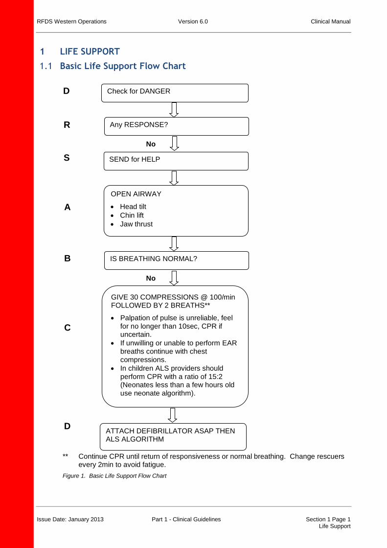
Issue Date: January 2013 Part 1 - Clinical Guidelines Section 1 Page 1 Life Support
1 LIFE SUPPORT
1.1 Basic Life Support Flow Chart
** Continue CPR until return of responsiveness or normal breathing. Change rescuers every 2min to avoid fatigue.
Figure 1. Basic Life Support Flow Chart
Check for DANGER
Any RESPONSE?
SEND for HELP
OPEN AIRWAY
Head tilt
Chin lift
Jaw thrust
IS BREATHING NORMAL?
GIVE 30 COMPRESSIONS @ 100/min FOLLOWED BY 2 BREATHS**
Palpation of pulse is unreliable, feel for no longer than 10sec, CPR if uncertain.
If unwilling or unable to perform EAR breaths continue with chest compressions.
In children ALS providers should perform CPR with a ratio of 15:2 (Neonates less than a few hours old use neonate algorithm).
ATTACH DEFIBRILLATOR ASAP THEN ALS ALGORITHM
No
No
D
A
S
R
B
D
C

RFDS Western Operations Version 6.0 Clinical Manual
Issue Date: January 2013 Part 1 - Clinical Guidelines Section 1 Page 2 Life Support
1.2 Newborn Life Support Flow Chart
If more than a few hours old use paediatric algorithm.
Figure 2. Newborn Life Support Flow Chart
Yes
At
all
sta
ge
s a
sk
: d
o y
ou
nee
d h
elp
?
Yes
Yes
Yes
Yes
No
Term gestation?
Breathing or Crying?
Good tone?
Prevent heat loss
Ensure open airway
Stimulate
HR below 100?
Gasping or apnoea?
Positive pressure ventilation
SpO2 monitoring
HR below 100?
Ensure open airway
Reduce leaks
Consider increasing pressure & oxygen
HR below 60?
Add chest compressions
3 compressions to each breath
100% oxygen
Consider intubation or LMA
HR below 60?
Venous access, Adrenaline
Consider volume expansion
Routine care:
Prevent heat loss
Ongoing evaluation
Laboured breathing
or persistent cyanosis?
Ensure open airway
SpO2 monitoring
Consider CPAP
Post-resuscitation care
Targeted pre-ductal SpO2 after birth
1min 60-70%
2min 65-85%
3min 70-90%
4min 75-90%
5min 80-90%
10min 85-90%
No
No
No
Adrenaline IV 10-30µg/kg (0.1-0.3mL/kg of
1:10,000 solution)

RFDS Western Operations Version 6.0 Clinical Manual
Issue Date: January 2013 Part 1 - Clinical Guidelines Section 1 Page 3 Life Support
1.3 Advanced Life Support (Adult)
Figure 3. Advanced Life Support (Adult)
During CPR
Airway adjunct (LMA/ETT) [See note 2]
Oxygen
Waveform capnography
IV/IO access
Plan actions before interrupting compressions. Charge defib
Drugs Shockable
Adrenaline 1mg after 2nd
shock (then every 2
nd
cycle)
Amiodarone 300mg after 3
rd shock
Drugs Non Shockable
Adrenaline 1mg immediately (then every
2nd
cycle)
Consider and Correct
Hypoxia
Hypovolaemia
Hyper/hypokalemia/metabolic disorders
Hypothermia/hyperthermia
Tension pneumothorax
Tamponade
Toxins
Thrombosis (pulmonary/coronary)
Post Resuscitation Care
Re-evaluate ABCDE
12 lead ECG
Treat precipitating causes
Re-evaluate oxygenation and ventilation
Temperature control (cool)
Start CPR
30 compressions @ 100/min : 2 breaths
Minimise Interruptions
Attach
Defibrillator / Monitor
Assess
Rhythm
Non
Shockable Shockable
Shock
CPR For
2 minutes CPR For
2 minutes
Return of
Spontaneous
Circulation?
Post Resuscitation Care

RFDS Western Operations Version 6.0 Clinical Manual
Issue Date: January 2013 Part 1 - Clinical Guidelines Section 1 Page 4 Life Support
Advanced Life Support (Adult) - Cont.
Notes
1. Good quality CPR with minimal interruptions essential. Return to CPR immediately after delivering a shock unless signs of life (breathing / moving), do not waste time checking rhythm or for a pulse.
2. Advanced airways (ETT, LMA) not required if successful BVM ventilation, if in place commence continuous chest compressions with no interruptions for ventilation (i.e. asynchronous ventilation). Securing advanced airway must not result in significant interruption to chest compressions and may be deferred to post resuscitation care.
3. ETCO2 ≤ 20mm Hg implies inadequate CPR or excessive ventilation.
4. Continue CPR whilst charging machine, charge as approaching end of 2 min cycle, stand clear only whilst shock is delivered.
5. Rhythm checks at two minutely intervals, if rhythm compatible with spontaneous circulation then check pulse.
6. Shockable rhythms are ventricular fibrillation and pulseless ventricular tachycardia.
7. Single shocks only, to be delivered (no stacked shocks). No precordial thump.
8. Energy levels; Monophasic 360J, Biphasic (use manufacturer’s default either 150 or 200J). Note All RFDS defibrillators are biphasic.
9. Best evidence is for uninterrupted CPR and early defibrillation, other interventions including drugs of less proven value.
10. There is no evidence for routinely giving buffers, atropine, calcium and magnesium in cardiac arrest. These drugs may be considered when treating potentially reversible causes of cardiac arrest.
a) Ca2+ (20mL of 10% calcium gluconate or 10mL of 10% calcium chloride) for hyperkalemia, hypocalcaemia, calcium channel blocker overdose.
b) Mg2+ (5mmol) for torsades de pointes, refractory VT / VF, hypokalemia, hypomagnesaemia.
c) Sodium bicarbonate for tricyclic overdose, hyperkalemia.
d) K+ (5mmol) for hypokalemia (in addition to giving mg).

RFDS Western Operations Version 6.0 Clinical Manual
Issue Date: January 2013 Part 1 - Clinical Guidelines Section 1 Page 5 Life Support
1.4 Advance Life Support (Paediatric)
Figure 4. Advanced Life Support (Paediatric)
During CPR
Airway adjunct (LMA/ETT) See note 3
Oxygen
Waveform capnography
IV/IO access
Plan actions before interrupting compressions. Charge defib to 4J/kg
Drugs Shockable
Adrenaline 10 µg/kg after 2nd
shock (then every 2
nd cycle)
Amiodarone 5mg/kg after 3rd
shock
Drugs Non Shockable
Adrenaline 10µg/kg immediately (then every 2
nd
cycle)
Consider and Correct
Hypoxia
Hypovolaemia
Hyper / hypokalemia / metabolic disorders
Hypothermia / hyperthermia
Tension pneumothorax
Tamponade
Toxins
Thrombosis
(pulmonary/coronary)
Post Resuscitation Care
Re-evaluate ABCDE
12 lead ECG
Treat precipitating causes
Re-evaluate oxygenation and ventilation
Temperature control (cool)
Adrenaline 10µg / kg
(immediately then every 2nd
cycle)
Start CPR
30 compressions @ 100/min : 2 breaths
Minimise Interruptions
Attach
Defibrillator / Monitor
Assess
Rhythm
Non
Shockable Shockable
Shock
CPR For
2 minutes CPR For
2 minutes Return of
Spontaneous
Circulation?
Post Resuscitation Care

RFDS Western Operations Version 6.0 Clinical Manual
Issue Date: January 2013 Part 1 - Clinical Guidelines Section 1 Page 6 Life Support
Advanced Life Support (Paediatric) – Cont.
Notes
1. Good quality CPR with minimal interruptions essential. Return to CPR immediately after delivering a shock unless signs of life (breathing / moving), do not waste time checking rhythm or for a pulse. Note, different ratio of compressions to ventilations reflects importance of hypoxia as cause of arrest.
2. Most paediatric arrests are asystole or PEA (pulseless electrical activity) secondary to hypoxia and hypovolaemia.
3. Advanced airways (ETT, LMA) not required if successful BVM ventilation, if in place commence continuous chest compressions with no interruptions for ventilation (i.e. asynchronous ventilation). Securing advanced airway must not result in significant interruption to chest compressions and may be deferred to post resuscitation care.
4. ETCO2 ≤20mmHg implies either inadequate CPR or excessive ventilation.
5. Continue CPR whilst charging machine, charge as approaching end of 2 min cycle, stand clear only whilst shock is delivered.
6. Rhythm checks at two minutely intervals, if rhythm compatible with spontaneous circulation then check pulse.
7. Shockable rhythms are ventricular fibrillation and pulseless ventricular tachycardia.
8. Single shocks only, to be delivered (no stacked shocks). No precordial thump.
9. Energy levels; Monophasic 4J/kg, Biphasic 4J/kg. Note All RFDS defibrillators are biphasic.
10. Best evidence is for uninterrupted CPR and early defibrillation, other interventions including drugs of less proven value.
11. There is no evidence for routinely giving buffers, atropine, calcium and magnesium in cardiac arrest. These drugs may be considered when treating potentially reversible causes of cardiac arrest.
i. Ca2+ (0.5mL/kg (max 20mL)of 10% calcium gluconate or 0.2mL/kg of 10% calcium chloride) for hyperkalemia, hypocalcaemia, calcium channel blocker overdose.
ii. Mg2+ (0.1-0.2mmol/kg) for torsades de pointes, refractory VT / VF, hypokalemia, hypomagnesaemia.
iii. Sodium bicarbonate (1mmol/kg for tricyclic overdose, hyperkalemia.
iv. K+ (0.1-0.2mmol/kg) for hypokalemia (in addition to giving Mg2+).
12. Neonates less than a few hours old refer to Neonate Life Support Algorithm.

RFDS Western Operations Version 6.0 Clinical Manual
Issue Date: January 2013 Part 1 - Clinical Guidelines Section 1 Page 7 Life Support
1.5 The Deteriorating Patient
Theory
At all times during the patient transport, from pre-flight assessment to handover of the patient at the receiving end it is important to recognise and act appropriately to signs of deterioration in the patient. This may sound like stating the obvious but a clearly defined action plan needs to be pre-determined for such events and staff need the confidence and support to be able to action such a plan.
For our purposes deterioration may either be physiological or behavioural, in either case an escalation of care may be required. Predictors of deterioration may be evident from the time of the pre-flight assessment, become evident whilst the patient is awaiting transport or during transport. The timing of deterioration will to a certain extent determine what actions may be required.
Physiological predictors of deterioration
In the remote and rural retrieval setting we are heavily reliant on simple parameters and clinical acumen to determine risk of deterioration, many of these parameters also form the basis of the more complex scoring systems. The measures available to us are more closely aligned to MET call or EWS scores and indeed less well resourced locations would have retrieval of the patient as part of their escalation process.
With this in mind the following limits are reminders of what may require an escalation in care.
Table 1. Physiological Predictors of Deterioration
<3 months 4-12 months 1-4 years 5-12 years 12 years-Adult
Adult
Nurse or Doctor Worried
Airway threat
Hypoxaemia SpO2 < 90% SpO2 < 90% SpO2 < 90% SpO2 < 90% SpO2 < 90% SpO2 < 90%
Respiratory distress, apnoea, cyanosis
Respiratory rate >60
<25
>50
<25
>40
<10
>30
<5
>30
<5
>30
<5
Heart rate <100
>180
<100
>180
<90
>160
<80
>140
<60
>130
<40
>130
Hypotension SBP <50 SBP <60 SBP <70 SBP < 80 SBP <90 SBP< 90
Acute change in neurological status, convulsion
Sudden fall in level of consciousness, GCS drop >2 points.
Repeated or extended seizures, or 1
st
seizure.
Sudden fall in level of consciousness, GCS drop >2 points.
Repeated or extended seizures, or 1
st
seizure.
Sudden fall in level of consciousness, GCS drop >2 points.
Repeated or extended seizures, or 1
st
seizure.
Sudden fall in level of consciousness, GCS drop >2 points.
Repeated or extended seizures, or 1
st
seizure.
Sudden fall in level of consciousness, GCS drop >2 points.
Repeated or extended seizures, or 1
st
seizure.
Sudden fall in level of consciousness, GCS drop >2 points.
Repeated or extended seizures, or 1
st
seizure.

RFDS Western Operations Version 6.0 Clinical Manual
Issue Date: January 2013 Part 1 - Clinical Guidelines Section 1 Page 8 Life Support
Escalation of care may comprise any or a number of the following actions:
1. Increase in priority of flight.
2. Doctor accompaniment.
3. Seeking additional orders, from RFDS doctor or clinical coordinator.
4. Diverting in flight for a higher level of assistance or resources (eg. To collect an RFDS doctor, or transfer patient to regional hospital emergency department, acquiring additional supplies like blood products.) Diversions ideally should be facilitated by the clinical coordinator.
5. Additional recommendations regarding pre-flight stabilisation (eg. Intubation, commencement of inotrope or other therapies.)
6. Planning to go in to a facility to stabilise a patient rather than have them bought out to the airport.
7. Planning a different destination for the patient (eg. An ICU bed)
8. Requesting a lights and sirens ambulance escort.
Predictors of Behavioural Disturbance
In-flight violence or behavioural disturbance is clearly a safety issue made all the more acute by the vulnerable environment in which we work. Every effort must be made to ensure the right personnel and armamentarium of chemical and physical restraint is available where required.
Behavioural disturbance may occur for psychiatric, organic and criminal reason. Whatever the cause, the pilot has an obligation to ensure the safety of the aircraft and is legally entitled to request restraint of a patient or passenger where required, medical authorisation for this restraint is not a pre-requisite rather an aviation safety duty of care if directed by the pilot. Patients referred under the mental health act will generally have medical authorisation for physical restraint in-flight.
Warning signs:
Facial and body language, suggesting anger and restlessness.
Physiological evidence of over-arousal (tachycardia, tachypnoea, muscle twitching, dilated pupils)
Increase volume of speech
Abnormal eye contact, refusal to communicate, fear, irritability
Unclear thought processes, poor concentration
Delusions and hallucinations with violent content, focussed on a particular person or command hallucinations
Overt hostility or suspiciousness
Verbal threats or gestures
Behaviour similar to that which preceded previous violent episodes
Reporting feeling anger or violent feelings
Risk factors:
History of violent behaviour
Alcohol or drug abuse
Reports of violence from carers
Expression of intent to harm
RFDS Western Operations Version 6.0 Clinical Manual
Issue Date: January 2013 Part 1 - Clinical Guidelines Section 1 Page 9 Life Support
Social rootlessness
Previous use of weapons
Previous dangerous impulsive acts
Denial of previous dangerous acts
Known personal trigger factors
Evidence of severe recent stress
Poor compliance with treatment
Antisocial, explosive or impulsive personality traits or personality disorders
In addition patients suffering delirium and dementia in particular may be worsened by unfamiliar environment and night-time conditions.
Escalation of care:
1. Ensure during pre-flight assessment that adequate history for warning signs and risk factors taken.
2. Ensure adequate pre-flight sedation including adequate antipsychotics for patients suffering from psychotic illness.
3. Doctor accompaniment
4. Additional escorts (eg police, cooperative relative)
5. Use of physical restraints
6. Ensure basic needs attended to ie nutrition, hydration, toileting.
7. Observe for and treat drug, alcohol and nicotine withdrawal
Nursing staff seeking assistance with either physiological or behavioural deterioration:
State clearly who and where you are.
Give a clear and concise background of the patient.
State clearly what the current problems are, if you have a specific concern, what you believe may be going on and your level of concern. (eg. The patient is violently resisting restraint and threatening to kill me. I am extremely worried for my safety.) (The patient has severe abdominal pain with a heart rate of 140 and systolic blood pressure of 80, I think he is bleeding and I am very worried.)
State clearly what action you would like to follow (eg. I want orders for sedation, I want the assistance of a doctor on the flight or to review this patient at the airport.)
References
National Institute for Health and Clinical Excellence. (2005). Violence: The short-term management of disturbed / violent behaviour in in-patient psychiatric settings and emergency departments. [CG25]. London. National Institute for Health and Clinical Excellence.
Tibballs J. Criteria for Activation of Medical Emergency Team. Clinical Practice Guidelines. September 2012. Royal Children’s Hospital, Melbourne.
Hillman K, Chen J, Cretikos M, Bellomo R, et al. Introduction of the medical emergency team (MET) system: a cluster-ransdomised controlled trial. The Lancet; Jun18-Jun24, 2005; 365, 9477
Fong N. Medical Emergency Response. Operational Directive OD 0040/07. Feb 2007. Department of Health Government of Western Australia.

RFDS Western Operations Version 6.0 Clinical Manual
Issue Date: January 2013 Part 1 - Clinical Guidelines Section 2 Page 1 Cardiovascular
2 CARDIOVASCULAR
2.1 Acute Coronary Syndromes
Theory
1. Acute Coronary Syndromes (ACS) cover a broad spectrum of acute presentations of ischaemic heart disease. This guideline covers the management of both ST Elevation Myocardial Infarction (STEMI) and Non-ST Elevation Acute Coronary Syndromes (NSTEACS).
2. ACS place a significant burden on health services and retrieval systems. Accurate diagnosis and consistency in management strategies are vital to appropriate use and equitable distribution of limited resources.
3. The diagnosis is based on history, 12 lead ECG findings and for NSTEACS, a serum Troponin.
STEMI
Consistent history plus any of the following:
Persistent ST elevation ≥ 1mm in 2 contiguous limb leads
Persistent ST elevation ≥ 2mm in 2 contiguous chest leads
New left bundle branch block (LBBB)
Changes consistent with posterior infarct (tall R in V1, deep anterior ST depression, ST elevation in V4 R)
ECG changes of right ventricular infarct (ST elevation in leads aVR and V4R)
NSTEMI
Consistent history without ECG changes consistent with STEMI, plus positive troponin and positive creatine kinase (CK).
Angina
High Risk, (positive troponin but negative CK)
Intermediate Risk, (needing further stratification after 8 hour troponin)
Low risk, negative troponin at 8 hours plus normal ECG.
4. Diagnosing a STEMI as soon as possible is vital, as reperfusion therapies must be given promptly to reduce morbidity and mortality. In the setting of a suggestive history, ECGs may need to be repeated at 10-15 minute intervals to diagnose an evolving STEMI.
5. Other ECG changes suggesting extreme risk include:
a) Total occlusion of left main coronary artery i. ECG
ST elevation in aVR ± aVL
Lesser ST elevation in V1
Marked ST depression in inferior leads ± left anterior fascicular block ii. May present with cardiogenic shock, significant ventricular arrhythmias or
cardiac arrest iii. Very high mortality rate
b) Total occlusion of proximal left anterior descending coronary artery (LAD) (Wellen’s Syndrome)
ECG – prominent T wave inversion in V1-V6. (mostly V1-V4)

RFDS Western Operations Version 6.0 Clinical Manual
Issue Date: January 2013 Part 1 - Clinical Guidelines Section 2 Page 2 Cardiovascular
These patients require URGENT percutaneous coronary angioplasty (PTCA) as they have a >60-70% mortality.
Pre–flight and In-flight Management
1. During the pre-flight assessment establish an accurate diagnosis.
View the 12 lead ECG yourself (faxed or scanned and emailed).
Provide assistance with making the diagnosis and instituting management. (It is expected that the assessing doctor takes responsibility for guiding management where the referring practitioner is uncertain).
Understand the plan for this patient and the rationale behind it. (Does this patient really need an urgent procedure or is the issue more one of convenience? Are they holding the catheter lab open for this patient, or not intending to do anything until tomorrow?).
2. Establish first line therapy for all ACS.
Aspirin loading dose 300mg orally (Medical Chest Item 62).
Glyceryl trinitrate (GTN) spray sublingual, titrated to pain and blood pressure (Medical Chest Item 190), may need patch or infusion.
Morphine titrated IV (2mg aquilots) or if unable to gain IV access then IM 5-10mg. (Medical Chest Item 188).
O2 to maintain normoxia or correct altitude hypoxia.
Clopidogrel 300mg loading dose.
Heparin infusion (5000 units loading dose followed by 1000 units per hour) or enoxaparin loading dose 1mg per kg subcutaneously. Use heparin if PTCA planned that day.
β- blocker if no contra-indications: atenolol 2.5mg-10mg IV OR 25-100mg orally. Alternatively metoprolol 5mg-15mg IV OR 25-100mg orally. Titrate to HR of 55-60bpm.
Correct electrolyte abnormalities that may predispose to arrhythmia (K+ and Mg2+ )
Maintain glycaemic control.
Consider a statin and an angiotensin converting enzyme inhibitor (ACEI).
Glycoprotein IIb / IIIa inhibitors (tirofiban) should be avoided after thrombolysis and should only be given with cardiologist advice (not widely available).
3. Reperfusion therapy.
STEMIs should be diagnosed early and reperfusion commenced as soon as possible provided no contraindications.
In practice no patients outside the metropolitan area are able to access PTCA in an appropriate time frame leaving thrombolysis the treatment of choice. Exceptions might exist for Rottnest Island and some inner rural locations where activation of an RFDS / SJA team on the FESA helicopter may be possible for a direct door to door transfer. The clinical coordinator in the RFDS Coordination Centre is best placed to judge the feasibility of this.
Second generation thrombolytics (tenecteplase or reteplase) are the agents of choice.
Verbal consent should be obtained from patients and this recorded.

RFDS Western Operations Version 6.0 Clinical Manual
Issue Date: January 2013 Part 1 - Clinical Guidelines Section 2 Page 3 Cardiovascular
Tenecteplase Dosage
Table 2. Tenecteplase Dosage
Patient weight (kg) Tenecteplase (IU) Tenecteplase (mg) Volume of reconstituted solution (mL)
<60 6,000 30 6
60 to <70 7,000 35 7
70 to <80 8,000 40 8
80 to <90 9,000 45 9
>90 10,000 50 10
Reteplase is given as 10 units intravenously, followed by 10 units after 30 minutes.
Streptokinase is rarely used these days and is unsuitable for indigenous patients who carry high
streptococcal antibody levels if it is used as a last resort the dose is 1.5 million units given over 60 minutes.
Contraindications to thrombolysis
Table 3. Contradindications to thrombolysis
Absolute Contraindications Relative Contraindications
Risk of Bleeding
Active bleeding or bleeding diathesis (excluding menses)
Significant closed head or facial trauma within 3 months
Suspected aortic dissection
Risk of Bleeding
Current use of anticoagulants: the higher the INR the higher the risk of bleeding
Non-compressible vascular punctures
Recent major surgery (< 3weeks)
Traumatic or prolonged (>10min) CPR
Recent (within 4 weeks) internal bleeding (e.g. gastro-intestinal or urinary)
Active peptic ulcer
Pregnancy
Risk of intracranial haemorrhage
Any prior intracranial haemorrhage
Ischaemic stroke within 3 months
Known structural cerebral vascular lesion
Known malignant intracranial neoplasm (primary or metastatic)
Risk of intracranial haemorrhage
History of chronic, severe, poorly controlled hypertension
Severe uncontrolled hypertension on presentation (>180mmHg SBP or >110mmHg DBP)
Ischaemic stroke more than 3 months ago, dementia, or known intracranial abnormality not covered in contraindications
Failure to reperfuse
Successful reperfusion is suggested by:
Patient is pain free
Haemodynamically stable
Cessation of arrhythmias
Reduction of ≥ 50% of maximum ST elevation by 60-90 minutes. Failure to reperfuse should result in a priority1 transfer for rescue angioplasty. (See Disposition).

RFDS Western Operations Version 6.0 Clinical Manual
Issue Date: January 2013 Part 1 - Clinical Guidelines Section 2 Page 4 Cardiovascular
4. Crew Mix
Figure 5. Crew Mix
5. Disposition and prioritisation
a) Chest pain in a primary location or hospital without doctor, tasked as P1 doctor accompanied
*May need to stage through regional centre.
b) Inter-hospital transport of ACS
*May need to stage through regional centre
Figure 6. Disposition and Prioritisation
Interhospital Transfer
NSTEMI +ve troponin
12 hours pain, arrhythmia, failure free, no GTN infusion
No doctor required
NSTEACS –ve troponin
Pain, arrhythmia, heart failure, GTN infusion
Doctor to accompany
Primary Chest pain Doctor to accompany
No pain No doctor required
STEMI Doctor to accompany
STEMI NSTEMI NSTEACS
Coronary Care Unit
Coronary
Care Unit*
High risk Low risk
Regional Hospital for differentiation, serial troponin.
Routine public transport for outpatient follow-up
Coronary
Care Unit*
STEMI NSTEMI NSTEACS
Coronary
Care Unit
P1 or P2 depending on reperfusion and complications: uncomplicated, reperfused at regional hospital may be P3
Coronary
Care Unit*
High risk Low risk
Coronary Care Unit*
P2 or P3 depending on local facilities and
patient status
Routine public transport for outpatient
follow-up

RFDS Western Operations Version 6.0 Clinical Manual
Issue Date: January 2013 Part 1 - Clinical Guidelines Section 2 Page 5 Cardiovascular
6. Ongoing management and communications
Patients should receive ongoing monitoring, before and during transport, with access to early defibrillation.
Provide oxygen to symptomatic patients and to correct altitude hypoxia.
Aim to achieve a pain free status inpatient (i.e. Remove any ongoing ischaemia).
Escalation in pain should result in repeat 12 lead ECG, some patients may require in-flight thrombolysis if a STEMI is evolving.
Patients requiring immediate access to PTCA should have their arrival times communicated to receiving hospitals to ensure the appropriate reception. Priority 1 ambulances may occasionally be required for these patients, operations staff need to be made aware of this requirement.
7. When RFDS stock of tenecteplase is used, the flight doctor must write a replacement script on a standard prescription form including the correct patient name and Medicare number. This must be provided to the flight nurse at the end of the flight. This is vital to maintain supply.
Medical Chest Items
GTN spray (Item 190), Aspirin 300mg tabs (Item 62), Morphine 10mg amps (Item 188)
References
National Heart Foundation of Australia “Guidelines for Management of Acute Coronary Syndromes” 2006 MJA Vol 184 No. 8 Supplement.
2011 Addendum to National Heart Foundation of Australia, Cardiac Society of Australia and NewZealand “Guidelines for The Management of Acute Coronary Syndromes 2006” March 2011
Australian Resuscitation Council. Acute Coronary Syndromes. Guideline 14.1 February 2011

RFDS Western Operations Version 6.0 Clinical Manual
Issue Date: January 2013 Part 1 - Clinical Guidelines Section 2 Page 6 Cardiovascular
2.2 Acute Pulmonary Oedema
Theory
Respiratory failure due to acute pulmonary oedema (APO) will be exacerbated by altitude hypoxia so supplemental oxygen is mandatory.
Pre–flight and In-flight Management
Flights are usually either priority 1 or 2, doctor accompanied, depending on the facilities at the referring location.
1. Diagnose and treat precipitating causes, including myocardial infarction, cardiac arrhythmias, pericardial effusion, hypertrophic cardiomyopathy and valvular heart disease. Consider non-cardiogenic pulmonary oedema.
2. Administer high flow oxygen with the patient sitting upright. Maximal supplemental oxygen followed by assisted ventilation should be used before resorting to a sea level cabin.
3. Give nitrates, either sublingually, or by infusion (commence at 10 g/min) (See Infusion Guideline). Maintain a systolic blood pressure ≥ 90mmHg. Topical application may not be reliable if sweaty or clammy.
4. Give IV frusemide 20 mg - 80 mg IV, repeating at 20 minutely intervals as necessary. Higher doses may be required if patient is taking frusemide regularly.
5. Note: A urinary catheter is essential to monitor output hourly.
6. If hypotensive, consider inotropic support (may then add vasodilator once in situ).
7. Consider the need for digoxin, especially if in atrial fibrillation.
8. If condition worsening consider NIV (Non Invasive Ventilation) or ventilation with ≥5 mmHg of PEEP and high dose oxygen. (See NIV Section).
9. Other less commonly used treatment modalities include venesection of 500 mL of blood (beware risk of hypovolaemia) or rotating tourniquets.
10. In dialysis patients who are overloaded, consider inducing diarrhoea with sorbitol / lactulose (difficult in the in-flight environment).
Special Notes
1. Intubation should be considered for all patients in APO who require high flow O2 at rest at the referring location. Especially look for confusion, exhaustion, a rising PCO2 and or relatively low PO2.
2. NIV may avoid the need for intubation but its use in the transport setting can be difficult (See NIV Section).
3. Avoid nitrates in patients who have received sildenafil (Viagra) in the previous 24 hours.
4. Morphine has been shown to adversely affect outcome in some studies and should be only be used judiciously.
Medical Chest Items
Frusemide tabs 40 mg (Item 85), Frusemide ampoules 20 mg/2mL (Item 120), IM if necessary, sublingual GTN spray (Item 190), Aspirin 300 mg tabs (Item 62).
RFDS Western Operations Version 6.0 Clinical Manual
Issue Date: January 2013 Part 1 - Clinical Guidelines Section 2 Page 7 Cardiovascular
References
Bersten A, Soni N. Oh’s Intensive Care Manual. 6th ed. Butterworth Heinemann. 2009
Cardiovascular drug guidelines [revised 2012 Feb]. In: eTG complete [CD-ROM]. Melbourne: Therapeutic Guidelines Limited; 2012 Jul.
Dr Mark Thomas. Orientation Manual for Renal Unit Medical Officers, Current 1999.
Royal Perth Hospital. CAPD Manual for Remote Nurses, Current 1999.
Graham CA. Pharmacological therapy of acute pulmonary oedema in the emergency department. Emergency Medicine, 16(1): 47-54. 2004.

RFDS Western Operations Version 6.0 Clinical Manual
Issue Date: January 2013 Part 1 - Clinical Guidelines Section 2 Page 8 Cardiovascular
2.3 Cardiac Arrhythmias
Theory
1. Cardiac arrhythmias are common and do not always require treatment in flight.
2. Diagnosis should be based on a 12 lead ECG where possible.
3. Priorities are always AIRWAY, BREATHING and CIRCULATION with application of supplemental oxygen and establishment of IV access. Patients should be fully monitored. If the patient is PULSELESS manage immediately according to the ALS algorithm. (See ALS guidelines).
4. Determine if the patient is STABLE or UNSTABLE. Unstable patients and patients with “stable VT” require immediate management. Features of instability are as follows:
a) Hypotension
b) Chest pain
c) Pulmonary oedema
d) Altered conscious state
e) Bradycardia <40 bpm , Tachycardia > 150 bpm
5. All antiarrhythmics have the potential to exacerbate dysrhythmias and cause myocardial depression.
Pre–flight and In-flight Management
1. During pre-flight assessment type of arrhythmia and stability should be established. A copy of the 12-lead ECG should be obtained. Advice regarding immediate management should be given including resuscitation, drugs and cardioversion.
2. Prioritisation will depend on skills and resources available at the referring location.
3. Unstable patients and those with a significant precipitating event (e.g. Acute coronary syndrome) and co-morbidities likely to result in an in-flight deterioration should be doctor accompanied.
4. All patients should have oxygen, venous access and be fully monitored.
5. Management is reliant on recognition of specific arrhythmias.
6. Sinus bradycardia and tachycardias require treatment of the underlying cause (e.g. hypovolaemia, fever, pain, left ventricular failure (LVF)) rather than specific antiarrhythmics or cardioversion.
7. For digoxin toxicity Digibind® may be required. Access at regional hospital or transport patient to regional hospital.

RFDS Western Operations Version 6.0 Clinical Manual
Issue Date: January 2013 Part 1 - Clinical Guidelines Section 2 Page 9 Cardiovascular
Bradycardias
Figure 7. Bradycardias
No
No
Yes
Yes
Yes UNSTABLE?
Atropine 500-600µg IV 3-
5 minutely up to 3mg.
Satisfactory response?
Interim measures:
Atropine 500µg to max 3mg
Adrenaline infusion 2-10µg/min
Alternative drugs* OR
Transcutaneous pacing
Seek expert help, transvenous
pacing.
*Alternatives include:
Isoprenaline (2-5µg/min)
Dopamine (2-5µg/kg/min)
Aminophylline
Glycopyrrolate
If β-Blocker or Ca2+
Channel blocker overdose consider glucagon or insulin/glucose/potassium infusion
Atropine contraindicated in cardiac transplant patients
Risk of asystole?
Recent asystole
Mobitz II AV block (constant PR interval with intermittent failure)
Complete heart block with broad QRS
Ventricular pause > 3sec
Observe

RFDS Western Operations Version 6.0 Clinical Manual
Issue Date: January 2013 Part 1 - Clinical Guidelines Section 2 Page 10 Cardiovascular
Tachycardias (with a pulse)
Figure 8. Tachycardias (with a pulse)
References
Australian Resuscitation Council. Guideline 11.9. Managing Acute Dysrhythmias. November 2009.
Cardiovascular drug guidelines [revised 2012 Feb]. In: eTG complete [CD-ROM]. Melbourne: Therapeutic Guidelines Limited; 2012 Jul.
No
UNSTABLE
Synchronised DC Shock Up to 3 attempts with sedation
Amiodarone 300mg IV over 10-20min then repeat shock, followed by:
Amiodarone 900mg over 24hr (if Torsades give Mg
2+, 2g over 10min)
Cardioversion of AF present for > 48hrs carries risk of stroke
Yes
Regular
No
Irregular
Broad
Seek expert help.
Regular
Seek expert help
STABLE Determine rhythm
Is the QRS narrow (<0.12s)?
Broad QRS
Regular?
Narrow QRS
Regular?
AF with bundle branch
block (treat as for narrow complex)
Pre-excited AF (consider amiodarone)
Polymorphic VT (Torsade de pointes) (give magnesium 2g over 10min)
If Ventricular Tachycardia (or uncertain rhythm): amiodarone 300mg IV over 20-60min then 900mg over 24 hr
If previously confirmed SVT with bundle branch block: Give adenosine as for regular narrow complex tachycardia
Use vagal manoeuvres
Adenosine 6mg rapid IV bolus
If unsuccessful give 12mg If unsuccessful give further 12mg.
Monitor ECG continuously
Normal sinus rhythm restored?
Irregular Narrow Complex Tachycardia Probable atrial fibrillation Control rate with:
β-blocker IV or digoxin IV
If onset < 48hr consider:
Amiodarone 300mg IV 20-
6-min, then 900mg IV over 24hr.
Probable re-entry PSVT:
Record 12-lead ECG in sinus rhythm
If recurs, give adenosine again & consider choice of anti-arrhythmic prophylaxis
Possible atrial flutter
Control rate (e.g. β-blocker)
Elective DC cardioversion with expert advice
RFDS Western Operations Version 6.0 Clinical Manual
Issue Date: January 2013 Part 1 - Clinical Guidelines Section 3 Page 1 Endocrine
3 ENDOCRINE
3.1 Diabetic Ketoacidosis
Theory
1. Diabetic ketoacidosis (DKA) is a state of relative or absolute deficiency of insulin, resulting in hyperglycaemia, ketosis, high anion gap metabolic acidosis and dehydration. The hyperglycaemia causes glycosuria, osmotic diuresis and progressive loss of fluid and electrolytes. The biochemical criteria are, venous pH <7.3 or bicarbonate <15 mmol/L plus blood or urinary ketones.
2. In a fully evolved hyperglycaemic coma, the most important clinical features are deep rapid breathing (Kussmaul respirations, secondary to acidosis), severe dehydration, circulatory insufficiency (hypotension, tachycardia) muscular weakness and a depressed level of consciousness.
3. Average deficits in diabetic ketoacidosis are 5-7 litres of water, 300-450 mmol of sodium and 3-5 mmol/kg of potassium. Correction of hyperglycaemia (4-8 hours) is more rapid than correction of acidosis (10-20 hours).
4. Underlying causes include; infection, newly diagnosed insulin dependent diabetes mellitus (IDDM), insufficient insulin (compliance, pump failure), infarction (myocardial, cerebral, gastrointestinal, peripheral vascular), intercurrent illness (e.g. diarrhoea and vomiting).
Pre-flight and In-flight Management
1. Pre-flight and in-flight management will be aimed at replacing fluid and electrolyte losses, correcting the ketosis and hyperglycaemia as well as commencing treatment for any underlying cause.
2. Flights are usually Priority 1 or 2, doctor accompanied, depending on the facilities at the referring location.
3. Ensure a secure airway, administer oxygen therapy and establish IV access.
4. Monitor GCS and vital signs frequently, BSL and urine output hourly (aim for >0.5mL/kg/hr) and consider NGT insertion. Blood gas, venous may be sufficient (pH, K+ and bicarb) monitoring 2 hourly for initial 6 hours.
5. Aim for 3mmol/hr fall in BSL, 3mmol/hr rise in bicarbonate, maintain potassium in normal range. Avoid hypoglycaemia.
6. IV fluids: a) Normal saline 500mL (10mL/kg for children) bolus over 10-15 mins if shocked or SBP
<90mmHg systolic , with dose repeated if required. If contemplating a third bolus in children seek advice, rarely required and potential for harm.
b) Subsequently or if SBP >90mmHg systolic use normal saline 1L in first hour, then
c) Normal saline (with 20mmol KCl) x 2 L over 4 hours, then
d) Normal saline (with KCl) 2L over 8 hours, then
e) Normal saline (with KCl) 1L over 6 hours,
f) Further litres dependent on vital signs, clinical hydration state and CVP.
g) Add 10% dextrose when BSL < 14mmol/L at 125 mL/hr. Normal saline may be given concurrently if still correcting volume. (Children: requirements dependent on degree of dehydration. Aim to give deficit [% dehydration x body weight] + maintenance over 48 hours).

RFDS Western Operations Version 6.0 Clinical Manual
Issue Date: January 2013 Part 1 - Clinical Guidelines Section 3 Page 2 Endocrine
7. Insulin infusion:
In adults give actrapid (50 units in 50mL N/Saline) at 0.1 IU/kg/hr IV, continue the patient’s usual long acting insulin if prescribed.
In children give actrapid 0.2 IU/kg SC then follow with 0.1 IU/kg SC every 2 hours, reduce to 0.1 IU/kg SC every 4-6/24 when BSL < 8 mmol/L.
8. If laboratory facilities available check electrolytes, glucose and blood culture; also request urea, creatinine, osmolarity, blood gases, 12-lead ECG, to look for signs of hyper- or hypokalaemia and acute myocardial ischaemia.
9. Potassium level may be high initially despite depleted stores. Take care with administration if the serum level is not known and do not commence potassium replacement in the presence of oliguria or if serum K+ is > 5.5mmol. Replace K+ at 40mmol/hr if K+ < 3.5mmol.
Special Notes
The endocrinology team at PMH prefer to be involved in management decisions for paediatric patients early.
References
Princess Margaret Hospital for Children. Emergency Department Clinical Guidelines. 1997.
Joint British Diabetes Societies Inpatient Care Group. The Management of Diabetic Ketoacidosis in Adults. NHS. March 2010.
Rosenbloom AL. “The management of diabetic ketoacidosis in children”. Diabetes Ther. 2010; 1(2): 103-120.
Diabetic Ketoacidosis- an Overview and update-presentation by Dr Malcolm Rivers Royal Flying Doctor Service. Jandakot. May 2011
Endocrinology drug guidelines [revised 2009 Jun]. In: eTG complete [CD-ROM]. Melbourne: Therapeutic Guidelines Limited; 2012 Jul.
Royal Children’s Hospital, Melbourne, Australia. Clinical practice Guideline on Diabetes mellitus, [Internet, cited 21/02/2012], available from: http://www.rch.org.au/clinicalguide/cpg.cfm?doc_id=5189

RFDS Western Operations Version 6.0 Clinical Manual
Issue Date: January 2013 Part 1 - Clinical Guidelines Section 3 Page 3 Endocrine
3.2 Hypoglycaemia
Theory
1. Hypoglycaemia needs to be considered as a differential diagnosis in all unconscious patients (especially but not exclusively diabetic patients), in all patients with abnormal behaviour and in all patients with unexplained neurological signs.
2. Moderate hypoglycaemia is characterised by tachycardia, sweating, clamminess, paraesthesia (face and hands), irritability, hunger and agitation.
3. Severe hypoglycaemia is characterised by mental confusion, bizarre behaviour, seizures, hypothermia and coma (hydrated, quiet and flaccid).
4. All symptoms may be blunted by alcohol, sedatives, patients on β-blockers and in the elderly.
5. The most common cause of hypoglycaemia is overdose of insulin or oral hypoglycaemics, particularly long acting sulphonylureas. Other causes include inadequate food intake, reactive (post-prandial), drugs (salicylates, iron, alcohol), status epilepticus and counter-regulatory (Addison's disease, hypopituitarism, myxoedema, severe cachexia, hepatic failure or severe renal failure).
Table 4. Hypoglycaemia – Normal Reference Range by Age
Age Normal Reference Range
0-6 months 2.2-5.0mmol/L
7 - 12months 1.9-8.0 mmol/L
2 years 2.8-7.2mmol/L
3 years 3.3-6.7 mmol/L
adults(fasting) 3.6-5.8 mmol/L
Note: Hypoglycaemic symptoms and signs may occur at almost normal levels if a patient is accustomed to high blood sugar levels.
Pre-flight and In-flight Management
1. If conscious:
Oral barley sugar, sweetened orange juice or sandwiches.
If cause was a long-acting insulin (NPH or Lente) or oral sulphonylurea, an IV infusion of 5% dextrose 100-125 mL/hr will need to be run for 24 hours and the patient will need to be admitted for observation.
2. If unconscious:
Resuscitation as for all unconscious patients, with attention to airway, breathing and circulation.
Establish IV access and give 50 mL of 50% dextrose in water at 10 mL/min. • May cause hypokalaemia if given too quickly.
Most patients recover in 5-10 mins unless hypoglycaemia was prolonged.
If chronic alcohol consumption is suspected, give 100 mg thiamine IM or IV before dextrose to prevent Wernicke's encephalopathy.
Treat fitting with diazepam 0.2 mg/kg IV, repeated as needed or PR diazepam 0.5 mg/kg

RFDS Western Operations Version 6.0 Clinical Manual
Issue Date: January 2013 Part 1 - Clinical Guidelines Section 3 Page 4 Endocrine
Special Notes
1. Neonates:
Give 20 mL of 5% dextrose orally or NGT or 1 mL/kg of 50% dextrose IV or 2 mL/kg of 25% dextrose IV.
2. Glucagon:
Can be given 1 mg IM or SC or IV, 0.03 mg/kg IM for children to a maximum dose of 1mg.
Side effects include nausea and vomiting, initially hyperkalaemia, then hypokalaemia.
3. In severe sulphonylurea overdose treatment is often prolonged requiring IV 10% glucose and frequent monitoring. Octreotide can reduce the duration of IV therapy and the need for additional boluses of 50% glucose. This should only be considered in severe overdose.
4. If indicated use: octreotide 50micrograms SC 8 hourly for up to 3 doses.
References
Dunn R, Dilley S, Brookes J, et al. The Emergency Medicine Manual. 5th ed. Tennyson, South Australia: Venom Publishing. 2010
Bersten A, Soni N. Oh’s Intensive Care Manual. 6th ed. Butterworth Heinemann. 2009.
Fulde G. Emergency Medicine, The Principles of practice. 5th ed. Australia: Saunders. 2009.
Endocrinology drug guidelines [revised 2009 Jun]. In: eTG complete [CD-ROM]. Melbourne: Therapeutic Guidelines Limited; 2012 Jul.
Australian Medicines Handbook [CD-ROM]. Adelaide: Australian Medicines Handbook Pty Ltd; January 2010.

RFDS Western Operations Version 6.0 Clinical Manual
Issue Date: January 2013 Part 1 - Clinical Guidelines Section 3 Page 5 Endocrine
3.3 Hypocalcaemia
Theory
1. This is a relatively common condition and, though rarely life-threatening, it needs to be recognised and treated appropriately if potentially serious problems are to be prevented.
2. Serum calcium is composed of 2 major fractions, 45% of the total serum calcium is bound to plasma proteins, chiefly albumin and the other 55% exists as ionised Ca2+. Changes in the ionised fraction result in the signs and symptoms of hypo-and hypercalcaemia. Acidosis increases the ionised fraction by displacing calcium from albumin whereas alkalosis decreases it; rapid changes in plasma acid-base status can therefore result in symptomatic hypocalcaemia. Examples include hyperventilation resulting in tetany and hypocalcaemia from massive transfusion of citrated blood.
Symptoms of Hypocalcaemia
In the early stages these include peripheral and peri-oral paraesthesiae and later tetany, carpo-pedal spasm, hyperreflexia, colicky abdominal pain, stridor due to laryngospasm and convulsions. In infants one may also see apnoeic spells and intermittent cyanosis.
Signs of Hypocalcaemia
1. These include Chvostek's sign (spasm of the ipsilateral facial muscles when the facial nerve is tapped over the parotid nerve), Trousseau's sign (carpo-pedal spasm caused by the reduction of the blood supply to the hand when a BP cuff above systolic pressure is applied to the forearm for 3 mins) and ECG changes which include lengthening of the QT interval and arrhythmias.
2. Hypocalcaemia may be associated with hypomagnesaemia and hypokalaemia; this can be of significance in aboriginal children and is probably related to renal and gastrointestinal losses.
Pre-flight and In-flight Management
1. The flight priority usually will depend on the underlying condition. If the patient is symptomatic and requiring active treatment then the flight will probably be Priority 1 or 2 and doctor accompanied.
2. Hypocalcaemia should always be considered in critically ill patients with sepsis, burns, acute renal failure, those who have been transfused with citrated blood, pancreatitis and those with hypoalbuminaemia.
3. Wherever possible blood electrolytes, including Ca2+, Mg2+, K+ and acid-base status should be known and all abnormalities corrected. The iStat analyser can provide valuable information and should be available in all cases where electrolyte and acid-base imbalances are present or are suspected. A 12-lead ECG should always be available as well.
4. In mild cases with minimal symptoms and no tetany, oral replacement therapy is appropriate.
5. In more severe cases the treatment for adults is IV calcium gluconate (20mL) or calcium chloride 10% solution (5-10 mL). Monitor the Pulse rate, BP and ECG.
6. For Infants use IV calcium gluconate 10% (0.22 mmol Ca2+/mL). Give 0.5mmol/kg slowly over 10-20 mins. If bradycardia develops then the infusion should be ceased immediately.

RFDS Western Operations Version 6.0 Clinical Manual
Issue Date: January 2013 Part 1 - Clinical Guidelines Section 3 Page 6 Endocrine
Special Notes
1. Normal values (PathWest): Total calcium 2.25-2.60 mmol/L; ionised calcium 1.12-1.32 mmol/L.
2. Calcium can precipitate or exacerbate digitalis toxicity therefore IV calcium must be given
very slowly in patients on digoxin and the ECG must be monitored continuously.
References
Edmond K. Guidelines for the investigation and treatment of children with diarrhoea and dehydration. Department of Paediatrics, Royal Darwin Hospital. May 1997
Dunn R, Dilley S, Brookes J, et al. The Emergency Medicine Manual. 5th ed. Tennyson, South Australia: Venom Publishing. 2010.
Marx J, Hockberger R, Walls R. Rosen’s Emergency Medicine. Concepts and Clinical Practice. 7th ed. Mosby. 2009.
Saunders C, Ho M. Current Emergency Diagnosis and Treatment. 4th ed. Appleton Lange. 1992.

RFDS Western Operations Version 6.0 Clinical Manual
Issue Date: January 2013 Part 1 - Clinical Guidelines Section 4 Page 1 Gastrointestinal
4 GASTROINTESTINAL
4.1 Acute Pancreatitis
Theory
1. A multi-system disease due to inflammation of the pancreas. Gallstones and alcohol abuse account for 75% of cases. Other causes include, mumps, vasculitis, trauma, drugs such as azothiaprine, antiretroviral and chemotherapeutic drugs, penetrating peptic ulcer and post ERCP. Overall mortality is 1.5%, up to 2% and usually restricted to patients with severe necrotizing pancreatitis.
2. Signs and symptoms depend on the amount of glandular destruction.
a) Mild to Moderate:
Epigastric pain, often of rapid onset and relieved by sitting forward, abdominal distension, nausea and vomiting, raised amylase and lipase, pain radiating to back, fever, tachycardia and hypotension.
b) Severe:
Hypotensive shock secondary to intraperitoneal blood and fluid loss, respiratory failure, acidosis, hypocalcaemia, abdominal mass.
3. Mild to moderate pancreatitis is usually self-limiting in 1 week. Complications, however, include chronic pancreatitis, diabetes, pancreatic insufficiency, ascites and cholelithiasis.
4. Severe pancreatitis can be complicated by acute respiratory distress syndrome (ARDS), metabolic acidosis, acute tubular necrosis, disseminated intravascular coagulation, shock, ileus, pleural effusions, severe vomiting, haemorrhage, sepsis or necrosis of the pancreas.
5. Pseudocysts can form; these are encapsulated fluid collections full of enzymes. Often multiple, if they are <6cm they tend to resolve. They may erode into blood vessels. A ruptured pseudocyst is a surgical emergency as it can cause massive bleeding.
6. Operative interventions if they are required may include laparotomy, endoscopic sphincterotomy and stone extraction if cholelithiasis. Total parenteral nutrition may be required if necrosis or infection is present. Occasionally debridement of necrotic pancreatic tissue and drainage of pseudocysts may be indicated.
7. Useful diagnostic tests, where available include amylase, lipase, FBC, U&E & creatinine, Ca2+, glucose, LFTs, bilirubin, arterial blood gases, coagulation studies, blood group and cross-match. A chest x-ray may show a raised left hemi-diaphragm, pleural effusions, atelectasis or ARDS. CT scan may show pancreatic necrosis or pseudocyst formation.
Pre-flight and In-flight Management
1. Flights are usually Priority 1 or 2 and doctor accompanied in severe cases.
2. Administer supplemental oxygen by mask:
Intubation should be considered prior to transfer if evidence of ARDS is present and high flow rates of oxygen are required at rest in the hospital.
3. Ensure adequate IV access prior to transfer, preferably with 2 IV lines. Fluid resuscitation should be aggressive with massive sequestration of fluid in severe cases.
4. A nasogastric tube is essential and urinary catheterization is desirable in all but the mildest of cases.
5. Treat shock according to guidelines for management of shock; A decreasing haematocrit may indicate haemorrhagic pancreatitis - transfusion may be required.
RFDS Western Operations Version 6.0 Clinical Manual
Issue Date: January 2013 Part 1 - Clinical Guidelines Section 4 Page 2 Gastrointestinal
6. Adequate analgesia is important. Narcotics should be given IV and titrated to effect. Metoclopramide 10 to 20 mg IV or ondansetron 4 to 8mg IV or buccal may be required for nausea/vomiting.
7. In-flight management of acid / base and electrolyte imbalances can be assisted by use of the iStat analyser. Insulin infusion should be considered if the blood glucose is >15 mmol/L. Also consider calcium gluconate for hypocalcaemia.
8. Antibiotics are rarely indicated in the acute phase as sepsis occurs later in the illness but if sepsis is present give IV metronidazole and ceftriaxone or cefotaxime.
9. ECG monitoring is recommended , non-specific ST-T wave changes and bradycardia due to toxins may be seen.
References
Dunn R, Dilley S, Brookes J, et al. The Emergency Medicine Manual. 5th ed. Tennyson, South Australia: Venom Publishing. 2010.
Bersten A, Soni N. Oh’s Intensive Care Manual. 6th ed. Butterworth Heinemann. 2009.
Fulde G. Emergency Medicine, The Principles of Practice. 5th ed. Australia: Saunders. 2009.
Gastrointestinal drug guidelines [revised 2011 Feb]. In: eTG complete [CD-ROM]. Melbourne: Therapeutic Guidelines Limited; 2012 Jul.

RFDS Western Operations Version 6.0 Clinical Manual
Issue Date: January 2013 Part 1 - Clinical Guidelines Section 4 Page 3 Gastrointestinal
4.2 Haematemesis and Melaena
Theory
1. Gastrointestinal tract (GIT) bleeding is a common medical emergency with significant morbidity and mortality. Despite treatment advances, mortality for patients presenting with upper gastrointestinal haemorrhage remains around 5-10%.
2. Haematemesis indicates bleeding proximal to the ligament of Treitz and occurs in only 50-66% of patients with upper GIT bleeding.
3. Melaena may mean haemorrhage from either the upper GIT or proximal colon.
4. Gastro-oesophageal varices account for 2-15% of all upper GIT bleeding. Bleeding will cease spontaneously in only 20-30% of cases but as bleeding is often more severe and recurrent, mortality approaches 25-40% for each episode of variceal haemorrhage.
Pre-flight and In-flight Management
1. Flights for patients will be usually priority 1 or 2. Flights where the patient has ongoing haemorrhage resulting in instability and/or requiring transfusion will be doctor accompanied.
2. Pre-flight management will include resuscitation of the patient and replacement of intravascular volume with isotonic crystalloid (normal saline or Hartmann's) or colloid (gelofusine or haemaccel).
3. Blood should be given if there is persistent haemodynamic instability despite 2 litres of crystalloid or colloid, if the initial Hb <8mg/dl, if there is significant re-bleeding, and in those patients with co-morbidities making them unable to tolerate periods of hypoperfusion or anaemia.
4. Oxygen should be administered to all patients.
5. Continuous ECG monitoring, non-invasive blood pressure monitoring and pulse oximetry should be instituted.
6. Patients should all have adequate IV access (two large bore cannulas 14 or 16g) Some patients may require inotropic support if still haemodynamically unstable despite adequate fluid therapy.
7. If possible blood (ideally cross matched type specific or if unable then uncrossed O neg) should be taken on the flight.
8. Octreotide infusion only has a place in the management of variceal haemorrhage. Dose is 50-100 µg bolus then infusion of 25 - 50 µg per hour. (See Drug Infusion Guidelines).
9. Fresh frozen plasma should be given when the prothrombin time is 3 seconds greater than the control or when large transfusions are required. Platelet transfusion is rarely required unless platelet count is less than 50 x 109 /L. (See transfusion guidelines).
10. If bleeding from a gastric ulcer is suspected then IV proton pump inhibitor therapy should be considered. Esomeprazole or omeprazole or pantoprazole 80mg over 15 to 30 mins
followed by infusion at 8mg/hour.
Special Notes
Balloon tamponade is not available to RFDS staff but occasionally patients from regional centres may have a Sengstaken-Blakemore tube or Minnesota tube in place, these patients are likely to be transported intubated. Effects of gas expansion during air transport must be considered. Only the gastric balloon should be inflated as inflation of the oesophageal balloon can cause severe oesophageal ulceration.

RFDS Western Operations Version 6.0 Clinical Manual
Issue Date: January 2013 Part 1 - Clinical Guidelines Section 4 Page 4 Gastrointestinal
References
Dunn R, Dilley S, Brookes J, et al. The Emergency Medicine Manual. 5th ed. Tennyson, South Australia: Venom Publishing. 2010.
Cameron P, Jelinek G, Kelly A, et al. Textbook of Adult Emergency Medicine. 3rd ed. Churchill Livingstone. 2009.
Gastrointestinal drug guidelines [revised 2011 Feb]. In: eTG complete [CD-ROM]. Melbourne: Therapeutic Guidelines Limited; 2012 Jul.

RFDS Western Operations Version 6.0 Clinical Manual
Issue Date: January 2013 Part 1 - Clinical Guidelines Section 4 Page 5 Gastrointestinal
4.3 Intestinal Obstruction
Theory
1. Intestinal obstruction may be caused by physical obstruction or absence of function (paralytic ileus).
2. Intestinal obstruction may occur in the small bowel or colon. Obstruction to the small bowel is acute; that of the colon, less so. Either may be partial or complete.
3. Patients with intestinal obstruction all have a quantity of gas trapped within the gut. Expansion of gas at altitude causes pain and may cause perforation of necrotic tissue. Patients with a complete intestinal obstruction, especially obstruction due to rigid external structures (e.g. hernia), are even more at risk.
4. Vomiting is common and changes in pressurisation can precipitate further vomiting with its attendant risk of aspiration.
5. Prolonged obstruction is complicated by perforation, sepsis and fluid/electrolyte disturbance. Patients should be resuscitated with appropriate fluids, K+ and antibiotics pre-flight.
Pre-flight and In-flight Management
1. The type and duration of the obstruction will determine the flight priority. Sicker patients, the very young and the very old may require a doctor accompanied flight.
2. All flights will require sea-level pressurisation. This, in most cases will preclude intermediary landings or 'meets' at airstrips whose elevation is considerably higher than sea-level.
The major changes in atmospheric pressure occur closest to the earth's surface in the first few thousand feet - if a patient is to become compromised it will occur there.
As in all things, however, occasionally a compromise is required if the risks (e.g. extra time taken to fly to Carnarvon rather than Meekatharra) outweigh the benefits. In some instances a patient with a bowel obstruction may suffer some additional pain on ascent to altitude but otherwise no serious adverse effects.
3. All patients will be 'nil by mouth', and will require IV fluids with close attention to be paid to hydration status. Patients who are vomiting or who have a small bowel obstruction require a nasogastric tube (NGT), which should be kept on straight drainage and aspirated at regular intervals. Sicker or elderly patients may require an indwelling catheter to allow more accurate monitoring of their urine output and fluid balance.
4. Where patients have been obstructed for some time, an up to date Na+, K+ levels and acid-base status can be provided by using the i-STAT analyser.
5. Analgesia should be provided with titrated IV narcotics. An anti-emetic is unnecessary and
may be potentially harmful. Nausea and vomiting should be treated with NGT aspiration.
References
MIMS Annual 23rd Ed 1999, Medimedia Australia Pty Ltd, p. 3-339
Marx J, Hockberger R, Walls R. Rosen’s Emergency Medicine. Concepts and Clinical Practice. 7th ed. Mosby. 2009

RFDS Western Operations Version 6.0 Clinical Manual
Issue Date: January 2013 Part 1 - Clinical Guidelines Section 5 Page 1 Genitourinary
5 GENITOURINARY
5.1 Acute / Chronic Renal Failure
Theory
1. Acute renal failure is defined as a rapid increase in metabolic waste products (urea, creatinine, K+) usually associated with marked decrease in urine output. Isolated acute renal failure has a mortality of approximately 10%, in the setting of multi -organ failure the figure approaches 40-80%.
2. Features of acute renal failure include:
A rise in creatinine of > 100 µmol/1/day,
Oliguria (urine output less than 0.5mL/kg/hr)
Either fluid depletion or fluid overload
Altered mental state
Hyperkalemia
Metabolic acidosis (exacerbates hyperkalemia and causes hypotension and nausea). More rapid onset in catabolic states (sepsis, GIT bleed, rhabdomyolysis)
Uraemic symptoms - nausea, hiccoughs, drowsiness, flap, foetor, pericarditis, bruising / bleeding, itch, hypotension, coma,
Death due to arrhythmia (secondary to high K+), pulmonary oedema, GIT bleed, pericardial tamponade.
Pre-flight and In-flight Management
1. Priority and the need for a doctor will need to be determined on an individual basis. Most patients in acute renal failure will be tasked as priority 1 or 2, doctor accompanied.
2. Determine the cause:
a) Pre-renal:
Absolute hypovolaemia – bleeding, vomiting, diarrhoea, diuresis, burns, inadequate intake.
Relative hypovolaemia – vasodilatation (sepsis, vasodilators), reduced oncotic pressure (cirrhosis, nephrosis, sepsis, malnutrition),
Reduced cardiac output – pulmonary embolism, pericardial tamponade, infarct, arrhythmia.
Reno-vascular – renal artery thrombosis, atheroemboli.
b) Renal:
Glomerulonephritis, acute tubular necrosis, acute interstitial nephritis.
c) Vascular:
Malignant hypertension, haemolytic uraemic syndrome, severe pre-eclampsia.
d) Post - renal:
Pelvocalyceal - ureteric ( solitary kidney, extrinsic (lymphoma), mural (stricture), luminal (stone, clot, sloughed papilla).
Vesicoureteric junction - bladder (Carcinoma of; bladder, cervix, bowel, stone).
Bladder neck - urethra (blocked catheter, stricture, prostate).
3. Determine type of renal failure and volume status:
a) Oliguric vs. non – oliguric?

RFDS Western Operations Version 6.0 Clinical Manual
Issue Date: January 2013 Part 1 - Clinical Guidelines Section 5 Page 2 Genitourinary
Oliguric - <20 mL/hr, 500 mL/day. Oliguric has a higher mortality, treat early and monitor U&E, volume.
b) Patient 'wet' vs. 'dry'. Assess volume:
In chronic renal failure check weight daily as a marker of total body water (1 kg =1 litre).
Low Na+ implies water overload not Na+ deficiency.
Extracellular fluid; oedema signifies > 2 kg of fluid overload.
Reduced skin turgor if dry (beware elderly)
Intravascular volume; i. JVP (aim for 2 cm at 45°), ii. BP (high if overloaded, postural drop present if dry) iii. Capillary return; increased if dry.
4. Renal Support:
Maintain intravascular volume: aim for a MAP of 75-80mmHg by replacing fluids (monitor JVP / CVP), +/- inotropes
Remove nephrotoxins (eg. NSAIDs)
Monitor urine output
There is no evidence to support the use of “renal or low-dose dopamine”
Use of loop diuretics (frusemide) may reduce the need for dialysis
5. Manage complications:
Hyperkalemia
Pulmonary oedema
Special Notes
1. For Chronic Renal Failure patients on or nearing dialysis;
Avoid catheterization
Avoid IV cannulation or blood letting in forearm veins (may be required for fistula)
No BP measurements, cannulae or venepuncture on arm with fistula
2. Continuous ambulatory peritoneal dialysis (CADP) complications:
a) Overload:
Hypertension, increased weight, pulmonary oedema – Treat with more frequent bag changes (if getting more fluid back than instilled) or increase strength of glucose (max 2.5%)
b) Dehydration:
Treat by reducing glucose strength of bag
c) Peritonitis:
Pain, fever, diarrhoea, cloudy bag – Send whole bag for MC&S, give vancomycin 2g and gentamicin 0.6mg/kg (max 50mg) into bag daily until culture results
d) Exit site infection:
Swab, then if Gram pos, vancomycin weekly for four weeks, or flucloxacillin 500mg 6 hourly for 2 weeks. If Gram neg, ciprofloxacin 500mg orally for 3 weeks.

RFDS Western Operations Version 6.0 Clinical Manual
Issue Date: January 2013 Part 1 - Clinical Guidelines Section 5 Page 3 Genitourinary
References
Antibiotic drug guidelines [revised 2010 Jun]. In: eTG complete [CD-ROM]. Melbourne: Therapeutic Guidelines Limited; 2012 Jul.
Bersten A, Soni N. Oh’s Intensive Care Manual. 6th ed. Butterworth Heinemann. 2009.
CAPD Manual for Remote Nurses, Royal Perth Hospital, current 1999.
Beed M, Sherman R, Mahajan R. Emergencies in Critical Care. Oxford: Oxford University Press. 2007

RFDS Western Operations Version 6.0 Clinical Manual
Issue Date: January 2013 Part 1 - Clinical Guidelines Section 6 Page 1 Infectious Diseases
6 INFECTIOUS DISEASES
6.1 Bacterial Meningitis
Theory
1. Acute Bacterial Meningitis is a life threatening emergency. The overall mortality is about 18% however this is raised at the extremes of age and in the immunocompromised.
2. The presentation may include a severe generalised headache more prominent over the occiput and worse with any manoeuvre that increases intracranial pressure. Other signs of meningeal irritation include photophobia and neck stiffness. Vomiting may be a prominent feature. Raised intracranial pressure may result in focal neurological signs, seizures, delirium and papilloedema.
Note: Lumbar punctures must not be performed where signs of raised intracranial pressure, focal neurological signs or a bleeding diathesis exist.
3. Patients with meningitis may be divided into two groups on the basis of presentation.
a) Acute presentation – Symptoms and signs have been present for less than 24 hours and are rapidly progressive. The causative organisms in these patients are usually pyogenic bacteria, and the mortality approaches 50%.
b) Subacute presentation – Symptoms and signs have been present for 1-7 days. Meningitis in this group of patients may be due to bacteria, viruses, or fungi, and the death rate in cases due to bacterial infection is much lower.
Pre-flight and In-flight Management
1. These flights would usually be doctor accompanied and priority 1 or 2.
2. Assess the airway. Patients may have an unprotected airway or hypoventilation from a depressed central nervous system and require intubation and ventilation.
3. Apply supplemental oxygen and monitor oxygenation (oximetry and ABGs).
4. Apply full cardiac monitoring. Note risk of bradycardia and other dysrhythmias with raised intracranial pressure.
5. Ensure IV access and normovolaemia. If hypovolaemic, resuscitate with normal saline. Check sodium, if low consider syndrome of inappropriate antidiuretic hormone (SIADH). (These patients may require fluid restriction to 2/3 maintenance).
6. Ensure normoglycaemia.
7. Attempt to gain blood cultures if possible but do not delay therapy if facility for this doesn’t exist.
8. Administer empirical antimicrobial therapy based on age and likely pathogens.
a) Under 3 months of age: Gram negatives, Group B streptococci, Staphylococci and Listeria
amoxicillin 50mg /kg IV 6 hourly plus
cefotaxime 50mg / kg IV 6 hourly (ceftriaxone if unavailable)
b) Over 3 months of age: Strep. pneumonia, Neisseria meningitides, Staphylococci, (Haemophilus influenzae now uncommon since immunisation)
dexamethasone 10mg IV (0.15mg /kg IV) prior antibiotics then 6 hourly
cefotaxime 2g IV (50mg / kg IV) 6 hourly or ceftriaxone 2g IV (50mg / kg IV) 12 hourly
RFDS Western Operations Version 6.0 Clinical Manual
Issue Date: January 2013 Part 1 - Clinical Guidelines Section 6 Page 2 Infectious Diseases
Plus
vancomycin 1.5g slow IV infusion (30mg / kg slow IV infusion) 12 hourly if Gram
positive diplococci or pneumococcal assay on CSF or concurrent otitis media, sinusitis, recent treatment with a β-lactam, or if viral or meningococcal infection unlikely. i. Immunosuppressed and elderly: Listeria
Add benzylpenicillin 2.4g IV (60mg/kg IV) 4 hourly. ii. Severe penicillin or cephalosporin allergy:
vancomycin 1.5g slow infusion IV (30mg/kg slow infusion IV) 12 hourly Plus
ciprofloxacin 400mg IV (10mg /kg IV) 6 hourly Or
moxifloxacin 400mg IV (10mg / kg IV) daily
Note: RFDS aircraft drug box does not carry some of the above drugs, they may need to be sourced from regional or local hospital.
Special Notes
1. Treat seizures aggressively with anticonvulsants.
2. If Neisseria meningitides confirmed contacts will need antibiotic prophylaxis. This is a notifiable disease.
a) Only medical or nursing staff who have performed or attempted mouth to mouth resuscitation or intubation or are in prolonged close contact with the infected patient require prophylaxis.
Medical Chest Items
Benzylpenicillin 600mg (item 167), water for injection 5mL (item 168), phenoxymethylpenicillin oral suspension 150mg/5mL (item 196), phenoxymethylpenicillin tablets 500mg (item 170), amoxycillin oral suspension 250mg / 5mL (item 130), amoxycillin capsules 500mg (item 172), cephalexin oral suspension 250mg/ 5mL (item 174), cephalexin capsules 500mg (item 175).
References
Sanders C, Ho M. Current Emergency Diagnosis and Treatment 4th Ed. Appleton and Lange, 1992
Fauci A, Braunwald E, Kasper D, et al. Harrison’s Principles of Medicine 14th Edition CD – ROM Version 1.2 McGraw Hill.
Watson C et al. Guidelines for control of meningococcal disease in Australia. NHMRC, AGPS 1996
McPhee S, Papadakis M, Tierney L. Current Medical Diagnosis and Treatment. 47th ed. Lange. 2007
Antibiotic drug guidelines [revised 2010 Jun]. In: eTG complete [CD-ROM]. Melbourne: Therapeutic Guidelines Limited; 2012 Jul.

RFDS Western Operations Version 6.0 Clinical Manual
Issue Date: January 2013 Part 1 - Clinical Guidelines Section 6 Page 3 Infectious Diseases
6.2 Meningococcal Infection
Theory
1. Neisseria meningitides is a gram negative diplococcus with many sero-groups, however 90% of disease is caused by sero-groups A,B or C. Transmission is via respiratory droplets. Up to 25% of the community are asymptomatic carriers. Risk factors include overcrowding, smoking, recent upper respiratory tract infection and complement deficiencies.
2. Meningococcal sepsis can occur with or without meningitis.
3. Features of systemic sepsis include:
Rapidly deteriorating (death can occur in under 2 hours) influenza like presentation with fever and myalgia.
Rash: Characteristic purpuric (although sometimes petechial) lesions on trunk and limbs that may coalesce to large ecchymoses.
Respiratory features: tachypnoea, hypoxia, pulmonary oedema and ARDS.
Cardiovascular collapse, distributive shock.
Neurological features: agitation, confusion, reduced level of consciousness.
Renal complications: Acute renal failure, metabolic acidosis
GIT: diarrhoea, vomiting
DIC
Septic arthritides
Pre-flight and In-flight Management
1. Most if not all flights for suspected meningococcal infection should be priority 1 doctor accompanied.
2. When suspected empirical therapy should commence immediately, do not wait for a definitive diagnosis, the patient is likely to be dead by then.
3. In a pre-hospital setting:
benzylpenicillin < 3yrs 300mg IV or IM* 1-9yrs 600mg IV or IM* >9 yrs 1.2g IV or IM*
*Preferably IV as shock may prevent absorption via the intramuscular route.
4. In remote setting such as nursing post or if allergic to Penicillin:
ceftriaxone 2g (50mg/kg) 12 hourly or 4g daily
5. In hospital setting:
ceftriaxone 2g (50mg/kg) 12 hourly or cefotaxime 2g (50mg/kg) 6 hourly
If penicillin allergy; ciprofloxacin 400mg (10mg/kg) 12 hourly
6. Manage shock aggressively:
2 x large bore peripheral cannulae or intra-osseous needle
Fluid boluses (note > than 30mL/kg pulmonary oedema is common)
Inotrope support very commonly required.
RFDS Western Operations Version 6.0 Clinical Manual
Issue Date: January 2013 Part 1 - Clinical Guidelines Section 6 Page 4 Infectious Diseases
Early goal directed treatment (See sepsis guideline) aim for: CVP 8-12mmHg), MAP ≥ 65mmHg, Urine output ≥ 0.5mL /kg/hour. (Central venous SO2 ≥70%, however this cannot be measure using our equipment). Adequate Hb.
7. Seek specialist advice regarding steroids, DVT prophylaxis, and GIT prophylaxis.
8. Maintain normoglycaemia and keep electrolytes within normal limits.
Special Notes
This is a notifiable disease, confirmed contacts will need antibiotic prophylaxis, this can be arranged through public health. Prophylaxis will consist of either; ceftriaxone 250mg IM stat (if pregnant), or ciprofloxacin 500mg orally stat (adults), or rifampicin 600mg orally (adult) 5mg /kg (neonate) 10mg/kg child 12 hourly for 2 days.
Only medical or nursing staff who have performed or attempted mouth to mouth resuscitation or intubation / airway suction, or are in prolonged close contact with the infected patient require prophylaxis.
Medical Chest Items
Benzylpenicillin 600mg vials / water for injection (items 167 / 168), phenoxymethylpenicillin tabs 500mg (item 170), phenoxymethylpenicillin oral suspension 150mg / 5mL (item 196).
References
NHMRC. Guidelines for the control of meningococcal disease in Australia. Oct.1996.
Bersten A, Soni N. Oh’s Intensive Care Manual. 6th ed. Butterworth Heinemann. 2009.
NHMRC. The Australian Immunisation Handbook. 8th Ed, 2008
Antibiotic drug guidelines [revised 2010 Jun]. In: eTG complete [CD-ROM]. Melbourne: Therapeutic Guidelines Limited; 2012 Jul.
Beed M, Sherman R, Mahajan R. Emergencies In Critical Care. Oxford: Oxford University Press 2007.
RFDS Western Operations Version 6.0 Clinical Manual
Issue Date: January 2013 Part 1 - Clinical Guidelines Section 6 Page 5 Infectious Diseases
6.3 Tuberculosis
Theory
1. Mycobacterium tuberculosis is transmitted in airborne droplet nuclei by people with pulmonary or laryngeal tuberculosis during expiratory efforts, such as coughing or sneezing. Even casual close exposure to an infection case has been known to lead to infection in a contact.
2. As a general rule persons with sputum positive for acid fast bacilli (AFB) on microscopy are considered most infectious while patients with extra-pulmonary disease are not.
3. There is no evidence to suggest that the risk of transmission of tuberculosis (TB) on aircraft is greater than in any other confined space (Including other forms of public transport) if the duration of transfer is the same. Risk of transmission seems particularly to be related to prolonged transfers (duration of flight >8 hours).
Pre-flight and in-flight management
1. Isolation is unnecessary for patients with tuberculosis whose sputum is bacteriologically negative, who do not cough or who are known to be on adequate therapy (based on known or probable drug susceptibility and a clear clinical response to therapy).
2. If the patient has or is suspected to have active pulmonary disease and he/she has not been given adequate therapy, the following precautions should be observed:
Patient to wear a mask capable of filtering submicron particles and to use tissues to cover their mouth and nose when coughing or sneezing; the tissues should then be placed in a disposable plastic bag that can be sealed.
Staff and other patients to wear a mask capable of filtering submicron particles when with the patient in a confined space, e.g. inside the aircraft or road ambulance.
People in an immunocompromised condition or on immunosuppressive therapy should not be carried on the same aircraft.
3. In suspected cases all undiagnosed patients with cavitations in the upper lungs, or haemoptysis should be considered as infectious in the absence of sputum results.
Special Notes
1. RFDS currently stock masks capable of filtering submicron particles. The only drawback is that to be efficient they may make breathing difficult for a sick patient. For these patients a mask with a one-way valve so that the air is only filtered when breathed out may be more suitable.
2. For the very rare occasion of transferring a person with active multi-drug resistant TB, a full hood type of respirator fitted with HEPA filter will be required for the accompanying medical staff.
3. For further information contact the Perth Chest Clinic on (08) 9325 3922.
References
Chin J. Control of Communicable Diseases Manual. 17th ed. American Public Health Association. 2000.
Centers for Disease Control and Prevention, Atlanta. Exposure of Passengers and Flight Crew to Mycobacterium tuberculosis on Commercial Aircraft, 1992-1995. Morbidity and Mortality Weekly Review. March 03, 1995 / 44(08);137-140.
Centre for Disease Control, Atlanta. Tuberculosis Risk on Aircraft http://www.cdc.gov/travel/tb_risk.html
Pang, Dr Shing. Perth Chest Clinic – personal communication, March 2000.

RFDS Western Operations Version 6.0 Clinical Manual
Issue Date: January 2013 Part 1 - Clinical Guidelines Section 6 Page 6 Infectious Diseases
6.4 Meliodosis
Theory
1. Meliodosis results from infection with the soil and water bacterium Burkholderia pseudomallei, usually by transmission through bare feet, although inhalation of aerosolised bacteria can occur and transmission through contaminated water supplies. Most cases occur in the “Wet Season” and the endemic regions are South East Asia and Northern Australia. Although a rare disease, it has a significant incidence (100 cases / year) in East Kimberley and the Northern Territory.
2. Clinical presentation depends on mode of infection, infecting dose of bacteria and host risk factors. Diabetes is the most important risk factor, followed by excessive alcohol consumption, diabetes, chronic renal disease and immune suppressive therapy. The usual incubation period is 1-21 days, although latency with subsequent reactivation does occur.
Clinical Presentation
Around half cases present with pneumonia, which can be part of a fatal septicaemia, a unilateral chest infection, or a chronic illness resembling TB. Severe cases often present as overwhelming sepsis with multiple organ abscesses. Prostatic abscess is common in males. Most of the cases we see come from remote Aboriginal communities in the Kununurra area.
Pre-flight and In-flight Management
1. A high index of suspicion will help to diagnose Meliodosis in susceptible individuals during the “Wet Season” (November – March). Suspected cases should be moved promptly to Darwin or Perth usually as P1 or 2 doctor accompanied flights.
2. Investigations should include, where possible:
a) Culture of blood, sputum, urine, skin lesions and abscesses. Throat and rectal swabs for culture in Ashdown’s media (should be available in Kimberley hospitals).
b) CXR
c) CT abdomen and pelvis
3. Manage according to “Early Goal Directed Treatment of Severe Sepsis Guidelines” (See guideline). Early antibiotic therapy, (do not wait for confirmation of diagnosis) with meropenum 1g IV (25mg/kg) 8 hourly. RFDS do not carry this routinely so it should be sourced from either local or regional hospital.
4. Advice regarding management is available from the Royal Darwin Hospital Infectious Diseases Team at RDH: (08) 8922 8888.
References
Currie BJ, Ward L, Cheng AC (2010) the Epidemiology and Clinical Spectrum of Meliodosis: 540 Cases from the 20 year Darwin Prospective Study. PLoS Neglected Tropical Diseases 4(11): e900.doi:10.1371/journal.pntd.0000900
Cheng AC, Currie BJ. Meliodosis: epidemiology, pathophysiology, and management. Clin Microbiol Rev 2005; 18(2):383-416

RFDS Western Operations Version 6.0 Clinical Manual
Issue Date: January 2013 Part 1 - Clinical Guidelines Section 6 Page 7 Infectious Diseases
6.5 Severe Sepsis
Theory
1. Severe sepsis is defined as the systemic response to an infection manifested by organ dysfunction from hypoperfusion or hypotension.
2. Features of severe sepsis include fever, tachycardia, tachypnoea, elevated white cell count and altered mental state. All of these signs can be masked in neonates.
3. The pathophysiology of sepsis is complex and results from effects of circulating bacterial products, mediated by released cytokines. Cytokines (Previously known as endotoxins) are responsible for the clinically observable effects of bacteremia on the host.
4. Sepsis is a common cause of morbidity and mortality across the world. With the use of modern cancer treatment, more available invasive procedures and more immunomodulating treatments it is likely that the prevalence and incidence of sepsis will increase. Developing bacterial resistance is also a factor to consider.
Pre-flight and in-flight management
1. Priority and need for doctor is to be assessed on an individual basis and depends on resources at referring location. Most patients with severe sepsis would be priority 1 or 2 and doctor accompanied.
2. Patients with severe sepsis need urgent empirical antibiotic treatment and urgent resuscitation to provide optimal organ perfusion and oxygenation.
3. Initial management of a suspected severe sepsis or septic shock:
Administer oxygen. Intubation and ventilation may be necessary to maintain adequate gas exchange. Aim to keep and inspiratory plateau pressures <30mmH2O in ventilated patients.
Establish good IV access, and if possible two sets of blood cultures and other appropriate cultures, recommended but should not delay giving empirical antibiotics.
Administer appropriate empirical antibiotics ideally within the first hour.
Administer IV fluid challenges of 500mL-1000mL Crystalloid or 300mL-500mL Colloid (child 10-20mL/kg) if the patient is hypotensive or if there are other signs of organ hypoperfusion such as oliguria, elevated lactate (>4mmol/l) or altered conscious state.
If hypotension does not respond to IV fluid resuscitation (aim CVP 8-12, 12 if mechanically ventilated) commence vasopressor infusion (usually Noradrenaline), preferably via a central line. Aim for a SBP>90mmHg or MAP > 65mmHg but remember that many of our patients require a higher MAP for adequate organ perfusion.
A urine output of over 0.5mL/kg/hour indicates adequate renal perfusion and can be used together with mental status, ABG and lactate to monitor progress.
Consider packed cell transfusion if Hb <7.0g/dl.
The use of corticosteroids remains controversial but may be indicated in patients unresponsive to fluid challenges and vasopressors.
Maintain adequate glycaemic control.

RFDS Western Operations Version 6.0 Clinical Manual
Issue Date: January 2013 Part 1 - Clinical Guidelines Section 6 Page 8 Infectious Diseases
Empirical treatment, no obvious source:
1. Immunocompetent adult: dicloxacillin or flucloxacillin 2g 6 hourly PLUS gentamicin 7mg/kg (ideal body wt) IV one dose, then adjust subsequent dose for renal function.
2. Immunocompetent adult with immediate hypersensitivity to penicillin: vancomycin 30mg/kg up to 1.5g 12 hourly PLUS gentamicin 7mg/kg IV one dose.
3. If meningococcal infection is suspected, add benzylpenicillin 60mg/kg up to 2.4g IV.
4. Immunocompetent adult where high prevalence of community-acquired MRSA or health care-associated MRSA, or known MRSA carrier of either type: vancomycin 30mg/kg up to 1.5g PLUS gentamicin 7mg/kg IV one dose.
5. Immunocompetent child under 6 months: amoxy/ampicillin 50mg/kg IV 6 hourly PLUS cefotaxime 50mg/kg IV 6 hourly or ceftriaxone 50mg/kg up to 2g 12 hourly.
6. Immunocompetent child over 6 months: dicloxacillin or flucloxacillin 50 mg/kg up to 2 g 6 hourly PLUS cefotaxime 50mg/kg up to 2 g IV 6 hourly OR ceftriaxone 50mg/kg up to 2g 12 hourly.
7. Neutropenic and immunosupressed patients: ceftazidime 2g (child 50 mg/kg) 8 hourly or OR piperacillin + tazobactam 4+0.5g (child 100+12.5mg/kg up to 4+0.5g) IV 8 hourly.
Consider meliodosis in the north of WA if risk factors exist: diabetes, excessive alcohol intake, chronic lung or renal disease, kava use, other (immune suppression / steroids, rheumatic heart disease, malignancy. Cover with meropenem 25mg/kg up to 1g IV.
References
Antibiotic drug guidelines [revised 2010 Jun]. In: eTG complete [CD-ROM]. Melbourne: Therapeutic Guidelines Limited; 2012 Jul.
Dellinger R, Levy M, Carlet J, et al. Surviving Sepsis Campaign: International guidelines for management of severe sepsis and septic shock: 2008. Crit Care Med. 2008;36:296-327
.
RFDS Western Operations Version 6.0 Clinical Manual
Issue Date: January 2013 Part 1 - Clinical Guidelines Section 7 Page 1 Mental Health
7 MENTAL HEALTH
7.1 Transfer of Mental Health Patients
Theory
1. Mental Health patient refers to patients who are being transferred principally because of an acute psychiatric disorder. This does not include patients being carried for other medical or surgical conditions who have an incidental psychiatric condition which is well controlled.
2. Other patients acutely affected by alcohol, illicit drugs or withdrawal symptoms, or who appear to pose a threat to the safety of a flight should be managed in accordance with these guidelines also, although they are not covered by the Mental Health Act.
3. Our CASA-approved Operations Manual requires us to ensure the safety of passengers and crew during flight. Conditions are imposed on the carriage of patients at risk of becoming disturbed or violent in flight.
4. In the event of an in-flight emergency there are limited resources on-board an aircraft. This must be borne in mind when planning the ‘least restrictive option’ for the management of a patient (as outlined in the Mental Health Act 1996).
5. The term ‘at risk’ is used to refer to those patients judged to be at risk of behavioural disturbance or violence during flight.
Pre-flight and In-flight Management
1. Flight Priority
Generally, flights are all classed initially as Priority 3 (routine inter-hospital transfer). However, in smaller hospitals, nursing posts and remote communities that priority may be upgraded at the RFDS doctor’s discretion. Patients who are known to be unstable or violent, or who have been awaiting transfer for more than 24 hours should be routinely re-assessed, and upgraded to a Priority 2 if appropriate.
2. ‘At risk’ status
The assessing RFDS doctor will determine if the patient is ‘at risk’ in conjunction with the referring doctor / mental health practitioner. Factors to be considered include past history, overt behavioural disturbance, agitation, confusion and delusional ideation. Most if not all patients with major affective disorders (especially schizophrenia and bipolar affective disorder) should be considered at risk.
a) If not ‘at risk’ of in-flight behavioural disturbance, restraint (chemical or physical) is inappropriate and the patient does not require an escort other than a flight nurse. Alternative means of transport (e.g. routine public transport) should be considered for these patients.
b) If ‘at risk’ the patient requires sedation, restraint and an additional escort. Only one such patient may be carried on an aircraft at any one time.
3. Sedation (chemical restraint)
IV access (cannula + bung) must be in place and well secured prior to transport (patients at risk of pulling out cannulae ought to have two cannulae). Our preferred regime for sedation is midazolam 2.5 - 5mg IV or diazepam 2.5 - 5mg IV supplemented with haloperidol 5mg IV titrated against the clinical response.
Careful airway management must be observed, and the patient should be monitored (at least SpO2). Encourage adequate use of pre-flight antipsychotics rather than over reliance on benzodiazepines, e.g. olanzapine 5mg p.o. 6 hourly, or zuclopenthixol acetate (Clopixol Accuphase) 50-150mg IM every 2-3 days.

RFDS Western Operations Version 6.0 Clinical Manual
Issue Date: January 2013 Part 1 - Clinical Guidelines Section 7 Page 2 Mental Health
4. Physical Restraints
Mechanical bodily restraint must be authorised in writing by a doctor or a senior Mental Health Practitioner. Restraint may only be used within the period for which the authorisation is given.
5. Escorts
All patients on a Form 3 (transport order) must be accompanied by a Police Officer.
‘At risk’ patients not on Form 3 require an alternative suitable escort provided by the referring location e.g. a community Mental Health Practitioner, Hospital Nurse, Hospital Orderly or a relative of the patient.
6. Carriage of psychiatric patients at night
Mentally disturbed patients are not normally carried at night due to limited resources and for safety reasons (risks of disorientation and lack of landing options in an emergency). Also, many patients would benefit from early sedation and an opportunity to reduce their arousal prior to transfer the next day. However, in exceptional circumstances, particularly where resources at the referring location are limited, a night flight may be authorised.
7. Intubation of mental patients for transport purposes
This is reserved for patients who are either not controllable with adequate, appropriate medication, whose sedation requirements put the security of their airway at risk, or have already aspirated as a result of heavy or prolonged sedation. Intubation and ventilation should be implemented by the doctor at the referring hospital after consultation with the assessing RFDS doctor, and with due reference to the probable flight arrival time. It should not be delayed until the RFDS plane arrives unless at a remote site with no doctor. Intubation should always be regarded as a last resort, and all ventilated cases must be notified as a Clinical Incident in order for management to inform the Chief Psychiatrist. Intubated patients do not require a police escort. Recurrent difficult mental health patients should be recorded in the register maintained for that purpose in the Co-Ordination Centre.
Special Notes
1. Copies of the relevant Mental Health Act forms appear in the following pages.
2. If a patient is classified as not ‘at risk’, yet appears to the RFDS crew on arrival as being ‘at risk’, then the duty RFDS doctor should be contacted and the procedures for ‘at risk’ patients followed.
3. Urinary catheterisation is helpful, particularly if the patient is sedated and restrained, the flight is a long one, or there is a risk of injury to the flight staff from a patient who is unrestrained for toileting requirements. Hospital staff are often reluctant to catheterise, because it may be perceived as assault, or liable to arouse an erstwhile docile patient. Our default position is that for any flight longer than two hours, catheterisation is usually kinder and safer and therefore mandatory, and departure from that position is by negotiation between the referring and transporting doctors.
4. Nicotine patches may prove invaluable in reducing agitation in smokers.
Medical Chest Items
Diazepam 10mg ampoules (Item 98), diazepam 2mg tabs (Item 191)
References
Clinician’s Guide to the Mental Health Act 1996, Mental Health Division, Health Department of Western Australia
WA Country Health Service. Sedation for Mental Health Patients Awaiting RFDS Transfer from Remote Regions Guideline. Effective: 18 November 2010.

RFDS Western Operations Version 6.0 Clinical Manual
Issue Date: January 2013 Part 1 - Clinical Guidelines Section 7 Page 3 Mental Health
Figure 9. Form 1 - front
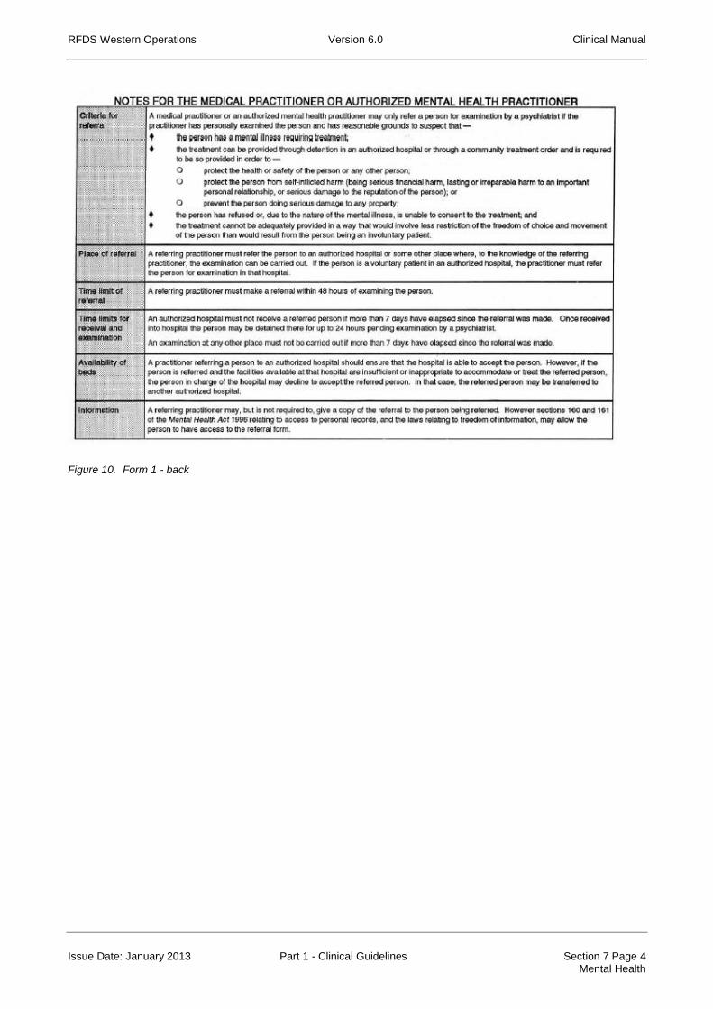
RFDS Western Operations Version 6.0 Clinical Manual
Issue Date: January 2013 Part 1 - Clinical Guidelines Section 7 Page 4 Mental Health
Figure 10. Form 1 - back

RFDS Western Operations Version 6.0 Clinical Manual
Issue Date: January 2013 Part 1 - Clinical Guidelines Section 7 Page 5 Mental Health
Figure 11. Form 3 - front

RFDS Western Operations Version 6.0 Clinical Manual
Issue Date: January 2013 Part 1 - Clinical Guidelines Section 7 Page 6 Mental Health
Figure 12. Form 3 - back

RFDS Western Operations Version 6.0 Clinical Manual
Issue Date: January 2013 Part 1 - Clinical Guidelines Section 7 Page 7 Mental Health
Figure 13. Record of Treatment

RFDS Western Operations Version 6.0 Clinical Manual
Issue Date: January 2013 Part 1 - Clinical Guidelines Section 8 Page 1 Miscellaneous
8 MISCELLANEOUS
8.1 Anaphylaxis
Theory
1. The most common causes of an anaphylactic reaction are bee stings, drugs (including antibiotics, streptokinase and anaesthetic agents), vaccines, snake antivenom and blood or blood products.
2. The clinical presentation is acute collapse with any of the following features:
Cutaneous: burning sensation, itching of lips, mouth or throat, flushing, angioedema,
urticaria, conjunctival injection or conjunctival oedema;
Cardiovascular: tachycardia, hypotension, shock;
Respiratory: rhinitis, bronchospasm, coughing, laryngeal oedema, choking sensation;
Gastro-intestinal: abdominal cramps, nausea, vomiting, diarrhoea;
CNS: Apprehension, metallic taste in mouth, loss of consciousness, convulsions.
Pre-flight and In-flight Management
1. Anticipate. Always question carefully about allergies before administering any drugs and exercise extra caution in known atopic individuals (asthma, hay fever, eczema).
2. Confirm the diagnosis. Rule out other causes for the patient’s signs and symptoms (e.g. vasovagal or hysterical reaction, asthma, pulmonary embolism, hypovolaemia, hypoglycaemia).
3. Adrenaline is the treatment of choice for severe allergic reactions.
The dose is 0.3 to 1.0mg IM in adults (0.3 – 1.0 mL of 1:1,000).
The dose in children is 10 µg/kg IM (e.g. 0.1 mL of 1:1,000 IM in a 10kg child).
In severe shock, or if IM injection has been ineffective, use adrenaline (1:10,000 Minijet) IV in similar doses. Monitor for arrhythmias and repeat adrenaline as necessary. Rarely with persistent shock adrenaline may be administered by titrated infusion.
4. Remove allergen.
Delay further absorption. Stop IV blood or drugs if suspected as cause.
5. Assess the airway.
If total obstruction intubate or create a surgical airway.
If airway obstruction with stridor give adrenaline IM as above then adrenaline 5mL 1:1000 nebulised.
6. Assess breathing.
Give Oxygen 6-10 litres/min via facemask, assist ventilation if necessary.
Treat bronchospasm with adrenaline IM as above.
Follow this with salbutamol 5mg nebulised every 15 minutes.
Consider other treatment modalities, e.g. further adrenaline, aminophylline or IV salbutamol.

RFDS Western Operations Version 6.0 Clinical Manual
Issue Date: January 2013 Part 1 - Clinical Guidelines Section 8 Page 2 Miscellaneous
7. Assess circulation.
If no pulse, treat as for cardiac arrest protocol.
If shocked, and no parental adrenaline already given, give adrenaline IV as above.
Give fluids: Haemaccel or Hartmann’s solution 500 to 1000 mL IV stat for an adult (20 mL/kg in children), further fluids according to clinical response.
8. Monitor response, especially heart rate and blood pressure (good index of response to treatment).
9. Consider other drugs to counteract histamine release and inflammatory response, e.g.
Steroids: hydrocortisone 4mg/kg (up to 200mg) IV 6 hourly or dexamethasone 0.1 - 0.25 mg/kg (up to 8mg IM or IV).
H2 receptor blockers (protracted cases): ranitidine 50mg IV 8-12 hourly.
Glucagon (especially for patients on blockers who have more severe reactions) 1mg IV every 5 mins.
10. Document on an Incident Report to permit follow up of blood cross-match, medical records, and for patient advice and Medic Alert warning.
Medical Chest Items
Adrenaline ampoules 1:1,000, 1mL (Item 99)
Promethazine mixture 5mg/ 5 mL (Item 119)
Loratadine tablets 10mg (Item 157)
Salbutamol Aerosol Spray 100g/dose (Item 107)
Aerosol Spacer (Item 229)
Prednisolone tabs 5mg (Item 151)
Oxygen (where available).
References
Bersten A, Soni N. Oh’s Intensive Care Manual. 6th ed. Butterworth Heinemann. 2009. Advanced Life Support Group. Advanced Paediatric Life Support. The Practical Approach. 5th ed. Blackwell
Publishing Ltd. 2005.
Cameron P, Jelinek G, Kelly A, et al. Textbook of Adult Emergency Medicine. 3rd ed. Churchill Livingstone. 2009.

RFDS Western Operations Version 6.0 Clinical Manual
Issue Date: January 2013 Part 1 - Clinical Guidelines Section 8 Page 3 Miscellaneous
8.2 Hyperkalaemia
Theory
1. Potassium is the principal intracellular cation and is largely responsible for the maintenance of the resting membrane potential. Potassium is shifted out of the cell in acidotic states
whereas insulin and 2 agonists promote potassium movement into the cell.
2. Hyperkalaemia is defined as a serum potassium > 5.0 mmol/l.
3. Causes include:
Factitious (longstanding or haemolysed specimen, very high WCC)
Tissue damage (multi-trauma, burns, rhabdomyolysis)
Decreased excretion (renal failure, Addison’s disease)
Drugs (indomethacin, spironolactone, ACEI)
Compartment shift (acidosis, insulin deficiency, digoxin overdose, suxamethonium, fluoride poisoning)
4. Clinical features include tingling, paraesthesiae, weakness and flaccid paralysis.
5. ECG features include heart block, peaked T waves, flattened P waves, prolonged P-R interval, widened QRS, sinus arrest or asystole.
Pre-flight and In-flight Management
1. Treat the underlying cause.
2. For life threatening hyperkalaemia give 50 gm dextrose (100 mL of 50% dextrose) with 5-10 units actrapid insulin SC followed by:
3. IV sodium bicarbonate 50-100 mmol over 30 min (if acidotic):
Avoid if patient has pulmonary oedema.
4. IV calcium gluconate 20 mL of 10% (2.2 mmol) or calcium chloride 10 mL of 10% solution;
Reduces the cardiotoxic effects of potassium.
Administer by slow IV (max 2 mL/min), repeat if necessary.
5. Salbutamol – 2 x 5 mg nebs 30 mins apart will reduce serum K+ by 1 mmol/L for 4 hours
Special Notes
1. Patients in renal failure need close attention to hydration status, consider diuresis with frusemide or inducement of diarrhoea with sorbitol / lactulose.
2. Oral and rectal resonium A 15-30 g 6/24 will reduce serum K+ in less acute cases.
Medical Chest Items
Salbutamol aerosol spray 100 g (item 107).
References
Bersten A, Soni N. Oh’s Intensive Care Manual. 6th ed. Butterworth Heinemann. 2009.
Dunn R, Dilley S, Brookes J, et al. The Emergency Medicine Manual. 5th ed. Tennyson, South Australia: Venom Publishing. 2010.
Thomas M. Orientation Manual For Renal Unit Medical Officers, RPH, current 1999.
CAPD Manual for Remote Nurses, Royal Perth Hospital, current 1999.

RFDS Western Operations Version 6.0 Clinical Manual
Issue Date: January 2013 Part 1 - Clinical Guidelines Section 8 Page 4 Miscellaneous
8.3 Hypokalaemia
Theory
1. Hypokalaemia is defined as a serum potassium < 3.5 mmol/L.
2. Potassium is the principal intracellular cation and is largely responsible for the maintenance of the resting membrane potential. Potassium is shifted out of the cell in acidotic states
whereas insulin and 2 agonists promote potassium movement into the cell.
3. Causes include:
Abnormal losses (vomiting and diarrhoea, drugs especially diuretics and corticosteroids, Conn's Syndrome, Cushing’s Syndrome)
Inadequate intake
Compartment shift (alkalosis, insulin, 2 agonists)
4. Clinical features include weakness, hypotonicity, depression, vasoconstriction, rhabdomyolysis, ileus and arrhythmias. Tetany if K+ < 2.5mmol/L
5. ECG changes include peaking of P waves, prolonged PR interval, T wave flattening or inversion, prominent U waves and atrial and ventricular tachyarrhythmias, especially torsade de pointes.
6. May be associated with hypomagnesaemia.
Pre-flight and In-flight Management
1. IV infusion of potassium chloride:
Maximum rate: 40 mmol/hr (1 g = 13 mmol)
ECG monitoring is required at high infusion rates
2. Correct Magnesium:
Mg2SO4 10mmol over 5min then 20-60mmol/day
References
Bersten A, Soni N. Oh’s Intensive Care Manual. 6th ed. Butterworth Heinemann. 2009.
Dunn R, Dilley S, Brookes J, et al. The Emergency Medicine Manual. 5th ed. Tennyson, South Australia: Venom Publishing. 2010.

RFDS Western Operations Version 6.0 Clinical Manual
Issue Date: January 2013 Part 1 - Clinical Guidelines Section 8 Page 5 Miscellaneous
8.4 Shock
Theory
1. Shock is defined as a state of inadequate tissue perfusion and oxygenation of tissues. In practice shock should be considered when systolic blood pressure is less than 90 mmHg.
2. Shock may be present due to hypovolaemia, reduced vascular tone and / or reduced cardiac output.
Causes of hypovolaemia (reduced intravascular volume), include haemorrhage (concealed or revealed), dehydration (loss of intra and extracellular fluids) due to losses or sequestration in the gut, excessive diuresis and excessive insensible losses (sweating).
Causes of reduced vascular tone include peripheral vasodilatation as occurs with septic shock, anaphylaxis, drugs, and autonomic neuropathy or spinal cord injury.
Causes of reduced cardiac output include cardiac failure secondary to arrhythmias, myocardial infarction, valvular disease, pulmonary embolus or cardiomyopathies; and reduced venous return as in venacaval obstruction due to abdominal masses (e.g. gravid uterus) or raised intra-thoracic pressure (eg. PEEP in ventilated patients).
Pre-flight and In-flight Management
1. Ideally shocked patients should be stabilized at the referring location prior to transfer. A patient that is unable to be stabilized due to inadequate local resources or ongoing problems should be a Priority 1 or 2 doctor-accompanied flight.
2. If hypotension develops in-flight:
Assess if treatment is necessary; does the patient have signs of shock such as pallor, sweating, cool mottled extremities, tachycardia, depressed level of consciousness and a reduced urine output? Can the hypotension (systolic blood pressure less than 90 mmHg) be regarded as normal for the patient or secondary to treatment? Common drugs implicated include vasodilators, narcotics (eg. Morphine), and antihypertensives. Falling blood pressure can occur in patients on Salbutamol infusions due to peripheral effects. Initial management may be to reduce the infusion or cease the suspected medication or observe further.
Administer high flow oxygen. A non-rebreathing mask should be considered.
3. Is patient hypovolemic, peripherally vasodilated or is there obstruction to venous return? Position supine and elevate lower limbs (e.g. pillows).
Lie in lateral position if pregnant.
4. Is the patient bleeding? If so attempt to:
Control haemorrhage – dressings, packing cavities, pressure bandages, suturing. Expedite transfer to surgeon.
Replace blood loss. Blood is preferable but sometimes not accessible. Two large bore IV lines (14 or 16 gauge) should be inserted and give Normal Saline (0.9% saline), Hartmann’s solution or colloid (e.g. gelofusine). Infuse rapidly at first (e.g. 500mL over 15 minutes) and continue until blood pressure improves, then reduce the rate.
5. Is the patient dehydrated?
Replace fluids. Insert at least one large bore IV cannula. Give a bolus of crystalloid (e.g. normal saline) or colloid (e.g. gelofusine) until blood pressure improves, then continue fluid replacement at a slower rate.

RFDS Western Operations Version 6.0 Clinical Manual
Issue Date: January 2013 Part 1 - Clinical Guidelines Section 8 Page 6 Miscellaneous
6. Is there reduced vascular tone?
Manage anaphylaxis with adrenaline and fluids. (See specific guidelines).
Manage septicaemia with fluids, antibiotics as indicated and inotropes if necessary. Inotropic infusions should only be used on doctor-accompanied flights. (See guidelines on sepsis).
Manage spinal cord injury with supine position and modest fluid replacement, may require vasopressors. (See guidelines on spinal cord injury).
7. Is there reduced cardiac output?
Manage acute left ventricular failure (pulmonary oedema) with oxygen, upright posture, frusemide and fluid restriction. (See specific guidelines).
Manage arrhythmias. (See specific guidelines).
Manage impaired venous return with patient positioning or reduction in intrathoracic pressure (e.g. reduce ventilator pressures, PEEP or release tension pneumothorax).
Medical Chest Items
Adrenaline ampoules 1: 1000, 1mL (Item 99).
References
Bersten A, Soni N. Oh’s Intensive Care Manual. 6th ed. Butterworth Heinemann. 2009.

RFDS Western Operations Version 6.0 Clinical Manual
Issue Date: January 2013 Part 1 - Clinical Guidelines Section 8 Page 7 Miscellaneous
8.5 Vascular Catastrophes
Theory
Ruptured abdominal aortic aneurysm (AAA)
1. Mortality 65-85%, 50% before reaching the operating theatre, making for precarious long distance aeromedical transfers.
2. Ruptured AAA heralded by triad of sudden onset mid-abdominal, flank or back pain (+/- scrotal radiation), shock and presence of pulsatile mass.
3. Degree of shock determined by location and size of rupture. A biphasic response may occur with an initial tear into retroperitoneal space followed by larger rupture hours later.
4. Emergency ultrasound 97% sensitive in diagnosis of AAA.
Aortic dissection
1. Most common aortic catastrophe: 2-3 x more common than ruptured AAA.
2. Associated with high mortality; particularly where diagnosis / transfer delayed.
3. Diagnosis can be difficult; estimated 38% of acute aortic dissections missed on initial evaluation.
4. Consideration dissection with any combination of:
Abrupt onset of thoracic or abdominal pain with sharp, tearing +/or ripping character.
Mediastinal +/or aortic widening on CXR.
Variation in pulse (absence of proximal extremity or carotid pulse) +/or BP (>20mmHg difference between right and left upper limbs).
Aortic incompetence.
5. Caution should be exercised with anticoagulation / thrombolysis if aortic dissection forms part of the differential diagnosis for presentations of atypical ischaemic chest pain.
Pre-flight and In-flight Management
General Principles:
1. Expeditious transfer. Priority 1, doctor accompanied.
2. Minimise investigations and procedures at referring hospital. Patient needs definitive vascular / cardiothoracic management.
3. Large bore IV access, monitoring (invasive blood pressure useful but should not delay transfer), indwelling catheter.
4. Consider need for blood products and reversal of anticoagulation.
5. Avoid hypothermia.
6. Aim for a quick airport handover (including an escort from the referring doctor).
Ruptured AAA
1. Avoid excessive fluid resuscitation (risk of loss of tamponade, stable retroperitoneal haemotoma becoming free intraperitoneal haemorrhage).
2. Aim for systolic blood pressure 90-100mmHg

RFDS Western Operations Version 6.0 Clinical Manual
Issue Date: January 2013 Part 1 - Clinical Guidelines Section 8 Page 8 Miscellaneous
Aortic Dissection
1. Control of blood pressure and heart rate essential to reduce shear stress on arterial wall (decreases propagation, tamponade rupture):
Adequate analgesia – IV morphine
β blocker – metoprolol 2.5-5mg IV boluses, or esmolol 0.5-1mg IV boluses; calcium channel blocker if β blocker contraindicated.
Nitroprusside infusion 2nd line. Difficulties with delivery (light sensitive) and availability, use with β blocker to avoid reflex tachycardia.
2. Aims: HR 60-80, BP 100-120 or lowest BP commensurate with vital organ perfusion.
3. Pericardiocentesis for tamponade may make things worse.
References
Sakalihasan N, Limet R, Defwe O. Abdominal Aortic Aneurysm. Lancet.2005; vol 365, issue 9470,1577-1589.
Hagan P, Nienaber C, Isselbacher E, et al. The International Registry of Acute Aortic Dissection: New insights into an old disease. JAMA 2000; 283:p897-903
Isselbacher E, Cigarroa J, Eagle K. Cardiac tamponade complicating proximal aortic dissection. Is pericardiocentesis harmful? Circulation. 1994;90:2375-2378.

RFDS Western Operations Version 6.0 Clinical Manual
Issue Date: January 2013 Part 1 - Clinical Guidelines Section 8 Page 9 Miscellaneous
8.6 Transfusion Medicine
Theory
1. An adequate haemoglobin is an essential element for oxygen delivery to the tissues. Haemoglobin is usually replaced by transfusing packed red blood cells (PRBC).
2. Clotting factors may be depleted or ineffectual due to a number of reasons; drugs (e.g. warfarin), toxins (e.g. snake bite envenomation), inadequate production (eg. liver failure, excessive consumption (e.g. disseminated intravascular coagulation), dilution of factors (e.g. major haemorrhage). Replacement clotting factors may be given in the forms of fresh frozen plasma (FFP) , prothrombinex –HT, fibrinogen concentrate (cryoprecipitate), recombinant Factor VIIa. (See below for what product to use in what circumstances).
3. Platelets play an important role in coagulation and may be depleted by bone marrow failure, auto-immune destruction or dilution.
4. A number of drugs are used for the reversal of anticoagulation or as antifbrinolytics; Vitamin K – role in activation of factors II, VII, IX,&X. Tranexamic acid – antifibrinolytic may have a role in major trauma. Protamine sulphate – for heparin reversal.
5. Using blood products must be weighed against potential adverse effects:
Transfusion related circulatory overload
Transmission of blood borne pathogens
Immune reactions
Transfusion related lung injury
Increase in plasma viscosity
Pre-flight and In-flight Management
Securing blood products
Wherever possible utilise crossed matched products from the hospital of origin, when assessing patients pre-flight ensure that the appropriate products are ordered and packed to accompany the patient.
If cross matched products will not be available it may be necessary to transport products from the base to the patient. In Jandakot 4 units of O negative PRBC are kept on site as is a supply of prothrombinex, above this products can be sourced from Red Cross Blood Transfusion Service (make phone request to their duty Haematologist and send faxed order form (appendix), available in RFDS Operations Centre). At rural bases blood products need to be sourced from the local hospital (See appendix with state wide product supplies).
FFP availability may be limited in rural areas and significant delays can be incurred awaiting thawing, order early.
For priority 1 flights the police will deliver blood products to Jandakot airport.
For flights inbound to Jandakot or meeting Jandakot crews elsewhere in the state additional blood products can be arranged to meet the patient. Please contact the duty Clinical Co-ordinator to arrange.
Note platelets are not available outside the metropolitan area unless we transport them to the patient.
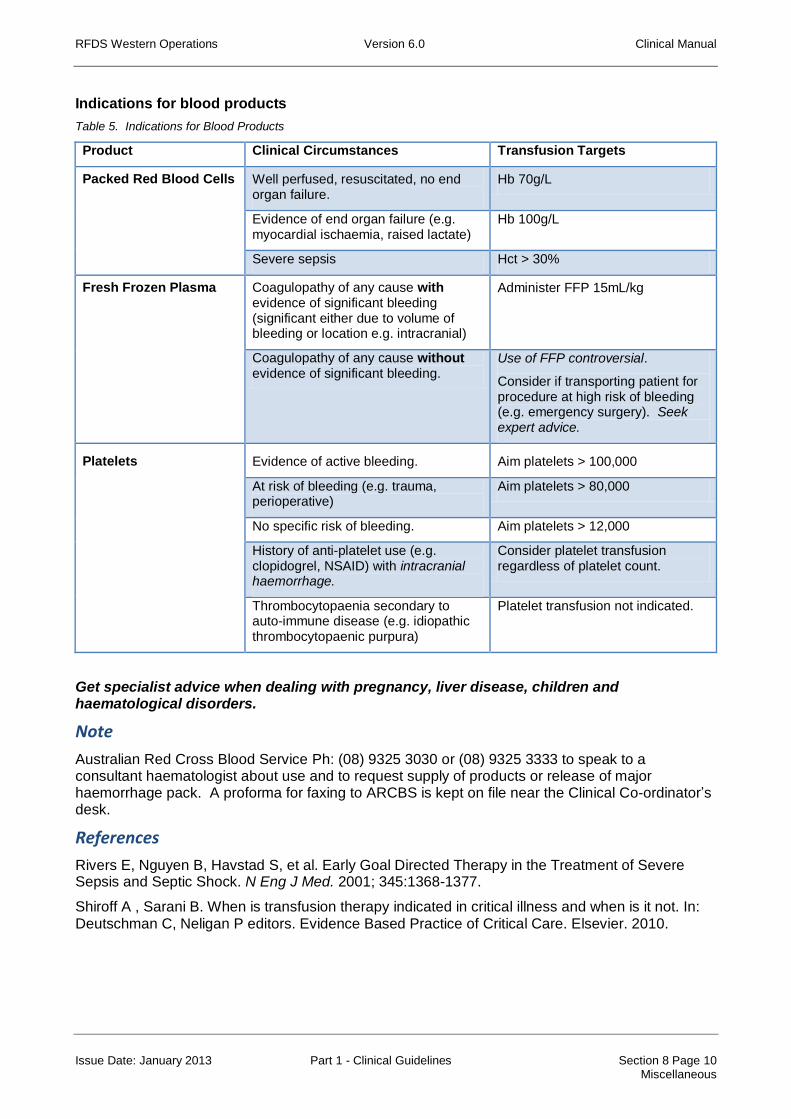
RFDS Western Operations Version 6.0 Clinical Manual
Issue Date: January 2013 Part 1 - Clinical Guidelines Section 8 Page 10 Miscellaneous
Indications for blood products
Table 5. Indications for Blood Products
Product Clinical Circumstances Transfusion Targets
Packed Red Blood Cells Well perfused, resuscitated, no end organ failure.
Hb 70g/L
Evidence of end organ failure (e.g. myocardial ischaemia, raised lactate)
Hb 100g/L
Severe sepsis Hct > 30%
Fresh Frozen Plasma Coagulopathy of any cause with evidence of significant bleeding (significant either due to volume of bleeding or location e.g. intracranial)
Administer FFP 15mL/kg
Coagulopathy of any cause without evidence of significant bleeding.
Use of FFP controversial.
Consider if transporting patient for procedure at high risk of bleeding (e.g. emergency surgery). Seek expert advice.
Platelets Evidence of active bleeding. Aim platelets > 100,000
At risk of bleeding (e.g. trauma, perioperative)
Aim platelets > 80,000
No specific risk of bleeding. Aim platelets > 12,000
History of anti-platelet use (e.g. clopidogrel, NSAID) with intracranial haemorrhage.
Consider platelet transfusion regardless of platelet count.
Thrombocytopaenia secondary to auto-immune disease (e.g. idiopathic thrombocytopaenic purpura)
Platelet transfusion not indicated.
Get specialist advice when dealing with pregnancy, liver disease, children and haematological disorders.
Note
Australian Red Cross Blood Service Ph: (08) 9325 3030 or (08) 9325 3333 to speak to a consultant haematologist about use and to request supply of products or release of major haemorrhage pack. A proforma for faxing to ARCBS is kept on file near the Clinical Co-ordinator’s desk.
References
Rivers E, Nguyen B, Havstad S, et al. Early Goal Directed Therapy in the Treatment of Severe Sepsis and Septic Shock. N Eng J Med. 2001; 345:1368-1377.
Shiroff A , Sarani B. When is transfusion therapy indicated in critical illness and when is it not. In: Deutschman C, Neligan P editors. Evidence Based Practice of Critical Care. Elsevier. 2010.
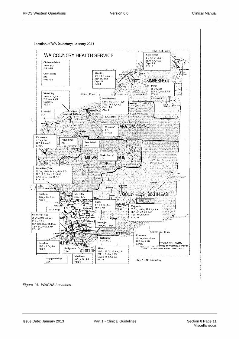
RFDS Western Operations Version 6.0 Clinical Manual
Issue Date: January 2013 Part 1 - Clinical Guidelines Section 8 Page 11 Miscellaneous
Figure 14. WACHS Locations

RFDS Western Operations Version 6.0 Clinical Manual
Issue Date: January 2013 Part 1 - Clinical Guidelines Section 8 Page 12 Miscellaneous
Figure 15. Request for Blood Components

RFDS Western Operations Version 6.0 Clinical Manual
Issue Date: January 2013 Part 1 - Clinical Guidelines Section 8 Page 13 Miscellaneous
8.7 Major Haemorrhage
Theory
1. Patients at risk of requiring massive transfusion should be identified early:
Haemostasis unlikely to be achieved at current location (e.g. Pelvic fracture, ruptured aortic aneurysm, variceal haemorrhage)
Prolonged PT / INR (either pharmacological or pathological)
Systolic BP < 110mmHg
Base deficit > 6
Hb <110 g/L
2. Early definitive haemostasis is the single most important factor in improving survival of patients with major haemorrhage. Staging patients via a regional centre with surgical facilities maybe required to achieve haemostasis, in consultation with regional and tertiary centres.
3. Ongoing resuscitation should include measures to avoid hypothermia, acidosis and coagulopathy.
4. Hypotension in the bleeding patient is primarily due to hypovolaemia and should be treated with volume replacement. There is little role for inotropes or vasopressors unless other causes of hypotension suspected (e.g. sepsis, myocardial injury)
5. The role of permissive hypotension has only been of proven benefit in penetrating trauma in an urban environment thus doesn’t apply to most RFDS patients, it is contraindicated in the presence of a concurrent brain injury.
Pre–flight and In–flight Management
1. Declare major haemorrhage early. These patients will be assessed as Priority 1 doctor accompanied patients.
2. Attend to airway, oxygenation and adequate access ( 2x large bore IV cannulae, or IO access).
3. Investigations FBP, Coagulation profile, ABG or VBG, lactate and cross match.
4. Volume resuscitation, crystalloids first line, early initiation of packed red cells.
5. Minimise time to definitive care / haemostasis by avoiding “going in” if airway breathing and adequate access managed. Maintain good communication with receiving hospitals to ensure appropriate reception (e.g. straight to theatre)
6. Correct and prevent coagulopathy:
Secure supply of FFP / arrange thawing and transport.
Aim to achieve PRBC : FFP ratio of 1:1 but don’t delay use of PRBC to awaiting FFP
Monitor for and correct Ca2+ levels.
Correct warfarin induced coagulopathy – vitamin K 10mg and prothrombinex 50 units /kg.
Discuss with haematologist (Red Cross) regarding tranexamic acid, cryoprecipitate, prothrombinex.
Consider platelet transfusion – discuss with duty haematologist, only available from Perth.
Consider tranexamic acid 1g over 10min within the first three hours of trauma likely to result in massive haemorrhage followed by 1g over the next 8 hours.

RFDS Western Operations Version 6.0 Clinical Manual
Issue Date: January 2013 Part 1 - Clinical Guidelines Section 8 Page 14 Miscellaneous
7. Correct and prevent acidosis.
Optimise tissue oxygenation (ensure Hb > 8g/dL, SBP >110 mm Hg, SpO2 > 98%), arterial line and IDC useful for monitoring tissue perfusion.
If using large volumes crystalloid CSL is preferable to normal saline in terms of preventing hyperchloraemic acidosis.
Avoid hypercapnoea (may need mechanical ventilation if patient hypoventilating).
Monitor Blood Gases and lactate if available.
8. Correct and Prevent Hypothermia
Actively warm patient.
Use warm IV fluids and blood products.
Special Notes
In the setting of massive transfusion access to blood products and haematology advice should be co-ordinated via the duty haematologist at Australian Red Cross Blood Service, they have knowledge of the State’s blood product supplies and for flights out of Jandakot can authorise the release of the RPH “Major Haemorrhage Pack” (4 units packed cells and 4 units pre-thawed FFP). Phone (08) 9325 3030 or (08) 9325 3333. A request form for blood products in on file at the Clinical Co-ordinator’s desk in the RFDS Operations Centre at Jandakot.
References
National Blood Authority. Patient Blood Management Guidelines: Module 1. Critical Bleeding Massive Haemorrhage. Australian Government 2011
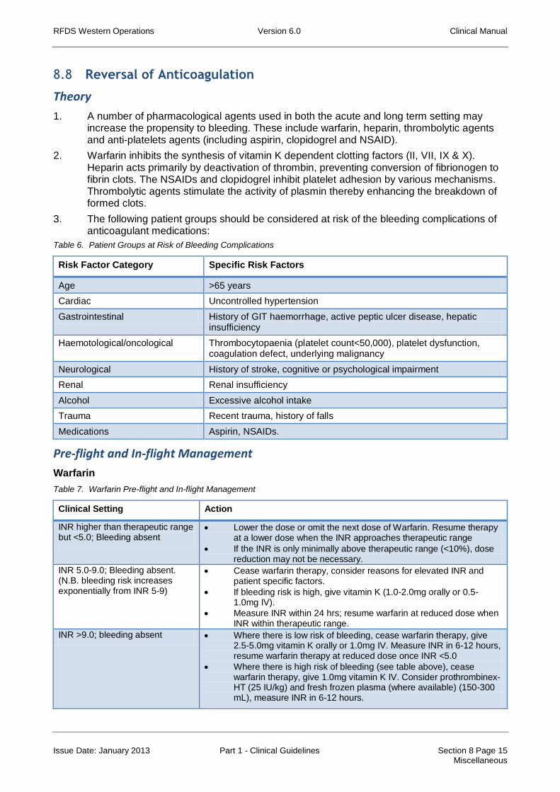
RFDS Western Operations Version 6.0 Clinical Manual
Issue Date: January 2013 Part 1 - Clinical Guidelines Section 8 Page 15 Miscellaneous
8.8 Reversal of Anticoagulation
Theory
1. A number of pharmacological agents used in both the acute and long term setting may increase the propensity to bleeding. These include warfarin, heparin, thrombolytic agents and anti-platelets agents (including aspirin, clopidogrel and NSAID).
2. Warfarin inhibits the synthesis of vitamin K dependent clotting factors (II, VII, IX & X). Heparin acts primarily by deactivation of thrombin, preventing conversion of fibrionogen to fibrin clots. The NSAIDs and clopidogrel inhibit platelet adhesion by various mechanisms. Thrombolytic agents stimulate the activity of plasmin thereby enhancing the breakdown of formed clots.
3. The following patient groups should be considered at risk of the bleeding complications of anticoagulant medications:
Table 6. Patient Groups at Risk of Bleeding Complications
Risk Factor Category Specific Risk Factors
Age >65 years
Cardiac Uncontrolled hypertension
Gastrointestinal History of GIT haemorrhage, active peptic ulcer disease, hepatic insufficiency
Haemotological/oncological Thrombocytopaenia (platelet count<50,000), platelet dysfunction, coagulation defect, underlying malignancy
Neurological History of stroke, cognitive or psychological impairment
Renal Renal insufficiency
Alcohol Excessive alcohol intake
Trauma Recent trauma, history of falls
Medications Aspirin, NSAIDs.
Pre-flight and In-flight Management
Warfarin
Table 7. Warfarin Pre-flight and In-flight Management
Clinical Setting Action
INR higher than therapeutic range but <5.0; Bleeding absent
Lower the dose or omit the next dose of Warfarin. Resume therapy at a lower dose when the INR approaches therapeutic range
If the INR is only minimally above therapeutic range (<10%), dose reduction may not be necessary.
INR 5.0-9.0; Bleeding absent. (N.B. bleeding risk increases exponentially from INR 5-9)
Cease warfarin therapy, consider reasons for elevated INR and patient specific factors.
If bleeding risk is high, give vitamin K (1.0-2.0mg orally or 0.5-1.0mg IV).
Measure INR within 24 hrs; resume warfarin at reduced dose when INR within therapeutic range.
INR >9.0; bleeding absent Where there is low risk of bleeding, cease warfarin therapy, give 2.5-5.0mg vitamin K orally or 1.0mg IV. Measure INR in 6-12 hours, resume warfarin therapy at reduced dose once INR <5.0
Where there is high risk of bleeding (see table above), cease warfarin therapy, give 1.0mg vitamin K IV. Consider prothrombinex-HT (25 IU/kg) and fresh frozen plasma (where available) (150-300 mL), measure INR in 6-12 hours.

RFDS Western Operations Version 6.0 Clinical Manual
Issue Date: January 2013 Part 1 - Clinical Guidelines Section 8 Page 16 Miscellaneous
Any clinically significant bleeding where warfarin induced coagulopathy is considered a contributing factor
Cease warfarin therapy, give 5.0-10.0 vitamin K IV plus prothrombinex-HT 25IU/kg and FFP (150-300 mL). OR
If FFP is unavailable, cease warfarin therapy, give 5.0-10mg vitamin K and prothrombinex-HT 25IU/kg. OR
If prothrombinex-HT is unavailable, give 5.0-10mg vitamin K and 10-15mL/kg of FFP
Heparin
The anticoagulant effects of Heparin may be reversed with protamine sulphate. Dose of protamine is dependent of dose of heparin to be reversed. Maximum protamine dose, regardless of heparin dose, is 50mg.
Protamine may precipitate cardiovascular collapse if administered rapidly (minimum infusion time – 10 minutes). It is derived from fish sperm and therefore patients with known hypersensitivity to fish may be at risk of reaction to Protamine.
Low molecular weight heparin (LMWH) e.g. enoxaparin – If within 8 hours of LMWH administration, use 1mg of protamine per 100 units of LMWH (consult LMWH product literature for number of units per mg – enoxaparin 100u/mg). If over 8 hours since LMWH administration, use 0.5mg of protamine per 100 units of LMWH.
Unfractionated heparin – 1mg protamine neutralises 100 units of unfractionated heparin when given within 15 minutes of heparin. If longer than 15 mins, less protamine may be required. Consult product literature for further information.
Post thrombolytic haemorrhage (major haemorrhage or intracerebral haemorrhage)
Manage as per major haemorrhage of other causes.
There is no specific agent for reversal of thrombolysis.
Be aware that a source of fibrinogen replacement may be required and therefore locate FFP or cryoprecipitate.
Many patients receiving thrombolysis will also receive heparin and anti-platelet agents – these may also require reversal.
Anti-platelet agents
There is limited evidence relating to the use of platelet infusions in the management of significant haemorrhage on anti-platelet agents.
Discuss with on-call haematologist.
References
Baker R, Coughlin P, Gallus A, et al. Warfarin Reversal: consensus guidelines on behalf of the Australasian Society of Thrombosis and Haemostasis. MJA. 181(9):492-497. 2004.
Bersten A, Soni N. Oh’s Intensive Care Manual. 6th ed. Butterworth Heinemann. 2009.
Shiroff A, Sarani B. When is transfusion therapy indicated in critical illness and when is it not? In: Deutschman C, Neligan P editors. Evidence Based Practice of Critical Care. Elsevier. 2010.

RFDS Western Operations Version 6.0 Clinical Manual
Issue Date: January 2013 Part 1 - Clinical Guidelines Section 8 Page 17 Miscellaneous
8.9 Mass Casualty Incidents
Theory
1. A Mass Casualty Incident (MCI) is defined as a situation where the event overwhelms the available resources. Sometimes it is immediately obvious that a disaster has occurred, but sometimes it only becomes apparent as further information comes in. Once a MCI has been declared, the methodology defined here comes into place, and this supersedes our usual RFDS mode of operation until the disaster has been completely dealt with.
2. The RFDS is an essential component of the overall Health Response to MCI in regional and remote WA. The Major Incident Medical Management and Support course (MIMMS) is used as a basis for planning, training and response to MCI’s in WA. The Emergency Management Act 2005 provides for the prompt and co-ordinated organisation of emergency management in the State. The RFDS and ambulance services are support agencies to the Department of Health.
RFDS Response
1. The primary role of the RFDS is to provide prompt advanced medical support at the scene or casualty clearing post (CCP) (this may be the nearest hospital), and collect accurate intelligence from the scene to provide back to the SHICC. RFDS MIMMS trained personnel will be assigned specific tasks to assist with command, control and communication. These roles may be on scene (Bronze level), at a command post or CCP (Silver level), or within the RFDS Co-ordination Centre or SHICC (Gold level).
2. RFDS will be tasked with co-ordinating transport of casualties by aeromedical assets.
3. RFDS Medical and Nursing staff first on scene of a MCI MUST NOT involve themselves with treatment or transport of casualties until a complete scene assessment and triage has been completed. This information must be recorded and relayed to SHICC (via RFDS SHICC Liaison Doctor or Clinical Co-ordinator).
4. All RFDS teams arriving on scene must report to the Health Commander for briefing, checking safety equipment and allocation of duties, upon completion of task report back for re-tasking. RFDS staff should not agree to tasks requested by other agencies without consent of their Health Commander.
5. The structure used for defining roles, priorities and chain of command is summarized as CSCATTT.

RFDS Western Operations Version 6.0 Clinical Manual
Issue Date: January 2013 Part 1 - Clinical Guidelines Section 8 Page 18 Miscellaneous
Table 8. Mass Casualty Incident Chain of Command
Command & Control
Command is vertical, know where you are in the health chain of command, to whom you report and who is reporting to you.
Control is horizontal across agencies and in most instances will be held by the Police. Requests for resources or assistance from other agencies should be sent up your chain of command to the Health Commander who will liaise with Commanders from other agencies. Failure to adhere to these lines of communication will result in chaos.
Command may occur at three levels:
Bronze at the incident site. Usually this will be managed by ambulance services.
Silver usually in close proximity to incident site however may utilize local nursing post or hospital, location where commanders and CCP will be set up. Access controlled by police. This is the usual level at which our staff will work, we are responsible for forwarding intelligence to Gold level command.
Gold at an administrative level remote from the location e.g. SHICC (e.g.State Health Incident Control Centre set up in Health Dept). Allocation of patients to various hospitals occurs at this level, there is no direct referral of patients by treating doctors to receiving hospitals.
Gold: State Health Incident Control Centre (SHICC), may or may not be convened at Royal St depending on nature of incident. RFDS should send liaison doctor (usually Director of Medical Services or his delegate), works with duty RFDS Clinical Co-Ordinator to ensure patients transported to destinations
determined by SHICC.
Silver:
Incident control centre (ICC) manned by:
Police Command.
Ambulance Commander (AC) and Health Commander (HC) (either WACHS or 1
st RFDS Dr on scene
depending on experience and MIMMS training, attached Treating doctor / nurse teams,
Ambulance Loading Officer reports to AC.
HC reports to SHICC and stays on site. Not involved in patient treatment.
Fire Command.
Casualty Clearing Post (CCP) manned by:
Senior Doctor (RFDS or WACHS), ideally not involved in patient treatment rather supervision and resourcing of teams, and patient flow.
Casualty Clearing Officer (ambulance).
Triage nurse.
Treating doctor/nurse teams.
Bronze: Ambulance forward Commander and ambulance teams (RFDS forward team may be assigned to AFC to assist, must report to AFC on entry and egress)

RFDS Western Operations Version 6.0 Clinical Manual
Issue Date: January 2013 Part 1 - Clinical Guidelines Section 8 Page 19 Miscellaneous
Table 8. Mass Casualty Incident Chain of Command (cont’d)
Safety Ensure safety of self, scene and survivors. The police will determine if the scene is safe to enter and may delegate scene safety to another agency, e.g. FESA.
Admission to scenes may be declined if not appropriately clad (See PPE).
Communication Determine how communication will occur and check the devices to be used (e.g. mobile phone, satellite phone, UHF radio). Do you know how to use your equipment, do you have back up power sources, do you know your call sign? Is RAVEN to be deployed?
Assessment This is a process of gathering information and determining what resources will be required to tackle the problem. See ETHANE report. Work with AC in determining suitable location for CCP.
Triage Triage will occur at many levels but it is important to use a consistent approach. The first level of triage (sieve) is to determine a priority for moving patients from the scene to a casualty clearing post. On arrival at a casualty clearing post the next level of triage (sort) determines a priority for treatment (See triage notes).
Treatment Treatment is generally confined to that essential to enable safe transport to a receiving hospital (clearly long transport times dictate more comprehensive treatment).
Transport A further element of triage occurs in determining order and mode of transport. Ensure that the right patient gets to the right place in the right time. Where patients go is determined by SHICC.
1. There is a structure for conveying information upwards to senior command and downwards when briefing staff, this is known as the ETHANE or METHANE report.
2. M Major incident (may be declared, on standby, or stood down).
3. E Exact location of incident (this may be of vital importance in the event that someone is reporting an incident in a primary location), GPS co-ordinates, nearest intersection, nearest airfield etc.
4. T Type of incident (e.g. bus roll over, passenger train derailment).
5. H Hazards present (fire, chemical, weather, traffic etc.).
6. A Access to the site (how should emergency services get there).
7. N Number and type of casualties.
8. E Emergency services present and resources required.
9. When RFDS staff are tasked to a mass casualty incident, they will be given an action card outlining their individual role and to whom they report.
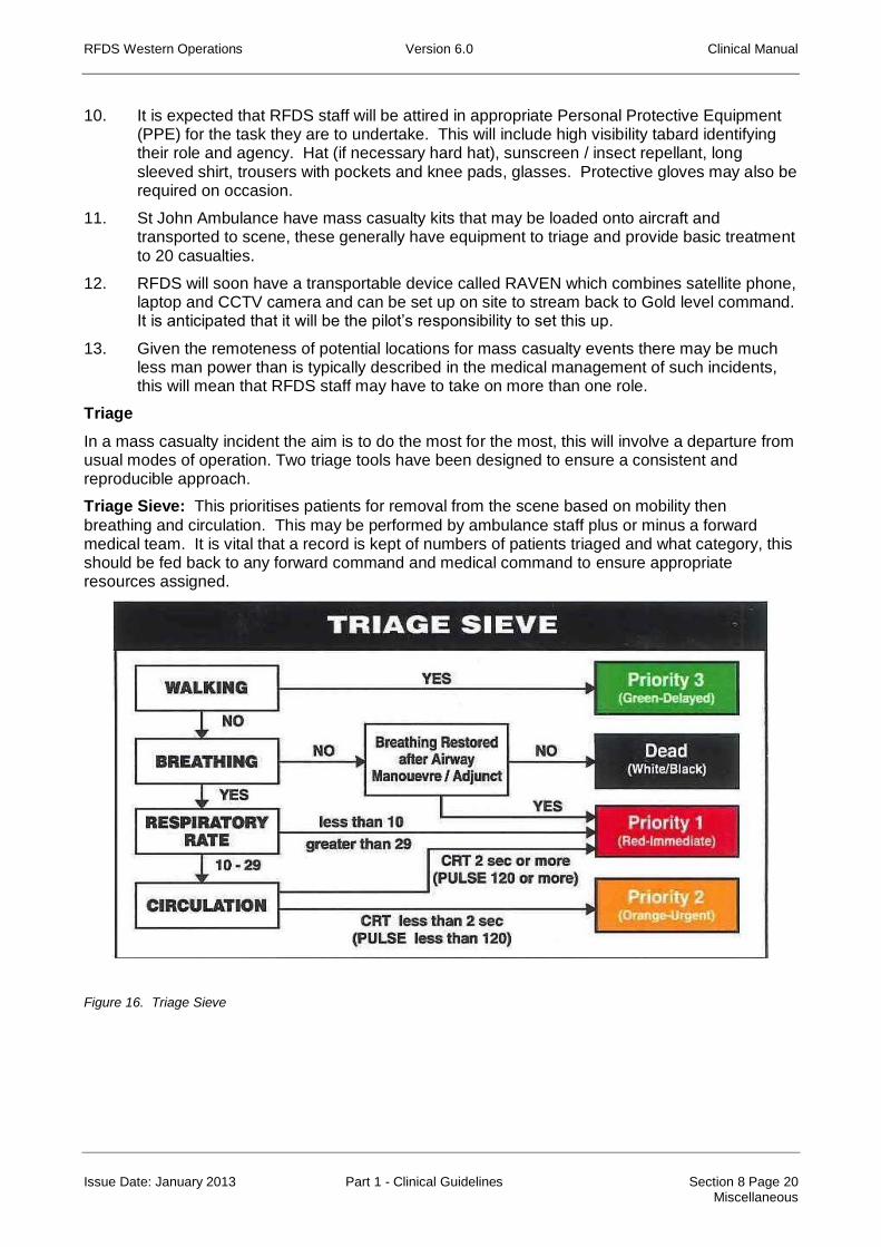
RFDS Western Operations Version 6.0 Clinical Manual
Issue Date: January 2013 Part 1 - Clinical Guidelines Section 8 Page 20 Miscellaneous
10. It is expected that RFDS staff will be attired in appropriate Personal Protective Equipment (PPE) for the task they are to undertake. This will include high visibility tabard identifying their role and agency. Hat (if necessary hard hat), sunscreen / insect repellant, long sleeved shirt, trousers with pockets and knee pads, glasses. Protective gloves may also be required on occasion.
11. St John Ambulance have mass casualty kits that may be loaded onto aircraft and transported to scene, these generally have equipment to triage and provide basic treatment to 20 casualties.
12. RFDS will soon have a transportable device called RAVEN which combines satellite phone, laptop and CCTV camera and can be set up on site to stream back to Gold level command. It is anticipated that it will be the pilot’s responsibility to set this up.
13. Given the remoteness of potential locations for mass casualty events there may be much less man power than is typically described in the medical management of such incidents, this will mean that RFDS staff may have to take on more than one role.
Triage
In a mass casualty incident the aim is to do the most for the most, this will involve a departure from usual modes of operation. Two triage tools have been designed to ensure a consistent and reproducible approach.
Triage Sieve: This prioritises patients for removal from the scene based on mobility then
breathing and circulation. This may be performed by ambulance staff plus or minus a forward medical team. It is vital that a record is kept of numbers of patients triaged and what category, this should be fed back to any forward command and medical command to ensure appropriate resources assigned.
Figure 16. Triage Sieve

RFDS Western Operations Version 6.0 Clinical Manual
Issue Date: January 2013 Part 1 - Clinical Guidelines Section 8 Page 21 Miscellaneous
Triage Sort: This prioritises patients for treatment, this triage occurs on entry to the Casualty Clearing Post and is based on physiological parameters and a scoring system.
Figure 17. Triage Sort
A “SMART TAG” triage and tracking system is hoped to be introduced into WA early 2012, until that time a colour coded cruciform triage card is used.
Patient Flow
The ideal flow of patients from an incident site is represented below.
Figure 18. Patient Flow
References
Advanced Life Support Group. Major Incident Medical Management and Support. 2nd
Ed, 2002. BMJ Publishing.

RFDS Western Operations Version 6.0 Clinical Manual
Issue Date: January 2013 Part 1 - Clinical Guidelines Section 8 Page 22 Miscellaneous
8.10 Morbid Obesity
Theory
Morbid obesity needs to be viewed as a multisystem pathology carrying a significant mortality risk, independent of the risk carried by associated dyslipidaemias, diabetes, ischaemic heart disease.
Complications to consider are:
1. Respiratory.
Obstructive Sleep Apnoea. Hypopnoea, long term physiological changes (hypoxia, hypercapnoea, polycythaemia, right heart failure).
Obesity Hypoventilation Syndrome. Desensitization to hypercapnoea leading to Type 2 respiratory failure.
Anatomical changes to the airway.
Reduced functional residual capacity.
Decreased lung compliance.
All these changes are more marked in the supine position.
2. Cardiovascular.
Hypertension
Ischaemic heart disease
Increased blood volume but disproportionately less per kg
Predisposition to arrhythmias precipitated by hypoxia and hypercapnoea
Obesity induced cardiac dysfunction. Left ventricular dilation and hypertrophy. Increased cardiac output. Limited capacity to increase cardiac output with exercise or stress
Pulmonary hypertension and right heart failure
Many of these problems may be undiagnosed as the patient’s usual sedentary lifestyle doesn’t test their cardiac function. The supine position may produce significant orthopnoea even cardiac arrest.
Pre-flight and In-flight Management
1. Loading and handling concerns.
Attempts must be made to get accurate patient weights. The best assessment of weight is provided by asking the patient if they cannot be weighed.
Estimations based on arm circumference and height can be calculated.
Women (kg) = (2.15 x arm circumference in cm) - 64.6 + (0.54 x height in cm)
Men (kg) = (3.29 x arm circumference in cm) – 93.2 + (0.43 x height in cm)
RFDS does not have a fixed upper limit of patient that can be carried (with the exception of patients carried on the Life Flight Jet of 130kg), patients are considered on individual merits. If a patient cannot fit on an AFTS stretcher then the obese patient restraint system must be used. The obese patient restraint is only rated to 250kg, beyond that, patients must meet mercy flight requirements so other forms of transport need to be considered and ruled out prior to using our aircraft. Where possible actual visualisation and measurement of the patient should occur before abandoning use of a stretcher or restraint.

RFDS Western Operations Version 6.0 Clinical Manual
Issue Date: January 2013 Part 1 - Clinical Guidelines Section 8 Page 23 Miscellaneous
Assistance with loading and unloading should be arranged through operations. This may include Fire and Emergency Services personnel.
Over 150kg then SJA should be asked to consider having the bariatric ambulance meet the aircraft at Jandakot. This requires two hours notice.
The obese patient restraint requires that the patient be supine, clearly this will pose a significant risk for a number of patients.
Patients with relatively benign conditions may still warrant a doctor accompanied flight as once loaded it is impossible to access the patient from the head end and side given the obstruction of the path between the two.
No patient should be refused carriage without the involvement of the Director of Medical Services or his deputy. The case may need to involve the input of the Chief Medical Officer for the state or his deputy (via RFDS Director of Medical Services). Non RFDS transport options (e.g. military) may need to be explored with or without the use of RFDS medical and nursing staff.
2. Induction of anaesthesia:
Invest in good preparation. Be prepared for potential difficult airway. Position patient in ramped position (external auditory meatus in line with angle of sternum, may require a number of pillows under shoulders, head and neck).
Adequate pre-oxygenation (consider using PEEP valve on BVM).
Use rapid sequence induction, be prepared for risk of aspiration (suction on and enough people to turn patient if necessary). Give acid prophylaxis if time permits.
Ventilate with PEEP and 45º head up. Tidal volume based on ideal body weight. Titrate pressures. Transport ventilators may struggle, hand ventilation may have to be resorted to.
Monitor ABG.
Beware sensitivity to opiates and sedatives.
Anticipate reduction in cardiac performance (prepare with fluids and or inotropes available).
Critically ill morbidly obese patients need adequate nutrition, ensure early dextrose +/- insulin.
Reference
Adams, JP. Murphy, PG. Obesity in anaesthesia and intensive care. BJA, 2000, Vol.85 No.1 91-108.
Crandall,CS. Gardner, S. Braude, DA. Estimation of Total Body Weight in Obese Patients. Air Medical Journal. May-June 2009, 28:3 139-145.

RFDS Western Operations Version 6.0 Clinical Manual
Issue Date: January 2013 Part 1 - Clinical Guidelines Section 8 Page 24 Miscellaneous
8.11 Diving Related Injury and Illness
Theory
1. There are a number of coastal locations in Western Australia where diving is a popular past-time, in addition some locations have industry dependant on diving. In particular Ningaloo Reef, Broome and some isolated pearl farms in the Kimberley, Bussleton / Dunsborough, Esperance, Geraldton.
2. Hyperbaric chambers exist in Broome, Fremantle Hospital and Garden Island (Naval use only).
3. A diving history is important to know about even if the patient is being transported for an unrelated problem as this will determine the flight profile required. Any symptoms developing within 48 hours of a dive should be considered as related until proven otherwise.
4. The diver may suffer from illness related to dysbarism, abnormal gas pressures and the general aquatic environment.
Barotrauma
Descent barotrauma (“squeeze”) commonly involves ENT injury including tympanic membrane rupture, round or oval window rupture leading to perilymph fistula resulting in acute dizziness, disorientation, tinnitus and deafness.
Ascent barotrauma, affects the lungs as a result of breath holding or airtrapping. This may result in mediastinal emphysema (presenting as hoarseness, neck swelling, retrosternal pain), pneumothorax / tension pneumothorax.
Decompression illness (DCI)
This is often called “The Bends” and is a result of dissolved nitrogen coalescing to form bubbles in the tissue or blood. These bubbles may cause problems either by obstructing small vessels causing ischaemia, placing direct mechanical pressure on tissues, or increasing pressure within tissues such that circulation is impaired. The bends can occur even if divers have been diving well within the limitation of their dive tables, predisposing factors include exercise, dehydration, hypothermia, age, alcohol, repetitive dives, multiple ascents, altitude exposure and obesity.
Clinical manifestations include skin rashes, joint pain, localized swelling through to neurological (spinal cord and central including altered mental state and ataxia), cardiac and pulmonary. Symptoms usually occur within 4 hours.
Arterial gas embolism (AGE), cerebral arterial gas embolism.(CAGE)
In this scenario pulmonary barotraumas is thought to be the origin of gas bubbles in the circulation. With CAGE symptoms occur as the diver surfaces resulting in loss of consciousness, fitting, chest pain and cardiovascular collapse. The sooner the onset of symptoms the greater severity.
Abnormal gas pressures
This includes nitrogen narcosis, hypoxia, hypercapnoea all of which resulting reduced mental performance and put the diver at risk of unsafe diving and drowning. Shallow water blackout is a hypoxic phenomenon that also occurs in snorklers (the diver hyperventilates to blow of CO2 and increase their breath hold time but may become apnoeic as a result).
The aquatic environment
Issues to consider are near-drowning and envenomation.

RFDS Western Operations Version 6.0 Clinical Manual
Issue Date: January 2013 Part 1 - Clinical Guidelines Section 8 Page 25 Miscellaneous
Pre-flight and In-flight Management
1. The priority and need for doctor will depend very much on the nature of the presentation.
2. All patients who have a history of diving in the last 24hours should be transported with a sea-level cabin even if their presentation is unrelated.
3. A strict sea-level cabin should be requested. Any meets must also occur at sea-level locations.
4. The patient must be kept flat (or head down although there is a suggestion that this might increase the risk of coronary bubbles forming) at all times to avoid bubbles rising up the cerebral circulation.
5. High flow oxygen must be given, if necessary the demand regulator on the aircraft can provide 100% oxygen with a good seal and divers are familiar with breathing via a regulator.
6. IV rehydration with normal saline will assist with tissue perfusion. Maintain a urine output of 2mL/kg/hr. A urinary catheter is advisable for the more severely affected.
7. Rewarming of the patient.
8. Aspirin is thought to help prevent platelet clumping.
9. A CXR should be requested if possible.
10. The patient must avoid straining (e.g. vomiting, coughing etc). An antiemetic is advisable.
11. If cerebral herniation is suspected temporizing with Mannitol may be useful.
12. The definitive treatment is recompression which involves transfer to a recompression chamber.
13. A severe diving injury in the Kimberley may ultimately need transfer to Perth but it is worthwhile exploring the possibility of an initial recompression in the Broome chamber prior transfer.
14. The dive profile and or computer and history will be useful for planning recompression therapy.
References
Dunn R, Dilley S, Brookes J, et al. The Emergency Medicine Manual. 5th ed. Tennyson, South Australia: Venom Publishing. 2010.
Edmonds C, Lowry C, Pennefather J, et al. Diving and Subaquatic Medicine. 4th ed. Hodder Arnold. 2005
Wyatt J, Illingworth R, Graham C, et al. Oxford Handbook of Emergency Medicine. 3rd ed. Oxford University Press. 2006

RFDS Western Operations Version 6.0 Clinical Manual
Issue Date: January 2013 Part 1 - Clinical Guidelines Section 9 Page 1 Neurological
9 NEUROLOGICAL
9.1 Status Epilepticus
Theory
1. Status epilepticus is a clinical or electrical seizure lasting longer than 5-10 minutes, or a series of seizures without complete recovery over the same period of time. After 30 minutes, the brain begins to suffer from hypoxia and acidosis, with depletion of local energy stores, cerebral oedema, and structural damage. Eventually, pyrexia, hypotension, respiratory depression, and even death may occur.
2. There can be a variety of causes of status epilepticus. Common causes include anticonvulsant withdrawal, alcohol withdrawal, cerebro-vascular accident, metabolic derangement (hypoxia, hyponatremia [< 120 mmol/l] hypoglycaemia, hyperosmolality [> 300 mosm/L]), trauma, drug toxicity (amphetamines, cocaine, salicylates, methanol,
ethanol), CNS infection, hyperthermia (> 41-42C) or tumour.
3. Search carefully for seizure activity in the comatose patient. Manifestations may be subtle, eg, deviation of head or eyes, repetitive jerking of fingers, hands, or one side of the face.
Pre-flight and In-flight Management
Flights for patients with status epilepticus will usually be doctor-accompanied and priority 1 or 2, depending on the facilities of the referring location.
1. Protect the Airway:
Roll the patient onto one side if possible. Endo-tracheal intubation may be necessary. Do not insert objects through clenched teeth, as it will not protect the airway and may cause broken teeth.
2. Obtain IV access:
If possible, take bloods for FBC, glucose, electrolytes, magnesium, and calcium determinations; hepatic and renal function tests; as well as extra tubes of blood for possible toxicology screen or drug levels (including anticonvulsants if patient is known or suspected to be taking them).
3. If IV access cannot be obtained:
diazepam may be given rectally at a dose of 0.2 – 0.7mg/kg or
midazolam can be given intranasally at a dose of 0.2mg/kg (max dose 10 mg) or
midazolam can be given IM at a dose of 0.1 – 0.4mg/kg (max dose 10mg).
4. Rule out hypoglycaemia:
Give glucose, 50 mL of 50% solution IV over 5 minutes.
If malnutrition or alcohol withdrawal is suspected, give thiamine, 100mg IV slowly prior to, or at the same time as glucose.
5. Give diazepam or midazolam:
Give diazepam 5-10mg (paediatrics 0.3-0.5mg/kg) IV over 1-2 minutes. This treatment is effective in 80-90% of cases of status epilepticus, although apnoea, bradycardia, or hypotension may rarely result.
Alternatively, IV midazolam may provide control of refractory status epilepticus; the suggested loading dose is 5 – 10mg (paediatrics 0.2mg/kg), followed by 0.05-0.2 mg/kg/hr.

RFDS Western Operations Version 6.0 Clinical Manual
Issue Date: January 2013 Part 1 - Clinical Guidelines Section 9 Page 2 Neurological
6. Administer a loading dose of phenytoin - regardless of the effect of diazepam, a maintenance drug is required:
Give phenytoin in normal saline, 15-20 mg/kg by IV infusion at a rate of 50 mg/min (paediatrics 2mg/kg/min) or slower.
Infusion of phenytoin at more rapid rates (especially if given into centrally placed IV lines) can precipitate cardiac arrhythmias or hypotension.
Phenytoin orally or IV should be given to all patients except those who have a short-term metabolic condition known to cause seizures, such as alcohol withdrawal or hypoglycaemia, which does not require or respond to phenytoin.
7. If these measures fail, general anaesthesia with ventilatory assistance and neuromuscular junction blockade will most likely be required. Use a rapid sequence induction technique with cricoid pressure, thiopentone (3 - 5mg/kg) and suxamethonium (1.5mg/kg).
8. Measure arterial blood gases and pH:
Arterial blood PCO2 is a sensitive indicator of the adequacy of ventilation (hypercapnia is present in proportion to the degree of hypoventilation). Metabolic acidosis due to lactic acidosis resulting from status epilepticus is commonly present for as long as 1 hour after a seizure, depending on the duration and vigour of muscular activity. This acidosis requires no treatment. Acidosis lasting longer than 1 hour should prompt a search for other causes of acidosis.
Special Notes
1. Consider Meningitis:
Commence appropriate antibiotics if meningitis is suspected, especially if fever (body temperature > 38.5o C) or nuchal rigidity is present. However, the muscle activity of status epilepticus alone produces transient fever higher than 38.5o C in 25% of patients. Status epilepticus may also produce a mild transient cerebrospinal fluid pleocytosis (< 100 cells/micro/L).
2. Prevent injury to the patient during the seizure by padding the environment. Do not use rigid restraint (fractures may result) or insert objects into the patient's mouth during the seizure.
Medical Chest Items
Diazepam ampoules 10mg/ 2 mL (Item 98), Diazepam tablets 2mg (Item 191).
References
Saunders C, Ho M. Current Emergency Diagnosis and Treatment. 4th Ed. Appleton Lange. 1992.
Hay W, Levin M, Sondheimer J, et al. Current Diagnosis and Treatment: Paediatrics. 20th ed. Lange. 2011.
McPhee S, Papadakis M, Tierney L. Current Medical Diagnosis and Treatment. 47th ed. Lange. 2007.
Dunn R, Dilley S, Brookes J, et al. The Emergency Medicine Manual. 5th ed. Tennyson, South Australia: Venom Publishing. 2010.
Lahat E, Goldman M, Barr J, et al. Comparison of intranasal midazolam with intravenous diazepam for treating febrile seizures in children: prospective randomised study. BMJ 2000; 321: 83-86

RFDS Western Operations Version 6.0 Clinical Manual
Issue Date: January 2013 Part 1 - Clinical Guidelines Section 9 Page 3 Neurological
9.2 Subarachnoid Haemorrhage
Theory
1. Defined as bleeding into the subarachnoid space. Seventy percent are due to rupture of an aneurysm in the Circle of Willis. Other causes are arterio-venous malformations, mycotic aneurysms, illicit drug use, bleeding diatheses and trauma. Risk factors include smoking, hypertension, alcohol, genetic factors, sympathomimetic drugs, oestrogen deficiency, anti-thrombotic therapy and statins.
2. Up to 80% of patients have a small “warning bleed” which precedes the major bleed. From the major bleeds, 50% of patients will die or be permanently incapacitated. A further 30% will die if not treated.
3. Important clinical symptoms and signs include: sudden onset of severe “worst ever” headache, vomiting, transient loss of consciousness, depressed conscious state and hypertension. Seizures occur in 20% but focal neurological signs are uncommon. Diagnosis is confirmed by CT scan (95% sensitive) and/or lumbar puncture.
4. Early neurosurgical or radiological intervention and the use of nimodipine (a calcium channel blocker which reduces vasospasm in cerebral arteries) give excellent outcomes for patients with warning bleeds and less severe SAH.
Pre-flight and In-flight Management
1. The priority assigned will be determined by the severity of the patient’s illness and accuracy of diagnosis, confirmed wherever possible with CT scan. Most flights will be Priority 2. Some critically ill patients in smaller centres may be Priority 1.
2. Flights should be doctor-accompanied if there is a significantly depressed conscious state, seizures or severe hypertension. Many patients with these criteria will benefit from early intubation and IPPV.
3. All patients should receive oxygen and be fully monitored. An IDC may be appropriate for very drowsy patients. Where possible, patients should be nursed with 30o head elevation to reduce cerebral oedema. Actions that induce sudden rises in intracranial pressure (e.g. coughing on ETT, vomiting or seizures) should be avoided as much as possible. Prophylactic anti-emetics and anticonvulsants may be appropriate for individual patients.
4. Moderate rises in blood pressure are necessary to maintain cerebral perfusion pressure, and do not require treatment. Severe hypertension should be treated initially with sedation and analgesia. If blood pressure remains grossly elevated, control with IV atenolol 1 mg/min or metoprolol (until target blood pressure reached). Do not correct rapidly. In patients with severe hypertension, invasive arterial pressure monitoring is helpful.
5. Nimodipine is used to prevent secondary cerebral vasospasm but is not stocked by RFDS and is generally only available in tertiary settings.
References
Dunn R, Dilley S, Brookes J, et al. The Emergency Medicine Manual. 5th ed. Tennyson, South Australia: Venom Publishing. 2010.
Bersten A, Soni N. Oh’s Intensive Care Manual. 6th ed. Butterworth Heinemann. 2009.
Neurology drug guidelines [revised 2011 Jun]. In: eTG complete [CD-ROM]. Melbourne: Therapeutic Guidelines Limited; 2012 Jul.
Cardiovascular drug guidelines [revised 2011 Jun]. In: eTG complete [CD-ROM]. Melbourne: Therapeutic Guidelines Limited; 2012 Jul.

RFDS Western Operations Version 6.0 Clinical Manual
Issue Date: January 2013 Part 1 - Clinical Guidelines Section 9 Page 4 Neurological
9.3 Delirium Tremens
Theory
1. Delirium tremens occurs as a result of withdrawal from alcohol. It is an acute organic psychosis that is usually manifest within 24-96 hours after the last drink, but may occur up to 7-10 days later. Delirium tremens is characterised by mental confusion, tremor, sensory hyperacuity, visual hallucinations (often terrifying), autonomic hyperactivity, diaphoresis, dehydration, electrolyte disturbances (especially hypokalaemia and hypomagnesaemia), seizures and cardiovascular abnormalities. Fever may be present.
2. Central nervous system depressant drugs are important in the treatment of delirium tremens. This applies whether the syndrome is the primary reason for transport of the patient, or whether it is a downstream effect of restricted access to habitual alcohol due to some other illness or injury.
Pre flight and In-flight Management
1. Priority will vary depending on the patient’s primary problem and the degree to which risks to the patient’s health are compounded by this severe alcohol withdrawal syndrome. A doctor-accompanied flight may be necessary if the patient is severely ill or if symptoms are difficult to control.
2. Pre-flight and in-flight management aims to provide supportive care: thiamine and multivitamin therapy (Ensure glucose not given until thiamine has been administered); fluid, electrolyte and glucose support, and benzodiazepine-based control of symptoms. Give diazepam 10mg to 20mg every 1 to 2 hours until control of symptoms is satisfactory and moderate sedation occurs. Doses may need to be high and extra supplies of benzodiazepines may need to be carried.
3. IV diazepam 5 – 10mg or midazolam 2.5-5mg IV may be needed to gain control of symptoms.
4. Other drugs with some potential application in delirium tremens include β-blockers (e.g.atenolol 100mg per day when the heart rate is above 100bpm and 50mg per day for heart rates between 50 and 100 bpm). Antiepileptic medication is not required unless there is an underlying seizure disorder. If an antipsychotic medication is required haloperidol or droperidol may be useful. Clonidine (e.g. 5μg/kg every two hours) has also been used for anxiolysis and to suppress cardiovascular symptoms.
Medical Chest Items
Diazepam 10mg/ 2 mL (Item 98), Diazepam tablets 2mg (Item 191).
References
McPhee S, Papadakis M, Tierny L. Current Medical Diagnosis and Treatment. 47th ed. Lange. 2007
Psychotropic drug guidelines [revised 2008 Oct]. In: eTG complete [CD-ROM]. Melbourne: Therapeutic Guidelines Limited; 2012 Jul

RFDS Western Operations Version 6.0 Clinical Manual
Issue Date: January 2013 Part 1 - Clinical Guidelines Section 10 Page 1 Obstetric
10 OBSTETRIC
10.1 Pre-term Labour and Tocolysis
Theory
1. Pre-term labour is defined as onset of labour before the 37th week of gestation. For RFDS purposes the emphasis is generally on women who are less than 36 weeks gestation where adequate paediatric management is not possible outside a tertiary (King Edward Memorial or Darwin Hospital) or in some cases regional setting. (Some regional paediatricians are able to manage neonates from 34 weeks gestation).
2. With careful assessment, prompt transfer and aggressive tocolytic therapy in flight, the majority of patients in pre-term labour can be transferred to an appropriate centre where optimal conditions exist for delivery and resuscitation of the neonate.
3. The neonatal transport service (NETS) in Western Australia is not intended to be a resuscitation service and should not be tasked for alleged imminent delivery without the consultation of a senior RFDS doctor with obstetric experience.
4. There is nothing to be gained by “going in” to hospitals to retrieve women in advanced labour, a rapid airport handover should be arranged.
5. Perinatal morbidity and mortality for low and extremely low birth weight infants is significantly improved by delivery and resuscitation in a tertiary setting.
6. Tocolysis is used to suppress labour to allow administration of corticosteroids for foetal lung maturation and ensure safe transfer in-utero to an appropriate facility.
7. Corticosteroids enhance foetal lung maturation and decrease the risk of neonatal intracerebral haemorrhage and necrotizing enterocolitis.
Pre-flight and In-flight Management
1. Diagnosis of labour is based on painful regular contractions accompanied by cervical change (effacement and dilatation). Information to be sought in the pre-flight assessment includes parity, frequency and strength of contractions, cervical dilatation and effacement, status of membranes (intact or ruptured), foetal heart rate (CTG if available), presentation, past obstetric history.
2. Examination of the cervix is essential, a digital examination unless contraindicated (PV bleed, SROM without labour). If contraindicated a sterile speculum examination is requested.
3. Encourage the use of foetal fibronectin swab testing. This test must be done prior to any vaginal examination and in the absence of ruptured membranes. A negative result less than 37 weeks in otherwise low risk women has a 96% predictive value for no delivery in the next week. This may mean a transfer is unnecessary and the woman can be monitored locally (depending on skill and experience of local staff).
4. Cervical length as determined by transvaginal ultrasound, if available, may be useful, a cervical length >30mm and undilated internal os are negative predictors of birth.
5. If < 34 weeks completed pregnancy give betamethasone (Celestone Chronodose) 11.4mg (equals 2 ampoules) IM. This may be contraindicated if chorioamnionitis.
6. Commence tocolysis immediately unless contraindicated. (Large APH or abruption may be a contraindication as is foetal death).
7. Instructions regarding choice of tocolytic should be clear:
a) < 4cm with intact membranes a trial of oral nifedipine may be considered.

RFDS Western Operations Version 6.0 Clinical Manual
Issue Date: January 2013 Part 1 - Clinical Guidelines Section 10 Page 2 Obstetric
The dose is 20mg PO at 30 minute intervals until contractions cease OR 60mg has been given.
Side effects of nifedipine include facial flushing, headache, nausea, tachycardia, dizziness, hypotension (very uncommon in normotensive patients), cardiac failure, raised liver enzymes.
b) If the woman is still laboring after 90 minutes salbutamol must be commenced.
c) 4cm commence salbutamol as per RFDS infusion guidelines:
Side effects of salbutamol include tachycardia, hypotension, tremor, pulmonary oedema, hyperglycaemia, hypokalemia.
d) If woman is term and the transfer time short it may be appropriate to avoid tocolysis and allow labour to progress.
e) Rarely alternative tocolytics may be considered:
glyceryl trinitrate – 5-10mg patch (max 20mg), may cause hypotension and headache.
indomethacin 100mg PO then 25mg PO 4 hourly. Risk of closure of foetal ductus arteriosus and renal impairment, gastric ulceration.
8. Magnesium for foetal neuro-protection is not routinely used in transport, the expectation is that it will be given on arrival at the tertiary centre as it is not given until 4 hours pre-delivery and then only for women less than 30 week gestation. Should a patient already have magnesium running a doctor accompanied flight will be required with cardiac, respiratory and patella reflex monitoring.
If used, a loading dose of 4g is given over 20min followed by 1g per hour infusion for 4 hours, cease if delivery occurs earlier.
If delivery is considered imminent discuss use with a consultant at KEMH.
9. The patient should be nursed in the left lateral position and given supplemental oxygen to compensate for altitude hypoxia.
10. A doctor will be required for women in more advanced labour where delivery is a risk. Priority will depend on stage of labour (i.e. threatened vs. established).
11. In the presence of prolonged rupture of membranes, Group B Streptococcus or urinary tract infection antibiotics should be given, benzylpenicillin 1.2g IV, if allergic to penicillin clindamycin 600mg IV.
12. If delivery is not imminent at the end of transfer then it may be acceptable to cease the salbutamol for ambulance transfer. If the woman is actively progressing and requiring ongoing salbutamol infusion she should have a nurse or doctor escort to the receiving hospital.
Special Notes
1. Nifedipine is the mainstay of tocolysis in the tertiary setting and consequently erroneous advice may be given regarding its use by hospital staff inexperienced in the retrieval setting. Whilst there is evidence supporting both the efficacy of nifedipine and its reduced side effect profile, this pertains to a very different population and circumstances with different clinical goals to those that apply to RFDS air transport over long distances in Western Australia.
2. All of the trials supporting nifedipine had a cut off of 4 cm dilatation, none of them involved long distance transport of women in advanced labour.
3. There are few if any other geographical locations in the world where women are transported in labour distances of thousands of kilometres. Many years of cumulative experience with using IV salbutamol in this setting has proven to be safe and resulted in

RFDS Western Operations Version 6.0 Clinical Manual
Issue Date: January 2013 Part 1 - Clinical Guidelines Section 10 Page 3 Obstetric
almost no risk of in-flight delivery. The advantage of using IV salbutamol by infusion is that it can be ceased, or titrated to effect.
References
King JF, Flenady V, Papatsonis D, et al. Calcium channel blockers for inhibiting preterm labour. Cochrane Database of Systematic Reviews 2003, Issue 1. Art. No.: CD002255.DOI:10.1002/14651858. CD002255.
Apl N, Newnham J, Nathan L, et al. Aeromedical transfer of women at risk of preterm delivery in remote and rural Western Australia. Why are there no births in flight? Australian and New Zealand J Obstetrics and Gynaecology. 2012 In press.
King Edward Memorial Hospital, Clinical Guidelines, 2.5 Preterm Labour, Section B. February 2011.

RFDS Western Operations Version 6.0 Clinical Manual
Issue Date: January 2013 Part 1 - Clinical Guidelines Section 10 Page 4 Obstetric
10.2 Pre-Eclampsia
Theory
1. Hypertension in pregnancy is defined as SBP≥140mmHg, DBP≥90mmHg, or a rise from booking blood pressure of >30/15mmHg.
2. Pre-eclampsia is a multi-system disorder characterized by hypertension developing after 20 weeks gestation and involvement of one or more other organ systems.
Proteinuria (random ACR >30mg/mmol)
Renal – rising creatinine, oliguria, uric acid >0.36mmol/l
Haematological – thrombocytopaenia, haemolysis, DIC
Liver – raised transaminases, RUQ pain, epigastric pain.
Neurological – headache, visual disturbances (flashes, floaters, cortical blindness, retinal vasospasm)
Hyperreflexia with clonus.
Convulsions (eclampsia)
CVA
Pulmonary oedema
IUGR
Abruption
3. Treatment in transport is aimed at controlling blood pressure and preventing convulsions. The definitive treatment is delivery of the foetus.
4. HELLP syndrome is a variant of severe pre-eclampsia (Haemolysis, Elevated Liver enzymes, Low Platelet count), this carries a maternal mortality as high as 2 %.
Pre-flight and In-flight Management
1. Patients with severe pre-eclampsia will require a priority 1 or 2 doctor accompanied flight. Asymptomatic hypertension may be a priority 2 or 3 nurse only flight.
2. Where possible laboratory evidence of disease severity should be sought (LFTs, Uric acid, platelet count, coagulation profile, and urinary ACR.
3. Direct questioning about symptoms should be documented (headache, epigastric or RUQ pain, visual disturbance, hyperreflexia).
4. Ensure IV access and oxygen therapy.
5. Consider antihypertensive therapy:
a) Mild( 140 -160 / 90-100) – Oral alpha methyl dopa 250-750mg tds or oral labetalol 100-400mg tds (contraindicated in asthma)
b) Severe (SBP ≥170 mm Hg or DBP ≥110 mm Hg)
1st line nifedipine 10mg p.o. repeated after 30 min if no effect.
2nd line labetolol 20-50mg IV over 2min (risk bradycardia, contraindicated in asthma). May repeat after 15-30min.
3rd line hydralazine 5-10mg IV over 2 min. May repeat after 30min. Blood pressure should not fall below 140/80.
6. Consider anticonvulsant therapy for anyone at risk of eclampsia, i.e. prodromal symptoms consistent with severe pre-eclampsia.

RFDS Western Operations Version 6.0 Clinical Manual
Issue Date: January 2013 Part 1 - Clinical Guidelines Section 10 Page 5 Obstetric
Mg2 SO4 seizure prophylaxis – Loading dose of 4g over 20 min followed by maintenance of 1g per hour. (See infusion guidelines). Monitor for loss of deep tendon reflexes and bradypnoea (< 12) with 15 minutely observations. Continuous cardiac monitoring and availability of calcium gluconate is required (2.2mmol in 10mL for magnesium toxicity). A magnesium infusion mandates a doctor accompanied flight.
7. Administer betamethasone 11.4mg IM to women 24-34 weeks in whom delivery is planned.
8. Restrict IV fluids to 80mL / hr. Pulmonary oedema is a common complication. Monitor hourly urine output via urinary catheter.
References
King Edward Memorial Hospital, Clinical Guidelines Section B; 2.2.1 – 2.2.3 Hypertension in Pregnancy – Medical Management. August 2011
Lowes S, Brown M, Dekker G, et al. Guidelines for the management of hypertensive disorders of pregnancy 2008. Society of Obstetric Medicine of Australia and New Zealand. 2008
.

RFDS Western Operations Version 6.0 Clinical Manual
Issue Date: January 2013 Part 1 - Clinical Guidelines Section 10 Page 6 Obstetric
10.3 Eclampsia
Theory
1. Eclampsia is a generalized tonic-clonic convulsion as a consequence of pregnancy induced hypertension or pre-eclampsia. It may occur pre, intra or post labour.
2. Eclampsia is associated with increased risk of maternal and foetal mortality and morbidity.
3. Prodromal symptoms and signs include a sharp rise in blood pressure, severe headache, drowsiness or confusion, visual disturbances, reduced urine output +/- increased proteinuria, twitchiness, upper abdominal pain, nausea or vomiting.
4. Complications include abruption, disseminated intravascular coagulation, brain haemorrhage, multiorgan failure involving cardiac renal and hepatic systems.
5. Eclamptic seizures are rarely prolonged and respond well to magnesium sulphate, there is little need for other anticonvulsants and need for intubation and ventilation is also rare.
6. Be sure to consider other causes of seizures in your differential diagnosis.
Pre-flight and In-flight Management
1. Flights for patients with eclampsia will usually be priority 1, doctor accompanied.
2. First line seizure management:
Nurse left lateral position.
Keep airway clear, suction if necessary.
Apply high flow oxygen.
Establish IV access.
3. Anticonvulsant therapy:
Magnesium sulphate 4g IV over 20 min followed by maintenance infusion of 1g per hour. (See infusion guidelines).
If a further seizure occurs an additional 2g can be given over 5 min.
If no IV access available, e.g. In a primary setting, Mg2SO4 can be given IM, 4g each buttock.
As a second line IV diazepam in 2mg boluses (max 10mg) may be considered.
4. Monitor for evidence of magnesium toxicity (respiratory depression, loss of deep tendon reflexes, cardiac dysrrythmias) and have calcium gluconate available to treat.
5. Manage hypertension by parenteral means as described in guideline for severe pre-eclampsia, e.g. labetolol or hydralazine.
6. Monitor hourly urine output.
7. Expedite delivery in suitable facility.
8. Monitor for complications and where possible determine platelet count, uric acid, clotting function, renal and liver function tests.
9. In the unlikely event that a patient is intubated be aware that KEMH does not have the capacity to manage a ventilated patient, communication with receiving units is paramount. It may be that the patient should be transported to an adult intensive care facility (preferably SCGH) with a view to sending an obstetric and paediatric team there to deliver her. Ensure that this is all determined before your arrival in Perth.
References
King Edward Memorial Hospital; Clinical Guidelines, Section B, 2.2 Hypertension in Pregnancy, May 2009

RFDS Western Operations Version 6.0 Clinical Manual
Issue Date: January 2013 Part 1 - Clinical Guidelines Section 10 Page 7 Obstetric
10.4 Antepartum Haemorrhage
Theory
1. The major causes of bleeding in the last half of pregnancy are placenta praevia, abruption, uterine scar disruption and ruptured vasa previa. Bleeding originating in the lower genital tract is also common but rarely significant.
2. Placenta praevia – implantation of the placenta over the lower segment of the uterus. Major implies the placenta covers partially or completely the internal os. Digital vaginal examination is contra-indicated, speculum examination is permissible. Delivery should be by caesarean section. Blood loss is generally painless.
3. Placental abruption – placental separation from the uterine wall. May be significant concealed blood loss. Pain is the key in diagnosis, especially pain persisting between contractions. The diagnosis is largely clinical and may be missed on ultrasound. Tocolysis may be a relative contraindication as a contracted uterus may tamponade some blood loss. The more severe grades may be associated with coagulopathy. Abruption may occur in the setting of pre-eclampsia.
4. Uterine scar disruption tends to occur in the setting of labour with sudden pain, bleeding cessation of contractions and loss of foetal heart sounds. Urgent caesarean delivery is required. Transfer may be required post operatively.
5. Vasa praevia is very rare and bleeding occurs with membrane rupture, blood loss is foetal with a 50% foetal mortality.
Pre-flight and in-flight management.
1. The priority is determined on a case by case basis but will usually be priority 1 or 2 with a midwife and doctor team.
2. Provide oxygen.
3. Look for evidence of shock, remember blood loss may be concealed.
4. Ensure two wide bore intravenous cannulae are inserted and blood cross matched to accompany the patient. If possible arrange for coagulation profile.
5. Assess cervix by speculum unless placenta known to be clear of the os, then digital examination appropriate.
6. Insert a urinary catheter as a measure of tissue perfusion.
7. Look for evidence of pre-eclampsia if abruption a possible diagnosis and manage accordingly. (See clinical guideline).
8. Ensure corticosteroids administered according to gestation and risk of delivery in next 24 hours.
9. Consider tocolysis if contracting, there is no contraindication with placenta praevia but relative contraindication with major abruption.
10. Enquire as to CTG result or foetal heart rate. If there is evidence of severe foetal distress delivery may need to be expedited at a regional centre then neonatal transfer arranged.
11. Enquire as to results of ultrasound examinations localising placenta and looking for evidence of abruption (remember abruption can be missed on ultrasound).
Reference
Damos J, Eisinger S. Advanced Life Support in Obstetrics Course Manual. 2006. American Academy of Family Physicians.

RFDS Western Operations Version 6.0 Clinical Manual
Issue Date: January 2013 Part 1 - Clinical Guidelines Section 10 Page 8 Obstetric
10.5 Post-Partum Haemorrhage
Theory
1. Occurs in 3-5% of all pregnancies, causes 5% of maternal deaths.
2. Primary vs Secondary.
a) Primary — ≥ 500mL loss within 24 hours of delivery. Causes (the 4 “T”’s):
Tone – uterine atony (grand multi, multiple pregnancy, polyhydramnios, prolonged, precipitate or dysfunctional labour, use of tocolytics, uterine infection).
Tissue – retained placenta or products of conception.
Trauma – tears uterine, vaginal or cervical.
Thrombin – coagulopathy (e.g. foetal death, abruption, HELLP syndrome, pre-eclampsia, amniotic fluid embolism).
b) Secondary — 1-6 weeks post partum, often related to infection +/- retained products. Less likely to be catastrophic.
Pre-flight and In-flight Management
1. All flight for primary PPH will be either priority 1 or 2 and doctor accompanied.
2. Ensure as precise a diagnosis and elucidation of cause as possible so as to guide management strategy, including a thorough examination of genital tract, inspection of placenta.
3. First line therapy / resuscitation:
Lie patient flat, may need to raise legs.
High flow oxygen.
Two large bore IV cannulae. Fluid resuscitation is as per guidelines for shock.
Have blood available, preferably cross-match supplied by referring hospital, if not carry O negative to patient. Consider early use of FFP and platelets (where available). (See guideline on major haemorrhage).
Monitor BP, HR, ECG, Urine output, Blood loss, Temp, O2 saturation.
4. Specific management :
a) Tone
Uterine massage
Empty bladder
Oxytocics – 1st line ergometrine 0.25mg IV (with antiemetic) or syntocinon 10 units IV if hypertensive. Followed by oxytocin infusion ( 40units in 1 litre CSL or N/saline over 4 hours. 2nd line prostaglandin F2 alpha (1mg/mL) 1mL intramyometrially or IM up to 5 mL.
OR misoprostol tabs 1mg ( 5 x 200µg) PR.
Bimanual compression.
Compression may also be provided by insertion and inflation of a Bakri balloon (with normal saline) +/- a B-Lynch suture.

RFDS Western Operations Version 6.0 Clinical Manual
Issue Date: January 2013 Part 1 - Clinical Guidelines Section 10 Page 9 Obstetric
Figure 19. Cannulae Placement
Tissue – Ensure placenta is delivered and complete or arrange same urgently.
Trauma – Repair or arrange urgent repair of tears. Early transfer to operating theatre may be required for ligation of major vessels or hysterectomy.
Thrombin – Where available check coagulation studies, replace as necessary.
Other (if stable) – Cervical and vaginal swabs for MC&S, antibiotics (amoxicillin / ampicillin 1g 8 hourly IV and metronidazole 500mg IV 8 hourly plus or minus gentamicin. Ultrasound. Analgesia may be required.
Medical Chest Items
Ergometrine maleate / Oxytocin ampoules 0.5mg/5IU /1mL. 1mL. (Item 162)
References
The Royal Women’s Hospital, Victoria, Australia. Clinical Practice Guidelines. 2006
American Academy of Family Physicians. Advanced Life Support in Obstetrics. Course Syllabus. 4th ed.

RFDS Western Operations Version 6.0 Clinical Manual
Issue Date: January 2013 Part 1 - Clinical Guidelines Section 10 Page 10 Obstetric
10.6 Epidurals In-Flight
Theory
1. A small number of patients each year are transported with epidural catheters in situ. It is not recommended that they be used in-flight for the following reasons.
2. Risk of movement of the catheter with patient transfer, results in the chance of intrathecal or intravascular injection.
3. Sub-optimal resuscitation conditions exist in-flight to manage a total spinal or local anaesthetic toxicity event.
Pre-flight and In-flight Management
1. An epidural may be “topped up” pre-flight by staff at the referring hospital if there is adequate time for post-top up observations.
2. If the flight is doctor accompanied and the doctor experienced in use of epidurals and confident of placement, they may give small incremental top ups.
3. IV narcotics and NSAIDS may be prescribed for flight nurse administration in the interim.

RFDS Western Operations Version 6.0 Clinical Manual
Issue Date: January 2013 Part 1 - Clinical Guidelines Section 10 Page 11 Obstetric
10.7 Obstetric Trauma
Theory
1. All major trauma in pregnancy should be managed with the same priorities as in the non pregnant, i.e. Airway and Cervical Spine, Breathing and Circulation. Some modifications in positioning will need to be made in the presence of a gravid uterus > 20 weeks gestation. What is best for mother is best for baby.
2. Only after initial assessment and stabilisation of mother should attention be turned to the foetus.
3. The greatest risks to the foetus are from maternal hypoxia and hypovolaemia.
4. Obstetric complications that may occur in the setting of trauma include foetal injury, foeto-maternal transfusion, pre-term labour, rupture of membranes, abruption and uterine rupture.
5. Anatomical and physiological changes to the airway, respiratory and circulatory systems occur in pregnancy which must be borne in mind when managing the pregnant patient.
Airway – laryngeal odema, soft tissue enlargement in neck and large breasts interfering with intubation.
Breathing – reduced functional residual capacity due to upward pressure on diaphragm from uterus. Increased oxygen demand. More rapid onset of hypoxia after induction.
Circulation – Increased blood volume, heart rate and relative anaemia. Effects of aorto-caval compression in the supine position.
Gastrointestinal – gastro-oesophageal reflux and increase risk of aspiration.
Pre-flight and In-flight Management
1. Priority and need for doctor will be assessed on a case by case basis however as per our usual major trauma guidelines most of these flights will either be a priority 1 or priority 2 with a medical team (doctor and midwife).
2. Ensure airway is secure, cervical spine is splinted where necessary. Provide supplemental oxygen.
3. Ensure two wide bore cannulae are inserted and cross matched blood arranged to accompany patient where necessary.
4. Manage the mother’s injuries according to usual EMST / ATLS principles.
5. Either a left lateral position or wedge under right hip to ensure left lateral tilt is required when 20 weeks gestation or greater.
6. After above priorities attended to the following obstetric considerations should be documented:
Fundal height
Uterine activity
CTG / FHR / FM
Vaginal loss blood or liquor
Cervical change (either speculum or digital exam)
Blood group, Keihauer, coags. Anti-D if Rh negative
Ultrasound
RFDS Western Operations Version 6.0 Clinical Manual
Issue Date: January 2013 Part 1 - Clinical Guidelines Section 10 Page 12 Obstetric
Steroids in delivery possible in next 24 hours
Tocolysis if indicated
7. All major trauma should be referred directly to the trauma centre (RPH). Any obstetric support will be provided by a team sent from KEMH / NETS. If there is evidence of foetal distress or imminent delivery good communication is the key, notifying trauma teams of the need for urgent obstetric intervention will ensure the most appropriate response is ready on your arrival at RPH.
8. Minor blunt abdominal trauma (no significant maternal injury) is unlikely to be referred for transport if CTG monitoring available, otherwise referral to a regional centre for a period of CTG monitoring, Kleihauer and ultrasound may be required.
Reference
Damos J, Eisinger S. Advanced Life Support in Obstetrics Course Manual. 2006. American Academy of Family Physicians.
Karczub A. State Trauma Guidelines For The Management of Injured Pregnant Women. Department of Health Western Australia 2012.
RFDS Western Operations Version 6.0 Clinical Manual
Issue Date: January 2013 Part 1 - Clinical Guidelines Section 11 Page 1 Paediatrics
11 PAEDIATRICS
11.1 Paediatric Upper Airway Obstruction
Theory
1. Usually children with upper airway obstruction are less than 5 years old, as their airways are narrower and more prone to swelling.
2. Most common causes are croup (viral laryngotracheobronchitis), laryngeal foreign body and epiglottitis. Epiglottitis is now rare in children since the Haemophilus influenza B immunization became available, but can still occur in unimmunized children or be due to other bacteria such as Streptococcus and staphylococcus.
3. Children with upper airway obstruction are in a life-threatening situation where rapid deterioration and sudden complete obstruction are possible.
4. Most children will be transported unintubated to either a regional hospital or directly to the nearest tertiary ICU.
Pre-flight and In-flight Management
1. Aetiology and severity can often be determined at the initial pre-flight assessment. Children with severe croup will often respond well to adrenaline by nebulizer. (dose 0.5mL/kg of 1:1000 (max 6mL), make up to 5mL with normal saline if necessary) and steroids (oral dexamethasone 0.2mL/kg or oral prednisolone 1mg/kg if unable to swallow or very severe dexamethasone 0.6mg/kg IM / IV (max 12mg)). Beware the possibility of rebound.
2. Referring medical staff should be advised to avoid any procedure that may distress the child and precipitate total obstruction, (e.g. throat examination, insert of IV, removing child from the parent or lying the child supine).
3. Flights will normally be Priority 1 and must be doctor-accompanied.
4. Children with severe respiratory distress may be better examined in the hospital or nursing post, in a calm, well lit environment, with resuscitation facilities at hand.
5. Children at risk of requiring intubation must be transferred to the nearest hospital with anaesthetic facilities and an experienced anaesthetist, as the child will require a careful inhalational induction prior to intubation. This could be done at a regional hospital in some cases, prior to transfer to a paediatric intensive care unit in a safer, controlled manner. Otherwise the child should be rapidly transferred to a tertiary centre with minimal handling or intervention.
6. Intubated children should be transferred as soon as possible to definitive care. Meticulous securing of the endotracheal tube (ETT) and positioning of the child’s neck and torso, as well as humidification of the ETT is essential to prevent tube displacement or obstruction. The child should have a NGT in situ, and be sedated and paralyzed with full monitoring as for any ventilated paediatric patient.
7. Unintubated children will usually be transported nursed upright by a parent. Oxygen is not required and can mask signs of impending obstruction. No attempt should be made to insert an IV. Monitoring of SaO2 can usually be done with minimal interruption. Note: A sudden drop in heart rate heralds complete airway obstruction preceding a fall in SaO2. Both are pre-terminal events. Intubation and needle cricothyroidotomy equipment (correct size for age) must accompany all stages of transport, as should a doctor. If the child completely obstructs his/her upper airway, the child should be placed supine on the stretcher with optimal positioning of the airway and gently bagged with 100% oxygen. Most children can be kept pink (oxygenated) as obstruction is usually from soft tissue. If not,

RFDS Western Operations Version 6.0 Clinical Manual
Issue Date: January 2013 Part 1 - Clinical Guidelines Section 11 Page 2 Paediatrics
one should move rapidly to intubation (using a smaller ETT than normal). If unsuccessful, needle cricothyroidotomy may be required to save the child’s life.
The RFDS doctor should ensure the ED and ICU consultants at the destination are aware, so preparation can be made to receive the child.
References
Strange G, Ahrens W, Lelyveld S, et al. Paediatric Emergency Medicine. 3rd ed. American College of Emergency Physicians. 2009.
Advanced Life Support Group. Advanced Paediatric Life Support. The Practical Approach. 5th ed. Blackwell Publishing Ltd. 2005.
Thompson K, Tey D, Marks M. Paediatric Handbook (Royal children’s Hospital, Melbourne). 8th Ed. Wiley and Blackwell 2009.
Duncan A. Part XV, Chapt. 96. Paediatric Intensive Care. BMJ Books. 1998.
Shann F. Drug Doses. (ICU, Royal Children’s Hospital, Melbourne). 15th Ed. 2010.

RFDS Western Operations Version 6.0 Clinical Manual
Issue Date: January 2013 Part 1 - Clinical Guidelines Section 11 Page 3 Paediatrics
11.2 Gastroenteritis / Dehydration In Children
Theory
1. Infectious gastroenteritis causes diarrhoea with or without vomiting.
2. Most cases can be managed with enteral hydration however the population base that we are often dealing with in our setting tend to present late with much more severe illness.
3. Be careful to consider sepsis and surgical causes of acute abdomen in the differential diagnosis.
4. Children at risk of severe illness include those with failure to thrive, congenital illnesses, and chronic disease.
5. Those who have had attempts at rehydration with inappropriate (hyper or hypotonic) solutions (sports drinks, soft drinks (diluted or otherwise), plain water require close attention and warrant checking electrolytes and glucose.
6. There is evidence to suggest that enteral rehydration has better outcomes than parenteral and that parenteral be reserved for correction of shock with early institution of enteral rehydration.
7. Assessing degree of dehydration can be difficult, mild dehydration carries no reliable clinical signs, though may be thirsty. Where available an accurate weight loss is very helpful.
Table 9. Dehydration in Children
Moderate dehydration (4-6%) Severe dehydration (≥ 7%)
Capillary refill time >2 sec Capillary refill time >3 sec
Increased respiratory rate Deep acidotic breathing
Mild decrease in tissue turgor Decreased tissue turgor
Signs of shock (tachycardia, irritable or reduced conscious level, hypotension, anuria)
Pre-flight and In-flight Management
1. Flights for children with moderate – severe dehydration will usually be Priority 1 or 2, doctor accompanied, depending on the facilities at the referring institution.
2. Assess the degree of dehydration clinically.
3. Calculate fluid requirements.
A. Deficit:
Estimate according to the clinical picture.
Use last known body weight when well (if available & recent) and current weight to calculate % dehydration. If shocked work on a basis of 10% dehydration after restoration of circulating volume with 20mL/kg IV bolus.
A useful formula for fluid deficit is
Fluid Deficit (mL) = [% dehydration] x [weight (kg)] x 10
eg. 10 kg child, 7.5% dehydrated, deficit = 7.5 x 10 x 10 = 750 mL B. Maintenance requirements:
First 10 kg body wt – 4 mL/kg/hr
Second 10 kg body wt – 2 mL/kg/hr
Every kg > 20kg – 1 mL/kg/hr

RFDS Western Operations Version 6.0 Clinical Manual
Issue Date: January 2013 Part 1 - Clinical Guidelines Section 11 Page 4 Paediatrics
C. Ongoing losses:
Difficult to estimate, therefore frequently review clinically and adjust rates accordingly.
D. Total fluid requirements = A + B + C
4. Method of Rehydration; Even vomiting children tolerate NGT rehydration well, with the vomiting often subsiding. Breast feeding should be continued.
a) Mild dehydration – usually able to rehydrate orally (10-20mL /kg /hour) or via NGT using oral rehydration solution (ORS) with 10mL/kg for every watery stool.
b) Moderate dehydration – Rapid NGT rehydration (50mL/kg (ORS) over 4 hrs) may be suitable for those > 6months. <6months correct more slowly with NGT (ORS) with ½ fluid in first 6 hours then remainder over next 18 hours.
c) Severe dehydration –
Correct shock with 20mL /kg bolus IV N/Saline (ICU admission likely if needs 40 mL/kg).
Check electrolytes.
For severe dehydration or those with severe vomiting, slower rehydration
aiming to replace ½ fluid over first 8 hours and ½ over next 16 hours (except with hypernatremia, aim to replace over 36-48 hrs).
If unable to commence NGT then use ½ N/Sal & 2.5% dextrose unless hypernatremic. If hypernatremic seek early paediatric advice.
Reassess regularly, noting ongoing losses, urine output and SG.
5. If no improvement consider other underlying disorders (e.g. sepsis, DKA) or severe electrolyte disturbance.
6. Investigations:
Beware hypoglycaemia – infants and small children may require a higher concentration of dextrose. If in doubt, discuss with Paediatric ED Consultant, or regional Paediatrician.
Hypokalaemia – common and frequently severe. Add KCl at a rate of 0.3 mmol/kg/hr. Child may appear floppy.
Sodium – beware hypo/hypernaetraemia.
7. There is some limited evidence for the use of a single dose of ondansetron with significant vomiting in children over 8 kg:
8-15kg 2mg wafer
15-30kg 4mg wafer
>30kg 8mg wafer
There is no place for antidiarrhoeals or maxolon.
Medical Chest Items
Gastrolyte Effervescent Tablets (Item 76)
References
Advanced Life Support Group. Advanced Paediatric Life Support. The Practical Approach. 5th ed. Blackwell Publishing
Ltd. 2005.
Royal Children’s Hospital, Melbourne, Australia, Clinical Practice Guideline on Gastroenteritis, [Internet, last updated ?2011; cited 13/12/2011], Available from: http://www.rch.org.au/clinicalguide/index.cfm
Shann. F. Drug Doses. ICU Royal Children’s Hospital, Melbourne. 15th Ed. 2010.
Princess Margaret Hospital for Children, Perth, Australia, Emergency Department Clinical Guideline on Gastroenteritis, Last reviewed December 2010.

RFDS Western Operations Version 6.0 Clinical Manual
Issue Date: January 2013 Part 1 - Clinical Guidelines Section 11 Page 5 Paediatrics
11.3 Neonate Retrievals
Theory
Generally patients up to 28 days of age (corrected for prematurity) are regarded as neonates. In Western Australia the Neonatal Emergency Transfer Service is charged with the clinical care for these babies utilising RFDS aircraft and flight nurses. This responsibility also applies to paediatricians employed by WACHS in Port Hedland and Broome.
Pre-flight and In-flight Treatment
1. The referral to RFDS is usually from the PMH Neonate Unit (NETS) or regional Paediatrician. The authorisation of the flight is the responsibility of the assessing RFDS medical officer, not PMH. If the request does not appear appropriate, consult the Director of Medical Services or Deputy.
2. If a request for such an infant comes from another source the assessing RFDS doctor should notify NETS or the regional Paediatrician of the case to ensure appropriate management.
3. All flight requests for a neonatal retrieval where the baby is not yet delivered must be discussed with the DMS or his Deputy. Nearly always if the mother is an active labour (even if advanced) and complicated or the baby is preterm, these patients are best transferred with baby in-utero. Flights should be Priority 1 or 2, accompanied by an RFDS doctor with tocolysis. Delivery at a tertiary centre with neonatal ICU facilities is likely to result in a better outcome for the baby than delivery elsewhere.
4. Flights for unborn babies are usually better conducted without a Paediatrician or cot, due to:
quicker response time
no room for the Paediatrician to resuscitate the infant as the neonatal cot occupies the 2nd stretcher
if the neonatal cot is on board, this limits access to the mother if she does deliver, especially if assistance is required.
5. Occasionally the Paediatrician will determine that a NETS retrieval is not necessary and the flight can be undertaken by RFDS staff alone. If this seems appropriate, we will undertake the flight, otherwise PMH or the regional Paediatrician (Port Hedland, Broome) should be requested to send their neonatal registrar or travel themselves. RFDS doctors are not trained in neonate retrieval per se and should not be coerced into performing retrievals outside their individual skill mix.
6. The Paediatrician may not have a lot of details on the baby and may not know the condition of the mother, so it is usual to contact the referring country doctor for more details, especially regarding the mother.
7. Neonatal retrievals should be prioritized as a Priority 1 or 2 as for other flights, even though response times for Priority 1 flights may be delayed awaiting arrival of the Paediatrician.
8. A sea level cabin may be required for babies with respiratory distress or GIT obstruction. The pilot may be asked to avoid turbulence for babies that are unstable or very preterm. Meets are contra-indicated in neonatal retrievals. The Paediatrician will always go into the hospital with the cot and Flight Nurse.
9. The RFDS Medical Officer should decide whether the mother is suitable to accompany the baby to PMH, however the demands of the baby may prevent adequate care of the mother if she is unwell. PMH have a small number of beds for mothers of sick neonates and have a visiting midwife. A mother can only stay there if a bed is available and she has had a normal delivery and is completely self-caring without complications. Otherwise it is usually

RFDS Western Operations Version 6.0 Clinical Manual
Issue Date: January 2013 Part 1 - Clinical Guidelines Section 11 Page 6 Paediatrics
best for the mother to remain in the country until discharged, then she can make her own way down. Sometimes sick mothers will be transferred to KEMH, but the baby still goes to PMH. If the mother is well, she travels as a passenger as she is not actually admitted to PMH.
10. The RFDS Medical Officer should also consider interim management when discussing the baby with the referring GP as he may or may not have received advice from a Paediatrician (for instance, he may only have spoken to a Nurse). Advice regarding oxygen therapy, checking blood sugar levels, fluid requirements and antibiotics may be required or the doctor can be referred back to the Paediatrician.
11. The Perth-based FESA helicopter cannot undertake neonate retrievals.
12. The Kimberley Paediatricians are based in Broome and are responsible for getting themselves to Derby in time for departure (Broome Health Service has an arrangement with a local air charter company to fly the Paediatrician to Broome)

RFDS Western Operations Version 6.0 Clinical Manual
Issue Date: January 2013 Part 1 - Clinical Guidelines Section 11 Page 7 Paediatrics
11.4 Intranasal Fentanyl
Theory
Atomized nasal medications are absorbed directly into the blood stream, avoiding first pass metabolism.
The MAD Nasal Drug Delivery Device is a fast and effective way to deliver medications without needles and is particularly useful for children.
Indications for use
Initial analgesia for children aged 1 year and older, in moderate to severe pain, with
Fractures and dislocations
Burns
Major lacerations
Painful procedures
Contraindications
Known Fentanyl hypersensitivity
Altered conscious state, sedated and not easily roused
Bilateral occluded nasal passage
Epistaxis
Dose
1. Use 100µg/2mL strength Fentanyl solution for intravenous use.
2. First dose - 1.5 µg / kg dose
3. A second dose may be administered 10 minutes after the first to provide adequate analgesia - 0.75 - 1.5µg/kg
4. After 2nd dose, if further analgesia is required, review and consider alternative or additional analgesia.
Administration
1. Draw up appropriate dose for weight (see table) plus 0.1mL extra to the first dose (to account for the dead space in the device).
2. Attach Mucosal Atomiser Device on to the end of the syringe.
3. With the child sitting at approximately 45 degrees or with head to one side, insert the device loosely into the nostril and press the plunger quickly.
4. Dose should be divided equally between nostrils.
5. Patient should be awake or easily rousable prior to each dose and standard observations including SaO2 taken regularly.
6. To improve effectiveness, minimize volume and maximize concentration. 1/3 mL per nostril is ideal, 1 mL is maximum. Use an appropriately concentrated drug.
7. Maximize total mucosal absorptive surface area. Atomize the drug (rather than drip it in) to cover a broad surface area. Use both nostrils to double the absorptive surface area.
8. Aim slightly up and outwards to cover the turbinates and olfactory mucosa.

RFDS Western Operations Version 6.0 Clinical Manual
Issue Date: January 2013 Part 1 - Clinical Guidelines Section 11 Page 8 Paediatrics
Dosage schedule
Table 10. Dosage Schedule for Intranasal Fentanyl
Side effects
Side effects are uncommon, but may include:
Respiratory depression
Hypotension
Nausea and vomiting
Pruritis
Chest wall rigidity (only reported in large intravenous doses)
Treatment of overdose includes:
Airway support and oxygen
Assist ventilation
Consider Naloxone bolus 0.1mg/kg IM or IV, maximum 2mg
References
Royal Children's Hospital, Clinical Practice Guidelines, (accessed 18 December 2012)
http://www.rch.org.au/clinicalguide/guideline_index/Intranasal_fentanyl/
Using the LMA MAD Nasal™ Mucosal Atomization Device. LMA 623-08/12. Wolfe Tory Medical Inc.
Weight estimate
(kg)
Initial dose
(1.5µg/kg) (µg)
Volume - Initial dose
(mL)
Top-up dose
(0.75 - 1.5µg) (µg)
Volume - Top-up dose
(mL)
10 15 0.3 7.5 - 15 0.15 - 0.3
12 18 0.35 9 - 18 0.2 - 0.35
14 20 0.4 10 - 20 0.2 - 0.4
16 24 0.5 12 - 24 0.25 - 0.5
18 27 0.55 13.5 - 27 0.25 - 0.55
20 - 24 30 0.6 15 - 30 0.3 - 0.6
25 - 29 37.5 0.75 18.75 - 37.5 0.35 - 0.75
30 - 34 40 0.8 20 - 40 0.4 - 0.8
35 - 39 52.5 1.05 26.5 - 52.5 0.5 - 1.5
40 - 44 60 1.2 30 - 60 0.6 - 1.2
45 - 49 67.5 1.35 67.5 0.65 - 1.35
> 50 75 1.5 37.5 - 75 0.75 - 1.5
RFDS Western Operations Version 6.0 Clinical Manual
Issue Date: January 2013 Part 1 - Clinical Guidelines Section 12 Page 1 Respiratory
12 RESPIRATORY
12.1 Pulmonary Embolism
Theory
1. Pulmonary embolism may be major (life-threatening) or minor. However, missed minor pulmonary embolism or venous thromboembolism (VTE) carries a mortality rate of 15-20%.
2. Minor pulmonary embolism may result in no symptoms or non-specific symptoms. These include tachypnoea, dyspnoea, tachycardia, mild pyrexia, pleuritic chest pain or pleural rub. Most reliable risk factors include:
previous history of VTE
family history of VTE
unilateral swelling of one lower limb
pregnancy (6-9 times increased risk), oestrogen use
recent surgery (x 4 weeks)
malignancy
known clotting tendency (e.g. protein C or S deficiency)
pleuritic chest pain
SaO2 < 95% on air
3. Major pulmonary embolism can cause sudden collapse or death. The patient may have syncopal symptoms and recover, or remain hypotensive. This is often associated with central chest heaviness or discomfort, dyspnoea and a sensation of impending doom. There may also be signs of pulmonary hypertension and or (R) heart failure. These include (R) parasternal heave, raised JVP and loud/delayed 2nd heart sound.
4. Investigations include:
a) clinical features or risks for DVT
b) ECG:
sinus tachycardia is most common
(R) axis deviation or (R) BBB
the classical S1, Q3, T3 pattern is uncommon and not specific for PE
c) D-dimer:
only available at regional hospitals
non-specific
d) ABGs:
Hypoxia and more commonly a low PCO2 ± respiratory alkalosis secondary to hyperventilation.
e) CXR:
not always available. Used mainly to exclude other diagnoses (e.g. pneumonia). May be normal or have subtle, non-specific changes (e.g. blunted costophrenic angle, raised hemidiaphragm or plate-like atelectasis. Less common is pulmonary infarctions.
f) Diagnosis usually requires CTPA and/or V/Q nuclear medicine scan.

RFDS Western Operations Version 6.0 Clinical Manual
Issue Date: January 2013 Part 1 - Clinical Guidelines Section 12 Page 2 Respiratory
Pre-flight and In-flight Treatment
1. The priority and necessity for a doctor will depend on the clinical status of the patient and local resources for treating and diagnosis.
2. Oxygen to correct hypoxia – sea level pressurisation not required.
3. Circulatory support including IV fluids +/or inotropes may be required.
4. Analgesia as required.
5. Anticoagulation:
a) Heparin
sodium heparin 5000 IUs followed by an infusion at 1000 IUs/hr. (See drug infusion guidelines).
b) Thrombolysis
Has been shown to decrease mortality in patients with massive pulmonary embolism and evidence of shock.
Tenecteplase is the thrombolytic carried by RFDS, most WACHS hospitals carry reteplase, trials specifically using these have not been undertaken however other thrombolytic agents (rtPA, streptokinase and urokinase) have been trialled at similar doses used for treating STEMI.
6. Communicate with ED/ICU consultants for patients with massive pulmonary embolism regarding patient’s condition and need for ICU intervention or bed.
References
Brown A, Cadogan D. Emergency Medicine: Emergency and Acute Medicine Diagnosis and Management. 5th Ed. Oxford University Press. 2006.
Clinical Evidence. Issue 9. 2003. BMJ Publishing Group
Shann, F. Drug Doses. ICU Royal Children’s Hospital, Melbourne 15th Ed. 2010.
Diepenbrock H. Quick Reference to Critical Care. 3rd ed. Lippincott Williams and Wilkins. 2008.

RFDS Western Operations Version 6.0 Clinical Manual
Issue Date: January 2013 Part 1 - Clinical Guidelines Section 12 Page 3 Respiratory
12.2 Acute Asthma
Theory
1. Definition – asthma is defined as recurrent bronchospasm (often with thick mucous plugging) that is reversible. It is very common in Australia. Previous episode(s) of severe asthma requiring ICU management or artificial ventilation are strong predictors of further episodes of acute severe asthma, as is under-treatment or poor compliance.
2. Recognition of acute severe asthma or life-threatening asthma:
a) Ensure correct diagnosis (“all that wheezes isn’t asthma”; similarly severe asthma may cause a “silent chest” due to little air movement in or out). Death is nearly always due to hypoxia.
b) Clinical signs of severity include:
severe respiratory distress with use of accessory muscles
inability to talk, or only speaks in words
chest hyperinflation
silence in auscultation
exhaustion resulting in reduced respiratory effort, and reduced respiratory rate
tachycardia (rarely due to drug treatment alone)
altered conscious state
SaO2 ≤ 92% or PaO2 ≤ 60 mmHg on high flow O2
normal or increasing PaCO2 causing respiratory acidosis
cyanosis is a very late sign indicating impending respiratory arrest
3. Intubation and ventilation is fraught with difficulty however:
should be considered for any of the above, not responding quickly to treatment.
should be carried out prior to impending or actual cardiorespiratory arrest
should be carried out prior to transport, if not responding to treatment and exhaustion is likely during transport duration.
Pre-flight and In-flight Management
1. Sit patient up with handrails to make it easier to use accessory muscles.
2. High flow O2 by mask, to keep SaO2 ≥ 92%
3. Frequent or continuous salbutamol by nebulizer, 5mg diluted with normal saline
4. IV or IM corticosteroids (e.g. IV hydrocortisone 1-2mg/kg)
5. Add ipratropin bromide (atrovent) -.5mg to nebulizer 4-6 hourly
6. If not improving consider adding:
IV salbutamol 250µg (Children 4-5µg/kg) IV as loading dose over 10 min, followed by infusion of 3-20 (Children 1-5µ/kg/min) µg/min (ref Drug Infusion Guidelines section)
IV Mg2SO4 25-100mg/kg over 20min, then 30mg/kg/hr.
IV aminophylline 5mg/kg loading dose over 1 hour, followed by infusion 0.5mg/kg/hr. May cause significant arrhythmias.
7. Consider Adrenaline
Inhaled (adrenaline 1%, 0.05 mL/kg diluted to 5mL with normal saline by nebulizer)
IM 0.01mg/kg
Consider the above early if patient in primary setting (e.g. station or nursing post)

RFDS Western Operations Version 6.0 Clinical Manual
Issue Date: January 2013 Part 1 - Clinical Guidelines Section 12 Page 4 Respiratory
8. If intubation required: a) Ketamine is recommended as induction agent 1-2mg/kg IV. NB requires separate
line as not compatible with above infusions.
b) Pre-oxygenate patient with 100% O2 sitting up. Aim for best SaO2 possible, which may taken several minutes. Only lie patient supine once anaesthetised. Avoid excessive bag valve mask ventilation as high airway resistance will result in a tendency to over inflate the stomach.
c) Once intubated, sedated and paralyzed patient may be difficult to ventilate, requiring high pressures and a low RR :
Dynamic hyperinflation (“auto-peep”) - air trapping results in breath-stacking (i.e. inspiration occurs before full expiration resulting in progressively greater lung volume), this can cause hypotension due to reduced venous return. May mimic tension pneumothorax. Maintain as great an expiratory time as possible (I:E ratio 1:3 – 1:5).
Avoid PEEP.
Permissive hypercapia is a strategy allowing the PaCO2 to increase (up to 80mmHg), whilst maintaining adequate oxygenation with slower or small tidal volumes (4-8mL/kg ideal body weight) rather than use of large tidal volumes and high pressure which will cause increased risk of pneumothorax, breath-stacking etc.
Patients may do better with slow, gentle hand bagging if problems with mechanical ventilation continue.
9. Cardiac arrest – usually a terminal event due to severe hypoxia, acidosis or tension pneumothorax (may be bilateral).
10. Paediatric patients – higher risk due to:
small airways – higher risk of mucus plugging
higher risk of oedema of mucosal lining of airways
diaphragmatic breathers – young children < 1- y.o. rely solely on their diaphragm to breath. Increased gastric inflation may cause diaphragmatic splinting early on
more prone to severe acidosis secondary to high PaCO2.
11. All patients with acute severe asthma should be Priority 1 or 2, doctor accompanied and may need a sea-level cabin pressure.
References
Bersten A, Soni N. Oh’s Intensive Care Manual. 6th ed. Butterworth Heinemann. 2009.
Australian Resuscitation Council. Advanced Life Support Manual p129-131. 201.
Shann. F. Drug Doses. ICU Royal Children’s Hospital, Melbourne 15th Ed. 2011.
Brown A, Cadogan M. Emergency Medicine. Emergency and Acute Medicine Diagnosis and Management. 5
th Ed. Oxford University Press. 2006.
Diepenbrick N. Quick Reference to Critical Care. Lipincott et al 3rd
Ed. 2008.
Siwik, J, Nowak R, Zoratti, E. The Evaluation and Management of Acute Severe Asthma. Medical Clinics of North America Vol 86 p1049-1071 2002.
National Asthma Council Australia. Asthma Management Handbook 2006. National Asthma Council Australia, Melbourne.
Beed M, Sherman R, Mahajan R. Emergencies in Critical Care. Oxford: Oxford University Press 2007.

RFDS Western Operations Version 6.0 Clinical Manual
Issue Date: January 2013 Part 1 - Clinical Guidelines Section 12 Page 5 Respiratory
12.3 Bronchiolitis
Theory
1. Inflammation and obstruction of the small airways (bronchioles) in infants (i.e. under 12 months). Mostly due to viral infection with RSV, parainfluenza or adenoviruses.
2. Clinical features include tachypnoea, tachycardia, respiratory distress, apnoeas and fine crepitations and wheezes on auscultation of the lungs.
3. Peek of severity usually around 2-3 days.
4. Indications for transfer are an inability to drink, increasing oxygen requirements and apnoeas. There should be a lower threshold for transferring younger infants or infants from primary sites.
5. Gas expansion at altitude and hypoxia during air transport poses a risk to patients with significant air trapping or hypoxemia.
6. High risk children include ex-premature, congenital heart disease, neurological conditions, chronic illness.
Pre-flight and In-flight Management
1. Pre-flight assessment should confirm the diagnosis and oxygen requirements of the child. Most flights will be Priority 1 or 2 and will be doctor-accompanied if child is very young, respiratory distress is severe or recurrent apnoeas are a problem.
2. The mainstay of treatment is supportive, ensuring oxygenation and hydration.
3. Smaller babies are best nursed in a Thermocot where oxygen concentrations and temperatures are more easily controlled. Older infants may be nursed on a parent’s lap with oxygen delivered by facemask. In both situations oxygen should be titrated to keep
SaO2 95% and to reduce respiratory distress.
4. Due to significant air trapping, children with more than mild bronchiolitis should be flown with sea level pressurisation.
5. In severely ill infants intubation and ventilation may be necessary, possibly requiring assistance from an Anaesthetist or Paediatrician. The administration of IV Aminophylline 10mkg/kg reduces respiratory muscle fatigue, stimulates the respiratory centre and prevents apnoeas. Advice from paediatric intensivist ought to be sought if considering these measures.
Special Notes
1. Children who are unable to drink should receive IV fluids.
2. There is an overlap between bronchiolitis and asthma. Older infants who have had recurrent episodes or who have a strong family history of asthma should be considered for
a trial of bronchodilator (e.g. salbutamol ipratropium bromide neb). Generally infants 9-12 months have not yet developed the receptors to respond to these drugs and nebulisers should be withheld as they can make younger infants severely hypoxic.
References
Strange G, Ahrens W, Lelyveld S, et al. Paediatric Emergency Medicine. 3rd ed. American College of Emergency Physicians. 2009.
Vaughan V, Behrman R. Nelson Textbook of Paediatrics. 13th ed. Saunders. 1987.

RFDS Western Operations Version 6.0 Clinical Manual
Issue Date: January 2013 Part 1 - Clinical Guidelines Section 13 Page 1 Toxicology
13 TOXICOLOGY
13.1 Snakebite
Theory
Australia’s venomous snakes are all elapids meaning they have front fangs. Although Australian snake venoms are among the most toxic in the world, a bite does not always result in venom being injected and if it is, it is usually subcutaneous and taken up by the lymphatic system. Therefore appropriate first aid includes a pressure immobilization bandage (PIB), splinting and resting the patient.
Australian snake venoms contain procoagulants which cause a severe coagulopathy, often in an otherwise asymptomatic patient. Some venoms may contain neurotoxins causing muscle paralysis which may result in respiratory failure and death from paralysis of the respiratory muscles.
Sudden hypotension and collapse, sometimes causing death, within an hour of being bitten is a rare complication of brown snake envenoming.
Pre-flight and In-flight Management
1. All patients with known or suspected snake bite require evacuation to a hospital with 24 hour resuscitation and laboratory facilities as well as stocks of antivenom. This is either a regional hospital or, for severely envenomed or complicated patients, a tertiary ICU. Flights are priority 1 or 2 depending on local facilities, and should always be doctor-accompanied, however logistics will sometimes result in the most expedient solution for a patient in a primary setting being on occasion a diversion of a nurse only flight, clearly this is a compromise.
2. The pre-flight assessment should include details of the bite and circumstances in which it occurred. Bites are often painless so snake bite should be considered in anyone presenting with unexplained collapse, vomiting and abdominal pain, coagulopathy or paralysis. Young children frequently cannot give a history so one must maintain a high index of suspicion.
3. Check airway, breathing (including ventilatory support if necessary) and that IV access has been obtained and hypotension corrected.
4. Confirm appropriate first aid (PIB should use an elastic rather than crepe bandage, include the whole limb and be applied as tightly as one would for a sprained ankle). Splinting and resting the patient can delay the spread of venom systemically for many hours.
5. Consider early contact with a consultant toxicologist by calling 13 11 26, or through any of the major teaching hospitals in Perth, as management of snake bite is constantly being updated as new research comes to light. A toxicologist can advise on the most likely type of snake, given the circumstances of the bite and geographical location. The toxicologist can then advise the most appropriate type(s) and quantities of monovalent antivenom(s). Polyvalent antivenom which contains antivenoms for all Australian snake types is rarely used and carries a higher risk of anaphylaxis, NB. children should receive the same amount of antivenom as adults (as the amount of venom injected is the same).
6. Venom Detection Kits (VDK) – these are used much less often now as they can be tricky to use and have many false positives. In WA the expert opinion of a toxicologist regarding snake type(s) is considered more reliable. If using the kit a saline swab from the bite site (cut a window in the PIB - do not remove) is most reliable. VDK’s only determine the type of snake from venom deposited on the skin at the bite site. They do NOT indicate whether or not the patient is envenomed, and alone are not an indication to give antivenom. Methods of identification of snake other than those above (e.g. description of the snake) are notoriously unreliable and should not be used.
RFDS Western Operations Version 6.0 Clinical Manual
Issue Date: January 2013 Part 1 - Clinical Guidelines Section 13 Page 2 Toxicology
7. Whole blood clotting time (WBCT) – This simple bedside test will detect any coagulopathy and can be carried out preflight or inflight and a positive result is considered sufficient evidence for the administration of antivenom. To perform a WBCT:
Obtain 10 mL of blood from patient by venepuncture
Place in a glass tube or container
Let stand for 20 minutes (do not agitate)
Then tilt container once only
If blood specimen is not clotted this implies a severe coagulopathy, i.e. patient is envenomed
8. Inflight management should include the following :
On arrival check appropriate first aid is in place – reinforce PIB if necessary but do not remove
Check history and for clinical signs of envenoming as above
Perform WBCT if not done
Consider calling Toxicologist for advice
Antivenom must be given immediately if indicated
9. Antivenom is only given for the following indications :
Coagulopathy – Spontaneous bleeding (e.g. from gums, nose or bite site) or positive WBCT or formal laboratory results. Point of care INR tests are not reliable in this setting and may default to normal in the setting of overwhelming coagulopathy
Note: Venom-induced consumptive coagulopathy is a late complication that does not respond to further antivenom and may require blood products to correct it (e.g. FFP)
Signs of neurotoxicity – Early signs are ptosis and diplopia, which can progress to general paralysis and respiratory failure due to paralysis of respiratory muscles, often insidiously
Collapse or hypotension where other causes are excluded
Evidence of acute renal failure secondary to rhabdomyolysis
Non-specific symptoms e.g. headache, sweating, vomiting, abdominal pain, are not alone indications for giving antivenom.
10. Antivenom must be given diluted with normal saline IV over 20 – 30 minutes. It should only be given in the presence of an RFDS Medical Officer and when resuscitation facilities are at hand. Although anaphylaxis is rare, Adrenaline should be drawn up but is not used as premedication, nor are antihistamines. Commence with 2 units of antivenom then repeat blood tests and/or consult a toxicologist regarding further antivenom or administration of blood products. PIB must not be removed until patient reaches his/her destination.
11. Administer tetanus prophylaxis if indicated.
References
Lecture notes by Prof. George Jelinek (WA Toxicology Service ) 2009
Lecture by Dr Ovidiu Pascu - RPH at RFDS base Jandakot 6 May 2010
Isbister G, Little M, Cull G, et al. Thrombotic microangiopathy from Australian brown snake (Pseudonaja) envenoming. Intern Med J.2007 Aug; 37(8): 523-528.
RFDS Western Operations Version 6.0 Clinical Manual
Issue Date: January 2013 Part 1 - Clinical Guidelines Section 13 Page 3 Toxicology
Isbister G, Scorgie F, Seldon M. Clinical relevance of Brown snake (Pseudonaja sp) factor V escaping haemostatic regulation. Blood: J of Am Soc Haematology.2009 Sept; 114 (12)
Shine R. Australian Snakes – A Natural History. New Holland Publishers. 1998
Sutherland S, Tibballs J. Australian Animal Toxins: The creatures, their toxins and care of the poisoned patient. Oxford University Press. Melbourne. 1983.
Sutherland S, King K. Management of Snakebite Injuries. Monograph ; Royal Flying Doctor Service of Australia. 1992.
Sutherland S. Antivenom Use in Australia: Premedication, adverse reactions and use of venom detection kits. Med J Aust. 1992 Dec 7-21;157(11-12): 734-739.
Wilson G. M. (1995). Depressed Myocardial Contractility following Massive Envenomation by an Unknown Snake. Emergency Medicine, 7(1): 23-26.

RFDS Western Operations Version 6.0 Clinical Manual
Issue Date: January 2013 Part 1 - Clinical Guidelines Section 13 Page 4 Toxicology
13.2 Red-back Spider Bite (RBSB)
Theory
1. The Australian Red-back Spider (Lactrodectus mactans hasselti) is widespread throughout Australia, both in bushland and around gardens and homes. Only bites from the female cause envenomation in man.
2. The venom causes the release of neurotransmitters from the neuromuscular junction, plus widespread release of catecholamines. Symptoms include a sharp pricking sensation followed by development of intense pain, redness and sweating at bite site. Pain may spread along a limb or commence at other sites (e.g. opposite limb) and is frequently associated with bizarre patches of sweating. Severe envenomation can involve severe hypertension, tachycardia, muscle fasciculations or weakness, and incoordination. Symptoms in infants include persistent screaming, tachycardia and redness. In pregnant women RBSB has been known to precipitate pre-term labour.
3. Envenomation is not life-threatening and resuscitation rarely required.
4. Morbidity is high and illness may become chronic in the absence of antivenom. Antivenom can be given days - weeks later for persistent symptoms. Deaths have been ascribed to RBSB prior to 1960 (when no antivenom was available). No deaths have occurred since 1960.
Pre–flight and In-flight Management
1. Not all patients with RBSB are envenomated, and many can be treated with antivenom locally. Flight requests are most likely to come from stations and nursing posts without antivenom. RBSB is rarely life threatening, and never immediately, so flights will generally be Priority 3, or occasionally 2.
2. Pre-flight assessment should cover areas such as recommended first aid (local crushed ice and water pack ± simple analgesics), need for antivenom and advice on how to give it.
3. Flight needs to be doctor accompanied if there is consideration being given to administering antivenom in flight (seriously envenomated patients only).
4. RFDS does not carry Red-back Spider antivenom, so this must be accessed from the regional or city hospital prior to departure.
5. In flight, all patients receiving antivenom require continuous ECG/NIBP/SpO2 monitoring, and IV. Premedication with adrenaline is not necessary. Antivenom is administered as a single dose of 2 ampoules (=1000 units) given IM or diluted in 100mL N Saline given over 20 minutes. It is common to require more than one dose, this may be repeated in 1-2 hours if symptoms persist. Antivenom is administered IV to those seriously envenomated or those patients in whom multiple IM doses have been incompletely effective.
6. Patients who have received antivenom should be watched or followed up at 24 hours prior to discharge, as symptoms necessitating further antivenom may recur.
7. In the event of an allergic reaction to antivenom, CEASE ANTIVENOM IMMEDIATELY, then follow the guideline on “Acute Anaphylaxis”.
8. Other treatment may include analgesics, diazepam and tetanus prophylaxis.

RFDS Western Operations Version 6.0 Clinical Manual
Issue Date: January 2013 Part 1 - Clinical Guidelines Section 13 Page 5 Toxicology
References
Sutherland S. Australian Animal Toxins: The Creatures, Their Toxins and Care of the Poisoned Patient. Oxford University Press, 1983.
Dunn R, Dilley S, Brookes J, et al. The Emergency Medicine Manual. 5th ed. Tennyson, South Australia: Venom Publishing. 2010.
Personal communication with Dr George Jelinek, Professor of Emergency Medicine, University of Western Australia, 1997.
Murray L, Daly F, Little M, et al. Toxicology Handbook. 2nd ed. Churchill Livingstone. 2011.

RFDS Western Operations Version 6.0 Clinical Manual
Issue Date: January 2013 Part 1 - Clinical Guidelines Section 13 Page 6 Toxicology
13.3 Irukandji Syndrome
Theory
The Irukandji syndrome is a collection of hypercatecholaminergic symptoms arising from jellyfish envenomation.
Signs and symptoms include severe generalised pain, associated with autonomic effects, cardiomyopathy and cardiogenic pulmonary oedema.
The syndrome is well documented in northern Queensland and in the Northern Territory, caused by the small jellyfish Carukia barnesi, amongst others. C. barnesi has not been found in Western Australia, but Irukandji syndrome is a significant clinical issue around the beaches of Broome and the Dampier Peninsula. The causative jellyfish in this region is yet to be identified.
Note: The large box jellyfish, Chironex fleckeri, which causes big welt marks with severe, immediate pain and can be fatal, is a separate entity and has not been found in Western Australia.
Mechanism of envenomation
Nematocysts are the stinging cells on the tentacles and bodies of jellyfish. These contain venom and a hollow shaft, which penetrates the skin of the victim upon contact, delivers the venom.
Symptoms and signs of envenomation
Symptoms: Generally minimal discomfort for 20-30 minutes after the sting, then symptoms are variable:
severe generalised pain involving particularly back, abdomen, chest and muscles
nausea, vomiting
headaches
anxiety, agitation and a feeling of impending doom
Signs: May include:
erythema at the sting site
diaphoresis
tachycardia
hypertension
Cardiac involvement in severe cases: i. ECG changes (T wave inversion and SR segment depression) ii. Progressing to myocardial depression with elevated troponin iii. Then cardiogenic pulmonary oedema and cardiogenic shock
The generalised pain usually takes 6-12 hours to resolve, and cardiac involvement may require ICU supportive care for 2-3 days.
Pre-flight and In-flight Management
First Aid:
1. Apply vinegar-soaked combine to the sting site for at least one minute, as vinegar can neutralise venom in nematocysts which have not been discharged.
2. Then immerse in hot water or hot shower.
3. If in a suitable location, skin scrapings can be taken for identification after the vinegar application. Skin scrapings are placed in 1-4% formalin for microscopy.

RFDS Western Operations Version 6.0 Clinical Manual
Issue Date: January 2013 Part 1 - Clinical Guidelines Section 13 Page 7 Toxicology
Treatment for local symptoms only:
Treat symptomatically. If no systemic symptoms after 1 hour the patient can be discharged +/- oral analgesia and antiemetic, with instructions to return if symptoms recur.
Treatment for systemic symptoms:
1. Give fentanyl 0.5 microgram/kg IV orm 0.05mg/kg IV every 10 mins until pain is controlled.
2. Give promethazine up to 25mg IV/IM for nausea.
3. Observe 15-minutely pulse BP, RR, SpO2.
Further management if pain or symptoms not controlled:
1. Perform ECG, CXR, troponins, U&E.
2. If hypertensive despite opiate, consider GTN infusion IV.
3. If pain not settling consider magnesium bolus and infusion (discuss with Toxicologist).
4. Be vigilant for pulmonary oedema and treat as per pulmonary oedema guidelines.
5. Repeat troponin 6 hourly.
6. Magnesium has been used in this setting but its use has not been supported by clinical trials.
Indications for transfer to tertiary facility:
1. Signs or symptoms of pulmonary oedema.
2. ECG/ biochemical evidence of cardiac dysfunction.
Transport of patients with Irukandji syndrome:
1. Flights should be doctor-accompanied, priority 2 or 1. There is a likelihood of massive opiate requirements for pain and a possibility of evolving pulmonary oedema and cardiac dysfunction.
2. RFDS doctors are advised to make contact with the on-call Toxicologist through the hospital switchboard or Poisons Information (ph 13 11 26).
3. The patient may require ventilation for cardiogenic pulmonary oedema, or to counteract respiratory depression from massive opiate requirements for severe pain.
4. Magnesium infusion is still contentious and should be discussed with the on-call Toxicologist.
References
Bailey PM, Little M, Jelinek GA, Wilce JA. Jellyfish envenoming syndromes:unknown toxic mechanisms and unproven theories. MJA 2003; 178: 34-37
Makrocanis CJ, Hall NL, Mein JK. Irukandji syndrome in Northern Western Australia: an emerging health problem. MJA 2004; 181 (11/12): 699-702
Isbitser GK. Managing injuries by venomous sea creatures in Australia. Aust Presc 2007;30:117-
21
Broome District Hospital Emergency Department protocol, 2006

RFDS Western Operations Version 6.0 Clinical Manual
Issue Date: January 2013 Part 1 - Clinical Guidelines Section 13 Page 8 Toxicology
13.4 An Approach To Poisoning
Theory
1. Despite a wide variety of potential poisons and envenomations that may present it is vital to have a standardized approach to the management of these patients.
2. Resuscitation is followed by risk assessment and more specific management decisions which may require consultation with a Toxicologist.
Pre-flight and In-flight management
1. Assess ABC. Airway may be compromised by direct trauma from the toxin or by respiratory depression from centrally acting agents. A GCS score alone is not a good predictor of risk of aspiration in this setting and an airway may need to be secured for a relatively high GCS. Ensure a safe airway, adequate oxygenation and ventilation. At least 2 secure IV cannulae should be in place for those requiring transport.
2. Many agents predisposed to arrhythmia, all patients should have a 12 lead ECG and continuous cardiac monitoring. Defibrillation may be unsuccessful without other specific managements (e.g. sodium bicarbonate for tricyclic antidepressant or Ca2+ for hydrofluoric acid poisoning). Patients requiring inotrope support may benefit from central venous access and invasive blood pressure monitoring.
3. Urinary catheterisation and nasogastric drainage may be required.
4. Identify agents that may cause seizure activity and be prepared to manage with IV benzodiazepines or phenobarbitone, phenytoin is not indicated in management of poisoning related seizures.
5. Monitor and manage hypoglycaemia in all patients.
6. Hyperthermia should be detected and managed (paralyzing and ventilating a patient may be required to assist with this).
7. Perform a thorough risk assessment identifying all potential agents then (form ingested e.g. slow release etc.) dose, time elapsed since ingestion, circumstances (e.g. geographic location, patients recent movements and behaviour), clinical features and course, patient factors (age, co-morbidities, regular medications, weight). Seek specialist advice from a toxicologist, the Poisons Information Centre phone 13 11 26 will connect you, identify
yourself as a treating doctor.
8. The mainstay of treatment will often be supportive, carefully identify potential complications and likely time frames in which they may occur, this will help determine the most appropriate care during transport and disposition of the patient.
9. Many specific antidotes are not routinely stocked by RFDS (due to prohibitive cost and rare use) and may need to be sourced from a regional or tertiary hospital for the transport. (eg. digoxin FAB fragments).
10. Gastrointestinal decontamination (e.g. ipecac, lavage, charcoal, whole bowel irrigation) is rarely used for cases where supportive care or antidote are unlikely to be effective. There are significant risks and little benefits, seek advice regarding the appropriateness in individual cases.
11. Enhanced elimination (charcoal, urinary alkalinisation, dialysis) may be required in some cases, again seek advice.
References
Murray L, Daly F, Little M, et al. Toxicology Handbook. 2nd ed. Churchill Livingstone. 2011

RFDS Western Operations Version 6.0 Clinical Manual
Issue Date: January 2013 Part 1 - Clinical Guidelines Section 13 Page 9 Toxicology
13.5 Paraquat Poisoning
Theory
1. Paraquat is a chemical herbicide available as a liquid in various concentrations up to 40+%. It also comes in water-soluble granules and as an aerosol (0.44%).
2. Serious poisoning by accidental or suicidal ingestion is nearly always fatal. Poisoning by other routes (e.g. skin absorption, inhalation) rarely causes fatalities.
3. Lethal dose is tiny - less than a mouthful (15mL) of the 20% solution is lethal. Death is due to pulmonary fibrosis (and this is made worse by the administration of oxygen which increases free radicals to attack the lungs) and renal failure. Paraquat is corrosive and can cause upper airway injury.
4. Acute toxicity may be:
Mild - patients asymptomatic or develop vomiting and diarrhoea, recovery is usual
Moderate - severe (e.g. ingestion of <15mL 20% solution)
Vomiting and diarrhoea is followed by renal and hepatic failure and then pulmonary fibrosis
Death occurs in the majority but may be delayed for two to three weeks
Acute fulminant - nausea, vomiting, extensive ulceration of oropharynx with acute multi-organ failure resulting in death from predominantly cardiogenic shock usually within one to four days.
5. Serum paraquat levels are important prognostically. Urine levels can also be performed.
6. There is no antidote for paraquat poisoning.
Pre-flight and In-flight Management
1. Pre-flight assessment should record specific details such as time of ingestion, circumstances of poisoning, name and concentration of formulation, co-ingestants, whether substance was diluted prior to ingestion, amount ingested, timing of vomiting and last meal in relation to ingestion.
2. This is the only circumstance where decontamination takes priority over resuscitation. Patients who have ingested paraquat should receive either Fuller’s earth or activated charcoal (50g) orally or even getting them to eat soil. Those who have been exposed should have been thoroughly decontaminated with soap and water, and removal of contaminated clothing.
3. Flights may be Priority 1 or 2 depending on resources available on the ground. Patients already showing symptoms of poisoning should be doctor-accompanied. There are no requirements for sea level pressurisation.
4. Patients should be fully monitored but must not receive supplementary oxygen in flight unless used to relieve dyspnoea in likely fatal cases. Lung transplantation for pulmonary fibrosis is ineffective due to fibrosis occurring in the newly transplanted lungs.
5. Treatment is that of complications (e.g. hypotension, pulmonary oedema, seizures or arrhythmias) and meticulous supportive care.
6. Ice cold fluids are used to relieve pain from oral ulceration - if not possible due to climatic conditions try diluted 1% lignocaine applied topically.
7. Death in-flight is unlikely but possible and should be handled as a Coroner’s case, plus incident reporting procedures as usual.
RFDS Western Operations Version 6.0 Clinical Manual
Issue Date: January 2013 Part 1 - Clinical Guidelines Section 13 Page 10 Toxicology
Note: Poisoned patients do not excrete paraquat and are therefore not a risk to others, even through close contact within the confines of the aircraft.
References
Ellenhorn M. Medical Toxicology Diagnosis and Treatment of Human Poisoning. 2nd ed. Williams and Wilkins. 1997
Saunders C, Ho M. Current Emergency Diagnosis and Treatment. 4th ed. Appleton Lange. 1992.
RFDS Western Operations Version 6.0 Clinical Manual
Issue Date: January 2013 Part 1 - Clinical Guidelines Section 13 Page 11 Toxicology
13.6 Serotonin Syndrome
Theory
1. A drug induced disorder characterised by altered cognitive behaviour, altered autonomic nervous system function and altered neuromuscular activity.
2. Aetiology;
Any drug combination that increases serotonin levels at postsynaptic serotonin-1A brain stem receptors (and perhaps serotonin-2 receptors also). Selective serotonin uptake inhibitors (SSRIs) and monoamine oxidase inhibitors (MAOIs) are the most common combination to cause this syndrome.
The syndrome occurs when a dose increase is made to a potent serotonin agonist (MAOI or SSRI) or soon after the addition of a second serotonergic agent. (e.g. lithium, amphetamines, cocaine, levodopa, bromocriptine, pethidine, dextromethorphan and venlafaxine).
Note: morphine and fentanyl are considered safe alternative IV analgesics rather than Pethidine but should be used in lower doses. Nonsteroidals, salicylates and paracetemol are safe with serotonin agonists.
3. Signs and Symptoms:
Rapid onset, non-specific, most commonly agitation, anxiety, restlessness, sinus tachycardia, mild hypertension, diaphoresis, hyperreflexia, myoclonus, shivering, tremor, diarrhoea and muscular rigidity. Less common symptoms include coma, seizures, VT, hyperthermia, decreased blood pressure, death.
4. Diagnosis is clinical and one of exclusion of other medical and psychiatric conditions. There are no laboratory tests and drug levels if assayed are generally normal.
Pre–flight and In–flight Management
1. Treatment is usually symptomatic and supportive if mild. Dramatic improvement generally occurs within 24 hours.
2. Monitor ECG, O2 saturation. Cease all serotonergic drugs.
3. Monitor for and treat hyperthermia.
4. There are no accepted guidelines for the use of serotonin antagonists, however, benzodiazepines are non-specific serotonin antagonists and can be used to decrease patient discomfort. Other agents that may be useful in moderate to severe cases include cyproheptadine, methysergide and propanolol. Cyproheptadine is the most potent. Dose. 4-8mg orally. Repeat dose 4-6 hourly if needed. Maximum dose 0.5mg/kg/day (32mg/day).
5. Barbiturates, neuromuscular paralysis, intubation and thermal control may be required.
Special Notes
Neurolept malignant syndrome is a differential diagnosis.
References
Tintinalli J, Stapczynski S, Cline D, et al. Tintinalli’s Emergency Medicine: A Comprehensive Study Guide. 7th
ed. The American College of Emergency Physicians. McGraw Hill. 2011
Fulde G. Emergency Medicine, The Principles of Practice. 5th ed. Australia: Saunders. 2009.
Dunn R, Dilley S, Brookes J, et al. The Emergency Medicine Manual. 5th ed. Tennyson, South Australia: Venom Publishing. 2010.

RFDS Western Operations Version 6.0 Clinical Manual
Issue Date: January 2013 Part 1 - Clinical Guidelines Section 13 Page 12 Toxicology
13.7 Cyanide Poisoning
Theory
1. Cyanide is widely used in the mining industry however acute exposure is often rapidly lethal with patients dying before reaching care.
2. Cyanide act by inhibiting oxidative metabolism resulting in lactic acidosis, pulmonary and coronary vasoconstriction and neurotransmitter release causing seizures. Loss of consciousness occurs in seconds with gas inhalation or 30-60 minutes with salt ingestion. Multiple systems are effected and symptoms may progress from nausea and vomiting to headache dyspnoea, cardiovascular collapse and agitation, seizures and coma.
Pre-flight and In-flight Management
1. Resuscitation and supportive care with immediate intubation and ventilation with 100% oxygen is indicated in severe poisoning.
2. A priority 1 doctor accompanied flight should be tasked however be mindful of the likelihood of standing a crew down given the rapid fatality of this poison.
3. Ensure the patient has been removed from the source and thoroughly decontaminated (washed with soap and water), clothing should be removed, placed in a sealed bag and not transported. DO NOT TRANSPORT PATIENT WITHOUT DECONTAMINATION AS THIS POSES A RISK TO CREW.
4. Activated charcoal is contraindicated if the airway is not secured by endotracheal tube.
5. Antidotes include;
Hydroxocobalamin (Vitamin B12a), the doses required as an antidote are massive and the drug is not widely available if at all. The dose is 5g in 200mL 5% dextrose over 30 min (the usual preparation available for pernicious anaemia is 1mg/mL so 5000 ampoules would be required) check with your regional hospital whether they carry this, this is generally followed by
Sodium thiosulphate 12.5g IV over 10min, if no improvement in 15 min both drugs are repeated.
Dicobalt edetate (Kelocyanor) is often mentioned as an antidote but has severe toxic side effects when administered to a patient without cyanide poisoning. 300mg is given IV over 1 minute, immediately followed by 50mL of 50% dextrose IV. Repeat if necessary. The diagnosis must be 100% certain and symptoms of poisoning severe. Should be followed by Sodium thiosulphate as above.
Some mine sites may carry kits containing amyl nitrate capsules (0.3mL each), crushed and inhaled 1 minutely until IV sodium nitrite (10mL of 3% solution (300mg) given over 2-3 min) followed by sodium thiosulphate as above. The value of amyl nitrite has been questioned but a lack of alternatives mean that it is still carried.
References
Murray L, Daly F, Little M, et al. Toxicology Handbook. 2nd ed. Churchill Livingstone. 2011.
Dunn R, Dilley S, Brookes J, et al. The Emergency Medicine Manual. 5th ed. Tennyson, South Australia: Venom Publishing. 2010
RFDS Western Operations Version 6.0 Clinical Manual
Issue Date: January 2013 Part 1 - Clinical Guidelines Section 14 Page 1 Trauma
14 TRAUMA
14.1 Burns
Theory
1. Burns may be either:
Superficial (involving the epithelium) – pink, red and painful.
Partial thickness (epithelium and part of the dermis) – mottled pink, painful, with hairs intact, red, blistered or oedematous.
Full thickness (through the skin to the underlying structures) – may be black or white and leathery, painless, no hairs, may have thrombosed blood vessels.
2. Partial and full thickness burns lose copious amounts of fluid and electrolytes, especially in the first few hours, which can result in hypovolemic shock.
3. Associated conditions may include carbon monoxide poisoning, cyanide poisoning (from burning plastics), smoke inhalation, trauma from explosions and falling debris, etc.
4. Calculation of area involved by the burn. NB. erythema alone is not included:
a) Adults, older children - “Rule of Nines”:
9% for head, each upper limb
18% for anterior trunk, posterior trunk, each lower limb
1% for perineum
b) Infants, small children – Lund-Browder Chart:
Figures below are for 1 year olds. For each additional year, subtract 1% from the head and add to lower extremes: i. 19% head ii. 12.5% each lower limb iii. other percentages as above
5. Burns greater than 25% result in a widespread systemic response and generalised oedema including pulmonary and airway even if no direct respiratory injury.

RFDS Western Operations Version 6.0 Clinical Manual
Issue Date: January 2013 Part 1 - Clinical Guidelines Section 14 Page 2 Trauma
Figure 20. Relative percentages of areas affected by growth (age in years)
Pre-flight and In-flight Management
1. Pre-flight management and advice will be directed at removing the offending agent, the ABC’s, cooling the burns (ensure 20 min of cooling with running water if possible) and establishing IV access and commencing fluid replacement. The patient should be covered to prevent heat loss. Remember coexisting injuries / illnesses.
2. Flights may be Priority 1 or 2 and may be doctor accompanied depending on the age of the patient, the extent of the burns and the medical facilities available locally.
3. Attempt to get as much information as possible about mechanism of injury.
4. Dressings:
For transport, all burns should be dressed with Acticote or SSD cream (not to the face), gauze, sheets or non-stick dressings and crepe bandages. Wet dressings and clingfilm slide off predisposing the patient to infection and hypothermia. Do not apply clingfilm in a circumfrential fashion.
5. Remove all clothing and jewellery.
6. Facial burns should be dressed with vaseline or sterile emollient, with chloromycetin ointment to the eyes and eyelids.
7. Fluid management (Parkland formula);
RFDS Western Operations Version 6.0 Clinical Manual
Issue Date: January 2013 Part 1 - Clinical Guidelines Section 14 Page 3 Trauma
2-4 mL/kg/%SA of burn in addition to maintenance fluids.
Give ½ in first 8 hours from time of burn. Give remainder over 16 hours.
Give all as crystalloid or half-crystalloid (Hartman’s) and half colloid (Haemaccel). For additional maintenance fluids in children use 2.5% dextrose, ½ normal saline with added 25mL 50% dextrose to each 500mL bag. Monitor BSL regularly especially in children.
8. Aim to maintain urine output >0.5mL/kg/hr (>1mL/kg/hr in children). Monitor for haemo / myoglobinuria fluids may need to be increased to push urine output to 2mL/kg/hr. Mannitol diuresis may be required.
9. Non invasive blood pressure measurements may be inaccurate, arterial lines are useful for large burns.
10. Monitoring pH and lactate may be useful measures of adequacy of tissue perfusion / resuscitation.
11. All patients with major burns should have an IDC inserted and NGT (gastroparesis is common).
12. Analgesia: Burns are very painful and administration of sufficient analgesia may necessitate intubation to protect the airway. IV morphine should be titrated or given by infusion pump.
13. Management of airway burns:
Suspect if there are substantial facial burns, oral erythema or blistering, carbonaceous sputum, hoarse voice or stridor.
Management includes early intubation before oedema makes this impossible. Suxamethonium can be used in a rapid sequence induction provided burns are < 5 days old.
14. Escharotomy: must be performed if circumferential burns are preventing adequate perfusion of the extremities (watch neurovasc obs, deep pain at rest or on passive movement of distal joints), or are impairing respiration. The eschar is split longitudinally with a scalpel, down to bleeding tissue. No analgesia / anaesthesia is required. Advice should be sought from burns consultant at the time to avoid unnecessary injury to peripheral nerves etc.
15. Elevate all involved limbs.
16. Persistent hypotension, myocardial ischaemia or arrhythmias;
17. Suspect and treat for carbon monoxide and cyanide poisoning.
18. All burns patients require tetanus prophylaxis.
19. Admission to a hospital with a specialist burns facility is recommended for;
burns > 10% BSA,
circumferential partial thickness or full thickness burns,
chemical or electrical burns or
burns to special areas i.e. face, neck, hands, feet, perineum, joints or inhalational burns.
Medical Chest Items
Silver sulphadiazine / chlorhexidine gluconate cream 1% / 0.2% / 100 gm (item 144).

RFDS Western Operations Version 6.0 Clinical Manual
Issue Date: January 2013 Part 1 - Clinical Guidelines Section 14 Page 4 Trauma
References
Dunn R, Dilley S, Brookes J, et al. The Emergency Medicine Manual. 5th ed. Tennyson, South Australia: Venom Publishing. 2010.
Barnden L. Assessment and treatment form for adult burn victims. Royal Perth Hospital, 1995.
Personal communication, CNS Joy Fong, Burns Unit, Royal Perth Hospital, 2000
Australian & New Zealand Burn Association. Emergency Management of Severe Burns Course Manual. 15th Ed February 2011.

RFDS Western Operations Version 6.0 Clinical Manual
Issue Date: January 2013 Part 1 - Clinical Guidelines Section 14 Page 5 Trauma
14.2 Hydrofluoric Acid Burns
Theory
1. Hydrofluoric acid (HF) is a strong inorganic acid often used to clean metal and in glass etching.
2. On contact with skin HF is absorbed systemically where it rapidly binds with calcium to form an insoluble precipitate. It lowers the plasma calcium level and can cause demineralisation of bone. It causes deep necrotic burns to skin and soft tissues.
3. Fatalities from systemic poisoning have occurred with as little as 2.5% body surface area exposure. Death is due to myocardial depression and arrhythmias secondary to intractable hypocalcaemia.
Pre-flight and In-flight Management
1. Pre-flight management advice should include copious lavage with water and the application of calcium gluconate gel topically. This will also provide pain relief.
2. Flights will usually be Priority 2. Where the burns are more than very minor, the flight should be doctor accompanied.
3. All patients should receive cardiac monitoring.
4. Continuing symptoms despite topical calcium gluconate indicates systemic absorption and further treatment should be commenced. This includes injection of 10% calcium gluconate subcut. with a 26G needle.
5. Severe or extensive burns require intra-arterial calcium gluconate titrated against the pain (via arterial lines to affected areas). Infuse 10mL calcium gluconate diluted with 40mL N/S into arterial line over 4/24. Follow with heparin flush. These patients require intensive care management.
6. Monitor serum calcium level with I-STAT analyser and give IV calcium gluconate as required. Amputation of a limb may be necessary to save life when hypocalcaemia is intractable.
References
Dreisbach R, True B. Dreisbach’s Handbook of Poisoning : Prevention, Diagnosis and Treatment. 13th ed. Parthenon Publishing. 2001.
Saunders C and Ho M, Current Emergency Diagnosis and Treatment. Appleton Lange. 4th Ed. 1992.
Ellenhorn M. Medical Toxicology Diagnosis and Treatment of Human Poisoning. 2nd ed. Williams and Wilkins. 1997.

RFDS Western Operations Version 6.0 Clinical Manual
Issue Date: January 2013 Part 1 - Clinical Guidelines Section 14 Page 6 Trauma
14.3 Identification and Management of Pelvic Fractures
Theory
The sacrum, ilium, ischium and pubis, along with a large number of ligamentous complexes, comprise the pelvis. Fractures and ligamentous disruptions of the pelvis suggest that major forces were applied to the patient, e.g. Ejection from a motor vehicle, crushing injury, pedestrian struck by moving vehicle or motorcycle collision. Pelvic fractures have a significant association with injuries to intra and retroperitoneal visceral and vascular structures. Therefore, hypotension may or may not be related to the pelvic fracture itself when blunt trauma is the mechanism for injury. Blood loss in a pelvic fracture is from the ends of the fractured bones, associated injuries to pelvic muscles, presacral veins and pelvic arteries
Pre-flight and In-flight management
1. These patients would usually be Priority 1 or 2 depending on the referring location and amount of pre transfer stabilisation. If there multiple injuries or the patient is unstable then the flight is likely to be doctor accompanied.
2. After checking airway, breathing and circulation ensure adequate IV access and fluid resuscitation, add supplemental oxygen.
3. Examination:
Inspect the pelvic area for ecchymosis, perineal or scrotal haematoma, or blood at the urethral meatus.
Inspect for leg-length discrepancy or rotational deformity.
If circumstances permit, in a male perform a rectal examination, as a high riding prostate gland is a sign of a significant pelvic fracture, note any palpable fracture, or the presence of gross or occult blood in the stool.
In a woman perform a vaginal examination, noting palpable fractures, the size and consistency of the uterus, or the presence of blood. Remember that women of childbearing age may be pregnant.
Palpate the bony pelvis to identify painful areas.
Determine pelvic stability by gently applying anterior-posterior compression and lateral-to-medial compression over the anterosuperior iliac crests. Testing for axial mobility by gently pushing and pulling on the legs will determine stability in a cranial-caudal direction. This should only be performed once as repeated testing for instability may dislodge clots from coagulated vessels and result in fatal haemorrhage.
4. Cautiously insert a urinary catheter if no blood is seen at the urethral meatus, otherwise a retrograde urethrogram will be needed.
5. Interpret the pelvic x-ray, giving special consideration to those fractures that are frequently associated with significant blood loss, eg, fractures that increase the pelvic volume.
6. Systematically evaluate the film for:
Width of the symphysis pubis - greater than 1 cm separation signifies significant posterior pelvic injury.
The integrity of the superior and inferior pubic rami bilaterally.
The integrity of the acetabula, as well as femoral heads and necks.
Symmetry of the ilium and width of the sacroiliac joints.
Symmetry of the sacral foramina by evaluating the arcuate lines.
RFDS Western Operations Version 6.0 Clinical Manual
Issue Date: January 2013 Part 1 - Clinical Guidelines Section 14 Page 7 Trauma
Fracture(s) of the transverse processes of L-5.
The pelvis is a ring that rarely sustains an injury in only one location. Displacement of ringed structures implies two fracture sites.
Remember, fractures that increase the pelvic volume, e.g. vertical shear and open-book fractures, are often associated with massive blood loss.
7. Techniques to Reduce Blood Loss from Pelvic Fractures.
Avoid excessive and repeated manipulation of the pelvis.
Internally rotate the lower legs to close an open-book type fracture. Pad bony prominences and tie the rotated legs together. These manoeuvres may reduce a displaced symphysis, decrease the pelvic volume, and be used as temporary measures until definitive treatment can be provided.
Apply pelvic splint such as T-pod device or a towel pulled tight around pelvis and fastened with safety pins.
Obtain early surgical and orthopaedic consultation to determine priorities.(Royal Perth Hospital Trauma line 1800-631-798)
References
American College of Surgeons. Advanced Trauma Life Support. Student Manual. 8th ed. 2008

RFDS Western Operations Version 6.0 Clinical Manual
Issue Date: January 2013 Part 1 - Clinical Guidelines Section 14 Page 8 Trauma
14.4 Crush Syndrome
Definition
The systemic manifestations of muscle ischaemia secondary to compartment syndrome.
Theory
1. First described in civilians buried under debris from collapsed buildings during the London Blitz in World War II. Other causes include earthquakes, mining accidents and prolonged unconsciousness (e.g. drug overdose) where the patient’s limbs are compressed by his own body weight. The diagnoses should be suspected in these circumstances.
2. The clinical presentation includes the following:
Signs of compartment syndrome: swollen, tense muscle compartments; pain in conscious patients, especially on passive stretching of affected muscles.
Pressure marks, including clothing patterns.
Reduced perfusion to peripheries. N.B. Peripheral pulses disappear late.
Cardiovascular: hypertension, tachycardia, arrhythmias secondary to hyperkalaemia.
Metabolic: metabolic acidosis, hyperkalaemia, and acute renal failure secondary to myoglobinuria, DIC.
Pre-flight and In-flight Management
1. These would usually be Priority 1 or 2 and Doctor-accompanied, depending on the facilities at the referring location.
2. There should be a low threshold for suspecting the diagnosis and treating early. Hyperkalaemia and metabolic acidosis can be confirmed using i-STAT. Urine may be discoloured dark red-brown from myoglobin, with dipstick positive for blood.
3. Good IV access with preferably two 16g cannulae, urinary catheterization, supplemental oxygen and splinting of any associated fractures should also be ensured.
4. Maintenance of high urinary output (>2 mL/kg/hr) with IV fluids (usually 1 litre/hr for first 4 hours) and IV mannitol, + dopamine infusion, unless anuric.
5. Urinary alkalisation with sodium bicarbonate 1mmol/kg.
6. Correct hyperkalaemia with 50% dextrose 25mL and actrapid insulin 5 units IV.
7. Transport early for definitive care, which includes haemodialysis, fasciotomy and debridement of dead muscle, and ICU management.
References
Dunn R, Dilley S, Brookes J, et al. The Emergency Medicine Manual. 5th ed. Tennyson, South Australia: Venom Publishing. 2010.

RFDS Western Operations Version 6.0 Clinical Manual
Issue Date: January 2013 Part 1 - Clinical Guidelines Section 14 Page 9 Trauma
14.5 Fractured Neck of Femur
Theory
1. Orthopaedic surgeons in Western Australia have indicated their desire to be able to have a patient with a fractured neck of femur on the operating table within four hours of the injury occurring, to maximise the success rate of maintaining the viability of the head of the femur.
2. In the real world obviously, this ideal is not always attainable, especially considering the long distances involved, and also the fact that often we are not notified by the referring location until well after this four hour period has passed.
Pre-flight Management
1. Check time of injury – if the fracture has occurred recently and it is feasible for us to retrieve the patient within four hours then it should be assessed as a priority 2 flight.
We still will not be able to get the patient to the operating table in four hours but this will minimise the delay – clinically it is not justified to make it a priority 1 flight based on an uncomplicated isolated fracture alone.
2. Check IV access – desirable to be able to use IV boluses without risk of needle-stick injury in flight and analgesia will be far more effective if titrated to patients needs. The patient should be fasting with appropriate maintenance fluids IV running.
3. Sometimes a femoral nerve block can effectively augment IV analgesia – it is worth asking if the referring doctor would consider performing a femoral nerve block prior to our retrieval.
4. Indwelling catheter – is recommended due to restricted mobility with this injury.
5. Splinting – ensure buddy splinting to the opposite leg. NB: Hare traction splints are not effective for fractured neck of femur – as the ring at the top sits directly under the fracture site. A vacuum mattress is an effective form of immobilisation during transfer.
6. Check that the neurovascular status of the leg distally is not compromised.
In-flight management
1. Routine monitoring with Propaq monitor.
2. Supplemental oxygen via Hudson’s mask as required.
3. IV analgesia titrated to patient needs – usually morphine or fentanyl boluses IV prn.
4. Antiemesis may need to be considered.

RFDS Western Operations Version 6.0 Clinical Manual
Issue Date: January 2013 Part 1 - Clinical Guidelines Section 14 Page 10 Trauma
14.6 Screening Adults With Suspected Cervical Spine Fractures
Theory
1. The presence of paraplegia or quadriplegia is presumptive evidence of spinal instability.
2. Assessment of the patient depends on their conscious state, neurological status and the presence or absence of midline tenderness.
3. Cervical spine injury is highly unlikely in patients with blunt trauma if the following 5 criteria are met:
A normal level of alertness,
No evidence of intoxication,
Absence of a focal neurological deficit,
Absence of tenderness at the posterior midline of the cervical spine and
The absence of clinically apparent pain that might distract the patient from the pain of a cervical spine injury.
4. Patients who fit the above criteria can be safely transferred without cervical immobilisation. All other patients should be transferred with the spine immobilised with a stiff neck collar until full radiographic assessment can be made.
5. Cervical spine radiographs should be assessed for
bony deformity
fracture of the vertebral body or processes
loss of alignment of the posterior aspect of the vertebral bodies (anterior extent of the vertebral canal),
increased distances between the spinous processes at one level,
narrowing of the vertebral canal, and
increased pre-vertebral soft-tissue space (>5 mm opposite C3).
References
American College of Surgeons Committee on Trauma, Advanced Trauma Life Support for Doctors: Student Course Manual, 6th Ed, American College of Surgeons Chicago, 1997.
Hoffman J, Mower W, Wolfson A, et al. Validity of a set of clinical criteria to rule out injury to the cervical spine in patients with blunt trauma. NEJM 2000 Jul 13;343(2):94-9.

RFDS Western Operations Version 6.0 Clinical Manual
Issue Date: January 2013 Part 1 - Clinical Guidelines Section 14 Page 11 Trauma
14.7 Acute Spinal Cord Injuries
Theory
1. The commonest presentation of acute spinal cord injury (SCI) (in the conscious patient) is neck or back pain with flaccid paralysis below the level of injury. The damage occurs at the time of injury. There is little that can be done to impact on this other than health promotion campaigns focussing on prevention and legislation to reduce the injury impact e.g. use of seatbelts.
2. It is the secondary damage that occurs in the subsequent hours following initial injury that has been the focus of much research within the SCI field over the last few years. The secondary damage occurs following a complex biomechanical cascade of events, resulting ultimately in a process called lipid peroxidisation, with subsequent further neurological deterioration. Additionally, the inflammatory process will result in oedema to the spinal cord, which has the ability to ascend the neurological deficit. The emphasis of research has therefore been to attempt to influence various points within this cascade and so positively affect outcomes for the patient.
3. The aim of treatment is to reduce or minimise cord oedema and prevent further cord damage.
A cord lesion is presumptive evidence of spinal fracture/dislocation which is unstable. All patients should be transferred with full spinal precautions immobilizing the entire spine.
Secondary cord damage can be minimised by ensuring adequate oxygenation, adequate spinal cord perfusion (adequate blood pressure) and the administration of methylprednisolone (although this is controversial).
Neural tissue can be further damaged by high blood sugar levels so it is important to control this and avoid dextrose containing IV fluids
4. Methylprednisolone is the only drug so far to have shown some benefit in acute spinal injuries. Researchers speculate that the drug has at least two main effects:
a) Suppresses the vigorous inflammatory responses at the site of injury, which may worsen its impact; and
b) Block the formation of free radicals. These charged, highly energetic ions can disrupt the membranes of cells (i.e. lipid peroxidation).
As spinal cord injuries are not common, there are few good large randomised trials, and its use is controversial. If used, it must be given in the first 8 hours after injury.
Pre-flight Management
1. Most flights will be Priority 2 and may not require a doctor. A doctor should accompany the flight if the cord lesion is high (and increasing oedema may affect innervation of respiratory muscles), if there are associated injuries requiring a doctor or patient is shocked. Pilots should be requested to avoid turbulence.
2. Pre-flight advice should include the immobilization of the entire spine, best done by placing the patient in a correctly applied Stifneck or Aspen rigid cervical collar and placed on a vacuum mattress with universal head immobilizer. Patients should receive adequate analgesia and/or sedation to allow them to lie still. Antiemetics, NGT and IDC are strongly recommended. Prophylaxis against gastric stress ulceration is also recommended. All patients should be transferred to Royal Perth Hospital.
3. Spinal shock occurs in cervical cord lesions where there is unopposed vagal response causing hypotension and bradycardia. It is important to distinguish from hypotension due

RFDS Western Operations Version 6.0 Clinical Manual
Issue Date: January 2013 Part 1 - Clinical Guidelines Section 14 Page 12 Trauma
to haemorrhagic shock, which usually causes peripheral shutdown and tachycardia. Spinal shock often does not respond to fluids and excess fluids can precipitate pulmonary oedema. If patients are very hypotensive they may require inotropes.
4. All patients should receive oxygen.
5. Methylprednisolone is not routinely administered, if there is any doubt consult Spinal Unit consultant urgently. It is administered as a bolus dose of 30mg/kg IV over 15 minutes, followed by 5.4mg/kg IV over 23 hours. (See drug infusion guidelines for details). It is contraindicated in open cord injuries.
6. Full spinal precautions should be maintained throughout all transfers.
7. Protect the skin – pressure relief and protection of bony prominences are important from the outset during transfer.
8. Check blood sugar levels four hourly if administering steroids – consult Spinal Unit consultant if elevated.
References
American College of Surgeons Committee on Trauma, Advanced Trauma Life Support for Doctors: Student Course Manual,6th Ed, American College of Surgeons Chicago,1997.
Bracken MB. Pharmacological Interventions for Acute Spinal Cord Injury (Cochrane Review).In: the Cochrane Library, Issue 1, 2001. Oxford.
See also RFDS Western Operations Clinical Manual 13.6 Guidelines for Screening Adult Patients with Suspected Cervical Spine Injury.

RFDS Western Operations Version 6.0 Clinical Manual
Issue Date: January 2013 Part 1 - Clinical Guidelines Section 14 Page 13 Trauma
14.8 Head Injury
Theory
The aims of patient management in severe head injury are to identify and treat life-threatening injuries and prevent secondary brain injury. Most morbidity results from delay in diagnosis and treatment of an intracranial haematoma or from failure to correct hypoxia and hypotension. Referral to a neurosurgical team improves outcome and this service should be consulted early.
Pre-flight and In-flight Management
1. Pre-flight information and advice is directed at airway management (with cervical spine control) and correction of hypovolaemia. Flights will usually be Priority 1 or 2, depending on facilities at the referring location and the severity of the injury. Any patient who may require intubation or other intervention must be doctor-accompanied.
2. Patients with open head injuries or pneumocephalus require sea level pressurisation. Whether sea level pressurisation is also required for patients with a fractured base of skull is controversial. Closed head injuries do not require pressurisation to sea level.
3. Assessment and resuscitation is carried out according to priorities taught in the EMST. Difficulties with intubation should be anticipated early as cervical spine stabilisation will be necessary. There may be significant facial injuries, foreign bodies in the airway and distortion of airway structures at laryngoscopy. Patients should be transferred when life-threatening extracranial injuries are controlled and there is no persisting hypotension.
4. Intubation is indicated to protect the airway from aspiration, correct hypoxia, allow controlled hyperventilation to reduce cerebral oedema, and to control the combative patient
to facilitate CT scanning or transport. Patients with a GCS 8 have been shown to benefit from early intubation, but the above criteria may include patients with GCS 8 – 12 as well. Rapid sequence induction and oral endotracheal intubation, with in-line stabilisation of the cervical spine, is the preferred method of intubation.
5. The induction agent of choice is thiopentone as this has been shown to assist in reduction of raised intracranial pressure, and its effects are predictable. Smaller doses than previously described are now recommended to avoid hypotension, e.g. 0.5–3.0mg/kg. Other agents (midazolam or propofol) can be used, but are less predictable in their effects and quite hypotensive.
6. To prevent the rise in intracranial pressure associated with laryngoscopy, lignocaine is no
longer recommended as it has not been shown to be effective. Fentanyl, in doses of 200g or higher has been shown to improve outcomes. In the hypotensive patient smaller doses of thiopentone and larger doses of fentanyl (eg. 1mg or more) are very effective and less likely to drop the BP.
7. Deteriorating conscious state, development of focal neurological signs, papilloedema or Cushing’s reflex (hypertension and bradycardia) are evidence of raised intracranial pressure and may be treated with:
a) intubation and controlled hyperventilation
b) 30 head elevation (ensure also the internal jugular vein has not been kinked by turning the head)
c) mannitol - 0.25 – 1.0 g/kg IV over 15 mins
d) steroids are not useful in the management of acute head injury.
8. Positioning the patient’s head up thirty degrees reduces intracranial pressure (ICP) without reducing cerebral perfusion pressure (CPP). Circumferential ETT ties are best avoided (use tape to face) as they increase ICP. Avoid pressure on Internal Jugular Vein. Keep the

RFDS Western Operations Version 6.0 Clinical Manual
Issue Date: January 2013 Part 1 - Clinical Guidelines Section 14 Page 14 Trauma
patient well sedated and paralysed to reduce cerebral oxygen demand and prevent coughing.
9. Hypovolaemia must be aggressively corrected to maintain cerebral perfusion pressure (CPP). The intracranial pressure (ICP, n = 5-15mmHg) cannot be measured in flight and thus the CPP can only be estimated (CPP = MAP – ICP). The MAP should be positioned to allow maintenance of CPP in the range 80-100mmHg.
10. Ensure adequate monitoring through invasive arterial blood pressure and central venous pressure measurement, oxygen saturation and cardiac monitors, use of expired air CO2 analyser, frequent ABG and blood glucose estimations, and hourly urine measure.
11. Patients with open head injuries require antibiotic prophylaxis. Flucloxacillin 1G IV 6 hourly is recommended.
12. Anticonvulsant prophylaxis may be indicated in severe neurotrauma. Give phenytoin 15-18mg/kg IV over 30-60 minutes.
13. Give thiamine early for cerebral protection in case glucose-containing IV solutions are required.
Special Notes
1. Aim for CVP = 10-12mmHg, MAP = 90-120mmHg, PaO2 > 100mmHg, PaCO2 = 35-40mmHg, normoglycaemia (use insulin/dextrose if necessary), urine output > 0.5mL/kg/hr.
2. The actual PaCO2 will be higher than the (end tidal) CO2 indicated on the monitor, regardless of where the probe is positioned (the difference is less if the probe is close to the patient).
3. Where ICP appears to be increasing (papilloedema, bradycardia, hypertension, loss of pupil reflexes) the use of hyperventilation to PaCO2 of < 35mmHg (reduces ICP but may reduce CPP) and the use of mannitol (reduces ICP but may cause cerebral oedema) should be discussed with the neurosurgical service.
References
Cameron P, Jelinek G, Kelly A, et al. Textbook of Adult Emergency Medicine. 3rd ed. Churchill Livingstone. 2009.
Monaghan M. Intravenous Lignocaine as Pretreatment for Intubation of Patients with Acute Severe Head Injury. Emerg Med Aust. Dec 2000; 12(4):337 - 343.
Allman K, Wilson I. Oxford handbook of anaesthesia. Oxford University Press. 2007.

RFDS Western Operations Version 6.0 Clinical Manual
Issue Date: January 2013 Part 1 - Clinical Guidelines Section 15 Page 1 Airway & Ventilation
15 AIRWAY & VENTILATION
15.1 Intubation of Patients - Overview
Theory
1. Indications for intubation are:
severe upper airway obstruction (e.g. epiglottitis, severe croup)
inability to clear or maintain the airway (e.g. depressed conscious state, profuse bleeding from airway)
sedation or anaesthesia is required to treat the patient or manage severely combative behaviour (after failure of other measures)
respiratory failure (e.g. trauma – flail chest, severe asthma)
to facilitate controlled ventilation (e.g. the management of head injury).
to maximise oxygenation in the presence of severe cardiovascular instability
2. One should always have a clear rationale behind a decision to intubate a patient and be aware that it is not a risk free procedure for the patient.
3. In the transport setting an elective intubation in a controlled environment before moving the patient is much safer than attempting to intubate in the uncontrolled environment of an aircraft or ambulance. Good judgement and an understanding of the likely course of the patients illness is required to avoid the dangers of an unplanned intubation.
4. Minimum requirements for anaesthesia and intubation:
an assistant
bed/stretcher capable of being placed head down
O2 supply, bag-valve-face mask (eg. Ambu bag)
Airway adjuncts (Guedel, nasopharyngeal)
suction apparatus
laryngoscope x 2 (1 spare in case of failure) and appropriate sized blades.
Magill’s forceps
endotracheal tubes in range of sizes
introducer +/- Frova bougie
drugs – for induction of anaesthesia, muscle relaxants, maintenance of anaesthesia, emergency drugs
Plan for failed intubation (“failed intubation drill”)
Equipment for difficult intubation (including Laryngeal Mask, needle and surgical airway options)
Tapes/ ties
Capnography and standard monitoring.
In the emergency setting, moribund patients can often be intubated without drugs. Other patients (and especially those with head injuries) should receive appropriate sedation and muscle relaxants. All emergency patients should have a rapid sequence induction with cricoid pressure to
prevent aspiration of stomach contents.

RFDS Western Operations Version 6.0 Clinical Manual
Issue Date: January 2013 Part 1 - Clinical Guidelines Section 15 Page 2 Airway & Ventilation
15.2 Rapid Sequence Induction
Theory
1. Defined as the administration of an induction agent followed immediately by a neuromuscular (NM) blocker, to induce a rapid onset of unconsciousness and paralysis to allow tracheal intubation. The "Gold standard" for emergency airway management.
2. Both the induction agent(s) and NM blocker are given as bolus, pre-determined doses (NOT titrated), in rapid sequence to cause rapid onset of best possible intubating conditions. This minimizes the time between loss of consciousness and airway reflexes, and intubation, reducing the risk of aspiration of gastric contents.
3. All emergency patients requiring intubation should be assumed to have full stomachs and hence require a rapid sequence induction (RSI) of anaesthesia.
4. Refer also to Clinical Guidelines "Preparation of Ventilated Patient for Transport", "Difficult Airway Algorithm" and "In-flight Checklist for Ventilated Patient" and Procedure "Adult Oral Intubation".
Procedure
Preparation
1. Assess patient’s airway for potential difficult intubation.
2. Position the patient to your best advantage (pillow under shoulders, “ramped position” for morbidly obese, inline stabilisation if c-spine injury a concern)
3. Apply monitoring equipment (ECG, NIBP, SaO2). ETCO2 monitor should be warmed up and ready. Observations should be recorded at 3-5 minutely intervals during induction.
4. Brief assistants. Demonstrate cricoid pressure if necessary and be sure assistant understands to wait until instructed to release. Ensure they understand the failed intubation plan.
5. Check equipment. Minimum requirements are:
Laryngoscopes X 2
BVM and oxygen
Suction - be sure it is running and accessible (under the pillow best).
Range of ETTs, introducer + 10mL syringe
IV line and fluids
Induction agents, NM blocker and emergency drugs (including vasopressors).
Resuscitation equipment
Difficult intubation equipment
Capnography and monitoring.
Pre-oxygenation
Administer 100% oxygen for a minimum of 3 mins.
If possible ETCO2 monitoring should be in situ.
Patient should breathe spontaneously. If respirations are inadequate the doctor should assist ventilation with cricoid pressure in situ. Using a PEEP attachment on the BVM may help improve oxygenation.
RFDS Western Operations Version 6.0 Clinical Manual
Issue Date: January 2013 Part 1 - Clinical Guidelines Section 15 Page 3 Airway & Ventilation
A brief period of Non Invasive Ventilation (see guideline for NIV) may improve pre-oxygenation.
In patients who are unable to co-operate with pre-oxygenation the “Delayed Sequence Induction” may be helpful. This involved very judicious use of sedation to ensure co-operation, agents which don’t blunt respiration such as ketamine would be best for this.
Paralysis and anaesthesia
Induction - assistant gives induction agent(s) immediately followed by NM blocker as IV push into running IV line. Ensure a flush between thiopentone and suxamethonium to prevent precipitation in the line.
Cricoid pressure – apply as soon as patient starts to lose consciousness.
Wait for loss of consciousness and onset of paralysis (once fasciculations have ceased if using suxamethonium).
DO NOT VENTILATE PATIENT – doing so increases the risk of aspiration. If properly pre-oxygenated will not require ventilation.
Pass the tube (Intubate)
as soon as patient fully relaxed.
Confirm position of the tube.
Gold Standard is:
a) continuous ETCO2 waveform b) seeing tube pass through cords
Other methods are unreliable.
Auscultate axillae and stomach.
Secure ETT
Secure with tape for transport.
Measure and record depth in centimeters at the teeth. (Lips may swell from trauma giving false measurement.)
A bite block may reduce tube movement.
Post-intubation management
Doctor’s first priority is always the safety of the tube
Ongoing maintenance of anaesthesia and relaxation
Continuous monitoring of vital signs and ETCO2. Consider Arterial Blood Gases.
Package for transport
Refer to Clinical Guideline "Preparation of the Ventilated Patient for Transport"
Choice of Drugs for Rapid Sequence Induction
Induction agents
Thiopentone – dose 1 – 3mg/kg
Gold standard for RSI due to rapid onset and predictable degree of anaesthesia
main side effect is hypotension (can be overcome by combining smaller doses with an opiate such as fentanyl)
beneficial in head injuries?(lowers ICP; anticonvulsant)

RFDS Western Operations Version 6.0 Clinical Manual
Issue Date: January 2013 Part 1 - Clinical Guidelines Section 15 Page 4 Airway & Ventilation
Propofol – dose 0.5 – 2mg/kg
profound respiratory depressant
more hypotensive compared with thiopentone (therefore not suitable for most emergency inductions)
useful as ongoing sedation for ventilated patient if plan is to awaken soon after admission
Midazolam – dose 0.15mg/kg
slow onset of action and hypotension has resulted in it no longer being considered suitable for RSI
has amnestic and anticonvulsant properties
useful in ongoing sedation of ventilated patient
Ketamine – dose 1-2mg/kg
dissociative anaesthetic (causes state of profound analgesia and anaesthesia where patient may appear awake)
airway reflexes are NOT preserved at induction doses
causes increase in sympathetic activity resulting in increased BP, increased ICP and bronchodilation
indications: acute asthma, profound hypovolaemic shock (eg. AAA)
contraindications: head injuries (relative), IHD
Fentanyl - dose 3µg/kg
short-acting opiate; profound respiratory depressant
useful in combination with other induction agents (allows smaller doses to be used, causing less hypotension)
avoid in shocked patients who are relying on their sympathetic drive to maintain their BP as fentanyl abolishes this and can result in severe hypotension
prevents the rise in ICP from laryngoscopy in head injured patients
high doses can cause chest wall muscle rigidity and difficulty in ventilation( treat with suxamethonium)
Morphine – dose 0.1 – 0.2mg/kg
sometimes used as an adjunct to other induction agents
less reliable respiratory depression and suppression of airway reflexes, slow onset and hypotension has meant it is no longer recommended for RSI
useful with midazolam as ongoing sedation of ventilated patient
Neuromuscular blockers
Suxamethonium – dose 1 -2mg/kg
non-competitive depolarizing NM blocker
IV administration leads to fasciculations 10 – 15 sec
max. paralysis 30 – 60 sec
return of spontaneous respirations 3 – 5 mins
full ventilatory capacity 8 -10 mins

RFDS Western Operations Version 6.0 Clinical Manual
Issue Date: January 2013 Part 1 - Clinical Guidelines Section 15 Page 5 Airway & Ventilation
Side effects:
fasciculations leading to increased intragastric, intraocular & intracranial pressures (possible clinical significance)
increased serum K+ (up to 0.5 mmol/l in average patient; up to 5-10mmol/l in patients with burns or crush injuries > 48 hrs, or those with NM disorders;
patients with renal failure who are not hyperkalaemic can be given suxamethonium)
bradycardia (especially children or repeated doses in adults)
Scoline apnoea (congenital absence of pseudocholinesterase results in prolongation of paralysis (hrs) – not a contraindication in most patients ventilated for transport as usually ventilated longer than this)
Malignant hyperthermia (genetic skeletal muscle abnormality triggered by inhalational anaesthetics and suxamethonium leads to muscle rigidity and breakdown, autonomic instability, hyperkalemia and acute renal failure. Often fatal.) Treated with dantrolene.
Vecuronium – dose 0.3mg/kg (intubating dose); 0.1mg/kg (ongoing relaxation)
competitive, non-depolarizing blocker
indications: intubation in patients where suxamethonium contraindicated, ongoing relaxation in ventilated patient
IV administration→ onset of paralysis 90 sec.
Intubating conditions in 21/2 mins.
return of spontaneous respirations 45 mins.
full ventilatory capacity ~ 60 mins.
no significant side effects or contraindications (difficult airway?)
Rocuronium – dose 1-1.5mg/kg (in RSI); 0.15mg/kg (ongoing relaxation)
competitive, non-depolarizing blocker
indications as above but more rapid onset makes it an attractive option where suxamethonium contraindicated and rapid intubation conditions desirable.
Intubating conditions in 60 sec.
Duration 10-40 minutes THIS MAKES ROCURONIUM A POOR CHOICE FOR THE INEXPERIENCED INTUBATOR. An antidote, sugammadex, exists but is currently not carried by RFDS.
Minimal side effects.
Special Cases
Head injured patient
Aim to prevent secondary brain injury by avoiding:
Hypoxia
Increased intracranial pressure - secondary to laryngoscopy; may be diminished by fentanyl but not lignocaine.
Hypotension (BP < 90 mmHg systolic)
best choice : thiopentone/fentanyl/suxamethonium

RFDS Western Operations Version 6.0 Clinical Manual
Issue Date: January 2013 Part 1 - Clinical Guidelines Section 15 Page 6 Airway & Ventilation
Haemodynamically unstable patient
aggressive fluid resuscitation +/or inotropes PRIOR to induction
best choice: small dose thiopentone ± fentanyl/suxamethonium or ketamine/suxamethonium
Hyperkalaemia
aggressive management of potassium level PRIOR to induction
AVOID suxamethonium
best choice: any induction agent/high dose vecuronium or rocuronium
Children
ensure well-oxygenated at all times
appropriately smaller doses of all drugs
pre-treat with atropine 0.02mg/kg (high risk of bradycardia secondary to suxamethonium/intubation)
Acute severe asthma
ensure well pre-oxygenated sitting up
once loss of consciousness occurs – lie down and intubate
ensure well sedated and paralysed post-intubation
best choice: ketamine/suxamethonium or thiopentone/fentanyl/suxamethonium
Upper airway obstruction
RSI CONTRAINDICATED
transport to specialist anaesthetist ASAP
consider surgical airway if obstruction imminent and hand ventilation /intubation fails
References
Criner G, Barnette R, D’Alonzo G. Critical Care Study Guide: Text and Review. 2nd ed. Springer. 2010.
Currie D, Ritchie E, Scott S. The management of head injuries – a practical guide for the emergency room. 2
nd ed. Oxford University Press. 2000.
Hunt C, Fletcher S. Tracheal Intubation in Head Injury : A Practical Guide
Part 1. Why intubate? When to intubate and choice of drugs. Trauma Grapevine. Mar 1999; 2(1):3 – 5.
Part 2. Intubation in head injury – How? Trauma Grapevine. Jul 1999; 2(2):2-3. Trauma Department Liverpool Hospital NSW.
Kunjappan V, Brown E, Alexander G. Rapid Sequence Induction Using Vecuronium. Anaes Analg. 1986 May; 65(5):503-6
Lafferty K, Kulkarni R, Windle M. Medications used in Tracheal Intubation. Updated: May16,2011. Medscape Reference. http://emedicine.medscape.com/article/109739-overview#showall (accessed Feb 2012)
Monaghan M.Seppelt I. Current Controversies in Trauma Airway Management. Indian J of Crit Care Med. 2004;8 :106 – 110.
Walls R, Murphy M. Manual Of Emergency Airway Management. 3rd
ed. Lippincott, Williams and Wilkins. 2008.
Weingart S. Preoxygenation, Reoxygenation, and Delayed Sequence Intubation in the Emergency
Department. J Emerg Med. 2011;40(6):661-7

RFDS Western Operations Version 6.0 Clinical Manual
Issue Date: January 2013 Part 1 - Clinical Guidelines Section 15 Page 7 Airway & Ventilation
15.3 Difficult Airway Algorithm
Theory
1. In the emergency and retrieval setting the risk of a difficult or failed intubation is increased by the nature of the patients and the settings in which intubation may be carried out.
2. Some features that might suggest potential for difficulty can be assessed prior intubation however a great many difficult intubations are unpredictable.
3. All doctors should have a well rehearsed plan to manage a difficult intubation.
4. All doctors and flight nurses should be intimately familiar with the equipment carried by RFDS for management of a difficult airway.
5. Communication needs to be clear in a crisis. Commands must be addressed to individuals, heard then repeated back to ensure they were heard correctly.
6. Ensure that as a team leader you are able to listen to suggestions from your team and utilize the skills of all that are available.
7. PATIENTS DO NOT DIE FROM FAILURE TO INTUBATE, THEY DIE FROM FAILURE TO OXYGENATE.
8. OXYGENATION DOES NOT REQUIRE VENTILATION.
9. GET HELP EARLY.
10. There is no room for hesitation in a “Can’t intubate, Can’t oxygenate” situation. Decisive progress to a surgical airway will save lives.
Pre-flight and In-flight management
Assessment of the patient
1. History. Congenital or acquired airway problems (rheumatoid, ankylosing spondylitis, pregnancy).Trauma of face, neck, or larynx. Radiotherapy. Previous difficulties (ask patient, check note, medic alert etc.)
2. Examination
a) Anatomy – small mouth, receding chin, high arched palate, big tongue, bull neck, morbid obesity, large breasts.
b) Scarring, radiotherapy fibrosis.
c) Neck flexion.
d) Poor dentition (gaps to get blade stuck in, awkwardly placed teeth)
e) C-spine immobilization, dental wiring.
f) Beard hiding abnormalities
g) Tests
Inter-incisor gap – distance between incisors with mouth fully open (<3cm intubation more difficult)
Mandible protrusion – if can’t protrude lower incisors anterior to upper likely to be more difficult.
Mallampati - examine oropharynx with moth fully open, tongue out and no phonating, if unable to see uvula more difficult.
Thyromental distance – with next extended measure distance from tip thyroid cartilage to tip of mandible < 6cm predicts 75% difficult laryngoscopies.

RFDS Western Operations Version 6.0 Clinical Manual
Issue Date: January 2013 Part 1 - Clinical Guidelines Section 15 Page 8 Airway & Ventilation
Preparation
a) Position patient to your best advantage. Neck flexed, head extended. Pillow under shoulders. Obese patients ramped (external auditory meatus in line with manubrium.) If necessary lower the bed or stand on a box to get the best view.
b) Prepare your equipment and assistants.
c) Use of introducer or bougie at the outset. Have capnography attached to the BVM.
Figure 21. Difficult airway algorithm
PLAN A
Can’t intubate, can oxygenate with BVM. Reposition and try again using BURP* maneuver or laryngeal manipulation, bougie, different blade. Ventilate between each attempt, no more than 4 attempts. If no ETCO2 trace quickly move on.
PLAN B
if PLAN A fails. LMA or Intubating LMA.
PLAN C
Wake patient up. If not possible move quickly to PLAN D.
Failure
PLAN D CAN’T INTUBATE, CAN’T OXYGENATE
Cannula cricothyroidotomy/tracheotomy
Palpable neck anatomy?
Scalpel Finger Cannula*
Melker 5.0 cuffed Seldinger
Technique
Scalpel Bougie*
Railroad 6.0 ETT
Consider awaken/ other upper airway techniques
Success
No
Yes
Jet oxygenate and stabilise
Jet oxygenate
and stabilise
Jet oxygenate and stabilise
FAILURE
Failure
Melker 5.0 cuffed Seldinger Technique
Failure

RFDS Western Operations Version 6.0 Clinical Manual
Issue Date: January 2013 Part 1 - Clinical Guidelines Section 15 Page 9 Airway & Ventilation
BURP – Moving the larynx by applying “Backwards, Up and to the Right Pressure”
Scalpel Finger Cannula Technique – Make a 8-10cm caudal to cranial vertical midline skin incision then use blunt dissection with fingers of both hands to separate strap muscles and identify trachea. A 14G cannula is then inserted directly into the airway as per needle cricothyroidotomy technique. Jet oxygenation can now be provided to reoxygenate then the cannula can be used to guide the Seldinger wire of the Melker kit. (See Part 3 - Procedures
Scalpel Finger Cannula Technique and Melker Cricothyroidotomy Conversion Technique).
Scalpel Bougie Technique - Midline anatomy can easily be identified, stabilize with non dominant hand and make a horizontal incision through cricothyroid membrane. Without removing blade, rotate 90º and pull towards yourself creating a triangular window through which you insert a Frova bougie feeling for tracheal rings. Jet oxygenation can be provided via the hollow bougie or a size 6 ETT can now be railroaded over the top. (See Part 3 -
Procedures Scalpel Bougie Technique)
References
Allman K, Wilson I. Oxford Handbook of Anaesthesia. 2nd Ed. Oxford University Press , 2007
Heard, A; Green, R; Eakins, P. The formulation and introduction of a “can’t intubate, can’t ventilate” algorithm into clinical practice. Anaesthesia, 2009; 64:601-608.

RFDS Western Operations Version 6.0 Clinical Manual
Issue Date: January 2013 Part 1 - Clinical Guidelines Section 15 Page 10 Airway & Ventilation
15.4 Preparation of Ventilated Patient for Transport
Theory
1. All ventilated patients by definition are critically ill and require careful attention to detail in supporting and monitoring all bodily functions.
2. Continuous monitoring of HR, BP, ventilator settings, SaO2 and ETCO2 is essential and must not be disrupted during preparation of the patient for transport or while loading/unloading patient from aircraft or ambulance.
3. Refer
a) Clinical Guideline: Rapid Sequence Induction
b) Clinical Guideline: In flight Checklist for Ventilated Patient
c) Procedures 1.5: Adult Oral Intubation
Pre-flight and In-flight Management
1. All ventilated patients must have a doctor, flight nurse team.
2. No more than one ventilated patient to be carried on aircraft at one time. Exceptions may occur in mass casualty events or when using the “Lifeflight Jet” if a 2nd RFDS doctor is available and on board.
3. Priority will depend on diagnosis and patient’s condition plus local resources. (e.g. ventilated trauma patient in small hospital may be Priority 1 whereas stable ventilated patient with overdose in regional hospital may be Priority2 or 3).
4. RFDS doctor and flight nurse will usually go into the referring hospital to package patient for transport and escort patient to airport. This is especially important if patient is unstable, requires intubation or other procedures. However, if patient is time-critical and already prepared, the referring doctor should be requested to bring the prepared patient to airport.
5. Packaging of patient for transport:
Airway
Safety of endotracheal tube (ETT) is doctor’s 1st priority at all times.
Check ETT position, patency and cuff pressure. If flying at altitude the cuff may need to be filled with saline rather than air.
Confirm ETT position (clinically, ETCO2, consider CXR (time permitting)).
Ensure ETT is well secured (consider bite block).
Immobilize cervical spine as required
Breathing
Connect ETT to flexible Cobbs connector then to ventilator hose.
Heat and moisture exchanger (HME) should be positioned closest to ETT, with ETCO2 sensor between HME and flexible connector, to prevent fogging.
Check all connections are secure.
Check Oxylog ventilator settings and set alarms as required before transferring patient to Oxylog.
Insert chest drains for all ventilated chest trauma, attach appropriate one way valve collection system (eg. Pneumostat, Heimlich valve)

RFDS Western Operations Version 6.0 Clinical Manual
Issue Date: January 2013 Part 1 - Clinical Guidelines Section 15 Page 11 Airway & Ventilation
Adult Ventilation
a) As a guide, for an adult : RR 8-10 breaths/min. TV 6-7 mL/kg BW and adjust according to SaO2, ETCO2 and/or ABGs.
b) Oxylog 1000
Check airmix (Fi02 0.6)/ no airmix (Fi02 1.0) is set as required.
Observe and record airway pressures.
Check TV using Wright spirometer
PEEP is added by connecting PEEP valve to expiratory valve of ventilator circuit after setting amount of PEEP required. Note Oxylog pressure gauge will not show PEEP due to non-return patient connector valve.
c) Oxylog 3000
Dial up FiO2 as required
Select ventilation mode (CMV, SIMV, PCV)
Depending on ventilation mode used check or set other parameters such as PEEP, PS, PIP, I:E ratio
Paediatric Ventilation
See separate guideline
Transferring between vehicles
To transfer to ambulance, the O2 line from the Oxylog must be disconnected from the hospital wall O2 and transferred to ambulance portable O2 supply.
Once in the ambulance the O2 should be connected to the ambulance ‘D’ cylinders, saving Oxyviva or spare cylinder for transfer to aircraft.
Before transferring to any O2 supply check contents are sufficient and supply is on. Constantly monitor O2 availability.
All movements should be coordinated and controlled by the person at the head end who is responsible for ETT security (i.e. doctor). It may be prudent to disconnect ETT from ventilator for transfers from bed to stretcher etc.
A back up form of ventilation must be available at all times (BVM, T-piece)
Circulation
Ensure two peripheral IVs, patent and well-secured are in place. Rapid access in transit must be possible.
Consider central and/or arterial lines as required. Perform only those procedures necessary bearing in mind delays to definitive care and risk of introducing iatrogenic complications.
Stop haemorrhage wherever possible.
Where necessary aggressive fluid resuscitation and/or inotropic support should be carried out to improve haemodynamic stability prior to moving the patient. If the patient is time–critical this should be carried out during transport.
Does the patient require blood?
HR/ECG should be monitored continuously, BP as required.

RFDS Western Operations Version 6.0 Clinical Manual
Issue Date: January 2013 Part 1 - Clinical Guidelines Section 15 Page 12 Airway & Ventilation
Drugs
a) Continuous infusion of sedation and muscle relaxant is preferable to intermittent bolus doses.
b) All ventilated patients should be adequately sedated and paralysed throughout the transfer. Usual choices include:
morphine : midazolam infusion (Ref. Drug Infusion Guidelines) or
propofol 4-15mg/kg/hr in adult and
vecuronium infusion (Ref. Drug Infusion Guidelines)
c) Note all drugs should be titrated to effect.
d) Other drugs may include inotropes, antiarrhythmics etc.
e) If patient develops bronchospasm, in-line salbutamol aerosol can be inserted into circuit as close to ETT as possible. Drug should be administered timed with inspiration.
f) Ensure adequate supplies of drugs for all phases of transport.
Environment
Monitor temperature (consider temperature probe inserted into oesophagus).
All ventilated patients require a nasogastric or orogastric tube to reduce gastric distension and risk of aspiration and prevent splinting of diaphragm, which can reduce venous return (especially children).
Protect eyes and pressure points.
Fluids
a) All ventilated patients require an IDC to monitor urine output and prevent incontinence.
b) Strict recording of fluid balance is required
c) Ideally urine output should be maintained at 0.5 – 1 mL/kg/hr (adults) or 2mL/kg/hr (children)
d) During all phases of transport, the doctor must have access to alternative means of ventilating patient in case of ventilator or oxygen failure (e.g. bag valve mask). He / she should also have intubation equipment and suction in case of dislodged or blocked ETT or cuff failure.
e) Escorts – all ventilated patients require 2 persons (one a doctor) to escort them at all times. If stable, doctor can escort patient with ambulance paramedic from Jandakot to tertiary hospital. If unstable, doctor should consider getting flight nurse to accompany him/her.
f) Communications – good communications between referring doctor and RFDS doctor; and RFDS doctor and receiving hospital ED or ICU is essential for a smooth transfer. RFDS doctor should consider early contact with receiving hospital and contact again with updated ETA and patient’s condition on landing at Jandakot.

RFDS Western Operations Version 6.0 Clinical Manual
Issue Date: January 2013 Part 1 - Clinical Guidelines Section 15 Page 13 Airway & Ventilation
15.5 Ventilated Patients—Continuing Management
Theory
1. The ventilated patient requires ongoing monitoring and vigilance to maintain normal physiology.
2. A habit of regular scanning of all monitoring, ventilator, tubes and lines is important to pick up anomalies early and address them.
3. Use of a checklist at the time of handover and at regular intervals during flight and after movement of the patient is advised.
Pre-flight and In-flight Management
1. Airway
Check ETT:
Position
Cuff
Security
Patency
Presence of throat pack?
2. Breathing
Check all circuit connections and ensure correct order and placement of HME and ETCO2.
Check ventilation: Chest expansion, capnography waveform and saturation.
Check and record ventilator settings including; FiO2, VT, RR, PAWP, PEEP, PS, mode of ventilation.
Monitor ETCO2 and SaO2 continuously.
Monitor arterial blood gases and tailor ventilation accordingly.
Check oxygen supply hose connections for leaks.
Check cylinder or aircraft oxygen supply.
Calculate oxygen requirements:
Flow rate x 1.5 journey time (min).
The Oxylog 3000 displays oxygen consumption in l/min, the Oxylog 1000 can be estimated at 8l/min for “air mix” and 15L/min for “no air mix”. Non ivasive ventilation can use very large amounts to compensate for leak (eg. 25L/min). When using ventilators add 1-2 L/min for driving gas.
A PC-12 aircraft carries approx 3000 litres plus 2 D cylinders (at 1600 litres each).
C (490litres) cylinders are used in oxyvivas and for transfer from aircraft to ambulance.

RFDS Western Operations Version 6.0 Clinical Manual
Issue Date: January 2013 Part 1 - Clinical Guidelines Section 15 Page 14 Airway & Ventilation
Table 11. Approximate Duration of O2 Cylinders
Size Flow Rate (L/min)
4 5 6 7 8 15 25
C 120 90 80 70 60 30 18
D 400 320 270 230 200 100 60
3. Airway and Breathing Back-up
Have bag valve mask and or demand resuscitator on hand.
Have means of re-intubating in case of extubation (laryngoscope and spare ETT)
These should be available at all stages including ambulance in intrahospital movement of patients.
4. Circulation
Adequate access (minimum 2x peripheral IV cannulae), patent and secure (visualise insertion site to be sure not extravasating.)
Spare injection port not occupied by ongoing infusions.
Ensure adequate perfusion, strict fluid balance, blood products and inotropes if needed.
Monitor urine output via IDC as a measure of organ perfusion.
Lactate may be a useful measure of the adequacy of organ perfusion.
Central lines and arterial lines for selected cases.
Monitor BP, HR, rhythm at 15 minutely (or less) intervals.
5. Drugs
Ensure adequate supplies for duration of transport, check infusion pumps functioning correctly.
Ensure timely delivery of ongoing intermittent drugs such as antibiotics.
Ensure no line disconnections.
6. Disability
Record pupil size and reactivity, consider possibility of awareness. Ensure pressure area and corneal protection.
Ensure c-spine immobilization as necessary.
NGT drainage.
7. Electrolytes, Hb and Glucose
Monitor electrolytes, replace as necessary.
Maintain normoglycaemia, if necessary insulin dextrose infusion.
Provide blood products as necessary.
8. Environment
Monitor core temperature with oesophageal or rectal probe.
RFDS Western Operations Version 6.0 Clinical Manual
Issue Date: January 2013 Part 1 - Clinical Guidelines Section 15 Page 15 Airway & Ventilation
15.6 Ventilation Strategies
Theory
A ventilation strategy will ensure that in addressing the patient’s underlying pathology no further lung damage is acquired. Even for patients with normal lungs the aim should be to minimise the risk of ventilator induced lung injury.
In order to deliver the most appropriate ventilation first determine if your patient is suffering from an obstructive problem (asthma, COPD, bronchiolitis), in which case use the obstructive strategy. For all other patients use a lung protective strategy.
Pre-flight and In-flight management using the Oxylog 30000
Lung Protective strategy
Aim for low tidal volumes, titrate PEEP and FiO2 concurrently to achieve adequate oxygenation, titrate RR to normalise pCO2 and pH.
Special circumstances:
Head injury: excessive PEEP can result in hypotension and reduced cerebral perfusion pressure. Default PEEP of 5cm H2O adequate. Nurse 30◦ head up. Aim for low normal pCO2.
Metabolic acidosis: RR≥ that which patient achieved. Allow additional pressure supported patient breaths (ASB = 10cm H2O, Trigger = 2).
Hypertensive APO: start PEEP=10cm H2O and rapidly titrate up whilst rapidly titrating GTN infusion to SBP≤140mmHg.
Cardiogenic shock: avoid high-level PEEP as worsens hypotension.
Pregnancy: Nurse left lateral position. Vt = 8ml/kg ideal body weight, RR 18-20 bpm aim for low normal pCO2 & normal pH.
Obstructive strategy
Aim for tidal volumes about 8ml/kg, low respiratory rates and high expiratory times with zero PEEP. Permissive hypercapnoea with pH>7.1.
Either volume controlled or pressure controlled modes may be used however if an uncuffed paediatric tube is in place use pressure controlled ventilation. The following charts are recommended starting points only and should be modified depending on hourly ABG’s and haemodynamics.
Rapidly Deteriorating Patients.
Remove from ventilator and bag ventilate. Get a feel for the lungs.
Check tube – displacement, obstruction, dislodged.
Check patient – pneumothorax, decompress.
Check ventilator
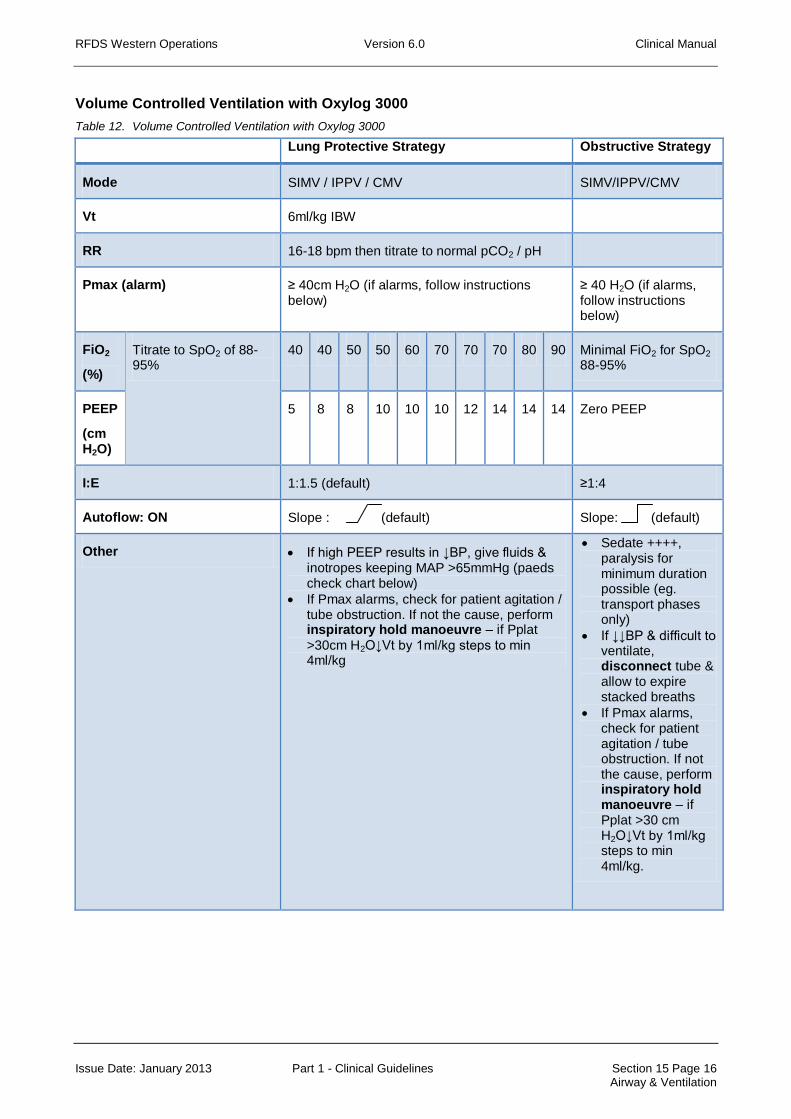
RFDS Western Operations Version 6.0 Clinical Manual
Issue Date: January 2013 Part 1 - Clinical Guidelines Section 15 Page 16 Airway & Ventilation
Volume Controlled Ventilation with Oxylog 3000
Table 12. Volume Controlled Ventilation with Oxylog 3000
Lung Protective Strategy Obstructive Strategy
Mode SIMV / IPPV / CMV SIMV/IPPV/CMV
Vt 6ml/kg IBW
RR 16-18 bpm then titrate to normal pCO2 / pH
Pmax (alarm) ≥ 40cm H2O (if alarms, follow instructions below)
≥ 40 H2O (if alarms, follow instructions below)
FiO2
(%)
Titrate to SpO2 of 88-95%
40 40 50 50 60 70 70 70 80 90 Minimal FiO2 for SpO2 88-95%
PEEP
(cm H2O)
5 8 8 10 10 10 12 14 14 14 Zero PEEP
I:E 1:1.5 (default) ≥1:4
Autoflow: ON Slope : (default) Slope: (default)
Other If high PEEP results in ↓BP, give fluids & inotropes keeping MAP >65mmHg (paeds check chart below)
If Pmax alarms, check for patient agitation / tube obstruction. If not the cause, perform inspiratory hold manoeuvre – if Pplat
>30cm H2O↓Vt by 1ml/kg steps to min 4ml/kg
Sedate ++++, paralysis for minimum duration possible (eg. transport phases only)
If ↓↓BP & difficult to ventilate, disconnect tube & allow to expire stacked breaths
If Pmax alarms, check for patient agitation / tube obstruction. If not the cause, perform inspiratory hold manoeuvre – if Pplat >30 cm H2O↓Vt by 1ml/kg steps to min 4ml/kg.
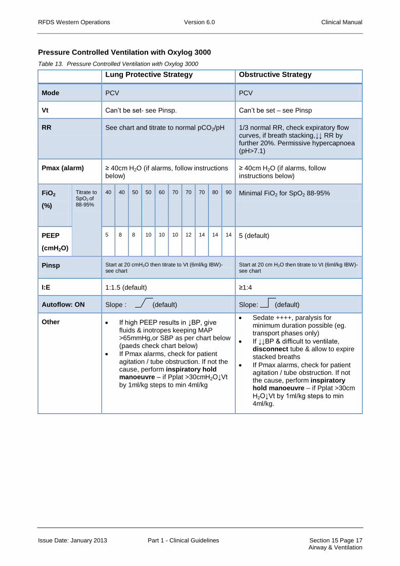
RFDS Western Operations Version 6.0 Clinical Manual
Issue Date: January 2013 Part 1 - Clinical Guidelines Section 15 Page 17 Airway & Ventilation
Pressure Controlled Ventilation with Oxylog 3000
Table 13. Pressure Controlled Ventilation with Oxylog 3000
Lung Protective Strategy Obstructive Strategy
Mode PCV PCV
Vt Can’t be set- see Pinsp. Can’t be set – see Pinsp
RR See chart and titrate to normal pCO2/pH 1/3 normal RR, check expiratory flow curves, if breath stacking,↓↓ RR by further 20%. Permissive hypercapnoea (pH>7.1)
Pmax (alarm) ≥ 40cm H2O (if alarms, follow instructions below)
≥ 40cm H2O (if alarms, follow instructions below)
FiO2
(%)
Titrate to SpO2 of 88-95%
40 40 50 50 60 70 70 70 80 90 Minimal FiO2 for SpO2 88-95%
PEEP
(cmH2O)
5 8 8 10 10 10 12 14 14 14 5 (default)
Pinsp Start at 20 cmH2O then titrate to Vt (6ml/kg IBW)- see chart
Start at 20 cm H2O then titrate to Vt (6ml/kg IBW)- see chart
I:E 1:1.5 (default) ≥1:4
Autoflow: ON Slope : (default) Slope: (default)
Other If high PEEP results in ↓BP, give fluids & inotropes keeping MAP >65mmHg,or SBP as per chart below (paeds check chart below)
If Pmax alarms, check for patient agitation / tube obstruction. If not the cause, perform inspiratory hold manoeuvre – if Pplat >30cmH2O↓Vt
by 1ml/kg steps to min 4ml/kg
Sedate ++++, paralysis for minimum duration possible (eg. transport phases only)
If ↓↓BP & difficult to ventilate, disconnect tube & allow to expire stacked breaths
If Pmax alarms, check for patient agitation / tube obstruction. If not the cause, perform inspiratory hold manoeuvre – if Pplat >30cm
H2O↓Vt by 1ml/kg steps to min 4ml/kg.

RFDS Western Operations Version 6.0 Clinical Manual
Issue Date: January 2013 Part 1 - Clinical Guidelines Section 15 Page 18 Airway & Ventilation
Age and Weight Modifications
Table 14. Age and Weight Modifications
Age /IBW
RR (lung protective)
RR (obstructive)
Vt (6ml/kg)
SBP
Term/3.5kg 40-60 13-20 20ml.
Note min Vt on paed circuit is 50ml, must hand ventilate or use paed ventilator.
≥50mmHg
3months/6kg 30-50 10-16 36ml.
Note min Vt on paed circuit is 50ml, must hand ventilate or use paed ventilator.
≥50mmHg
6months/8kg 30-50 10-16 48ml.
Note min Vt on paed circuit is 50ml, may need to hand ventilate or use paed ventilator.
≥60mmHg
1 year/10kg 30-40 10-13 60ml ≥65mmHg
2years/13kg 20-30 7-9 78ml ≥65mmHg
4years/15kg 20 7 90ml ≥70mmHg
6years/20kg 16 6 120ml ≥75mmHg
8years/25kg 16 6 150ml ≥80mmHg
10years/30kg 16 6 180ml ≥85mmHg
12years/40kg 16 6 240ml ≥90mmHg
14years/50kg 16 6 300ml ≥90mmHg
17years/70kg 16 6 420ml ≥90mmHg
References
Nickson, C. Own The Oxylog 3000. September 2011.
http://lifeinthefastlane.com/2011/09/own-the-oxylog-3000/
Weingart, S. How to Dominate the Ventilator, Part 1. May 2010.
http://emcrit.org/lectures/vent-part-1/
Weingart, 2. How to Dominate the Ventilator, Part 2. June 2010.
http://emcrit.org/podcasts/vent-part-2/

RFDS Western Operations Version 6.0 Clinical Manual
Issue Date: January 2013 Part 1 - Clinical Guidelines Section 15 Page 19 Airway & Ventilation
15.7 Non-Invasive Ventilation
Theory
1. In selected patients, non-invasive ventilatory support may prevent the need for intubation and mechanical ventilation. Complications of failed intubation, ventilator acquired pneumonia, tracheostomy and respiratory muscle wastage are avoided and the patient can continue to communicate. Positive pressure reduces the work of breathing and increases functional residual capacity by recruiting collapsed alveoli. Improvements in lung compliance can also be gained.
2. Left ventricular function can be improved by reduction in preload and afterload.
3. Two forms of NIV can be delivered with the Oxylog 3000: CPAP (continuous positive airway pressure) and BIPAP (biphasic positive airway pressure). CPAP is preferred for the management of acute pulmonary oedema whereas hypoventilatory respiratory failure (eg. from COPD) may benefit from BIPAP or CPAP with pressure support.
4. Traditionally NIV has not been considered in transport medicine due to lack of suitable equipment and the risk of intubation during transport if NIV fails. The Oxylog 3000 in RFDS service, now provides the opportunity to deliver NIV to selected patients.
Pre-flight and In-flight Management
1. Consider the opportunity for NIV when assessing the flight request. Patients already undergoing NIV should prompt consideration of using this technique for ventilatory support. The technique is likely to be less suitable for longer flight times as risk of failure increases with time.
2. Prepare to go in to the referring hospital and take the Oxylog 3000, CPAP mask (with attachment points) and a harness.
3. Time in the hospital is required to establish if treatment will work (at least ½ hour). Does the patient tolerate it, do parameters such as blood gases improve, is patient likely to become fatigued? NIV may not be a good option with restricted pilot hours.
4. Gas consumption during NIV is greater than that for IPPV (eg. up to 30L/min). Have you got enough oxygen?
Patient Selection
Contraindications
1. Not fully conscious or cooperative.
2. Risk of, or actual, airway obstruction or deterioration in conscious state.
3. Facial abnormalities, trauma, recent surgery or burns.
4. Suffering from excessive secretions, vomiting or bowel obstruction.
5. Having a high oxygen requirement or suffering life threatening hypoxia.
6. Profoundly acidaemic.
7. Haemodynamically unstable, suffering dysrhythmias or other severe co-morbidities.
8. Pneumothoraces (unless ICC in situ).
9. Recent upper GI surgery.
Indications
1. Acute pulmonary oedema.
2. Obstructive sleep apnoea.

RFDS Western Operations Version 6.0 Clinical Manual
Issue Date: January 2013 Part 1 - Clinical Guidelines Section 15 Page 20 Airway & Ventilation
3. Acute exacerbation of COPD.
4. Ventilator weaning.
5. Respiratory failure in immunocompromised (e.g. Neutropaenic) patients at high risk of ventilator acquired pneumonia.
6. Other acute respiratory failure where no contraindications.
7. Chronic respiratory failure e.g. Neuromuscular disease where it is desirable to avoid intubation as weaning would be difficult.
Complications
1. Mask intolerance (25%)
2. Skin damage.
3. Gastric distension and aspiration. (Routine gastric decompression is not indicated however.)
4. Patient may still become obtunded and lose airway.
5. Sinus pain, nasal congestion.
6. Raised intraocular pressure.
7. Raised intracranial pressure.
8. Hypotension if hypovolaemic.
Ventilator set up
1. Use face mask and harness and connect to ventilator hose.
2. NIV can be delivered in CPAP and PCV modes on the ventilator. In NIV mode mask leakages will be detected and compensated and included in measured values for Vt and MV.
3. Switch on NIV by pressing Settings key, then scroll to page 2/2. On NIV line, change to ON and confirm.
4. Biphasic Positive Airway Pressure (BIPAP) can be delivered using NIV in the PCV mode. This is like giving CPAP at two alternating pressures.
5. Pressure support (a gas flow triggered by inspiratory effort to a set pressure) can be provided using the ASB (assisted spontaneous breathing) function in either BIPAP or CPAP.
Tips to aid patient compliance
1. If patient able, allow the patient to hold the mask initially until used to it then apply harness.
2. Start with lower pressures and titrate up, the pressure should make the work of breathing easier rather than result in the patient fighting it. Provide an antiemetic.
BIPAP
For hypoventilatory respiratory failure (e.g. acute exacerbation COPD)
Set PCV
Turn NIV on
I:E ratio 1:2
PEEP = 4cm H2O
Pinsp = 10cm H2O

RFDS Western Operations Version 6.0 Clinical Manual
Issue Date: January 2013 Part 1 - Clinical Guidelines Section 15 Page 21 Airway & Ventilation
Titrate FiO2 to SaO2 >90%
Adjust Trigger to maximise synchronisation with the patients breathing.
Repeat ABG at 30 minutes
i. If PCO2 decreased by 10-20% then PEEP = 4cm H2O, Pinsp = 16 cm H2O ii. If PCO2 decreased by <10% then PEEP = 6cm H2O, Pinsp = 20 cm H2O iii. If PCO2 rising or clinically no improvement consider intubation.
For acute pulmonary oedema
Set PCV
Turn NIV on
I:E ratio 1:2
PEEP = 8cm H2O
Pinsp = 10 cm H2O
Commence FiO2 100%
Adjust trigger to maximise synchronisation with patients breathing.
Repeat ABG at 30 minutes
i. If no improvement clinically and with ABG consider intubation. ii. Titrate PEEP to 10cm H2O and Pinsp to 15cm H2O
If no improvement after 1 hour consider intubation pre-flight.
CPAP
For acute pulmonary oedema
Set CPAP
Turn NIV on
I:E ratio 1:2
PEEP = 10 cm H2O
PS = 0
Commence FiO2 100%
Repeat ABG at 30 min and assess patient clinically.
i. If no improvement consider intubation ii. Titrate PEEP to effect (may need 15-20cm H2O)
For hypoventilatory respiratory failure
Set CPAP
Turn NIV on
I:E ratio 1:2
PEEP = 4 cm H2O
PS = 15 cm H2O
Titrate FiO2 to SaO2 >90%
Adjust trigger to maximise synchronisation with patient breathing.
Repeat ABG at 30 min.

RFDS Western Operations Version 6.0 Clinical Manual
Issue Date: January 2013 Part 1 - Clinical Guidelines Section 15 Page 22 Airway & Ventilation
i. If no improvement consider intubation ii. Titrate pressure support (ASB) to effect
If no improvement after 1 hour consider intubation pre-flight.
References
Bersten A, Soni N. Oh’s Intensive Care Manual. 6th ed. Butterworth Heinemann. 2009.
Drager Medical. Oxylog 3000 Instructions for Use.
Beed M, Sherman R, Mahajan R. Emergencies in Critical Care. Oxford: Oxford University Press. 2007.
Dunn R, Dilley S, Brookes J, et al. The Emergency Medicine Manual. 5th ed. Tennyson, South Australia: Venom Publishing. 2010.

RFDS Western Operations Version 6.0 Clinical Manual
Issue Date: January 2013 Part 1 - Clinical Guidelines Section 15 Page 23 Airway & Ventilation
15.8 Paediatric Induction, Intubation and Ventilation
Theory
1. Attention to differences in paediatric anatomy, physiology and psychology is required for the safe induction, intubation and ventilation of children.
2. Airway differences include; high anterior larynx, floppy larynx, large tongue, large occiput, short trachea, cricoid is narrowest part of airway. To overcome some of these differences there are a number of strategies; straight laryngoscope blades under 6 months for picking up the tip of the epiglottis, positioning the child with a sandbag / IV fluid bag under shoulders, avoid hyperextension of neck, avoid pressing on soft tissues of floor of mouth, when applying face mask (keep fingers on bony parts), use of CPAP with Ayre’s T-piece.
3. The impact of an over distended stomach from mask ventilation is very significant, early decompression of the stomach is essential to successful ventilation.
4. A short trachea means that flexion or extension of the head can result in either right main bronchus intubation or extubation, the head and neck should be immobilized to avoid this. The tube should be fixed to the maxilla rather than the mandible. A guedel airway may help splint the tube.
5. Doses of drugs should be carefully calculated and checked.
6. Pre-medication with atropine is common 10µg/kg IV to prevent the bradycardia associated with suxamethonium and laryngoscopy. At the very least this should be drawn up and ready to give.
7. In the emergency and transport setting inductions are most commonly IV. Whilst the agents used are the same as for adults please not that doses per kg may differ with faster metabolism or greater sensitivity. Doses may need to be significantly reduced for those with altered conscious level or haemodynamic instability.
thiopentone 4-6mg/kg
propofol 3-5mg/kg
ketamine 2mg/kg (useful for haemodynamicaly compromised)
+/- fentanyl 1-2µg/kg
suxamethonium 1-2mg/kg
8. Useful formulae:
ETT size age/4 +4 or size of nostril or little finger, if using cuffed ETT’s a half size smaller may be a better fit. In all cases have a size above and a size below the expected available.
Tube length age/2+12, ETT’s are marked with a black line to indicate the depth the tube should pass to.
Weight (age +4)x2
9. Whilst cuffed ETTs are now recommended for those above a size 3, some caution needs to be heeded. Inflation of the cuff should be reserved as a means of compensating for a leak that is interfering with ventilation when higher pressures are required. Even an uninflated cuff pressure can reach >30cm H2O at altitude. High volume low pressure cuffs should
minimize any risk of tracheal trauma however care needs to be taken to only inflate the cuff to the point where the leak is abolished and no more. Air is to be used so that pressure can be closely monitored and adjusted. A manometer is supplied to ensure the cuff pressure is kept at 20-30cm H2O, the cuff pressure should be monitored during ascent, at the top of the climb, during descent and on landing. A half size smaller than expected with a traditional

RFDS Western Operations Version 6.0 Clinical Manual
Issue Date: January 2013 Part 1 - Clinical Guidelines Section 15 Page 24 Airway & Ventilation
uncuffed tube is likely to be required. The impact of cuff expansion at altitude is greater than for the the adult.
10. The use of a cuffed tube helps eliminate need for tube changes, and difficulties ventilating with a large leak around the tube. These ventilation difficulties are more likely to be experienced with lung pathology that requires higher ventilation pressures (eg. aspiration, pneumonia, asthma etc). A cuffed ETT may also be a little less likely to extubate or migrate down the right main bronchus with movement of the patient’s head.
11. It is likely that we will still be taking handover of patients with uncuffed ETT’s, the decision to change to a cuffed ETT will be a matter of clinical judgment and experience after assessing the adequacy of ventilation and degree of leak.
12. Neither the Oxylog 1000 nor the Oxylog 3000 are paediatric ventilators. Very careful attention must be paid to the adequacy of ventilation. For children under 15kg in particular there are significant challenges such that in some instances manual ventilation may be preferred. All effort must be made to minimize dead space, including not using catheter mounts, ensuring paediatric heat moisture exchange filter (HME) is used and no other extraneous connectors in the circuit. A constant eye must be kept on chest excursion (hand on chest), ETCO2, SaO2, and ABG’s.
13. Modifications to the Oxylog 3000 ventilator software will be made to enable ventilation with much smaller tidal volumes and a paediatric (low dead space) circuit, this circuit will be suitable for tidal volumes of 50-250mL. Recommended tidal volumes are 5-7mL/kg. When switching the ventilator on you will be asked to chose between paediatric and adult circuits, the device must then run through a check with the circuit to be used in situ.
14. Pressure controlled ventilation is commonly used in the paediatric setting using inspiratory pressures of 16-20cm H2O with an appropriate rate and 4cm PEEP. PCV will however, not compensate for bronchospasm or tube obstruction.
Reference
Allman K, Wilson I. Oxford Handbook of Anaesthesia. 2nd Ed. Oxford University Press. 2007
Weiss M, Dullenkopf A, Fischer J, et al. Prospective randomized controlled multi-centre trial of cuffed or uncuffed endotracheal tubes in small children. B J Anaes. 2009; 103 (6): 867-873.
Clements R, Steel A, Bates A, et al. Cuffed endotracheal tube use in paediatric prehospital intubation: challenging the doctorine? Emerg Med J 2007; 24:57-58.
Newth C, Rachman B, Patel N, et al. The use of cuffed versus uncuffed endotracheal tubes in pediatric intensive care. J Pediatr. 2004 Mar;144(3):333-7.
Knight , G; Consultant ICU Princess Margaret Hospital for Children, Subiaco Western Australia – Personal Communication

RFDS Western Operations Version 6.0 Clinical Manual
Issue Date: January 2013 Part 1 - Clinical Guidelines Section 15 Page 25 Airway & Ventilation
15.9 Paediatric Leak Attachment
Description
Cylindrical aluminium connection with a small hole drilled through one wall. The hole is sited between ridges to prevent accidental obstruction. The device has a 22mm female connection at one end for connection to the ventilator and a 15mm female/22mm male connector for connection to the circuit.
Theory
1. This device was produced for use with the Oxylog 1000 ventilator. It is not required for use with the Oxylog 3000 ventilator.
2. At a set rate of 30 breaths per minute and the lowest minute ventilation setting, the tidal volume delivered by the Oxylog ventilator is approximately 100mL. This limits use of the ventilator to infants of approximately 10kgs or greater.
3. Insertion of a ‘deliberate’ leak into the circuit allows delivery of smaller tidal volumes. Using this adaptor PMH staff have successfully ventilated infants down to 4kgs.
4. The volume of gas leaked will be determined by the airway pressures and inspiratory time.
5. The device is recommended for use when ventilating infants less than 10kgs. It is the responsibility of the medical practitioner caring for the infant to ensure safe use of the device, with appropriate ventilator, circuit and settings, and for the adequacy of ventilation.
Technique
1. The device is inserted between the ventilator and patient circuit.
2. As with all infant mechanical ventilation, a ventilation rate is set and then the minute ventilation increased gradually from the minimum setting until adequate ventilation is achieved, gauged by degree of chest expansion, airway pressure, pulse oximetry and capnography. Beware rising pCO2 resulting in increasing acidosis.
3. The device does not remove the need for frequent re-assessment of the patient and adequacy of ventilation and readjustment of parameters as required.
4. Small infants between 4-10kg may be better if hand ventilated with either a Laerdal bag/valve mask (self-inflating) or T-piece, depending on the experience of the flight doctor.
Reference
Prepared by: Dr D. McConville, RFDS Western Operations, Port Hedland Base.
Reviewed by: Dr A. Duncan, ICU, Princess Margaret Hospital, Perth.
Figure 22. Paediatric Leak Attachment

RFDS Western Operations Version 6.0 Clinical Manual
Issue Date: January 2013 Part 1 - Clinical Guidelines Section 16 Page 1 Occupational & Administrative
16 OCCUPATIONAL & ADMINISTRATIVE
16.1 Occupational Exposure to Blood and Bodily Fluids
Theory
1. HIV, Hepatitis B and C may be transmitted by significant exposure to blood or other body fluids.
2. Prevention is the mainstay of protection, so standard infection control practices must be adhered to.
3. Risk of transmission is dependent on the type of injury sustained. A thorough risk assessment of each exposure must be performed by an RFDS medical officer.
4. Those exposed to a source positive for a blood-borne virus must be referred to an infectious diseases expert or clinical immunologist.
Risks
1. Risk of Hepatitis B infection carries the highest risk after exposure to a positive source (10-40%). Hepatitis B vaccine is advised for all at risk staff (Nurses, Doctors, Pilots and Engineers); this vaccine has a 90% rate of protection after 3 doses.
2. Risk of Hepatitis C transmission after needle-stick injury from positive source is 1.8%. Transmission from mucous membrane exposure is rare.
3. Risk of HIV transmission after percutaneous exposure from positive source is 0.3% and 0.09% from mucous membrane exposure.
4. High risk injuries are:
Deep injury from device visibly contaminated with blood.
Injury associated with hollow bore needle.
Source patient has late stage HIV or high viral load
Source patient with Hep B who is HBeAg +ve, HBV DNA detectable, high viral load.
Source patient with Hep C who is HCV RNA PCR detectable
Procedure following injury or exposure
1. First aid. If skin is exposed the area should be washed well immediately with soap and water. If water is not available, use 60-90% alcohol hand cleanser (such as located in red IV roll in the aircraft.)
2. If the injury is to mucous membranes (eyes, mouth etc.) flush with copious water or normal saline.
3. Report to RFDS doctor immediately. Notify the doctor directly if on the flight, or by satellite telephone or radio, irrespective of your location. RFDS doctor should follow these guidelines:
Perform a risk assessment (based on history of incident, knowledge of patient)
Arrange baseline blood tests from source and recipient.
Counsel recipient (regarding risk, required follow-up, precautions.)
Seek advice or arrange referral to infectious diseases or immunology expert (the Immunology registrar at RPH is on call all hours via switch board.)
Complete a Clinical Incident Report (Notify Director of Medical Services or Assistant Director of Medical Services immediately).

RFDS Western Operations Version 6.0 Clinical Manual
Issue Date: January 2013 Part 1 - Clinical Guidelines Section 16 Page 2 Occupational & Administrative
Seek consent of recipient to forward follow-up results to office of Director of Medical Services.
Worker's Compensation Report. Complete notification for a work-related injury.
Risk Assessment
Table 15. Risk Assessment for Exposure to Bodily Fluids
Non parenteral exposure (low risk) Intact skin
Doubtful exposure (low risk) Superficial (not bleeding) intradermal injury with device thought NOT to be contaminated with bodily fluid.
Contamination of prior wound with substance other than blood.
Mucous membrane contact with substance other than blood.
Possible exposure (low to moderate risk) Superficial (not bleeding) intradermal injury from device thought to be contaminated with blood.
Prior wound contamination with blood.
Mucous membrane contact with blood.
Definite exposure (moderate risk) Penetrating injury with needle contaminated with blood or bodily fluid.
Injection of <1mL of blood or bodily fluid.
Laceration caused by visibly contaminated instrument.
In lab setting inoculation with HIV, HCV, HBV +ve tissues.
Massive exposure (high risk) Blood transfusion.
Injection of >1mL blood or bodily fluid.
Parenteral exposure to lab specimens containing high titre of virus.

RFDS Western Operations Version 6.0 Clinical Manual
Issue Date: January 2013 Part 1 - Clinical Guidelines Section 16 Page 3 Occupational & Administrative
Medical Management
Table 16. Medical Management for Exposure to Bodily Fluids
Source negative for blood borne virus
Source status unknown, or high risk, but negative
Source Hep B positive
Source Hep C positive Source HIV positive
Offer repeat blood at 3 months
If high risk injury treat as for positive source.
Note: It is impossible to test a sharp.
If immune, no further action.
Non-immune - see post-exposure prophylaxis.
Rpt bloods at 3 and 6 months.
Provide behavioural counselling.
No treatment is available.
Refer to clinical microbiologist or hepatologist with Hep C expertise.
Repeat bloods, HCV RNA PCR @ 4, 8, 12 wks.
HCV ab @ 3 and 6 months.
Arrange counselling / support.
Provide behavioural counselling.
Immediate referral to immunologist (24 hr call) for advice on PEP. Truvada ® available from regional and private hospitals.
PEP is best started with in 24hrs.
Expect immunologist to advise on risk of infection, signs and symptoms, effectiveness of PEP, side effects of PEP, advice on pregnancy, breast feeding and comorbidities.
Repeat testing at 6wks, 3 and 6 months.
Ensure ongoing counselling.
Provide behavioural counselling.
PEP = Post-exposure prophylaxis
Hep B PEP for the non-immune
1. Unvaccinated
If source HBsAg +ve, give HBIG and initiate Hep B vaccination within 24 hrs.
If source is unknown, initiate Hep B vaccination within 24 hrs.
2. Previously vaccinated.
If adequate immunity, no treatment.
Non-responder (had 3 doses and re-immunised with 3 doses but still no response) should have 2 doses of HBIG (1st within 24 hrs, second in 1 month).
Response to previous immunisation unknown, test anti Hep B Ab’s, if inadequate give 1 dose HBIG and vaccine booster. If adequate no further action.
Risk Counselling
The nature of the injury and the status of the source must be ascertained in order to give accurate advice. Consider transmissibility information in “Risk Assessment” given at start of this guideline.
Behavioural Counselling
Hep C exposure.
May not donate blood, plasma, tissues or semen.
Do not need to modify sexual practices, avoid pregnancy or refrain from breast feeding.
Continue standard precautions with work practices.

RFDS Western Operations Version 6.0 Clinical Manual
Issue Date: January 2013 Part 1 - Clinical Guidelines Section 16 Page 4 Occupational & Administrative
Hep B exposure in non-immune
If high risk injury may not donate blood, plasma, tissues or semen. Should avoid pregnancy until outcome known.
Continuation of breastfeeding and sexual activity will depend on immune status of baby or partner.
Work practices may need to be modified according to the nature of the work. (See DoH Policy for Health Care Workers with BBV Infections)
HIV exposure
For 12 months may not donate tissues, blood, plasma, breast milk or semen.
Sexual abstinence or protected sex (condoms) for a minimum of 3 months.
Avoid pregnancy till 6 month surveillance complete.
Cease breast feeding.
Do not share needles, razors, toothbrushes.
Cover open wounds with waterproof dressings.
References
Department of Health, Western Australia. Operational Directive Number: OD 0091/07, dated 20 December 2007. Management of Occupational Exposure to Blood and Body Fluids in the Health Care Setting.
NHMRC. Australian Immunisation Handbook (2007) 9th Ed.

RFDS Western Operations Version 6.0 Clinical Manual
Issue Date: January 2013 Part 1 - Clinical Guidelines Section 16 Page 5 Occupational & Administrative
16.2 Deceased Patients
Theory
1. The death of a patient at any stage from pre-flight assessment or consultation through to admission at a receiving hospital must be recorded by filing a clinical incident form, this allows for accurate mortality reporting and where necessary appropriate compilation of records for coronial inquests.
2. All doctors are expected to know when and how a patient must be referred to the coroner, when it is acceptable to fill out a death certificate and how to fill out same.
3. RFDS aircraft generally should not be tasked to retrieve deceased persons as this makes the asset unable to respond to other emergencies.
4. The RFDS Director of Medical Services (or nominated deputy) must be notified at the time of death.
Patients to be referred to the coroner
Deaths as a result of trauma, violence, criminal action, suicide.
Death appears unexpected or unnatural.
Deaths where the cause is not definitely known.
Deaths where medical mismanagement is suspected.
Deaths where you have not been the treating doctor in the most recent illness.
Deaths where the doctor is unwilling to complete a death certificate.
Death during or due to anaesthetic.
Death during care (e.g. wards of the state) or custody, whilst detained under mental health act.
To refer a patient to the coroner either call the “Coronial Investigation Unit” (Police) or for remote areas the local police. The police are responsible for custody of the body, they may transport the body themselves or commonly arrange the government contracted undertaker to do so.
If a patient dies in flight, request operations staff contact the “Coronial Investigation Unit” (Police) or local police to notify them of a sudden death. It should be requested that the aircraft be met to take over custody of the deceased. If there is no medical officer on board arrangements should be made for a medical officer to meet the aircraft. A medical officer must certify life extinct in the observation and treatment chart (this is not a death certificate merely documenting that the patient is dead). All lines and tubes should be left in situ, copies of all documentation should be made for the police or their contracted undertaker. Permission should be sought from the police to remove the patient from the aircraft if needed to free the aircraft up for further tasking and /or get body and staff out of the heat.
For any queries contact the Coroner’s Office on 1800 671 994.
Completing a death certificate
1. You must have been the treating (for at least 30 min) doctor during the last illness.
2. You must have a definitive diagnosis. Give the cause of death not the mode of dying. (i.e. not respiratory failure when the patient had pneumonia). Causative organisms should be recorded for infectious diseases and histological diagnosis plus location of primary for neoplastic causes.
3. The cause of death and all contributing factors must be documented.
4. Do not use abbreviations.
RFDS Western Operations Version 6.0 Clinical Manual
Issue Date: January 2013 Part 1 - Clinical Guidelines Section 16 Page 6 Occupational & Administrative
5. The death certificate should be handed to the undertaker responsible for the body.
6. Note there is a special perinatal (of at least 20wks gestation or any death in the first 28 days of life) death certificate though in our practice we would refer most of these to the coroner.
Destination of patients who have died in flight
If a patient dies in flight the patient should be returned to the originating hospital or flown on to the destination depending on which is closest and logistically most appropriate.
For patients who have a completed death certificate an undertaker should be contacted, preferably attempt to find out who the family would prefer, otherwise in Perth, Donald Chipper and Son (the family can liaise with them if they wish to change). Operations staff should do this to ensure the deceased can be handed over promptly on landing.
Death at referring location and prior departure
Should a death occur in presence of RFDS staff without having actually taken off with the patient, responsibility for notification of coroner or death certification can be passed back to the referring doctor, unless this occurs in a primary location in which case this remains an RFDS responsibility.
Notification of next of kin
The RFDS medical officer certifying life extinct should contact the referring doctor and ask them to notify the next of kin. For primary retrievals this may be more difficult, the RFDS doctor may need to directly notify family, remote area nurse or request the police locate and notify the next of kin. Relatives who are accompanying patients who die in flight should have appropriate explanations, support and assistance offered on arrival.
Deaths in remote locations
It is common to receive calls regarding patients found deceased, or for whom resuscitation is in progress. Ensure BLS / ALS protocols have been followed as appropriate. Follow-up support by telephone after the event is very important to debrief those involved and answer any questions they may have. We do not fly to retrieve deceased persons.
References
Turnbull, R. The Coronial Process in Western Australia, A Handbook for Medical Practitioners and Medical Students. Office of the State Coroner. June 2010
Bird, S. “How to Complete a Death Certificate, A Guide for GPs.” Australian Family Physician Vol 40, No.6. June 2011