Embed Size (px)
Citation preview
ClinicalTHYROIDOLOGY
A publication of the American Thyroid Association
JUNE 2011VOLUME 23 l ISSUE 6
METFORMIN SHRINKS THYROID NODULES IN PATIENTS WITH INSULIN RESISTANCE . . . .2Rezzonico J, Rezzonico M, Pusiol E, Pistoia F, Niepomniszcze H. Metformin treatment for small benign thyroid nodules in patients with insulin resistance . Metab Syndr Relate Disord 2011;9:69-75. Epub December 3, 2010.
THYROID AUTOIMMUNITY IS ASSOC- IATED WITH AN INCREASED RISK OF SPONTANEOUS MISCARRIAGE IN EUTHYROID WOMEN . . . . . . . . . . . . . . . . . . . . .4Chen L, Hu R. Thyroid autoimmunity and miscarriage: a meta-analysis . Clin Endocrinol (Oxf) 2011;74:513-9.
THE MEDIAN URINARY IODINE IS LOW IN AMERICAN PREGNANT WOMEN . WHY HAVE WE NOT IMPROVED IODINE INTAKE IN THIS VULNERABLE POPULATION? . . . . . . . .7Caldwell KL, Makhmudov A, Ely E, Jones RL, Wang RY. Iodine status of the U .S . Population, National Health and Nutrition Examination Survey, 2005-2006 and 2007-2008 . Thyroid. February 16, 2011 [Epub ahead of print].
DOES EXTREME OBESITY AFFECT THYROID HORMONE METABOLISM? . . . . . . . . . .9Michalaki MA, Gkotsina MI, Mamali I, Markantes GK, Faltaka A, Kalfarentzos F, Vagenakis AG, Markou KB. Impaired pharmacokinetics of levothyroxine in severely obese volunteers . Thyroid. March 21, 2011 [E-pub ahead of print]. doi: 10.1089/thy.2010.0149.
SELENIUM MAY PREVENT GOITER AND THYROID NODULES . . . . . . . . . . . . . . . . . . . 11Rasmussen LB, Schaumburg L, Kohrle J, Pedersen IB, Hollenbach B, Hög A, Ovesen L, Perrild H, Laurberg P. Selenium status, thyroid volume, and multiple nodule formation in an area with mild iodine deficiency . Eur J Endocrinol 2011;164:585-90. Epub January 17, 2011.
REOPERATION CURES ONLY HALF OF THE PATIENTS WITH LOCALLY RECURRENT PAPILLARY THYROID CANCER . . . . . . . . . . . . . . . . . . . . . . . . .13Yim JH, Kim WB, Kim EY, Kim WG, Kim TY, Ryu JS, Gong G, Hong SJ, Shong YK. The outcomes of first reoperation for locoregionally recurrent/persistent papillary thyroid carcinoma in patients who initially underwent total thyroidectomy and remnant ablation . J Clin Endocrinol Metab. April 20, 2011 [Epub ahead of print].
LOW BASAL SERUM THYROGLOBULIN IN A SENSITIVE ASSAY IS AN EXCELLENT PREDICTOR OF LOW RECURRENCE OF DIFFERENTIATED THYROID CANCER . . . . . . . .15Malandrino P, Latina A, Marescalco S, Spadaro A, Regalbuto C, Fulco RA, Scollo C, Vigneri R, Pellegriti G. Risk-adapted management of differentiated thyroid cancer assessed by a sensitive measurement of basal serum thyroglobulin . J Clin Endocrinol Metab. March 30, 2011 [Epub ahead of print].
WITH THE RISING INCIDENCE OF SECOND CANCERS, SHOULD YOU GIVE RAI TO A LOW-RISK (T1NO) THYROID CANCER PATIENT? . . . . . . . . . . . . . . . . . . . . . . . .17Iyher NG, Morris LG, Tuttle RM, Shaha AR, Ganly I. Rising incidence of second cancers in patients with low-risk (T1N0) thyroid cancer who receive radioactive iodine therapy . Cancer. March 22, 2011 [Epub ahead of print]. doi: 10.1002/cncr.26070.
OFFICIAL ITALIAN TRANSLATION of the 2009 American Thyroid Association Management Guidelines for Patients with Thyroid Nodules and Differentiated Thyroid Cancer . . . . . . . . . . . .19
TO THE EDITOR: DAVID S . COOPER, MD . . . . .21
DR . WEISSEL RESPONDS . . . . . . . . . . . . . . . . . . . .23
Editor-in ChiefJerome M. Hershman, MDVA Greater Los Angeles Healthcare System and UCLA School of Medicine Endocrinology 111D 11301 Wilshire Blvd Los Angeles, CA 90073 Telephone: 310-268-3852 Fax: 310-268-4879 Email: [email protected]
Associate Editors:Albert G. Burger, MDProfessor, University of Geneva Geneva, Switzerland Email: [email protected]
Stephanie L. Lee, MD, PhDDirector of the Thyroid Health Center Boston University Medical Center Boston, MA Telephone: 617-638-8530 Fax: 617-638-7221 Email: [email protected]
Jorge H. Mestman, MDProfessor of Clinical Medicine and OB/GYN University of Southern California Keck School of Medicine Los Angeles, CA Telephone: 323-442-6179 Email: [email protected]
Stephen W. Spaulding, MDDepartment of Medicine, University of Buffalo Buffalo NY Telephone: 716-862-6530 Fax: 716-862-6526 Email: [email protected]
PresidentGregory A. Brent, MD
Secretary/Chief Operating OfficerRichard T. Kloos, MD
TreasurerDavid H. Sarne, MD
President-ElectJames A. Fagin, MD
Secretary-ElectJohn C. Morris, MD
Past-PresidentTerry F. Davies, MD
Executive DirectorBarbara R. Smith, CAEAmerican Thyroid Association 6066 Leesburg Pike, Suite 550 Falls Church, VA 22041 Telephone: 703-998-8890 Fax: 703-998-8893 Email: [email protected]
Designed ByKaren Durland ([email protected])
Clinical ThyroidologyCopyright © 2011 American Thyroid Association, Inc. Printed in the USA. All rights reserved.
ClinicalTHYROIDOLOGY
Back to Contents
SUMMARY
BACKGROUNDIn a prior study, these authors found that individuals with hyperinsulinemia had an increased thyroid volume as well as an increased number of thyroid nodules.
METHODSEighty women who were thyroid peroxidase antibody–negative and living in an iodine-sufficient area and who had insulin resistance (IR) and solid, benign, hyperplastic thyroid nodules were prospectively evaluated for nodule shrinkage using metformin, levothyroxine, or a combination of the two. Fourteen women did not complete follow-up, leaving 66 women with 75 thyroid nodules. IR was evaluated by homeostasis model assessment (HOMA; fasting serum insulin in microunits per milliliter multiplied by plasma glucose in millimoles per liter divided by 22.5); a HOMA index of >2.5 indicated IR. Nodule volume determined using ultrasound was calculated with the elliptical shape volume formula. Women who qualified were randomly assigned to four treatment groups and followed for 6 months: group 1 (n = 14; 19 nodules) was treated with metformin alone; group 2 (n = 18; 21 nodules) was treated with metformin and levothyroxine; group 3 (n = 19; 20 nodules) was treated with levothyroxine alone; and group 4 (n = 15; 15 nodules) were controls. The metformin dose was 1000 mg twice daily; the dose of levothyroxine was adjusted to keep the serum thyrotropin (TSH) level at 0.11 to 0.99 mU/L. Patients were treated for 6 months and then reevaluated using ultrasound.
RESULTSThe characteristics of the patients in the groups were similar at baseline (mean age, 43; mean weight, 80 kg; mean body-mass index [the weight in kilograms divided by the square of the height in meters], 31; mean HOMA score, 3.3; mean TSH, 2.55; median nodule volume, 298 mm3). Patients treated with levothyroxine had a significant decrease in TSH (mean, 0.59 mU/L). Patients taking metformin had a significant decrease in their HOMA scores into the normal range. All patients on active treatment (groups 1 to 3) had
METFORMIN SHRINKS THYROID NODULES IN PATIENTS WITH INSULIN RESISTANCE
Rezzonico J, Rezzonico M, Pusiol E, Pistoia F, Niepomniszcze H. Metformin treatment for small benign thyroid nodules in patients with insulin resistance . Metab Syndr Relate Disord 2011;9:69-75. Epub December 3, 2010.
continued on next page
VOLUME 23 • ISSUE 6 JUNE 2011
CLINICAL THYROIDOLOGY l JUNE 2011 3 VOLUME 23 l ISSUE 6
Back to Contents
METFORMIN SHRINKS THYROID NODULES Rezzonico J, et. al.IN PATIENTS WITH INSULIN RESISTANCE
a significant reduction in thyroid volume, with no significant difference in the posttreatment thyroid volume between these three groups. Nodule size was markedly and significantly reduced in both groups treated with metformin (from 364 to 75 mm3 in group 1 and from 336 to 126 mm3 in group 2; 74% of nodules were reduced with metformin alone, 95% were reduced with metformin plus levothyroxine treatment), whereas nodule size was unchanged in the other treatment groups. Patients treated with metformin alone had a significantly greater reduction in nodule size than those treated with levothyroxine
alone or those with no treatment. Similar nodule shrinkage with metformin was seen with small (<1 cm) and larger lesions (1 to 2 cm).
CONCLUSIONSIn patients with small hyperplastic thyroid nodules, metformin produced a significant decrease in nodule size, and the combination of metformin plus levothyroxine showed the best reduction in nodule volume, whereas levothyroxine alone reduced nodule growth but not nodule volume.
COMMENTARY
This small, relatively brief study reveals a marked reduction in thyroid nodule volume in 74% of these insulin-resistant subjects who were being treated with metformin. The overall reduction in thyroid volume was less impressive, perhaps because only small lesions were included in this study, hence making up a relatively small percentage of total thyroid volume. Combination therapy with metformin plus levothyroxine was associated with a reduction in nodule size in 95% of lesions. In contrast, suppressive therapy with levothyroxine alone did not result in nodule shrinkage, perhaps because the TSH suppression was briefer and TSH was not suppressed to grossly hyperthyroid levels as has been done in prior successful trials (1,2). Past studies evaluating shrinkage of thyroid nodules with levothyroxine suppression have had mixed results, and the
2009 American Thyroid Association management guidelines for thyroid nodules do not recommend levothyroxine suppressive therapy for patients with benign nodules who live in iodine-sufficient areas (3). In this study, metformin treatment appears to offer a tantalizing reduction in thyroid nodule volume, with efficacy seen in a striking percentage of treated patients. As insulin-resistant patients often have multiple associated risk factors for surgery, this would be an important group for further study of non-operative therapies, especially for larger nodules in which cosmesis may be an issue. As the authors point out, we need more studies done with metformin to reproduce these results.
—JaneWeinreb,MDDivision of Endocrinology, Diabetes and Metabolism
VA Greater Los Angeles Healthcare SystemLos Angeles, CA 90073
References
1. Wémeau J, Caron P, Schvartz C, et al. Effects of thyroid-stimulating hormone suppression with levothyroxine in reducing the volume of solitary thyroid nodules and improving extranodular no palpable changes: a randomized, double-blind, placebo-controlled trial by the French Thyroid Research Group. J Clin Endocrinol Metab 2002;87:4928-34.
2. Papini E, Petrucci L, Guglielmi R, et al. Long-term changes in nodular goiter: a 5-year prospective randomized trial of levothyroxine suppressive therapy for benign cold thyroid nodules. J Clin Endocrinol Metab 1998;83:780-3.
3. Cooper DS, Doherty GM, Haugen BR, et al. Revised American Thyroid Association management guidelines for patients with thyroid nodules and differentiated thyroid cancer. Thyroid 2009;19:1167-214.
CLINICAL THYROIDOLOGY l JUNE 2011 4 VOLUME 23 l ISSUE 6
Back to Contents
then calculated. Likewise, the abortion rates for TA-positive and TA-negative groups were compared, and the pooled relative risk (RR) and its 95% CI were calculated.
Inclusion criteria for the analysis were as follows:
1. All published studies were included, regardless of their publication language or date.
2. The cohort studies had to be prospectively designed.
3. Overt thyroid dysfunction in gestation was excluded, while euthyroidism was defined as a serum thyrotropin (TSH) level between 0.3 and 5.0 mIU/L.
4. TAI was defined as one or more types of positive TA. TA refers to thyroid microsome antibody, thyroglobulin antibody, and thyroid peroxidase antibody.
5. OR or RR and its 95% CI were calculated from the raw data extracted from the original literature.
6. The studies involving multiple in vitro fertili-zation procedures were excluded.
RESULTSThe search strategy identified 53 potentially relevant studies, 22 of which were included in the meta-analysis; 8 of these were case–control studies and 14 cohort studies.
Case–Control Studies: The 8 case–control studies included in the systematic review were published between 1993 and 2008. They reported data on 1077 women who had had recurrent spontaneous abortions, all of whom were white. Most of these women had had two or more consecutive miscarriages (n = 1341), while women with no history of abortion (n = 747), female blood donors (n = 300), and parous women (n = 284) served as most of the controls.
SUMMARY
BACKGROUNDAccording to epidemiologic research, 31% of pregnancies end in abortion. Because human chori-onic gonadotropin is measured to detect early gestation, about one third of miscarriages could be discovered in time. The prevalence of two consecutive abortions is approximately 2% to 4%, whereas the prevalence of three consecutive abortions is <1%. Many factors have been associated with miscarriage, such as hereditary defects, developmental deform-ities, peripartum infection, environmental exposure (smoking, alcohol abuse, and intoxication) and some endocrine disorders (diabetes mellitus, thyroid diseases, and hyperprolactinemia).
Recurrent spontaneous abortion has been associated with several autoimmune diseases, especially systemic lupus erythematous and antiphospholipid syndrome.
In 1990, Stagnaro-Green (1) initially reported that euthyroid women with positive thyroid autoantibodies (TA) are more liable to have a spontaneous abortion. Since then, numerous studies on the association between thyroid autoimmunity (TAI) and miscarriage have been published. Considering the heterogeneity of the design and methodology of these studies and the conflicting study results, the authors have conducted a meta-analysis to provide more persuasive evidence for clinical practice.
METHODSA systematic evaluation of the association between TAI and miscarriage was based on a search and data analysis of published case–control studies and cohort studies. TAI prevalence was compared between women who had had a spontaneous abortion and controls in case–control studies. The pooled odds ratio (OR) and its 95% confidence interval (CI) were
THYROID AUTOIMMUNITY IS ASSOCIATED WITH AN INCREASED RISK OF SPONTANEOUS MISCARRIAGE IN EUTHYROID WOMEN
Chen L, Hu R. Thyroid autoimmunity and miscarriage: a meta-analysis . Clin Endocrinol (Oxf) 2011;74:513-9.
ClinicalTHYROIDOLOGY
continued on next page
CLINICAL THYROIDOLOGY l JUNE 2011 5 VOLUME 23 l ISSUE 6
Back to Contents
THYROID AUTOIMMUNITY IS ASSOCIATED Chen L, Hu R.WITH AN INCREASED RISK OF SPONTANEOUS MISCARRIAGE IN EUTHYROID WOMEN
In six studies, both thyroglobulin antibody (TgAb) and thyroid peroxidase antibody (TPOAb) were detected. Only in one study were thyroid microsome antibody (TmAb), TgAb, and TPOAb all tested. One study measured only TPOAb.
Six studies reported the prevalence of TAI in women with recurrent spontaneous abortion (RSA, three or more consecutive miscarriages), of which five studies concluded that the prevalence of TAI in women with RSA is significantly higher than in controls. Only one study claimed that the prevalence of TAI in RSA women is lower than in controls. Overall, the incidence of consecutive abortions was 25.3%, as compared to 11.8% in the control group (OR, 2.55; 95% CI, 1.42 to 4.57).
Cohort Studies: Fourteen prospective cohort studies published from 1990 to 2009 assessed the influence of TAI on pregnancy outcome. In all, 598 TA-positive pregnancies and 4870 TA-negative pregnancies were followed. The result was that 120 of 598 women in the TA-positive group (20.1%) and 313 of 4870 women in the TA-negative group (6.4%) had a spontaneous abortion (RR, 2.31; 95% CI, 1.90 to 20.82). Only one study was undertaken in Asia. Among the 14 cohort studies, four observed pregnancies
in which assisted reproduction technology had been used, while the rest surveyed spontaneous pregnancies.
Diversity in the methods applied to measure thyroid autoantibodies was also observed. Both TPOAb and TgAb were determined in five studies. In three studies, TgAb, TmAb, and TPOAb were detected, whereas only TPOAb was tested in five studies. In another study, only TmAb was detected. Nine studies drew a conclusion that TAI increased the risk of miscarriage. However, according to the remaining five studies, the abortion rate in the TAI group was higher than in controls, but not significantly different.
Meta-analysis: The pooled OR was 2.55 (95% CI, 1.42 to 4.57; P = 0.002), which means thyroid autoimmunity was more prevalent in women with RSA. The pooled RR was 2.31 (95% CI, 1.90 to 2.82; P<0.00001), which implies that thyroid-autoantibody-positive women were more liable to have a miscarriage.
CONCLUSIONSBased on the currently available evidence, it appears that the presence of thyroid autoimmunity is associated with an increased risk of spontaneous miscarriage in euthyroid women.
COMMENTARY
The present meta-analysis attempts to provide an estimate of the risk of miscarriage for euthyroid women in the presence of thyroid autoantibodies. The authors rightly state that although the presence of positive thyroid antibodies is significantly associated with miscarriage, it does not mean that the relationship is causal. Because thyroid antibodies may coexist with other autoimmune diseases, such as systemic lupus erythematosus and antiphospholipid syndrome, with spontaneous abortion rates of 7% to 8% and 22%, respectively, subjects affected with these conditions were excluded from the present analysis.
Although the authors considered the possibility that titer values of thyroid antibodies could be related to miscarriages, this association was mentioned in only one study.
One important point in this type of analysis is the definition of thyroid dysfunction in the first trimester of pregnancy. It is recommended that gestational-age-specific reference ranges be used in the diagnosis of thyroid dysfunction, particularly early in pregnancy. However, the range in values according to gestational age is not usually reported by the manufacturers or by commercial laboratories. Therefore, it is
continued on next page
CLINICAL THYROIDOLOGY l JUNE 2011 6 VOLUME 23 l ISSUE 6
Back to Contents
recommended that medical practitioners follow the recommendations suggested by the Endocrine Society Guidelines in Thyroid Disorders in Pregnancy (2); a serum TSH over 2.5 mIU/L in the first trimester of gestation is considered diagnostic of hypothyroidism and is known to potentially affect the outcome of pregnancies, including miscarriages. In the present meta-analysis, serum TSH in the selected articles ranged from 0.3 to 5.0 mIU/L, therefore many of the miscarriages occurred in women suffering from mild hypothyroidism. It has to be kept in mind that many of the studies were published before 2007, when the recommendation for a serum TSH range was given. Serum TSH levels in the antithyroid-antibody-positive women were 0.61 mIU/L higher than in the antithyroid-antibody-negative women (95% CI, 0.51-0.71; P<0.00001).
The other issue discussed by the authors is patient age; women with positive thyroid antibodies in the 9 studies considered were, on average, 1.29 years older than those with negative antibodies; it is known that there is an increase in the incidence of spontaneous miscarriage with age.
In summary, the meta-analysis done by Chen and Hu supports the general medical concept of an increased rate of spontaneous miscarriages in women harboring positive thyroid antibodies, although other factors, such as age, mild thyroid dysfunction, and higher serum TSH values, albeit within normal limits, could contribute to it.
The dilemma for the practitioner in the presence of a euthyroid, antithyroid–antibody-positive woman who wishes to become pregnant or who is in her first trimester is to treat or not to treat with levothyroxine. This is a controversial issue among endocrinologists and obstetricians, with some benefit of treatment reported in the literature (3-5). Considering the higher risk for miscarriages in euthyroid women with thyroid autoimmunity and the potential benefits of levothyroxine therapy, could 50 or 75 µg daily of levothyroxine be harmful to a healthy young woman?
—JorgeH.Mestman,MD
References
1. Stagnaro-Green A, Roman SH, Cobin RH, el-Harazy E, Alvarez-Marfany M, Davies TF. Detection of at-risk pregnancy by means of highly sensitive assays for thyroid autoantibodies.
JAMA. 1990;264:1422-5.
2. Abalovich M, Amino N, Barbour LA, et al. Management of thyroid dysfunction during pregnancy and postpartum: an Endocrine Society Clinical Practice Guideline. J Clin Endocrinol Metab 2007; 92(8 Suppl):S1-S47.
3. Negro R, Formoso G, Mangieri T, et al.
Levothyroxine treatment in euthyroid pregnant women with autoimmune thyroid disease: effects on obstetrical complications. J Clin Endocrinol Metab 2006;91:2587-91. Epub April 18, 2006.
4. Negro R, Schwartz A, Gismondi R, et al: Universal screening versus case finding for detection and treatment of thyroid hormonal dysfunction during pregnancy. J Clin Endocrinol Metab 2010; 95:1699-707.
5. Alexander EK. Here’s to you, baby! A step forward in support of universal screening of thyroid function during pregnancy. J Clin Endocrinol Metab 2010;95:1575-7.
THYROID AUTOIMMUNITY IS ASSOCIATED Chen L, Hu R.WITH AN INCREASED RISK OF SPONTANEOUS MISCARRIAGE IN EUTHYROID WOMEN
CLINICAL THYROIDOLOGY l JUNE 2011 7 VOLUME 23 l ISSUE 6
Back to Contents
stable and similar to NHANES III in 1988–1994 (1). Similar to prior NHANES, the urinary iodine intake was highest in the children and in persons >70 years of age. In both surveys in this report, children 6 to 11 years of age had median UI concentrations >200 µg/L, but about 5% had severe iodine sufficiency, with a UI of <50 µg/L. All pregnant women (sample size, 184) surveyed during 2005–2008 had a median UI of 125 µg/L (95% confidence interval, 86 to 198) with 56.9% having a UI <150 µg/L. UI concentrations were lower among non-Hispanic black participants and non-Hispanic white and Mexican-American subjects.
CONCLUSIONSThese findings confirm the stabilization of UI concentration and adequate iodine nutrition in the general U.S. population since 2000. However, certain groups, such as pregnant women, most likely do not have a sufficient dietary iodine intake according to the World Health Organization (WHO). The clinical impact of this insufficiency is not clear and further study is recommended.
SUMMARY
BACKGROUND Urinary Iodine (UI) for the general population is examined in this report for the periods 2005–2006 and 2007–2008. Two National Health and Nutrition Examination Surveys (NHANES), NHANES III (1988–1994) and NHANES I (1971–1974) that demonstrated a significant reduction in the median UI concentration, raising the fear that the United States was heading toward becoming an iodine-deficient population.
METHODSDuring 2005–2006 and 2007–2008, participants were selected to participate in the NHANES that reflected the general U.S. population by age, sex, and race/ethnicity. Approximately one third of the 2005–2006 participants, 6 years of age or older, were selected to provide a UI sample, while all the participants in 2007–2008 were tested.
RESULTSThe median UI for the general U.S. population in 2005–2006 and 2007–2008 was 164 µg/L, which is
THE MEDIAN URINARY IODINE IS LOW IN AMERICAN PREGNANT WOMEN . WHY HAVE WE NOT IMPROVED IODINE INTAKE IN THIS VULNERABLE POPULATION?
Caldwell KL, Makhmudov A, Ely E, Jones RL, Wang RY. Iodine status of the U .S . Population, National Health and Nutrition Examination Survey, 2005-2006 and 2007-2008 . Thyroid. February 16, 2011 [Epub ahead of print].
ClinicalTHYROIDOLOGY
COMMENTARY
The WHO defines adult iodine nutrition as excessive iodine intake, >300 µg/L; more than adequate intake, 200–299 µg/L; adequate intake, 100–199 µg/L; mild iodine deficiency, 50–99 µg/L; moderate iodine deficiency, 20–49 µg/L and severe iodine deficiency, <20 µg/L (2). Recommended iodine intake for pregnant women is higher, with a suggested median UI concentration of 150–249 µg/L. The Recommended Daily Allowance (RDA) for iodine of the Institute of Medicine (IOM) Food and Nutrition Board is 90 mg/day for children, 150 mg/day for adults, and 220 mg/day for pregnant women (3). At the level of the general
U.S. population, it is a relief that the decrease in iodine intake that occurred between NHANES I and NHANES III did not continue to decline and has stayed stable, currently at 164 mg/L. The epidemiologists would say that that is adequate, since it means that as a population, the U.S. getting sufficient iodine. But the high iodine intake is in the very young and the elderly, which means there are segments of the population who have, on average, an insufficient iodine intake. U.S. pregnant women (median UI, 125 µg/L) as a population do not have an adequate intake according to the WHO (recommended, 150-249 mg/L), and they most likely have an insufficient intake as an
continued on next page
CLINICAL THYROIDOLOGY l JUNE 2011 8 VOLUME 23 l ISSUE 6
Back to Contents
individual as compared to the IOM recommended RDA of 220 µg/day. Of concern is that more than half of the women surveyed had a urinary iodine less than 150 µg/L, which is clearly lower than the RDA. I am concerned that despite the widely publicized need for adequate iodine intake in vulnerable populations such as pregnant women, we have not made much headway in eradicating inadequate iodine intake in this group. For your patients who are planning to become or are pregnant, it is important that their prenatal vitamin contain iodine. The vast majority of prenatal vitamins that contain iodine will have 150 µg
THE MEDIAN URINARY IODINE IS LOW IN AMERICAN Caldwell KL, et. al.PREGNANT WOMEN . WHY HAVE WE NOT IMPROVED IODINE INTAKE IN THIS VULNERABLE POPULATION?
per daily dose, which combined with a dietary sources is likely to be adequate. But note that only 51% of prenatal vitamins contain iodine (4), and it is critical that you or your patient check the nutrition label to confirm that her prenatal vitamin contains iodine. This recommendation concurs with the 2006 statement from the American Thyroid Association that women receive 150-µg iodine supplements daily during pregnancy and lactation and that all prenatal vitamin/mineral preparations contain 150 µg of iodine (5).
—StephanieL.Lee,MD,PhD
References
1. Hollowell JG, Staehling NW, Hannon WH, Flanders DW, Gunter EW, Maberly GF, Braverman LE, Pino S, Miller DT, Garbe PL, DeLozier DM, Jackson RJ. 1998 Iodine nutrition in the United States: trends and public health implications: iodine excretion data from National Health and Nutrition Examination Surveys I and III (1971–1974 and 1988–1994). J Clin Endocrinol Metab 1998;83:3401-8.
2. World Health Organization. Iodine status worldwide: WHO global database on iodine deficiency. Geneva, Switzerland: World Health Organization, 2004.
3. Institute of Medicine Panel on Micronutrients. DRI: dietary reference intakes for vitamin A, vitamin K, arsenic, boron, chromium, copper, iodine, iron, manganese, molybdenum, nickel, silicon, vanadium, and zinc. Washington, DC: National Academies Press, 2002.
4. Leung AM, Pearce EN, Braverman LE. Iodine content of prenatal multivitamins in the United States. N Engl J Med 2009;360:939-40.
5. Becker DV, Braverman LE, Delange F, Dunn JT, Franklyn JA, Hollowell JG, Lamm SH, Mitchell ML. Iodine supplementation for pregnancy and lactation-United States and Canada: recommendations of the American Thyroid Association. Thyroid 2006;16:949-51.
CLINICAL THYROIDOLOGY l JUNE 2011 9 VOLUME 23 l ISSUE 6
Back to Contents
subjects were given 600 of µg L-T4 in the morning, and blood was sampled for the next 4 hours.
RESULTSAdministering L-T4 caused the mean serum T3 to fall 7% in the morbidly obese subjects (from 137±16 to128±14) at 2 hours, whereas it rose 17% (from 133±19 to 155±21) in the controls (P<0.001). The serum T4 level rose in both groups, but it peaked later in the obese group (2.5±0.9 vs. 1.9±0.1 hr, P<0.01). Based on estimated plasma volume and the assumption that giving exogenous L-T4 did not suppress the release of endogenous T4 and TSH and that the endogenous production of T4 remained constant during the study period, the calculated area under the curve was lower for obese subjects (13.5±7.4 vs. 19.1±5.4 P< 0.01), as was the maximum concentration of T4 (5.1±2.3 vs. 6.4±1.8, P<0.05) as compared with controls.
CONCLUSIONSThe authors concluded that severely obese individuals may require higher doses of L-T4 than normal and that impaired L-T4 pharmacokinetics such as delayed gastrointestinal absorption might be involved. The reason for the small decrease in serum T3 was not clear.
SUMMARY
BACKGROUNDThese authors previously reported that euthyroid but severely obese subjects have higher levels of triiodothyronine (T3), thyroxine (T4) and thyrotropin (TSH) than normal controls, and suggested that the morbidly obese have reset their “central thyrostat” (1).
METHODSThe authors assessed the kinetics of serum T3 and T4 after giving 600 µg of levothyroxine (L-T4) as an oral solution to 16 men and 22 women who were apparently euthyroid but severely obese (mean [±SD] body-mass index [BMI; the weight in kilograms divided by the square of the height in meters] 48.6±8.3) and to 24 age-matched controls (mean BMI, 23.3±1.7). None of the study participants was taking any medication, had positive anti-thyroid peroxidase antibodies or any thyroid disease, or had other co-morbidities. There was no mention of smoking status. The mean basal serum TSH was 2.3±1.0 µIU/ml and the mean total T4 was 8.7±1.3 µg/dl in the obese subjects, as compared with 1.3±0.9 and 7.5±1.3 in the controls (both P<0.001). After a 12-hour fast, the
DOES EXTREME OBESITY AFFECT THYROID HORMONE METABOLISM?
Michalaki MA, Gkotsina MI, Mamali I, Markantes GK, Faltaka A, Kalfarentzos F, Vagenakis AG, Markou KB. Impaired pharmacokinetics of levothyroxine in severely obese volunteers . Thyroid. March 21, 2011 [E-pub ahead of print]. doi: 10.1089/thy.2010.0149.
ClinicalTHYROIDOLOGY
COMMENTARY
In view of reports of accelerated gastric emptying in morbidly obese patients, it would have been helpful if there was some other evidence of jejunoileal malabsorption to support the authors’ suggestion that delayed L-T4 absorption was involved. Knowing the smoking history of the subjects would have been helpful, in view of the strong correlation between current smoking, BMI, and TSH levels in Norwegian men and women (2). That study also found a strong correlation between BMI and “subclinical hypothyroidism” in current smokers, as well as in never-smokers (2).
Many studies have found a direct correlation between BMI and TSH levels in apparently healthy individuals whose thyroid hormone and TSH levels are in the normal range. There is controversy concerning whether some of these individuals have “subclinical hypothyroidism,” however. It has been suggested that elevated antithyroid antibodies need to be present before entertaining the diagnosis in such cases (3).
The L-T4 dose required to maintain euthyroidism in thyroidectomized subjects has been reported to be directly related to circulating interleukin (IL)-6 levels (4), and cells in adipose tissue from obese subjects
continued on next page
CLINICAL THYROIDOLOGY l JUNE 2011 10 VOLUME 23 l ISSUE 6
Back to Contents
DOES EXTREME OBESITY AFFECT Michalaki MA, et. al.THYROID HORMONE METABOLISM?
www.thyroid.org
American Thyroid Association
Prevent Diagnose Treat
Support valuable patient education and crucial thyroid research!
are known to release more IL-6. A recent study on a variety of human cells reported that IL-6 blocks their conversion of T4 to T3 while increasing their inactivation of both T3 and T4, apparently by depletion of an intracellular thiol co-factor (5). Could the higher
levels of IL-6 in severely obese individuals affect the way such individuals metabolize L-T4?
—StephenW.Spaulding,MD
References
1. Michalaki MA, Vagenakis AG, Leonardou AS, Argentou MN, Habeos IG, Makri MG, Psyrogiannis Ai, Kalfarentzos FE,. Kyriazopoulou VE. Thyroid function in humans with morbid obesity. Thyroid 2006:16:73-8.
2. Åsvold BO, Bjøro T, Vatten LJ. Association of serum TSH with high body mass differs between smokers and never-smokers. J Clin Endocrinol Metab 2011;94:5023-7. Epub October 21, 2009; doi:10.1210/jc.2009-1180.
3. Rotondi M, Magri F, Chiovato L. Thyroid and obesity: not a one-way interaction. J Clin Endocrinol Metab 2011;96:344-6.
4. Papanas N, Papatheodorou K, Papazoglou D, Gioka T, Antonoglou C, Kotsiou S, Maltezos E. Post-thyroidectomy thyroxine replacement dose in patients with or without compensated heart failure: the role of cytokines. Cytokine 2008;41:121-6. Epub December 31, 2007.
5. Wajner SM, Goemann IM, Bueno AL, Larsen PR, Maia AL. IL-6 promotes nonthyroidal illness syndrome by blocking thyroxine activation while promoting thyroid hormone inactivation in human cells. J Clin Invest 2011;121:1834-45. E-pub April 11, 2011; doi:10.1172/JCI44678.
CLINICAL THYROIDOLOGY l JUNE 2011 11 VOLUME 23 l ISSUE 6
Back to Contents
in iodine-sufficient populations. The structure of the gland was classified as normal, diffuse (no registered nodules), solitary nodule >10 mm in diameter (one thyroid nodule), or multinodular (more than one thyroid nodule). In the event of more than three nodules in a lobe, only the three largest nodules were registered.
All participants were asked to give a urine sample that was analyzed for iodine.
Blood samples were obtained for measurement of serum selenium concentration. The limit of detection was 15 µg/L. Human serum concentrations typically range between 50 and 200 µg/L. Interassay variation was 7%, and intraassay variation was 5% during the determinations.
RESULTSThyroid volume decreased significantly from the first to the second cross-sectional study. Before fortifi-cation, thyroid volume was 12.5 ml, versus 11.9 after fortification (P<0.05). Urinary iodine excretion increased at the same time, 97 µg/day before fortification to 148 µg/day after fortification (P<0.001).
Median serum selenium concentration decreased slightly, but significantly, by 5% during this period. It was 99.2 µg/L before fortification and 95.0 µg/L after fortification (P<0.05). Iodine excretion and serum selenium concentration were weakly but significantly positively correlated (r = 0.2, P<0.001).
In the combined cohort, the serum selenium con-centration was significantly negatively associated with thyroid volume. The significant negative association between serum selenium concentration and thyroid volume was found in the entire group of women, but did not reach significance in the subgroup of women 60 to 65 years of age. In men (all 60 to 65 years),
SUMMARY
BACKGROUNDSelenium plays an important role in the function of the thyroid gland. It is found in high concentration in the thyroid gland, is incorporated as selenocysteine into a number of antioxidant selenoproteins, and contributes to the antioxidant defense by protecting the thyrocytes from any excess hydrogen peroxide that is produced during thyroid hormone biosynthesis. The association between serum selenium concentration and thyroid volume and goiter development is controversial, with positive, negative, and no association reported.
METHODS The objective was to study the associations between serum selenium concentration and thyroid volume, both before (C1) and after (C2) iodine fortification was introduced in Denmark. Additional objectives were: (a) to examine the association between serum selenium concentration and prevalence of thyroid nodules and (b) to investigate associations between serum selenium concentration and risk for enlarged thyroid gland.
Subjects were participants in the Danish Investigation of Iodine Intake and Thyroid Diseases (DanThyr) study. Two cross-sectional studies were carried out, the first (C1) in 1997–1998 and the second (C2) in 2004–2005. Two groups, one of women and one of men, were randomly selected: women 18 to 22, 40 to 45, and 60 to 65 years of age and men 60 to 65 years of age. Analyses were performed on 805 participants (405 from C1 and 400 from C2), from whom all the variables of interest were available
Thyroid ultrasounds before and after iodine fortification were interpreted by the same two sonographers. Thyroid enlargement was defined as a thyroid volume >18 ml for women and >25 ml for men, which corresponds to the mean +3 SD values
SELENIUM MAY PREVENT GOITER AND THYROID NODULES
Rasmussen LB, Schaumburg L, Kohrle J, Pedersen IB, Hollenbach B, Hög A, Ovesen L, Perrild H, Laurberg P. Selenium status, thyroid volume, and multiple nodule formation in an area with mild iodine deficiency . Eur J Endocrinol 2011;164:585-90. Epub January 17, 2011.
ClinicalTHYROIDOLOGY
continued on next page
CLINICAL THYROIDOLOGY l JUNE 2011 12 VOLUME 23 l ISSUE 6
Back to Contents
SELENIUM MAY PREVENT GOITER Rasmussen LB, et. al.AND THYROID NODULES
there was no association between thyroid volume and serum selenium concentration).
The serum selenium concentration was significantly negatively associated with risk for enlarged thyroid gland; the association was significant before iodine fortification, but not after iodine fortification. Furthermore, the risk for an enlarged thyroid gland increased with lower serum selenium concentrations in women but not in men. Low serum selenium concentrations tended to increase the risk for multiple thyroid nodules of more than 10 mm in diameter. In contrast, serum selenium did not influence the risk for solitary nodules.
CONCLUSIONSThe serum selenium concentration has an effect on thyroid volume and probably multiple nodule formation in areas with mild iodine deficiency; however, the association is weak and appears to be confined to women. The results of the present study as well as some previous studies suggest that sufficient selenium intake is one of the environmental factors that may add to the prevention of goiter and thyroid nodules. Prospective intervention studies are needed to evaluate the potential role of selenium in patients suffering from goiter and thyroid nodularity.
COMMENTARY
The majority of the literature on the effect of selenium on thyroid hormone metabolism and of selenium supplementation therapy originates in Europe. As discussed by the authors, the role of selenium supplementation in preventing goiter or reducing thyroid volume is controversial, perhaps sex- and age-dependent, and related to iodine in the diet. Two recent studies demonstrated the beneficial effect of selenium supplementation in reducing thyroid peroxidase (TPO) antibody titer in women with Hashimoto’s thyroiditis. In one study from Greece, l-selenomethionine 200 µg a day given for 12 months caused a significant decrease in serum anti-TPO levels in the first 6 months of treatment, with an additional 8% decrease when the treatment was extended for another 6 months, while withdrawal of the drug caused a 4.8% increase in anti-TPO concentrations (1). In a study from Italy, selenium, 200 µg daily, was given to women during pregnancy
and the postpartum period, who were then compared to control pregnant TPO-positive women who were being given placebo therapy. Postpartum thyroiditis and permanent hypothyroidism were significantly lower in the group receiving therapy as compared with the control group (28.6 vs. 48.6%, P<0.01; and 11.7 vs. 20.3%, P<0.01, respectively) (2). Let me add a note of caution on the potential adverse effects of selenium supplementation. During a mean (±SD) follow-up of 7.7±2.7 years in a group of persons without diabetes who were receiving selenium supplementation or placebo, type 2 diabetes developed in 58 selenium recipients and in 39 placebo recipients (hazard ratio, 1.55; 95% confidence interval, 1.03 to 2.33). The conclusion by the authors was that selenium supple-mentation does not seem to prevent type 2 diabetes, and it may increase the risk for the disease (3).
—JorgeH.Mestman,MD
REFERENCES
1. Mazokopakis EE, Papadakis JA, Papadomanolaki MG, et al. Effects of 12 months treatment with L-selenomethionine on serum anti-TPO levels in patients with Hashimoto’s thyroiditis. Thyroid 2007;17:609-12.
2. Negro R, Greco G, Mangieri T, Pezzarossa A, Dazzi D, Hassan H. The influence of selenium
supplementation on postpartum thyroid status in pregnant women with thyroid peroxidase autoantibodies. J Clin Endocrinol Metab 2007;92(4):1263-8. Epub February 6, 2007.
3. Stranges S, Marshall JR, Natarajan R, et al. Effects of long-term selenium supplementation on the incidence of type 2 diabetes: a randomized trial. Ann Intern Med 2007;147:217-23. Epub July 9, 2007.
CLINICAL THYROIDOLOGY l JUNE 2011 13 VOLUME 23 l ISSUE 6
Back to Contents
section in 62 patients and modified radical neck dissection in 20. The median duration of the relapse-free interval until the first recurrence was 2.3 years (range, 1 to 10). Forty-two patients (51%) experienced a biochemical remission. Those in remission had a lower preoperative serum Tg as compared with those who did not have a remission, but there were no other significant clinical differences between those who experienced remission and those who did not. Only 2 of 10 patients with a stimulated Tg >100 ng/ml experienced a biochemical remission after reoperation. The magnitude of the change in stimulated Tg, comparing preoperative and postoperative values, was proportional to the number of tumor-positive lymph nodes removed. A postoperative stimulated Tg <5 ng/ml was highly predictive of no clinical recurrence. Three of 13 patients with clinical recurrence had stimulated Tg <1 ng/ml after the first reoperation.
CONCLUSIONSReoperation is effective for treating locally recurrent PTC. Stimulated Tg is a useful marker for evaluating the efficacy of reoperation.
SUMMARY
BACKGROUNDRecurrence of papillary thyroid cancer (PTC) in cervical lymph nodes is common. The guidelines of the ATA recommend reoperation as the initial treatment when distant metastases are not present. The purpose of this study was to analyze the success of this form of treatment.
METHODSThis Korean study analyzed 83 patients who underwent reoperation for locoregionally recurrent PTC; only patients without antibodies to thyroglobulin (Tg) were selected. All patients had a total thyroidectomy performed by a single surgeon and also received 131I ablation. Patients were followed using neck ultrasound, other imaging methods, and measurement of serum Tg to detect recurrence. Reoperation was performed by the same surgeon. After reoperation, a biochemical cure was defined as a thyrotropin-stimulated Tg less than 1 ng/ml. The median follow-up after reoperation was 5.4 years.
RESULTSThe initial surgery consisted of central-neck dis-
REOPERATION CURES ONLY HALF OF THE PATIENTS WITH LOCALLY RECURRENT PAPILLARY THYROID CANCER
Yim JH, Kim WB, Kim EY, Kim WG, Kim TY, Ryu JS, Gong G, Hong SJ, Shong YK. The outcomes of first reoperation for locoregionally recurrent/persistent papillary thyroid carcinoma in patients who initially underwent total thyroidectomy and remnant ablation . J Clin Endocrinol Metab. April 20, 2011 [Epub ahead of print].
ClinicalTHYROIDOLOGY
COMMENTARY
Minor elevations of serum Tg are common in patients with PTC after initial thyroidectomy and radioiodine ablation. Many patients will have undetectable “suppressed” serum Tg but a stimulated value >2 ng/ml with no readily detectable neck disease on cervical ultrasound or disease that is found elsewhere on radioiodine scans. This report suggests that it may be worthwhile to search more vigorously for neck disease
as the cause of the elevated serum Tg, because the reoperation achieved a biochemical cure in half of the patients. Unfortunately, the method for detecting the recurrent cervical disease was not stated, so one may speculate that disease was detected by ultrasound and biopsy of lesions. In contrast with this report, a major U.S. medical center achieved a biochemical cure in only 23% of patients with the first operation for recurrent neck disease using the same criterion of a stimulated
continued on next page
CLINICAL THYROIDOLOGY l JUNE 2011 14 VOLUME 23 l ISSUE 6
Back to Contents
Tg <1 ng/ml (1). In 9% of the patients in the recent U.S. study, no PTC was found in the resected nodes, and none of this group had a biochemical remission despite the identification of recurrent disease based on ultrasound and fine-needle aspiration biopsy of suspected lesions (1). Also worrisome is that about one fourth of those with clinical recurrence in the Korean study had a postoperative stimulated Tg <1 ng/ml. It is not surprising that only 20% of those with a larger burden of disease, indicated by a stimulated
Tg >100 ng/ml, had a biochemical remission. Based on the experience of this Korean group, perhaps it is worthwhile to follow patients with observation only if the stimulated Tg is <5 ng/ml because only one fourth to one half of these patients have a biochemical cure and clinical recurrence was not detected in a 5-year follow up in the patients with a stimulated Tg <5 ng/ml after the reoperation.
—JeromeM.Hershman,MD
REOPERATION CURES ONLY HALF OF THE PATIENTS Yim JH, et. al.WITH LOCALLY RECURRENT PAPILLARY THYROID CANCER
REFERENCE
1. Al-Saif O, Farrar WB, Bloomston M, Porter K, Ringel MD, Kloos RT. Long-term efficacy of lymph node reoperation for persistent papillary thyroid cancer. J Clin Endocrinol Metab 2010;95:2187–94. Epub March 23, 2010.
CLINICAL THYROIDOLOGY l JUNE 2011 15 VOLUME 23 l ISSUE 6
Back to Contents
RESULTSThe 425 patients were divided into three groups based on the Tg assay: 331 with a basal Tg <0.1 ng/ml (group 1), 80 with a basal Tg of 0.11 to 0.50 ng/ml (group 2), and 14 with a basal Tg of 0.51 to 1.0 ng/ml (group 3). A stimulated serum Tg higher than 2.0 ng/ml was found in three patients (0.9%) in group 1, in 23 patients (29%) in group 2, and in 10 patients (71%) in group 3. When the basal Tg was <0.15 ng/ml, only 1.4% had recurrent disease during a 4-year follow-up; with this low basal Tg, recurrence was found in none of the patients in the low-risk group, 1% in the intermediate–risk groups, and 2.7% in the high-risk groups. Only 5 (1.4%) of the 356 patients with basal Tg levels no higher than 0.15 ng/ml had stimulated Tg values higher than 2.0 ng/ml, and none of them had recurrences. In contrast, 33 of 69 with a basal Tg >0.15 ng/ml had recurrences.
CONCLUSIONSWith the use of a sensitive Tg assay, basal serum Tg may be sufficient to assess the risk-adapted treatment of patients with differentiated thyroid cancer. When the Tg was <0.15 ng/ml, the negative predicted value for recurrence was 98.6%.
SUMMARY
BACKGROUNDThe two aims of the present study were: (1) to apply basal serum thyroglobulin (Tg) levels measured with a second-generation assay to discriminate between patients with and those without thyroid malignant disease after a total thyroidectomy and 131I remnant ablation and (2) to evaluate the role of the Tg assay in the treatment of these patients.
METHODSThis is a retrospective study of 545 patients with differentiated thyroid cancer; 95% had papillary cancer and 5% had follicular cancer. The investigators used a Tg assay with a sensitivity of 0.1 ng/ml and measured Tg before (basal) and after the administration of recombinant thyrotropin (TSH). Excluded from further analysis were the 18% of these patients who had positive tests for Tg antibody and the 3.9% who had basal Tg levels >1 ng/ml, leaving 425 study participants. The patients were divided into low (52 patients), intermediate (235) and high (138) risk categories based on histopathologic criteria.
LOW BASAL SERUM THYROGLOBULIN IN A SENSITIVE ASSAY IS AN EXCELLENT PREDICTOR OF LOW RECURRENCE OF DIFFERENTIATED THYROID CANCER
Malandrino P, Latina A, Marescalco S, Spadaro A, Regalbuto C, Fulco RA, Scollo C, Vigneri R, Pellegriti G. Risk-adapted management of differentiated thyroid cancer assessed by a sensitive measurement of basal serum thyroglobulin . J Clin Endocrinol Metab. March 30, 2011 [Epub ahead of print].
ClinicalTHYROIDOLOGY
COMMENTARY
The improved sensitivity of commercial Tg assays makes them an excellent tool for following patients with differentiated thyroid carcinoma. The data from this report are similar to those of Smallridge et al., who also showed that a basal Tg <0.1 ng/ml was highly predictive of a stimulated Tg <2 ng/ml, thus eliminating the necessity for use of recombinant TSH in a large proportion of patients (1). Recently Spencer et al., using a sensitive Tg assay, reported that only
0.3% of a group of 655 patients with basal Tg <0.1 had a stimulated Tg >2 ng/ml (2). The other point of the current paper is that the basal Tg data must be placed in the context of the risk of the patient based on response to therapy and follow-up, a concept similar to that elaborated by Tuttle and colleagues summarized in the February 2011 issue of Clinical Thyroidology (3).
—JeromeM.Hershman,MD
CLINICAL THYROIDOLOGY l JUNE 2011 16 VOLUME 23 l ISSUE 6
Back to Contents
References
1. Smallridge RC, Meek SE, Morgan MA, Gates GS, Fox TP, Grebe S, Fatourechi V. Monitoring thyroglobulin in a sensitive immunoassay has comparable sensitivity to recombinant human tsh-stimulated thyroglobulin in follow-up of thyroid cancer patients. J Clin Endocrinol Metab 2007;92:82-7.
2. Spencer C, Fatemi S, Singer P, Nicoloff J, Lopresti J. Serum basal thyroglobulin measured by a second-generation assay correlates with the recombinant human thyrotropin-stimulated thyroglobulin response in patients treated for differentiated thyroid cancer. Thyroid 2010;20:587-95.
LOW BASAL SERUM THYROGLOBULIN IN A SENSITIVE Malandrino P, et. al.ASSAY IS AN EXCELLENT PREDICTOR OF LOW RECURRENCE OF DIFFERENTIATED THYROID CANCER
3 Tuttle RM, Tala H, Shah J, Leboeuf R, Ghossein R, Gonen M, Brokhin M, Omry G, Fagin JA, Shaha A. Estimating risk of recurrence in differentiated thyroid cancer after total thyroidectomy and radioactive iodine remnant ablation: using response to therapy variables to modify the initial risk estimates predicted by the new American Thyroid Association staging system. Thyroid 2010;20:1341-9. Epub October 29, 2010.
We invite you to join the ATA!
Are You Intrigued by the Study of the Thyroid? You Belong in the ATA!• ATAmembersareleadersinthyroidologywhopromoteexcellenceand
innovationinclinicalcare,research,education,andpublicpolicy.
• Joinusasweadvanceourunderstandingofthecausesandimprovetheclinicalmanagementofthyroiddiseasesinthiseraofrapidpacebiomedicaldiscovery.
• Aclose-knit,collegialgroupofphysiciansandscientists,theATAisdedicatedtothereseachandtreatmentofthyroiddiseases.ATA'srichhistorydatesbackto1923anditsmembersarerespectedworldwideasleadersinthyroidology.
• TheATAencouragesyoutoapplyformembership.Wewantyoutoexperiencethewealthofknowledgeandenjoythebenefitsofbeingactiveinthishighlyspecializedandregardedsociety.TheATAlooksforwardtohavingyouasamember!
www.thyroid.org
CLINICAL THYROIDOLOGY l JUNE 2011 17 VOLUME 23 l ISSUE 6
Back to Contents
WITH THE RISING INCIDENCE OF SECOND CANCERS, SHOULD YOU GIVE RAI TO A LOW-RISK (T1NO) THYROID CANCER PATIENT?
Iyher NG, Morris LG, Tuttle RM, Shaha AR, Ganly I. Rising incidence of second cancers in patients with low-risk (T1N0) thyroid cancer who receive radioactive iodine therapy . Cancer. March 22, 2011 [Epub ahead of print]. doi: 10.1002/cncr.26070.
ClinicalTHYROIDOLOGY
SUMMARY
BACKGROUND An increase in secondary primary malignancies (SPM) after radioactive iodine (RAI) therapy for differentiated thyroid is a risk that has been seriously recognized for the past 8 years, since the initial report by Rubino et al. (1). During this time, several papers (2) explored this question using the Surveillance, Epidemiology, and End Results (SEER) database of the National Cancer Institute, but the purpose of this reanalysis is to determine specifically the pattern in the increase in use of RAI and the pattern of increase of SPM in low-risk patients with differentiated thyroid cancer.
METHODSThe SEER 13 cohort database (1973–2006) was queried for patients diagnosed with well-differentiated thyroid cancer (WDTC) including papillary, follicular variant of papillary, and follicular thyroid carcinoma. Patients were excluded if any of the following data were missing: tumor size, extrathyroidal extension, lymph-node status, metastasis status, or RAI administration. Low-risk tumors were defined as those <2 cm intrathyroidal tumors without extrathyroidal extension, lymph-node metastases, or distant metastases in patients <45 years of age. An SPM was defined as a solid or hematologic malignancy that occurs >6 months after diagnosis. The risk of SPM is the standardized incidence ratio (SIR)—the ratio of observed to expected (O/E) second cancers. The excess absolute risk represents the absolute number of additional second cancers attributable to RAI treatment and is calculated as the excess (O/E) number of second cancers in patients per 10,000 person-years at risk.
RESULTSWDTC has increased in incidence from 3.5 per 100,000 population in 1973 to 11.4 per 100,000 in 2007. Thyroid tumor size has been recorded since 1983. Since 1983,
the incidence of low-risk WDTC has increased from 1.2 to 5.2 per 100,000, which accounts for the majority (55.6%) of the increased incidence of WDTC. Among patients <45 years of age who have low-risk tumors, RAI treatment increased from 3.3% in 1973 to 38.1% in 2006. As expected, the rate of overall survival among patients with low-risk WDTC has remained constant during this period, at nearly 100%. For all patients with WDTC, the SIR of SPM at any site was 1.18 (95% confidence interval [CI], 1.10 to 1.25). The SPMs with significantly elevated risk because of RAI were cancers of the salivary gland (SIR, 3.84; 95% CI, 1.66 to 7.56) and kidneys (SIR, 2.62; 95% CI, 1.94 to 3.47) and leukemia (SIR, 2.09; 95% CI, 1.49 to 2.86). The risk of leukemia was especially high in patient <45 year of age (SIR, 5.32; 95% CI, 2.75 to 9.30) as compared with older patients (SIR, 2.26; 95% CI, 1.43 to 3.39).
CONCLUSIONSIn the past three decades, the incidence of thyroid cancer has increased more than threefold. Many groups have argued that this increase is due to incidental detection from the increased use of head and neck imaging studies, but nearly 50% of the increased incidence is from tumors >2 cm, suggesting that a significant number of patients have clinically important tumors. The proportion of tumors that were considered low risk in 2007 was 45.6%. Despite the increase in proportion of low-risk tumors, the use of RAI for WDTC has remarkably increased, according to this report to nearly 40% in the low-risk category. Although the rates of RAI increased, overall survival remains excellent, at nearly 100%. This report demonstrates that there were no excess SPMs during the time that RAI was less commonly used in 1973–1981, and the rise in SPM was stepwise over the time that the use of RAI was increasing. The fact that the shapes of the curves of the rise in SPM and the use of RAI are not similar suggests that there may be other factors involved in the development of SPM.
continued on next page
CLINICAL THYROIDOLOGY l JUNE 2011 18 VOLUME 23 l ISSUE 6
Back to Contents
COMMENTARY
We are faced with increasing numbers of new cases of thyroid cancer (3) and need to make a decision about treatment. Consideration for RAI ablation must include not only the increase in risk of chronic complications including sialadenitis, dry mouth, and tooth loss, but also the increased risk of SPM in low-risk patients. This is especially worrisome with the increased risk of leukemia in patients <45 years old, as compared with older patients. Since the mid-1980s when I did my fellowship, we practiced in a way to destroy every molecule of thyroid cancer, and nearly every patient I saw received an ablation dose of RAI. I now have changed my practice because of the evidence of SPM after RAI. In 2011, I think we must consider not using RAI ablation in our patients with low-risk thyroid cancer and realize that the complications do not justify the ability to use a post-
therapy thyroglobulin as a tumor marker or an RAI whole-body scan to detect tumor recurrence. No studies, including this one, have shown a significant reduction in the already low mortality in patients with low-risk (<2 cm intrathyroidal) WDTC with RAI ablation. I am actively changing my practice as I have more carefully reviewed the literature and do not give RAI ablation to patients with small tumors (<1.5 cm) without extrathyroidal or capsule invasion, positive nodes, or distant disease. Further, when I do treat, I am reducing the whole-body radiation exposure by decreasing the dose of RAI and administering remnant ablation after recombinant human TSH stimulation. I have not yet jumped to not using RAI therapy in primary tumors >2 cm, but I will consider that as additional data are published.
—StephanieL.Lee,MD,PhD
WITH THE RISING INCIDENCE OF SECOND CANCERS, Iyher NG, et. al.SHOULD YOU GIVE RAI TO A LOW-RISK (T1NO) THYROID CANCER PATIENT?
References
1. Rubino C, de Vathaire F, Dottorini ME, Hall P, Schvartz C, Couette JE, Dondon MG, Abbas MT, Langlois C, Schlumberger M.. Second primary malignancies in thyroid cancer patients. Br J Cancer 2003;89:1638-44.
2. Brown AP, Chen J, Hitchcock YJ, Szabo A, Shrieve DC, Tward JD. The risk of second primary
malignancies up to three decades after the treatment of differentiated thyroid cancer. J Clin Endocrinol Metab 2008;93:504-15. Epub November 20, 2007.
3. Davies L, Welch HG. Increasing incidence of thyroid cancer in the United States, 1973-2002. JAMA 2006;295:2164-7.
CLINICAL THYROIDOLOGY l JUNE 2011 19 VOLUME 23 l ISSUE 6
Back to Contents
OFFICIAL ITALIAN TRANSLATION of the 2009 American Thyroid Association Management Guidelines for Patients with Thyroid Nodules and Differentiated Thyroid Cancer
American Thyroid Association (ATA) Guidelines Taskforce on Thyroid Nodules and Differentiated Thyroid Cancer . Revised American Thyroid Association management guidelines for patients with thyroid nodules and differentiated thyroid cancer. Thyroid 2009;19:1167-214.
ClinicalTHYROIDOLOGY
American Thyroid Association (ATA) Acknowledgement to the TranslatorsThe American Thyroid Association (ATA) wishes to acknowledge the contributions of ATA members Sebastiano Filetti, MD (University of Roma La Sapienza, Rome, Italy), Giuseppe Costante, MD (University of Catanzaro “Magna Græcia”, Catanzaro, Italy), Cosimo Durante, MD (University of Roma La Sapienza, Rome, Italy), and Furio Pacini, M.D. (The University of Siena, Siena, Italy) who have contributed their time and expertise to the translation of the Linee Guida dell’American Thyroid Association sulla Gestione dei Pazienti con Noduli Tiroidei e Carcinoma Differenziato della Tiroide from the original English Revised American Thyroid Association management guidelines for patients with thyroid nodules and differentiated thyroid cancer published by the American Thyroid Association (ATA) Guidelines Taskforce on Thyroid Nodules and Differentiated Thyroid Cancer in THYROID 2009; 19:1167-1214 with corrections published in THYROID 2010; 20:674-675.
The Translators efforts are aimed to facilitate understanding of the guidelines and their rationale to Italian physicians not well acquainted with English language. The document is being presented as it was received by the ATA without additional review or proofreading. The ATA extends deep appreciation to the translators.
American Thyroid Association (ATA)The American Thyroid Association (ATA) is the leading worldwide organization dedicated to the advancement, understanding, prevention, diagnosis and treatment of thyroid disorders and thyroid cancer. ATA is an international individual membership organization with over 1,400 members from 43 countries around the world. Celebrating its 88th
anniversary, ATA delivers its mission through several key endeavors: the publication of highly regarded monthly journals, THYROID, Clinical Thyroidology and Clinical Thyroidology for Patients; annual scientific meetings; biennial clinical and research symposia; research grant programs for young investigators, support of online professional, public and patient educational programs; and the development of guidelines for clinical management of thyroid disease.
ATA Guidelines DisclaimerThe American Thyroid Association develops Clinical Practice Guidelines to provide guidance and recommendations for particular practice areas concerning thyroid disease and thyroid cancer. The Guidelines are not inclusive of all proper approaches or methods, or exclusive of others. The Guidelines do not establish a standard of care and specific outcomes are not guaranteed.
Treatment decisions must be made based on the independent judgment of health care providers and each patient's individual circumstances. A guideline is not intended to take the place of physician judgment in diagnosing and treatment of particular patients.
The ATA develops guidelines based on the evidence available in the literature and the expert opinion of the task force in the recent timeframe of the publication of the guidelines. Management issues have not been and cannot be comprehensively addressed in randomized trials; therefore, the evidence cannot be comprehensive. Guidelines cannot always account for individual variation among patients. Guidelines cannot be considered inclusive of all proper methods of care or exclusive of other treatments reasonably directed at obtaining the same results.
continued on next page
CLINICAL THYROIDOLOGY l JUNE 2011 20 VOLUME 23 l ISSUE 6
Back to Contents
It is our pleasure to present the official Italian translation of the 2009 American Thyroid Association management guidelines for patients
with thyroid nodules and differentiated thyroid cancer. The original English version of this document (published in 2009) provides the most up-to-date, evidence-based guide to clinical practice in this field. Each of the recommendations is rooted in an extensive, critical review of the literature in this field by a 11-member task force of experts from the United States, as well as two renowned endocrinologists from Europe, Drs. Martin Schlumberger (France) and Furio Pacini (Italy). As a result of their contributions, the new ATA Guidelines also reflect the important and sometimes differing viewpoints of European endocrinology. The goal of our translation is to both extend and enhance access to this valuable resource by Italian health-care professionals working in this field. Not only those who feel that they have a limited command of the English language, but also even the more experienced readers of English will find this translation a useful tool for exploring the highly important, finer shades of meaning contained in the guidelines. Our ultimate aim is to foster the diffusion in our country of common protocols for the diagnosis, treatment, and follow-up of thyroid disease, approaches based on scrupulously reviewed, carefully weighed scientific evidence. It is our sincere hope that our efforts will spark similar initiatives
in other countries as well (and not only within the European Union). In today’s world, where patients, physicians, and researchers are increasingly on the move, widespread familiarity with these guidelines and active attempts to apply the recommendations they contain will help to improve our ability to offer high-quality, cost-effective care to our patients.
—SebastianoFiletti,MDRome, Italy
TO READ THE GUIDELINES AND ACKNOWLEDGMENT IN ITALIAN, PLEASE OPEN THE PDFs BELOW .
Ringraziamento della American Thyroid Association (ATA) ai traduttori (Acknowledgment) (PDF File, 80.3 KB)
Linee Guida dell’American Thyroid Association sulla Gestione dei Pazienti con Noduli Tiroidei e Carcinoma Differenziato della Tiroide (PDF File, 1.01 MB)
The guidelines were reprinted with the permission of the publisher, Mary Ann Liebert, Inc.
Therefore, the American Thyroid Association considers adherence to this guideline to be voluntary, with the ultimate determination regarding its application to be made by the treating physician and health care professionals with the full consideration of the individual patient’s clinical history and physical status. In addition, the guideline concerns the therapeutic interventions used in clinical practice and do not pertain to clinical trials. Clinical trials are a
separate matter, designed to research new and novel therapies, and the guidelines are not necessarily relevant to their purpose.
Guideline development includes an identification of areas for future study and research, indicating the focus for future investigational therapy based on the findings reviewed and synthesized from the latest literature.
OFFICIAL ITALIAN TRANSLATION of the 2009 American Thyroid Association Management Guidelines for Patients with Thyroid Nodules and Differentiated Thyroid Cancer
CLINICAL THYROIDOLOGY l JUNE 2011 21 VOLUME 23 l ISSUE 6
Back to Contents
ClinicalTHYROIDOLOGY
To the Editor:
women, and in acknowledgment of this fact, the drug is approved for use in lactation, just like PTU, by the American Academy of Pediatrics (14).
Recent concerns raised by the American Thyroid Association and the Food and Drug Administration about potential PTU-induced hepatotoxicity have resulted in recommendations that limit the use of PTU (15,16) to individuals who cannot take MMI, persons with life-threatening thyrotoxicosis, and hyperthyroid women in the first trimester of pregnancy. Breast-feeding is not included in the indications. Because breast-feeding can last for many months or even years (17), the duration of PTU exposure of the mother could place her at increased risk for severe liver toxicity, while a safer drug (i.e., MMI) is available that would not put the baby at increased risk for adverse health consequences. While there are no case reports of infants breast-feeding from mothers taking PTU in whom hepatotoxicity subsequently developed, there are instances of babies with presumed in utero hepatotoxicity from PTU (18).
In light of the foregoing, MMI should be the drug of choice in breast-feeding women. Doses up to 40 mg per day are safe, although the minimum dose that is required to maintain normal thyroid function should be used. This was also the conclusion of a recent extensive review of the available literature (19). Interestingly, the need for an antithyroid drug in the postpartum period is associated with a decreased likelihood that a woman will breast-feed or be advised to breast-feed by a physician (20), indicating a need for greater physician education about the safety of antithyroid drugs in lactation.
—DavidS.Cooper,MDProfessor of Medicine
The Johns Hopkins University School of Medicine Baltimore, MD 21287
In his brief review on propylthiouracil (“Propylthiouracil Is an Old but Still Useful Drug”) in the April 2011 issue of Clinical Thyroidology, Dr. Weissel makes one erroneous statement and one personal recommendation with which I disagree. The incorrect statement is that propylthiouracil (PTU)-related agranulocytosis is “independent of the age of the patient.” The study he cited (1) and others (e.g., 2) show that it is actually older persons who are more susceptible to agranulocytosis.
The personal recommendation made by Dr. Weissel that is troubling is the statement that “PTU should be used in preference to MMI [methimazole] also in lactating mothers, since its secretion in the milk is less than that of MMI.” While it is true that PTU crosses breast epithelium less well than MMI does because it is highly protein-bound and ionized at physiologic pH (3), the quantities of both drugs that an infant might ingest are, in fact, quite low. For PTU, it has been estimated that a child might ingest approximately 150 µg per day from a woman taking 200 mg of PTU three times a day (4).It has been calculated that a baby could receive 70 µg of MMI over an 8-hour period after the mother received a single 40-mg dose (5). Because this small amount conceivably could have an effect on a nursing baby’s thyroid function, it was suggested that PTU might be preferred in nursing mothers (6). However, subsequent data from Azizi and colleagues (7-12) show that infants exposed to MMI in breast milk have normal thyroid function (7-9), as well as normal IQ and growth (10-12). These investigators also measured serum MMI levels by high-performance liquid chromatography in 8 infants 2 hours after they breast-fed from mothers who were taking 10 mg of MMI two or three times a day. The serum MMI levels were undetectable in 6 of the 8 babies and were at the limit of detection (0.03 µg/ml) in the other two (12). Serum MMI levels at least 10-fold higher than this are seen in serum in adults after a 10-mg oral dose (13). Therefore, there is no reason not to use methimazole in breast-feeding
continued on next page
CLINICAL THYROIDOLOGY l JUNE 2011 22 VOLUME 23 l ISSUE 6
Back to Contents
REFERENCES
1. Cooper DS, Goldminz D, Levin AA, Ladenson PW, Daniels GH, Molitch ME, Ridgway EC. Agranulocytosis associated with antithyroid drugs: effects of patient age and drug dose. Ann Intern Med 1983;98:26-9.
2. Tajiri J, Noguchi S, Murakami T, Murakami N. Antithyroid drug-induced agranulocytosis: the usefulness of routine white blood cell count monitoring. Arch Intern Med 1990;150:621-4.
3. Mandel SJ, Cooper DS. The use of antithyroid drugs in pregnancy and lactation. J Clin Endocrinol Metab 2001;86:2354-9.
4. Kampmann JP, Johansen K, Hansen JM, Helweg J. Propylthiouracil in human milk: revision of a dogma. Lancet 1980;1:736-7.
5. Cooper DS, Bode HH, Nath B, Saxe V, Maloof F, Ridgway EC. Methimazole pharmacology in man: studies using a newly developed radioimmunoassay for methimazole. J Clin Endocrinol Metab 1984;58:473-9.
6. Cooper DS. Which anti-thyroid drug? Am J Med 1986;80(6):1165-8.
7. Azizi F. Effect of methimazole treatment of maternal thyrotoxicosis on thyroid function in breast-feeding infants. J Pediatr 1996;128:855-8.
8. Azizi F, Khoshniat M, Bahrainian M, Hedayati M. Thyroid function and intellectual development of infants nursed by mothers taking methimazole. J Clin Endocrinol Metab 2000;85:3233-8.
9. Azizi F, Hedayati M. Thyroid function in breast-fed infants whose mothers take high doses of methimazole. J Endocrinol Invest 2002;25:493-6.
10. Azizi F, Khamseh ME, Bahreynian M, Hedayati M. Thyroid function and intellectual development of children of mothers taking methimazole during pregnancy. J Endocrinol Invest 2002;25:586-9.
11. Azizi F, Bahrainian M, Khamseh ME, Khoshniat M. Intellectual development and thyroid function in children who were breast-fed by thyrotoxic mothers taking methimazole. J Pediatr Endocrinol Metab 2003;16:1239-243.
12. Azizi F. Thyroid function in breast-fed infants is not affected by methimazole-induced maternal hypothyroidism: results of a retrospective study. J Endocrinol Invest 2003;26:301-4.
13. Okamura Y, Shigemasa C, Tatsuhara T. Pharmacokinetics of methimazole in normal subjects and hyperthyroid patients. Endocrinol Jpn 1986;33:605-15.
14. American Academy of Pediatrics Committee on Drugs. Transfer of drugs and other chemicals into human milk. Pediatrics. 2001;108:776-89.
15. Bahn RS, Burch HS, Cooper DS, Garber JR, Greenlee CM, Klein IL, Laurberg P, McDougall IR, Rivkees SA, Ross D, Sosa JA, Stan MN. The role of propylthiouracil in the management of Graves’ disease in adults: report of a meeting jointly sponsored by the American Thyroid Association and the Food and Drug Administration. Thyroid 2009;19:673-4.
16. FDA drug safety communication: new boxed warning on severe liver injury with propylthio-uracil. Accessed May 16, 2011, at http://www.fda.gov/Drugs/DrugSafety/Postmarket DrugSafetyInformationforPatientsandProvid-ers/ucm209023.htm.
17. Shealy KR, Scanlon KS, Labiner-Wolfe J, Fein SB, Grummer-Strawn LM. Characteristics of breastfeeding practices among US mothers. Pediatrics 2008;122(Suppl 2):S50-S55.
18. Cooper DS, Rivkees SA. Putting propylthiouracil in perspective. J Clin Endocrinol Metab 2009;94:1881-2.
19. Karras S, Tzotzas T, Kaltsas T, Krassas GE. Pharmacological treatment of hyperthyroidism during lactation: review of the literature and novel data. Pediatr Endocrinol Rev 2010;8:25-33.
20. Lee A, Moretti ME, Collantes A, Chong D, Mazzotta P, Koren G, Merchant SS, Ito S. Choice of breastfeeding and physicians’ advice: a cohort study of women receiving propylthiouracil. Pediatrics 2000;106(1 Pt 1):27-30.
To the Editor, continued
CLINICAL THYROIDOLOGY l JUNE 2011 23 VOLUME 23 l ISSUE 6
Back to Contents
Dr . Weissel responds:
My statement on page 3, 1st paragraph was ... “The prevalence (of agranulocytosis) seems to be independent of the age of the patient and of the dose used.” Dr. Cooper claims that is incorrect. Let me briefly quote from Cooper’s review article (1) that I cited (contrary to Cooper’s suggestion, I did not quote the 1983 paper by Cooper et al.) “Some, but not all, studies have suggested that the risk of agranulocytosis is greater in older patients and that they have a higher rate of death.” In his contribution to the textbook, The Thyroid: A Fundamental and Clinical Text, Cooper states: “Elderly patients may be more susceptible to agranulocytosis” (p. 671). I conclude from this repeated cautious phrasing that the author admits uncertainty about the influence of age on this side effect of propylthiouracil (PTU). Dai et al. did not find any correlation of PTU-induced agranulocytosis with age (3).
My personal preference for PTU in lactating women troubles Dr. Cooper. My argument was—and is—that PTU is secreted less in the milk than is MMI. Cooper agrees that this is the case, but he presents a calculation that might give the impression that the infants are at risk of ingesting considerable amounts
of PTU when the lactating mother’s hyperthyroidism is treated with PTU. However, the mother’s dose per day on which his calculation is based is extremely high. His calculation (based on a paper by Momotami et al. [4]) that a baby could receive as much as 150 µg of PTU per day is, at least in Europe, unrealistic. Doses of 600 mg per day for a lactating mother would be necessary for such concentrations, doses that are not given in Europe, where tablets contain only 20 mg in some countries (e.g., Austria). The usual starting dose of PTU in Austria is 300 mg per day for newly diagnosed hyperthyroidism. This dose is subsequently rapidly tapered to a dose of about 60 mg per day. Moreover, Momotami et al. conclude in their article: “Mothers can breast-feed while taking PTU at doses as high as 750 mg daily without adverse effects on thyroid status in their infants.” I am not aware of any paper reflecting the experience of equivalent high doses of methimazole.
—MichaelWeissel,MD,Medical Clinic III
Medical University of Vienna, Austria
References
1. Cooper DS. Antithyroid drugs. N Engl J Med 2005;352:905-17.
2. Cooper DS. Treatment of thyrotoxicosis. Werner and Ingbar’s The thyroid: a fundamental and clinical text (9th ed., Braverman LE, Utiger RD, eds. Philadelphia: Lippincott, Williams & Wilkins, 2004, pp. 665-94.
3. Dai WX, Zhang JD, Zhan SW, Xu BZ, Jin H, Yao Y, Xin WC, Bai Y. Retrospective analysis of 18 cases of antithyroid drug (ATD)-induced agranulocytosis. Endocr J 2002; 49:29-33.
4. Momotami N, Yamashita R, Makino F, Noh JY, Ishikawa N, Ito K. Thyroid function in wholly breast-feeding infants whose mothers take high doses of propylthiouracil. Clin Endocrinol 2000;53:177-81.
ClinicalTHYROIDOLOGY
CLINICAL THYROIDOLOGY l JUNE 2011 24 VOLUME 23 l ISSUE 6
Back to Contents
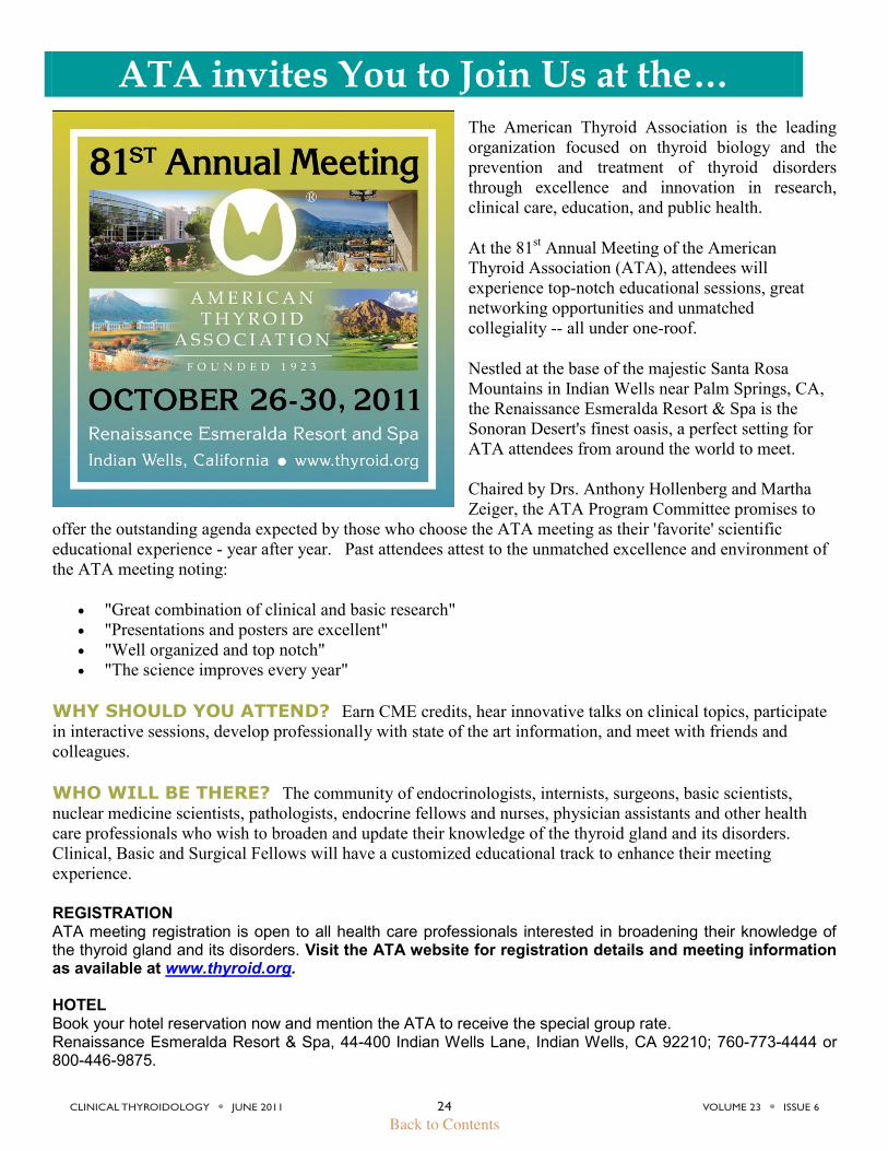
ATA invites You to Join Us at the… The American Thyroid Association is the leading organization focused on thyroid biology and the prevention and treatment of thyroid disorders through excellence and innovation in research, clinical care, education, and public health.
At the 81st Annual Meeting of the American Thyroid Association (ATA), attendees will experience top-notch educational sessions, great networking opportunities and unmatched collegiality -- all under one-roof.
Nestled at the base of the majestic Santa Rosa Mountains in Indian Wells near Palm Springs, CA, the Renaissance Esmeralda Resort & Spa is the Sonoran Desert's finest oasis, a perfect setting for ATA attendees from around the world to meet. Chaired by Drs. Anthony Hollenberg and Martha Zeiger, the ATA Program Committee promises to
offer the outstanding agenda expected by those who choose the ATA meeting as their 'favorite' scientific educational experience - year after year. Past attendees attest to the unmatched excellence and environment of the ATA meeting noting:
"Great combination of clinical and basic research" "Presentations and posters are excellent" "Well organized and top notch" "The science improves every year"
WHY SHOULD YOU ATTEND? Earn CME credits, hear innovative talks on clinical topics, participate in interactive sessions, develop professionally with state of the art information, and meet with friends and colleagues.
WHO WILL BE THERE? The community of endocrinologists, internists, surgeons, basic scientists, nuclear medicine scientists, pathologists, endocrine fellows and nurses, physician assistants and other health care professionals who wish to broaden and update their knowledge of the thyroid gland and its disorders. Clinical, Basic and Surgical Fellows will have a customized educational track to enhance their meeting experience.
REGISTRATION ATA meeting registration is open to all health care professionals interested in broadening their knowledge of the thyroid gland and its disorders. Visit the ATA website for registration details and meeting information as available at www.thyroid.org. HOTEL Book your hotel reservation now and mention the ATA to receive the special group rate. Renaissance Esmeralda Resort & Spa, 44-400 Indian Wells Lane, Indian Wells, CA 92210; 760-773-4444 or 800-446-9875.
ATA • 6066 Leesburg Pike, Suite 550 • Falls Church, VA 22041 • 703 998-8890
Call for Abstracts
www.thyroid.org Call for Abstracts Submission Deadlines
Regular call: Site opens – Wednesday, April 27, 2011 Site closes – Wednesday, June 22, 2011 Acceptance notification – Monday, July 25, 2011
Short call: Site opens – Wednesday, September 7, 2011 Site closes – Wednesday, September 21, 2011 Acceptance notification – Monday, September 26, 2011
ATA Abstract Submission Policy and Responsibilities of the Author: The ATA requests submission of abstracts for consideration at ATA scientific meetings to feature new data presented as posters or oral presentations. The ATA goal is to provide the audience and the media with new data that are unpublished (in print or electronic) which are being publicly presented for the first time. Authors are asked to strictly comply with this requirement; data that are to become available to the public in the setting of a national or international meeting before their presentation at the ATA meeting are not eligible for presentation at the ATA meeting. Data may be submitted for publication before or after abstract submission to the ATA. However, data accepted for publication prior to the ATA meeting would REQUIRE the authors to request the publisher to embargo their publication (electronic and print) until 8:00 am local time the first day of the meeting, or would REQUIRE the authors to withdraw their abstract from the ATA meeting. Many editors are favorable to embargo requests because of the attention that may be drawn to the publication after original presentation of the data at a major meeting. Further, the authors are welcome to announce the date and place of their anticipated publication if known. Authors that do not comply with this policy may be restricted from future abstract submissions for a term to be determined by the ATA Executive Committee. Arbitration, if needed, will occur via the ATA Board of Directors. Abstracts are reviewed in confidence by the ATA program committee with possible ad hoc members.
Additional policies: CHARACTER LIMIT: There is a limit of 2,245 characters (approx. 300 words) for the text of your
submission. Authors of accepted posters are required to be present during the assigned poster sessions. Scientific materials presented at the ATA Annual Meeting must not have been submitted for publication
at the time of abstract submission or presented at a scientific meeting before the 81st Annual Meeting of the ATA (local and regional meetings excluded).
All abstracts must be filed electronically via the American Thyroid Association website www.thyroid.org. Submissions will not be accepted by fax or mail.
All materials must arrive on or before the abstract deadlines noted above. Authorship on multiple abstracts is permitted. Short Call Abstracts Short Call Abstracts are reserved for the presentation of the very latest, important thyroid-related research
with high impact. Submission of a Short Call Abstract does not guarantee acceptance for presentation. (Please note that regular research reports should be submitted by the Regular Abstract deadline.)
Only Six (6) Short Call Abstracts will be selected for 10-minute oral presentations during a special symposium. Selected additional Short Call Abstracts may be presented as special posters. All other submissions will not be published.
Acceptance notices for those selected will be e-mailed on or before September 26, 2011. Online confirmation is required.
CLINICAL THYROIDOLOGY l JUNE 2011 26 VOLUME 23 l ISSUE 6
Back to Contents
American Thyroid Association CME Meet-the-Professor Seminars Online
Register now at www.thyroid.org
Increase your understanding of clinical management of thyroid disease and thyroid cancer. Join the American Thyroid Association (ATA) for monthly educational seminars. Spend one hour a month taking advantage of this unique opportunity to hear top thyroid experts in "Meet-the-Professor" style seminars in the convenience of your office. Earn CME Credit (1 AMA PRA Category 1 Credit™ available per course).
UPCOMING COURSES: Clinical Trials for Progressive Thyroid Carcinoma
Steven I Sherman, MD The University of Texas M. D. Anderson Cancer Center Tuesday, June 21, 2011, 11:00 AM ET 1 CME Credit Available
Traditional cytotoxic chemotherapies have been ineffective and rarely used for the treatment of advanced or progressive thyroid cancers. Recent developments in understanding the biology of thyroid cancers combined with emergence of appropriately targeted therapies are providing exciting new opportunities for patients to be treated. Hear an update on results of the most recent clinical trials from a leader in this field.
Controversies in Surgery for Well Differentiated Thyroid Cancer Ralph P. Tufano, MD, FACS Johns Hopkins University School of Medicine SPECIAL TIME: Tuesday, July 12, 2011, 4:00 PM ET 1 CME Credit Available
Thyroid cancer has the most rapidly rising incidence of all cancers in the United States. The American Thyroid Association Management Guidelines for Patients in Thyroid Nodules and Differentiated Thyroid Cancer have provided recommendations for the extent of surgery based on the best evidence available. Despite these guidelines, controversy exists on what is the appropriate extent of surgery for well differentiated thyroid cancer. This seminar will discuss those controversies and the rational for the ATA guideline recommendations addressing these areas which will include: extent of thyroidectomy as well as indications for and extent of central and lateral neck dissection.
Costs: $119 for ATA members per webinar/$149 for non-members per webinar. Register to participate in the live streaming programs or purchase recordings if you have a schedule conflict with the live broadcast. Target Audience: Endocrinologists, internists, surgeons, basic scientists, nuclear medicine scientists, pathologists, endocrine and surgery fellows, nurses, physician assistants and other health care professionals who wish to broaden and update their knowledge of the thyroid gland and its disorders. Disclosures: Disclosures for presenting faculty and content controllers will be provided to attendees on-screen at the live webinar activity. Accreditation and Designation Statement: intellyst® Medical Education is accredited by the Accreditation Council for Continuing Medical Education to provide continuing medical education for physicians. intellyst® Medical Education designates each of these live activities for a maximum of 1 AMA PRA Category 1 Credit™. Physicians should only claim credit commensurate with the extent of their participation in the activity.
(703) 998-8890 www.thyroid.org [email protected] Dedicated to scientific inquiry, clinical excellence, public service, education & collaboration