Embed Size (px)
Citation preview

Clinical Guide toCardiology
The Clinical Guides Series
Series Editor: Christian Camm
The Clinical Guides are a brand new resource for junior doctors and medical students.
They provide practical and concise day-to-day information on common conditions, symp-toms and problems faced in the clinical environment. They are easy to navigate and allowswift access to information as it is needed, with step-by-step guidance on decision mak-ing, investigations and interventions, and how to survive and thrive on clinical rotationand attachment.
This new title is also available as an e-book.For more details, please seewww.wiley.com/buy/9781118755334or scan this QR code:
Clinical Guide toCardiology
Edited by
Christian F. CammJohn Radcliffe Hospital, Oxford, UK
A. John CammSt. George’s University of London, London, UK
This edition first published 2016 © 2016 by John Wiley & Sons, Ltd
Registered office: John Wiley & Sons, Ltd, The Atrium, Southern Gate, Chichester, West Sussex, PO19 8SQ, UK
Editorial offices: 9600 Garsington Road, Oxford, OX4 2DQ, UKThe Atrium, Southern Gate, Chichester, West Sussex, PO19 8SQ, UK111 River Street, Hoboken, NJ 07030-5774, USA
For details of our global editorial offices, for customer services and for information about how to apply for permission toreuse the copyright material in this book please see our website at www.wiley.com/wiley-blackwell
The right of the authors to be identified as the authors of this work has been asserted in accordance with the UKCopyright, Designs and Patents Act 1988.
All rights reserved. No part of this publication may be reproduced, stored in a retrieval system, or transmitted, in any formor by any means, electronic, mechanical, photocopying, recording or otherwise, except as permitted by the UK Copyright,Designs and Patents Act 1988, without the prior permission of the publisher.
Designations used by companies to distinguish their products are often claimed as trademarks. All brand names andproduct names used in this book are trade names, service marks, trademarks or registered trademarks of their respectiveowners. The publisher is not associated with any product or vendor mentioned in this book. It is sold on the understandingthat the publisher is not engaged in rendering professional services. If professional advice or other expert assistance isrequired, the services of a competent professional should be sought.
The contents of this work are intended to further general scientific research, understanding, and discussion only and arenot intended and should not be relied upon as recommending or promoting a specific method, diagnosis, or treatment byhealth science practitioners for any particular patient. The publisher and the author make no representations or warrantieswith respect to the accuracy or completeness of the contents of this work and specifically disclaim all warranties, includingwithout limitation any implied warranties of fitness for a particular purpose. In view of ongoing research, equipmentmodifications, changes in governmental regulations, and the constant flow of information relating to the use of medicines,equipment, and devices, the reader is urged to review and evaluate the information provided in the package insert orinstructions for each medicine, equipment, or device for, among other things, any changes in the instructions or indicationof usage and for added warnings and precautions. Readers should consult with a specialist where appropriate. The factthat an organization or Website is referred to in this work as a citation and/or a potential source of further informationdoes not mean that the author or the publisher endorses the information the organization or Website may provide orrecommendations it may make. Further, readers should be aware that Internet Websites listed in this work may havechanged or disappeared between when this work was written and when it is read. No warranty may be created orextended by any promotional statements for this work. Neither the publisher nor the author shall be liable for any damagesarising herefrom.
Library of Congress Cataloging-in-Publication Data
Clinical guide to cardiology / edited by Christian F. Camm, A. John Camm.p. ; cm.
Includes bibliographical references and index.ISBN 978-1-118-75533-4 (pbk.)I. Camm, Christian F. (Christian Fielder), editor. II. Camm, A. John, editor.[DNLM: 1. Heart Diseases–diagnosis. 2. Heart Diseases–therapy. WG 141]RC682616.1′2–dc23
2015025343
A catalogue record for this book is available from the British Library.
Wiley also publishes its books in a variety of electronic formats. Some content that appears in print may not be available inelectronic books.
Cover image: iStockphoto/© Nerthuz
Set in 8.5/10.5pt Frutiger Light by Aptara Inc., New Delhi, India
1 2016
Contents
Contributors, vii
Acronyms and Abbreviations, viii
About the Companion Website, xiv
PART 1 Examination Techniques, 1
1. Examination Techniques, 3
PART 2 Approach to Presenting Complaints, 11
2. Chest Pain, 13
3. Shortness of Breath, 28
4. Loss of Consciousness, 47
5. Palpitations, 62
6. Cardiac Murmurs, 72
7. Shock, 92
8. Oedema, 106
PART 3 Conditions, 117
9. Acute Coronary Syndrome, 119
10. Stable Angina, 138
11. Heart Failure, 147
12. Infective Endocarditis, 160
13. Arrhythmias, 172
14. Valvular Heart Disease, 193
15. Cardiomyopathy, 214
16. Hypertension, 232
17. Pericardial Disease, 245
18. Congenital Heart Disease, 261
PART 4 Imaging, 277
19. Electrocardiogram, 279
20. Transoesophageal Echocardiogram, 293
21. Trans-Thoracic Echocardiogram, 299
v
vi Contents
22. Cardiac MRI, 306
23. Cardiac CT, 313
24. Cardiac Catheterization, 320
PART 5 Interventional Therapies, 327
25. Pacemakers and Implantable Cardiac Defibrillators, 329
26. Percutaneous Coronary Intervention and Angioplasty, 338
27. Valvuloplasty, 348
28. Transcatheter Aortic Valve Implantation, 352
29. Cardiac Ablation, 359
PART 6 Pharmacology, 365
30. Anti-Arrhythmic Agents, 367
31. Beta-Blockers, 375
32. Calcium-Channel Blockers, 380
33. Nitrates, 385
34. Drugs Targeting the Angiotensin Axis, 389
35. Diuretics, 393
36. Anticoagulants, 399
37. Antiplatelets, 409
38. Lipid Regulation, 414
Index, 417
Contributors
Laura Ah-KyeKing’s College Hospital NHS Foundation Trust,London, UK
Kristopher BennettWhipps Cross Hospital, London, UK
Christian F. CammJohn Radcliffe Hospital, Oxford, UK
Lucy CarpenterBarts Health NHS Trust, London, UK
Yang ChenImperial College Healthcare NHS Trust,London, UK
Ji-Jian ChowImperial College Healthcare NHS Trust,London, UK
James CranleyPapworth Hospital NHS Foundation Trust,Cambridge, UK
George DaviesOxford University Hospitals NHS Trust, Oxford, UK
Akshay GargKing’s College Hospital NHS Foundation Trust,London, UK
Harminder S. GillKing’s College Hospital NHS Foundation Trust,London, UK
Katie GloverGuy’s and St Thomas’ NHS Foundation Trust,London, UK
Stephanie HicksKing’s College Hospital NHS Foundation Trust,London, UK
Fritz-Patrick JahnsKing’s College Hospital NHS Foundation Trust,London, UK
Sophie MaxwellWalsall Manor Hospital, Walsall, UK
Blair MerrickHammersmith Hospital, London, UK
Madeline MooreKing’s College Hospital NHS Foundation Trust,London, UK
Sarah MorrowChelsea and Westminster Hospital, London,UK
Rahul K. MukherjeeKing’s College Hospital NHS Foundation Trust,London, UK
Anna RobinsonKing’s College Hospital NHS Foundation Trust,London, UK
Arvind SinghalChelsea and Westminster Hospital NHSFoundation Trust, London, UK
Nicholas SunderlandKing’s College Hospital NHS Foundation Trust,London, UK
Anneline te RieleUniversity Medical Centre, Utrecht,the Netherlands
Maria TsakokHammersmith Hospital, London, UK
Robert A. WatsonImperial College Healthcare NHS Trust,London, UK
vii
Acronyms and Abbreviations
2D two-dimensional3D three-dimensionalA2 aortic valve component of heart sound 2AAA abdominal aortic aneurysmABG arterial blood gasABPM ambulatory blood pressure monitoringACC American College of CardiologyACE angiotensin-converting enzymeACEi angiotensin-converting enzyme inhibitorACR albumin:creatinine ratioACS acute coronary syndromeACTH adrenocorticotropic hormoneADP-P2Y adenosine diphosphate-P2Y receptorAF atrial fibrillationAHA American Heart AssociationAI angiotensin IAII angiotensin IIAKI acute kidney injuryALD alcoholic liver diseaseALP alkaline phosphataseALS advanced life supportAMA American Medical AssociationAMB acute marginal branchAMTS Abbreviated Mental Test scoreAPS antiphospholipid syndromeaPTT activated partial thromboplastin timeAR aortic regurgitationARB angiotensin-II receptor blockerARDS acute respiratory distress syndromeAS aortic stenosisASD atrial septal defectAST aspartate aminotransferaseATP adenosine triphosphateAV atrioventricularAVN atrioventricular nodeAVNRT AV-nodal re-entrant tachycardiaAVPU alert/responsive to voice/responsive to pain/unresponsiveAVRT atrioventricular re-entrant tachycardiaAVSD atrioventricular septal defectBAH bilateral adrenocortical hyperplasiaBAV balloon aortic valvuloplastyBCS British Cardiovascular SocietyBD twice a dayBE base excessbeta-hCG beta human chorionic gonadotrophinBM Boehringer-Mannheim – capillary glucose testBMI body mass indexBNP brain natriuretic peptide
viii
Acronyms and Abbreviations ix
BP blood pressureBPH benign prostatic hyperplasiabpm beats per minuteCABG coronary artery bypass graftCAC coronary artery calciumCAD coronary artery diseaseCath Lab (coronary) catheterization laboratoryCCB calcium-channel blockerCCF congestive cardiac failureCCP cyclic citrullinated peptideCCU cardiac care unitCK-MB creatine kinase – MB isoformCKD chronic kidney diseaseCMV cytomegalovirusCN coagulase negativeCNS central nervous systemCO cardiac outputCoA coarctation of the aortaCOPD chronic obstructive pulmonary diseaseCOX cyclo-oxygenaseCPAP continuous positive airway pressureCPR cardiopulmonary resuscitationCRP C-reactive proteinCRT cardiac resynchronization therapyCRT-D cardiac resynchronization therapy + cardiac defibrillatorCT computed tomographyCTPA computed tomography pulmonary angiogramCTR cardiothoracic ratioCV(S) cardiovascular (system)CVA cerebrovascular accidentCVD cerebrovascular diseaseCVP central venous pressureCXA X-ray coronary angiographyCXR chest X-rayDAPT dual anti-platelet therapyDC direct currentDCM dilated cardiomyopathyDH drug historyDHP dihydropyridineDKA diabetic ketoacidosisDM diabetes mellitusDVLA Driver and Vehicle Licensing AgencyDVT deep vein thrombosisEBV Epstein–Barr virusECG electrocardiogramecho echocardiogramED emergency departmentEDV end-diastolic volumeEEG electroencephalogramEF ejection fractionEGDT early goal-directed therapyeGFR estimated glomerular filtration rateELR external loop recorderEPS electrophysiological studyESC European Society of Cardiology
x Acronyms and Abbreviations
ESM ejection systolic murmurESR erythrocyte sedimentation rateESV end-systolic volumeEVAR endovascular aneurysm repairFAST focused assessment with sonography for traumaFBC full blood countFFP fresh frozen plasmaFFR fractional flow reserveFH family historyFY2 foundation year 2 doctorG6PD glucose-6-phosphate dehydrogenaseGCS Glasgow coma scaleGFR glomerular filtration rateGI gastrointestinalGMP guanosine monophosphateGORD gastro-oesophageal reflux diseaseGP general practitionerGRA glucorticoid-remediable aldosteronismGRACE Global Registry of Acute Coronary EventsGTN glyceryl trinitrateGZA glycyrrhizic acidHACEK organisms associated with culture-negative infective endocarditisHb haemoglobinHbA1c glycated haemoglobinHBPM home blood pressure monitoringHCG human chorionic gonadotrophinHCM hypertrophic cardiomyopathyHDL high density lipoproteinHDU high dependency unitHF heart failureHF-PEF heart failure with preserved ejection fractionHF-REF heart failure with reduced ejection fractionHIT heparin-induced thrombocytopeniaHIV human immunodeficiency virusHOCM hypertrophic obstructive cardiomyopathyHPC history of the presenting complaintHR heart rateHTN hypertensionIABP intra-aortic balloon pumpIC intercostalICD implantable cardioverting defibrillatorICH intracerebral haemorrhageIE infective endocarditisIGG immunoglobulin GIHD ischaemic heart diseaseILR internal loop recorderIM intramuscularINR international normalized ratioISMN isosorbide mononitrateITU intensive therapy unitIV intravenousIVCD intraventricular conduction delayIVDU intravenous drug userIVUS intravascular ultrasoundJVP jugular venous pulse/pressure
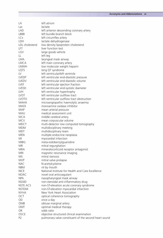
Acronyms and Abbreviations xi
LA left atriumLac lactateLAD left anterior descending coronary arteryLBBB left bundle branch blockLCx left circumflex arteryLDH lactate dehydrogenaseLDL cholesterol low density lipoprotein cholesterolLFT liver function testLGV large goods vehicleLL left legLMA laryngeal mask airwayLMCA left main coronary arteryLMWH low molecular weight heparinLQTS long QT syndromeLV left ventricular/left ventricleLVEDP left ventricular end-diastolic pressureLVEDV left ventricular end-diastolic volumeLVEF left ventricular ejection fractionLVESD left ventricular end-systolic diameterLVH left ventricular hypertrophyLVOT left ventricular outflow tractLVOTO left ventricular outflow tract obstructionMAHA microangiopathic haemolytic anaemiaMAOI monoamine oxidase inhibitorMAP mean arterial pressureMAU medical assessment unitMCA middle cerebral arteryMCV mean corpuscular volumeMDCT multi-detector row computed tomographyMDM multidisciplinary meetingMDT multidisciplinary teamMEN multiple endocrine neoplasiaMI myocardial infarctionMIBG meta-iodobenzylguanidineMR mitral regurgitationMRA mineralocorticoid receptor antagonistMRI magnetic resonance imagingMS mitral stenosisMVP mitral valve prolapseNAC N-acetylcysteineNBM nil by mouthNICE National Institute for Health and Care ExcellenceNOAC novel oral anticoagulantNPA nasopharyngeal mask airwayNSAID non-steroidal anti-inflammatory drugNSTE ACS non-ST-elevation acute coronary syndromeNSTEMI non-ST-elevation myocardial infarctionNYHA New York Heart AssociationOCT optical coherence tomographyOD once a dayOMB obtuse marginal arteryOMT optimal medical therapyOR odds ratioOSCE objective structured clinical examinationP2 pulmonary valve constituent of the second heart sound
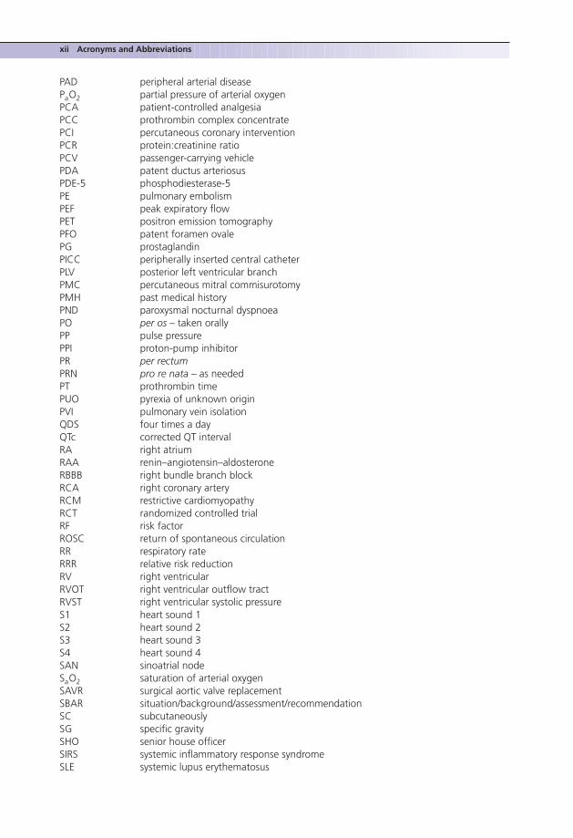
xii Acronyms and Abbreviations
PAD peripheral arterial diseasePaO2 partial pressure of arterial oxygenPCA patient-controlled analgesiaPCC prothrombin complex concentratePCI percutaneous coronary interventionPCR protein:creatinine ratioPCV passenger-carrying vehiclePDA patent ductus arteriosusPDE-5 phosphodiesterase-5PE pulmonary embolismPEF peak expiratory flowPET positron emission tomographyPFO patent foramen ovalePG prostaglandinPICC peripherally inserted central catheterPLV posterior left ventricular branchPMC percutaneous mitral commisurotomyPMH past medical historyPND paroxysmal nocturnal dyspnoeaPO per os – taken orallyPP pulse pressurePPI proton-pump inhibitorPR per rectumPRN pro re nata – as neededPT prothrombin timePUO pyrexia of unknown originPVI pulmonary vein isolationQDS four times a dayQTc corrected QT intervalRA right atriumRAA renin–angiotensin–aldosteroneRBBB right bundle branch blockRCA right coronary arteryRCM restrictive cardiomyopathyRCT randomized controlled trialRF risk factorROSC return of spontaneous circulationRR respiratory rateRRR relative risk reductionRV right ventricularRVOT right ventricular outflow tractRVST right ventricular systolic pressureS1 heart sound 1S2 heart sound 2S3 heart sound 3S4 heart sound 4SAN sinoatrial nodeSaO2 saturation of arterial oxygenSAVR surgical aortic valve replacementSBAR situation/background/assessment/recommendationSC subcutaneouslySG specific gravitySHO senior house officerSIRS systemic inflammatory response syndromeSLE systemic lupus erythematosus
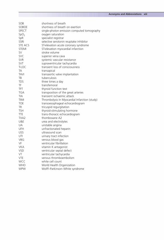
Acronyms and Abbreviations xiii
SOB shortness of breathSOBOE shortness of breath on exertionSPECT single-photon emission computed tomographySpO2 oxygen saturationSpR specialist registrarSSRI selective serotonin reuptake inhibitorSTE ACS ST-elevation acute coronary syndromeSTEMI ST-elevation myocardial infarctionSV stroke volumeSVC superior vena cavaSVR systemic vascular resistanceSVT supraventricular tachycardiaT-LOC transient loss of consciousnessTA transapicalTAVI transaortic valve implantationTB tuberculosisTDS three times a dayTF transfemoralTFT thyroid function testTGA transposition of the great arteriesTIA transient ischaemic attackTIMI Thrombolysis In Myocardial Infarction (study)TOE transoesophageal echocardiogramTR tricuspid regurgitationTSH thyroid-stimulating hormoneTTE trans-thoracic echocardiogramTXA2 thomboxane A2U&E urea and electrolytesUA unstable anginaUFH unfractionated heparinUSS ultrasound scanUTI urinary tract infectionVBG venous blood gasVF ventricular fibrillationVKA vitamin K antagonistVSD ventricular septal defectVT ventricular tachycardiaVTE venous thromboembolismWCC white cell countWHO World Health OrganizationWPW Wolff–Parkinson–White syndrome
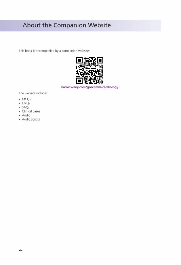
About the Companion Website
This book is accompanied by a companion website:
www.wiley.com/go/camm/cardiology
The website includes:
� MCQs� EMQs� SAQs� Clinical cases� Audio� Audio scripts
xiv

PART 1
Examination Techniques

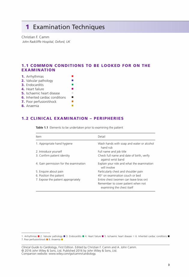
1 Examination TechniquesChristian F. CammJohn Radcliffe Hospital, Oxford, UK
1.1 COMMON CONDITIONS TO BE LOOKED FOR ON THEEXAMINATION
1. Arrhythmias �
2. Valvular pathology �
3. Endocarditis �
4. Heart failure �
5. Ischaemic heart disease �
6. Inherited cardiac conditions �
7. Poor perfusion/shock �
8. Anaemia �
1.2 CLINICAL EXAMINATION – PERIPHERIES
Table 1.1 Elements to be undertaken prior to examining the patient
Item Detail
1. Appropriate hand hygiene Wash hands with soap and water or alcoholhand rub
2. Introduce yourself Full name and job title3. Confirm patient identity Check full name and date of birth, verify
against wrist band4. Gain permission for the examination Explain your role and what the examination
will involve5. Enquire about pain Particularly chest and shoulder pain6. Position the patient 45◦ on examination couch or bed7. Expose the patient appropriately Entire chest (women can leave bras on)
Remember to cover patient when notexamining the chest itself
1. Arrhythmias � 2. Valvular pathology � 3. Endocarditis � 4. Heart failure � 5. Ischaemic heart disease � 6. Inherited cardiac conditions �7. Poor perfusion/shock � 8. Anaemia �
3
Clinical Guide to Cardiology, First Edition. Edited by Christian F. Camm and A. John Camm.© 2016 John Wiley & Sons, Ltd. Published 2016 by John Wiley & Sons, Ltd.Companion website: www.wiley.com/go/camm/cardiology.
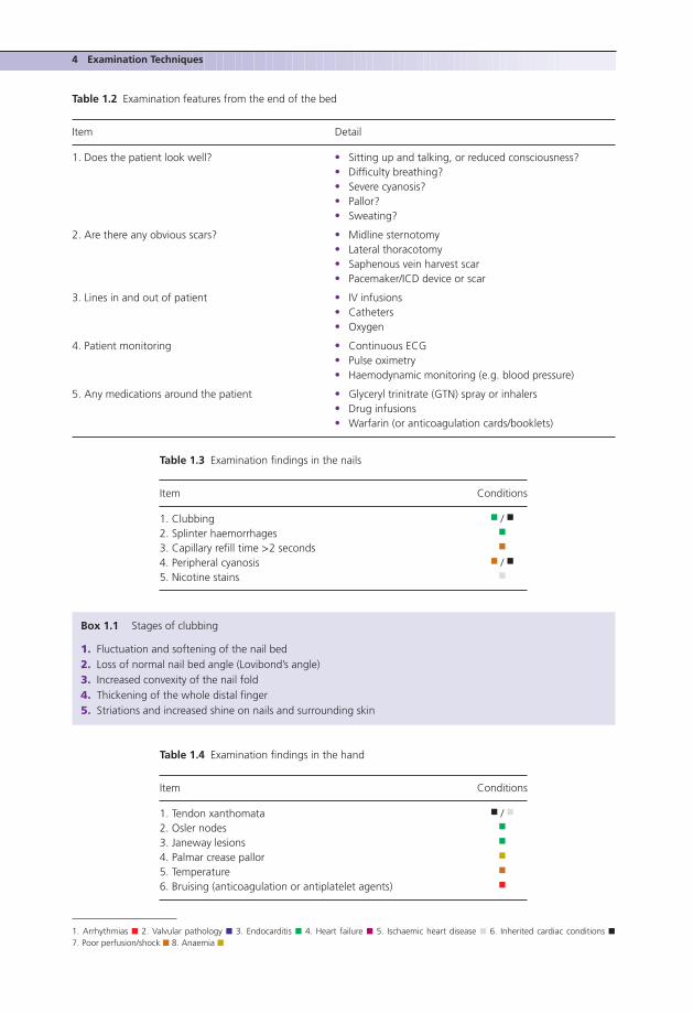
4 Examination Techniques
Table 1.2 Examination features from the end of the bed
Item Detail
1. Does the patient look well? � Sitting up and talking, or reduced consciousness?� Difficulty breathing?� Severe cyanosis?� Pallor?� Sweating?
2. Are there any obvious scars? � Midline sternotomy� Lateral thoracotomy� Saphenous vein harvest scar� Pacemaker/ICD device or scar
3. Lines in and out of patient � IV infusions� Catheters� Oxygen
4. Patient monitoring � Continuous ECG� Pulse oximetry� Haemodynamic monitoring (e.g. blood pressure)
5. Any medications around the patient � Glyceryl trinitrate (GTN) spray or inhalers� Drug infusions� Warfarin (or anticoagulation cards/booklets)
Table 1.3 Examination findings in the nails
Item Conditions
1. Clubbing � / �
2. Splinter haemorrhages �
3. Capillary refill time >2 seconds �
4. Peripheral cyanosis � / �
5. Nicotine stains �
Box 1.1 Stages of clubbing
1. Fluctuation and softening of the nail bed2. Loss of normal nail bed angle (Lovibond’s angle)3. Increased convexity of the nail fold4. Thickening of the whole distal finger5. Striations and increased shine on nails and surrounding skin
Table 1.4 Examination findings in the hand
Item Conditions
1. Tendon xanthomata � / �
2. Osler nodes �
3. Janeway lesions �
4. Palmar crease pallor �
5. Temperature �
6. Bruising (anticoagulation or antiplatelet agents) �
1. Arrhythmias � 2. Valvular pathology � 3. Endocarditis � 4. Heart failure � 5. Ischaemic heart disease � 6. Inherited cardiac conditions �7. Poor perfusion/shock � 8. Anaemia �
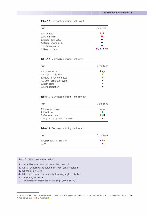
Examination Techniques 5
Table 1.5 Examination findings in the wrist
Item Conditions
1. Pulse rate � / �
2. Pulse rhythm �
3. Radio-radial delay �
4. Radio-femoral delay �
5. Collapsing pulse �
6. Blood pressure � / � / � / �
Table 1.6 Examination findings in the eyes
Item Conditions
1. Corneal arcus �/age2. Conjunctival pallor �
3. Petechial haemorrhages �
4. Xanthelasma over eyelids �
5. Roth spots �
6. Lens dislocation �
Table 1.7 Examination findings in the mouth
Item Conditions
1. Hydration status general2. Dentition �
3. Central cyanosis � / �
4. High arched palate (Marfan’s) �
Table 1.8 Examination findings in the neck
Item Conditions
1. Carotid pulse – character � / �
2. JVP �
Box 1.2 How to examine the JVP
1. Located between heads of sternocleidomastoid2. JVP has double pulse (rather than single found in carotid)3. JVP can be occluded4. JVP may be made more visible by lowering angle of the bed5. Hepato-jugular reflux6. Height measured from the sternal angle (angle of Louis)
1. Arrhythmias � 2. Valvular pathology � 3. Endocarditis � 4. Heart failure � 5. Ischaemic heart disease � 6. Inherited cardiac conditions �7. Poor perfusion/shock � 8. Anaemia �
6 Examination Techniques
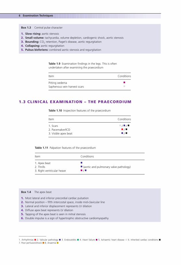
Box 1.3 Central pulse character
1. Slow rising: aortic stenosis2. Small volume: tachycardia, volume depletion, cardiogenic shock, aortic stenosis3. Bounding: CO2 retention, Paget’s disease, aortic regurgitation4. Collapsing: aortic regurgitation5. Pulsus bisferiens: combined aortic stenosis and regurgitation
Table 1.9 Examination findings in the legs. This is oftenundertaken after examining the praecordium
Item Conditions
Pitting oedema �
Saphenous vein harvest scars �
1.3 CLINICAL EXAMINATION – THE PRAECORDIUM
Table 1.10 Inspection features of the praecordium
Item Conditions
1. Scars � / � / �
2. Pacemaker/ICD � / �
3. Visible apex beat � / �
Table 1.11 Palpation features of the praecordium
Item Conditions
1. Apex beat �
2. Thrills � (aortic and pulmonary valve pathology)3. Right ventricular heave � / �
Box 1.4 The apex beat
1. Most lateral and inferior precordial cardiac pulsation2. Normal position – fifth intercostal space, inside mid-clavicular line3. Lateral and inferior displacement represents LV dilation4. Diffuse apex beat represents LV dilation5. Tapping of the apex beat is seen in mitral stenosis6. Double impulse is a sign of hypertrophic obstructive cardiomyopathy
1. Arrhythmias � 2. Valvular pathology � 3. Endocarditis � 4. Heart failure � 5. Ischaemic heart disease � 6. Inherited cardiac conditions �7. Poor perfusion/shock � 8. Anaemia �
Examination Techniques 7
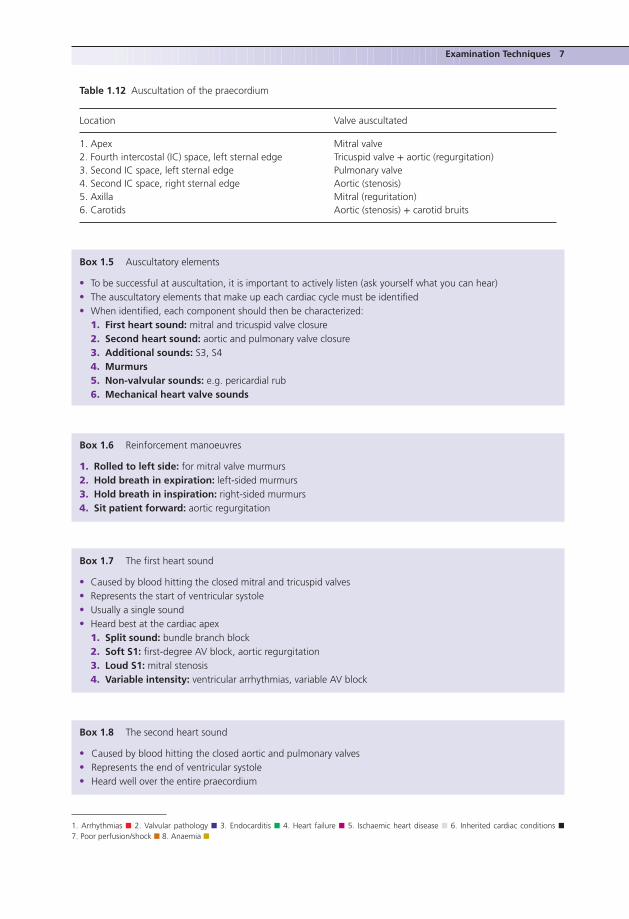
Table 1.12 Auscultation of the praecordium
Location Valve auscultated
1. Apex Mitral valve2. Fourth intercostal (IC) space, left sternal edge Tricuspid valve + aortic (regurgitation)3. Second IC space, left sternal edge Pulmonary valve4. Second IC space, right sternal edge Aortic (stenosis)5. Axilla Mitral (reguritation)6. Carotids Aortic (stenosis) + carotid bruits
Box 1.5 Auscultatory elements
� To be successful at auscultation, it is important to actively listen (ask yourself what you can hear)� The auscultatory elements that make up each cardiac cycle must be identified� When identified, each component should then be characterized:
1. First heart sound: mitral and tricuspid valve closure2. Second heart sound: aortic and pulmonary valve closure3. Additional sounds: S3, S44. Murmurs5. Non-valvular sounds: e.g. pericardial rub6. Mechanical heart valve sounds
Box 1.6 Reinforcement manoeuvres
1. Rolled to left side: for mitral valve murmurs2. Hold breath in expiration: left-sided murmurs3. Hold breath in inspiration: right-sided murmurs4. Sit patient forward: aortic regurgitation
Box 1.7 The first heart sound
� Caused by blood hitting the closed mitral and tricuspid valves� Represents the start of ventricular systole� Usually a single sound� Heard best at the cardiac apex
1. Split sound: bundle branch block2. Soft S1: first-degree AV block, aortic regurgitation3. Loud S1: mitral stenosis4. Variable intensity: ventricular arrhythmias, variable AV block
Box 1.8 The second heart sound
� Caused by blood hitting the closed aortic and pulmonary valves� Represents the end of ventricular systole� Heard well over the entire praecordium
1. Arrhythmias � 2. Valvular pathology � 3. Endocarditis � 4. Heart failure � 5. Ischaemic heart disease � 6. Inherited cardiac conditions �7. Poor perfusion/shock � 8. Anaemia �
8 Examination Techniques
� Usually a split sound on inspiration� Pulmonary component follows aortic
1. Widely split: right bundle branch block2. Fixed splitting: atrial septal defects3. Soft aortic component: aortic stenosis
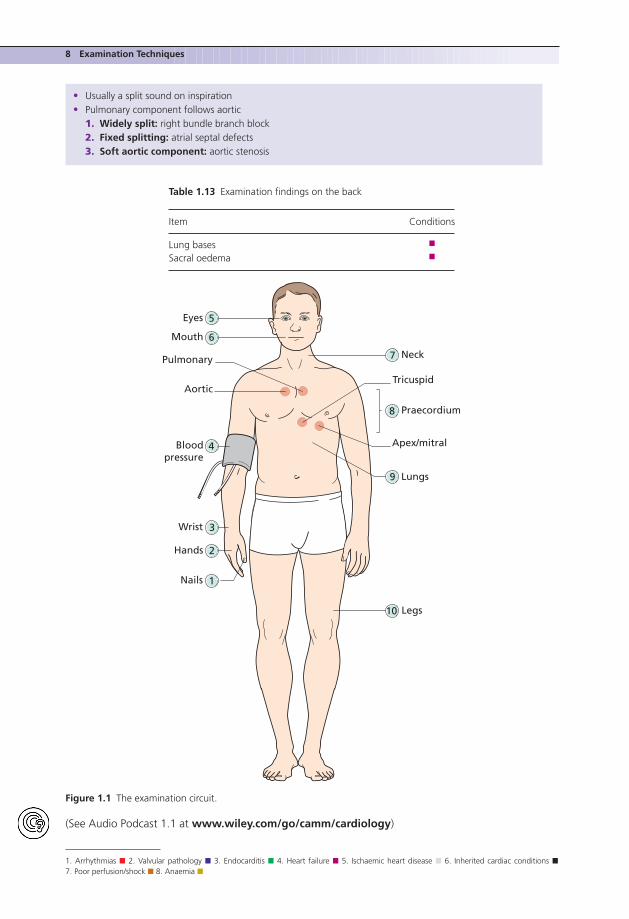
Table 1.13 Examination findings on the back
Item Conditions
Lung bases �
Sacral oedema �
1Nails
2Hands
3Wrist
5Eyes
Neck
6
7
Mouth
Lungs
8
9
Praecordium
10 Legs
Tricuspid
Pulmonary
Aortic
Apex/mitral4Bloodpressure
Figure 1.1 The examination circuit.
(See Audio Podcast 1.1 at www.wiley.com/go/camm/cardiology)
1. Arrhythmias � 2. Valvular pathology � 3. Endocarditis � 4. Heart failure � 5. Ischaemic heart disease � 6. Inherited cardiac conditions �7. Poor perfusion/shock � 8. Anaemia �
Examination Techniques 9
1.4 HOW TO PRESENT YOUR FINDINGS
Safety first approachDetails� An approach that works well when not sure of your findings� Useful for objective structured clinical examinations (OSCEs) to ensure that information is not missed� Discuss the positive findings (and key negatives) in the order that you examined� Give a potential diagnosis after presenting findings
ExampleI examined this 52-year-old patient. He presented with shortness of breath and leg swelling. On inspec-tion he was clearly dyspnoeic but otherwise appeared well. He was alert. There was a well healed midlinesternotomy scar. His pulse was regular at 80 bpm. His blood pressure was 110/80 mmHg. The patient waswell hydrated. The JVP was raised by 8 cm. There were no additional peripheral signs elucidated. On thepraecordium he had no additional scars. His apex beat was not inappropriately located. On auscultationS1 and S2 were both heard. Additionally a third heart sound was heard across the praecordium. Therewere no additional sounds. There were inspiratory crackles at the lung bases and some sacral oedema.A clear scar along the course of the long saphenous vein was seen on the left leg, this was combinedwith bilateral pitting oedema reaching the mid-calf.
In conclusion, this patient presents with shortness of breath and signs suggestive of heart failure.
Ward-round basedDetails� An approach to be used when you are confident or pressed for time� Give your suspected diagnosis first� Discuss the examination findings that support the diagnosis and help to exclude others� Discuss findings in the order of most supportive to least supportive of your diagnosis
ExampleI examined this 52-year-old patient. He presented with shortness of breath and leg swelling. Examinationrevealed a patient with a clinical picture of congestive heart failure. This was supported by findings ofinspiratory crackles at the lung bases, pitting oedema in the sacral region and bilaterally in the legs up tothe mid-calf level. In addition, the JVP was raised to 8 cm above the angle of Louis. On auscultation S1and S2 were clearly heard with the addition of a third heart sound. The patient has a history of coronaryartery bypass surgery as supported by the midline sternotomy scar and long saphenous vein graft scaron the left leg. Given these findings, this suggests a history of heart failure potentially secondary toischaemic heart disease.
10 Examination Techniques
1.5 EPONYMOUS SIGNS AND SYMPTOMS
Table 1.14 Eponymous signs in cardiology
Eponym Details
Austin Flint murmur Low-pitched rumbling murmur in mid-diastole due to aortic regurgitationcausing mitral stenosis
Beck’s triad Three signs associated with cardiac tamponade:i. Low arterial blood pressure
ii. Distended neck veinsiii. Muffled heart sounds
Corrigan’s pulse A large-volume pulse which collapses away due to aortic regurgitation –observed at the carotid
De Musset’s sign Rhythmic nodding of the head due to increased pulse pressure in aorticregurgitation
Duroziez’s sign Compression of the femoral artery with the bell of the stethoscope leads to anaudible diastolic murmur – aortic regurgitation
Ewart’s sign Collection of signs at the left lung base due to pericardial effusion:i. ‘Woody’ dullness to percussion
ii. Increased vocal resonanceiii. Bronchial breath sounds
Friedreich’s sign Significant drop in JVP during the diastolic phase due to constrictive pericarditisGraham Steell murmur Pulmonary regurgitant murmur heard in the left 2nd intercostal spaceJaneway lesions Non-tender, small erythematous nodular lesions on the palms/soles indicative
of endocarditisKussmaul’s sign Paradoxical rise in JVP on inspiration, indicative of reduced right ventricular
filling (e.g. right heart failure or constrictive pericarditis)Mayne’s sign A drop >15 mmHg in diastolic blood pressure when the arm is raised – aortic
regurgitationMuller’s sign Bobbing of the uvula due to wide pulse pressure of aortic regurgitationOliver’s sign Downward tug of the trachea during systole – aneurysm of the aortic archOsler nodes Painful, raised lesions on the hands/feet caused by immune complex deposition
and suggestive of infective endocarditisOsler’s sign Falsely elevated blood pressure due to calcification of the vesselsQuinke’s pulse Alternating blushing and blanching of the fingernails – aortic regurgitationRoth spots Retinal haemorrhages with a pale fibrin centre caused by immune complex
deposition and suggestive of infective endocarditisStill’s murmur Innocent flow murmurWatson’s waterhammer pulse As with Corrigan’s pulse, but observed over the radial artery
For additional resources and to test your knowledge, visit the companion website at:
www.wiley.com/go/camm/cardiology

PART 2
Approach to Presenting Complaints

2 Chest PainMaria TsakokHammersmith Hospital, London, UK
2.1 DEFINITION
Any pain or discomfort that is felt to originate in and around the thorax.
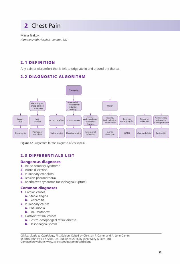
2.2 DIAGNOSTIC ALGORITHM
Chest pain
Pleuritic pain:sharp pain on
breathing
Cough,SOB
PneumoniaPulmonaryembolism
SOB,cyanosis
Myocardial:- retrosternal- radiation- crushing
Occurs on effort
Stable angina Unstable anginaMyocardialinfarction
Aorticdissection
GORD Musculoskeletal Pericarditis
Occurs at rest
Severeprolonged pain,
autonomicfeatures
Other
Tearing,back radiation,sudden onset
Burning,worse lying flat
Tender topalpation
Central pain,relieved on
leaning forward
Figure 2.1 Algorithm for the diagnosis of chest pain.
2.3 DIFFERENTIALS LIST
Dangerous diagnoses1. Acute coronary syndrome2. Aortic dissection3. Pulmonary embolism4. Tension pneumothorax5. Boerhaave’s syndrome (oesophageal rupture)
Common diagnoses1. Cardiac causes
a. Stable anginab. Pericarditis
2. Pulmonary causesa. Pneumoniab. Pneumothorax
3. Gastrointestinal causesa. Gastro-oesophageal reflux diseaseb. Oesophageal spasm
13
Clinical Guide to Cardiology, First Edition. Edited by Christian F. Camm and A. John Camm.© 2016 John Wiley & Sons, Ltd. Published 2016 by John Wiley & Sons, Ltd.Companion website: www.wiley.com/go/camm/cardiology.
14 Chest Pain
4. Musculoskeletal causesa. Rib contusions/fracturesb. Intercostal muscle strainsc. Costochondritis (including Tietze and Bornholm syndromes)
Diagnoses to consider1. Psychiatric causes2. Herpes zoster
2.4 KEY HISTORY FEATURES
(See Audio Podcast 2.1 at www.wiley.com/go/camm/cardiology)
Dangerous diagnosis 1Diagnosis: Acute coronary syndrome
Questionsa. Is the pain crushing or heavy in nature?
These are the typical descriptions, but the pain may also be described as tight, gripping or pressing.
b. Does the pain radiate to the left arm or jaw?
These distinctive sites of radiation are highly suggestive of myocardial pain.
c. Are there associated autonomic symptoms?
Commonly nausea/vomiting and sweating.
d. Are there any cardiac risk factors?
See Box 2.1.
Box 2.1 Cardiac risk factors
Non-modifiable:
1. Increasing age2. Male gender3. Family history4. Previous cardiovascular events5. Diabetes
Modifiable:
1. Smoking2. Hypertension3. Obesity4. Low physical activity
Dangerous diagnosis 2Diagnosis: Aortic dissection
Questionsa. Is the pain tearing, central and extremely severe?
Interscapular when involving the descending aorta, anterior when involving the ascending aorta.
b. Does the pain radiate through to the back?
The pain may also radiate to the abdomen; these sites help distinguish dissection from ACS.
c. Sudden onset?
The pain occurs very suddenly, as the layers of the aorta are rapidly forced apart.