Embed Size (px)
Citation preview

Clinical Guidance
Acute Kidney Injury Summary This guideline outlines the management of patients with acute kidney injury from risk assessment and initial management to referral and transfer criteria. It was developed by the London AKI network which has representation from Renal, General Medicine and Critical Care from the major hospitals in London and their referring hospitals. The development and launch was supported by NHS Kidney Care. The aim is to streamline the management of patients with AKI across London which will not only improve care but also education and training. In addition, it offers an opportunity for joint audits.
Document Detail Document type Clinical Guideline Document name Acute Kidney Injury Document location GTi Clinical Guidance Database Version 1.0 Effective from March 2012 Review date March 2015 Owner Clinical Lead, Nephrology
Author(s) Marlies Ostermann, Consultant in Nephrology and Intensive Care Taryn Pile, Consultant Nephrologist London Acute Kidney Injury Network
Approved by, date London Acute Kidney Injury Network, March 2012 Superseded documents Related documents Clinical management guideline for adult patients with
hyperkalemia Keywords Acute, kidney, injury, renal, failure Relevant external law, regulation, standards
Change History
Date Change details, since approval Approved by

London AKI Network Manual

London AKI Network is a multidisciplinary collaboration of healthcare professionals and organisations in London and the referring regions
Our aim is to deliver equitable, high quality acute kidney injury prevention and care through the clinical network model
Sponsored by NHS Kidney CareThis information is also available on our website at: www.londonaki.net and our ‘London AKI’ iPhone app. Hospital guidelines databases can be linked directly to: http://www.londonaki.net/clinical
These guidelines are consistent with available national guidelines (UK Renal Association, Intensive Care Society, NCEPOD, National Imaging Board and NICE Clinical
Guideline 50 on acute admissions)
Content will be updated annually by the London Acute Kidney Injury Network. The next update is due in September 2013 following publication of the NICE guideline
on acute kidney injury. Please give feedback by emailing [email protected]

The manual collates available evidence, national guidelines and clinical standards into clear AKI patient pathways and accessible, practical advice. It is designed for those managing AKI in general ward areas. It also aims to clarify the interaction between general wards, local critical care and regional kidney unit services. The availability of a written guideline with this content is an NCEPOD standard for each NHS Trust.
Use of the guidelines and manual is optional, as is membership of the network. Trusts are encouraged to link guideline databases to our clinical guideline (http://www.londonaki.net/clinical). We will guarantee that this guideline will be quality assured, updated on an annual basis and that the feedback of network members will inform its development.
The AKI care bundle (management and investigation) should be instituted for all patients classified as AKI i.e. 1.5 x rise from the most recent baseline Cr (in the last year) or 6 hours of oliguria (0.5mls/kg/hr). While the bundle may be instituted earlier this is a basic standard of care for patients who have even mild AKI.
Patients who progress to, or have, AKI 3 which represents >80% loss of kidney function, should be discussed with the local nephrology or ITU team unless a rapidly treatable cause for AKI has been identified, and that treatment is deliverable by the base team.
If transfer to critical care is required this should be as soon as possible. Transfer target to kidney unit is 24 hours, but there are currently heavy demands on acute renal bed useage at some sites.
Patients with even AKI 1 and 2 should be referred if there is blood and protein +++ on dipstick or the clinical team suspect the patient may have primary renal pathology (eg glomerulonephritis, tubulointerstitial nephritis, haemolytic uraemic syndrome). Such patients need specialist nephrology diagnosis (possibly including renal biopsy) and management.
Patients with evolving multi-organ failure should be managed locally in critical care. They will generally not meet transfer safety criteria. Guidelines for this and who should be referred from ITU to renal are in the manual.
The basic panel of investigations is USS, dipstick and routine haematology and biochemistry. More specialist tests (anti-GBM etc) may be done but results delivery should not delay the referral process.
USS should be performed<24 hours for all non-recovering AKI where the cause is not obvious. The target is <6 hours where urinary obstruction with infection is suspected. (National Imaging Board standard).
In general single organ support should be provided within the regional renal unit. Some patients need stabilisation prior to transfer as outlined in the guideline. In some patients having ongoing specialist care (e.g. complex surgery or cancer care) it may be preferable to manage the patient in the local ITU to maintain continuity with the base speciality teams.
Temporary lowering of K with insulin and dextrose does not facilitate safe transfer (as there may be rebound in transit) and hyperkalaemic patients should have onsite CVVH or bicarbonate prior to transfer such that the K lowering is likely to be sustained.
We would recommend early discussion with your nephrology or critical care teams when there is any uncertainty regarding the most appropriate clinical plan.
These are guidelines rather than binding protocols. Guidelines inform and harmonise practice but are not a substitute for the proper clinical assessment of individual cases. We will guarantee that our materials represent consensus, National guidelines, available evidence and are up to date. We cannot assume clinical responsibility for the consequences of deployment of these guidelines, appropriately or otherwise.
•
•
•
•
•
•
•
•
•
•
•
•
•
Overview
website: www.londonaki.netemail: [email protected]
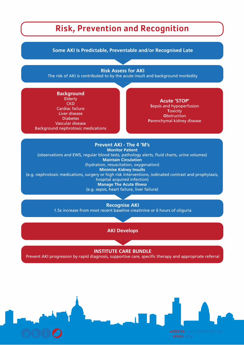
Prevent AKI - The 4 ‘M’sMonitor Patient
(observations and EWS, regular blood tests, pathology alerts, fluid charts, urine volumes)Maintain Circulation
(hydration, resuscitation, oxygenation)Minimise Kidney Insults
(e.g. nephrotoxic medications, surgery or high risk interventions, iodinated contrast and prophylaxis, hospital acquired infection)Manage The Acute Illness
(e.g. sepsis, heart failure, liver failure)
Risk, Prevention and Recognition
website: www.londonaki.netemail: [email protected]
BackgroundElderly
CKDCardiac failureLiver disease
DiabetesVascular disease
Background nephrotoxic medications
Some AKI Is Predictable, Preventable and/or Recognised Late
Risk Assess for AKIThe risk of AKI is contributed to by the acute insult and background morbidity
Recognise AKI1.5x increase from most recent baseline creatinine or 6 hours of oliguria
INSTITUTE CARE BUNDLEPrevent AKI progression by rapid diagnosis, supportive care, specific therapy and appropriate referral
AKI Develops
Acute ‘STOP’Sepsis and hypoperfusion
ToxicityObstruction
Parenchymal kidney disease

website: www.londonaki.netemail: [email protected]
Institute in all patients with a 1.5 X rise in creatinine or oliguria (<0.5mls/kg/hr) for >6 hours
This is a Medical Emergency
Causes Think ‘STOP AKI’Sepsis and hypoperfusion, Toxicity (drugs/contrast), Obstruction, Parenchymal kidney disease (acute GN)
Full set of physiological observationsAssess for signs of shock/hypoperfusion
If MEWS triggering give oxygen, begin resuscitation and contact critical care outreach team
Fluid therapy in AKIAssess heart rate, blood pressure, jugular venous pressure,
capillary refill (should be <3 secs), conscious level.If hypovolaemic give bolus fluids (e.g. 250-500mls) until volume replete with regular review of response.
Middle grade review if >2 litres filling in oliguria.If the patient is euvolaemic give maintenenance fluids (estimated output plus 500mls)
and set daily fluid target.
Monitoring in AKIDo arterial blood gas and lactate if venous bicarbonate is low or evidence of severe sepsis or hypoperfusion.
Consider insertion of urinary catheter and measurement of hourly urine volumes.Measure urea, creatinine, bone, other electrolytes and venous bicarbonate
at least daily while creatinine rising.Measure daily weights, keep a fluid chart and perform a minimum of 4 hourly observations.
Perform regular fluid assessments and check for signs of uraemia.
Investigation of AKIInvestigate the cause of all AKI unless multi-organ failure or obvious precipitant
Urine dipstick. If proteinuria is present perform urgent spot urine protein creatinine ratio (PCR).USS should be performed within 24 hours unless AKI cause is obvious or AKI is recovering
or within 6 hours if obstruction with infection (pyonephrosis) is suspected.Check liver function (hepatorenal), CRP and CK (rhabdomyolysis). If platelets low do blood
film/LDH/Bili/retics (HUS/TTP). If PCR high, consider urgent Bence Jones protein & serum free light chains.
Supportive AKI careTreat sepsis - in severe sepsis intravenous antibiotics should be administered within 1 hour of recognition.
Stop NSAID/ACE/ARB/metformin/K-sparing diuretics and review all drug dosages.Give proton pump inhibitor and perform dietetic assessment.
Stop anti-hypertensives if relative hypotension. If hypovolaemic consider stopping diuretics.Avoid radiological contrast if possible. If given follow prophylaxis protocol.
AKI Care Bundle

Patient Name: ................................................................................
No: ............................................ DOB: ...........................................
AKI Care Bundle Checklist
website: www.londonaki.netemail: [email protected]
URGENT ASSESSMENTABC and full set of observationsOxygen therapy?Early warning system triggeringCritical care outreach called (if triggering)
YES NO N/A
MONITORING IN AKIPhysiological monitoring plan decided (minimum 4 hourly) Arterial blood gas and lactate if indicatedUrinary catheter and hourly volumes (if indicated)Twice daily blood tests while creatinine risingDaily weights instituted Fluid chart instituted
YES NO N/A
SUPPORTIVE AKI CARESepsis treated (IV antibiotics <1 hour if severe)Drug chart and dosages reviewed NSAIS/ACE/ARB/K-sparing diuretics/metformin stopped Proton pump inhibitor givenAntihypertensives stopped if relative hypotension dietetic assessment
YES NO N/A
INVESTIGATION OF AKIUrine dipstick and documentation of resultIf proteinuria, protein creatinine ratio checkedUSS <24hrs requested (aki not recovering or cause not clear)Bone, liver function, CK, CRPMyeloma screen (if appropriate)Autoimmune screen (if appropriate)If platelets low, microangiopathy screen (blood film, retics, LDH)
YES NO N/A
FLUID THERAPY IN AKIClinical assessment of volume status and perfusion? Bolus fluids with reassessment to achieve euvolaemia Maintenance fluid requirements estimated and prescribed
YES NO N/A
AKI REFERRALReferral pathway reviewed Referral nephrologyReferral critical care
YES NO N/A
Signed: .............................................................
Date: .................................................................
Position: ...........................................................
Fix patient sticker here

‘STOP’ AKI and Checklist
website: www.londonaki.netemail: [email protected]
SEPSIS & HYPOPERFUSION Severe SepsisHaemorrhageDehydrationCardiac FailureLiver FailureRenovascular Insult (E.G. Aortic Surgery)
The London AKI Network has Developed the ‘STOP’ Acronym to Improve Awareness of AKI Causes
YES NO N/A
OBSTRUCTIONBladder OutflowStonesTumourSurgical Ligation Of UretersExtrinsic Compression (E.G. Lymph Nodes)Retroperitoneal Fibrosis
YES NO N/A
PARENCHYMAL KIDNEY DISEASEGlomerulonephritisTubulointersitial NephritisRhabdomyolysisHaemolytic Uraemic SyndromeMyeloma KidneyMalignant Hypertension
YES NO N/A
Sepsis & hypoperfusion Toxicity Obstruction Parenchymal kidney disease
TOXICITYNephrotoxic DrugsIodinated Radiological Contrast
YES NO N/A
Patient Name: ................................................................................
No: ............................................ DOB: ...........................................
Signed: .............................................................
Date: .................................................................
Position: ...........................................................
Fix patient sticker here
website: www.londonaki.netemail: [email protected]
Hyperkalaemia, Acidosis, Pulmonary Oedema, Reduced Conscious Level
Begin Medical Therapy and Get Help
Local Critical Care Teamand
Local Nephrology Team (if onsite)
These are Holding Measures Prior to Specialist Help from Critical Care or Nephrology Services
HyperkalaemiaMedical therapy of hyperkalaemia is a transient measure pending imminent recovery in renal function
or transfer to kidney unit or critical care for renal replacement therapy.If ECG changes give calcium gluconate 10mls 10%.
If bicarbonate <22mmol/L and no fluid overload give 500mls 1.26% sodium bicarbonate over 1 hour.K>6.5mmol/L or ECG changes give insulin 10IU in 50mls of 50% dextrose over 15 minutes & salbutamol
10mg nebulised (caution with salbutamol in tachycardia or ischaemic heart disease).Insulin/dextrose and salbutamol reduce ECF potassium for <4 hours only.
AcidosisMedical therapy of acidosis with bicarbonate should be reserved fo emergency management
of hyperkalaemia (as above) pending specialist help.pH<7.15 requires immediate critical care referral.
Pulmonary OedemaSit the patient up and give oxygen (60-100% unless contraindicated)
If haemodynamically stable give furosemide 80mg IV. Consider repeat bolus and infusion at 10mg/hourIf haemodynamically stable commence GTN 1-10mg/hour titrating dose.
Reduced Conscious LevelManage uraemic coma as per all reduced consciousness (airway management) pending
critical care transfer and emergency renal replacement therapy.
AKI Complications

Local Renal Teamor
Local Critical Care Team(essential if the patient is developing multi-organ failure)
Dataset Needed for Kidney Unit ReferralsU and E, Calcium, Phosphate, ABG/lactate, FBC, coagulation, LFTs.Heart rate, respiratory rate, blood pressure, Oxygen saturations.
AVPU or GCS score.Urine output.
AKI grade and pre-morbid Cr level.Urine dipstick.
USS if obtained.Co-morbid history.
MRSA status (if known).
If the Patient Is Too Ill To Transfer (see AKI Transfer Policy)Contact Local Critical Care Team
Institute AKI Care Bundle While Transfer Pending
Referral from Ward
website: www.londonaki.netemail: [email protected]
All AKIwith
Blood and protein +++ on dipstick Possible autoimmune disease/ glomerulonephritis,
myeloma Possible HUS/TTP, hypertension
Poisoning.
Local Renal TeamIf transfer decided see
AKI transfer policy
All AKIwith
Obstruction on USS(NB partially obstructed patients may have
normal or high urine volumes).
LOCAL UROLOGY TEAMIf nephrostomy or stenting
required proceed immediately
Progression to AKI 3 Or AKI 3 at Recognition or AKI Complicationsand Imminent Recovery Unlikely

Referral from Ward to Kidney Unit Checklist
website: www.londonaki.netemail: [email protected]
The following data are required for referral to your local renal servicePlease use this checklist to ensure you have all this essential information
This checklist is also available on the London AKI iPhone App
Urea and Electrolytes
Calcium
Phosphate
Arterial Blood Gases and Lactate
Urine Dipstick
USS Result (if performed)
Baseline Renal Function (if known)
Past Medical History
Blood Pressure
Heart Rate
Oxygen Saturations
Respiratory Rate
Avpu or GCS Assessment of Conscious Level
Current Urine Volume
Mrsa Status
Whether Diarrhoea Last 48 Hours
YES NO N/A
Signed: .............................................................
Date: .................................................................
Position: ...........................................................
Fix patient sticker here
Patient Name: ................................................................................
No: ............................................ DOB: ...........................................
Local Renal Teamor
Local Critical Care Team(essential if the patient is developing multi-organ failure)
Dataset Needed for Kidney Unit ReferralsU and E, Calcium, Phosphate, ABG/lactate, FBC, coagulation, LFTs.Heart rate, respiratory rate, blood pressure, Oxygen saturations.
AVPU or GCS score.Urine output.
AKI grade and pre-morbid Cr level.Urine dipstick.
USS if obtained.Co-morbid history.
MRSA status (if known).
If the Patient Is Too Ill To Transfer (see AKI Transfer Policy)Contact Local Critical Care Team
Institute AKI Care Bundle While Transfer Pending
Referral from Ward
website: www.londonaki.netemail: [email protected]
All AKIwith
Blood and protein +++ on dipstick Possible autoimmune disease/ glomerulonephritis,
myeloma Possible HUS/TTP, hypertension
Poisoning.
Local Renal TeamIf transfer decided see
AKI transfer policy
All AKIwith
Obstruction on USS(NB partially obstructed patients may have
normal or high urine volumes).
LOCAL UROLOGY TEAMIf nephrostomy or stenting
required proceed immediately
Progression to AKI 3 Or AKI 3 at Recognition or AKI Complicationsand Imminent Recovery Unlikely

website: www.londonaki.netemail: [email protected]
NeurologicalAlert on AVPU score or GCS >12.
If Criteria not Met Emergency Referral to Local Critical CareOnce stabilised follow ITU to acute kidney unit transfer policy.
HyperkalaemiaNo ECG changes.K < 6.0mmol/L.
If K lowered to <6.0 after presentation this must be potentially sustained (e.g bicarbonate therapy ordialysis/CVVH) not transient therapy (insulin and dextrose).
CirculatoryHeart rate > 50/min and < 120/min.Blood pressure > 100mmHg systolic.
MAP > 65MMHg.Lactate < 4mmol/L.
(lower BP values may be accepted if it has been firmly established these are pre-morbid).
Renal AcidosispH >7.2.
Venous bicarbonate >12mmol/L.Lactate < 4mmol/L.
RespiratoryRespiratory rate >11 and < 26/min.
Oxygen saturations >94% on not more than 35% oxygen.If patient required acute CPAP must have been independent of this treatment for 24 hrs.
Transfer From Ward to Kidney Unit(interhospital transfer)
The following is a guideline for whether patients are safe to transfer from a ward to a kidney unit in another hospital.
All AKI 3 patients or patients with complications should also be assessed as safe for transfer by a middle grade doctor and if necessary by the home critical care team.

Transfer from Ward to Kidney Unit Checklist
website: www.londonaki.netemail: [email protected]
The following is to enable renal teams to screen referrals for transfer safetyAll AKI interhospitals should have a bedside assessment by at least a middle grade
doctor and be deemed safe prior to interhospital transferThis checklist is also available on the London AKI iPhone App
Potassium <6.0mmol/l
pH>7.2
Venous Bicarbonate >12mol/l
Calcium (ionised > 1mmol/l, total > 2mmol/l)
Lactate (< 4 mmol/l)
Blood Pressure (>100mmHg)
MAP (>65mmhg)
Heart Rate (>50/min and <120/min)
Oxygen Saturations (>94% on not more than 35% O2)
Respiratory Rate (>11/min and <26/min)
AVPU Alert or GCS > 12
Clinical Assessment of Transfer Safety by Middle Grade Doctor
at Referring Site
MRSA Status
Whether Diarrhoea in Last 48 Hours
YES NO N/A
Patient Name: ................................................................................
No: ............................................ DOB: ...........................................
Signed: .............................................................
Date: .................................................................
Position: ...........................................................
Fix patient sticker here

For step down care see: AKI transfers policy critical care to kidney unit
Requests for nephrology advice (not-transfer) on critical care patients should be made to liaison nephrologist for the hospital or, if unavailable, to local on-call renal team.
Referral for nephrology opinion is at the discretion of the consultant intensivist and generally not necessary in patients
with AKI in the context of multi-organ failure.
Referral is recommended if
Possibility of AKI as an initiating event (with subsequent systemic decompensation) - i.e AKI 3 early in illness.
Single organ failure. AKI with possible vasculitis, lupus or autoimmune disease.
AKI in myeloma or malignancy or tumour lysis. AKI with unexplained pulmonary infiltrates
or pulmonary haemorrhage. HUS/TTP.
AKI in pregnancy. AKI with urological abnormalities. AKI with malignant hypertension.
AKI with poisoning.
Referral from Critical Care to Nephrology
website: www.londonaki.netemail: [email protected]

website: www.londonaki.netemail: [email protected]
Phone Local Renal Team
If the Patient is Accepted for Transfer, a Handover to Critical Care inReceiving Hospital Should be Done and Critical Care Outreach Informed
Further discussion with receiving hospital intensivist not required if condition stable or improving
NeurologicalAlert AVPU (unless stable, chronic neurological impairment).
Below is a Guideline for What Would be Considered a Safe ITU to Kidney UnitTransfer. These Transfers Should be Discussed at a Senior Level.
MetabolicK < 6.0, ionised Ca > 1mmol/L.
pH normal.Bicarbonate > 16mmol/L.
Lactate normal.
CirculatoryHeart rate > 50/min and < 120/min.
BP > 100mmHg systolic.MAP > 65MMHg.
If given inotropes given must have been inotrope independent > 24 hours.
RespiratoryRespiratory rate >11/min and < 26/min.
Saturations > 94% on not more than 35% oxygen.If patient required acute CPAP must have been independent of this treatment for 24 hrs.
If ventilated <1 week should have been independent of respiratory support for 48hrs.If longer term invasive ventilation should have been independent of all respiratory support
for 1 day for each week ventilated and for a period of not less than 48 hours.
Transfer From Critical Care to Kidney Unit(interhospital transfer)
Monitor Function To 72 Hours in High Risk CasesIf oliguria or rising creatinine early referral to local renal team.
NB there is no-proven role for N-Acetyl cysteine, post-contrast dialysis/CVVHor routine cessation of metformin or ACE inbitors.
High volume (>100mls) iodinated contrast procedureand
CKD with eGFR<60 (particularly diabetic nephropathy)or AKI
Other risk factors dehydration, heart failure, severe sepsis,cirrhosis, nephrotoxins (NSAIDS, aminoglycosides).
Risk factors are multiplicative.
Give Prophylaxis if High RiskVolume expansion (unless hypervolaemic) with normal saline or or 1.26% bicarbonate
Sample regimensIV Na bicarbonate 1.26% 3mls/Kg/hr for 1 hour pre-procedure and 6 hours post-procedure
orIV 0.9% normal saline 1ml/kg/hr 12 hours pre and 12 hours post procedure
Contrast Induced Nephropathy (CIN) Prophylaxis
website: www.londonaki.netemail: [email protected]
Assess Risk
Is Contrast Procedure Necessary?
Resuscitate to Euvolaemia
Minimise contrast, use low or iso-osmolar contrast
Yes
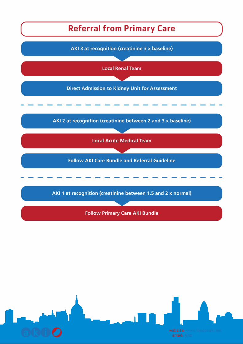
Referral from Primary Care
website: www.londonaki.netemail: [email protected]
AKI 3 at recognition (creatinine 3 x baseline)
Direct Admission to Kidney Unit for Assessment
Local Renal Team
AKI 2 at recognition (creatinine between 2 and 3 x baseline)
Follow AKI Care Bundle and Referral Guideline
Local Acute Medical Team
AKI 1 at recognition (creatinine between 1.5 and 2 x normal)
Follow Primary Care AKI Bundle

Monitor Function To 72 Hours in High Risk CasesIf oliguria or rising creatinine early referral to local renal team.
NB there is no-proven role for N-Acetyl cysteine, post-contrast dialysis/CVVHor routine cessation of metformin or ACE inbitors.
High volume (>100mls) iodinated contrast procedureand
CKD with eGFR<60 (particularly diabetic nephropathy)or AKI
Other risk factors dehydration, heart failure, severe sepsis,cirrhosis, nephrotoxins (NSAIDS, aminoglycosides).
Risk factors are multiplicative.
Give Prophylaxis if High RiskVolume expansion (unless hypervolaemic) with normal saline or or 1.26% bicarbonate
Sample regimensIV Na bicarbonate 1.26% 3mls/Kg/hr for 1 hour pre-procedure and 6 hours post-procedure
orIV 0.9% normal saline 1ml/kg/hr 12 hours pre and 12 hours post procedure
Contrast Induced Nephropathy (CIN) Prophylaxis
website: www.londonaki.netemail: [email protected]
Assess Risk
Is Contrast Procedure Necessary?
Resuscitate to Euvolaemia
Minimise contrast, use low or iso-osmolar contrast
Yes

ASA score, consider pre-operative CPEX testing.Pre-morbid factors: CKD, diabetes, vascular disease, cardiac failure, liver failure.
In emergency surgery consider current patient stability/illness severity.Type of surgery: If ‘major’ operation or known high risk (e.g. cardiac bypass, likely heavy blood loss
or involving pelvis or renal tract).
Consider pre-optimisation in ward or critical care area and scheduled post-operative admission to critical care.
There is no role for the routine use of dopamine or frusemide in perioperative AKI prevention.DIscontinue or avoid nephrotoxic drugs if possible.
If risk of long-term renal insufficiency (e.g. nephrectomy in CKD discuss with nephrology team).Optimise circulation and oxygenation during surgery.
Consider and Treat Specific Surgical CausesBlood loss, hypovolaemia, surgical sepsis, hypotension due to epidural or opiate anaesthesia, postoperative urinary retention or obstruction of the renal tract as a surgical complication.
Perioperative AKI
website: www.londonaki.netemail: [email protected]
Preoperative AKI Risk Assessment(anaesthetic and surgical teams) in pre-assessment clinic or ward
Postoperative AKI Risk Assessment
MonitorObservations (blood pressure, heart rate, urine volumes, regular blood tests)
If postoperative AKI develops (oliguria 6 hours or 1.5 rise from baseline creatinine) Institute AKI Care Bundle and Referral Pathway
Postoperative resuscitation as appropriate
As per pre-op assessment. Assess surgery undertaken, blood loss, perioperative haemodynamic stability,perioperative oxygenation and perioperative oliguria.

Fluids
website: www.londonaki.netemail: [email protected]
Adult Maintenance Fluids
Baseline Requirements50-100mmol sodium, 40-80mol potassium
and 1.5-2.5L water per 24 hoursOral, enteral or parenteral route
Give According to Clinical Scenario
General Volume Replacementor Expansion
Give balanced crystalloid solutions(Hartman’s solution/Ringer’s lactate)
These contain small amounts of potassium.Avoid in hyperkalaemia. If AKI only use these
if close (HDU) monitoring of potassiumor
ColloidsAvoid high molecular weight (>200kDa
starches in severe sepsis due to risk of AKIAssess vital signs, postural blood pressure,capillary refill, JVP and consider invasive
or non-invasive measurement using flow-based technology
Adjust estimated requirements according to changes in sensible
or insensible losses
Regular assessment of volume and hydration status
Daily weightsFluid charts
Measured electrolytes
HaemorrhageGive blood and blood productsBalanced crystalloid or colloid
may be given while blood awaitedClinical assessment as above
Severe Free Water Losses(hypernatraemia)
5% dextroseor 4%/0.18% dextrose/saline
Hypochloraemia(vomiting, NG drainage)
Give normal saline(Potassium repletion usually also required)
Available parenteral solutions (if required)
Hartmans solution/Ringer’s lactateNormal Saline5% dextrose
0.4%/0.18% dextrose/salinePotassium usually added additionally
SensibleLosses
(measurable)Surgical drains
VomitingDiarrhoea
Urine(variable amounts of
electrolytes)
InsensibleLosses
RespirationPerspirationMetabolism
Increase in pyrexiaor tachypnoea(Mainly water)
Adult Resuscitation or Replacement Fluids
ASA score, consider pre-operative CPEX testing.Pre-morbid factors: CKD, diabetes, vascular disease, cardiac failure, liver failure.
In emergency surgery consider current patient stability/illness severity.Type of surgery: If ‘major’ operation or known high risk (e.g. cardiac bypass, likely heavy blood loss
or involving pelvis or renal tract).
Consider pre-optimisation in ward or critical care area and scheduled post-operative admission to critical care.
There is no role for the routine use of dopamine or frusemide in perioperative AKI prevention.DIscontinue or avoid nephrotoxic drugs if possible.
If risk of long-term renal insufficiency (e.g. nephrectomy in CKD discuss with nephrology team).Optimise circulation and oxygenation during surgery.
Consider and Treat Specific Surgical CausesBlood loss, hypovolaemia, surgical sepsis, hypotension due to epidural or opiate anaesthesia, postoperative urinary retention or obstruction of the renal tract as a surgical complication.
Perioperative AKI
website: www.londonaki.netemail: [email protected]
Preoperative AKI Risk Assessment(anaesthetic and surgical teams) in pre-assessment clinic or ward
Postoperative AKI Risk Assessment
MonitorObservations (blood pressure, heart rate, urine volumes, regular blood tests)
If postoperative AKI develops (oliguria 6 hours or 1.5 rise from baseline creatinine) Institute AKI Care Bundle and Referral Pathway
Postoperative resuscitation as appropriate
As per pre-op assessment. Assess surgery undertaken, blood loss, perioperative haemodynamic stability,perioperative oxygenation and perioperative oliguria.

AKI Teaching Materials
website: www.londonaki.netemail: [email protected]
“STOP” - causes of AKISepsis and hypoperfusion (hypovolaemia, heart failure, hepatorenal)
Toxicity (drugs, contrast)Obstruction
Parenchymal kidney disease (myeloma, rhabdomyolysis, RPGN, HUS, TIN)
Rising creatinine = rising mortality
Stage Serum creatinine
rise ≥ 26 µmol/L within 48hrsor rise ≥1.5- to 1.9 X baseline SCr
<0.5 mL/kg/hr for > 6 consecutive hrs
1
rise ≥ 2 to 2.9 X baseline SCr <0.5 mL/kg/hr for > 12 hrs
2
rise ≥3 X baseline SCR or rise 354 µmol/L or commenced on renal replacement therapy (RRT) irrespective of stage
<0.3 mL/kg/hr for > 24 hrsor anuria for 12 hrs
3
Urine output
KDIGO Staging System for Acute Kidney Injury
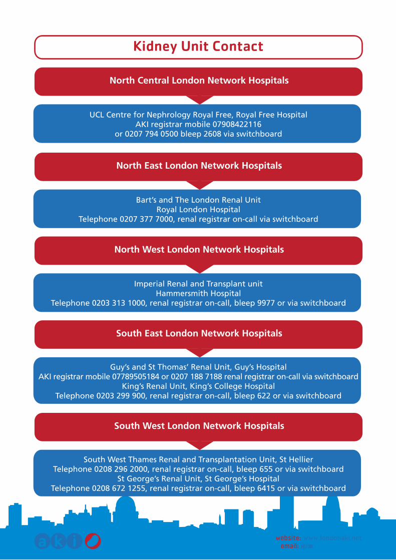
UCL Centre for Nephrology Royal Free, Royal Free HospitalAKI registrar mobile 07908422116
or 0207 794 0500 bleep 2608 via switchboard
Kidney Unit Contact
website: www.londonaki.netemail: [email protected]
North Central London Network Hospitals
Bart’s and The London Renal UnitRoyal London Hospital
Telephone 0207 377 7000, renal registrar on-call via switchboard
North East London Network Hospitals
Imperial Renal and Transplant unitHammersmith Hospital
Telephone 0203 313 1000, renal registrar on-call, bleep 9977 or via switchboard
North West London Network Hospitals
Guy’s and St Thomas’ Renal Unit, Guy’s Hospital AKI registrar mobile 07789505184 or 0207 188 7188 renal registrar on-call via switchboard
King’s Renal Unit, King’s College HospitalTelephone 0203 299 900, renal registrar on-call, bleep 622 or via switchboard
South East London Network Hospitals
South West Thames Renal and Transplantation Unit, St HellierTelephone 0208 296 2000, renal registrar on-call, bleep 655 or via switchboard
St George’s Renal Unit, St George’s HospitalTelephone 0208 672 1255, renal registrar on-call, bleep 6415 or via switchboard
South West London Network Hospitals
AKI Teaching Materials
website: www.londonaki.netemail: [email protected]
“STOP” - causes of AKISepsis and hypoperfusion (hypovolaemia, heart failure, hepatorenal)
Toxicity (drugs, contrast)Obstruction
Parenchymal kidney disease (myeloma, rhabdomyolysis, RPGN, HUS, TIN)
Rising creatinine = rising mortality
Stage Serum creatinine
rise ≥ 26 µmol/L within 48hrsor rise ≥1.5- to 1.9 X baseline SCr
<0.5 mL/kg/hr for > 6 consecutive hrs
1
rise ≥ 2 to 2.9 X baseline SCr <0.5 mL/kg/hr for > 12 hrs
2
rise ≥3 X baseline SCR or rise 354 µmol/L or commenced on renal replacement therapy (RRT) irrespective of stage
<0.3 mL/kg/hr for > 24 hrsor anuria for 12 hrs
3
Urine output
KDIGO Staging System for Acute Kidney Injury

Recognition
website: www.londonaki.netemail: [email protected]
Adding Insult to Injury. A review of the care of patients who died in hospital with a primary diagnosis of acute kidney injury (acute renal failure). National Confidential Enquiry into Patient Outcome and Death (NCEPOD). 2009.
UK Renal Association Clinical Practice Guideline on Acute Kidney Injury. 2011.
Kidney Disease Improving Global Outcomes (KDIGO) Clinical Practice Guideline for Acute Kidney Injury. 2011.
National Institute For Clinical Excellence (NICE) Clinical Guideline 50: Recognition and Response to Acute Illness in Adults in Hospital.
Imaging for Acute Kidney Injury (acute renal failure): Good Practice Recommendations from the National Imaging Board. 2010.
British Consensus Guidelines on Intravenous Fluid Therapy for Adult Surgical Patients.BAPEN Medical, the Association for Clinical Biochemistry, the Association of Surgeons of Great Britain and Ireland, the Society of Academic and Research Surgery, the UK Renal Association and the Intensive Care Society. 2008 - update 2011.
Pre-operative Assessment and Patient Preparation: The Role of The Anesthetist. The Association of Anesthetists of Great Britain and Ireland. 2010.
Guidelines for the Transfer of the Critically Ill Adult. UK Intensive Care Society. 3rd Edition 2011.
References:
1
2
3
4
5
6
7
8
We would like to thank Chris Kirwan for his help in preparing the sections on perioperative acute kidney injury
We would like to thank Nick Macartney and Jeremy Dawson for for their help with the bundle checklist
We would like to thank NHS Kidney Care for Sponsoring this Document
Acknowledgements
•
•
•
• Design by: UCL Medical Illustration Services 03/2012

www.londonaki.net