Embed Size (px)
Citation preview
Volume 2 • Issue 2 • 1000123J Clinic Experiment CardiolISSN:2155-9880 JCEC, an open access journal
Research Article Open Access
Clinical & Experimental
CardiologyDuning et al. J Clinic Experiment Cardiol 2011, 2:2
http://dx.doi.org/10.4172/2155-9880.1000123
Keywords: Atrial fibrillation; Poincaré analysis; Atrial remodeling; Ischemic stroke; Prevention
Abbreviations: AF: Atrial Fibrillation; EPA: Extended Poincaré Analysis; PAF: Paroxysmal Atrial Fibrillation; SD: Standard Deviation; MIT: Massachusetts Institute of Technology
Introduction One fifth of all strokes are caused by atrial fibrillation (AF) [1,2]. Oral
anticoagulation is highly efficacious for both primary and secondary prevention of stroke in patients with AF [3], and thus detection of AF is essential. However, as in up to one third of cases AF is intermittent, appropriate diagnosis - and thus treatment - is frequently missed [4]. Even when suspected, diagnosis of paroxysmal AF (PAF) is estimated to be missed in more than 50% of patients [5,6]. Relative to standard ECG, 24h-ECG doubles the detection rate of PAF but still misses about one third of cases later identified by ambulatory 7-day ECG and up to 44% of cases detected by long-term event recorders [7]. The number of patients needed to screen to detect one case of PAF has been calculated to be 20 [8].
In patients having survived cerebral infarctions the cause of stroke remains undetermined in up to 40 % of cases despite extensive diagnostic work-up [9-11]. Because of the high frequency of PAF and the poor sensitivity of 24h-ECG, a substantial number of these patients are likely to have undetected PAF. These patients fail to receive effective preventive therapy although they carry as a high a risk of cardiogenic stroke as those with permanent AF [12,13].
Because limited time and resources do not allow monitoring all patients with cryptogenic stroke beyond a 24h-ECG, measures are needed to identify patients with an increased risk for paroxysmal AF. Since even short-duration AF can induce electrical and contractile atrial remodeling, previous episodes of PAF may increase the variability of the
atrial electrical wavelength also in the absence of fibrillation, i.e. during sinus rhythm [14,15]. This phenomenon may manifest as increased R-R interval dynamics on ECG. Combined with an adequate analysis tool, it could help to identify patients with an increased likelihood of PAF who would profit from extended ECG monitoring.
Here we tested whether patients with previously diagnosed PAF show abnormalities of R-R dynamics during sinus rhythm. Assessment was based on automated extended Poincaré analysis (EPA) trained on an independent set of ECG from patients with PAF.
MethodsPatients
We enrolled 38 patients from our cardiologic department (mean age 68 years, range 44 to 85 years; 14 females). Twenty-nine had an established diagnosis of paroxysmal or persistent AF, identified as self-terminating episodes of AF on past conventional ECG analyses (< 1 year before start of the study, minimum of two ECGs). Fifteen of these
*Corresponding author: Thomas Duning, University Hospital of Muenster, Department of Neurology, Albert-Schweitzer-Str. 33, 48149 Muenster, Germany, Tel: 49-251-83-45301; Fax: 49-251-83-45313; E-mail: [email protected]
Received January 04, 2011; Accepted February 09, 2011; Published February 11, 2011
Citation: Duning T, Kirchhof P, Wersching H, Hepp T, Reinhardt R, et al. (2011) Extended Electrocardiographic Poincare Analysis (EPA) for Better Identification of Patients with Paroxysmal Atrial Fibrillation. J Clinic Experiment Cardiol 2:123. doi:10.4172/2155-9880.1000123
Copyright: © 2011 Duning T, et al. This is an open-access article distributed under the terms of the Creative Commons Attribution License, which permits unrestricted use, distribution, and reproduction in any medium, provided the original author and source are credited.
AbstractBackground: Atrial fibrillation (AF) – be it permanent or paroxysmal – is the most frequent and most effectively
treatable cause of stroke. However, when paroxysmal, even electrocardiography for 24 hours (24h-ECG) misses AF in more than 50% of cases. We assessed whether extended Poincaré analysis of ECG R-R intervals (EPA) can help to identify electrocardiographic remodeling suggestive of paroxysmal AF (PAF).
Methods: Twenty-nine patients with previously diagnosed PAF were re-assessed by 24h-ECG using conventional analysis and EPA based on a previously trained algorithm considering among other ratios of R-R interval duration, number of premature atrial complexes, approximate entropy, and standard deviation of Poincaré plots-axes. 24h-ECG from 21 healthy subjects without a history of AF served as negative, and 9 patients with permanent AF as positive controls.
Results: PAF during 24h-ECG was detected in 4 out of 29 (14%) patients with a history of PAF by conventional analysis. EPA classified ECGs of these and 22 additional patients with a history of PAF, i.e. a total 90%, as suggestive of PAF. All patients with permanent AF were identified by both tools. EPA additionally classified the ECG in 4 out of 21 control subjects as suggestive of PAF.
Conclusions: Extended Poincaré analysis in patients with a history of PAF is more sensitive to electrocardiographic abnormalities than is conventional 24h- ECG analysis. These findings warrant prospective studies of EPA in patients with a high likelihood of PAF, i.e. with stroke of undetermined origin.
Extended Electrocardiographic Poincare Analysis (EPA) for Better Identification of Patients with Paroxysmal Atrial FibrillationThomas Duning1*, P. Kirchhof2, H. Wersching1, T. Hepp4, R. Reinhardt4, H. Heuer3, E. B. Ringelstein1 and S. Knecht1
1Depatment of Neurology, University Hospital of Muenster, Albert- Schweitzer-Str. 33, 48149 Muenster, Germany 2Depatment of Cardiology, University Hospital of 2Muenster, Albert- Schweitzer-Str. 33, 48149 Muenster, Germany3Depatment of Cardiology, Johannes Hospital, Johannesstr. 9-17, 44137 Dortmund, Germany4Apoplex Medical Technologies GmbH, Delaware Ave. 1-3, 66953 Pirmasens, Germany
Citation: Duning T, Kirchhof P, Wersching H, Hepp T, Reinhardt R, et al. (2011) Extended Electrocardiographic Poincare Analysis (EPA) for Better Identification of Patients with Paroxysmal Atrial Fibrillation. J Clinic Experiment Cardiol 2:123. doi:10.4172/2155-9880.1000123
Page 2 of 7
Volume 2 • Issue 2 • 1000123J Clinic Experiment CardiolISSN:2155-9880 JCEC, an open access journal
patients had recurrences of AF after cardioversion in the past. Nine patients had a diagnosis of permanent atrial fibrillation and served as positive controls.
As negative controls, 21 volunteers (mean age 62 years, range 53 to 81 years; 11 females) without a diagnosis of arrhythmia or structural heart disease were included. These had undergone previous Holter ECG analyses and showed no episodes of AF. Twenty-six patients (68 %) and 12 control subjects (55 %) were treated with _-blockers for blood pressure control. Patients with pacemakers and patients treated with antiarrhythmic drugs (sodium or potassium channel blockers or amiodarone) were excluded. Clinical characteristics of patients are listed in table 1.
Additionally, a 66 year old male patient with an infraction of the right cerebral artery of undetermined cause from our stroke-unit was screened. Standard 12- lead ECG on admission was unrevealing, so were transesophageal echocardiography and laboratory analyses. Extracranial and transcranial doppler/duplex sonography showed bilateral low grade atherosclerosis of the carotid arteries without hemodynamic relevance. No history of heart disease or arrhythmia and prior ischemic cardiovascular events was noted.
Electrocardiographic recording
All patients were assessed by standard 12-lead ECG. Additionally, patients underwent a 24h-ECG with 6-channel recorders (H12+; Mortara Instruments). The median ECG recording time was 19.5 hours (SD ± 2.5, range 15.3 to 22.9 hours). Data were edited and cleaned from artefacts using the Pathfinder Software (Vers. V8.257, Reynolds). Channel 2 of the ECG recording was used to generate R-R lists with the same software. The results were analyzed, Interpreted, and additionally revised visually by two experienced cardiologists using the H-SCRIBE
software (version 4.0; Mortara Instruments). The cardiologists were blinded to the patient’s medical history and the results of any other ECG recordings of the patient. The detection criteria require >1:1 AV conduction for a minimum of 24 ventricular cycles.
Extended Poincaré analysis (EPA)
Onset of atrial fibrillation is often preceded by premature atrial complexes, atrial tachycardia, and other ectopic activities. While the origin of these phenomena is manifold, most of them cause changes in ventricular response due to alterations in atrioventricular nodal conduction [16]. Non-linear algorithms can reveal abnormalities in cardiac regulation not detected by traditional single or linear measures of heart rate variability [17]. We used a combination of published linear and non linear parameters for EPA [18-24]. We then trained the resulting algorithm to discriminate between AF, PAF and sinus rhythm using the MIT data set “Atrial Fibrillation” [http://www.physionet.org/physiobank/database/nsrdb/] which contains data from 4 patients with permanent atrial fibrillation (40 hours), 19 patients with PAF (189 hours), and 18 control patients without arrhythmias (385 hours). The following parameters were considered for classification.
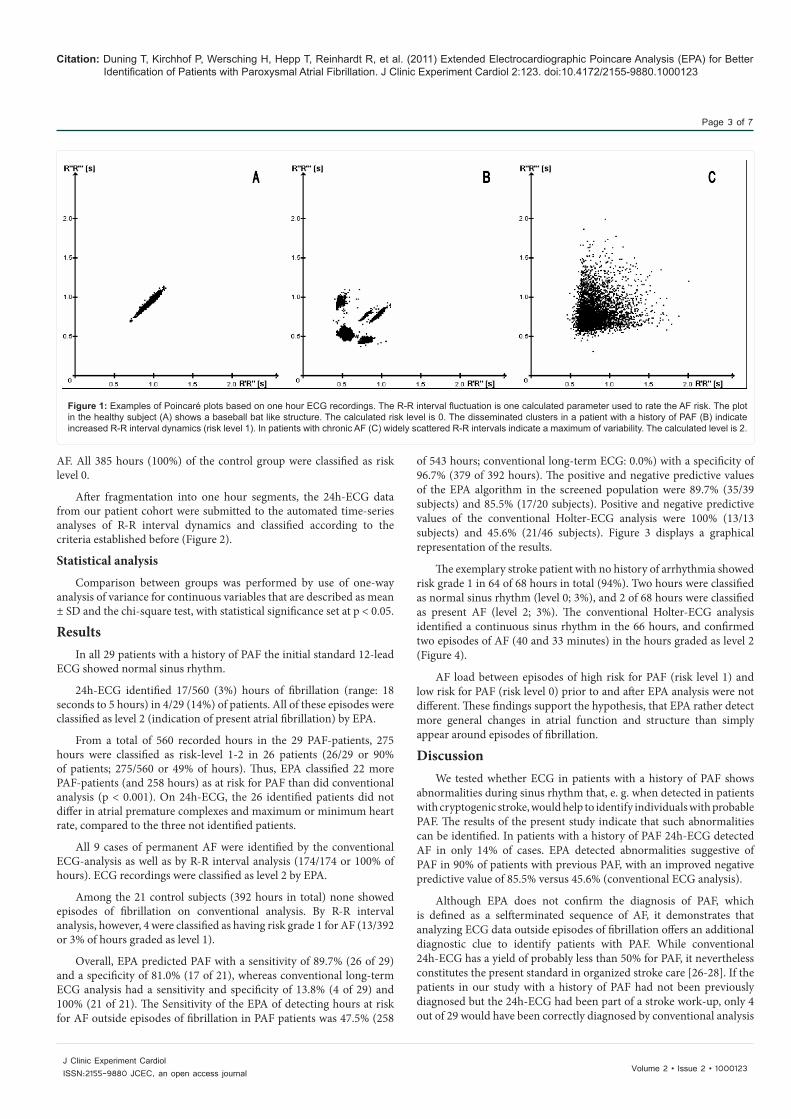
First, principle component analysis [18] was used to calculate the standard deviation (SD) of the minor axis (SD1), and of the major axis (SD2) of the Poincaré plots, and the ratio SD1/SD2 [20]. Dynamics of R-R interval fluctuations were assessed by creating R-R difference plots. Instead of plotting an R-R interval against its previous one, the differences between two consecutive R-R intervals were plotted and normalized by dividing them with the mean of the two corresponding R-R intervals [(Ri-Ri+1)/(Ri+Ri+1); (Figure 1)]. Krstacic et al. have shown that the ratio between the shortest and longest interval of maximal six consecutive R-R intervals might be a predictive parameter for PAF [19]. Since premature atrial complexes are known to play a major role in triggering AF, the number of these complexes was included in the analysis. As proposed by Thong et al., the number of complexes that trigger the risk for PAF depend on the preceding normal beats, the type of the premature atrial complexes (normal complexes with sinus node reset or abnormal complexes: interpolated, full compensatory pause, delayed sinus node reset) and also on the rhythm of the subsequent beats [21]. Thus, not all detected atrial premature complexes were rated equally, and only premature atrial complexes without sinus nodal reset were included in the analysis. Finally, regularity was analysed by calculating the approximate entropy of R-R interval data, which is a measure of complexity in time series analysis of ECG data [25].
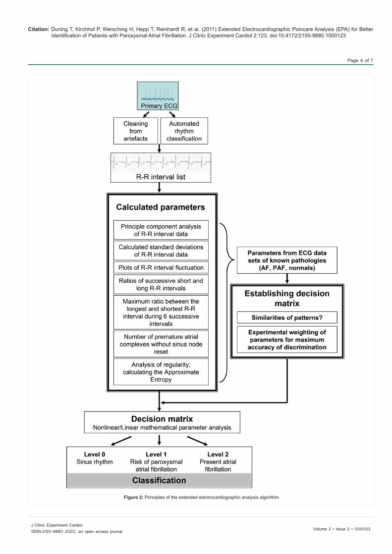
Based on the MIT data sets, these mathematical parameters were weighted in an iterative procedure to establish cut off values, to predefine increasing risk of AF and to maximise the discriminating accuracy between the three groups. Each ECG data segment of one hour duration was classified using the following categories (Figure 2).
• Risk level 0 Sinus rhythm
• Risk level 1 Increased R-R interval dynamics, being at risk of paroxysmal atrial fibrillation
• Risk level 2 Indication of present atrial fibrillation
After establishing classification criteria, all 40 hours (100%) of permanent AF of the MIT data set were classified as present fibrillation (risk level 2). In the PAFpatient group 153 of 189 hours (81%) were classified as being at risk of paroxysmal AF or having present episodes of fibrillation (level 1 or 2). All 105 hours including episodes of fibrillation were classified as risk level 2 by algorithm. Thus, 48 hours without fibrillating episodes were correctly rated as being at risk of paroxysmal
Clinical characteristics of the study population
PAF Group (29) Chronic AF Group (9) Control Group (21)
Age 67 (± 9.8) 69 (± 9.6) 62 (± 15.3) Gender male/female 18/11 6/3 10/11Sinus bradycardia 1 (3%)Hypertensive crisis 2 (7%)Coronary 1 vessel disease 3 (10%) 3 (14%)
Coronary 2 vessel disease 1 (3%) 1 (5%)
Coronary 3 vessel disease 3 (10%)
Ischemic cardiomyophathy 1 (11%)
Unselected chronic heart disease 1 (3%) 2 (10%)
Diabetes mellitus 1 (3%)Sinus tachycardia 1 (11%)Orthostatic disorder 1 (11%)Medication Beta-blocking-agents 22 (76%) 4 (44%) 12(48%)Calcium-channel-blockers 1 (3%) 1 (11%)
ACE inhibitor 3 (10%)AT1 inhibitor 3 (10%)Digitalis 2 (7%) 1 (11%)No medication 3 (14%)
Table 1: Clinical characteristics of patients. PAF = paroxysmal atrial fibrillation; Chronic AF = chronic atrial fibrillation; ACE = angiotensin-converting enzyme; AT1 inhibitor = angiotensin II type 1 receptor antagonist.
Citation: Duning T, Kirchhof P, Wersching H, Hepp T, Reinhardt R, et al. (2011) Extended Electrocardiographic Poincare Analysis (EPA) for Better Identification of Patients with Paroxysmal Atrial Fibrillation. J Clinic Experiment Cardiol 2:123. doi:10.4172/2155-9880.1000123
Page 3 of 7
Volume 2 • Issue 2 • 1000123J Clinic Experiment CardiolISSN:2155-9880 JCEC, an open access journal
AF. All 385 hours (100%) of the control group were classified as risk level 0.
After fragmentation into one hour segments, the 24h-ECG data from our patient cohort were submitted to the automated time-series analyses of R-R interval dynamics and classified according to the criteria established before (Figure 2).
Statistical analysis
Comparison between groups was performed by use of one-way analysis of variance for continuous variables that are described as mean ± SD and the chi-square test, with statistical significance set at p < 0.05.
ResultsIn all 29 patients with a history of PAF the initial standard 12-lead
ECG showed normal sinus rhythm.
24h-ECG identified 17/560 (3%) hours of fibrillation (range: 18 seconds to 5 hours) in 4/29 (14%) of patients. All of these episodes were classified as level 2 (indication of present atrial fibrillation) by EPA.
From a total of 560 recorded hours in the 29 PAF-patients, 275 hours were classified as risk-level 1-2 in 26 patients (26/29 or 90% of patients; 275/560 or 49% of hours). Thus, EPA classified 22 more PAF-patients (and 258 hours) as at risk for PAF than did conventional analysis (p < 0.001). On 24h-ECG, the 26 identified patients did not differ in atrial premature complexes and maximum or minimum heart rate, compared to the three not identified patients.
All 9 cases of permanent AF were identified by the conventional ECG-analysis as well as by R-R interval analysis (174/174 or 100% of hours). ECG recordings were classified as level 2 by EPA.
Among the 21 control subjects (392 hours in total) none showed episodes of fibrillation on conventional analysis. By R-R interval analysis, however, 4 were classified as having risk grade 1 for AF (13/392 or 3% of hours graded as level 1).
Overall, EPA predicted PAF with a sensitivity of 89.7% (26 of 29) and a specificity of 81.0% (17 of 21), whereas conventional long-term ECG analysis had a sensitivity and specificity of 13.8% (4 of 29) and 100% (21 of 21). The Sensitivity of the EPA of detecting hours at risk for AF outside episodes of fibrillation in PAF patients was 47.5% (258
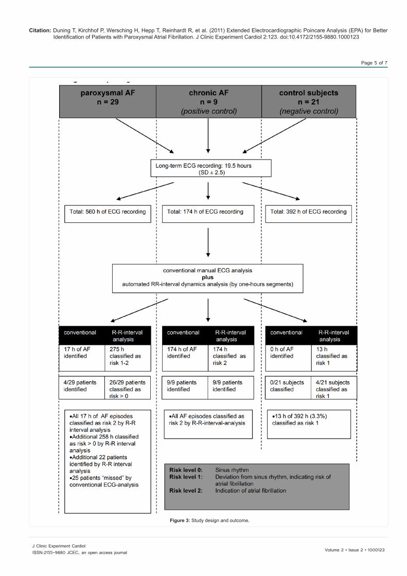
of 543 hours; conventional long-term ECG: 0.0%) with a specificity of 96.7% (379 of 392 hours). The positive and negative predictive values of the EPA algorithm in the screened population were 89.7% (35/39 subjects) and 85.5% (17/20 subjects). Positive and negative predictive values of the conventional Holter-ECG analysis were 100% (13/13 subjects) and 45.6% (21/46 subjects). Figure 3 displays a graphical representation of the results.
The exemplary stroke patient with no history of arrhythmia showed risk grade 1 in 64 of 68 hours in total (94%). Two hours were classified as normal sinus rhythm (level 0; 3%), and 2 of 68 hours were classified as present AF (level 2; 3%). The conventional Holter-ECG analysis identified a continuous sinus rhythm in the 66 hours, and confirmed two episodes of AF (40 and 33 minutes) in the hours graded as level 2 (Figure 4).
AF load between episodes of high risk for PAF (risk level 1) and low risk for PAF (risk level 0) prior to and after EPA analysis were not different. These findings support the hypothesis, that EPA rather detect more general changes in atrial function and structure than simply appear around episodes of fibrillation.
DiscussionWe tested whether ECG in patients with a history of PAF shows
abnormalities during sinus rhythm that, e. g. when detected in patients with cryptogenic stroke, would help to identify individuals with probable PAF. The results of the present study indicate that such abnormalities can be identified. In patients with a history of PAF 24h-ECG detected AF in only 14% of cases. EPA detected abnormalities suggestive of PAF in 90% of patients with previous PAF, with an improved negative predictive value of 85.5% versus 45.6% (conventional ECG analysis).
Although EPA does not confirm the diagnosis of PAF, which is defined as a selfterminated sequence of AF, it demonstrates that analyzing ECG data outside episodes of fibrillation offers an additional diagnostic clue to identify patients with PAF. While conventional 24h-ECG has a yield of probably less than 50% for PAF, it nevertheless constitutes the present standard in organized stroke care [26-28]. If the patients in our study with a history of PAF had not been previously diagnosed but the 24h-ECG had been part of a stroke work-up, only 4 out of 29 would have been correctly diagnosed by conventional analysis
Figure 1: Examples of Poincaré plots based on one hour ECG recordings. The R-R interval fluctuation is one calculated parameter used to rate the AF risk. The plot in the healthy subject (A) shows a baseball bat like structure. The calculated risk level is 0. The disseminated clusters in a patient with a history of PAF (B) indicate increased R-R interval dynamics (risk level 1). In patients with chronic AF (C) widely scattered R-R intervals indicate a maximum of variability. The calculated level is 2.
Citation: Duning T, Kirchhof P, Wersching H, Hepp T, Reinhardt R, et al. (2011) Extended Electrocardiographic Poincare Analysis (EPA) for Better Identification of Patients with Paroxysmal Atrial Fibrillation. J Clinic Experiment Cardiol 2:123. doi:10.4172/2155-9880.1000123
Page 4 of 7
Volume 2 • Issue 2 • 1000123J Clinic Experiment CardiolISSN:2155-9880 JCEC, an open access journal
Figure 2: Principles of the extended electrocardiographic analysis algorithm.
Citation: Duning T, Kirchhof P, Wersching H, Hepp T, Reinhardt R, et al. (2011) Extended Electrocardiographic Poincare Analysis (EPA) for Better Identification of Patients with Paroxysmal Atrial Fibrillation. J Clinic Experiment Cardiol 2:123. doi:10.4172/2155-9880.1000123
Page 5 of 7
Volume 2 • Issue 2 • 1000123J Clinic Experiment CardiolISSN:2155-9880 JCEC, an open access journal
Figure 3: Study design and outcome.

Citation: Duning T, Kirchhof P, Wersching H, Hepp T, Reinhardt R, et al. (2011) Extended Electrocardiographic Poincare Analysis (EPA) for Better Identification of Patients with Paroxysmal Atrial Fibrillation. J Clinic Experiment Cardiol 2:123. doi:10.4172/2155-9880.1000123
Page 6 of 7
Volume 2 • Issue 2 • 1000123J Clinic Experiment CardiolISSN:2155-9880 JCEC, an open access journal
and treated accordingly. In addition to these four patients, EPA would have indicated that in another 22 patients R-R dynamics were highly suggestive of PAF and could have prompted extended monitoring in order to pick up episodes of PAF.
In organized stroke care ECG signals are often monitored continuously, but without automatic data analysis or storage. Such data would be amenable to EPA and allow automatic analysis of the full length of time a patients is monitored by ECG. Figure 4 gives an example of continuously monitored ECG in a patient with cryptogenic stroke. There was no AF during the first 24 hours but EPA was suggestive of PAF throughout during this time. It was only after 37 hours that an AF episode occurred.
Dynamics of cardiac excitation in PAF often change because even short periods of AF alter atrial function and structure [14,15]. Concomitant structural changes in the atrioventricular node additionally affect cardiac excitation [29,30]. Both mechanisms can increase ectopic activity and thereby enhance R-R interval dynamics outside periods of fibrillation. However, EPA provided false negative results in 10% of patients with an established history of PAF. Although these false negative results show that the premise of electrical remodeling due to PAF and its detection by ECG does not hold in all cases, this false negative rate is considerably less than that of conventional ECG analysis (86%). Conversely, EPA provided false positive results (grade one risk) in 4 out of 21 individuals without a known history of paroxysmal atrial fibrillation. These false positive results caution
that analysis of R-R interval dynamics is complex. Grade one risk in controls without arrhythmia may reflect noise or artefacts in ECG recording as well as low-grade pathology or signs of remodeling due to undetected PAF, other paroxysmal arrhythmias or occult structural heart disease. However, all correctly identified hours with episodes of atrial fibrillation (in chronic
AF patients as well as in patients with PAF) were graded as risk level two. Thus, using “level 2” would have allowed a clear distinction between individuals without and with present episodes of fibrillation, even if appearing only for seconds.
EPA also detects atrial extra systoles, and the number of these supraventricular ectopic complexes was included in the risk analysis. Although premature atrial complexes are a rather non-specific marker of an underlying heart disease, some authors have hypothesized that these complexes are the main single predictor of the presence of PAF and should be regarded as a masked type of paroxysmal atrial fibrillation [31-33]. In the current study, the number of atrial premature complexes did not differ between identified and not identified PAF-patients. However, this study did not assess whether manual determination of atrial extra systoles is similarly sensitive as EPA for the detection of a “risk for AF”. From a practical point such manual analysis would be more than difficult to install in managed stroke care.
A limitation of this study is the exclusion of patients with class I-III antiarrhythmics because here possible interferences with cardiac
Figure 4: Example of a long-term ECG-recording of a stroke patient with an acute cerebral infarction of undetermined cause. A total of 68 hours was recorded, whereof 2 hours were classified as risk level 0 (sinus rhythm), 2 hours as risk level 2 (risk of atrial fibrillation) and 64 hours of risk level 1 (indication risk for AF). Above, showcases of the heart rhythm of hour 53 (risk level 1, A) and hour 38 (risk level 2, B) are displayed.Given that only 2 hours of the ECG showed AF, a conventional Holter-ECG would likely have missed the diagnosis of paroxysmal AF, i.e. when monitored during the first 37 hours. Conversely, EPA graded 62 of 66 hours of sinus rhythm as having a risk for AF, along with classifying the 2 hours of AF correctly as level 2.

Citation: Duning T, Kirchhof P, Wersching H, Hepp T, Reinhardt R, et al. (2011) Extended Electrocardiographic Poincare Analysis (EPA) for Better Identification of Patients with Paroxysmal Atrial Fibrillation. J Clinic Experiment Cardiol 2:123. doi:10.4172/2155-9880.1000123
Page 7 of 7
Volume 2 • Issue 2 • 1000123J Clinic Experiment CardiolISSN:2155-9880 JCEC, an open access journal
electric excitation and, consequently, heart rate dynamics exist. The sensitivity and specificity of our procedure in these patients still has to be assessed.
In conclusion, EPA may allow identifying patients with a high likelihood for atrial fibrillation. An abnormal result from EPA should not be interpreted as “presence of AF” or “risk for stroke”, but as an indication for further cardiological evaluation by serial long-term ECG, event-recording ECG or an insertable cardiac monitor. Even if the underlying pathomechanisms remain nonspecific, the method might offer a sufficient predictive power in a selected group of patients, e. g. stroke patients. EPA might help to focus extended and costly long-term ECG recordings in these patients. Prospective studies are needed to determine the reliability, validity, and finally sensitivity and negative predictive value of automated EPA to identify patients who later show PAF on extended ECG monitoring – and those who will not.Acknowledgment
This work was founded by the BMBF-Competence Network Atrial Fibrillation and by a grant from Apoplex Medical Technologies.
References
1. Lin HJ, Wolf PA, Benjamin EJ, Belanger AJ, D’Agostino RB (1995) Newly diagnosed atrial fibrillation and acute stroke. The Framingham Study. Stroke 26: 1527-1530.
2. Straus SE, Majumdar SR, McAlister FA (2002) New evidence for stroke prevention: scientific review. JAMA 288: 1388-1395.
3. Hart RG, Pearce LA, McBride R, Rothbart RM, Asinger RW (1999) Factors associated with ischemic stroke during aspirin therapy in atrial fibrillation: analysis of 2012 participants in the SPAF I-III clinical trials. The Stroke Prevention in Atrial Fibrillation (SPAF) Investigators. Stroke 30: 1223-1229.
4. Lloyd-Jones DM, Wang TJ, Leip EP, Larson MG, Levy D, et al. (2004) Lifetime risk for development of atrial fibrillation: the Framingham Heart Study. Circulation 110: 1042-1046.
5. Humphries KH, Kerr CR, Connolly SJ, Klein G, Boone JA, et al. (2001) New-onset atrial fibrillation: sex differences in presentation, treatment, and outcome. Circulation 103: 2365-2370.
6. Hylek EM, Go AS, Chang Y, Jensvold NG, Henault LE, et al. (2003) Effect of intensity of oral anticoagulation on stroke severity and mortality in atrial fibrillation. N Engl J Med 349: 1019-1026.
7. Roche F, Gaspoz JM, Da Costa A, Isaaz K, Duverney D, et al. (2002) Frequent and prolonged asymptomatic episodes of paroxysmal atrial fibrillation revealed by automatic long-term event recorders in patients with a negative 24-hour Holter. Pacing Clin Electrophysiol 25: 1587-1593.
8. Jabaudon D, Sztajzel J, Sievert K, Landis T, Sztajzel R (2004) Usefulness of ambulatory 7-day ECG monitoring for the detection of atrial fibrillation and flutter after acute stroke and transient ischemic attack. Stroke 35: 1647-1651.
9. Lee BI, Nam HS, Heo JH, Kim DI (2001) Yonsei Stroke Registry. Analysis of 1,000 patients with acute cerebral infarctions. Cerebrovasc Dis 12: 145-151.
10. Rothrock JF, Lyden PD, Brody ML, Taft-Alvarez B, Kelly N, et al. (1993) An analysis of ischemic stroke in an urban southern California population. The University of California, San Diego, Stroke Data Bank. Arch Intern Med 153: 619-624.
11. Sacco RL, Ellenberg JH, Mohr JP, Tatemichi TK, Hier DB, et al. (1989) Infarcts of undetermined cause: the NINCDS Stroke Data Bank. Ann Neurol 25: 382-390.
12. Hart RG, Pearce LA, Rothbart RM, McAnulty JH, Asinger RW, et al. (2000) Stroke with intermittent atrial fibrillation: incidence and predictors during aspirin therapy. Stroke Prevention in Atrial Fibrillation Investigators. J Am Coll Cardiol 35: 183-187.
13. Risk factors for stroke and efficacy of antithrombotic therapy in atrial fibrillation.
Analysis of pooled data from five randomized controlled trials (1994) Arch Intern Med 154: 1449-1457.
14. Niwano S, Wakisaka Y, Kojima J, Yumoto Y, Inuo K, et al. (2003) Monitoring the progression of the atrial electrical remodeling in patients with paroxysmal atrial fibrillation. Circ J 67: 133-138.
15. Schotten U, Duytschaever M, Ausma J, Eijsbouts S, Neuberger HR, et al. (2003) Electrical and contractile remodeling during the first days of atrial fibrillation go hand in hand. Circulation 107: 1433-1439.
16. Mangin L, Vinet A, Page P, Glass L (2005) Effects of antiarrhythmic drug therapy on atrioventricular nodal function during atrial fibrillation in humans. Europace 7 Suppl 2: 71-82.
17. Vikman S, Makikallio TH, Yli-Mayry S, Pikkujamsa S, Koivisto AM, et al. (1999) Altered complexity and correlation properties of R-R interval dynamics before the spontaneous onset of paroxysmal atrial fibrillation. Circulation 100: 2079-2084.
18. Castells F, Cebrian A, Millet J (2007) The role of independent component analysis in the signal processing of ECG recordings. Biomed Tech (Berl) 52: 18-24.
19. Krstacic G, Gamberger D, Smuc T, Krstacic A (2001) Some Important R-R Interval Based Paroxysmal Atrial Fibrillation Predictors. Computers in Cardiology 23: 107-112.
20. Lerma C, Infante O, Perez-Grovas H, Jose MV (2003) Poincare plot indexes of heart rate variability capture dynamic adaptations after haemodialysis in chronic renal failure patients. Clin Physiol Funct Imaging 23: 72-80.
21. Thong T, McNames J, Aboy M, Goldstein B (2004) Prediction of paroxysmal atrial fibrillation by analysis of atrial premature complexes. IEEE Trans Biomed Eng 51: 561- 569.
22. Maier C, Bauch M, Dickhaus H (2001) Screening and prediction of paroxysmal atrial fibrillation by analysis of heart rate variability parameters. Computers in Cardiology 28: 129-132.
23. Poli S, Barbaro V, Bartolini P, Calcagnini G, Censi F (2003) Prediction of atrial fibrillation from surface ECG: review of methods and algorithms. Ann Ist Super Sanita 39: 195-203.
24. Seely AJ, Macklem PT (2004) Complex systems and the technology of variability analysis. Crit Care 8: R367 R384.
25. Pincus SM (1991) Approximate entropy as a measure of system complexity. Proc Natl Acad Sci U S A 88: 2297-2301.
26. Hobbs FD, Fitzmaurice DA, Mant J, Murray E, Jowett S, et al. (2005) A randomized controlled trial and cost-effectiveness study of systematic screening (targeted and total population screening) versus routine practice for the detection of atrial fibrillation in people aged 65 and over. The SAFE study. Health Technol Assess 9:1-74.
27. Schaer BA, Zellweger MJ, Cron TA, Kaiser CA, Osswald S (2004) Value of routine holter monitoring for the detection of paroxysmal atrial fibrillation in patients with cerebral ischemic events. Stroke 35: e68-e70.
28. Shafqat S, Kelly PJ, Furie KL (2004) Holter monitoring in the diagnosis of stroke mechanism. Intern Med J 34: 305-309.
29. Liu Y, Zeng W, Delmar M, Jalife J (1993) Ionic mechanisms of electronic inhibition and concealed conduction in rabbit atrioventricular nodal myocytes. Circulation 88: 1634-1646.
30. Meijler FL, Jalife J, Beaumont J, Vaidya D (1996) AV nodal function during atrial fibrillation: the role of electrotonic modulation of propagation. J Cardiovasc Electrophysiol 7: 843-861.
31. Agarwal SK, Heiss G, Rautaharju PM, Shahar E, Massing MW, et al. (2010) Premature ventricular complexes and the risk of incident stroke: the Atherosclerosis Risk In Communities (ARIC) Study. Stroke 41: 588-593.
32. Binici Z, Intzilakis T, Nielsen OW, Kober L, Sajadieh A (2010) Excessive supraventricular ectopic activity and increased risk of atrial fibrillation and stroke. Circulation 121: 1904-1911.
33. Todo K, Moriwaki H, Saito K, Naritomi H (2009) Frequent premature atrial contractions in stroke of undetermined etiology. Eur Neurol 61: 285-288.


![[ Home ] [ PAF News ] [ Checklist ] [ PAF Feedback ] [ PAF ...€¦ · [ Home ] [ PAF News ] [ Checklist ] [ PAF Feedback ] [ PAF Search Page ] [ General aspects ] [ Case studies](https://img.pdfslide.us/doc/110x75/5f2c6cae89a8d014356437ba/-home-paf-news-checklist-paf-feedback-paf-home-paf-news.jpg)