Embed Size (px)
Citation preview
2/26/2015
1
NERVE
ENTRAPMENT
Antonio Stecco, MD
University of Padua, Department of Physical
Medicine and Rehabilitation, Padua, Italy.
Entrapment neuropathies are common
clinical entities
� Among the most prevalent are median nerve
entrapment at the wrist and ulnar nerve entrapment
at the elbow.
� “Other nerve entrapments and their presenting
syndromes pose more difficult diagnostic challenges
and may often be confused with more common clinical
conditions.”Yang LJ, Gala VC, McGillicuddy JE.; Superficial peroneal nerve syndrome: an
unusual nerve entrapment. Case report.; J Neurosurg. 2006 May;104(5):820-3.
The presence of a fascia could explain
the suprascapular nerve entrapment.
“In pathologic and post-trauma conditions, the fascia can
be retracted or thickened and the suprascapular nerve
may be entrapped along its course in the supraspinatus
fossa, between the suprascapular notch and the
spinoglenoid notch.”
Duparc F, Coquerel D, Ozeel J, Noyon M, Gerometta A, Michot C.; Anatomical basis of
the suprascapular nerve entrapment, and clinical relevance of the supraspinatus
fascia.; Surg Radiol Anat. 2010 Mar;32(3):277-84.
Fibrous bands structures
� “We suggest that certain fibrous and muscular
structures could also be an anatomical basis for
supraclavicular nerve entrapment syndrome.”
Jelev L, Surchev L.; Study of variant anatomical structures (bony canals, fibrous
bands, and muscles) in relation to potential supraclavicular nerve
entrapment.; Clin Anat. 2007 Apr;20(3):278-85.
2/26/2015
2
Fibrous tunnel within the medial
intermuscular septum
“…the ulnar nerve (UN) passes through a fibrous tunnel
within the medial intermuscular septum into the posterior
compartment of the upper arm in more complicated
patterns than those described in anatomy textbooks.
Given that these unreported patterns might be related
to the idiopathic (UN) entrapment at the midarm”
Won HS, Han SH, Oh CS, Chung IH, Kim SM, Lim SY.;Topographic relationship
between the medial intermuscular septum and the ulnar nerve in the upper
arm.; J Neurosurg. 2011 Jun;114(6):1534-7.
Primary ulnar entrapment
neuropathy in the midarm
“Stimulation of the ulnar nerve showed a motor
conduction block at a distance of 7.5-10 cm proximal
to the medial epicondyle, where the nerve was
compressed by the medial intermuscular septum.”
Nakajima M, Ono N, Kojima T, Kusunose K.;Ulnar entrapment neuropathy
along the medial intermuscular septum in the midarm.; Muscle Nerve. 2009
May;39(5):707-10.
Thickening of brachial fascia
“The authors report two anatomic cases of median nerve
entrapment, which can be one of the causes of carpal
tunnel syndrome. The first case was the thickening of
brachial fascia that resembles the Struther's ligament.
The second case was the thickening of the bicipital
aponeurosis.”
Piyawinijwong S, Khampremsri N, Ongsiriporn M, Roongruangchai J; Cadaveric
study of median nerve entrapment in the arm: report of two anatomical
cases. ; J Med Assoc Thai. 2011 Nov;94(11):1405-9.
Mechanical compression where the
nerve pierces the fascia
“Superficial peroneal nerve syndrome is an entrapment
neuropathy that results from mechanical compression of
the nerve at or near the point where the nerve pierces
the fascia to travel within the subcutaneous tissue.”
Yang LJ, Gala VC, McGillicuddy JE.; Superficial peroneal nerve syndrome: an
unusual nerve entrapment. Case report.; J Neurosurg. 2006
May;104(5):820-3.
2/26/2015
3
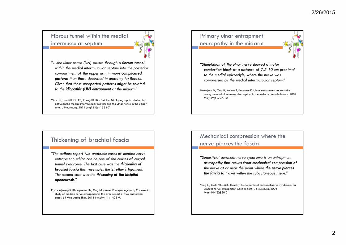
Superficial peroneal
nerve
SUPERFICIAL FASCIA
DEEP FASCIA
MUSCLE
EPIMYSIUM
SUPERFICIAL FASCIA
Deep fascia entrapment
Around the nerve there are
always fat and loose
connective tissue
NERVE
Large nerve fibres and deep fascia
The larger nerve fibres are often
surrounded by different layers of loose
connective tissue that preserves the
nerve from traction to which the fascia is
subjected.
Layers
physiology
� longitudinal movement of
nerve
Loose connective tissue: GAG, adipose tissue, hyaluronic acid
2/26/2015
4
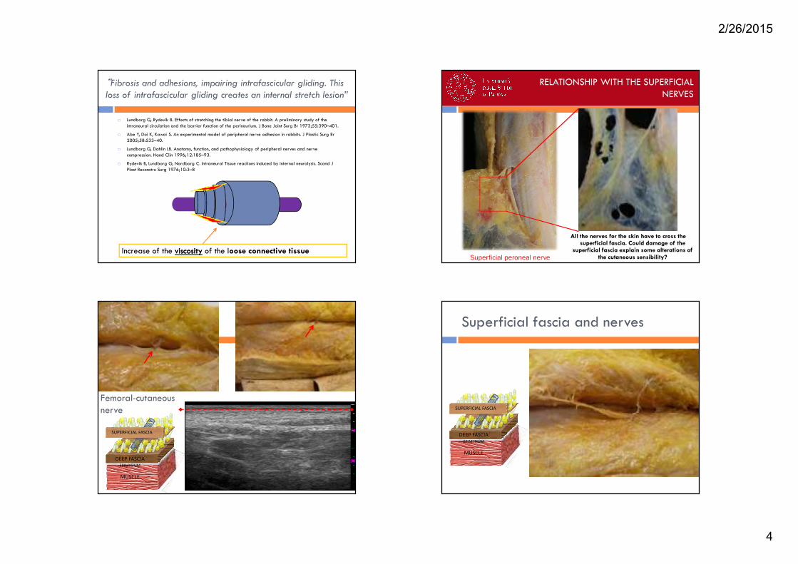
“Fibrosis and adhesions, impairing intrafascicular gliding. This
loss of intrafascicular gliding creates an internal stretch lesion”
� Lundborg G, Rydevik B. Effects of stretching the tibial nerve of the rabbit. A preliminary study of the
intraneural circulation and the barrier function of the perineurium. J Bone Joint Surg Br 1973;55:390–401.
� Abe Y, Doi K, Kawai S. An experimental model of peripheral nerve adhesion in rabbits. J Plastic Surg Br
2005;58:533–40.
� Lundborg G, Dahlin LB. Anatomy, function, and pathophysiology of peripheral nerves and nerve
compression. Hand Clin 1996;12:185–93.
� Rydevik B, Lundborg G, Nordborg C. Intraneural Tissue reactions induced by internal neurolysis. Scand J
Plast Reconstru Surg 1976;10:3–8
Increase of the viscosityviscosity of the loose connective tissue
RELATIONSHIP WITH THE SUPERFICIAL
NERVES
Superficial peroneal nerve
All the nerves for the skin have to cross the superficial fascia. Could damage of the
superficial fascia explain some alterations of the cutaneous sensibility?
Femoral-cutaneous
nerve
DEEP FASCIA
MUSCLE
EPIMYSIUM
SUPERFICIAL FASCIA
Superficial fascia and nerves
DEEP FASCIA
MUSCLE
EPIMYSIUM
SUPERFICIAL FASCIA
2/26/2015
5
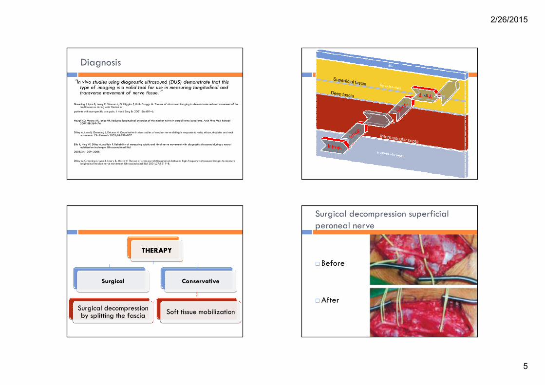
Diagnosis
“In vivo studies using diagnostic ultrasound (DUS) demonstrate that this type of imaging is a valid tool for use in measuring longitudinal and transverse movement of nerve tissue.”
Greening J, Lynn B, Leary R, Warren L, O’Higgins P, Hall- Craggs M. The use of ultrasound imaging to demonstrate reduced movement of the median nerve during wrist flexion in
patients with non-specific arm pain. J Hand Surg Br 2001;26:401–6.
Hough AD, Moore AP, Jones MP. Reduced longitudinal excursion of the median nerve in carpal tunnel syndrome. Arch Phys Med Rehabil 2007;88:569–76.
Dilley A, Lynn B, Greening J, DeLeon N. Quantitative in vivo studies of median nerve sliding in response to wrist, elbow, shoulder and neck movements. Clin Biomech 2003;18:899–907.
Ellis R, Hing W, Dilley A, McNair P. Reliability of measuring sciatic and tibial nerve movement with diagnostic ultrasound during a neural mobilisation technique. Ultrasound Med Biol
2008;34:1209–2008.
Dilley A, Greening J, Lynn B, Leary R, Morris V. The use of cross-correlation analysis between high-frequency ultrasound images to measure longitudinal median nerve movement. Ultrasound Med Biol 2001;27:1211–8.
THERAPYTHERAPY
Surgical
Surgical decompression by splitting the fascia
Conservative
Soft tissue mobilization
Surgical decompression superficial
peroneal nerve
� Before
�After
2/26/2015
6
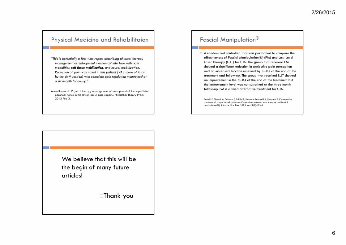
Physical Medicine and Rehabilitaion
“This is potentially a first-time report describing physical therapy
management of entrapment mechanical interface with pain
modalities, soft tissue mobilization, and neural mobilization.
Reduction of pain was noted in this patient (VAS score of 0 cm
by the sixth session) with complete pain resolution maintained at
a six-month follow-up.”
Anandkumar S.; Physical therapy management of entrapment of the superficial
peroneal nerve in the lower leg: A case report.; Physiother Theory Pract.
2012 Feb 2.
Fascial Manipulation®
� A randomized controlled trial was performed to compare the
effectiveness of Fascial Manipulation(®) (FM) and Low-Level
Laser Therapy (LLLT) for CTS. The group that received FM
showed a significant reduction in subjective pain perception
and an increased function assessed by BCTQ at the end of the
treatment and follow-up. The group that received LLLT showed
an improvement in the BCTQ at the end of the treatment but
the improvement level was not sustained at the three month
follow-up. FM is a valid alternative treatment for CTS.
Pratelli E, Pintucci M, Cultrera P, Baldini E, Stecco A, Petrocelli A, Pasquetti P. Conservative
treatment of carpal tunnel syndrome: Comparison between laser therapy and fascial
manipulation(®). J Bodyw Mov Ther. 2015 Jan;19(1):113-8.
We believe that this will be
the begin of many future
articles!
�Thank you