Embed Size (px)
Citation preview

R E V I E WAustralian Dental Journal 2012; 57: 2–10
doi: 10.1111/j.1834-7819.2011.01640.x
Clinical considerations for increasing occlusal verticaldimension: a review
J Abduo,* K Lyons�
*Faculty of Dentistry, The University of Western Australia, Crawley, Western Australia, Australia.�Department of Oral Rehabilitation, Faculty of Dentistry, University of Otago, Dunedin, New Zealand.
ABSTRACT
The purpose of this article is to discuss the clinical considerations related to increasing the occlusal vertical dimension(OVD) when restoring a patient’s dentition. Thorough extraoral and intraoral evaluations are mandatory to assess thesuitability of increasing OVD. In the literature, multiple techniques have been proposed to quantify OVD loss. However, thetechniques lack consistency and reliability, which in turn affects the decision of whether to increase the OVD. Therefore,increasing OVD should be determined on the basis of the dental restorative needs and aesthetic demands. In general, aminimal increase in OVD should be applied, though a 5 mm maximum increase in OVD can be justified to provide adequateocclusal space for the restorative material and to improve anterior teeth aesthetics. The literature reflects the safety ofincreasing the OVD permanently, and although signs and symptoms may develop, these are usually of an interim nature.Whenever indicated, the increase in OVD should be achieved with fixed restorations rather than a removable appliance, dueto the predictable patient adaptation. The exception to this is for patients with TMD, where increasing the OVD should stillbe achieved using removable appliances to control TMD-associated symptoms before considering any form of irreversibleprocedure.
Keywords: Occlusal vertical dimension, facial aesthetics, temporomandibular disorder, tooth wear, occlusion.
Abbreviations and acronyms: CLS = crown lengthening surgery; IORS = interocclusal rest space; OVD = occlusal vertical dimension;TMD = temporomandibular disorder; TMJ = temporomandibular joint.
(Accepted for publication 15 September 2011.)
INTRODUCTION
The Glossary of Prosthodontic Terms defines thevertical dimension as the distance between two selectedanatomic points.1 The vertical dimension when themandibular teeth are occluding with the maxillary teethis defined as the occlusal vertical dimension (OVD).The OVD for dentate individuals is mainly determinedby the remaining dentition, hence loss of tooth sub-stance might influence the OVD. A loss of OVD cansignificantly affect patient function, comfort andaesthetics.2
Several authors have commented on the dyna-mic nature of the dentoalveolar complex and masti-catory system.3–6 So, whilst the loss of OVD is apossible consequence of tooth wear, the originalOVD can be preserved by a dentoalveolar compen-satory mechanism involving the extrusion of wornteeth.3–6
Increasing the OVD from the clinical perspective hasbeen reported to facilitate the treatment of patientspresenting with generalized and complex dentalabnormalities such as generalized tooth wear andsignificant occlusal irregularities.7–9 However, there isstill considerable debate in the literature abouttreatment modalities used to increase OVD. Someauthors have assumed that the OVD is constantthroughout an individual’s life, and any alteration ofthe OVD will subsequently interfere with thephysiology of the masticatory system and the patient’sability to adapt.10,11 The reported consequences ofincreasing the OVD are hyperactivity of the masticatorymuscles, elevation in occlusal forces, bruxism andtemporomandibular disorders (TMDs).2,10,11 On thecontrary, other authors have reported that suchsymptoms are transitory.12–15
Although evidence regarding the implications ofincreasing OVD is still lacking, the rehabilitative
2 ª 2012 Australian Dental Association
Australian Dental JournalThe official journal of the Australian Dental Association
procedures involving the increase in OVD should beapproached with caution. The aim of this narrativereview article is to discuss the clinical considerationsrelated to increasing the OVD.
CLINICAL EVALUATION
In contemporary dentistry, emphasis should be placedon conservative management strategies.16 Since increas-ing the OVD by restorative means involves multipleteeth in at least one arch, it is regarded as an extensive,costly and time-consuming procedure. Prevention strat-egies and conservative measures should be the clini-cian’s main priority. Conservative management forpatients with reduced vertical tooth height includesdietary counselling, fluoride application, exclusion ofdietary disorders, controlling parafunctional habits andmanagement of gastro-oesophageal reflux disorder.As the prevention of tooth wear is not the purpose ofthis article, the readers are referred to other referenceson this topic.7,8,17,18 Nevertheless, it is important tostate that increasing the OVD should only be consid-ered where comprehensive prosthodontic rehabilitationis justified.
Comprehensive extraoral and intraoral assessmentsare mandatory before considering an increase in theOVD. This is important since increasing the OVD isnormally part of a comprehensive rehabilitation ratherthan a single treatment modality. A thorough assess-ment process should reveal the merits of altering theOVD and allow the clinician to consider suitabletreatment options. Given that the standard patientexamination procedure is followed, the followingextraoral and intraoral assessments should be consid-ered for patients in need of an increase in OVD.
Extraoral considerations
The literature suggests several extraoral factors beconsidered prior to the clinical decision to increase theOVD. These include the magnitude of OVD loss, facialprofile and aesthetics, and status of the TMJ.
Magnitude of OVD loss
Many authors recommend an evaluation of an actualversus apparent loss of OVD.2,19,20 One means ofevaluation is the use of interocclusal rest space (IORS),i.e. the difference in vertical dimension between whenthe mandible is at rest and when the mandible is inocclusion.1 For dentate individuals, the initial referenceis the OVD of the existing dentition. Subsequently, thevertical dimension when the mandible is at rest can beevaluated clinically. The rationale behind measuring theIORS is to determine how much to increase the OVD.An IORS of 2 mm has been suggested as the physiolog-
ical space, and therefore an IORS of more than 2 mmindicates that the OVD can be safely increased.2
However, the literature suggests that there are fourlimitations associated with positioning the mandible atrest: (1) for the same individual, different mandibularpositions can be obtained at different examinationperiods. This has been attributed to the influence ofmuscle activity and fatigue.21,22 A suggestion has beenmade that the true rest position of the mandible, whereall the muscles are relaxed, does not exist;23 (2) loss ofOVD is associated with a parallel loss of the verticaldimension when the mandible is at rest. This means theIORS is vulnerable to a similar loss in dimension to theOVD.24,25 Such a phenomenon would underestimatethe IORS and, subsequently, the loss in OVD; (3) themandibular rest position occurs at a zone rather than aspecific level. This finding is supported by clinical studiesthat have confirmed the ability of the patient to adaptafter increasing the OVD;12–15,26,27 and (4) there issubstantial variation between clinicians in evaluatingthe resting position of the mandible. Clinically, anaccurate determination of the vertical dimension isdifficult when the landmarks are located on movableskin tissues,28 and where the mean facial measurementcould account for only half the skeletal movement.29
Two questions would seem relevant for any givenclinical situation: what is the most reliable technique fordetermining OVDloss? And what is the significance of anysuch loss? Unfortunately, both questions have not beenanswered in the literature. Table 1 presents the availableclinical techniques to determine the loss of OVD. Ingeneral, many of the proposed techniques have beenadapted from complete dentures fabrication procedures.Although all the stated techniques have been found to beuseful, none have been assessed to be scientifically moreaccurate than another.30 It has been suggested that inorder to improve the accuracy of the recording procedure,more than one method should be used.19
The available clinical trials that increased theOVD beyond IORS (4–5 mm inter-incisally) did notreveal patient maladaptation or pathological reac-tions.12–15,26,27 On this basis, it could be stated thatthe determination of the OVD increase should not bebased on IORS values.
Facial aesthetics
The determinants of facial aesthetics are the sagittalprofile, facial tissues appearance, lip morphology andteeth display.31 Sagittal assessment of the face can revealmandibular pseudo-prognathism which might be a signof OVD loss and overclosure of the mandible. Thisobservation has been confirmed clinically7 and anthro-pologically.32 On the basis of a cephalometric analysisof dry skulls, Fishman found that tooth wear resultedin a reduction of arch width and gonial angle that
ª 2012 Australian Dental Association 3
Increasing occlusal vertical dimension

may contribute to the overall mandibular pseudo-prognathism.33 Likewise, Varrela found that a worndentition is associated with a reduced gonial angle andreduced face height.34 Crothers anticipated mandibularpseudo-prognathism to develop from one or more of thefollowing factors: loss of OVD and subsequent forwardrotation of the mandible; dentofacial bone remodellingafter tooth wear; an edge-to-edge anterior tooth rela-tionship after loss of vertical tooth height; and anteriorpositioning of the mandible due to the loss of anteriortooth guidance.24 The severity of mandibular pseudo-prognathism can be subjectively assessed by reviewing anold photograph of a patient’s facial profile. Althoughincreasing the OVD reduces the pseudo-prognathism ofthe mandible,24 the significance of this effect is doubtfulsince increasing the OVD for dentate individuals islimited to 5 mm inter-incisally, which may not besufficient to induce facial alterations.
From the frontal view, several facial implications canmanifest after loss of OVD including altered facialcontour, narrowed vermillion borders and an overclosedcommissure.24 These implications are exacerbated byincreased mandibular pseudo-prognathism.24 As long asthe lip competence is not compromised, it is thought that
increasing the OVD might reverse the consequence ofOVD loss and restore facial morphology.28,35 Mohindraand Bulman reported an improvement in facial aestheticsby the insertion of complete dentures constructed at anincreased OVD.36 However, Gross et al. reported thatafter experimental increase of the OVD by 2–6 mm fordentate individuals, there was an insignificant extraoralimprovement of facial tissues appearance.37 This findingcan be attributed to the significant loss in OVD foredentulous individuals without compensation in com-parison to dentate individuals. In addition to increasingOVD, the effect complete dentures have on facialaesthetics could be related to horizontal support of thefacial tissues from the dentures.
The upper lip position in relation to the incisal edgesof maxillary anterior teeth determines the teeth displaywhile smiling and at rest.31 Insufficient display of themaxillary anterior teeth can be improved by loweringthe occlusal surface of the maxillary teeth. Further,increasing the OVD allows the establishment of anincisal overjet that can augment the support of themaxillary lips. Subsequently, an overbite can be incor-porated which can allow the maxillary incisal edge tobe placed parallel to the lower lip, rendering a more
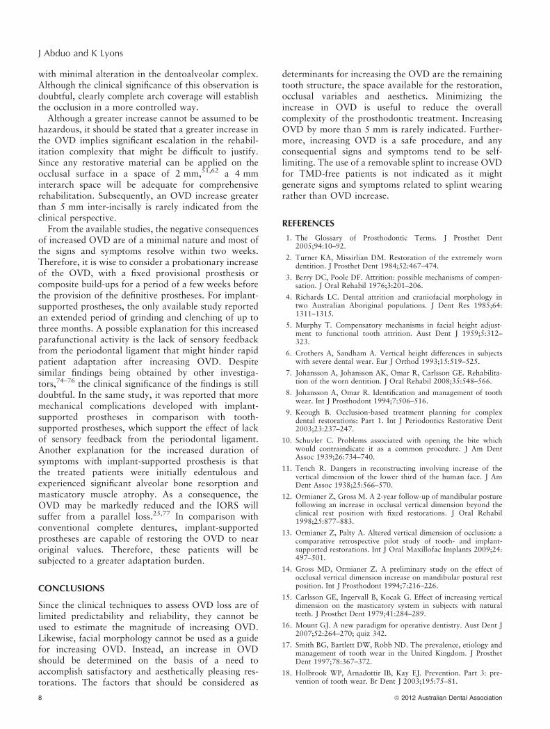
Table 1. Described clinical techniques for assessment of OVD loss
Technique Description Advantages Disadvantages
Pre-treatmentrecord
– Visual assessment of old diagnosticmodels
– Previous photograph
– Approximates the loss of clinicalcrown height78
– Formulates baseline record8
– Old models are rarely availablebefore treatment79
Incisors heightmeasurement
– The distance between the gingivalmargins of the maxillary andmandibular anterior teeth whenthey are in occlusion. A distance ofless than 18 mm indicates loss ofOVD
– Approximates the loss of clinicalcrown height
– Applicable clinically– Aesthetically relevant– Measures the severity of tooth
wear80
– Poorly represents the actual loss ofOVD5
– Affected by the original anteriortooth relationship
Phoneticevaluation
– S sound to measure the closestspeaking space
– F sound to locate the incisal edgesof anterior maxillary teeth
– M sound to locate the mandible inrest position
– Reproducible81
– Applicable clinically– Indicates patient adaptation after
loss of tooth tissues– Indicates incisal tooth relationship– Locates the incisal edges of maxillary
anterior teeth in relation to lowerlip77
– Variable outcome for patients withClass II and III occlusions19
– Poorly represents the actual loss ofOVD82
– More useful for complete denturesconstruction28,77
Patientrelaxation
– Mandible positioning at rest – Applicable clinically– Visualizes the facial appearance
at rest83
– Ensures the lips are meeting
– Minor muscles tension will lead toinaccurate measurements28,84
Assessmentof facialappearance
– Evaluation of facial tissues andmusculature at rest
– Applicable clinically– Visualizes the facial appearance
at rest83
– Ensures the lips are meeting
– Arbitrary evaluation of the facialaesthetics28,84
Radiographicevaluation
– Cephalometric assessment ofmaxillomandibular relationship
– Highly accurate andreproducible85,86
– Indicates incisal tooth relationship87
– Controlled setting is mandatory– Additional equipment and
radiation85
Neuromuscularevaluation
– Recording EMG muscle activitieswhere minimal muscle activityindicates the mandible is at restposition
– Useful clinical and research tool forOVD evaluation88,89
– Accurate and reproducible90,91
– The devices are rarely available in theclinical setting
– Great expertise is required– Rigorously controlled recording
conditions are necessary89
4 ª 2012 Australian Dental Association
J Abduo and K Lyons

aesthetic appearance.31 On the contrary, excessivedisplay of the gingival tissues will not be improved byincreasing OVD. Rather, aesthetic crown lengtheningsurgery (CLS) should be considered.38,39
It could be speculated that although the loss of OVDcan lead to changes in sagittal profile and facial tissuesappearance, there is no compelling evidence thatincreasing the OVD for dentate individuals by restor-ative means reverses these morphological changes.Therefore, it is important to emphasize that increasingOVD is not indicated to improve facial aesthetics.Nevertheless, teeth display might improve by loweringthe maxillary occlusal plane after increasing OVD.
Temporomandibular joint status
The prevalence of temporomandibular joint disorders(TMDs) has been reported to be 7–10% within thepopulation.40,41 Therefore, it is not uncommon toencounter patients with signs and symptoms of TMDseeking routine dental care. However, TMD has beenfound to primarily affect young and middle agedadults.40,42 Considering that this group of patientsmight not suffer from significant loss of OVD,43 itcould be speculated that the development of TMD isnot associated with the loss of OVD. This assumptionis supported by the clinical observation that attrition isnot associated with an increased prevalence of TMD.44
Through routine clinical assessment, it is critical toassess the status of the temporomandibular joint (TMJ)before intervention therapy. TMJ evaluation is com-prised of assessment of joint and muscle pain, mandib-ular movement and associated sounds.7,8 Despite thelack of compelling evidence supporting a relationshipbetween the OVD and TMD, TMJ evaluation willallow observation of the initial TMJ status of thepatient. Even if increasing OVD may not exacerbateTMD signs and symptoms, patient adaptation might bemasked by the pre-existing discomfort. Therefore,comprehensive restorative treatment involving anincrease in OVD should be approached with cautionfor patients with TMD. Multiple authors have sug-gested stabilizing TMD patients and minimizing thesigns and symptoms with a removable occlusal appli-ance before the commencement of irreversible prosth-odontic treatment.7,45
To date, there is more evidence to support conserva-tive management of TMD such as with occlusalappliances, behavioural therapy, physiotherapy andjaw exercises than permanent occlusal alteration thathas not yet been proven.46–48 Where there is a genuineneed to increase OVD, it should be carried out using aconservative method such as with an occlusal appli-ance.46,47,49 Therefore, for patients with TMD, theocclusal appliance has a dual purpose: stabilizing theTMD and increasing OVD. The intended permanent
increase in the OVD can be incorporated into theocclusal appliance. On the basis of patient adaptationto the occlusal appliance, permanent restoration at theincreased OVD can then be performed.45,50
Intraoral considerations
Intraoral assessment involves examining the followingparameters: remaining tooth structure and occlusion.
Remaining tooth structure
The prognosis of a dental restoration is directlydetermined by the amount of remaining tooth struc-ture.51 For generalized loss of vertical tooth height, theclinician is faced with the dilemma of limited remainingtooth structure that is necessary for adequate retentionand resistance of the restoration. The original toothheight determines the active preparation height,which can be defined as the vertical distance betweenthe preparation margin and the occlusal-axial lineangle. In order to avoid compromising the preparationheight, increasing the OVD should be considered toprovide adequate space to accommodate the restorativematerial. The merit behind this technique is moreprominent in generalized loss of tooth height mani-fested from tooth wear. As a result of this approach, theteeth will be subjected to less pulpal trauma. Inaddition, by utilizing the available vertical height ofthe tooth, the indication for adjunctive crown length-ening surgery is minimized.
Given that tooth preparation taper for a crown is10–20 degrees for a posterior tooth, according toParker’s et al. calculations, 3 mm is the minimalpreparation height.52 Similar findings were confirmedby Maxwell et al. regarding anterior teeth andpremolars.53 Since only 46% of prepared molarsexhibit an adequate resistance form,54 according toGoodacre et al.,51 at least 4 mm is recommended as theminimal preparation height. If this height is notavailable, then auxiliary retention and resistancefeatures should be incorporated. Therefore, withincreasing OVD, it is possible to crown teeth with anoriginal clinical crown height of 3 mm withoutadjunctive therapy. As a result, it appears that the finalpreparation height is a critical determinant of the needand the magnitude of the OVD increase.
When there is limited vertical tooth height, analternative approach to increasing OVD is CLS.2
However, the possible sequelae of CLS of multipleteeth in an arch are loss of a significant amount of softand hard tissues, the effect on the emergence profile andthe development of a black triangle. The exposure ofroot surfaces excludes the use of adhesive restorations,and necessitates restoring the crown lengthened teethwith full coverage restorations. In relation to aesthetics
ª 2012 Australian Dental Association 5
Increasing occlusal vertical dimension

of the anterior teeth, CLS is an excellent procedure toimprove the contour of the gingival tissues and enhancethe aesthetic display of the anterior teeth for patientswith a high or average lip line when smiling.38,39
However for a low lip line, there will be minimalimprovement of the aesthetic display unless the OVD isincreased. Further, CLS by itself will not improve therelationship of the anterior teeth. One of the concernsassociated with CLS is the increase in the crown-to-rootratio that might be attributed to increased teethmobility and a compromised prognosis. However, thereis no compelling evidence regarding the negative effectof an increased crown-to-root ratio.55 A recent system-atic review reported that a severely reduced but healthyperiodontal support is not a compromising factor forthe longevity of teeth utilized as abutments for fixeddental prostheses.56
For a clinical crown height of less than 3 mm, CLS isthe only means of providing for adequate preparationheight by exposing more tooth structure. Nevertheless,for excessively short teeth, the rehabilitative treatmentcan be a combination of increasing OVD and CLS asan adjunctive treatment. The clinician should decideon the best compromise of the multiple treatmentoptions to minimize the invasiveness of the overalltreatment.
With the continuous development of adhesive tech-nologies, it is possible to bond an onlay restoration tothe remaining tooth structures, even if the remainingstructure is less than 3 mm. The advantages of adhesiverestorations are the conservative nature of the operativeprocedure in relation to the tooth and periodontaltissues, and less clinical time required for the applica-tion and completion of the treatment. However,significant care should be taken while bonding therestoration to dentine and the maximum amount ofenamel should be used.57 The available materials forbonding are metal, ceramic and composite resin.
Chana et al. reported a 89% survival rate of resinbonded metal veneers for a duration of 60 months.58
Likewise, Jamous et al. found that 80% of resinbonded metal restorations survived after seven years.59
In relation to ceramic onlays, Wagner et al. reportedthat the survival of ceramic onlays was 81% in sevenyears. In the same study, they found that the perfor-mance of ceramic onlays is comparable to metalonlays.60 Similarly, Otto and Schneider found thesurvival rate for ceramic onlays to be 89% up to17 years.61 As a simpler option, Hemmings et al. haveshown favourable short- to medium-term performanceof direct composite resin restorations when placed in athickness of 2 mm or more.62 Poyser et al. reported asurvival rate of 94% after two years for composite resinrestorations placed at an increased OVD.63 Compositeresin restorations have the advantages of ease of repairor modification. However, they still suffer from wear,
margin staining, marginal fracture and surface rough-ness.64 Therefore, it appears that increasing the OVDby direct composite resin restorations is a predictablemedium-term option, while metal or ceramic onlays aremore adequate as long-term options.
Occlusion
Clinically, unopposed teeth have been reported to beprone to overeruption, which can create occlusalinterferences.65 For some patients, increasing OVDfacilitates occlusion reorganization and the achieve-ment of an even occlusal plane.9 Subsequently, aninvasive sacrifice of tooth structure can be avoided.
As a result of a worn anterior dentition, the mandibletends to be habitually located more anteriorly. Byrecording the difference in the horizontal mandibularposition when the mandible is in centric relation andmaximal intercuspation, a horizontal space can beobtained inter-incisally.66 This space can be utilized toprovide adequate room for restoration of the anteriorteeth. The advantage of using this method is thefeasibility of restoring worn anterior teeth withoutincreasing the OVD.
Loss of posterior tooth support has been cited asprobably the main cause for loss of OVD in dentateindividuals.2 The implications of losing the posteriorteeth are the overloading of the remaining anterior teethand increasing the potential to wear. A nine-yearclinical trial comparing the occlusal stability of patientswith complete dental arches and shortened dentalarches revealed that patients of both groups exhibiteda similar overbite and occlusal tooth wear.67 Moreanterior teeth in the shortened dental arch group werein occlusion. Since the occlusion of the shortened dentalarch group exhibited relative stability, the authorsconcluded that a new occlusal equilibrium wasobtained.67 On the contrary, one cross-sectional studyconfirmed that patients with an extremely diminishedposterior tooth support (0 to 2 occluding units) tendedto exhibit an anterior dentition with more prominentspacing, heavier occlusal contacts, occlusal wear,mobility and vertical overlap.68 All of these findingscan eventually lead to the loss of OVD. Therefore, forpatients with extremely shortened dental arch, it isimportant to eliminate the potential cause of OVD lossby achieving a stable posterior occlusion before con-sidering increasing the OVD.
Patients with a worn anterior dentition suffer from aloss of clinical crown height and the possibility ofdevelopment of an edge-to-edge incisal relationship.6,7
As a result, the aesthetic appearance is affected and theanterior guidance is lost.69 In addition to an aestheticimprovement, increasing the OVD rectifies the anteriortooth relationship, by re-establishing an overjet andoverbite, and facilitating the establishment of anterior
6 ª 2012 Australian Dental Association
J Abduo and K Lyons

tooth guidance.9,69 According to the modern theories ofocclusion, anterior tooth guidance is desirable as it isbelieved to protect the posterior teeth in eccentricmovements.70–72
Patients with a steep anterior tooth guidance canbenefit significantly from increasing OVD as it willalleviate the broad area of anterior tooth contacts andprovide shallower and less constrained angle of disclu-sion.9,69 Even though a steep anterior tooth guidancedoes not appear to be contributory to the developmentof pathological signs and symptoms, it still poses adaunting challenge for the restoration of anteriorteeth.69
Therefore, increasing the OVD facilitates reorgani-zation of the occlusion by elimination of occlusalinterferences, provision of adequate overjet and over-bite, and alleviation of steep anterior tooth guidance.
FEASIBILITY OF INCREASING OVD
Increasing OVD has been considered by some authorsto be a hazardous procedure that can violate a patient’sdental physiology and adaptation.10,11 The basis ofsuch claims is the thought that OVD occurs at a specificlevel that should be maintained through an individual’slife.2
In the literature, multiple articles have challenged thehypothesis of the negative implications of increasingOVD beyond the IORS.12–15,26,27 In general, theiroutcomes reflect the safety, patient adaptation andpredictability of increasing the OVD. This is true inrelation to TMJ and masticatory muscle health. How-ever, the available studies suffer from a lack ofrandomization and control group. In addition, signif-icant variation exists in relation to the subjectivemethods to assess patient adaptation. All the availablestudies had a limited number of participants and itcould be assumed that they are not representative of thewhole population.
Carlsson et al. increased the OVD by 4 mm for sixparticipants with removable appliances temporarilycemented on the occlusal surface of the mandibularposterior teeth and the canines. After seven days,despite all the participants reported subjective symp-toms, five of them reported resolution of the symptomswithin two days. One participant could not adapt to theintervention.15 However, the maldaptation could havebeen due to the appliance design and associatedbulkiness rather than the increase in OVD. In twostudies, Dahl and Krogstad increased the OVD for 20participants up to 4.7 mm by using anterior removablesplints. All symptoms resolved within two weeks, withlisping being the most common symptom.26,27 Like-wise, Gross and Ormianer reported resolution of minorsymptoms after two weeks of increasing the OVD up to4.5 mm with fixed prostheses.14 The eight participants
were reviewed in a follow-up study that confirmed thelong-term patient adaptation after increasing OVD.12
More recently, in a retrospective study by Ormianerand Palty, the OVD was increased up to 5 mm for 30patients requiring whole arch prostheses supported byteeth or implants.13 Despite all the patients adapted tothe increase in OVD, a few patients with implant-supported prostheses suffered from prolonged grindingthat resolved within 2–3 months after administering anocclusal splint.
In relation to the method of increasing OVD, thestudies that increased OVD with fixed prostheses12,14
indicated less symptom severity than the studies thatincreased OVD with a removable appliance.15,26,27
This outcome could be attributed to the fixed prosthe-ses having the advantages of being fixed in the mouth,mimicking natural tooth morphology, minimizingbulkiness with reduced interference with speech andimproved overall comfort. In addition, the fixed natureof the prosthesis may enhance patient compliance andacceptance of the treatment. Therefore, wheneverpossible, the increase in OVD should be performedfor TMD-free patients with fixed restorations ratherthan with a removable appliance. Removable appli-ances could be a source of patient maladaptation due tofactors other than increased OVD.
In relation to the magnitude of increasing the OVD, anincrease of up to 5 mm inter-incisally is a feasiblealteration.12–15,26,27 Such outcomes support the assump-tion of other investigations that physiological OVDoccurs at a range, commonly known as the comfort zone,rather than a specific constant level. Subsequently, itcould be expected that the patient can adapt to analteration in OVD as long as it is confined to this zone.
The possible adaptation mechanisms to an increasedOVD could be masticatory muscle lengthening andrelaxation, dentoalveolar maturation, or a combinationof these two mechanisms. In a two-year study, afterincreasing OVD by covering the whole arch, Ormianerand Gross found that relapse of the OVD to its originalvalue was minimal.12 This finding supports the theorythat muscle relaxation and changes in muscle lengthwere the primary adaptation mechanisms,73 rather thanreturning to the original OVD by dentoalveolar matu-ration. Further, this outcome is in accordance with thefinding of Ormianer and Palty that reported patientadaptation even when implant-supported prostheseswere utilized.13 On the contrary, after increasing OVDby covering the anterior teeth only, Dahl and Krogstadreported that occlusal stability was obtained orthodon-tically by intrusion of the occluding segments of thearch and extrusion of the non-occluding segments ofthe arch.27 Therefore, it could be assumed that an OVDincrease by partial arch coverage will lead to dento-alveolar alteration, while the complete arch coveragewill lead to immediate establishment of an occlusion
ª 2012 Australian Dental Association 7
Increasing occlusal vertical dimension
with minimal alteration in the dentoalveolar complex.Although the clinical significance of this observation isdoubtful, clearly complete arch coverage will establishthe occlusion in a more controlled way.
Although a greater increase cannot be assumed to behazardous, it should be stated that a greater increase inthe OVD implies significant escalation in the rehabil-itation complexity that might be difficult to justify.Since any restorative material can be applied on theocclusal surface in a space of 2 mm,51,62 a 4 mminterarch space will be adequate for comprehensiverehabilitation. Subsequently, an OVD increase greaterthan 5 mm inter-incisally is rarely indicated from theclinical perspective.
From the available studies, the negative consequencesof increased OVD are of a minimal nature and most ofthe signs and symptoms resolve within two weeks.Therefore, it is wise to consider a probationary increaseof the OVD, with a fixed provisional prosthesis orcomposite build-ups for a period of a few weeks beforethe provision of the definitive prostheses. For implant-supported prostheses, the only available study reportedan extended period of grinding and clenching of up tothree months. A possible explanation for this increasedparafunctional activity is the lack of sensory feedbackfrom the periodontal ligament that might hinder rapidpatient adaptation after increasing OVD. Despitesimilar findings being obtained by other investiga-tors,74–76 the clinical significance of the findings is stilldoubtful. In the same study, it was reported that moremechanical complications developed with implant-supported prostheses in comparison with tooth-supported prostheses, which support the effect of lackof sensory feedback from the periodontal ligament.Another explanation for the increased duration ofsymptoms with implant-supported prosthesis is thatthe treated patients were initially edentulous andexperienced significant alveolar bone resorption andmasticatory muscle atrophy. As a consequence, theOVD may be markedly reduced and the IORS willsuffer from a parallel loss.25,77 In comparison withconventional complete dentures, implant-supportedprostheses are capable of restoring the OVD to nearoriginal values. Therefore, these patients will besubjected to a greater adaptation burden.
CONCLUSIONS
Since the clinical techniques to assess OVD loss are oflimited predictability and reliability, they cannot beused to estimate the magnitude of increasing OVD.Likewise, facial morphology cannot be used as a guidefor increasing OVD. Instead, an increase in OVDshould be determined on the basis of a need toaccomplish satisfactory and aesthetically pleasing res-torations. The factors that should be considered as
determinants for increasing the OVD are the remainingtooth structure, the space available for the restoration,occlusal variables and aesthetics. Minimizing theincrease in OVD is useful to reduce the overallcomplexity of the prosthodontic treatment. IncreasingOVD by more than 5 mm is rarely indicated. Further-more, increasing OVD is a safe procedure, and anyconsequential signs and symptoms tend to be self-limiting. The use of a removable splint to increase OVDfor TMD-free patients is not indicated as it mightgenerate signs and symptoms related to splint wearingrather than OVD increase.
REFERENCES
1. The Glossary of Prosthodontic Terms. J Prosthet Dent2005;94:10–92.
2. Turner KA, Missirlian DM. Restoration of the extremely worndentition. J Prosthet Dent 1984;52:467–474.
3. Berry DC, Poole DF. Attrition: possible mechanisms of compen-sation. J Oral Rehabil 1976;3:201–206.
4. Richards LC. Dental attrition and craniofacial morphology intwo Australian Aboriginal populations. J Dent Res 1985;64:1311–1315.
5. Murphy T. Compensatory mechanisms in facial height adjust-ment to functional tooth attrition. Aust Dent J 1959;5:312–323.
6. Crothers A, Sandham A. Vertical height differences in subjectswith severe dental wear. Eur J Orthod 1993;15:519–525.
7. Johansson A, Johansson AK, Omar R, Carlsson GE. Rehabilita-tion of the worn dentition. J Oral Rehabil 2008;35:548–566.
8. Johansson A, Omar R. Identification and management of toothwear. Int J Prosthodont 1994;7:506–516.
9. Keough B. Occlusion-based treatment planning for complexdental restorations: Part 1. Int J Periodontics Restorative Dent2003;23:237–247.
10. Schuyler C. Problems associated with opening the bite whichwould contraindicate it as a common procedure. J Am DentAssoc 1939;26:734–740.
11. Tench R. Dangers in reconstructing involving increase of thevertical dimension of the lower third of the human face. J AmDent Assoc 1938;25:566–570.
12. Ormianer Z, Gross M. A 2-year follow-up of mandibular posturefollowing an increase in occlusal vertical dimension beyond theclinical rest position with fixed restorations. J Oral Rehabil1998;25:877–883.
13. Ormianer Z, Palty A. Altered vertical dimension of occlusion: acomparative retrospective pilot study of tooth- and implant-supported restorations. Int J Oral Maxillofac Implants 2009;24:497–501.
14. Gross MD, Ormianer Z. A preliminary study on the effect ofocclusal vertical dimension increase on mandibular postural restposition. Int J Prosthodont 1994;7:216–226.
15. Carlsson GE, Ingervall B, Kocak G. Effect of increasing verticaldimension on the masticatory system in subjects with naturalteeth. J Prosthet Dent 1979;41:284–289.
16. Mount GJ. A new paradigm for operative dentistry. Aust Dent J2007;52:264–270; quiz 342.
17. Smith BG, Bartlett DW, Robb ND. The prevalence, etiology andmanagement of tooth wear in the United Kingdom. J ProsthetDent 1997;78:367–372.
18. Holbrook WP, Arnadottir IB, Kay EJ. Prevention. Part 3: pre-vention of tooth wear. Br Dent J 2003;195:75–81.
8 ª 2012 Australian Dental Association
J Abduo and K Lyons

19. Rivera-Morales WC, Mohl ND. Restoration of the verticaldimension of occlusion in the severely worn dentition. Dent ClinNorth Am 1992;36:651–664.
20. Brown KE. Reconstruction considerations for severe dentalattrition. J Prosthet Dent 1980;44:384–388.
21. Tzakis M, Carlsson GE, Kiliaridis S. Effect of chewing trainingon mandibular postural position. J Oral Rehabil 1989;16:503–508.
22. Kiliaridis S, Katsaros C, Karlsson S. Effect of masticatory musclefatigue on cranio-vertical head posture and rest position of themandible. Eur J Oral Sci 1995;103:127–132.
23. Rugh JD, Drago CJ. Vertical dimension: a study of clinical rest po-sition and jaw muscle activity. J Prosthet Dent 1981;45:670–675.
24. Crothers AJ. Tooth wear and facial morphology. J Dent 1992;20:333–341.
25. Tallgren A, Lang BR, Walker GF, Ash MM Jr. Roentgen ceph-alometric analysis of ridge resorption and changes in jaw andocclusal relationships in immediate complete denture wearers.J Oral Rehabil 1980;7:77–94.
26. Dahl BL, Krogstad O. The effect of a partial bite raising splint onthe occlusal face height. An x-ray cephalometric study in humanadults. Acta Odontol Scand 1982;40:17–24.
27. Dahl BL, Krogstad O. Long-term observations of an increasedocclusal face height obtained by a combined orthodontic ⁄prosthetic approach. J Oral Rehabil 1985;12:173–176.
28. Toolson LB, Smith DE. Clinical measurement and evaluation ofvertical dimension. J Prosthet Dent 1982;47:236–241.
29. Tryde G, McMillan DR, Christensen J, Brill N. The fallacy offacial measurements of occlusal height in edentulous subjects.J Oral Rehabil 1976;3:353–358.
30. Rivera-Morales WC, Mohl ND. Relationship of occlusal verticaldimension to the health of the masticatory system. J ProsthetDent 1991;65:547–553.
31. Tjan AH, Miller GD, The JG. Some esthetic factors in a smile.J Prosthet Dent 1984;51:24–28.
32. Kaidonis JA. Tooth wear: the view of the anthropologist. ClinOral Investig 2008;12(Suppl 1):S21–S26.
33. Fishman LS. Dental and skeletal relationships to attritionalocclusion. Angle Orthod 1976;46:51–63.
34. Varrela J. Dimensional variation of craniofacial structures inrelation to changing masticatory-functional demands. Eur JOrthod 1992;14:31–36.
35. Kois JC, Phillips KM. Occlusal vertical dimension: alterationconcerns. Compend Contin Educ Dent 1997;18:1169–1174,1176-1167; quiz 1180.
36. Mohindra NK, Bulman JS. The effect of increasing verticaldimension of occlusion on facial aesthetics. Br Dent J 2002;192:164–168.
37. Gross MD, Nissan J, Ormianer Z, Dvori S, Shifman A. The effectof increasing occlusal vertical dimension on face height. Int JProsthodont 2002;15:353–357.
38. Jorgensen MG, Nowzari H. Aesthetic crown lengthening. Peri-odontol 2000 2001;27:45–58.
39. Wang HL, Greenwell H. Surgical periodontal therapy. Peri-odontol 2000 2001;25:89–99.
40. LeResche L. Epidemiology of temporomandibular disorders:implications for the investigation of etiologic factors. Crit RevOral Biol Med 1997;8:291–305.
41. List T, Wahlund K, Wenneberg B, Dworkin SF. TMD in childrenand adolescents: prevalence of pain, gender differences, andperceived treatment need. J Orofac Pain 1999;13:9–20.
42. Magnusson T, Egermarki I, Carlsson GE. A prospective investi-gation over two decades on signs and symptoms of temporo-mandibular disorders and associated variables. A final summary.Acta Odontol Scand 2005;63:99–109.
43. Van’t Spijker A, Rodriguez JM, Kreulen CM, Bronkhorst EM,Bartlett DW, Creugers NH. Prevalence of tooth wear in adults.Int J Prosthodont 2009;22:35–42.
44. Seligman DA, Pullinger AG, Solberg WK. The prevalence of dentalattrition and its association with factors of age, gender, occlusion,and TMJ symptomatology. J Dent Res 1988;67:1323–1333.
45. De Boever JA, Carlsson GE, Klineberg IJ. Need for occlusaltherapy and prosthodontic treatment in the management oftemporomandibular disorders. Part II: Tooth loss and prosth-odontic treatment. J Oral Rehabil 2000;27:647–659.
46. List T, Axelsson S, Leijon G. Pharmacologic interventions in thetreatment of temporomandibular disorders, atypical facial pain,and burning mouth syndrome. A qualitative systematic review.J Orofac Pain 2003;17:301–310.
47. De Boever JA, Carlsson GE, Klineberg IJ. Need for occlusaltherapy and prosthodontic treatment in the management oftemporomandibular disorders. Part I. Occlusal interferences andocclusal adjustment. J Oral Rehabil 2000;27:367–379.
48. Carlsson GE. Critical review of some dogmas in prosthodontics.J Prosthodont Res 2009;53:3–10.
49. Dao TT, Lavigne GJ. Oral splints: the crutches for temporo-mandibular disorders and bruxism? Crit Rev Oral Biol Med1998;9:345–361.
50. Davies SJ, Gray RM, Whitehead SA. Good occlusal practice inadvanced restorative dentistry. Br Dent J 2001;191:421–424,427-430, 433-424.
51. Goodacre CJ, Campagni WV, Aquilino SA. Tooth preparationsfor complete crowns: an art form based on scientific principles.J Prosthet Dent 2001;85:363–376.
52. Parker MH, Calverley MJ, Gardner FM, Gunderson RB.New guidelines for preparation taper. J Prosthodont 1993;2:61–66.
53. Maxwell AW, Blank LW, Pelleu GB Jr. Effect of crown prepa-ration height on the retention and resistance of gold castings.Gen Dent 1990;38:200–202.
54. Parker MH, Malone KH III, Trier AC, Striano TS. Evaluation ofresistance form for prepared teeth. J Prosthet Dent 1991;66:730–733.
55. Grossmann Y, Sadan A. The prosthodontic concept of crown-to-root ratio: a review of the literature. J Prosthet Dent 2005;93:559–562.
56. Lulic M, Bragger U, Lang NP, Zwahlen M, Salvi GE. Ante’s(1926) law revisited: a systematic review on survival rates andcomplications of fixed dental prostheses (FDPs) on severelyreduced periodontal tissue support. Clin Oral Implants Res2007;18(Suppl 3):63–72.
57. Tyas MJ, Burrow MF. Adhesive restorative materials: a review.Aust Dent J 2004;49:112–121.
58. Chana H, Kelleher M, Briggs P, Hooper R. Clinical evaluation ofresin-bonded gold alloy veneers. J Prosthet Dent 2000;83:294–300.
59. Jamous I, Sidhu S, Walls A. An evaluation of the performance ofcast gold bonded restorations in clinical practice, a retrospectivestudy. J Dent 2007;35:130–136.
60. Wagner J, Hiller KA, Schmalz G. Long-term clinical performanceand longevity of gold alloy vs ceramic partial crowns. Clin OralInvestig 2003;7:80–85.
61. Otto T, Schneider D. Long-term clinical results of chairside CerecCAD ⁄ CAM inlays and onlays: a case series. Int J Prosthodont2008;21:53–59.
62. Hemmings KW, Darbar UR, Vaughan S. Tooth wear treated withdirect composite restorations at an increased vertical dimension:results at 30 months. J Prosthet Dent 2000;83:287–293.
63. Poyser NJ, Briggs PF, Chana HS, Kelleher MG, Porter RW, PatelMM. The evaluation of direct composite restorations for theworn mandibular anterior dentition – clinical performance andpatient satisfaction. J Oral Rehabil 2007;34:361–376.
ª 2012 Australian Dental Association 9
Increasing occlusal vertical dimension

64. Redman CD, Hemmings KW, Good JA. The survival and clinicalperformance of resin-based composite restorations used to treatlocalised anterior tooth wear. Br Dent J 2003;194:566–572.
65. Craddock HL, Youngson CC, Manogue M, Blance A. Occlusalchanges following posterior tooth loss in adults. Part 1: a studyof clinical parameters associated with the extent and type ofsupraeruption in unopposed posterior teeth. J Prosthodont2007;16:485–494.
66. Dahl BL, Carlsson GE, Ekfeldt A. Occlusal wear of teeth andrestorative materials. A review of classification, etiology, mech-anisms of wear, and some aspects of restorative procedures. ActaOdontol Scand 1993;51:299–311.
67. Witter DJ, Creugers NH, Kreulen CM, de Haan AF. Occlusalstability in shortened dental arches. J Dent Res 2001;80:432–436.
68. Sarita PT, Kreulen CM, Witter DJ, van’t Hof M, Creugers NH.A study on occlusal stability in shortened dental arches. Int JProsthodont 2003;16:375–380.
69. Vence BS. Predictable esthetics through functional design: the roleof harmonious disclusion. J Esthet Restor Dent 2007;19:185–191.
70. Pokorny PH, Wiens JP, Litvak H. Occlusion for fixed prostho-dontics: a historical perspective of the gnathological influence.J Prosthet Dent 2008;99:299–313.
71. Becker CM, Kaiser DA. Evolution of occlusion and occlusalinstruments. J Prosthodont 1993;2:33–43.
72. Carlsson GE. Dental occlusion: modern concepts and theirapplication in implant prosthodontics. Odontology 2009;97:8–17.
73. Goldspink G. The adaptation of muscle to a new functionallength. In: Anderson DJ, Mathews B, eds. Mastication. Bristol:John Wright & Sons Ltd, 1976:90–99.
74. Gartner JL, Mushimoto K, Weber HP, Nishimura I. Effect ofosseointegrated implants on the coordination of masticatorymuscles: a pilot study. J Prosthet Dent 2000;84:185–193.
75. Hsieh WW, Luke A, Alster J, Weiner S. Sensory discrimination ofteeth and implant-supported restorations. Int J Oral MaxillofacImplants 2010;25:146–152.
76. Weiner S, Sirois D, Ehrenberg D, Lehrmann N, Simon B, Zohn H.Sensory responses from loading of implants: a pilot study. Int JOral Maxillofac Implants 2004;19:44–51.
77. Fayz F, Eslami A. Determination of occlusal vertical dimension: aliterature review. J Prosthet Dent 1988;59:321–323.
78. Wright WH. Use of intra-oral jaw relation wax records in com-plete denture prosthesis. J Am Dent Assoc 1939;26:542–557.
79. Levin EI. Dental esthetics and the golden proportion. J ProsthetDent 1978;40:244–252.
80. Johansson A, Haraldson T, Omar R, Kiliaridis S, Carlsson GE. Asystem for assessing the severity and progression of occlusal toothwear. J Oral Rehabil 1993;20:125–131.
81. Burnett CA. Reproducibility of the speech envelope and interoc-clusal dimensions in dentate subjects. Int J Prosthodont 1994;7:543–548.
82. Burnett CA. Clinical rest and closest speech positions in thedetermination of occlusal vertical dimension. J Oral Rehabil2000;27:714–719.
83. Samant A, Martin JO, Cinotti WR, Moy F. Vertical dimension ofthe face and muscle tone. Compendium 1986;7:755, 758–759.
84. Carossa S, Catapano S, Scotti R, Preti G. The unreliability offacial measurements in the determination of the vertical dimen-sion of occlusion in edentulous patients. J Oral Rehabil 1990;17:287–290.
85. Atwood DA. A critique of research of the rest position of themandible. J Prosthet Dent 1966;16:848–854.
86. Orthlieb JD, Laurent M, Laplanche O. Cephalometric estimationof vertical dimension of occlusion. J Oral Rehabil 2000;27:802–807.
87. Edwards CL, Richards MW, Billy EJ, Neilans LC. Using com-puterized cephalometrics to analyze the vertical dimension ofocclusion. Int J Prosthodont 1993;6:371–376.
88. Ferrario VF, Sforza C, D’Addona A, Miani A Jr. Reproducibilityof electromyographic measures: a statistical analysis. J OralRehabil 1991;18:513–521.
89. Baba K, Tsukiyama Y, Clark GT. Reliability, validity, and utilityof various occlusal measurement methods and techniques.J Prosthet Dent 2000;83:83–89.
90. Throckmorton GS, Teenier TJ, Ellis E III. Reproducibility ofmandibular motion and muscle activity levels using a commercialcomputer recording system. J Prosthet Dent 1992;68:348–354.
91. Hannam AG, DeCou RE, Scott JD, Wood WW. The kinesio-graphic measurement of jaw displacement. J Prosthet Dent 1980;44:88–93.
Address for correspondence:Dr Jaafar Abduo
Faculty of DentistryThe University of Western Australia
35 Stirling HighwayCrawley WA 6009
Email: [email protected]
10 ª 2012 Australian Dental Association
J Abduo and K Lyons