Embed Size (px)
Citation preview
1
CLINICAL CASE PRESENTATIONS FROM THE ENDOCRINE SERVICE OF GARRAHAN PEDIATRIC HOSPITAL – Buenos Aires, Argentina THYROID TUMORAL PATHOLOGY IN PEDIATRICS
1. THYROID CARCINOMA
2. FUNGAL THYROIDITIS
1. Different forms of presentation of Papil lary Thyroid Cancer:
CLINICAL CASE 1. Cervical Adenopathy Noelia Dujovne, Viviana Herzovich Endocrinology Service. Garrahan Pediatric Hospital, Buenos Aires, Argentina A 5-year-old patient, from Tierra del Fuego Province, presented one year earlier a 2-cm-diameter latero-cervical tumor located in the anterior border of the right sternocleidomastoid muscle. The local pediatrician indicated treatment with a first generation cephalosporin. Comments: Adenomegaly is the abnormal increase in lymph node volume, defined by a 1.5 diameter length or the finding of a node not previously detected. In children lymph node are easily palpated and their size depends on child age. A complete physical examination should include definition of extension (regional o generalized), location, size, consistency, mobility, presence of pain. Pain is present in inflammatory/infectious pathology and it is absent in tumor pathology or tuberculosis. The following studies are recommended in the presence of an adenomegaly: blood cell counting, erythrocyte sedimentation, Epstein-Bar and toxoplasmosis serology, PPD 2 UT. Thorax radiology and ultrasound imaging of the neck are also recommended. In case of detection of a primary focus specific therapy is indicated. Most frequent etiology: bacterial infections (Staphylococcus aureus, beta hemolytic estreptococcus, anaerobic germs). Usual therapy: cephalosporin, 50-100 mg/kg/day during 10-20 days. Follow up at 48 hours, 7 and 14 days.
2
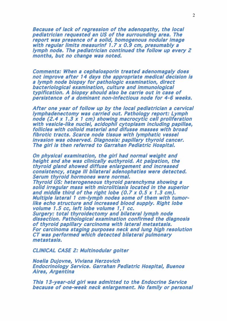
Because of lack of regression of the adenopathy, the local pediatrician requested an US of the surrounding area. The report was presence of a solid, homogenous nodular image with regular l imits measurinf 1.7 x 0.9 cm, presumably a lymph node. The pediatrician continued the follow up every 2 months, but no change was noted. Comments: When a cephalosporin treated adenomagaly does not improve after 14 days the appropriate medical decision is a lymph node biopsy for pathologic examination, direct bacteriological examination, culture and immunological typification. A biopsy should also be carrie out in case of persistence of a dominant non-infectious node for 4-6 weeks. After one year of follow up by the local pediatrician a cervical
lymphadenectomy was carried out. Pathology report: Lymph node (2.4 x 1.3 x 1 cm) showing macrocytic cell proliferation with vesicle-like nuclei, acidophil cytoplasm including papillae, foll icles with colloid material and difusse masses with broad fibrotic tracts. Scarce node tissue with lymphatic vessel invasion was observed. Diagnosis: papil lary thyroid cancer. The girl is then referred to Garrahan Pediatric Hospital. On physical examination, the girl had normal weight and height and she was clinically euthyroid. At palpation, the thyroid gland showed diffuse enlargement and increased consistency, stage II I bilateral adenophaties were detected. Serum thyroid hormones were normal. Thyroid US: heterogeneous thyroid parenchyma showing a solid irregular mass with microlitiasis located in the superior and middle third of the right lobe (0.7 x 0.5 x 1.3 cm). Multiple lateral 1 cm-lymph nodes some of them with tumor-like echo structure and increased blood supply. Right lobe volume 1.5 cc, left lobe volume 1,1 cc. Surgery: total thyroidectomy and bilateral lymph node dissection. Pathological examination confirmed the diagnosis of thyroid papil lary carcinoma with lateral metastasis. For carcinoma staging purposes neck and lung high resolution CT was performed which detected bilateral pulmonary metastasis. CLINICAL CASE 2: Multinodular goiter Noelia Dujovne, Viviana Herzovich Endocrinology Service. Garrahan Pediatric Hospital, Buenos Aires, Argentina This 13-year-old girl was admitted to the Endocrine Service because of one-week neck enlargement. No family or personal
3
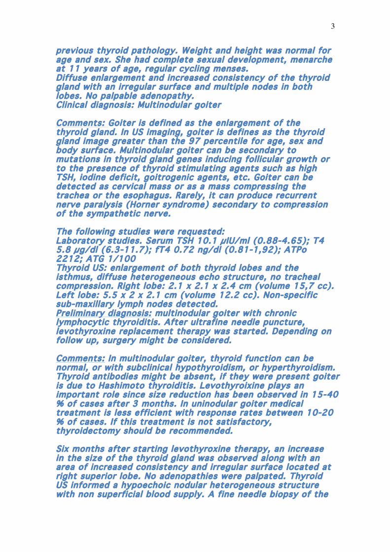
previous thyroid pathology. Weight and height was normal for age and sex. She had complete sexual development, menarche at 11 years of age, regular cycling menses. Diffuse enlargement and increased consistency of the thyroid gland with an irregular surface and multiple nodes in both lobes. No palpable adenopathy. Clinical diagnosis: Multinodular goiter Comments: Goiter is defined as the enlargement of the thyroid gland. In US imaging, goiter is defines as the thyroid gland image greater than the 97 percentile for age, sex and body surface. Multinodular goiter can be secondary to mutations in thyroid gland genes inducing foll icular growth or to the presence of thyroid stimulating agents such as high TSH, iodine deficit, goitrogenic agents, etc. Goiter can be detected as cervical mass or as a mass compressing the trachea or the esophagus. Rarely, it can produce recurrent nerve paralysis (Horner syndrome) secondary to compression of the sympathetic nerve. The following studies were requested: Laboratory studies. Serum TSH 10.1 µIU/ml (0.88-4.65); T4 5.8 µg/dl (6.3-11.7); fT4 0.72 ng/dl (0.81-1,92); ATPo 2212; ATG 1/100 Thyroid US: enlargement of both thyroid lobes and the isthmus, diffuse heterogeneous echo structure, no tracheal compression. Right lobe: 2.1 x 2.1 x 2.4 cm (volume 15,7 cc). Left lobe: 5.5 x 2 x 2.1 cm (volume 12.2 cc). Non-specific sub-maxillary lymph nodes detected. Preliminary diagnosis: multinodular goiter with chronic lymphocytic thyroiditis. After ultrafine needle puncture, levothyroxine replacement therapy was started. Depending on follow up, surgery might be considered. Comments: In multinodular goiter, thyroid function can be normal, or with subclinical hypothyroidism, or hyperthyroidism. Thyroid antibodies might be absent, if they were present goiter is due to Hashimoto thyroiditis. Levothyroixine plays an important role since size reduction has been observed in 15-40 % of cases after 3 months. In uninodular goiter medical treatment is less efficient with response rates between 10-20 % of cases. If this treatment is not satisfactory, thyroidectomy should be recommended. Six months after starting levothyroxine therapy, an increase in the size of the thyroid gland was observed along with an area of increased consistency and irregular surface located at right superior lobe. No adenopathies were palpated. Thyroid US informed a hypoechoic nodular heterogeneous structure with non superficial blood supply. A fine needle biopsy of the
4
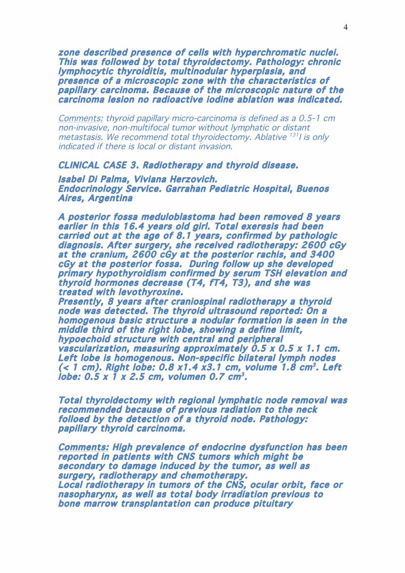
zone described presence of cells with hyperchromatic nuclei. This was followed by total thyroidectomy. Pathology: chronic lymphocytic thyroiditis, multinodular hyperplasia, and presence of a microscopic zone with the characteristics of papil lary carcinoma. Because of the microscopic nature of the carcinoma lesion no radioactive iodine ablation was indicated. Comments: thyroid papillary micro-carcinoma is defined as a 0.5-1 cm non-invasive, non-multifocal tumor without lymphatic or distant metastasis. We recommend total thyroidectomy. Ablative 131I is only indicated if there is local or distant invasion. CLINICAL CASE 3. Radiotherapy and thyroid disease. Isabel Di Palma, Viviana Herzovich. Endocrinology Service. Garrahan Pediatric Hospital, Buenos Aires, Argentina A posterior fossa meduloblastoma had been removed 8 years earlier in this 16.4 years old girl. Total exeresis had been carried out at the age of 8.1 years, confirmed by pathologic diagnosis. After surgery, she received radiotherapy: 2600 cGy at the cranium, 2600 cGy at the posterior rachis, and 3400 cGy at the posterior fossa. During follow up she developed primary hypothyroidism confirmed by serum TSH elevation and thyroid hormones decrease (T4, fT4, T3), and she was treated with levothyroxine. Presently, 8 years after craniospinal radiotherapy a thyroid node was detected. The thyroid ultrasound reported: On a homogenous basic structure a nodular formation is seen in the middle third of the right lobe, showing a define limit, hypoechoid structure with central and peripheral vascularization, measuring approximately 0.5 x 0.5 x 1.1 cm. Left lobe is homogenous. Non-specific bilateral lymph nodes (< 1 cm). Right lobe: 0.8 x1.4 x3.1 cm, volume 1.8 cm3. Left lobe: 0.5 x 1 x 2.5 cm, volumen 0.7 cm3. Total thyroidectomy with regional lymphatic node removal was recommended because of previous radiation to the neck folloed by the detection of a thyroid node. Pathology: papil lary thyroid carcinoma. Comments: High prevalence of endocrine dysfunction has been reported in patients with CNS tumors which might be secondary to damage induced by the tumor, as well as surgery, radiotherapy and chemotherapy. Local radiotherapy in tumors of the CNS, ocular orbit, face or nasopharynx, as well as total body irradiation previous to bone marrow transplantation can produce pituitary
5
insufficiency. The hypothalamus seems to be more sensitive than the pituitary gland. The deleterious biological effects of irradiation depend on total dose, fractional dose, α/β ratio (α, β radio-biological parameters of cell survival), duration of radiotherapy, and patient´s age. Incidence of central hypothyroidism after cranial (CIR) and craniospinal irradiation (CSI) in central nervous tumors varies widely among different studies. Children receiving radiotherapy for Hodgkin lymphoma, CSI, or total body irradiation have risk for thyroid disease because of thyroid sensitivity to radiation damage. Primary hypothyroidism is the most frequent thyroid dysfunction after gland irradiation. Time lag for hypothyroidism development shows a negative relation with radiation dosage. Primary hypothyroidism can be masked in children who received craniospinal irradiation because of co-existence of central hypothyroidism. The incidence of thyroid nodules or thyroid carcinoma varies greatly among different reports, depending on duration of follow up. The risk of developing thyroid cancer seems to depend on dose of irradiation. This risk is even greater in small children Thyroid Cancer___________________ Thyroid cancer is the most frequent endocrine tumor in children, with an incidence of 0.5-1.5 % of childhood tumors. It is the most common malignant tumor of head and neck in youngsters. Histologically, it is similar to adult thyroid cancer but presentation and survival are different in children and adolescents. In young people, tumors tend to be larger, more invasive to surrounding areas, and they show faster lymph node metastasis as well as distant metastasis. Present evidence suggests that this biological behavior is more aggressive below 6 years of age. Risk factors for thyroid cancer are: ectopic thyroid, multinodular goiter, radiation exposure, low iodine diet. It is associated with Gardner syndrome, Cowdens syndrome and Carney complex. Clinically, thyroid cancer can be presented as a mass in the neck or a solitary nodule in the thyroid gland detected in a routine physical examination or being discovered by parents or patients themselves. Other initial symptoms are dysphagia,
6
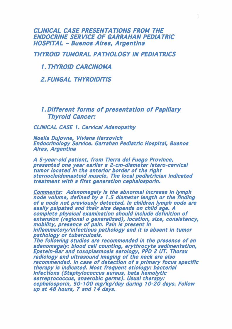
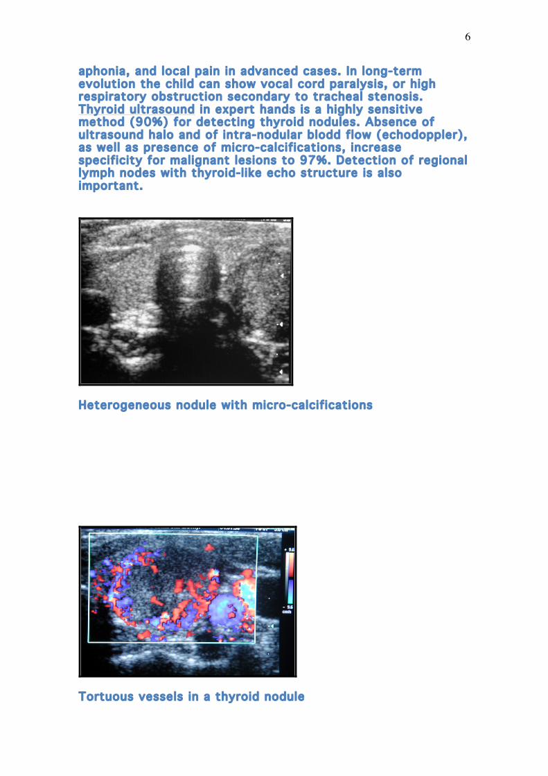
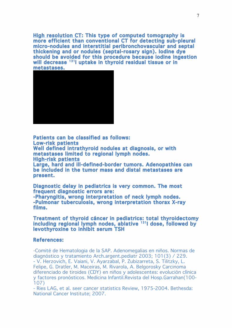
aphonia, and local pain in advanced cases. In long-term evolution the child can show vocal cord paralysis, or high respiratory obstruction secondary to tracheal stenosis. Thyroid ultrasound in expert hands is a highly sensitive method (90%) for detecting thyroid nodules. Absence of ultrasound halo and of intra-nodular blodd flow (echodoppler), as well as presence of micro-calcifications, increase specificity for malignant lesions to 97%. Detection of regional lymph nodes with thyroid-like echo structure is also important.
Heterogeneous nodule with micro-calcifications
Tortuous vessels in a thyroid nodule
7
High resolution CT: This type of computed tomography is more efficient than conventional CT for detecting sub-pleural micro-nodules and interstitial peribronchovascular and septal thickening and or nodules (septal-rosary sign). Iodine dye should be avoided for this procedure because iodine ingestion will decrease 131I uptake in thyroid residual tissue or in metastases.
Patients can be classified as follows: Low-risk patients Well defined intrathyroid nodules at diagnosis, or with metastases limited to regional lymph nodes. High-risk patients Large, hard and il l-defined-border tumors. Adenopathies can be included in the tumor mass and distal metastases are present. Diagnostic delay in pediatrics is very common. The most frequent diagnostic errors are: -Pharyngitis, wrong interpretation of neck lymph nodes. -Pulmonar tuberculosis, wrong interpretation thorax X-ray films. Treatment of thyroid cáncer in pediatrics: total thyroidectomy including regional lymph nodes, ablative 131I dose, followed by levothyroxine to inhibit serum TSH References: -Comité de Hematologia de la SAP. Adenomegalias en niños. Normas de diagnóstico y tratamiento Arch.argent.pediatr 2003; 101(3) / 229. - V. Herzovich, E. Vaiani, V. Ayarzabal, P. Zubizarreta, S. Tilitzky, L. Felipe, G. Dratler, M. Maceiras, M. Rivarola, A. Belgorosky Carcinoma diferenciado de tiroides (CDY) en niños y adolescentes: evolución clínica y factores pronósticos. Medicina Infantil.Revista del Hosp.Garrahan(100-107) - Ries LAG, et al. seer cancer statistics Review, 1975-2004. Bethesda: National Cancer Institute; 2007.
8
- Iorcansky S, Herzovich V, Zantleifer D, et al. Cancer papilar detiroides infanto juvenile. Re-evaluación de factores pronósticosy nueva propuesta de estadificación. Medicina Infantil. 2000;VII: 173-79. - Thyroid Cancer: A comprehensive Guide to Clinical Management.Capitulo10 Merrily Poth Edicion 2006 - Hung W, Sarlis NJ. Current Controversies in the Management ofediatric Patients with Well-differentiated nonmedullary ThyroidCancer: A Review. Thyroid, 2002; 8: 683-702. - Dinauer C, Breuer C and Rivkees S. Differentiated Thyroid Cancer in Children: Diagnosis and Management, Current Opinion InOncology 2008, 20:59-65. - Thyroid Cancer: A comprehensive Guide to Clinical Management.Capitulo 41 A Bauer and M Poth.Edicion 2006. Barbara Jarzab, Daria Handkiewicz-Junak. Differentiated thyroidcancer in children and adults: same or distinct disease?HORMONES 2007, 6(3):200-209. - Lazar L, Lebenthal Y, Steinmetz A, Yackobovitch-Gavan M, PhillipM. J Pediatr. 2009;154 (5):708-14. Differentiated thyroid carcinomain pediatric patients: comparison of presentation andcourse between pre-pubertal children and adolescent. - Schlumberger M, De Vathaire F, Travagli JP, Vassal G, LemerleJ, Parmentier C, Tubiana M,- Differentiated thyroid carcinoma inchildhood: long term follow-up of 72 patients. J Clin Endocrinol Metab,1987;65(6):1088-94. - Yamashita S, Saenko V. Mechanisms of disease: molecular geneticsof childhood thyroid cancers. Nat Clin Pract EndocrinolMetab, 2007; 3:422–429. - Steliarova-Foucher E, Stiller CA, Pukkala E, Lacour B, Plesko I,Parkin DM. Thyroid cancer incidence and survival among Europeanchildren and adolescents (1978-1997): report from the AutomatedChildhood Cancer Information System project. Eur JCancer, 2006;42: 2150-2169. - Tamimi DM. The association between chronic lymphocytic thyroiditisand thyroid tumors. Int J Surg Pathol,2002;10(2):141-6. -Powers PA, Dinauer CA, Tuttle RM, Robie DK, McClellan DR,Francis GL Tumor size and extent of disease at diagnosis predictthe response to initial therapy for papillary thyroid carcinomachildren and adolescents. J Pediatr Endocrinol Metab.2003;16(5):693-702. - Jarzab B, Handkiewicz-Junak D, Wloch J, Kalemba B, RoskoszJ, Kukulska A, Puch Z.. Multivariate analysis of prognostic factorsfor differentiated thyroid carcinoma in children. Eur J NuclMed, 2000 ; 27: 833-841. - Catherine Dinauer, Gary L. Francis. Thyroid Cancer in Children.Endocrinol Metab Clin N Am 2007, 36 ;779–806. Cohen L. E. Endocrine late affects of cancer treatment Endocrinol Metab Clin N Am 2005; 34: 769-789 -Schmiegelow M., Felt-Rasmussen U., Poulsen H. S., Muller J. J Clin Endocrinol Metab 2003; 88:136-140. 2. FUNGAL THYROIDITIS IN A PATIENT WITH ACUTE LYMPHOBLASTIC LEUKEMIA
9
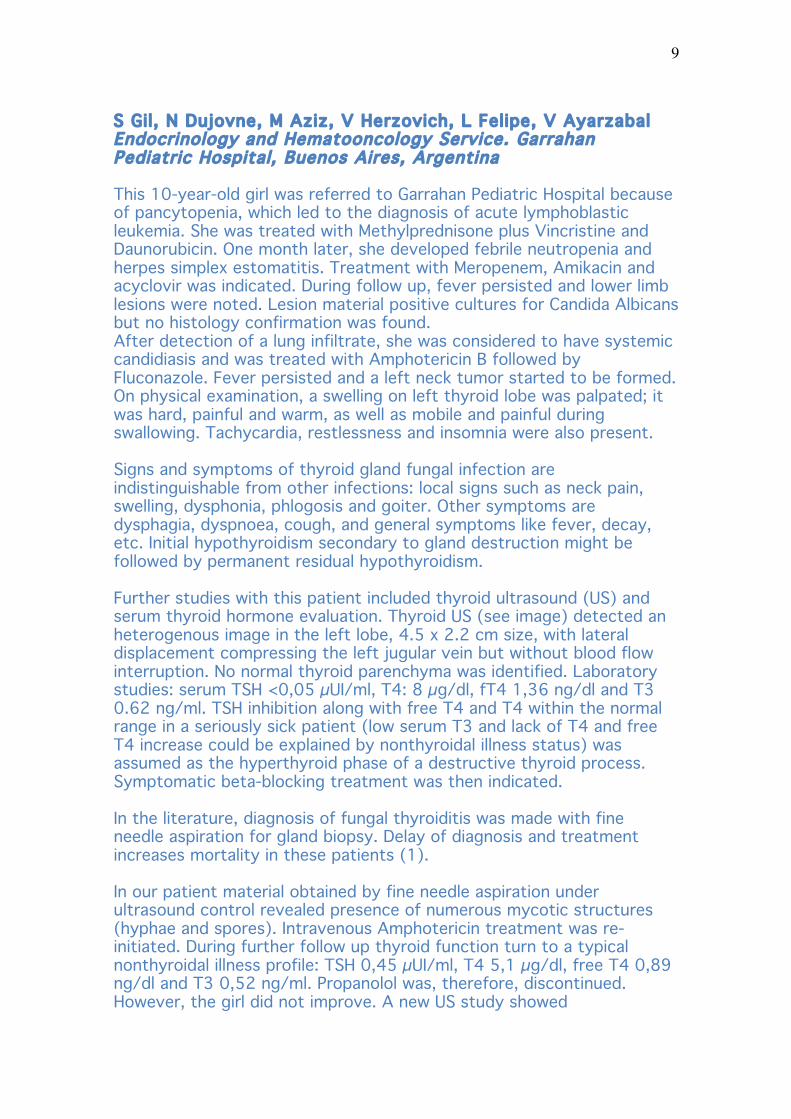
S Gil, N Dujovne, M Aziz, V Herzovich, L Felipe, V Ayarzabal Endocrinology and Hematooncology Service. Garrahan Pediatric Hospital, Buenos Aires, Argentina This 10-year-old girl was referred to Garrahan Pediatric Hospital because of pancytopenia, which led to the diagnosis of acute lymphoblastic leukemia. She was treated with Methylprednisone plus Vincristine and Daunorubicin. One month later, she developed febrile neutropenia and herpes simplex estomatitis. Treatment with Meropenem, Amikacin and acyclovir was indicated. During follow up, fever persisted and lower limb lesions were noted. Lesion material positive cultures for Candida Albicans but no histology confirmation was found. After detection of a lung infiltrate, she was considered to have systemic candidiasis and was treated with Amphotericin B followed by Fluconazole. Fever persisted and a left neck tumor started to be formed. On physical examination, a swelling on left thyroid lobe was palpated; it was hard, painful and warm, as well as mobile and painful during swallowing. Tachycardia, restlessness and insomnia were also present. Signs and symptoms of thyroid gland fungal infection are indistinguishable from other infections: local signs such as neck pain, swelling, dysphonia, phlogosis and goiter. Other symptoms are dysphagia, dyspnoea, cough, and general symptoms like fever, decay, etc. Initial hypothyroidism secondary to gland destruction might be followed by permanent residual hypothyroidism. Further studies with this patient included thyroid ultrasound (US) and serum thyroid hormone evaluation. Thyroid US (see image) detected an heterogenous image in the left lobe, 4.5 x 2.2 cm size, with lateral displacement compressing the left jugular vein but without blood flow interruption. No normal thyroid parenchyma was identified. Laboratory studies: serum TSH <0,05 µUI/ml, T4: 8 µg/dl, fT4 1,36 ng/dl and T3 0.62 ng/ml. TSH inhibition along with free T4 and T4 within the normal range in a seriously sick patient (low serum T3 and lack of T4 and free T4 increase could be explained by nonthyroidal illness status) was assumed as the hyperthyroid phase of a destructive thyroid process. Symptomatic beta-blocking treatment was then indicated. In the literature, diagnosis of fungal thyroiditis was made with fine needle aspiration for gland biopsy. Delay of diagnosis and treatment increases mortality in these patients (1). In our patient material obtained by fine needle aspiration under ultrasound control revealed presence of numerous mycotic structures (hyphae and spores). Intravenous Amphotericin treatment was re-initiated. During further follow up thyroid function turn to a typical nonthyroidal illness profile: TSH 0,45 µUI/ml, T4 5,1 µg/dl, free T4 0,89 ng/dl and T3 0,52 ng/ml. Propanolol was, therefore, discontinued. However, the girl did not improve. A new US study showed
10
extension of the lesion to right lobe. Caspofungin was added as medication but without improvement. Anti-fungal medications can be used in the treatment of fungal thyroiditis and abscesses. But in cases of unsatisfactory response a total thyroidectomy should be carried out in order to eliminate the infection and avoid disease progression (2). Persistence of neutropenic fever and clinical deterioration resulted in surgical intervention. At initial incision, a purulent material was obtained which included pre-thyroid muscles, the left lobe and the inferior pole of the right lobe: total thyroidectomy was then carried out. After surgery, the patient showed clinical improvement and cessation of fever. Post-operative hypocalcemia developed and patient was treated with calcitriol and extra calcium, as well as levothyroxine. Culture material was negative but pathological study confirmed the diagnosis of infectious thyroiditis. Recovery was complete. Patient could then re-initiated chemotherapy for her acute lymphoblastic leukemia. Four months after surgery, recovery of parathyroid function was documented.
Final Discussion Fungal infection of the thyroid gland is very rare. The thyroid gland has protective mechanisms which confer a relative resistance to infection such as a rich blood and lymphatic flow as well as separation from other structures of the neck by a thick fibrous capsule (3). Moreover, iodine can act as bactericide agent contributing to thyroid resistance to infections (3). Patients with high risk of fungal thyroiditis are those under immune-suppression such as HIV, leukemia, auto-immune disease, and transplanted subjects under immunosuppressive medication (1, 2). Forty cases of fungal thyroiditis have been published, agents include Aspergillus, Candida, Histoplasma Capsulatum, Cryptococcus neoformans, Coccidiodes inmitis and Pseudallescheria Boydii. Candida
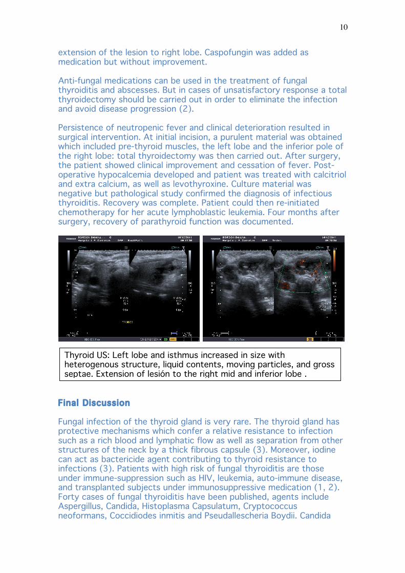
Thyroid US: Left lobe and isthmus increased in size with heterogenous structure, liquid contents, moving particles, and gross septae. Extension of lesión to the right mid and inferior lobe .
11
infections are mostly mono-cellular. Predisposition factors are antibiotics and catheterization (3). Seven cases of Candida Albicans thyroiditis have been published, all in immune-compromised subjects. The youngest was a 17-year-old subject while all the others were adults, 2 male and 4 female patients; one of them died with a disseminated infection, thyroid involvement was found at autopsy (1-4). Finally, in immune-suppressed patients with prolonged fever syndrome and lack of response to antibiotic therapy fungal infection should be kept in mind and the possibility of thyroid involvement taken into consideration. REFERENCES 1-Gandhi RT, Tollin SR, Seely EW. Diagnosis of Candida thyroiditis by fine needle aspiration. J Infect 1994 28:77-81. 2-Fernandez J, Anaissie J, Vassilopoulou Sellin, Samann NA. Case Report: Acute fungal thyoiditis in a patient with acute myelogenous leukemia. J Internal Medicine 1991 230:539-41. 3-Golani LZ, Zavaski AP, Maia AL. Fungal thyroiditis: An overview. Mycopathologia 2006 161:129-39. 4-Daas N, Lossosos IS, Yahalom, Rund, Wolf, Ben-Yehuda A. Candida abscess of the thyroid in a patient with acute lymphocytic leukemia. Eur J Med Res 1997 2:365-6.