Embed Size (px)
Citation preview
Research ArticleClinical Application of Artificial Dermis and Autologous Skin inRepairing Skin and Soft Tissue Defects of Hands and Feet withBone Exposure Injuries
Chengke Li,1 Weihai Song,2 Yanwen Lei,3 Songgen Peng,3 Weiying Chu,1
and Guochao Deng 4
1Department of Orthopedics, Shunde Hospital affiliated to Jinan University (Foshan Shunde Second People’s Hospital,Foshan Shunde Feng Yaojing Memorial Hospital), Shunde District, Foshan, Guangdong, China2Department of Orthopedics, Municipal Hospital Affiliated to Taizhou University, Taizhou, Zhejiang, China3Department of Orthopedics, Shunde Peace Surgical Hospital Co., Ltd, Foshan, Guangdong, China4Department of Orthopedics, Linhai Second People’s Hospital, Linhai, Zhejiang, China
Correspondence should be addressed to Guochao Deng; [email protected]
Received 5 September 2021; Accepted 1 October 2021; Published 16 October 2021
Academic Editor: Songwen Tan
Copyright © 2021 Chengke Li et al. .is is an open access article distributed under the Creative Commons Attribution License,which permits unrestricted use, distribution, and reproduction in any medium, provided the original work is properly cited.
Patients with skin and soft tissue defects are very common. Mild trauma often causes mild skin damage, while severe injuries areoften accompanied by bone and tendon exposure, which brings great pain to patients. For the defect of skin and soft tissue, thetraditional treatment methods are mostly medium or full-thickness skin or skin flap transplantation. .ese methods are effectivein wound repair, but there are still many problems. In recent years, with the improvement of tissue engineering technology, the useof artificial skin to repair various skin wounds is gradually becoming clinical, and the key technology of skin tissue engineering liesin the development of dermal substitutes. .e appearance of artificial dermis not only solves the shortage of autologous skinsource but also makes the operation simple and easy. .e purpose of this study was to investigate the clinical effect of artificialdermis combined with autologous skin grafts in repairing hand and foot skin and soft tissue defects with bone exposure. .eresults show that the use of artificial dermis combined with autogenous blade thick skin to treat patients with hand and foot softtissue injury with bone exposure has a good clinical effect, and the skin is alive and has fewer complications, which is worthyof promotion.
1. Introduction
With the continuous development of modern society andthe acceleration of industrialization and mechanization, theprobability of traffic accident injury, severe burn, and highfalling injury in daily life is increased, and the number ofpatients with limb skin and soft tissue defects is graduallyincreased. Mild trauma often causes mild skin damage, andsevere cases are often accompanied by bone and tendonexposure, bringing great pain to patients [1, 2]. .erefore,how to effectively clean up the wound to control infectionand repair skin and soft tissue defects is a major clinicalproblem. For skin and soft tissue defects, the traditional
treatment is mostly medium-thickness or full-thickness skinor skin flap transplantation [3, 4]. .e wound repair effect ofthese methods is acceptable, but there are still manyproblems. It is difficult for the medium-thickness skin toeffectively resist the contracture, and the scar contracture ofthe wound is serious in the later stage, which affects thefunction again [5, 6]. In addition, mild scar or pigmentationmay occur in the donor site, so the thick skin is often notused in the repair of joint defects. However, full-thicknessskin often has insufficient skin source, especially for patientswith extensive burns, skin is extremely valuable. After full-thickness skin grafting on a slightly larger wound surface, thedonor area is difficult to be sutured directly, and skin
HindawiEvidence-Based Complementary and Alternative MedicineVolume 2021, Article ID 1202826, 8 pageshttps://doi.org/10.1155/2021/1202826
grafting is needed again for repair, causing secondarytrauma to the patient. In addition, after full-thickness skin isimplanted in the skin donor area, skin necrosis is oftencaused by improper blood supply and improper bandagingand fixation, and the success rate of transplantation is rel-atively low. Autologous flap transplantation has a good effecton repairing exposed bone and tendon wounds and caneffectively cover the wounds, but the operation is difficult,and the doctor’s workload and psychological pressure areheavy. Similarly, the donor flap area of flap surgery needsskin grafting to repair, and the scar is obvious in the laterstage, and the risk of vascular crisis and necrosis of thetransplanted flap is high [7, 8]. In recent years, with theimprovement of tissue engineering technology, the use ofartificial skin to repair various skin wounds is slowly movingtowards the clinic, and the key to skin tissue engineeringtechnology lies in the development of dermal substitutes. Atpresent, the artificial dermis commonly used in clinicalpractice is a double-layer structure composed of collagensponge without terminal and silica gel layer extracted fromthe Achilles tendon of pig. .e silica gel mold can not onlyisolate the invasion of external bacteria but also prevent themoisture evaporation of the collagen sponge layer. Whencapillaries and fibroblasts grow into the sponge layer, der-mal-like tissue is formed. At this time, the silica gel mem-brane is removed and the autologous thin skin layer isimplanted, which can achieve good repair effect [9, 10]. .eappearance of artificial dermis not only solves the shortage ofautologous skin source but also makes the operation simpleand easy. .e purpose of this study was to investigate theclinical effect of artificial dermis combined with autologousskin grafts in repairing hand and foot skin and soft tissuedefects with bone exposure. .e specific report is as follows.
2. Materials and Methods
2.1. Patients. From March 2017 to June 2020, 92 patientswith skin and soft tissue defects of hands and feet with boneexposure injury were selected as the research object. Amongthem, 54 were males and 38 were females, aged 20 to 48 yearsold, with an average age of (33.41± 8.64) years old. .ecauses of injury were as follows: machine crush injury in 30cases, cutting defect injury in 26 cases, rolling in 24 cases,and the other 12 cases. Injured parts: 40 cases of hands and52 cases of feet. Inclusion criteria: hospitalization time afterinjury <24 h; skin and soft tissue defects; bone/tendon-ex-posed wounds with fractures after trauma; clinical data arecomplete. Exclusion criteria: patients with severe bone ex-posure infection and chronic osteomyelitis; chronic woundswith exposed bone or tendons; patients with poor nutritionalstatus and difficult to tolerate surgery; patients with mentalillness; patients with diabetes who have poor blood sugarcontrol for many years; patients who fell off during follow-up.
2.2. Operation Method. All patients underwent artificialdermis combined with autologous skin repair treatment.After regional nerve block anesthesia, the wound was
debrided. .e wound and inflammatory granulationtissue were excised at a distance of 1 cm from the edge ofthe wound to completely remove the exposed and ne-crotic tendons; the necrotic tissue on the surface of theexposed bone was chiseled away, and the soft tissue withpoor vitality around the dead bone was enlarged to creategood blood supply for the wound surface. .e woundsurface was electrocoagulated to stop the blood, and theuse of silk ligation was reduced. .e wound surface wasrepeatedly rinsed with normal saline, hydrogen peroxide,iodophor, or povidone iodine and covered with gauze..e operation was divided into two stages, and the ar-tificial dermal graft was performed in stage I: the artificialdermis was immersed in physiological saline until thewhole layer was wet and then cut according to the size ofthe wound so that it could cover the wound to be repairedand the exposed bone surface and keep close to the woundwithout leaving gaps. .e silica gel of the artificial dermisfaces outwards; holes are punched on the surface (topromote drainage) so that the collagen surface is close tothe wound surface, and as far as possible, the hematomaand air under the skin are squeezed out. .e suture orskin suture device is fixed on the wound edge so that itclosely fits the wound edge, and the appropriate pressureis applied. .e external dressing was replaced for the firsttime 3 to 5 days after the operation. If there is fluid orblood under the artificial dermis, cut small holes on thesurface of the silicone membrane for drainage and thenchange the external dressing every 2 to 3 days. After 2 to 3weeks, the artificial dermis appears pink with the growthof cell components and capillaries and the deposition ofcollagen components. At this time, the silica gel mem-brane and the collagen layer are gradually separated,indicating that the dermal-like tissue has basicallyformed, and autologous skin transplantation can beperformed. For wounds with a large exposed area of boneand/or tendon, one artificial dermal covering operationusually cannot completely granulate the wound. If thewound is still exposed, two or more artificial dermalgrafts are required until the wound is completely coveredwith granulation.
Autologous skin grafting for stage II: after routinedisinfection, the silicone membrane on the surface of theartificial dermis was removed, the surface of the woundwas gently scraped with a scalpel, and the surface secre-tions and infected and edema granulation tissue wereremoved. After washing with normal saline, povidoneiodine or iodophor solution was applied to clean thewound, and appropriate wet compress was applied. .inslice skin from the lower limb or upper limb was applied tothe wound surface, and the wound skin was sutured with3-0 silk thread or skin stapler. Bandage and fix according tothe conventional method, change the external dressing 5 to7 days after the operation, and observe the survival of theskin graft. After operation, the affected limb was raised andthe dressing was changed 3∼5 days later. In addition toantibiotic treatment, analgesic drugs were used for 3 days.2∼3 days after skin donor site operation, the outer dressingwas replaced, and the inner oil gauze was kept to keep the
2 Evidence-Based Complementary and Alternative Medicine
wound clean and dry, and the inner oil gauze could fall offby itself.
2.3. Observation Index. All patients were followed up for6–12 months to observe their recovery. With reference [11] tothe literature, the doctors in charge of our department willmake the following judgments on the growth of the skin in thedonor area and the graft area: they will observe the skingrafting area’s flatness and appearance color changes andevaluate them as “good” and “bad” and describe the colorchange of the donor site, including no obvious change, loss ofpigment, pigmentation, and redness; according to the degreeof scar hyperplasia in the skin graft area, the evaluation ismade as “none,” “very light,” “mild,” and “obvious”; they willobserve the scar changes in the donor area and evaluate themas “none,” “very mild,” “mild,” and “obvious.” Based on theabove results, the flatness and appearance color of the skingrafts in the skin grafting area, scar hyperplasia in the skingrafting area, and the skin donor area were considered as thefour main factors. .e clinical results were evaluated as“excellent” (no factor abnormality), “good” (1 factor abnor-mality), “average” (2 factors abnormality), and “poor” (3 ormore factor abnormality). Excellent and good rate �
(excellent + good) cases/total cases × 100%. .ey will alsoobserve and record the survival of the patient’s skin and theoccurrence of complications during the follow-up period.
3. Results
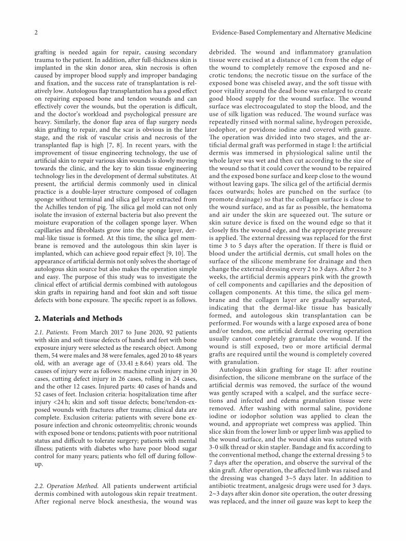
3.1. Comprehensive Evaluation Results. All 92 patientscompleted the second skin graft surgery, and the skinssurvived well. .e smoothness of skin graft area was “good”in 92 cases and “poor” in 0 case. .ere were 74 cases of“none” and 12 cases of “extremely mild,” 6 cases of “mild,”and 0 case of “obvious” scar hyperplasia in the skin graftarea. .ere were 77 cases of “none,” 10 cases of “extremelymild,” 5 cases of “mild,” and 0 case of “obvious” scar hy-perplasia in the donor site. .ere were 80 cases with noobvious change in the color of the skin donor site and 12cases with obvious change. In the comprehensive evaluation,35 cases were excellent, 50 cases were good, 7 cases wereaverage, and 0 cases were poor, with the excellent and goodrate of 92.39% (85/92), as shown in Figure 1.
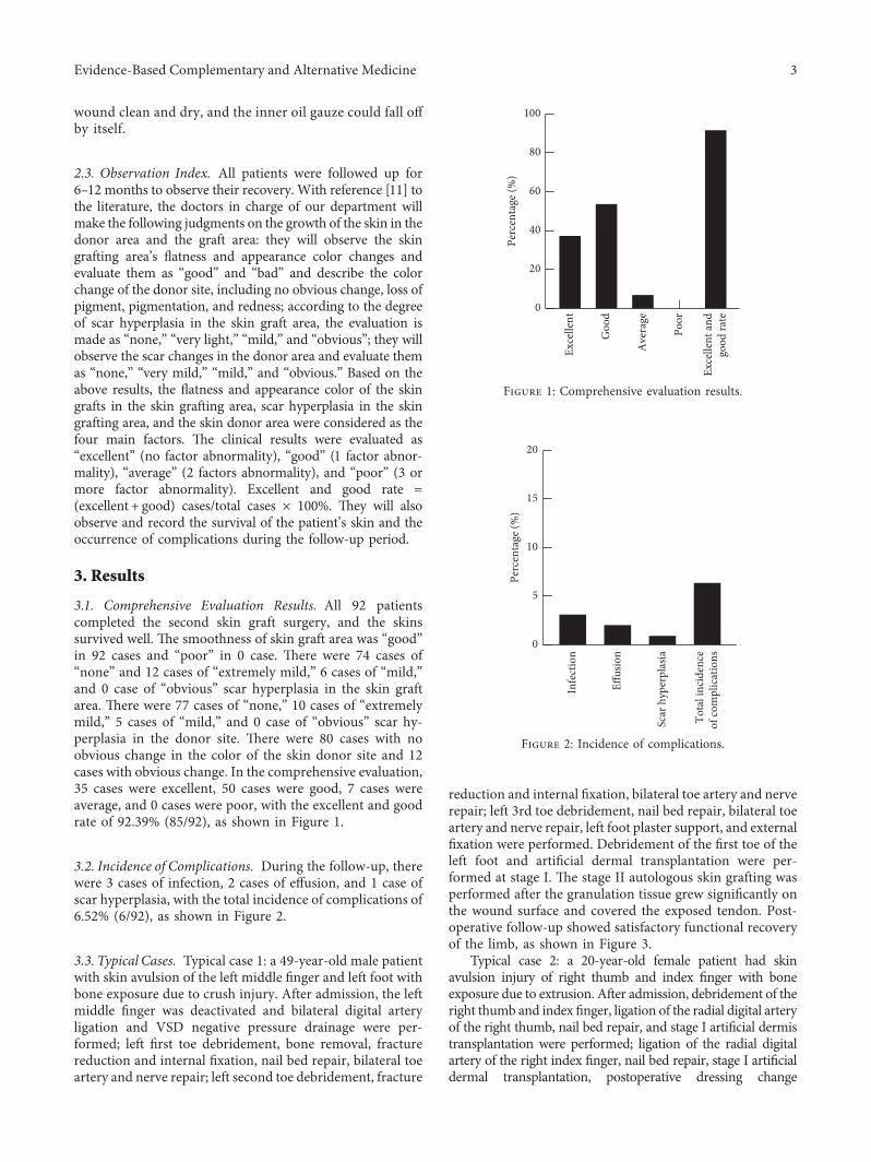
3.2. Incidence of Complications. During the follow-up, therewere 3 cases of infection, 2 cases of effusion, and 1 case ofscar hyperplasia, with the total incidence of complications of6.52% (6/92), as shown in Figure 2.
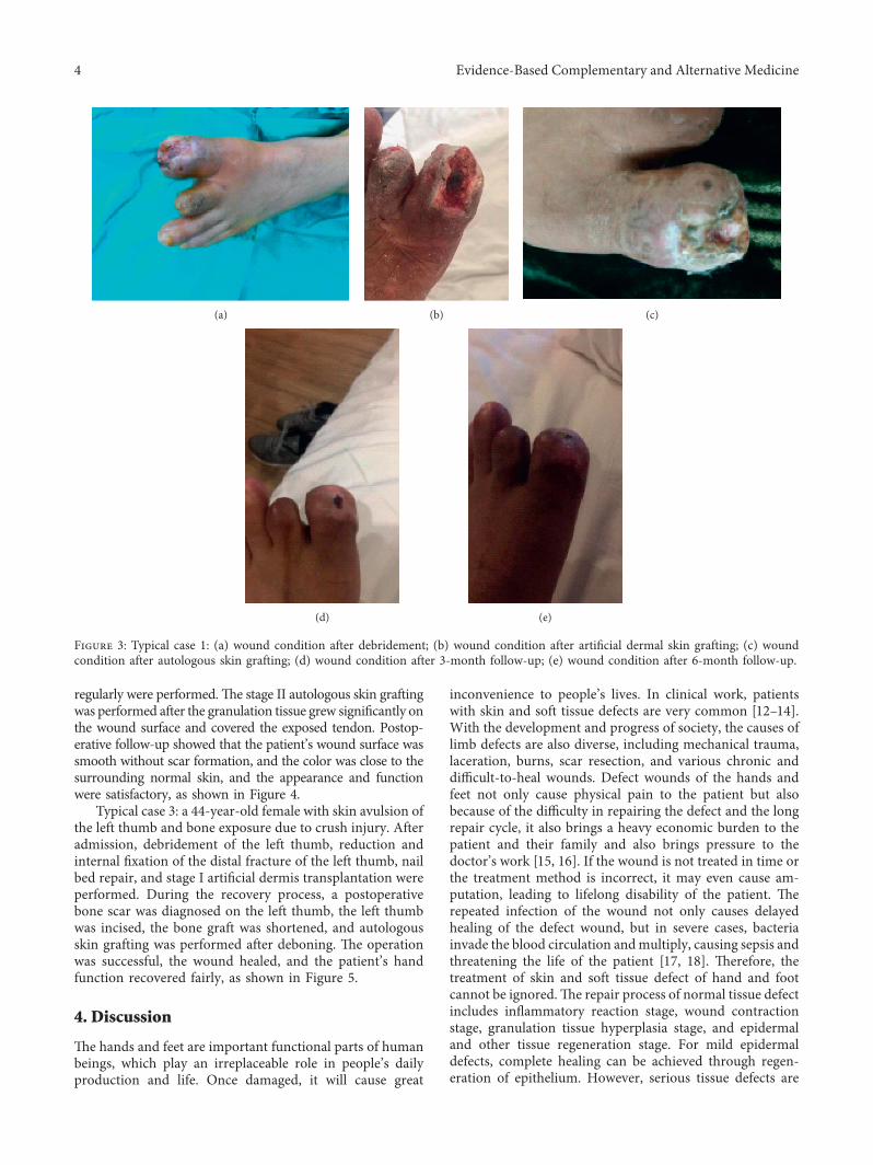
3.3. Typical Cases. Typical case 1: a 49-year-old male patientwith skin avulsion of the left middle finger and left foot withbone exposure due to crush injury. After admission, the leftmiddle finger was deactivated and bilateral digital arteryligation and VSD negative pressure drainage were per-formed; left first toe debridement, bone removal, fracturereduction and internal fixation, nail bed repair, bilateral toeartery and nerve repair; left second toe debridement, fracture
reduction and internal fixation, bilateral toe artery and nerverepair; left 3rd toe debridement, nail bed repair, bilateral toeartery and nerve repair, left foot plaster support, and externalfixation were performed. Debridement of the first toe of theleft foot and artificial dermal transplantation were per-formed at stage I. .e stage II autologous skin grafting wasperformed after the granulation tissue grew significantly onthe wound surface and covered the exposed tendon. Post-operative follow-up showed satisfactory functional recoveryof the limb, as shown in Figure 3.
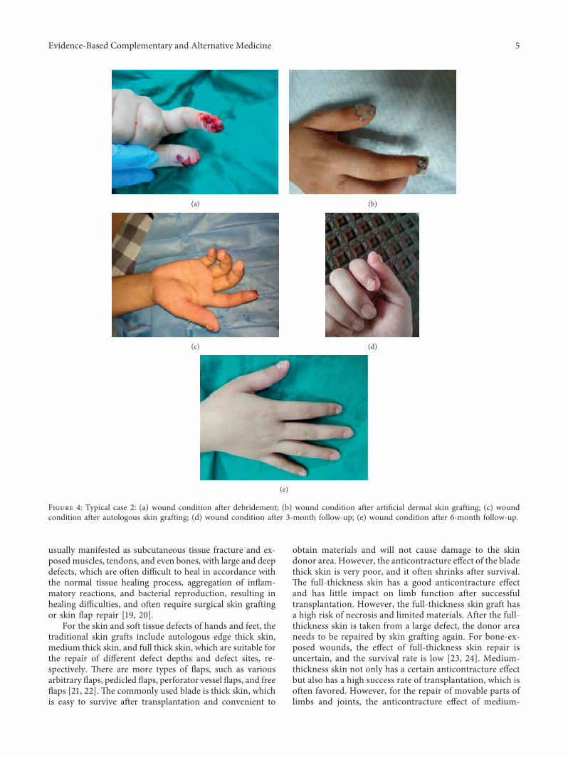
Typical case 2: a 20-year-old female patient had skinavulsion injury of right thumb and index finger with boneexposure due to extrusion. After admission, debridement of theright thumb and index finger, ligation of the radial digital arteryof the right thumb, nail bed repair, and stage I artificial dermistransplantation were performed; ligation of the radial digitalartery of the right index finger, nail bed repair, stage I artificialdermal transplantation, postoperative dressing change
Poor
Exce
llent
Goo
d
Ave
rage
Exce
llent
and
good
rate
0
20
40
60
80
100
Perc
enta
ge (%
)
Figure 1: Comprehensive evaluation results.
Infe
ctio
n
Effus
ion
Scar
hyp
erpl
asia
Tota
l inc
iden
ceof
com
plic
atio
ns
0
5
10
15
20
Perc
enta
ge (%
)
Figure 2: Incidence of complications.
Evidence-Based Complementary and Alternative Medicine 3
regularly were performed..e stage II autologous skin graftingwas performed after the granulation tissue grew significantly onthe wound surface and covered the exposed tendon. Postop-erative follow-up showed that the patient’s wound surface wassmooth without scar formation, and the color was close to thesurrounding normal skin, and the appearance and functionwere satisfactory, as shown in Figure 4.
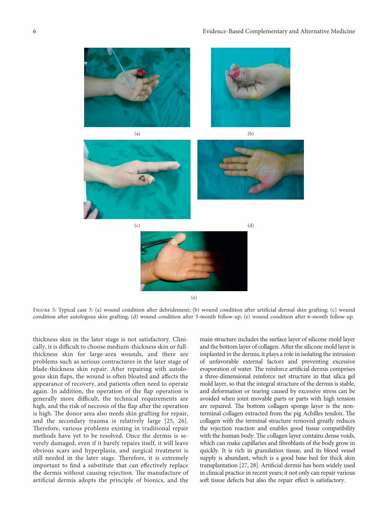
Typical case 3: a 44-year-old female with skin avulsion ofthe left thumb and bone exposure due to crush injury. Afteradmission, debridement of the left thumb, reduction andinternal fixation of the distal fracture of the left thumb, nailbed repair, and stage I artificial dermis transplantation wereperformed. During the recovery process, a postoperativebone scar was diagnosed on the left thumb, the left thumbwas incised, the bone graft was shortened, and autologousskin grafting was performed after deboning. .e operationwas successful, the wound healed, and the patient’s handfunction recovered fairly, as shown in Figure 5.
4. Discussion
.e hands and feet are important functional parts of humanbeings, which play an irreplaceable role in people’s dailyproduction and life. Once damaged, it will cause great
inconvenience to people’s lives. In clinical work, patientswith skin and soft tissue defects are very common [12–14].With the development and progress of society, the causes oflimb defects are also diverse, including mechanical trauma,laceration, burns, scar resection, and various chronic anddifficult-to-heal wounds. Defect wounds of the hands andfeet not only cause physical pain to the patient but alsobecause of the difficulty in repairing the defect and the longrepair cycle, it also brings a heavy economic burden to thepatient and their family and also brings pressure to thedoctor’s work [15, 16]. If the wound is not treated in time orthe treatment method is incorrect, it may even cause am-putation, leading to lifelong disability of the patient. .erepeated infection of the wound not only causes delayedhealing of the defect wound, but in severe cases, bacteriainvade the blood circulation andmultiply, causing sepsis andthreatening the life of the patient [17, 18]. .erefore, thetreatment of skin and soft tissue defect of hand and footcannot be ignored..e repair process of normal tissue defectincludes inflammatory reaction stage, wound contractionstage, granulation tissue hyperplasia stage, and epidermaland other tissue regeneration stage. For mild epidermaldefects, complete healing can be achieved through regen-eration of epithelium. However, serious tissue defects are
(a) (b) (c)
(d) (e)
Figure 3: Typical case 1: (a) wound condition after debridement; (b) wound condition after artificial dermal skin grafting; (c) woundcondition after autologous skin grafting; (d) wound condition after 3-month follow-up; (e) wound condition after 6-month follow-up.
4 Evidence-Based Complementary and Alternative Medicine
usually manifested as subcutaneous tissue fracture and ex-posed muscles, tendons, and even bones, with large and deepdefects, which are often difficult to heal in accordance withthe normal tissue healing process, aggregation of inflam-matory reactions, and bacterial reproduction, resulting inhealing difficulties, and often require surgical skin graftingor skin flap repair [19, 20].
For the skin and soft tissue defects of hands and feet, thetraditional skin grafts include autologous edge thick skin,medium thick skin, and full thick skin, which are suitable forthe repair of different defect depths and defect sites, re-spectively. .ere are more types of flaps, such as variousarbitrary flaps, pedicled flaps, perforator vessel flaps, and freeflaps [21, 22]. .e commonly used blade is thick skin, whichis easy to survive after transplantation and convenient to
obtain materials and will not cause damage to the skindonor area. However, the anticontracture effect of the bladethick skin is very poor, and it often shrinks after survival..e full-thickness skin has a good anticontracture effectand has little impact on limb function after successfultransplantation. However, the full-thickness skin graft hasa high risk of necrosis and limited materials. After the full-thickness skin is taken from a large defect, the donor areaneeds to be repaired by skin grafting again. For bone-ex-posed wounds, the effect of full-thickness skin repair isuncertain, and the survival rate is low [23, 24]. Medium-thickness skin not only has a certain anticontracture effectbut also has a high success rate of transplantation, which isoften favored. However, for the repair of movable parts oflimbs and joints, the anticontracture effect of medium-
(a) (b)
(c) (d)
(e)
Figure 4: Typical case 2: (a) wound condition after debridement; (b) wound condition after artificial dermal skin grafting; (c) woundcondition after autologous skin grafting; (d) wound condition after 3-month follow-up; (e) wound condition after 6-month follow-up.
Evidence-Based Complementary and Alternative Medicine 5
thickness skin in the later stage is not satisfactory. Clini-cally, it is difficult to choose medium-thickness skin or full-thickness skin for large-area wounds, and there areproblems such as serious contractures in the later stage ofblade-thickness skin repair. After repairing with autolo-gous skin flaps, the wound is often bloated and affects theappearance of recovery, and patients often need to operateagain. In addition, the operation of the flap operation isgenerally more difficult, the technical requirements arehigh, and the risk of necrosis of the flap after the operationis high. .e donor area also needs skin grafting for repair,and the secondary trauma is relatively large [25, 26]..erefore, various problems existing in traditional repairmethods have yet to be resolved. Once the dermis is se-verely damaged, even if it barely repairs itself, it will leaveobvious scars and hyperplasia, and surgical treatment isstill needed in the later stage. .erefore, it is extremelyimportant to find a substitute that can effectively replacethe dermis without causing rejection. .e manufacture ofartificial dermis adopts the principle of bionics, and the
main structure includes the surface layer of silicone mold layerand the bottom layer of collagen. After the siliconemold layer isimplanted in the dermis, it plays a role in isolating the intrusionof unfavorable external factors and preventing excessiveevaporation of water. .e reinforce artificial dermis comprisesa three-dimensional reinforce net structure in that silica gelmold layer, so that the integral structure of the dermis is stable,and deformation or tearing caused by excessive stress can beavoided when joint movable parts or parts with high tensionare repaired. .e bottom collagen sponge layer is the non-terminal collagen extracted from the pig Achilles tendon. .ecollagen with the terminal structure removed greatly reducesthe rejection reaction and enables good tissue compatibilitywith the human body..e collagen layer contains dense voids,which can make capillaries and fibroblasts of the body grow inquickly. It is rich in granulation tissue, and its blood vesselsupply is abundant, which is a good base bed for thick skintransplantation [27, 28]. Artificial dermis has been widely usedin clinical practice in recent years; it not only can repair varioussoft tissue defects but also the repair effect is satisfactory.
(a) (b)
(c) (d)
(e)
Figure 5: Typical case 3: (a) wound condition after debridement; (b) wound condition after artificial dermal skin grafting; (c) woundcondition after autologous skin grafting; (d) wound condition after 3-month follow-up; (e) wound condition after 6-month follow-up.
6 Evidence-Based Complementary and Alternative Medicine
In this study, artificial dermis combined with autologousthick skin graft was used to treat hand and foot soft tissuedefect with bone exposure. 92 patients were found to havecompleted the second skin graft operation and the skingraft survived well. In the comprehensive evaluation, 35cases were excellent, 50 cases were good, 7 cases wereaverage, and 0 cases were poor, with the excellent and goodrate of 92.39% (85/92). During the follow-up, there were 3cases of infection, 2 cases of effusion, and 1 case of scarhyperplasia, with the total incidence of complications of6.52% (6/92). It shows that the use of artificial dermiscombined with autogenous blade thick skin to treat pa-tients with hand and foot soft tissue injury with boneexposure has a good clinical effect, and the skin is alive andhas fewer complications. .e reason is that compared withtraditional treatment methods, artificial dermal surgery issimpler, requires lower requirements for doctors, and iseasy to popularize; there was no obvious secondary traumaduring the operation, and scar-free healing was achieved inthe donor area, which made up for the shortcomings of thetraditional method of “removing the east wall and repairingthe west wall” and greatly reduced the patient’s pain in thetreatment process, without the forced position of flapsurgery; patient compliance is high.
In this study, the comprehensive evaluation of sevenpatients after surgery failed to achieve good results. .ereason was analyzed as follows: the selection of skin graftthickness can affect the recovery of patients..erefore, whenchoosing the skin graft thickness, the effect of thick-edgedskin graft cannot be believed unilaterally, and the repair ofthe damaged area must be considered. It is necessary toensure the complete recovery of the skin graft area as muchas possible while considering the reduction of injury in theskin donor area. .e degree of injury and infection will alsoaffect the recovery of patients. In severe cases, the functionalrecovery of patients’ limbs is restricted, while infection willdeepen the wound injury. As artificial dermis is susceptibleto infection, it is not conducive to wound recovery, sopreoperative debridement should be strictly carried out. .ephysicians in the author’s department conducted repeatedoperations, learned and discussed with each other after theoperation, and summarized the experience during the op-eration. We believed that the artificial dermis had poor anti-infection ability. Once the infection occurred, the operationfailure rate was extremely high. .erefore, it is forbidden forthose who have not controlled the wound infection. Bloodoozing and leakage will cause blood stasis under the artificialdermis, separating the dermis from the basement, therebyaffecting the growth of capillaries and fibroblasts, which willdissolve the dermis over time..erefore, the wound must bestrictly hemostasised, and those with coagulation dysfunc-tion are prohibited.
5. Conclusion
.e use of artificial dermis combined with autogenous bladethick skin to treat patients with hand and foot soft tissueinjury with bone exposure has a good clinical effect, and theskin is alive and has fewer complications.
Data Availability
.e data can be obtained from the corresponding authorupon reasonable request.
Conflicts of Interest
All the authors declare no conflicts of Interest.
Authors’ Contributions
Chengke Li and Weihai Song are co-first author.
References
[1] U. Kneser, A. Arkudas, and J. P. Beier, “Extended skin and softtissue defects after vascular wounds: plastic surgical con-cepts,” Zentralbl Chir, vol. 138, no. 5, pp. 536–542, 2013.
[2] Q.Wu, Z. Shao, Y. Li et al., “A novel skin-stretching device forclosing large skin-soft tissue defects after soft tissue sarcomaresection,” World Journal of Surgical Oncology, vol. 18, no. 1,p. 247, 2020.
[3] M. D. Liu, X. K. Yang, and F. Han, “Strategy for wound repairof skin and soft tissue defect and systematic rehabilitationtreatment for functional reconstruction of patients with se-vere burn or trauma on knees,” Chinese Journal of Burns,vol. 34, no. 5, pp. 266–270, 2018.
[4] P. Gentile and S. Garcovich, “Systematic review: adipose-derived mesenchymal stem cells, platelet-rich plasma andbiomaterials as new regenerative strategies in chronic skinwounds and soft tissue defects,” International Journal ofMolecular Sciences, vol. 22, no. 4, 2021.
[5] D. L. Lee, A. Y. Ryu, and S. C. Rhee, “Negative pressure woundtherapy: an adjuvant to surgical reconstruction of large ordifficult skin and soft tissue defects,” International WoundJournal, vol. 8, no. 4, pp. 406–411, 2011.
[6] F. C. Iwuagwu, S. K. Orkar, and A. Siddiqui, “Reconstructionof volar skin and soft tissue defects of the digits including thepulp: experience with the free SUPBRA flap,” Journal ofPlastic, Reconstructive & Aesthetic Surgery, vol. 68, no. 1,pp. 26–34, 2015.
[7] J.-W. Lee, Y.-C. Jang, and S.-J. Oh, “Use of the artificial dermisfor free radial forearm flap donor site,” Annals of PlasticSurgery, vol. 55, no. 5, pp. 500–502, 2005.
[8] M. Shimizu, H. Matsumine, and M. Takeuchi, “Reconstruc-tion of chopart’s amputation stump using artificial dermiscombined with free anterolateral thigh flap,” Plastic andReconstructive Surgery - Global Open, vol. 3, no. 11, p. e558,2015.
[9] S. Namgoong, J. E. Jung, S.-K. Han, S.-H. Jeong, andE.-S. Dhong, “Potential of tissue-engineered and artificialdermis grafts for fingertip reconstruction,” Plastic and Re-constructive Surgery, vol. 146, no. 5, pp. 1082–1095, 2020.
[10] S. Eo, Y. Kim, and S. Cho, “Vacuum-assisted closure improvesthe incorporation of artificial dermis in soft tissue defects:terudermis and Pelnac,” International Wound Journal, vol. 8,no. 3, pp. 261–267, 2011.
[11] S. Suzuki, K. Kawai, F. Ashoori, N. Morimoto, Y. Nishimura,and Y. Ikada, “Long-term follow-up study of artificial dermiscomposed of outer silicone layerand inner collagen sponge,”British Journal of Plastic Surgery, vol. 53, no. 8, pp. 659–666,2000.
[12] X. R. Wu, P. J. Wei, and Y. H. Zhao, “Effects of ilioinguinalcomposite tissue flaps in repairing skin and soft tissue defects
Evidence-Based Complementary and Alternative Medicine 7
on hand or foot,” Chinese Journal of Burns, vol. 36, no. 8,pp. 722–725, 2020.
[13] D. Elliot, R. Adani, S. Hyun Woo, and J. B. Tang, “Repair ofsoft tissue defects in finger, thumb and forearm: less invasivemethods with similar outcomes,” Journal of Hand Surgery,vol. 43, no. 10, pp. 1019–1029, 2018.
[14] C. Klein, P. Marie-Christine, F. Deroussen, E. Haraux, andR. Gouron, “Treatment options for soft tissue defects in severefoot trauma in children,” Journal of Wound Care, vol. 30,no. 6, pp. 432–438, 2021.
[15] T. Fujitani, Y. Zenke, M. Shinone, K. Menuki, K. Fukumoto,and A. Sakai, “Negative pressure wound therapy with surgicalgloves to repair soft tissue defects in hands,” Journal of UOEH,vol. 37, no. 3, pp. 185–190, 2015.
[16] H. M. Schubert, M. Brandstetter, F. Ensat, H. Kohlosy, andA. H. Schwabegger, “Split thickness skin graft for coverage ofsoft tissue defects,” Operative Orthopadie und Traumatologie,vol. 24, no. 4-5, pp. 432–438, 2012.
[17] K. Yuan, B. Zhao, T. Cooper et al., “.e management ofdegloving injuries of the limb with full thickness skin graftingusing vacuum sealing drainage or traditional compressiondressing: a comparative cohort study,” Journal of OrthopaedicScience, vol. 24, no. 5, pp. 881–887, 2019.
[18] J.-F. Zhang, L. Wang, R.-Z. Hao, Y.-X. Huo, H.-Y. Yang, andY.-C. Hu, “Treatment of fingertip avulsion injuries using twoperiposition pedicled flaps,” Journal of Plastic, Reconstructive& Aesthetic Surgery, vol. 72, no. 4, pp. 628–635, 2019.
[19] R. Latifi, H. El-Hennawy, A. El-Menyar et al., “.e therapeuticchallenges of degloving soft-tissue injuries,” Journal of Emer-gencies, Trauma, and Shock, vol. 7, no. 3, pp. 228–232, 2014.
[20] S. Lo, Y.-T. Lin, C.-H. Lin, and F. C.Wei, “A new classificationto aid the selection of revascularization techniques in majordegloving injuries of the upper limb,” Injury, vol. 44, no. 3,pp. 331–335, 2013.
[21] E. Yuce, K. Z. Sevim, M. V. Kiyak et al., “Neutrophil elastaseinhibitor increases flap survival in experimental deglovinginjuries,” Sisli Etfal Hastanesi tip bulteni, vol. 54, no. 2,pp. 169–175, 2020.
[22] Y. H. Kim, S. Youn, I. H. Sung, J. T. Kim, and K. T. Hwang,“Latissimus dorsi flap coverage of soft tissue defect followingbelow-knee amputation: emphasis on flap design and recip-ient vessels,” European Journal of Orthopaedic Surgery andTraumatology, vol. 23, no. 5, pp. 603–610, 2013.
[23] A. Berg, S. Kaul, G. E. Rauscher, M. Blatt, and S. Cohn,“Successful full-thickness skin regeneration using epidermalstem cells in traumatic and complex wounds: initial experi-ence,” Cureus, vol. 12, no. 9, Article ID e10558, 2020.
[24] H. Yan, S. Liu, W. Gao et al., “Management of deglovinginjuries of the foot with a defatted full-thickness skin graft,”Journal of Bone and Joint Surgery, vol. 95, no. 18, pp. 1675–1681, 2013.
[25] S. Hasatsri, A. Angspatt, and P. Aramwit, “Randomized clinicaltrial of the innovative bilayered wound dressing made of silk andgelatin: safety and efficacy tests using a split-thickness skin graftmodel,” Evidence-based Complementary and Alternative Medi-cine: eCAM, vol. 2015, Article ID 206871, 2015.
[26] R. Adani, L. Rossati, L. Tarallo, andM. Corain, “Use of integraartificial dermis to reduce donor site morbidity after pedicleflaps in hand surgery,” <e Journal of Hand Surgery, vol. 39,no. 11, pp. 2228–2234, 2014.
[27] J. C. Jang, R.-J. Choi, S.-K. Han, S.-H. Jeong, and W.-K. Kim,“Effect of fibroblast-seeded artificial dermis on wound heal-ing,” Annals of Plastic Surgery, vol. 74, no. 4, pp. 501–507,2015.
[28] K. L. Ou, Y. S. Tzeng, H. H. Liu et al., “Negative pressurewound therapy in conjunction with artificial dermis forburned hand reconstruction,” Annals of Plastic Surgery,vol. 86, no. 2S Suppl 1, pp. S13–S17, 2021.
8 Evidence-Based Complementary and Alternative Medicine