Embed Size (px)
Citation preview
www.eda-egypt.org • Codex : 102/21.01 • DOI : 10.21608/edj.2020.51002.1361
Print ISSN 0070-9484 • Online ISSN 2090-2360
Oral Medicine, X-Ray, Oral Biology and Oral Pathology
EGYPTIANDENTAL JOURNAL
Vol. 67, 433:446, January, 2021
* Lecturer, Department of Oral Medicine and Periodontology, Faculty of Dentistry, Cairo University, Cairo, Egypt. ** Associate Professor, Department of Oral and Maxillofacial Radiology, Faculty of Dentistry, Cairo University, Cairo, Egypt.
CLINICAL AND RADIOGRAPHIC EVALUATION OF OPEN FLAP DEBRIDEMENT WITH OR WITHOUT NANOCRYSTALLINE HYDROXYAPATITE BONE GRAFT IN MANAGEMENT OF
PERIODONTAL INTRABONY DEFECTS
Weam Elbattawy* and Dina Fahim Ahmed**
ABSTRACT
Background: Nanocrystalline hydroxyapatite (n-HA) bone graft could improve proliferation and differentiation of periodontal ligament fibroblasts. The osteoconductive and biomimetic properties incorporated into the early stage of human physiological bone turnover assumed that deposition of these molecules promoted early osteogenesis in and around Nano Bone.
Methods: 20 periodontitis patients with intra-bony defects participated in this clinical trial and treated with either open flap debridement (OFD) alone as control group or OFD and n-HA bone graft as intervention group. Clinical outcomes included plaque index (PI), gingival index (GI), probing depth (PD), clinical attachment level (CAL) and gingival recession (GR). Bone defect area (BDA) was measured by digital periapical radiographs and linear measurements were used to calculate the radiographic bone fill. Measurements at baseline and 6-months postoperatively were subjected to statistical analysis.
Results: Both OFD and OFD with n-HA bone substitute resulted in statistically significant improvement in all clinical and radiographic parameters from baseline to 6 months. But no significant difference was observed between both groups. After 6 months, OFD showed 2.4(±0.5)mm PD reduction and 1.2(±0.8)mm CAL gain, while n-HA group showed 3.2(±1.1)mm PD reduction and 2.7(±1.3)mm CAL gain. The BDA was reduced by 1.7(±1)mm2 with OFD and by 3.4(±2.2)mm2 in n-HA group.
Conclusion: Both OFD and OFD with n-HA showed significant reduction in clinical and radiographic outcomes after 6 months with no statistically significant difference between them.
Clinical relevance: Both treatment modalities are successful procedures for treating intrabony defects
KEYWORDS: Nanocrystalline Hydroxyapatite bone substitute, open flap debridement, intrabony defects, periodontal regeneration
(434) Weam Elbattawy and Dina Fahim AhmedE.D.J. Vol. 67, No. 1
INTRODUCTION
Periodontitis is a localized inflammatory response resulting from infection of the periodontal pocket with accumulation of subgingival plaque and opportunistic bacteria residing in the oral cavity (Sculean et al., 2004). Loss of support within the periodontium could be manifested as clinical attachment loss (CAL) and radiographically through alveolar bone loss. Hence, deep pockets associated with periodontal intrabony defects have been an anatomical sequela to periodontal disease (Papapanou et al., 2018).
The morphologic features of the alveolar bone could be altered by periodontal disease through reducing the bone height. Horizontal, vertical or osseous craters have been identified as different patterns of bone loss in periodontitis. Vertical or angular defects occurred in an oblique direction, leaving a hollowed-out trough in the bone alongside the root with the base of the defect being located apical to the surrounding bone (Papapanou and Tonetti, 2000). While, healing and prognosis of the intrabony defects after treatment depends on several factors specifically the defect morphology which was found to affect the availability of cellular and vascular elements essential for regeneration of such defects (Cortellini and Tonetti, 2015). Therefore, the greatest prognosis and regeneration potential has been associated with 2 and 3- wall defects, while there were no predictable results for 1-wall defects (Reynolds et al., 2015).
The chief goals of treating periodontitis have always targeted reducing pocket depth, bleeding on probing and inflammation, CAL gain and stop further loss of attachment. Nevertheless, periodontal regeneration has been an ultimate goal whenever possible to provide long term periodontal stability (Feres et al., 2015). The success of cause-related therapy could control the periodontal disease, however surgical treatment of intrabony defects has been suggested to reconstruct the anatomy of the
periodontium. Thus, it could sustain a post treatment periodontal health (Lang et al., 2000).
Open flap debridement (OFD) has been one among different surgical techniques used to achieve the ideal circumstances needed for periodontal regeneration (Ramfjord and Nissle, 1974; Caffesse, Sweeney and Smith, 1986). OFD could result in successful treatment of intrabony defects (Graziani et al., 2012) and achieved significant clinical benefits when used as control surgical procedure in several clinical trials that evaluated periodontal regeneration (Cortellini et al., 1996; Needleman et al., 2005).
Subsequent to the thorough debridement of bony periodontal defects, bone graft substitutes could be placed to enhance the final regenerative outcome with greater predictability (Cortellini and Tonetti, 2015). An ideal bone graft should be biocompatibe with no immunogenic response elicited within the host, with osteoinductive and osteoconductive capacity to provide a surface structure allowing for attachment, migration and proliferation of cells (Reynolds et al., 2010). Also, an ideal bone graft should have a physicochemical structure similar to the natural bone to improve angiogenesis and fibro vascular tissue ingrowth with integration of the graft material with new bone. It should also undergo the remodelling process with a resorption rate similar to that of the new bone formation to result in an augmented site regenerated with host bone alone (Bartold et al., 2016).
Synthetic hydroxyapatite (HA) bone graft has been used for bone regeneration due to its composition and structural similarity to the mineralized natural bone. A chemical bond could form between HA based bone grafts and bone once embedded forming an initial bone matrix that could be either of globular deposits or an organized collagen fibers network, thus improving the bonding properties of bone matrix to the HA (de Bruijn et al., 1995; Wang et al., 2007; Sheikh et al., 2017).
NANOCRYSTALLINE HYDROXYAPATITE BONE SUBSTITUTE FOR TREATING PERIODONTAL INTRABONY DEFECTS (435)
The main component of mineral bone was found to be nano-crystalline HA (n-HA) constituting the key inorganic components of hard tissues in teeth and bone. While the significance of nanotechnology was postulated to generate materials that could mimic the natural nanostructure of the living human tissues. Consequently, nanotechnology has been testified as being able to increase adhesion of the bone forming osteoblasts on nano grained materials in comparison to conventional micron grained materials (Shi et al., 2009). Moreover, the nano-sized particles deliberate special properties to n-HA as a large surface-to volume ratio, the presence of a surface hydrated layer and being non-apatitic in nature. This hydrated layer could participate in the interaction with macromolecules through its ability for ion exchange and its capacity for adsorption. Intrinsically, it is presumed that this layer on the bone mineral nanoparticles is actively involved in the process of homeostasis in addition to other procedures involved in the regulation of osteogenesis (Kumta et al., 2005; Cunniffe et al., 2010; Dziak et al., 2019).
Therefore, the regenerative potential of n-HA bone graft has been a point of debate whether it could attain a true regeneration or not. Hence, this clinical trial aimed to assess clinical and radiographic evaluation of n-HA together with open flap debridement versus open flap debridement alone in managing deep periodontal intrabony defects.
MATERIALS AND METHODS
Study design
This study was designed as a non-randomized clinical trial comparing the clinical and radiographic outcomes of open flap debridement and nano-crystalline hydroxyapatite bone graft versus open flap debridement alone in management of periodontal intrabony defects. The study protocol was registered in ClinicalTrials.gov (ID: NCT04643288).
Study population
This clinical trial included 20 patients (11 females and 9 males, aged 36 to 56 years) suffering from periodontitis stages III or IV, where 16 patients were diagnosed as stage III periodontitis, while only 4 patients were diagnosed as stage IV. Recruitement of subjects was done from the outpatient clinic, Department of Oral Medicine and Periodontology, Faculty of Dentistry, Cairo University between September 2018 and November 2019 meeting the following eligibility criteria: 1) healthy patients (Abramson, 1966); 2) diagnosed with periodontitis stages III or IV with interdental CAL at two or more non adjacent teeth (Caton et al., 2018); 3) with at least one site with probing pocket depth (PPD) ≥ 6 mm, clinical attachment loss (CAL) ≥ 5mm and two or three-walled intra-bony defects ≥ 3mm; 3) intrabony defects seen in periapical radiographs; and 4) patients who agreed to participate in the clinical trial and signed a written informed consent. Exclusion criteria included: 1) teeth with suprabony defects or 1-wall intrabony defects; 2) pregnant or lactating women; 3) taking any medication 3 months prior to the study; 4) patients who received any periodontal treatment 6 months prior to study initiation; 5) former or current smokers.
Power and sample size calculation
This clinical trial was meant to assess clinically and radiographically the possible predictable results regarding the use of nanocrystalline hydroxyapatite bone graft substitute together with open flap debridement versus open flap debridement alone in the treatment of periodontal intrabony defects. Sample size was calculated according to the change in pocket depth from baseline to six months in the study by Kasaj et al., (2008). Using G-power program, the effect size between both groups was found to be 1.4 using power of 80% and 5% significance level giving a total sample size of 18 patients (9 patients in each group). This number was to be increased to a total sample size of 20
(436) Weam Elbattawy and Dina Fahim AhmedE.D.J. Vol. 67, No. 1
(10 patients per group) to compensate for possible losses or drop out during the follow up.
Pretreatment phase
Full mouth probing and radiographic examination was performed for the recruited patients at the initial examination phase. Proper motivation and education were offered to the patients to perform oral hygiene measures as twice daily tooth brushing with soft toothbrush using modified bass brushing technique and once daily interdental cleaning with dental floss and interdental brushes for wide interproximal embrasure spaces.
Full mouth supra and subgingival debridement was performed using ultrasonic device* with supragingival scaling inserts** followed by universal and Gracey’s curettes*** for proper subgingival debridement. Patient preparation was completed over 2-3 visits, in two weeks. Local anesthesia was used when necessary for patient comfort. 0.12 % chlorhexidine HCL**** mouth rinse was prescribed twice daily for 2 weeks.
Re-evaluation of the recruited patients was done after 4-6 weeks from the initial periodontal therapy to confirm the need for periodontal surgery. The criteria that indicated the necessity of surgical intervention included persistence of interproximal site with PPD ≥ 5 mm, CAL ≥ 4 mm, and interproximal intra-bony defects assessed by periapical radiographs.
Surgical phase
After the pretreatment phase and re-evaluation, patients were recalled and allocated to receive either OFD alone as the control group or OFD and nano-
crystalline hydroxyapatite bone graft (n-HA)*****
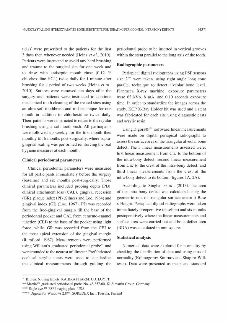
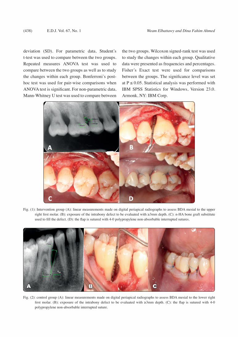
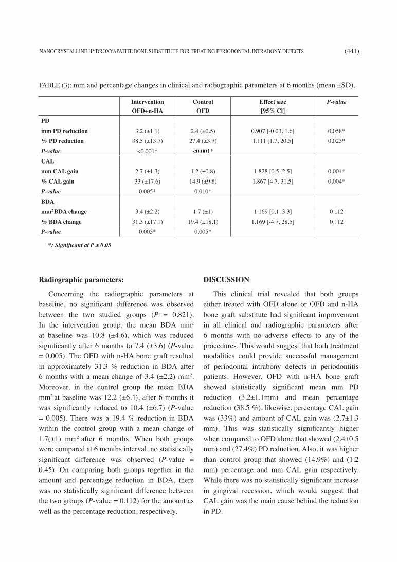
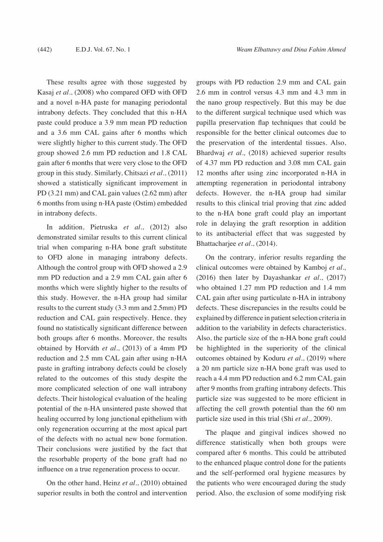
as the intervention group. Under local anesthesia an intrasulcular incision was performed with #15c blade elevating a full-thickness mucoperiosteal flap. The access flap was raised buccally and lingually to expose 2-3 mm of the alveolar crest and thorough debridement was performed using area-specific curettes (Hu-Friedy, USA). After exposing the intrabony defect, it was evaluated to confirm that it was a 2- or 3-wall defect with ≥3mm depth (figures 1B, 2B).
OFD was performed for the control group which received meticulous debridement and root planning of the intrabony defect area to remove all the inflammatory granulation tissue (figure 2B). While the intervention group received the same surgical technique (OFD), and n-HA bone graft substitute was used to fill the defect by gradual placement of incremental fragments till the defect space was completely filled avoiding any air voids from incorporating the grafted material. Bone graft substitute (n-HA) was placed up to the level of the alveolar bone crest (figure 1C). Then the mucoperiosteal flap was returned back to its position in both groups and sutured with 4-0 polypropylene non-absorbable interrupted sutures****** (figures 1D, 2C).
Postsurgical instructions
Postoperatively, all patients were instructed to take systemic antibiotics (Amoxicillin 500 mg t.d.s)******* orally for 5 days to prevent postsurgical infection (Singh et al., 2012). Also, oral analgesics and anti-inflammatory drugs (Ibuprofen 600 mg
* Woodpecker UDS-P with LED, China.** EMS Woodpecker ultrasonic scaler tip, Woodpecker, China*** Hu-Friedy universal and Gracey’s curette; Hu-Friedy, Chicago, USA.**** Hexitol: Chlorhexidine HCL mouthwash, The Arab Drug Company for pharmaceutical & CHEM. IND. CO. Cairo-Egypt. ***** Nano Bone, Dentaurum, Germany. ****** Polypropylene blue 4-0 Assut sutures of Switzerland, Assut Medical Sàrl, Switzerland. ******* E-mox 500 mg Cap., E.I.P.I.C.O., Egyptian Int. Pharmaceutical Industrial Co., A.R.E.
NANOCRYSTALLINE HYDROXYAPATITE BONE SUBSTITUTE FOR TREATING PERIODONTAL INTRABONY DEFECTS (437)
t.d.s)* were prescribed to the patients for the first 3 days then whenever needed (Heinz et al., 2010). Patients were instructed to avoid any hard brushing and trauma to the surgical site for one week and to rinse with antiseptic mouth rinse (0.12 % chlorhexidine HCL) twice daily for 1 minute after brushing for a period of two weeks (Heinz et al., 2010). Sutures were removed ten days after the surgery and patients were instructed to continue mechanical tooth cleaning of the treated sites using an ultra-soft toothbrush and roll technique for one month in addition to chlorhexidine twice daily. Then, patients were instructed to return to the regular brushing using a soft toothbrush. All participants were followed up weekly for the first month then monthly till 6 months post-surgically, where supra-gingival scaling was performed reinforcing the oral hygiene measures at each month.
Clinical periodontal parameters
Clinical periodontal parameters were measured for all participants immediately before the surgery (baseline) and six months post-surgically. Those clinical parameters included probing depth (PD), clinical attachment loss (CAL), gingival recession (GR), plaque index (PI) (Silness and Löe, 1964) and gingival index (GI) (Löe, 1967). PD was recorded from the free-gingival margin till the base of the periodontal pocket and CAL from cemento-enamel junction (CEJ) to the base of the pocket using light force, while, GR was recorded from the CEJ to the most apical extension of the gingival margin (Ramfjord, 1967). Measurements were performed using William’s graduated periodontal probe** and were rounded to the nearest millimeter. Prefabricated occlusal acrylic stents were used to standardize the clinical measurements through guiding the
periodontal probe to be inserted in vertical grooves within the stent parallel to the long axis of the tooth.
Radiographic parameters
Periapical digital radiographs using PSP sensors size 2*** were taken, using right angle long cone parallel technique to detect alveolar bone level, Planmeca X-ray machine, exposure parameters were 63 kVp, 8 mA, and 0.10 seconds exposure time. In order to standardize the images across the study, KCP X-Ray Holder kit was used and a stent was fabricated for each site using diagnostic casts and acrylic resin.
Using Digora®**** software, linear measurements were made on digital periapical radiographs to assess the surface area of the triangular alveolar bone defect. The 3 linear measurements assessed were: first linear measurement from CEJ to the bottom of the intra-bony defect; second linear measurement from CEJ to the crest of the intra-bony defect; and third linear measurements from the crest of the intra-bony defect to its bottom (figures 1A, 2A).
According to Singhal et al., (2013), the area of the intra-bony defect was calculated using the geometric rule of triangular surface area= ó Base x Height. Periapical digital radiographs were taken immediately preoperative (baseline) and six months postoperatively where the linear measurements and surface area were carried out and bone defect area (BDA) was calculated in mm square.
Statistical analysis
Numerical data were explored for normality by checking the distribution of data and using tests of normality (Kolmogorov-Smirnov and Shapiro-Wilk tests). Data were presented as mean and standard
* Brufen, 600 mg tablets, KAHIRA PHARM. CO. EGYPT. ** Martin™ graduated periodontal probe No. 43-357-00, KLS martin Group, Germany. *** Eagle eye ™ PSP Imaging plate, USA**** Digora For Windows 2.8™, SOREDEX Inc., Tuusula, Finland
(438) Weam Elbattawy and Dina Fahim AhmedE.D.J. Vol. 67, No. 1
deviation (SD). For parametric data, Student’s t-test was used to compare between the two groups. Repeated measures ANOVA test was used to compare between the two groups as well as to study the changes within each group. Bonferroni’s post-hoc test was used for pair-wise comparisons when ANOVA test is significant. For non-parametric data, Mann-Whitney U test was used to compare between
the two groups. Wilcoxon signed-rank test was used to study the changes within each group. Qualitative data were presented as frequencies and percentages. Fisher’s Exact test were used for comparisons between the groups. The significance level was set at P ≤ 0.05. Statistical analysis was performed with IBM SPSS Statistics for Windows, Version 23.0. Armonk, NY: IBM Corp.
Fig. (1): Intervention group (A): linear measurements made on digital periapical radiographs to assess BDA mesial to the upper right first molar. (B): exposure of the intrabony defect to be evaluated with ≥3mm depth. (C): n-HA bone graft substitute used to fill the defect. (D): the flap is sutured with 4-0 polypropylene non-absorbable interrupted sutures.
Fig. (2): control group (A): linear measurements made on digital periapical radiographs to assess BDA mesial to the lower right first molar. (B): exposure of the intrabony defect to be evaluated with ≥3mm depth. (C): the flap is sutured with 4-0 polypropylene non-absorbable interrupted suture.
NANOCRYSTALLINE HYDROXYAPATITE BONE SUBSTITUTE FOR TREATING PERIODONTAL INTRABONY DEFECTS (439)
RESULTS
Demographic data
The mean age values and gender distribution are shown in table (1), showing no statistically significant difference between the mean age values in both the control and intervention groups. There was also no statistically significant difference between gender distributions in both groups.
TABLE (1): Mean, standard deviation (SD), frequencies (n), percentages and results of Student’s t-test and Fisher’s Exact test for comparisons between demographic data in the two groups
Intervention
(n = 10)
Control
(n = 10)P-value
Age (Years)
Mean (SD) 46 (6.5) 42.3 (5.2) 0.176
Gender [n (%)]
Male 6 (60%) 3 (30%) 0.370
Female 4 (40%) 7 (70%)
*: Significant at P ≤ 0.05
Clinical periodontal parameters:
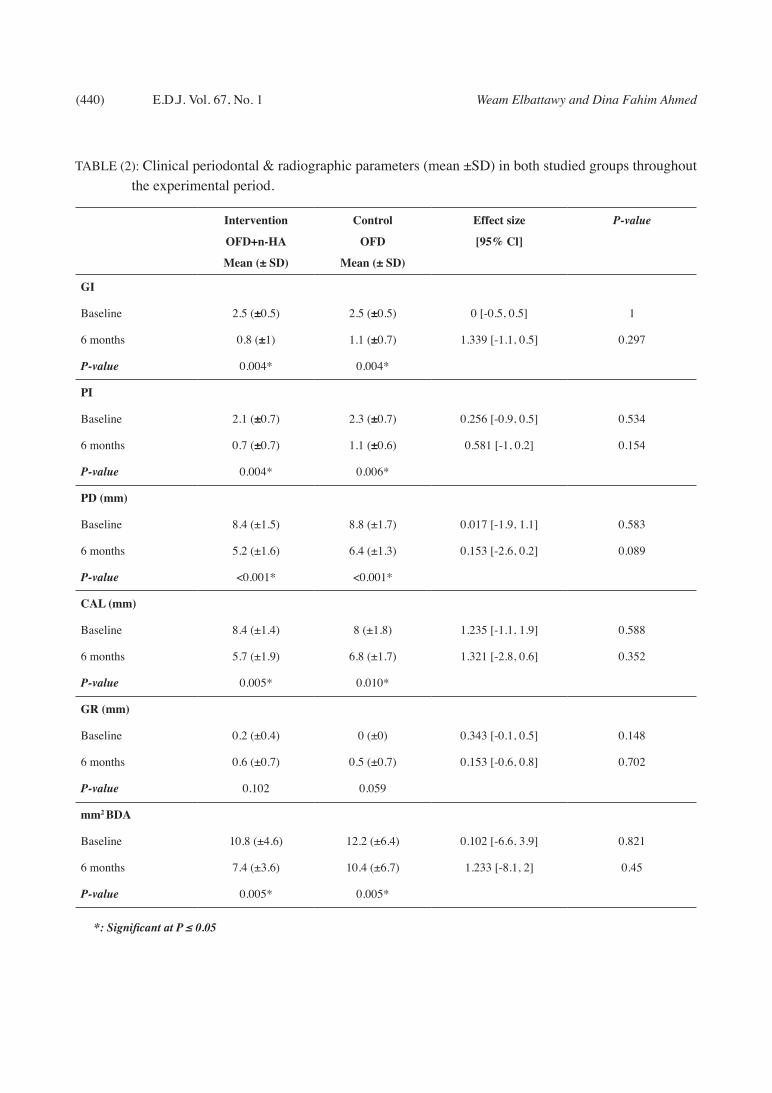
The clinical and radiographic parameters recorded for the control (OFD) and intervention groups (OFD+n-HA) throughout the study period are shown in table (2), while the mm reduction and percentage change in PD, CAL and BDA are represented in table (2).
The results of this clinical trial showed a statistically significant reduction in GI after 6 months in both groups (P = 0.004) but with no statistically significant difference between both groups at baseline as well as after six months (P = 1 and P = 0.297), respectively. Similarly, there was a statistically significant reduction in PI scores after 6 months in intervention and control groups (P = 0.004, P = 0.006), respectively, but also with
no statistically significant difference between both groups at baseline and after 6 months (P = 0.534 and P = 0.154), respectively.
After 6 months, a significant improvement was observed in PD values from baseline in both inter-vention and control groups (P <0.001). However, there was no statistically significant difference on comparing both groups at baseline as well as at 6 months (P-value = 0.583 and P = 0.089), respec-tively (table 2). Similarly, there was a statistically significant gain in CAL values from baseline to six months in intervention group (P = 0.005) and in control group (P = 0.010) but with no statistically significant difference on comparing baseline values (P = 0.588) as well as after 6 months (P = 0.352) between both groups as shown in table (2).
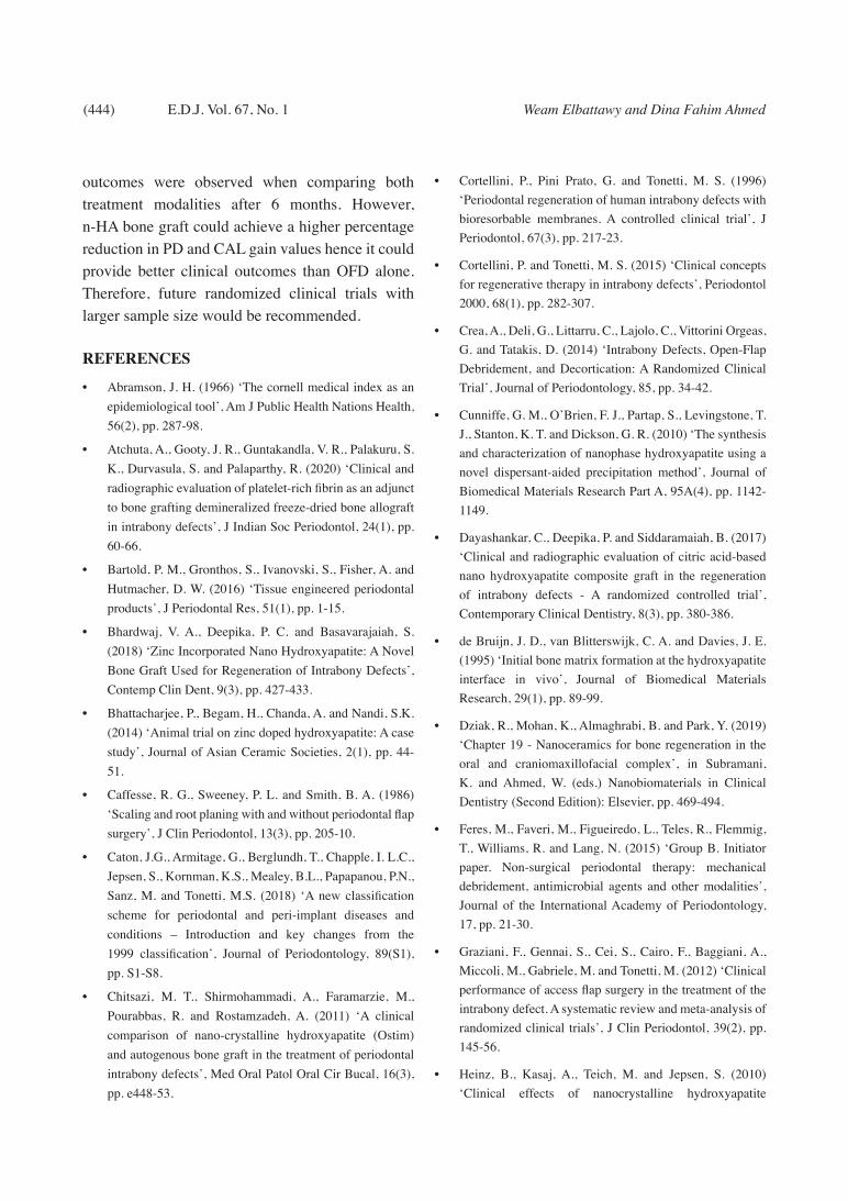
As regards the reduction in PD; there was a statistically significant mm and percentage reduction of PD from baseline to 6 months in each of the control and intervention groups (P <0.001). Also, there was a statistically significant mm and percentage gain in CAL within the control and intervention groups after 6 months (P = 0.010, P = 0.005), respectively as shown in table (3).
On comparing the mm and percentage reduction in PD between the two groups after 6 months; the n-HA intervention group showed a higher statistically significant mean mm reduction (P = 0.058) and higher mean percentage reduction (P = 0.023) than the OFD control group. Similarly, the comparison between the amount of mm and percentage gain in CAL revealed that the intervention group showed a statistically significantly higher amount and percentage gain in CAL than control group (P = 0.004 and P = 0.004), respectively (table 3).
Regarding the gingival recession values, there was no statistically significant change in gingival recession (GR) after 6 months for each group. Also, there was no statistically significant difference between both groups at baseline values and after 6 months (P=0.148, P= 0.702), respectively.
(440) Weam Elbattawy and Dina Fahim AhmedE.D.J. Vol. 67, No. 1
TABLE (2): Clinical periodontal & radiographic parameters (mean ±SD) in both studied groups throughout the experimental period.
Intervention
OFD+n-HA
Mean (± SD)
Control
OFD
Mean (± SD)
Effect size
[95% Cl]
P-value
GI
Baseline 2.5 (±0.5) 2.5 (±0.5) 0 [-0.5, 0.5] 1
6 months 0.8 (±1) 1.1 (±0.7) 1.339 [-1.1, 0.5] 0.297
P-value 0.004* 0.004*
PI
Baseline 2.1 (±0.7) 2.3 (±0.7) 0.256 [-0.9, 0.5] 0.534
6 months 0.7 (±0.7) 1.1 (±0.6) 0.581 [-1, 0.2] 0.154
P-value 0.004* 0.006*
PD (mm)
Baseline 8.4 (±1.5) 8.8 (±1.7) 0.017 [-1.9, 1.1] 0.583
6 months 5.2 (±1.6) 6.4 (±1.3) 0.153 [-2.6, 0.2] 0.089
P-value <0.001* <0.001*
CAL (mm)
Baseline 8.4 (±1.4) 8 (±1.8) 1.235 [-1.1, 1.9] 0.588
6 months 5.7 (±1.9) 6.8 (±1.7) 1.321 [-2.8, 0.6] 0.352
P-value 0.005* 0.010*
GR (mm)
Baseline 0.2 (±0.4) 0 (±0) 0.343 [-0.1, 0.5] 0.148
6 months 0.6 (±0.7) 0.5 (±0.7) 0.153 [-0.6, 0.8] 0.702
P-value 0.102 0.059
mm2 BDA
Baseline 10.8 (±4.6) 12.2 (±6.4) 0.102 [-6.6, 3.9] 0.821
6 months 7.4 (±3.6) 10.4 (±6.7) 1.233 [-8.1, 2] 0.45
P-value 0.005* 0.005*
*: Significant at P ≤ 0.05
NANOCRYSTALLINE HYDROXYAPATITE BONE SUBSTITUTE FOR TREATING PERIODONTAL INTRABONY DEFECTS (441)
Radiographic parameters:
Concerning the radiographic parameters at baseline, no significant difference was observed between the two studied groups (P = 0.821). In the intervention group, the mean BDA mm2
at baseline was 10.8 (±4.6), which was reduced significantly after 6 months to 7.4 (±3.6) (P-value = 0.005). The OFD with n-HA bone graft resulted in approximately 31.3 % reduction in BDA after 6 months with a mean change of 3.4 (±2.2) mm2. Moreover, in the control group the mean BDA mm2 at baseline was 12.2 (±6.4), after 6 months it was significantly reduced to 10.4 (±6.7) (P-value = 0.005). There was a 19.4 % reduction in BDA within the control group with a mean change of 1.7(±1) mm2 after 6 months. When both groups were compared at 6 months interval, no statistically significant difference was observed (P-value = 0.45). On comparing both groups together in the amount and percentage reduction in BDA, there was no statistically significant difference between the two groups (P-value = 0.112) for the amount as well as the percentage reduction, respectively.
DISCUSSION
This clinical trial revealed that both groups either treated with OFD alone or OFD and n-HA bone graft substitute had significant improvement in all clinical and radiographic parameters after 6 months with no adverse effects to any of the procedures. This would suggest that both treatment modalities could provide successful management of periodontal intrabony defects in periodontitis patients. However, OFD with n-HA bone graft showed statistically significant mean mm PD reduction (3.2±1.1mm) and mean percentage reduction (38.5 %), likewise, percentage CAL gain was (33%) and amount of CAL gain was (2.7±1.3 mm). This was statistically significantly higher when compared to OFD alone that showed (2.4±0.5 mm) and (27.4%) PD reduction. Also, it was higher than control group that showed (14.9%) and (1.2 mm) percentage and mm CAL gain respectively. While there was no statistically significant increase in gingival recession, which would suggest that CAL gain was the main cause behind the reduction in PD.
TABLE (3): mm and percentage changes in clinical and radiographic parameters at 6 months (mean ±SD).
InterventionOFD+n-HA
ControlOFD
Effect size[95% Cl]
P-value
PDmm PD reduction 3.2 (±1.1) 2.4 (±0.5) 0.907 [-0.03, 1.6] 0.058*% PD reduction 38.5 (±13.7) 27.4 (±3.7) 1.111 [1.7, 20.5] 0.023*P-value <0.001* <0.001*CALmm CAL gain 2.7 (±1.3) 1.2 (±0.8) 1.828 [0.5, 2.5] 0.004*% CAL gain 33 (±17.6) 14.9 (±9.8) 1.867 [4.7, 31.5] 0.004*P-value 0.005* 0.010*BDAmm2 BDA change 3.4 (±2.2) 1.7 (±1) 1.169 [0.1, 3.3] 0.112% BDA change 31.3 (±17.1) 19.4 (±18.1) 1.169 [-4.7, 28.5] 0.112P-value 0.005* 0.005*
*: Significant at P ≤ 0.05
(442) Weam Elbattawy and Dina Fahim AhmedE.D.J. Vol. 67, No. 1
These results agree with those suggested by Kasaj et al., (2008) who compared OFD with OFD and a novel n-HA paste for managing periodontal intrabony defects. They concluded that this n-HA paste could produce a 3.9 mm mean PD reduction and a 3.6 mm CAL gains after 6 months which were slightly higher to this current study. The OFD group showed 2.6 mm PD reduction and 1.8 CAL gain after 6 months that were very close to the OFD group in this study. Similarly, Chitsazi et al., (2011) showed a statistically significant improvement in PD (3.21 mm) and CAL gain values (2.62 mm) after 6 months from using n-HA paste (Ostim) embedded in intrabony defects.
In addition, Pietruska et al., (2012) also demonstrated similar results to this current clinical trial when comparing n-HA bone graft substitute to OFD alone in managing intrabony defects. Although the control group with OFD showed a 2.9 mm PD reduction and a 2.9 mm CAL gain after 6 months which were slightly higher to the results of this study. However, the n-HA group had similar results to the current study (3.3 mm and 2.5mm) PD reduction and CAL gain respectively. Hence, they found no statistically significant difference between both groups after 6 months. Moreover, the results obtained by Horváth et al., (2013) of a 4mm PD reduction and 2.5 mm CAL gain after using n-HA paste in grafting intrabony defects could be closely related to the outcomes of this study despite the more complicated selection of one wall intrabony defects. Their histological evaluation of the healing potential of the n-HA unsintered paste showed that healing occurred by long junctional epithelium with only regeneration occurring at the most apical part of the defects with no actual new bone formation. Their conclusions were justified by the fact that the resorbable property of the bone graft had no influence on a true regeneration process to occur.
On the other hand, Heinz et al., (2010) obtained superior results in both the control and intervention
groups with PD reduction 2.9 mm and CAL gain 2.6 mm in control versus 4.3 mm and 4.3 mm in the nano group respectively. But this may be due to the different surgical technique used which was papilla preservation flap techniques that could be responsible for the better clinical outcomes due to the preservation of the interdental tissues. Also, Bhardwaj et al., (2018) achieved superior results of 4.37 mm PD reduction and 3.08 mm CAL gain 12 months after using zinc incorporated n-HA in attempting regeneration in periodontal intrabony defects. However, the n-HA group had similar results to this clinical trial proving that zinc added to the n-HA bone graft could play an important role in delaying the graft resorption in addition to its antibacterial effect that was suggested by Bhattacharjee et al., (2014).
On the contrary, inferior results regarding the clinical outcomes were obtained by Kamboj et al., (2016) then later by Dayashankar et al., (2017) who obtained 1.27 mm PD reduction and 1.4 mm CAL gain after using particulate n-HA in intrabony defects. These discrepancies in the results could be explained by difference in patient selection criteria in addition to the variability in defects characteristics. Also, the particle size of the n-HA bone graft could be highlighted in the superiority of the clinical outcomes obtained by Koduru et al., (2019) where a 20 nm particle size n-HA bone graft was used to reach a 4.4 mm PD reduction and 6.2 mm CAL gain after 9 months from grafting intrabony defects. This particle size was suggested to be more efficient in affecting the cell growth potential than the 60 nm particle size used in this trial (Shi et al., 2009).
The plaque and gingival indices showed no difference statistically when both groups were compared after 6 months. This could be attributed to the enhanced plaque control done for the patients and the self-performed oral hygiene measures by the patients who were encouraged during the study period. Also, the exclusion of some modifying risk
NANOCRYSTALLINE HYDROXYAPATITE BONE SUBSTITUTE FOR TREATING PERIODONTAL INTRABONY DEFECTS (443)
factors as smoking which was proved to have an effect on retardation of the process of regeneration and healing outcomes could be another important factor in enhancing the clinical outcomes (Patel et al., 2012).
This clinical trial achieved a percentage reduction in BDA of 31.3 % in the n-HA group versus 19.4% in the OFD group where each group showed a statistically significant reduction in BDA from baseline. However, there was no statistically significant difference when both groups were compared. Superior results were achieved by Kamboj et al., (2016) with a 59.8% bone fill measured by CBCT and by Dayashankar et al., (2017) where citric acid-based n-HA bone graft was used reaching a 39.89% bone fill. This superiority could be attributed to the different materials used or the more accurate radiographic imaging technique than the digital standard radiography. Also, the zinc incorporated n-HA could increase the bone fill up to 54.7% versus 40.2 % with the n-HA group which were also superior to this study. This could be related to the longer follow up interval of 9 months that might permit further bone deposition in addition to the zinc that is thought to improve the bioactivity of n-HA bone graft material.
It has been common in the literature for radio-graphic outcomes to be parallel with the clinical ones (Reynolds et al., 2003). This was the case in this clinical trial which is also comparative to Pi-etruska et al., (2012) who observed a statistically significant reduction in the depth and width of bone defect after both OFD and OFD with n-HA bone substitute. However, their study achieved a better bone defect depth reduction in the n-HA group than with OFD alone. Noteworthy, in the radiographic evaluation a narrow gap was noted between the graft and the surrounding bone walls. The authors explained this by the histological findings proved by Heinz et al., (2010) who confirmed that bone graft substitutes usually heal by connective tissue
encapsulation rather than true regeneration in the periodontium, where only minimal true bone for-mation could be found at the most apical part of the intrabony defect zone.
The success of OFD alone as a conservative surgical approach for treating periodontal intrabony defects has been agreed upon in many clinical trials which were included in a systematic review and subjected to metanalysis by Graziani et al., (2012) confirming that OFD is a successful surgical technique that could improve clinical and radiographic outcomes. Therefore, it should not be ignored that the OFD surgical procedure could enhance the percentage reduction of the BDA significantly even when performed alone without any bone substitute added. This agrees with Crea et al., (2014) who achieved a statistically significant improvement in the radiographic defect depth with OFD alone after 12 months. It also agrees with Atchuta et al., (2020) who reached a 20.24% reduction in radiographic defect depth after OFD of intrabony defects. On the other hand, space maintenance by bone grafts were proved in the literature to be effective in the process of regeneration by acting as structural scaffolds. That was confirmed by other reports which proved that filling intrabony defects with biomaterial could enhance the clinical results making them superior to those achieved with OFD procedure alone (Reynolds et al., 2003). Thus, n-HA bone graft would be an easily available affordable bone substitute and hence with all its advantages the hypotheses to be behind the enhanced clinical outcomes even when no actual true bone was proved to occur.
CONCLUSIONS
Within the limitations of this study as it is a non-randomized clinical trial, it is concluded that both OFD alone and OFD with n-HA bone substitute could be considered as a successful intervention for treating periodontal intrabony defects, yet no statistically significant differences in radiographic
(444) Weam Elbattawy and Dina Fahim AhmedE.D.J. Vol. 67, No. 1
outcomes were observed when comparing both treatment modalities after 6 months. However, n-HA bone graft could achieve a higher percentage reduction in PD and CAL gain values hence it could provide better clinical outcomes than OFD alone. Therefore, future randomized clinical trials with larger sample size would be recommended.
REFERENCES
• Abramson, J. H. (1966) ‘The cornell medical index as an epidemiological tool’, Am J Public Health Nations Health, 56(2), pp. 287-98.
• Atchuta, A., Gooty, J. R., Guntakandla, V. R., Palakuru, S. K., Durvasula, S. and Palaparthy, R. (2020) ‘Clinical and radiographic evaluation of platelet-rich fibrin as an adjunct to bone grafting demineralized freeze-dried bone allograft in intrabony defects’, J Indian Soc Periodontol, 24(1), pp. 60-66.
• Bartold, P. M., Gronthos, S., Ivanovski, S., Fisher, A. and Hutmacher, D. W. (2016) ‘Tissue engineered periodontal products’, J Periodontal Res, 51(1), pp. 1-15.
• Bhardwaj, V. A., Deepika, P. C. and Basavarajaiah, S. (2018) ‘Zinc Incorporated Nano Hydroxyapatite: A Novel Bone Graft Used for Regeneration of Intrabony Defects’, Contemp Clin Dent, 9(3), pp. 427-433.
• Bhattacharjee, P., Begam, H., Chanda, A. and Nandi, S.K. (2014) ‘Animal trial on zinc doped hydroxyapatite: A case study’, Journal of Asian Ceramic Societies, 2(1), pp. 44-51.
• Caffesse, R. G., Sweeney, P. L. and Smith, B. A. (1986) ‘Scaling and root planing with and without periodontal flap surgery’, J Clin Periodontol, 13(3), pp. 205-10.
• Caton, J.G., Armitage, G., Berglundh, T., Chapple, I. L.C., Jepsen, S., Kornman, K.S., Mealey, B.L., Papapanou, P.N., Sanz, M. and Tonetti, M.S. (2018) ‘A new classification scheme for periodontal and peri-implant diseases and conditions – Introduction and key changes from the 1999 classification’, Journal of Periodontology, 89(S1), pp. S1-S8.
• Chitsazi, M. T., Shirmohammadi, A., Faramarzie, M., Pourabbas, R. and Rostamzadeh, A. (2011) ‘A clinical comparison of nano-crystalline hydroxyapatite (Ostim) and autogenous bone graft in the treatment of periodontal intrabony defects’, Med Oral Patol Oral Cir Bucal, 16(3), pp. e448-53.
• Cortellini, P., Pini Prato, G. and Tonetti, M. S. (1996) ‘Periodontal regeneration of human intrabony defects with bioresorbable membranes. A controlled clinical trial’, J Periodontol, 67(3), pp. 217-23.
• Cortellini, P. and Tonetti, M. S. (2015) ‘Clinical concepts for regenerative therapy in intrabony defects’, Periodontol 2000, 68(1), pp. 282-307.
• Crea, A., Deli, G., Littarru, C., Lajolo, C., Vittorini Orgeas, G. and Tatakis, D. (2014) ‘Intrabony Defects, Open-Flap Debridement, and Decortication: A Randomized Clinical Trial’, Journal of Periodontology, 85, pp. 34-42.
• Cunniffe, G. M., O’Brien, F. J., Partap, S., Levingstone, T. J., Stanton, K. T. and Dickson, G. R. (2010) ‘The synthesis and characterization of nanophase hydroxyapatite using a novel dispersant-aided precipitation method’, Journal of Biomedical Materials Research Part A, 95A(4), pp. 1142-1149.
• Dayashankar, C., Deepika, P. and Siddaramaiah, B. (2017) ‘Clinical and radiographic evaluation of citric acid-based nano hydroxyapatite composite graft in the regeneration of intrabony defects - A randomized controlled trial’, Contemporary Clinical Dentistry, 8(3), pp. 380-386.
• de Bruijn, J. D., van Blitterswijk, C. A. and Davies, J. E. (1995) ‘Initial bone matrix formation at the hydroxyapatite interface in vivo’, Journal of Biomedical Materials Research, 29(1), pp. 89-99.
• Dziak, R., Mohan, K., Almaghrabi, B. and Park, Y. (2019) ‘Chapter 19 - Nanoceramics for bone regeneration in the oral and craniomaxillofacial complex’, in Subramani, K. and Ahmed, W. (eds.) Nanobiomaterials in Clinical Dentistry (Second Edition): Elsevier, pp. 469-494.
• Feres, M., Faveri, M., Figueiredo, L., Teles, R., Flemmig, T., Williams, R. and Lang, N. (2015) ‘Group B. Initiator paper. Non-surgical periodontal therapy: mechanical debridement, antimicrobial agents and other modalities’, Journal of the International Academy of Periodontology, 17, pp. 21-30.
• Graziani, F., Gennai, S., Cei, S., Cairo, F., Baggiani, A., Miccoli, M., Gabriele, M. and Tonetti, M. (2012) ‘Clinical performance of access flap surgery in the treatment of the intrabony defect. A systematic review and meta-analysis of randomized clinical trials’, J Clin Periodontol, 39(2), pp. 145-56.
• Heinz, B., Kasaj, A., Teich, M. and Jepsen, S. (2010) ‘Clinical effects of nanocrystalline hydroxyapatite
NANOCRYSTALLINE HYDROXYAPATITE BONE SUBSTITUTE FOR TREATING PERIODONTAL INTRABONY DEFECTS (445)
paste in the treatment of intrabony periodontal defects: a randomized controlled clinical study’, Clinical Oral Investigations, 14(5), pp. 525-531.
• Horváth, A., Stavropoulos, A., Windisch, P., Lukács, L., Gera, I. and Sculean, A. (2013) ‘Histological evaluation of human intrabony periodontal defects treated with an unsintered nanocrystalline hydroxyapatite paste’, Clinical Oral Investigations, 17(2), pp. 423-430.
• Kamboj, M., Arora, R. and Gupta, H. (2016) Journal of Indian Society of Periodontology, 20(4), pp. 423-428.
• Kasaj, A., Röhrig, B., Zafiropoulos, G.-G. and Willershausen, B. (2008) ‘Clinical Evaluation of Nanocrystalline Hydroxyapatite Paste in the Treatment of Human Periodontal Bony Defects – A Randomized Controlled Clinical Trial: 6-Month Results’, Journal of Periodontology, 79(3), pp. 394-400.
• Koduru, S., Aghanashini, S., Nadiger, S., Apoorva, S., Bhat, D. and Puvvalla, B. (2019) ‘A clinical and radiographic evaluation of the efficacy of nanohydroxyapatite (Sybograf <sup>™</sup>) versus bioactive calcium phosphosilicate putty (Novabone<sup>®</sup>) in the treatment of human periodontal infrabony defects: A randomized clinical trial’, Contemporary Clinical Dentistry, 10(1), pp. 16-23.
• Kumta, P. N., Sfeir, C., Lee, D.-H., Olton, D. and Choi, D. (2005) ‘Nanostructured calcium phosphates for biomedical applications: novel synthesis and characterization’, Acta Biomaterialia, 1(1), pp. 65-83.
• Lang, N. P., Hämmerle, C., Oesch, B., AG, P. and Schenk, R. K. (2000) ‘Risk of Transmission of Agents Associated with Creutzfeldt-Jakob Disease and Bovine Spongiform Encephalopathy’, Plastic and Reconstructive Surgery, 105(6), pp. 2273-2274.
• Löe, H. (1967) ‘The Gingival Index, the Plaque Index and the Retention Index Systems’, J Periodontol, 38(6), pp. Suppl:610-6.
• Needleman, I., Tucker, R., Giedrys-Leeper, E. and Worthington, H. (2005) ‘Guided tissue regeneration for periodontal intrabony defects--a Cochrane Systematic Review’, Periodontol 2000, 37, pp. 106-23.
• Papapanou, P.N., Sanz, M., Buduneli, N., Dietrich, T., Feres, M., Fine, D.H., Flemmig, T.F., Garcia, R., Giannobile, W. V., Graziani, F., Greenwell, H., Herrera, D., Kao, R. T., Kebschull, M., Kinane, D. F., Kirkwood, K.L., Kocher, T., Kornman, K.S., Kumar, P. S., Loos, B.-------G., Machtei, E., Meng, H., Mombelli, A., Needleman, I., Offenbacher,
S., Seymour, G. J., Teles, R. and Tonetti, M. S. (2018) ‘Periodontitis: Consensus report of workgroup 2 of the 2017 World Workshop on the Classification of Periodontal and Peri-Implant Diseases and Conditions’, Journal of Clinical Periodontology, 45(S20), pp. S162-S170.
• Papapanou, P. N. and Tonetti, M. S. (2000) ‘Diagnosis and epidemiology of periodontal osseous lesions’, Periodontol 2000, 22, pp. 8-21.
• Patel, R. A., Wilson, R. F. and Palmer, R. M. (2012) ‘The effect of smoking on periodontal bone regeneration: a systematic review and meta-analysis’, J Periodontol, 83(2), pp. 143-55.
• Pietruska, M., Skurska, A., Pietruski, J., Dolińska, E., Arweiler, N., Milewski, R., Duraj, E. and Sculean, A. (2012) ‘Clinical and radiographic evaluation of intrabony periodontal defect treatment by open flap debridement alone or in combination with nanocrystalline hydroxyapatite bone substitute’, Annals of Anatomy - Anatomischer Anzeiger, 194(6), pp. 533-537.
• Ramfjord, S. P. (1967) ‘The Periodontal Disease Index (PDI)’, The Journal of Periodontology, 38(6), pp. 602-610.
• Ramfjord, S. P. and Nissle, R. R. (1974) ‘The modified widman flap’, J Periodontol, 45(8), pp. 601-7.
• Reynolds, M. A., Aichelmann-Reidy, M. E. and Branch-Mays, G. L. (2010) ‘Regeneration of Periodontal Tissue: Bone Replacement Grafts’, Dental Clinics of North America, 54(1), pp. 55-71.
• Reynolds, M. A., Aichelmann-Reidy, M. E., Branch-Mays, G. L. and Gunsolley, J. C. (2003) ‘The Efficacy of Bone Replacement Grafts in the Treatment of Periodontal Osseous Defects. A Systematic Review’, Annals of Periodontology, 8(1), pp. 227-265.
• Reynolds, M. A., Kao, R. T., Nares, S., Camargo, P. M., Caton, J. G., Clem, D. S., Fiorellini, J. P., Geisinger, M. L., Mills, M. P., Nevins, M. L. and Rosen, P. S. (2015) ‘Periodontal Regeneration — Intrabony Defects: Practical Applications From the AAP Regeneration Workshop’, Clinical Advances in Periodontics, 5(1), pp. 21-29.
• Sculean, A., Donos, N., Schwarz, F., Becker, J., Brecx, M. and Arweiler, N. B. (2004) ‘Five-year results following treatment of intrabony defects with enamel matrix proteins and guided tissue regeneration’, Journal of Clinical Periodontology, 31(7), pp. 545-549.
• Sheikh, Z., Hamdan, N., Ikeda, Y., Grynpas, M., Ganss,
(446) Weam Elbattawy and Dina Fahim AhmedE.D.J. Vol. 67, No. 1
B. and Glogauer, M. (2017) ‘Natural graft tissues and synthetic biomaterials for periodontal and alveolar bone reconstructive applications: a review’, Biomaterials Research, 21(1), pp. 9.
• Shi, Z., Huang, X., Cai, Y., Tang, R. and Yang, D. (2009) ‘Size effect of hydroxyapatite nanoparticles on proliferation and apoptosis of osteoblast-like cells’, Acta Biomaterialia, 5(1), pp. 338-345.
• Silness, J. and Löe, H. (1964) ‘Periodontal disease in pregnancy. ii. Correlation between oral hygiene and periodontal condition.’, Acta Odontol Scand, 22, pp. 121-35.
• Singh, B., Pandey, N., Mehrotra, B., Srivastava, A., Chowdhury, A. and Tiwari, S. (2012) ‘Development
of Comprehensive Satisfaction Index (ComSI) and its association with WHOQOL-BREF’, Journal of Indian Society of Periodontology, 16(4), pp. 562-568.
• Singhal, R., Nandlal, Kumar, A. and Rastogi, P. (2013) ‘Role of Space Provision in Regeneration of Localized Two-Wall Intrabony Defects Using Periosteal Pedicle Graft as an Autogenous Guided Tissue Membrane’, Journal of Periodontology, 84(3), pp. 316-324.
• Wang, H., Li, Y., Zuo, Y., Li, J., Ma, S. and Cheng, L. (2007) ‘Biocompatibility and osteogenesis of biomimetic nano-hydroxyapatite/polyamide composite scaffolds for bone tissue engineering’, Biomaterials, 28(22), pp. 3338-3348.