Embed Size (px)
Citation preview

REVIEW
Clinical and Biological Principles of Cold AtmosphericPlasma Application in Skin Cancer
Jesus Gay-Mimbrera . Maria Carmen Garcıa . Beatriz Isla-Tejera .
Antonio Rodero-Serrano . Antonio Velez Garcıa-Nieto . Juan Ruano
Received: March 21, 2016 / Published online: May 3, 2016� The Author(s) 2016. This article is published with open access at Springerlink.com
ABSTRACT
Plasma-based electrosurgical devices have long
been employed for tissue coagulation, cutting,
desiccation, and cauterizing. Despite their
clinical benefits, these technologies involve
tissue heating and their effects are primarily
heat-mediated. Recently, there have been
significant developments in cold atmospheric
pressure plasma (CAP) science and engineering.
New sources of CAP with well-controlled
temperatures below 40 �C have been designed,
permitting safe plasma application on animal
and human bodies. In the last decade, a new
innovative field, often referred to as plasma
medicine, which combines plasma physics, life
science, and clinical medicine has emerged.
This field aims to exploit effects of mild plasma
by controlling the interactions between plasma
components (and other secondary species that
can be formed from these components) with
specific structural elements and functionalities
of living cells. Recent studies showed that CAP
can exert beneficial effects when applied
selectively in certain pathologies with minimal
toxicity to normal tissues. The rapid increase in
new investigations and development of various
devices for CAP application suggest early
adoption of cold plasma as a new tool in the
biomedical field. This review explores the latest
major achievements in the field, focusing on
the biological effects, mechanisms of action,
and clinical evidence of CAP applications in
areas such as skin disinfection, tissue
regeneration, chronic wounds, and cancer
Enhanced content To view enhanced content for thisarticle go to www.medengine.com/Redeem/E2C4F06001B9A0F0.
J. Gay-Mimbrera � B. Isla-Tejera �A. V. Garcıa-Nieto � J. Ruano (&)Instituto Maimonides de Investigacion Biomedicade Cordoba (IMIBIC), Hospital Universitario ReinaSofıa, University of Cordoba, Cordoba, Spaine-mail: [email protected]
M. C. GarcıaDepartment of Applied Physics, University ofCordoba, Cordoba, Spain
B. Isla-TejeraDepartment of Pharmacy, Hospital UniversitarioReina Sofıa, Cordoba, Spain
A. Rodero-SerranoDepartment of Physics, School of EngineeringScience of Belmez, University of Cordoba, Cordoba,Spain
A. V. Garcıa-Nieto � J. RuanoDepartment of Dermatology, Hospital UniversitarioReina Sofıa, Cordoba, Spain
Adv Ther (2016) 33:894–909
DOI 10.1007/s12325-016-0338-1

treatment. This information may serve as a
foundation for the design of future clinical trials
to assess the efficacy and safety of CAP as an
adjuvant therapy for skin cancer.
Keywords: Apoptosis; Cold atmospheric
plasma; Dermatology; Keratinocytes;
Melanoma; Non-melanoma skin cancer;
Oncology
CHARACTERISTICS OF COLDATMOSPHERIC PLASMA
Plasma is an ionized gas that is composed of
ions, electrons, photons, and neutrals (radicals
as well as excited atoms and molecules). All are
active species capable of inducing different
physical phenomena and chemical reactions.
There are many examples of plasma in nature,
such as plasmas that are generated in stars and
the polar aurora. Plasmas can also be created in
the laboratory setting; here, plasmas are
maintained by applying an external source of
energy, generally an electromagnetic field, to a
gas. Plasma technology has gained much
interest in recent years owing to its various
applications in fields such as microelectronics,
waste elimination, lighting, and textile. Several
nonthermal plasma sources have been recently
developed. These plasma sources can be well
controlled and open to the air, allowing for the
maintenance of CAPs with temperatures below
40 �C. These developments have encouraged
therapeutic application of CAPs and the
emergence of plasma medicine technology.
Nonetheless, plasma application on the
human body for medical purposes has a
significant history. In the mid-nineteenth
century, electrotherapy was introduced as a
therapeutic approach, and spark and flash
discharges were employed to treat several
diseases. Later, electrosurgical techniques
based on the use of plasmas were developed.
In electrosurgery, selective coagulation or tissue
cutting is achieved by tissue heating which
results in desiccation of cells, denaturation of
proteins, or devitalization of tissues [1]. Argon
plasma coagulation (APC) is a well-established
method in the field and commonly employed
today for tissue coagulation during endoscopy
(in gastroenterology, general and visceral
surgery, urology, or gynecology) [2]. APC is a
monopolar technique introduced in the 1970s,
in which electrical energy is transferred to target
tissue as current by means of argon plasma. This
technique competes with traditional laser
ablation. Comparative studies have shown
that APC is more effective for tissue
destruction owing to its superior energy
concentration. Furthermore, PlasmaJet�,
another electrosurgical technique, possesses
activity that is mostly mediated by thermal
(thus destructive) interaction with living
tissues. It consists of a bipolar electrode system
with low-flow argon as the process gas. This
technique is typically employed to cut or
coagulate tissues in a well-defined and
localized manner. These direct plasma
applications on living tissues in electrosurgery
are based on extreme interactions of plasma
with cells or tissue, which may lead to cellular
destruction and local ‘‘sealing’’ of tissue.
In the last decade, with the development of
CAPs, modern plasma medicine has emerged.
This branch of medicine aims to exploit the
effects of mild plasma by utilizing the distinct
interaction of plasma components (and other
secondary species than can be formed from
these) with specific structural elements as well
as functionalities of living cells [1]. These
interactions may lead to either stimulation or
inhibition of cellular function; thus, this
technique may be employed for various
Adv Ther (2016) 33:894–909 895
therapeutic purposes [3]. While most clinical
studies have been conducted in the area of
dermatology, interests in CAP technology have
also emerged from other disciplines, such as
oncology, surgery, otolaryngology,
gastroenterology, and odontology [4, 5].
Sources of Cold Atmospheric Plasma
New CAP sources used in plasma medicine can
be classified into three types [6]:
1. Direct plasma sources These plasmas use the
human body (such as the skin, internal
tissues, etc.) as an electrode; thus, the
current produced by plasmas has to pass
through the body. The most utilized
technology in this category is the
dielectric barrier discharge (DBD) plasma
source.
2. Indirect plasma sources These plasmas are
generated between two electrodes. Active
species that are created by the plasmas are
subsequently transported to target
application areas. Several devices are
available, ranging from very narrow
plasma needles or jets to larger plasma
torches such as the kINPen� MED,
Atmospheric Pressure MicroPlasma Jet
(APMPJ), and MicroPlaSter� a and b.
3. Hybrid plasma sources These plasmas
combine the benefits of the two
aforementioned plasma source types (e.g.,
using the plasma production technique of
direct plasma sources and the essentially
current-free property of indirect plasma
sources). This is achieved by introducing a
grounded wire mesh electrode, which has a
significantly smaller electrical resistance
than that of the tissue; thus, in principle,
all current can pass through the wire mesh.
The MiniFlatPlaSter is an example of a
hybrid plasma source.
Nevertheless, plasmas can also be generated by
discharges in air, noble gases, or any desired
mixture. They can be excited by various means,
including radio frequency, microwave
frequencies, and high voltage AC or DC, in
continuous or pulsed mode in order to produce
a chemical cocktail of active species for
biomedical applications. Table 1 summarizes the
properties of various types of plasma reactors that
have been employed for dermatological purposes.
In cold atmospheric pressure plasma sources, the
main reactive components are reactive neutral
species (including free radicals and few ground
state molecules such as peroxides and ozone) and
UV radiation. Because CAP sources operate at
atmospheric pressure while in contact with air,
they generate copious quantities of reactive
oxygen species (ROS) and reactive nitrogen
species (RNS), collectively referred to as RONS. A
list of RONS was provided in a recent review,
which also highlighted the role of these species in
redox biology and their implications for
therapeutic applications of plasma [7]. RONS can
strongly influence cellular biochemistry and are
known to be important in animal and plant
immune systems, supporting the notion that
they are indeed key mediators in CAP
therapeutics.
Here, we review the latest evidence of the
biological effects, mechanisms of action, and
clinical benefits of CAP application in
dermatology. This review also highlights the
potential application of CAP as co-adjuvant
therapy for the treatment of skin tumor.
Analyses described in this review article were
based on previouslyconducted studies. No studies
involving human subjects or animals were
performed for the publication of this review.
896 Adv Ther (2016) 33:894–909

Table 1 Summary of plasma sources used in dermatology
Type ofplasmasource
Plasmasource
Device Gas Frequency Flow Power/dischargevoltage
Treatment
Direct
plasma
source
DBD PlasmaDerm�,
CINOGY
GmbH
Ar DC
Pulsed
100–400 Hz
8 slm 0.17–0.24 W/
14 kV
Melanoma [36]
Chronic ulcer [42]
Skin moisture [47]
Custom design Ar ? O2 DC
Pulsed 60 Hz
No
flow
0.9 W/20 kV Keratinocytes effect [15]
Custom design Air DC
Pulsed
255–518 Hz
No
flow
0.2–0.4 W/
5–6 kV
Wound healing [49]
Custom design
(point to plane
arrangement)
Air 100 kHz
Pulsed
300 Hz
No
flow
14 kV Skin treatment (physical
model) [50]
DBD, INP
Greifswald
Air 31 kHz No
flow
0.4–1.6 W/
13 kV
Study of skin irritation
[51]
DBD plasma strip Air 6.6 kHz No
flow
0.7 W/3.5 kV Infected wound [52]
Direct
DBD-Bioplasma
cell modulation
Air 15–20 kHz
Pulsed
10–110 Hz
No
flow
40 W/
6–7 kV
Acne and aesthetic
treatment [53]
Surface DBD Air/Ar 20–21 kHz No
flow/
0.5
slm
0.14–0.3 W/
3.5–10 kV
Wound healing [54]
Volumen DBD Ar 33 kHz 0.5 slm 6 W/
9–10 kV
Wound healing [54]
Indirect
plasma
source
Jets kINPen� MED,
neoplas tools
GmbH
Ar 1–1.5 MHz 3–8
slm
1–6 kV Skin decontamination
[55]
Psoriasis [43]
Melanoma [50]
Skin moisture [31]
Plasma effect in stratum
corneum [56]
Wound healing [4, 18,
57, 58]
Adv Ther (2016) 33:894–909 897
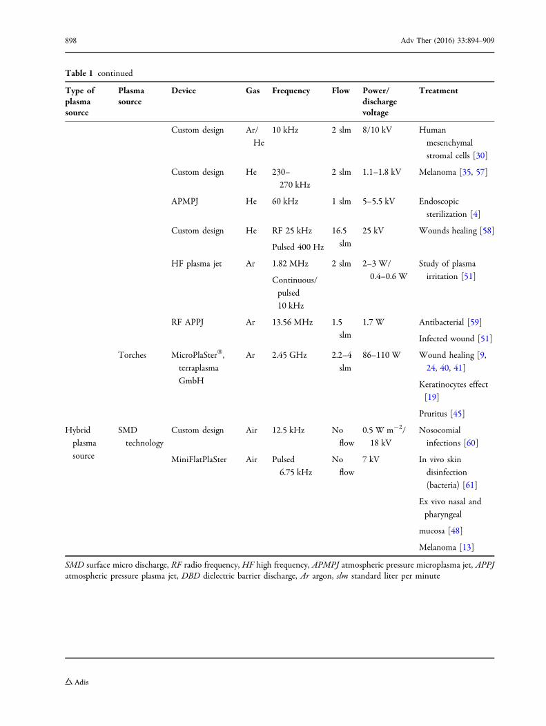
Table 1 continued
Type ofplasmasource
Plasmasource
Device Gas Frequency Flow Power/dischargevoltage
Treatment
Custom design Ar/
He
10 kHz 2 slm 8/10 kV Human
mesenchymal
stromal cells [30]
Custom design He 230–
270 kHz
2 slm 1.1–1.8 kV Melanoma [35, 57]
APMPJ He 60 kHz 1 slm 5–5.5 kV Endoscopic
sterilization [4]
Custom design He RF 25 kHz
Pulsed 400 Hz
16.5
slm
25 kV Wounds healing [58]
HF plasma jet Ar 1.82 MHz
Continuous/
pulsed
10 kHz
2 slm 2–3 W/
0.4–0.6 W
Study of plasma
irritation [51]
RF APPJ Ar 13.56 MHz 1.5
slm
1.7 W Antibacterial [59]
Infected wound [51]
Torches MicroPlaSter�,
terraplasma
GmbH
Ar 2.45 GHz 2.2–4
slm
86–110 W Wound healing [9,
24, 40, 41]
Keratinocytes effect
[19]
Pruritus [45]
Hybrid
plasma
source
SMD
technology
Custom design Air 12.5 kHz No
flow
0.5 W m-2/
18 kV
Nosocomial
infections [60]
MiniFlatPlaSter Air Pulsed
6.75 kHz
No
flow
7 kV In vivo skin
disinfection
(bacteria) [61]
Ex vivo nasal and
pharyngeal
mucosa [48]
Melanoma [13]
SMD surface micro discharge, RF radio frequency, HF high frequency, APMPJ atmospheric pressure microplasma jet, APPJatmospheric pressure plasma jet, DBD dielectric barrier discharge, Ar argon, slm standard liter per minute
898 Adv Ther (2016) 33:894–909
BIOLOGICAL EFFECTSAND MECHANISMS OF ACTIONOF COLD ATMOSPHERIC PLASMA
CAP is a gas composed of multiple chemically
active species. It induces physical and chemical
changes on biological surfaces upon
application. In recent years, several studies
have demonstrated biological effects of these
changes. Elucidation of the key mechanism
behind the effects of CAP in cancerous cells
will be crucial to determine the optimal dose of
CAP for clinical use.
Oxidative Stress
The level of intracellular ROS and RNS (RONS) is
tightly regulated by small antioxidant
molecules and scavenging enzymes. At low
concentrations, RONS are involved in various
biological processes such as bacterial
destruction by macrophages and endothelial
nitric oxide-mediated vasodilatation. However,
when their levels exceed the capacity of the
redox balance control system, a state referred to
as oxidative stress, they can be cytotoxic and
cause cell death. Oxidative stress is involved in
the development of various diseases such as
psoriasis, chronic ulcers, and cancer.
Cancer cells display weaker antioxidant
mechanisms when compared to normal cells.
This property can facilitate selective attack of
cancer cells by CAP mediated by the
extracellular RONS, resulting in severe
oxidative damage and cell death. Schmidt
et al. observed that alterations in redox state
due to CAP treatment caused changes in cellular
morphology and mobility, but not cell viability
[8]. The authors also found that oxidative stress
induced by CAP can modify the expression of
nearly 3000 genes encoding structural proteins
and inflammatory mediators, such as growth
factors and cytokines.
Gene Expression and Epigenetic Changes
Numerous studies have assessed the effects of
CAP on gene expression and epigenetics in
several cells lines. Application of CAP for 2 min
with the MicroPlaSter b� device on a fibroblast
culture and in a wound healing mouse model
increased the expression of type I collagen and
genes encoding proteins involved in wound
healing processes (interleukin 6 [IL-6], IL-8,
chemokine [C–C motif] ligand 2 [CCL2],
transforming growth factor beta 1 [TGF-b1],
TGF-b2, CD40 ligand, chemokine [C–X–C
motif] ligand 1 [CXCL1], interleukin 1 receptor
antagonist [IL-1RA], and plasminogen activator
inhibitor-1 [PAI-1]) without affecting cellular
migration, proliferation, and apoptosis [9].
Zhong et al. demonstrated the downregulation
of IL-12 and upregulation of IL-1b, IL-6, IL-8,
IL-10, tumor necrosis factor a (TNFa), interferon
gamma (IFNc), and vascular endothelial growth
factor (VEGF) mRNAs when CAP was applied to
keratinocyte cultures [10]. Park et al.
demonstrated for the first time changes in
DNA methylation pattern following CAP
application in a breast cancer cell line
expressing the estrogen receptor (MCF-7) and
one that does not express it (MDA-MB-231).
Epigenetic modifications were more extensive
in MCF-7 cells, affecting the promoter region of
genes related to ‘‘cell mobility’’, ‘‘connective
tissue function and development’’, ‘‘motility
development’’, ‘‘cell–cell communication and
cell–cell interaction’’, and ‘‘cell survival and cell
death’’ [11].
Adv Ther (2016) 33:894–909 899
Mitochondria, Cell Cycle, and Apoptosis
Apoptosis is a type of programmed cell death,
and mitochondria act as the major regulator of
apoptosis. Various intracellular and
extracellular signals induced by CAP-mediated
oxidative stress converge in mitochondria,
increasing their transmembrane potential and
promoting the release of pro-apoptotic factors
including cytochrome c. This process is
regulated by the Bcl-2 protein family and
ultimately leads to the activation of the
caspase cascade [12]. Arndt et al. showed that
when CAP was applied for 2 min to a melanoma
cell line, pro-apoptotic changes such as Rad17
and tumor suppressor p53 phosphorylations,
cytochrome c release, and caspase-3 activation
were initiated [13].
The cell cycle is a series of events leading to
cell replacement in tissues. RONS produced
following high dose application of CAP can
alter the cell cycle, which typically leads to
apoptosis. However, lower doses of CAP can also
inhibit cell proliferation by inducing cell
senescence, especially when most cells in the
tissue are in the proliferative phase, as observed
in most tumors [13]. Typically, normal tissues
differ from tumor in the proportion of cells in
each cell cycle phase at a given time. In fact, this
could be the biological mechanism behind the
high selectivity of CAP to induce apoptosis of
these cells while preserving viability of
non-tumor cells. Yan et al. demonstrated that
CAP increased the percentage of apoptotic
tumor cells by blocking the cell cycle at the
G2/M checkpoint, and this effect was mediated
by reduced intracellular cyclin B1 and
cyclin-dependent kinase 1 (Cdc2), increased
p53 and cyclin-dependent kinase inhibitor 1
(p21), and increased Bcl-2-like protein 4 (Bax)/
B cell lymphoma 2 (Bcl-2) ratio [14]. However, it
is important to mention that the viability of
non-tumor cells can also be altered if cells are
exposed to CAP for a longer period of time [15].
EFFECTS OF COLD ATMOSPHERICPLASMA ON NORMAL SKIN CELLS
In a laboratory setting, several studies have been
performed to determine the effect of CAP applied
to cells that are part of the epidermal (i.e.,
keratinocytes and melanocytes) or dermal
(fibroblasts) cytoarchitecture. In these studies,
dose-dependent effects of CAP on cells were
observed. CAP application for less than 2 min
on keratinocytes and fibroblasts was not
associated with increased cell toxicity or
apoptosis. However, lower or higher doses may
stimulate or inhibit cell migration (fibroblasts)
and proliferation (fibroblasts and keratinocytes),
respectively. Most of these studies utilized
normal melanocytes as a control for melanoma
cells. The latest evidence of the effects of CAP on
skin cells is summarized in this review.
The antiproliferative effects of CAP have been
associated with increased numbers of
keratinocytes in the G2/M1 phase [16]. Wende
etal. evaluated 40-sCAP application on an invitro
model of wound healing based on culture of
human keratinocytes colonized by Staphylococcus
epidermidis [17]. Bacterial load reduction and
closure of artificial wound were improved
following CAP application when compared to
the control. Hasse et al. investigated the ex vivo
effects of CAP applied to healthy human skin
samples for a longer period of time. In this study,
while epidermis integrity and expression pattern
of various keratins remained unchanged, basal
proliferation of keratinocytes was found to be
increased after 1–3 min of CAP exposure.
Apoptosis was induced only when CAP was
applied for 3–5 min [18]. This proliferative effect
achieved using short exposure time may be
900 Adv Ther (2016) 33:894–909
beneficial to expedite healing processes. Other
studies have demonstrated increased expression
of IL-8, TGB-1b/TGB-b2, and b-defensin mRNA
24–48 h following keratinocytes exposure to CAP
for 2 min, with no observable modification of cell
proliferation, migration, or apoptosis [19].
Wound healing is a complex and dynamic
biological process that requires the sequential
coordination of cells, cytokines, chemokines, and
proteins of the renin–angiotensin system. The
time when resident fibroblasts achieve the
capacity to produce growth factors and generate
a collagen network is a critical point in the tissue
restoration process [20]. Shashurin et al. observed
that fibroblast adhesion and migration were
halved following 5-min CAP application, which
appeared concurrently with the downregulation
of a and b integrins (10% and 22%, respectively)
[21]. A subsequent study, in which CAP was
applied on a human skin-derived fibroblast
culture for less than 1 min, showed absence of
cell proliferation and apoptosis changes [22].
However, opposing effects were observed in
other studies. Tipa and Kroesen applied CAP on
a cell culture-based wound model for 5–15 s and
found that fibroblasts were able to cover the
artificial wound more rapidly without any
observable cytotoxic effects [23]. The observed
increase in the proliferative and migration
capacities of fibroblasts may be linked to
peroxisome proliferator-activated receptor
gamma (PPARc) activation mediated by elevated
intracellular ROS [19].
STANDARDIZATIONOF PROCEDURES AND SAFETYASSESSMENT
Several factors can influence the interpretation
of the effects of CAP on cells and tissues,
making it challenging to compare results
obtained by different researchers. A
standardized system of procedures related to
plasma sources, devices, and treatment doses
used in each study is necessary. Currently, only
the DIN General Requirements of Plasma
Sources in Medicine (DIN SPEC 91315, 2014),
which was presented at the 5th International
Conference on Plasma Medicine (ICPM5), has
been published.
While most studies conducted in human
subjects have described the short-term safety
profile of the plasma device, there is currently
no consensus on which strategy should be used
to address this issue. In several studies, a tissue
tolerable plasma (TTP) was determined. For
example, Isbary et al. evaluated the tolerability
and safety of CAP applied using the FlatPlaSter
2.0 and MiniFlatPlaSter devices through
histology, electron microscopy, and DNA
damage evaluations [24]. Ma et al. determined
that the intracellular mechanisms were the
most effective in protecting cells from
oxidative stress induced by plasma by reducing
cell death [25]. Lademann et al. focused on
assessing the effects of UV radiation and
temperature on the skin following CAP
applications in several patients and healthy
volunteers. They showed that UV radiation
emitted by CAP was an order of magnitude
lower than the minimal erythemal dose (the
minimum dose needed to produce sunburn in
the skin in vivo) and no thermal damage was
observed in the CAP-treated areas [26]. Wende
et al. recently used standardized procedures to
evaluate the mutagenic potential of kIN-Pen�
MED plasma in the clinic [27]. They
demonstrated that RONS generated by the
plasma were not able to interact directly with
DNA or were found in low concentrations,
which should allow for DNA damage repair by
cellular mechanisms. Thus, plasma was
determined to be non-genotoxic to human
Adv Ther (2016) 33:894–909 901
cells in vitro. Lastly, it should be highlighted
that despite these attempts, in vivo studies
evaluating potential long-term side effects of
CAP have yet to be conducted.
ANTITUMOR EFFECTS OF CAPSAND THEIR POTENTIALAPPLICATION IN DERMATOLOGY
CAP has shown a significant anticancer capacity
over a wide range of cancer types. Several
studies have found that tumor cells are more
sensitive to CAP compared to normal cells; thus,
this technology should be considered for an
ideal cancer treatment.
As described previously, CAP can selectively
induce apoptosis of tumor cells [28]. This
feature supports CAP as a new therapeutic tool
that complements the clinical benefits obtained
with conventional treatments, as the latter may
result in the damage of surrounding healthy
tissues and are associated with greater treatment
costs and/or risk of adverse effects. In contrast
to chemotherapy and radiotherapy, the most
attractive feature of CAP is its selective capacity
for killing cancer cells. To date, several studies
have demonstrated the benefit of CAP jet
application on culture of cells obtained from
human tumors or in immortal cell lines and
animal models (Table 2). The selectivity of CAP
was not only observed in cancer cells but also in
various cancer cell lines. The killing capacity of
CAP is dose-dependent and inversely
proportional to the growth speed of cancer
cells. Different studies have examined the
effects of CAP on cell adhesion, migration,
and invasive capacities. CAP can decrease cell
adhesion without causing necrosis. In fact, CAP
was able to induce detachment of certain cells
through the action of ROS on the outer cell
membrane without necessarily causing any
intracellular changes [29]. This effect appeared
to be reversible; thus, it can serve as the basis of
future CAP applications in tumor microsurgery.
Data related to the percentage or timing of
the improvement achieved after CAP treatment
vary widely among studies. There are many
methodological differences (type of CAP,
exposure time, and distance to cells) that
make it difficult to compare the results (i.e.,
cell apoptosis after CAP application varies from
20% to 40% of melanoma cells and \10% of
melanocytes) [13]. Related to the time needed
to achieve the improvement, most of the
reviewed studies were performed after aplying
CAP for 1–180 s on cell cultures (Table 2).
Short-time effects were assessed after 1–3 days
in most of the cases; longer periods are needed
to explore chronic effects, but in that case is not
possible to use cell cultures because of
confluency of cells; thus, animal models
should be used for that purpose.
In most studies, CAP was applied directly to
cells or tissues. However, over the past 4 years,
CAP-irradiated media have also been found to
effectively kill cancer cells. These media were
used on mesenchymal stroma and LP-1
myeloma cell lines, and the majority of the
observed effects were mediated by H2O2 and O2
species [30]. Yan et al. recently determined
H2O2 as the main reactive species and cysteine
as the central target molecule of CAP-irradiated
media used on glioblastoma and breast cancer
cells [31]. Plasma-activated water (PAW), an
example of a CAP-irradiated media, is a
promising anticancer therapeutic that has
several advantages over the direct CAP
application. These advantages should be
accounted for if PAW implementation in the
clinic is considered. PAW can be stored in a
refrigerator for 1 week without losing its
anticancer properties [32]. This feature will
902 Adv Ther (2016) 33:894–909
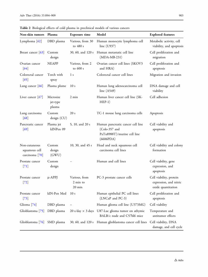
Table 2 Biological effects of cold plasma in preclinical models of various cancers
Non-skin tumors Plasma Exposure time Model Explored features
Lymphoma [62] DBD plasma Various, from 30
to 480 s
Human monocytic lymphoma cell
line (U937)
Metabolic activity, cell
viability, and apoptosis
Breast cancer [63] Custom
design
30, 60, and 120 s Human metastatic cell line
(MDA-MB-231)
Cell proliferation and
migration
Ovarian cancer
[64]
NEAPP Various, from 2
to 600 s
Ovarian cancer cell lines (SKOV3
and HRA)
Cell proliferation and
apoptosis
Colorectal cancer
[65]
Torch with
spray
1 s Colorectal cancer cell lines Migration and invasion
Lung cancer [66] Plasma plume 10 s Human lung adenocarcinoma cell
line (A549)
DNA damage and cell
viability
Liver cancer [67] Microsize
jet-type
plasma
2 min Human liver cancer cell line (SK-
HEP-1)
Cell adhesion
Lung carcinoma
[68]
Custom
design (CU)
20 s TC-1 mouse lung carcinoma cells Apoptosis
Pancreatic cancer
[69]
Plasma jet
kINPen 09
5, 10, and 20 s Human pancreatic cancer cell line
(Colo-357 and
PaTu8988T)/murine cell line
(6606PDA)
Cell viability and
apoptosis
Non-cutaneous
squamous cell
carcinoma [70]
Custom
design
(GWU)
10, 30, and 45 s Head and neck squamous cell
carcinoma cell lines
Cell viability and colony
formation
Prostate cancer
[71]
Custom
design
– Human and cell lines Cell viability, gene
expression, and
apoptosis
Prostate cancer
[72]
l-APPJ Various, from
2 min to
20 min
PC-3 prostate cancer cells Cell viability, protein
expression, and nitric
oxide quantitation
Prostate cancer
[73]
kIN-Pen Med 10 s Human epithelial PC cell lines
(LNCaP and PC-3)
Cell proliferation and
apoptosis
Glioma [74] DBD plasma – Human glioma cell line (U373MG) Cell viability
Glioblastoma [75] DBD plasma 20 s/day 9 3 days U87-Luc glioma tumor on athymic
BALB/c nude and C57bl6 mice
Temperature and
antitumor effects
Glioblastoma [76] SMD plasma 30, 60, and 120 s Human glioblastoma cancer cell lines Cell viability, DNA
damage, and cell cycle
Adv Ther (2016) 33:894–909 903

permit the central production of PAW in
hospitals using a single plasma generator. PAW
can then be packaged and distributed to various
operating rooms as needed for cancer
treatments within the same day. Moreover,
PAW can be applied topically on tumor
surfaces or injected into the tumor [33]. This
feature is significant in dermatology because
skin tumors are easily accessible using these
approaches. However, basic principles to guide
PAW application in cancer, specifically to treat
skin cancer, remain undefined.
Oxidative stress may play various roles in the
pathogenesis of human skin cancers. The most
clinically relevant dermatological tumors are
the basal cell carcinoma, cutaneous squamous
cell carcinoma, and malignant melanoma.
Melanoma cells exhibit increased oxidative
stress, which could damage surrounding tissue,
thereby supporting the progression of
metastasis [34]. When CAP was applied on an
immortal melanoma cell line for 2 min,
apoptosis was induced in cancer cells, but not
in non-neoplastic melanocytes [13]. CAP
treatment for 1 min did not induce apoptosis,
although a prolonged inhibition of cell
proliferation was observed, promoting cell
senescence. Importantly, this demonstrated
the ability of CAP application to remove
tumor cells from the proliferative phase of the
cell cycle. Ishaq et al. also observed a similar
effect by comparing a line of melanoma cells to
melanocytes in culture [35]. Elevated
intracellular ROS induced the expression of
genes involved in cellular apoptosis mediated
by TNFa and apoptosis signal-regulating kinase
(ASK). When cells were pretreated with
N-acetylcysteine and an antibody against
TNFa, the apoptosis signal was inhibited.
Recently, Daeschlein et al. evaluated the
antitumor efficacy of CAP administered in
conjunction with a bleomycin-based
electrochemotherapy in a mouse model of
melanoma. The combinational therapy
improved mice survival significantly when
compared to the electrochemotherapy alone
[36].
To date, experimental and/or clinical studies
of CAP use in non-melanoma skin cancer (such
as basal cell carcinoma and squamous cell
carcinoma) have yet to be conducted. In
non-melanoma skin cancer, diminished
Table 2 continued
Non-skin tumors Plasma Exposure time Model Explored features
Glioblastoma [77,
78]
Custom
design
(GWU)
Various, from 60
to 180 s
Human glioblastoma cancer cell line
(U87)
Cell viability, cell cycle,
and apoptosis
Glioblastoma [79] DBD plasma 30, 60, 90, and
180 s
Glioma cell lines (U87, U373, A172),
human normal astrocytes E6/E7,
and HUVEC
Cell viability, cell cycle,
and apoptosis
Neuroblastoma
[80]
Helium-based
plasma
0, 30, 60, and
120 s
Neuro2a cells murine neuroblastoma Metabolic activity and
apoptosis
HUVEC human umbilical vein endothelial cells, l-APPJ micro-scaled atmospheric pressure plasma jet, GWU GeorgeWashington University, NEAPP non-equilibrium atmospheric pressure plasma, DBD dielectric barrier discharge, CUClemson University, SMD surface micro discharge
904 Adv Ther (2016) 33:894–909
antioxidant defense caused by chronic UV
exposure may contribute to carcinogenesis
[34]. A related study, assessing the use of CAP
in cultured explants of human low-degree
non-cutaneous epidermoid carcinoma of the
head and neck, demonstrated that plasma can
decrease cell viability and increase DNA
fragmentation and cell apoptosis [37].
CLINICAL USE OF CAP IN HUMANSKIN
Few studies have addressed the effects of CAP in
humans, and studies related to skin cancer have
not been performed. Most clinical trials evaluated
the efficacy and safety of cold plasma application,
mainly as adjuvant treatment of chronic
cutaneous ulcers. This is due to the frequent
bacterial colonization or wound infection in
chronic cutaneous ulcers that may affect
adequate restitution of tissue structure and
function. Furthermore, development of bacterial
resistance is unlikely based on CAP mechanism of
action [38, 39]. Study design and information
obtained on CAP efficacy and safety can serve as a
foundation to develop future clinical trials to
assess CAP as a treatment option for skin cancer
[40–42]. Virtually all clinical studies of CAP have
been performed using medical devices designed
for topical application (MicroPlaSter� a,
MicroPlaSter� b, and PlasmaDerm� VU 2010),
following a phase I or II clinical trial design using
small numbers of healthy volunteers or patients
(from 14 to 70). In all cases, treatment was well
tolerated and no significant differences in adverse
effect frequency were observed in the CAP group
as compared to the controls. Results obtained
from other studies on CAP application in human
subjects are circumstantial; most were
communicated as case reports or small series of
cases, and all studies were performed in
non-cancerous skin diseases. In most studies,
CAP treatment was not more effective than the
placebo; however, it was well tolerated with no
relevant adverse events [43–45]. Recently,
Metelmann et al. published a retrospective
review of 12 patients with head and neck
non-cutaneous advanced squamous cell
carcinoma that were treated with CAP to
decontaminate infected cancer ulcerations.
When they evaluated anticancer effects,
superficial partial remission of tumor was
observed in some cases following CAP exposure
[46].
CONCLUSION
The recent development of new plasma sources
and devices for simple CAP application on the
skin paralleled numerous studies performed
in vitro, ex vivo, and in human subjects. When
applied directly or indirectly on cell cultures or
disease models (in vitro or in vivo using animal
models), CAP has been shown to reduce cell
proliferation, adhesion, and migration, and
induce selective apoptosis of neoplastic cells
without damaging normal cells. These selective
effects may be due to differences in the
intracellular oxidative status and cell cycle
phase between normal and tumor tissues.
Furthermore, excessive levels of oxidative
radicals induced by CAP can induce DNA
damage and cell cycle exit into senescence,
apoptosis, or necrosis. The antitumor capacity
of CAP treatment can been regulated by
controlling treatment time, gas source
composition, gas flow rate, and supply voltage.
When CAP is utilized to irradiate a medium or
water to obtain WAS, the distance between CAP
source and the liquid, and the final volume
should be taken into consideration. Despite
mounting evidence supporting its use, studies
Adv Ther (2016) 33:894–909 905

of CAP in human subjects are still limited. In the
majority of these studies, CAP was shown to be
well tolerated without any observable short-term
adverse effects. Therefore, we suggest further
investigation of CAP, including the use of PAW,
as potential adjuvant therapies for skin tumors
such as basal cell carcinoma, squamous cell
carcinoma, and malignant melanoma.
ACKNOWLEDGMENTS
No funding or sponsorship was received for this
study or publication of this article. All authors
meet the International Committee of Medical
Journal Editors (ICMJE) criteria for authorship,
take responsibility for the integrity of the work
as a whole, and have given final approval for the
version to be published. The authors would like
to thank Editage (http://www.editage.com) for
English-language editing.
Disclosures. Jesus Gay-Mimbrera, Maria
Carmen Garcıa, Beatriz Isla-Tejera, Antonio
Rodero-Serrano, Antonio Velez Garcıa-Nieto,
and Juan Ruano have nothing to disclose.
Compliance with Ethics
Guidelines. Analyses described in this review
article were based on previously conducted
studies. No studies involving human subjects
or animals were performed for the publication
of this review.
Open Access. This article is distributed
under the terms of the Creative Commons
Attribution-NonCommercial 4.0 International
License (http://creativecommons.org/licenses/
by-nc/4.0/), which permits any noncommer-
cial use, distribution, and reproduction in any
medium, provided you give appropriate credit
to the original author(s) and the source, provide
a link to the Creative Commons license, and
indicate if changes were made.
REFERENCES
1. von Woedtke Th, Reuter S, Masur K, Weltmann KD.Plasmas for medicine. Phys Rep. 2013;530:291–320.
2. Raiser J, Zenker M. M. Argon plasma coagulation foropen surgical and endoscopic applications: state ofthe art. J Phys D Appl Phys. 2006;39:3520–3.
3. Heslin C, Boehm D, Milosavljevic V, Laycock M,Cullen PJ, Bourke P. Quantitative assessment ofblood coagulation by cold atmospheric plasma.Plasma Med. 2014;4:153–63.
4. Zuo X, Wei Y, Wei Chen L, Dong Meng Y.Non-equilibrium atmospheric pressuremicroplasma jet: an approach to endoscopictherapies. Phys Plasmas. 2013;20:083507.
5. Kong MG, Kroesen G, Morfill G, et al. Plasmamedicine: an introductory review. New J Phys.2009;11:115012.
6. Isbary G, Zimmermann JL, Shimizu T, et al.Non-thermal plasma-more than five years ofclinical experience. Clin Plasma Med.2013;1:19–23.
7. Graves DB. The emerging role of reactive oxygenand nitrogen species in redox biology and someimplications for plasma applications to medicineand biology. J Phys D Appl Phys. 2012;45:263001.
8. Schmidt A, von Woedtke T, Bekeschus S. Periodicexposure of keratinocytes to cold physicalplasma–an in vitro model for redox-relateddiseases of the skin. Oxid Med Cell Longev.2016;2016:9816072.
9. Arndt S, Unger P, Wacker E, et al. Cold atmosphericplasma (CAP) changes gene expression of keymolecules of the wound healing machinery andimproves wound healing in vitro and in vivo. PLoSone. 2013;8:e79325.
10. Zhong S, Dong Y, Liu D, et al. Surface air plasmainduced cell death and cytokines release of humankeratinocytes in the context of psoriasis. Br JDermatol. 2016;174:542–52.
11. Park S-B, Kim B, Bae H, et al. Differential epigeneticeffects of atmospheric cold plasma on MCF-7 andMDA-MB-231 breast cancer cells. PLoS One.2015;10:e0129931.
906 Adv Ther (2016) 33:894–909
12. Ahn HJ, Kim KI, Kim G, Moon E, Yang SS, Lee JS.Atmospheric-pressure plasma jet induces apoptosisinvolving mitochondria via generation of freeradicals. PLoS One. 2011;6:e28154.
13. Arndt S, Wacker E, Li YF, et al. Cold atmosphericplasma, a new strategy to induce senescence inmelanoma cells. Exp Dermatol. 2013;22:284–9.
14. Yan X, Zou F, Zhao S, et al. On the mechanism ofplasma inducing cell apoptosis. IEEE Trans PlasmaSci. 2010;38:2451–7.
15. Kim KC, Piao MJ, Madduma Hewage SRK, et al.Non-thermal dielectric-barrier discharge plasmadamages human keratinocytes by inducingoxidative stress. Int J Mol Med. 2016;37:29–38.
16. Blackert S, Haertel B, Wende K, von Woedtke T,Lindequist U. Influence of non-thermalatmospheric pressure plasma on cellular structuresand processes in human keratinocytes (HaCaT).J Dermatol Sci. 2013;70:173–81.
17. Wende K, Landsberg K, Lindequist U, WeltmannK-D, von Woedtke T. Distinctive activity of anonthermal atmospheric-pressure plasma jet oneukaryotic and prokaryotic cells in a cocultivationapproach of keratinocytes and microorganisms.IEEE Trans Plasma Sci. 2010;38:2479–85.
18. Hasse S, Duong Tran T, et al. Induction ofproliferation of basal epidermal keratinocytes bycold atmospheric-pressure plasma. Clin ExpDermatol. 2016;41(2):202–9.
19. Arndt S, Landthaler M, Zimmermann JL, et al.Effects of cold atmospheric plasma (CAP) onb-defensins, inflammatory cytokines, andapoptosis-related molecules in keratinocytesin vitro and in vivo. PLoS One. 2015;10:e0120041.
20. Brun P, Pathak S, Castagliuolo I, et al. Heliumgenerated cold plasma finely regulates activation ofhuman fibroblast-like primary cells. PLoS One.2014;9:e104397.
21. Shashurin A, Stepp MA, Hawley TS, et al. Influenceof cold plasma atmospheric jet on surface integrinexpression of living cells. Plasma Process Polym.2010;7:294–300.
22. Lopes BB, De Paula Leite Kraft MB, Rehder J, BatistaFRX, Puzzi MB. The interactions betweennon-thermal atmospheric pressure plasma andex vivo dermal fibroblasts. Proc Eng.2013;59:92–100.
23. Tipa RS, Kroesen GMW. Plasma-stimulated woundhealing. IEEE Trans Plasma Sci. 2011;39:2978–9.
24. Isbary G, Koritzer J, Mitra A, et al. Ex vivo humanskin experiments for the evaluation of safety of newcold atmospheric plasma devices. Clin Plasma Med.2013;1:36–44.
25. Ma R, Feng H, Li F, et al. An evaluation ofanti-oxidative protection for cells againstatmospheric pressure cold plasma treatment. ApplPhys Lett. 2012;100:3–7.
26. Lademann J, Richter H, Alborova A, et al. Riskassessment of the application of a plasma jet indermatology. J Biomed Opt. 2009;14:054025.
27. Wende K, Bekeschus S, Schmidt A, et al. Riskassessment of a cold argon plasma jet in respect toits mutagenicity. Mutat Res Genet Toxicol EnvironMutagen. 2016;798–799:48–54.
28. Keidar M. Plasma for cancer treatment. Plasma SourSci Technol. 2015;24:033001.
29. Kieft IE, Kurdi M, Stoffels E. Reattachment andapoptosis after plasma-needle treatment of culturedcells. IEEE Trans Plasma Sci. 2006;34:1331–6.
30. Xu D, Liu D, Wang B, et al. In situ OH generationfrom O2
- and H2O2 plays a critical role inplasma-induced cell death. PLoS One.2015;10:e0128205.
31. Yan D, Talbot A, Nourmohammadi N, et al.Principles of using cold atmospheric plasmastimulated media for cancer treatment. Sci Rep.2015;17(5):18339.
32. Adachi T, Tanaka H, Nonomura S, et al.Plasma-activated medium induces A549 cell injuryvia a spiral apoptotic cascade involving themitochondrial-nuclear network. Free Radic BiolMed. 2015;79:28–44.
33. Utsumi F, Kajiyama H, Nakamura K, et al. Effect ofindirect nonequilibrium atmospheric pressureplasma on anti-proliferative activity againstchronic chemo-resistant ovarian cancer cellsin vitro and in vivo. PLoS One. 2013;8(12):e81576.
34. Sander CS, Hamm F, Elsner P, Thiele JJ. Oxidativestress in malignant melanoma and non-melanomaskin cancer. Br J Dermatol. 2003;148(5):913–22.
35. Ishaq M, Kumar S, Varinli H, et al. Atmospheric gasplasma-induced ROS production activatesTNF-ASK1 pathway for the induction ofmelanoma cancer cell apoptosis. Mol Biol Cell.2014;25:1523–31.
36. Daeschlein G, Scholz S, Lutze S, et al. Comparisonbetween cold plasma, electrochemotherapy and
Adv Ther (2016) 33:894–909 907

combined therapy in a melanoma mouse model.Exp Dermatol. 2013;22:582–6.
37. Welz C, Emmert S, Canis M, et al. Cold atmosphericplasma: a promising complementary therapy forsquamous head and neck cancer. PLoS One.2015;10:e0141827.
38. Maisch T, Shimizu T, Li Y-F, et al. Decolonisation ofMRSA, S. aureus and E. coli by cold-atmosphericplasma using a porcine skin model in vitro. PLoSOne. 2012;7:e34610.
39. Klampfl TG, Isbary G, Shimizu T, et al. Coldatmospheric air plasma sterilization against sporesand other microorganisms of clinical interest. ApplEnviron Microbiol. 2012;78:5077–82.
40. Isbary G, Morfill G, Schmidt HU, et al. A firstprospective randomized controlled trial to decreasebacterial load using cold atmospheric argon plasmaon chronic wounds in patients. Br J Dermatol.2010;163:78–82.
41. Isbary G, Heinlin J, Shimizu T, et al. Successful andsafe use of 2 min cold atmospheric argon plasma inchronic wounds: results of a randomized controlledtrial. Br J Dermatol. 2012;167:404–10.
42. Brehmer F, Haenssle HA, Daeschlein G, et al.Alleviation of chronic venous leg ulcers with ahand-held dielectric barrier discharge plasmagenerator (PlasmaDerm(�) VU-2010): results of amonocentric, two-armed, open, prospective,randomized and controlled trial (NCT01415622).J Eur Acad Dermatol Venereol. 2015;29:148–55.
43. Klebes M, Lademann J, Philipp S, et al. Effects oftissue-tolerable plasma on psoriasis vulgaristreatment compared to conventional localtreatment: a pilot study. Clin Plasma Med.2014;2:22–7.
44. Isbary G, Morfill G, Zimmermann J, Shimizu T,Stolz W. Cold atmospheric plasma: a successfultreatment of lesions in Hailey-Hailey disease. ArchDermatol. 2011;147:388–90.
45. Heinlin J, Isbary G, Stolz W, et al. A randomizedtwo-sided placebo-controlled study on the efficacyand safety of atmospheric non-thermal argonplasma for pruritus. J Eur Acad DermatolVenereol. 2013;27:324–31.
46. Metelmann HR, Nedrelow DS, Seebauer C, et al.Head and neck cancer treatment and physicalplasma. Clin Plasma Med. 2015;3:17–23.
47. Daeschlein G, Scholz S, Ahmed R, et al. Cold plasmais well-tolerated and does not disturb skin barrier orreduce skin moisture. J Dtsch Dermatol Ges.2012;10:509–15.
48. Welz C, Becker S, Li Y-F, et al. Effects of coldatmospheric plasma on mucosal tissue culture.J Phys D Appl Phys. 2013;46:045401.
49. Tiede R, Hirschberg J, Daeschlein G, von WoedtkeT, Vioel W, Emmert S. Plasma applications: adermatological view. Contrib Plasma Phys.2014;2:118–30.
50. Rajasekaran P, Mertmann P, Bibinov N, Wandke D,Viol W, Awakowicz P. DBD plasma source operatedin single-filamentary mode for therapeutic use indermatology. J Phys D Appl Phys. 2009;42:225201.
51. Bender C, Matthes R, Kindel E, et al. The irritationpotential of nonthermal atmospheric pressureplasma in the het-CAM. Plasma Process Polym.2010;7:318–26.
52. Boekema BKHL, Vlig M, Guijt D, et al. A newflexible DBD device for treating infected wounds:in vitro and ex vivo evaluation and comparisonwith a RF argon plasma jet. J Phys D Appl Phys.2016;49:044001.
53. Chutsirimongkol C, Boonyawan D, Polnikorn N,Techawatthanawisan W, Kundilokchai T.Non-thermal plasma for acne treatment andaesthetic skin improvement. Plasma Med.2014;4:79–88.
54. Haertel B, von Woedtke T, Weltmann K-D,Lindequist U. Non-thermal atmospheric-pressureplasma possible application in wound healing.Biomol Ther 2014;22:477–90.
55. Daeschleia G, Scholza S, Ahmed R, et al. Skindecontamination by low-temperature atmosphericpressure plasma jet and dielectric barrier dischargeplasma. J Hosp Infect. 2012;81:177–83.
56. Fluhr JW, Sassning S, Lademann O, et al. In vivoskin treatment with tissue-tolerable plasmainfluences skin physiology and antioxidant profilein human stratum corneum. Exp Dermatol.2011;21:130–4.
57. Ishaq M, Bazaka K, Ostrikov K. Pro-apoptotic NOXAis implicated in atmospheric-pressureplasma-induced melanoma cell death. J Phys DAppl Phys. 2015;48:464002.
58. Jacofsky MC, Lubahn C, McDonnell C, et al.Spatially resolved optical emission spectroscopy ofa helium plasma jet and its effects on woundhealing rate in a diabetic murine model. PlasmaMed. 2014;4:177–91.
59. Boekema BKHL, Hofmann SS, van Ham BJT,Bruggeman PJ, Middelkoop E. Antibacterial plasmaat safe levels for skin cells. J Phys D Appl Phys.2013;46:422001.
908 Adv Ther (2016) 33:894–909
60. Morfill G, Shimizu T, Steffes B, Schmidt H-U.Nosocomical infections-a new approach towardspreventive medicine using plasmas. New J Phys.2009;11:115019.
61. Li YF, Taylor D, Zimmermann JL, et al. In vivo skintreatment using two portable plasma devices:comparison of a direct and an indirect coldatmospheric plasma treatment. Clin Plasma Med.2013;1:35–9.
62. Kaushik N, Kumar N, Kim CH, Kaushik NK, ChoiEH. Dielectric barrier discharge plasma efficientlydelivers an apoptotic response in humanmonocytic lymphoma. Plasma Process Polym.2014;11:1175–87.
63. Wang M, Holmes B, Cheng X, Zhu W, Keidar M,Zhang LG. Cold atmospheric plasma for selectivelyablating metastatic breast cancer cells. PLoS One.2013;8:e73741.
64. Iseki S, Nakamura K, Hayashi M, et al. Selectivekilling of ovarian cancer cells through induction ofapoptosis by nonequilibrium atmospheric pressureplasma. Appl Phys Lett. 2012;100:113702.
65. Kim CH, Kwon S, Bahn JH, et al. Effects ofatmospheric nonthermal plasma on invasion ofcolorectal cancer cells. Appl Phys Lett. 2010;96.
66. Joh HM, Choi JY, Kim SJ, Chung TH, Kang T-H.Effect of additive oxygen gas on cellular response oflung cancer cells induced by atmospheric pressurehelium plasma jet. Sci Rep. 2014;4:6638.
67. Gweon B, Kim M, Bee Kim D, et al. Differentialresponses of human liver cancer and normal cells toatmospheric pressure plasma. Appl Phys Lett.2011;99:063701.
68. Kim JY, Ballato J, Foy P, et al. Apoptosis of lungcarcinoma cells induced by a flexible opticalfiber-based cold microplasma. Biosens Bioelectron.2011;28:530–8.
69. Partecke LI, Evert K, Haugk J, et al. Tissue tolerableplasma (TTP) induces apoptosis in pancreaticcancer cells in vitro and in vivo. BMC Cancer.2012;12:1–10.
70. Guerrero-Preston R, Ogawa T, Uemura M, et al.Cold atmospheric plasma treatment selectivelytargets head and neck squamous cell carcinomacells. Int J Mol Med. 2014;34:941–6.
71. Hirst AM, Frame FM, Maitland NJ, Connell DO.Low temperature plasma: a novel focal therapy forlocalized prostate cancer. Biomed Res Int.2014;2014:878319.
72. Gibson AR, McCarthy HO, Ali A, O’Connell D,Graham WG. Interactions of a non-thermalatmospheric pressure plasma effluent with PC-3prostate cancer cells. Plasma Process Polym.2014;11:1142–9.
73. Weiss M, Gumbel D, Hanschmann E-M, et al. Coldatmospheric plasma treatment inducesanti-proliferative effects in prostate cancer cells byredox and apoptotic signaling pathways. PLoS One.2015;10:e0130350.
74. Conway GE, Casey A, Milosavljevic V, Liu Y, HoweO, Cullen PJ, et al. Non-thermal atmosphericplasma induces ROS-independent cell death inU373MG glioma cells and augments thecytotoxicity of temozolomide. Br J Cancer.2016;114(4):435–43.
75. Vandamme M, Robert E, Pesnel S, et al. Antitumoreffect of plasma treatment on U87 gliomaxenografts: preliminary results. Plasma ProcessPolym. 2010;7:264–73.
76. Koritzer J, Boxhammer V, Al E. Restoration ofsensitivity in chemo-resistant glioma cells by coldatmospheric plasma. PLoS One. 2013;8:1–10.
77. Cheng X, Murphy W, Recek N, et al. Synergisticeffect of gold nanoparticles and cold plasma onglioblastoma cancer therapy. J Phys D Appl Phys.2014;47:335402.
78. Cheng X, Sherman J, Murphy W, Ratovitski E,Canady J, Keidar M. The effect of tuning coldplasma composition on glioblastoma cell viability.PLoS One. 2014;9:1–9.
79. Siu A, Volotskova O, Cheng X, et al. Differentialeffects of cold atmospheric plasma in the treatmentof malignant glioma. PLoS One. 2015;10:e0126313.
80. Walk RM, Snyder J, Srinivasan P, et al. Coldatmospheric plasma for the ablative treatment ofneuroblastoma. J Pediatr Surg. 2013;48:67–73.
Adv Ther (2016) 33:894–909 909


![Homemade an Atmospheric Pressure Cold Plasma Sterilization ... · plasma reactor shown in Fig. 1 [2]. By our DBD plasma at atmospheric pressure system, that can succeed in . E. coli](https://img.pdfslide.us/doc/110x75/6087616ab358a048a252f115/homemade-an-atmospheric-pressure-cold-plasma-sterilization-plasma-reactor-shown.jpg)